Dual interaction of factor H with C3d and glycosaminoglycans in host–nonhost discrimination by complement Tommi Kajander a , Markus J. Lehtinen b,1 , Satu Hyvärinen b,1 , Arnab Bhattacharjee b,1 , Elisa Leung c , David E. Isenman c , Seppo Meri b,d , Adrian Goldman a,2 , and T. Sakari Jokiranta b,2 a Institute of Biotechnology, University of Helsinki, Viikinkaari, FIN-00014, Helsinki, Finland; b Haartman Institute, University of Helsinki, Haartmaninkatu, FIN-00014, Helsinki, Finland; c Department of Biochemistry, University of Toronto, Toronto, ON, Canada M5S 1A8; and d Division of Microbiology, Helsinki University Central Hospital Laboratory (HUSLAB), Haartmaninkatu, FIN-00029, Helsinki, Finland Edited* by Douglas T. Fearon, University of Cambridge School of Clinical Medicine, Cambridge, United Kingdom, and approved January 5, 2011 (received for review November 12, 2010) The alternative pathway of complement is important in innate immunity, attacking not only microbes but all unprotected biological surfaces through powerful amplification. It is unresolved how host and nonhost surfaces are distinguished at the molecular level, but key components are domains 19–20 of the complement regulator factor H (FH), which interact with host (i.e., nonactivator surface glycosaminoglycans or sialic acids) and the C3d part of C3b. Our structure of the FH19–20:C3d complex at 2.3-Å resolution shows that FH19–20 has two distinct binding sites, FH19 and FH20, for C3b. We show simultaneous binding of FH19 to C3b and FH20 to nonactivator surface glycosaminoglycans, and we show that both of these interactions are necessary for full binding of FH to C3b on nonactivator surfaces (i.e., for target discrimination). We also show that C3d could replace glycosaminoglycan binding to FH20, thus pro- viding a feedback control for preventing excess C3b deposition and complement amplification. This explains the molecular basis of atyp- ical hemolytic uremic syndrome, where mutations on the binding interfaces between FH19–20 and C3d or between FH20 and glycosa- minoglycans lead to complement attack against host surfaces. structure and function | X-ray crystallography | hemolysis | kidney diseases | human mutations P reviously unencountered microbes invading a human body must be rapidly recognized and eliminated. This is the function of in- nate immunity, which includes the alternative pathway (AP) of complement. AP components can attack targets with hydroxyl or amine groups (i.e., all biological surfaces). This is a powerful defense mechanism, because there is rapid amplification leading to efficient opsonization or target lysis by the membrane attack complex (MAC). The AP attack is, therefore, also potentially dangerous for the host if one’s cells and acellular structures are not protected. The AP activation is based on spontaneous hydrolysis of C3 in plasma leading to production of C3b, which then randomly attaches onto any surface hydroxyl or amine group through a re- active thioester located on the C3d part [i.e., thioester domain (TED)] of C3b. If these surface-attached C3b molecules are not quickly inactivated to iC3b and C3d, C3b deposition is rapidly amplified by a positive enzymatic feedback loop, leading to op- sonophagocytosis and formation of the lytic membrane attack complex. On host surfaces, which are naturally nonactivators of the AP, efficient down-regulation of bound C3b occurs in three ways: factor I-mediated cleavage of C3b to inactive iC3b, acceleration of the decay of the preformed C3 convertases, or inhibition of factor B binding to C3b. Factor H (FH) is required for all these. It also down-regulates C3b deposition on noncellular surfaces, such as the heparan sulfate-rich glomerular basement membrane. FH is, thus, essential for restricting AP attack against host surfaces while allowing AP attack against foreign surfaces (i.e., for target dis- crimination) (1). A long-standing central question in complement research has been how does FH distinguish the two types? It is known that sialylation of erythrocyte surface makes the cells nonactivators by enhanced FH binding (2, 3), and glycosamino- glycans on endothelial cells participate in making the latter non- activators in the same way (4). FH is composed of 20 homologous domains. Domains 1–4 are essential for various regulatory activities such as cofactor and de- cay accelerating activity (5, 6), whereas domains 19 and 20 (FH19– 20) are essential for target discrimination (7, 8) and bind the C3d part of C3b (9), glycosaminoglycans (10), and sialic acids (11). Mutations in FH19–20 lead to a severe systemic disease, atypical hemolytic uremic syndrome (aHUS). aHUS, which often leads to end-stage renal disease, is characterized by damage to eryth- rocytes, thrombocytes, and endothelial cells (12). The character- istic cell damage in aHUS occurs when the mutated FH is unable to efficiently recognize C3b on these cells. The molecular mech- anism of AP target discrimination is a major unresolved question, because some mutations interfere with binding of FH19–20 to cell surfaces and others interfere with C3b/C3d binding (13–15); the mutations are spread over both domains 19 and 20. We have determined the X-ray crystal structure of FH19–20: C3d complex and performed extensive mutagenesis, binding, and functional studies. Results of these indicate how only two com- plement proteins, C3b and FH, can perform efficient target rec- ognition. aHUS mutations in FH19, FH20, or two different parts of C3d all lead to cell destruction, because FH19–20 has two binding sites for C3d and C3d has two binding sites for FH19–20. In our comprehensive model of target discrimination, FH20 binds to glycosaminoglycans on host surfaces and FH19 binds to C3b, thus leading to cofactor activity. In addition, FH20 can also bind to any already deposited C3d on the surface, again allowing FH19 to bind another C3b and leading to its inactivation. Results Structure of the FH19–20:C3d Complex. We expressed and purified C3d and FH19–20 and crystallized the complex (Materials and Methods). The structure was initially determined by molecular re- placement with C3d at 3.8 Å but refined against a higher-resolution Author contributions: T.K., M.J.L., A.G., and T.S.J. designed research; T.K., M.J.L., S.H., A.B., and E.L. performed research; S.H., A.B., E.L., and D.E.I. contributed new reagents/ analytic tools; T.K., M.J.L., S.H., D.E.I., S.M., A.G., and T.S.J. analyzed data; T.K., M.J.L., D.E.I., S.M., A.G., and T.S.J. wrote the paper; and T.S.J. coordinated the project. The authors declare no conflict of interest. *This Direct Submission article had a prearranged editor. Data deposition: The crystallography, atomic coordinates, and structure factors have been deposited in the Protein Data Bank, www.pdb.org (PDB ID code 2XQW). 1 M.J.L., S.H., and A.B. contributed equally to this work. 2 To whom correspondence may be addressed. E-mail: adrian.goldman@helsinki.fi or sakari.jokiranta@helsinki.fi. This article contains supporting information online at www.pnas.org/lookup/suppl/doi:10. 1073/pnas.1017087108/-/DCSupplemental. www.pnas.org/cgi/doi/10.1073/pnas.1017087108 PNAS | February 15, 2011 | vol. 108 | no. 7 | 2897–2902 IMMUNOLOGY Downloaded by guest on March 17, 2020
Transcript
Dual interaction of factor H with C3d andglycosaminoglycans in host–nonhostdiscrimination by complementTommi Kajandera, Markus J. Lehtinenb,1, Satu Hyvärinenb,1, Arnab Bhattacharjeeb,1, Elisa Leungc, David E. Isenmanc,Seppo Merib,d, Adrian Goldmana,2, and T. Sakari Jokirantab,2
aInstitute of Biotechnology, University of Helsinki, Viikinkaari, FIN-00014, Helsinki, Finland; bHaartman Institute, University of Helsinki, Haartmaninkatu,FIN-00014, Helsinki, Finland; cDepartment of Biochemistry, University of Toronto, Toronto, ON, Canada M5S 1A8; and dDivision of Microbiology, HelsinkiUniversity Central Hospital Laboratory (HUSLAB), Haartmaninkatu, FIN-00029, Helsinki, Finland
Edited* by Douglas T. Fearon, University of Cambridge School of Clinical Medicine, Cambridge, United Kingdom, and approved January 5, 2011 (received forreview November 12, 2010)
The alternative pathway of complement is important in innateimmunity, attacking not only microbes but all unprotected biologicalsurfaces through powerful amplification. It is unresolved how hostand nonhost surfaces are distinguished at the molecular level, butkey components are domains 19–20 of the complement regulatorfactor H (FH), which interact with host (i.e., nonactivator surfaceglycosaminoglycans or sialic acids) and the C3d part of C3b. Ourstructure of the FH19–20:C3d complex at 2.3-Å resolution showsthat FH19–20 has two distinct binding sites, FH19 and FH20, forC3b. We show simultaneous binding of FH19 to C3b and FH20 tononactivator surface glycosaminoglycans, and we show that bothof these interactions are necessary for full binding of FH to C3b onnonactivator surfaces (i.e., for target discrimination). We also showthat C3d could replace glycosaminoglycan binding to FH20, thus pro-viding a feedback control for preventing excess C3b deposition andcomplement amplification. This explains themolecular basis of atyp-ical hemolytic uremic syndrome, where mutations on the bindinginterfaces between FH19–20 and C3d or between FH20 and glycosa-minoglycans lead to complement attack against host surfaces.
structure and function | X-ray crystallography | hemolysis | kidneydiseases | human mutations
Previously unencounteredmicrobes invading a human bodymustbe rapidly recognized and eliminated. This is the function of in-
nate immunity, which includes the alternative pathway (AP) ofcomplement. AP components can attack targets with hydroxyl oramine groups (i.e., all biological surfaces). This is a powerful defensemechanism, because there is rapid amplification leading to efficientopsonizationor target lysis by themembraneattackcomplex (MAC).The AP attack is, therefore, also potentially dangerous for the hostif one’s cells and acellular structures are not protected.The AP activation is based on spontaneous hydrolysis of C3
in plasma leading to production of C3b, which then randomlyattaches onto any surface hydroxyl or amine group through a re-active thioester located on the C3d part [i.e., thioester domain(TED)] of C3b. If these surface-attached C3b molecules are notquickly inactivated to iC3b and C3d, C3b deposition is rapidlyamplified by a positive enzymatic feedback loop, leading to op-sonophagocytosis and formation of the lytic membrane attackcomplex. On host surfaces, which are naturally nonactivators of theAP, efficient down-regulation of bound C3b occurs in three ways:factor I-mediated cleavage of C3b to inactive iC3b, acceleration ofthe decay of the preformed C3 convertases, or inhibition of factorB binding to C3b. Factor H (FH) is required for all these. It alsodown-regulates C3b deposition on noncellular surfaces, such asthe heparan sulfate-rich glomerular basement membrane. FH is,thus, essential for restricting AP attack against host surfaces whileallowing AP attack against foreign surfaces (i.e., for target dis-crimination) (1). A long-standing central question in complementresearch has been how does FH distinguish the two types? It is
known that sialylation of erythrocyte surface makes the cellsnonactivators by enhanced FH binding (2, 3), and glycosamino-glycans on endothelial cells participate in making the latter non-activators in the same way (4).FH is composed of 20 homologous domains. Domains 1–4 are
essential for various regulatory activities such as cofactor and de-cay accelerating activity (5, 6), whereas domains 19 and 20 (FH19–20) are essential for target discrimination (7, 8) and bind the C3dpart of C3b (9), glycosaminoglycans (10), and sialic acids (11).Mutations in FH19–20 lead to a severe systemic disease, atypicalhemolytic uremic syndrome (aHUS). aHUS, which often leadsto end-stage renal disease, is characterized by damage to eryth-rocytes, thrombocytes, and endothelial cells (12). The character-istic cell damage in aHUS occurs when the mutated FH is unableto efficiently recognize C3b on these cells. The molecular mech-anism of AP target discrimination is a major unresolved question,because some mutations interfere with binding of FH19–20 to cellsurfaces and others interfere with C3b/C3d binding (13–15); themutations are spread over both domains 19 and 20.We have determined the X-ray crystal structure of FH19–20:
C3d complex and performed extensive mutagenesis, binding, andfunctional studies. Results of these indicate how only two com-plement proteins, C3b and FH, can perform efficient target rec-ognition. aHUS mutations in FH19, FH20, or two different partsof C3d all lead to cell destruction, because FH19–20 has twobinding sites for C3d and C3d has two binding sites for FH19–20.In our comprehensive model of target discrimination, FH20 bindsto glycosaminoglycans on host surfaces and FH19 binds to C3b,thus leading to cofactor activity. In addition, FH20 can also bind toany already deposited C3d on the surface, again allowing FH19 tobind another C3b and leading to its inactivation.
ResultsStructure of the FH19–20:C3d Complex. We expressed and purifiedC3d and FH19–20 and crystallized the complex (Materials andMethods). The structure was initially determined by molecular re-placement with C3d at 3.8 Å but refined against a higher-resolution
Author contributions: T.K., M.J.L., A.G., and T.S.J. designed research; T.K., M.J.L., S.H.,A.B., and E.L. performed research; S.H., A.B., E.L., and D.E.I. contributed new reagents/analytic tools; T.K., M.J.L., S.H., D.E.I., S.M., A.G., and T.S.J. analyzed data; T.K., M.J.L.,D.E.I., S.M., A.G., and T.S.J. wrote the paper; and T.S.J. coordinated the project.
The authors declare no conflict of interest.
*This Direct Submission article had a prearranged editor.
Data deposition: The crystallography, atomic coordinates, and structure factors have beendeposited in the Protein Data Bank, www.pdb.org (PDB ID code 2XQW).1M.J.L., S.H., and A.B. contributed equally to this work.2To whom correspondence may be addressed. E-mail: [email protected][email protected].
This article contains supporting information online at www.pnas.org/lookup/suppl/doi:10.1073/pnas.1017087108/-/DCSupplemental.
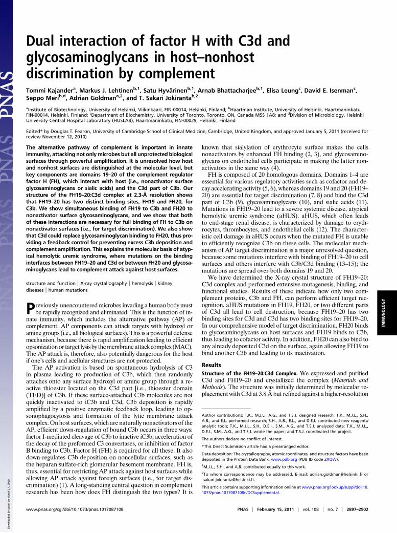
dataset at 2.3Å (Table S1 and Fig. S1). The asymmetric unit revealsa surprising FH19–20:C3d2 heterotrimer, where two sites on a sin-gle FH19–20 bind two separate C3ds in different ways and at dif-ferent sites (Fig. 1A). The overall structures of both C3d moleculesare unchanged (16), whereas there are small local conformationalchanges in FH19–20 (13) because of binding. Both binding sites onC3d are far from the thioester site used for covalent attachment ofC3b to targets (16) (Fig. 1).Of the two interfaces on FH that bind to C3d, one interface (the
FH20 site) is formed by the tip of the domain 20, which binds ina strongly electrostatic manner in a negatively charged cleft on theconcave top of the α/α-helical bundle of C3d (the C3dFH20 site)(Fig. 1B and Fig. S2A andC). The side chain of Arg1203 on FH20protrudes into the cleft and forms an ion pair with C3d Glu160(Glu1153 in the full sequence of C3). The total buried surfacearea of 490 Å2 includes three ion pairs and an extensive ionicnetwork (Fig. S2 B and D), which presumably compensates forthe low shape complementarity (Sc) (17) of 0.43, consistent withthe observed micromolar affinities. There are local changes in theloops around the FH20 site: Lys1188 turns almost 180° to point toC3d, and Thr1184 and Arg1171 pack against C3d. The other in-terface (the FH19 site) is larger (620 Å2) and less charged (Fig. 1Cand Fig. S2B) but with a higher Sc (0.70). This interface is formedby residues from both FH19 and FH20 that bind to the side of C3dat helices 104–118 and 170–189 and the Pro121 loop (the C3dFH19
site) (Fig. 1C and Fig. S2D).The most important and striking feature of this complex is that
the two sites are discontinuous and do not allow one FH to bindsimultaneously one C3d through both interfaces. In addition, theC3dFH20 site is partially buried in the full C3bmolecule (see below).
aHUS-Associated Mutations Are Found in Both Interfaces. The un-expected structure with two binding sites explains why the 15 re-ported aHUS-associated point mutations in surface residues oc-cur discontinuously in both FH domains 19 and 20 (Fig. 1A andTable 1). Of these, our structure suggests that 2 of 3 mutations inFH19 (Asp1119Gly and Tyr1142Asp) and 5 of 12 mutations inFH20 (Arg1182Ser, Trp1183Arg/Leu, Thr1184Arg, Arg1203Trp,and Gly1204Glu) would lead to impaired binding of FH19–20 toC3d/C3b. Five of the other eight residues affect binding of FH19–20 to heparin and/or endothelial cells (13–15) (Table 1 and Table
S2). The remaining two mutations (Glu1135Asp and Ile1170Val)are partially buried and presumably affect FH folding.Some aHUS patients have mutations in C3. All five published
or C3dFH20 sites. Two mutations (Asp122Asn and Gln168Lys) affect
B CFH19
C3d
A
C3dFH20
Fig. 1. Structure of the FH19–20:C3d complex. (A) The asymmetric unit contains one FH19–20 (gray) and two C3d molecules (green and blue). The residuesmutated in aHUS patients and located at the interfaces are annotated and shown as green/blue spheres (C3d) or gray spheres (FH19–20). The thioester siteresidues are in yellow. (B) Close-up view of the FH20–C3d interface and (C) the FH19–C3d interface: FH19–20 is in gray, C3d is in green or blue, the maininterface residues are sticks, disulphide bridges are in yellow, and hydrogen bonds are dashed lines. Structure figures were prepared using PyMol (version 1.3;Schrödinger, LLC).
Table 1. Location and reported functional consequences ofsurface-exposed aHUS-associated mutations within FH19–20 andC3d part of C3b
Location in FH19–20:C3d Mutations
Reported functionaldefects*
FH–C3b/d FH–heparin
aHUS mutations on FH19–20 surfaceFH19 site Asp1119Gly† ↓ ↔FH19 site Tyr1142Asp/Cys† nt ntFH20 site Arg1182Ser† ↓ ↓FH20 site Trp1183Leu† ↓ ↓FH20 site Thr1184Arg† ↓ ↑FH20 site Arg1203Trp† ↓ ↓FH20 site Gly1204Glu† nt ntHeparin site? Gly1194Asp† nt ntHeparin site Arg1206Cys ↓ ↓Heparin site Arg1210Cys ↓ ↓Heparin site Arg1215Gln/Gly ↓ ↓Next to heparin site? Leu1189Arg ↔ ↑Next to heparin site? Glu1198Ala ↔ ↑
aHUS mutations on C3d surfaceC3dFH19 Asp122Asn† ↓C3dFH19 Gln168Lys† ↓C3dFH20 Arg49Leu† ntC3dFH20 Ala101Val† ↓Next to C3dFH19 Ile164Thr† nt
*Details of functional analyses and references are found in Tables S2 and S3.↓, binding diminished because of the mutation; ↔, no effect; ↑, enhancedbinding; nt, not tested.†Mutants discussed in this report. Buried mutations likely to disrupt the foldare not listed: Val1134Gly, Glu1135Asp, Trp1157Arg, Cys1163Trp, Val1168Glu,Ile1169Leu, Ile1170Val, Ser1191Leu/Trp, Val1197Ala, Phe1199Ser, andPro1226Ser for FH19–20 and Cys165Trp for the C3d part of C3b.
2898 | www.pnas.org/cgi/doi/10.1073/pnas.1017087108 Kajander et al.
residues in contact with the FH19 binding site, one mutation(Ile164Thr) is near that site, and two mutations (Arg49Leu andAla101Val) affect residues in contact with the FH20 binding siteon C3d (Table 1 and Table S3). Three of these mutations(Asp122Asn,Gln168Lys, andAla101Val) are known to impair FHbinding (18), whereas two have yet to be studied (19).
Verification of the Two FH-C3d Interfaces by Mutagenesis. Thepresence of aHUS mutations on both sides of the FH19:C3d andFH20:C3d interfaces strongly suggests that neither interface is anartifact. To prove this, we analyzed published data and studied fur-thermutations in both FH19–20 andC3d.All of the published pointmutations in theFH19 (Asp1119Gly,Gln1139Ala, andLys1188Ala)or FH20 site (Arg1182Ser/Ala, Trp1183Leu, Thr1184Arg, andArg1203Ala) led to reduced C3d/C3b binding (13–15, 20) (Fig. 2Aand Table 1). Next, we tested binding of seven C3d mutants toFH19–20. Both the mutations in the C3dFH19 site (Glu117Ala andAsp122Ala) and all four mutations in the C3dFH20 site (Asp36Ala,Glu160Ala, Asp163Ala, and Lys291Ala) significantly reduced theaffinity of the interaction (Fig. 2B–D), whereas the controlmutation(Glu37Ala) had no effect.
Availability of the Binding Sites on C3b.We then generatedmultiplemutations in FH19–20 to delete either the FH19 or FH20 site,FH19Del-20 (Gln1137Ala-Gln1139Ala-Tyr1142Ala) and FH19–
20Del (Thr1184Gly-Lys1202Ala-Arg1203Ala-Tyr1205Ala), andtested their binding toC3d andC3b. C3d boundWTFH19–20witha twofold higher affinity than FH19Del-20 and a sixfold higher af-finity than FH19–20Del (Fig. 2E). This indicates that the FH19 andFH20 sites bind C3d independently and with similar affinities.FH19–20Del bound similarly to C3d and C3b (apparent KD = 1.14vs. 1.48 μM), indicating that the C3bFH19 site is fully available, asexpected from the X-ray structure of C3b (21). Conversely, C3bbound WT FH19–20 or FH19Del
–20 with a three to four timeslower affinity than C3d did. This, although unanticipated based onprior information, is also consistent with the C3b structure (21),because the C3bFH20 site is partially hidden (Fig. 2F). Thus, con-formational change in C3b would be required for full bindingthrough the C3b:FH20 interface (Fig. S3). Furthermore, theC3dFH20 site also overlaps with the binding site of FH4 in theFH1–4:C3b structure (22). Despite this, we were unable to inhibitthe cofactor or decay-accelerating functions of FH1–4 or full FHbyeither FH19–20 orFH19Del
–20 (Fig. S4), indicating that FH4 is notessential for FH1–4 functions in the fluid phase, which is alsoconsistent with earlier studies (5). Overall, it seems that FH19–20binds C3d primarily through the FH20 site (Fig. 2E) (apparentKD = 0.41 vs. 1.84 μM) but binds C3b through the FH19 site. Thishas important implications for understanding how FH functions intarget discrimination (see below).
B C
D E
Apparent KD (µM)
C3d C3b
FH19-20 0.18 0.54
FH19Del-20 0.41 1.84
FH19-20Del 1.14 1.48
F
A
C3d fingerprint on FH19 site
C3d fingerprint on FH20 site
(D1119)
Q1139
K1188
T1184
R1203K1230
FH19 sitefingerprint on C3d
FH20 site fingerprint on C3d
D36
E160
E117D122
D163
K291
FH20 site
C3d
FH19 site
Fig. 2. FH19–20 binding sites on C3d and C3b. (A) Mutations on FH19–20 that reduce affinity to C3d or C3b (13–15, 20) are in darker color and annotated. (B)Binding of C3dg mutants to solid-phase FH19–20 by surface plasmon resonance (SPR) normalized to WT C3dg. Bars indicate SD (n = 3). *P < 0.05; **P < 0.01;***P < 0.001. (C) Binding of 125I-labeled FH19–20 to microtitre plate-coated C3dg mutants (SD indicated; n = 3) and (D) C3d mutations that impaired FH19–20binding (darker blue and bright green). (E) Apparent affinities of FH19–20, FH19Del–20, and FH19–20Del to C3d and C3b measured using SPR. (F) Location ofthe exposed FH19 and partially occluded FH20 binding sites in C3b. FH19–20 bound to the FH20 binding site is shown in green, FH19–20 bound to the FH19binding site is in blue, and C3b is in gray.
Kajander et al. PNAS | February 15, 2011 | vol. 108 | no. 7 | 2899
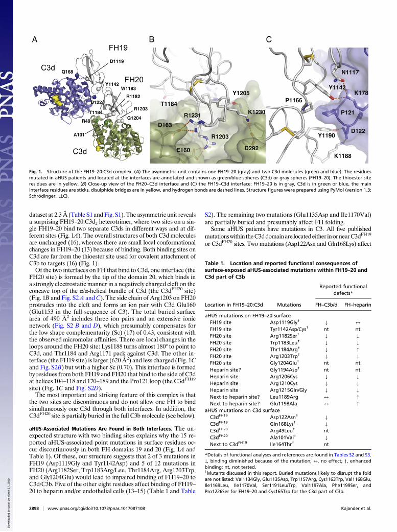
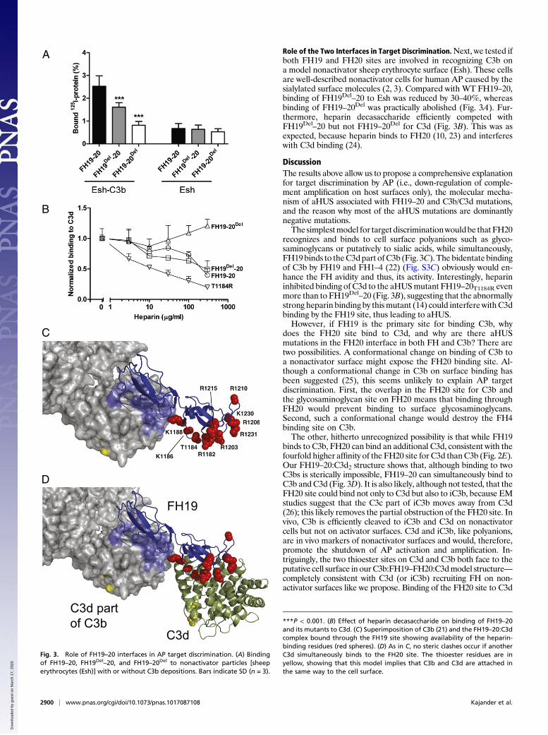
Role of the Two Interfaces in Target Discrimination.Next, we tested ifboth FH19 and FH20 sites are involved in recognizing C3b ona model nonactivator sheep erythrocyte surface (Esh). These cellsare well-described nonactivator cells for human AP caused by thesialylated surface molecules (2, 3). Compared with WT FH19–20,binding of FH19Del
–20 to Esh was reduced by 30–40%, whereasbinding of FH19–20Del was practically abolished (Fig. 3A). Fur-thermore, heparin decasaccharide efficiently competed withFH19Del
–20 but not FH19–20Del for C3d (Fig. 3B). This was asexpected, because heparin binds to FH20 (10, 23) and interfereswith C3d binding (24).
DiscussionThe results above allow us to propose a comprehensive explanationfor target discrimination by AP (i.e., down-regulation of comple-ment amplification on host surfaces only), the molecular mecha-nism of aHUS associated with FH19–20 and C3b/C3d mutations,and the reason why most of the aHUS mutations are dominantlynegative mutations.The simplestmodel for target discriminationwouldbe thatFH20
recognizes and binds to cell surface polyanions such as glyco-saminoglycans or putatively to sialic acids, while simultaneously,FH19 binds to theC3dpart ofC3b (Fig. 3C). The bidentate bindingof C3b by FH19 and FH1–4 (22) (Fig. S3C) obviously would en-hance the FH avidity and thus, its activity. Interestingly, heparininhibited binding of C3d to the aHUSmutant FH19–20T1184R evenmore than to FH19Del
–20 (Fig. 3B), suggesting that the abnormallystrong heparin binding by thismutant (14) could interferewithC3dbinding by the FH19 site, thus leading to aHUS.However, if FH19 is the primary site for binding C3b, why
does the FH20 site bind to C3d, and why are there aHUSmutations in the FH20 interface in both FH and C3b? There aretwo possibilities. A conformational change on binding of C3b toa nonactivator surface might expose the FH20 binding site. Al-though a conformational change in C3b on surface binding hasbeen suggested (25), this seems unlikely to explain AP targetdiscrimination. First, the overlap in the FH20 site for C3b andthe glycosaminoglycan site on FH20 means that binding throughFH20 would prevent binding to surface glycosaminoglycans.Second, such a conformational change would destroy the FH4binding site on C3b.The other, hitherto unrecognized possibility is that while FH19
binds to C3b, FH20 can bind an additional C3d, consistent with thefourfold higher affinity of the FH20 site for C3d thanC3b (Fig. 2E).Our FH19–20:C3d2 structure shows that, although binding to twoC3bs is sterically impossible, FH19–20 can simultaneously bind toC3b andC3d (Fig. 3D). It is also likely, although not tested, that theFH20 site could bind not only to C3d but also to iC3b, because EMstudies suggest that the C3c part of iC3b moves away from C3d(26); this likely removes the partial obstruction of the FH20 site. Invivo, C3b is efficiently cleaved to iC3b and C3d on nonactivatorcells but not on activator surfaces. C3d and iC3b, like polyanions,are in vivo markers of nonactivator surfaces and would, therefore,promote the shutdown of AP activation and amplification. In-triguingly, the two thioester sites on C3d and C3b both face to theputative cell surface in ourC3b:FH19–FH20:C3dmodel structure—completely consistent with C3d (or iC3b) recruiting FH on non-activator surfaces like we propose. Binding of the FH20 site to C3d
A
B
C
C3d part of C3b
FH19
C3d
D
Fig. 3. Role of FH19–20 interfaces in AP target discrimination. (A) Bindingof FH19–20, FH19Del–20, and FH19–20Del to nonactivator particles [sheeperythrocytes (Esh)] with or without C3b depositions. Bars indicate SD (n = 3).
***P < 0.001. (B) Effect of heparin decasaccharide on binding of FH19–20and its mutants to C3d. (C) Superimposition of C3b (21) and the FH19–20:C3dcomplex bound through the FH19 site showing availability of the heparin-binding residues (red spheres). (D) As in C, no steric clashes occur if anotherC3d simultaneously binds to the FH20 site. The thioester residues are inyellow, showing that this model implies that C3b and C3d are attached inthe same way to the cell surface.
2900 | www.pnas.org/cgi/doi/10.1073/pnas.1017087108 Kajander et al.
or iC3b could, thus, replace binding of FH20 to nonactivator surfacepolyanions, further explaining why the C3d and polyanion sitesoverlap on FH20. The spatial organization of these complexes issimilar (Fig. 3 C and D).Our results suggest a comprehensive molecular mechanism for
target discrimination of AP (Fig. 4) and provide evidence fornegative feedback control of activation on nonactivator surfaces.On an activator (nonhost) surface, the carboxyl terminus of FHbinds C3b monovalently and weakly through either the FH19binding site or the partially occluded FH20 binding site on C3b,leading to relatively weak avidity of FH for C3b, poor control ofC3b or C3bBb by FH1–4, subsequent AP activation, and elimi-nation of the target. On nonactivator (host) surfaces, a dual con-tact of FH19–20 occurs: binding to C3b through the FH19 site andto cell surface polyanions through the FH20 glycosaminoglycanbinding site. This increases the avidity of FH for nonactivatorsurface-bound C3b, leading to FH1–4 binding and rapid down-regulation of AP. This also implies negative feedback control ac-tivity. Breakdown of C3b toC3d (or iC3b) on nonactivator surfaceswill increase the number of binding sites for FH19–20. More FHcan be recruited, leading to a more rapid breakdown of C3b toiC3b and C3d. This also suggests why FH mutants can be domi-nantly negative; any mutant that prevents binding or slows downthis regulatory feedback loop would lead to disease.This model explains all published data on aHUS point muta-
tions in FH19–20 and the C3d part of C3b (Fig. 4). Mutations inFH19 affect C3b binding (Fig. 2A), and mutations in FH20 affect,variously, glycosaminoglycan binding and C3d binding (the effectof theGly1194Aspmutation is speculative, because it has not beenexperimentally validated) (Figs. 2A and 3B and Table S1). Allmutations in the C3d domain affect either FH19 or FH20 binding(Fig. 2 B–D). Our model explains how all these mutations reducecontrol of C3b on nonactivator surfaces, leading to aHUS (Fig. 4).The model suggests intriguing future directions for study of bio-compatibility of plasma-exposed materials and complement eva-sion by several FH19–20 binding pathogenic microbes. It alsoprovides a platform for designing therapies for alternative path-way-associated diseases such as aHUS.
Materials and MethodsProteins and Site-Directed Mutagenesis. Mutagenesis, expression, and purifi-cationhave beenpreviously described for FH19–20 (27),most FH19–20mutants(13), andC3d (or C3dg) and C3dgmutants (28). FH constructs were expressed inPichia pastoris and C3d or C3dg constructs in Escherichia coli. New point
mutations were introduced into the FH19–20 similarly to the previously de-scribed mutants using the QuikChange technique (Stratagene) (14). The tem-plate for generating the FH19Del-20 mutant was the Gln1139Ala mutant, andthe primer used to generate the triplemutantwas CATCAGTTGAGTACGCATGCG CGAACT TGG CTC AAC TTGAGGG. The template for FH19–20Del was theArg1203Ala mutant, and the primers used to generate the quadruple mutantwere CAG TTG AAT TTG TGT GTG CAG CGG GAG CTC GTC TTT CAT CAC G andCATAGCATTAAGGTGGGGAGCCAAACAGAAGCT TTA TTCG. The FH19–20mutants were expressed in P. pastoris (14), and FH19Del–20 was purified usingheparin affinity chromatography; however, FH19–20Del was purified with theResource S cation exchange column (GE Healthcare) because of the lack ofheparin binding. C3 and FHwere purified from plasma, and C3bwas preparedfrom C3 as described (9, 29).
A fluorescence thermal shift assay to confirm mutant stability relative toWT was performed by adding 2.5 μL of 1:100 diluted Sypro Orange (Mo-lecular Probes), 2.5 μL of 0.7 mg/mL protein, 20 μL of 0.1 M Hepes, and 20 μLof 0.15 M NaCl, pH 7.5, to wells of a 48-well thin-wall PCR plate (Bio-Rad).The plates were sealed with Optical-Quality Sealing Tape (Bio-Rad) andheated in MiniOpticon RT-PCR equipment (Bio-Rad) from 20 °C to 95 °C inincrements of 1 °C. The wavelengths for excitation and emission were 490and 575 nm, respectively. Tm values for FH19–20Del and FH19Del–20 de-creased by 4 °C and 6 °C, respectively, compared with WT FH19–20 (66 °C).
Crystallization and Solution of the Structure. Despite extensive trials, we wereunable to crystallize FH19–20 with C3d, because FH19–20 always crystallizedby itself as a tetramer (13, 20) in various crystal forms. We reasoned thatinhibiting tetramer formation would help crystallization of the complex, andtherefore, we used the FH19–20 double mutant Asp1119Gly–Gln1139Aladesigned to break the tetramer interface (13). The mutant still bound C3dwell but crystallized poorly by itself. This allowed us to crystallize FH19–20D1119G–Q1139A:C3d complex because the homotetrameric crystals did notform. Small-angle X-ray scattering (SAXS) data also showed that the FH19–20D1119G–Q1139A mutant is clearly monomeric, whereas the WT protein hasa tendency to form higher oligomers and aggregates.
The FH19–20D1119G–Q1139A:C3d complex was crystallized at 22 °C from 0.1M Hepes, pH 7.5, containing 14% PEG 4000 (160 μM FH19–20, 200 μM C3d).Crystals appeared in 3–4 d. All data were collected at the European Syn-chrotron Radiation Facility (ESRF). The initial dataset, to 3.8-Å resolution,was collected at ID14-2; optimization of cryocooling conditions with 10%glycerol enabled us to collect data to 2.3-Å resolution at ID23-1 (Table S1).The initial molecular replacement solution was found with the 3.8-Å data.PHASER (30) readily found two molecules of C3d but not FH19–20. However,closer inspection of Fo–Fc and σA-weighted maps revealed very clear featuresof continuous extra density consistent with a single FH19–20, and therefore,we manually built FH20 into the density between the two C3d molecules,giving an asymmetric unit with two C3d molecules and a single FH19–20.Successive rounds of building with Coot (31) and refinement with REFMAC(32) or PHENIX (33) against the 2.3-Å dataset allowed us to build an essen-tially complete model of the full heterotrimeric complex. The R factors(Rwork/Rfree) are 20.4%/24.3% with good geometry (Table S1). The C3ds areessentially unchanged from earlier structures [root mean square deviations(rmsds)/Cα = 0.27–0.5 Å], whereas there are local conformational changes inthe FH (rmsd/Cα = 1.04 Å). Modeling of the two mutated side chains D1119and Q1139 to the crystal structure (Fig. S5) indicated that Gln1139 wouldhydrogen bond to the Ile115 carbonyl group and Ser171, whereas Asp1119would form an intermolecular helix cap with C3d (Fig. S5). The mutantshould bind C3d less well than WT, which was observed.
Surface Plasmon Resonance Analyses. The binding of C3dgmutants to FH19–20was analyzed on a Biacore 3000 instrument (Biacore/GE Healthcare) essentiallyas described earlier using buffer (pH 7.2) containing 10mMHepes, 0.15 M NaCl,3 mM EDTA, and 0.005% P20 surfactant (28). WT FH19–20 was coupled to CM-5sensor chips to achieve immobilized protein level of∼1,900 resonance units (RU).The concentration of the C3dg mutants in the fluid phase was 2.5 μM.
The surface plasmon resonance (SPR) analyses for testing affinity of theFH19–20 constructs to C3d and C3b were performed on a Biacore 2000 in-strument using CM-5 sensor chips coupled with 500 RU C3d or 2,300 RU C3bto obtain an equal number of coupled molecules. Kinetic analyses wereperformed at 22 °C using PBS as the running buffer at 30 μL/min flow and1.25–40 μM FH19–20 or its mutants.
Radioligand Assays. To test binding of C3dgmutants to FH19–20, 80 μL C3dg orC3dg mutant (10 μg/mL) were coupled overnight onto Nunc Polysorp Break-Apart wells, and the binding of 125I-FH19–20 (6 μg/mL) was measured. To testthe effect of heparin on the interaction, C3d was coupled onto the wells, and
A B C
Defects in aHUS
Nonself surface(activator)
Self surface(nonactivator)
Self surface(nonactivator)
FHFHFH
Fig. 4. Model of regulation of complement amplification and its implica-tions to pathogenesis of aHUS. (A) Weak binding on nonhost cells leads toactivation. (B) In host cells, strong binding to glycosaminoglycans and C3bleads to regulation, except in mutated states (Lower). (C) In host cells, strongbinding to previously deposited C3d (or possibly iC3b) and C3b also leads todown-regulation, except in mutated states in aHUS (Lower). Red and yellowdenote loss- and gain-of-function mutations, respectively.
Kajander et al. PNAS | February 15, 2011 | vol. 108 | no. 7 | 2901
125I-labeled FH19–20 or FH19–20 mutant (6 μg/mL) was added in the presenceof increasing amounts of dextran (Sigma-Aldrich) or heparin decasaccharide(Neoparin, Inc.); dextran had no effect (Fig. S6). The assays were performed in1% BSA in PBS or 1/2 PBS (70 mM NaCl, 5 mM phosphate).
Cell Binding Analyses. Binding of 125I-FH19–20 or FH19–20 mutants to C3b-coated or uncoated sheep erythrocytes was measured after coating the cellswith approximately 25,000 C3b molecules/cell using purified C3, B, and D aspreviously described (29). The amount of cell-bound C3b was calculated usinga trace amount of 125I-labeled C3 in the reaction mixtures. The C3b-coatedcells were incubatedwith 125I-FH19–20 or FH19–20mutants (2 μg/mL) for 2 h at37 °C. The assay was performed in 1% BSA in veronal-buffered saline (VBS) aspreviously described (14).
Statistical Analyses. Statistics and error bars are shown for independent experi-ments.Datawere subjected toone-wayANOVA(n=3or4;α-levels 0.05, 0.01, and0.001) followedbyDunnett’s posttest to compare themeanvaluesobtainedwiththe WT protein vs. the mutant proteins using GraphPad Prism 5.01 software.
ACKNOWLEDGMENTS. We thank Seija Mäki and Katja Rosti for technicalassistance in crystal screening and Marjatta Ahonen and Kirsti Widing fortechnical assistance in protein expression. T.K. is funded by the Academy ofFinland. M.J.L. was partially funded by the Maud Kuistila Memorial Founda-tion, the Emil Aaltonen Foundation, and the Finnish Cultural Foundation.S.M. and T.S.J. are funded by the Academy of Finland, the Sigrid JuséliusFoundation, and the Helsinki University Hospital Funds (EVO). A.G. is fundedby the Academy of Finland and the Sigrid Jusélius Foundation.
1. Meri S, Pangburn MK (1990) Discrimination between activators and nonactivators ofthe alternative pathway of complement: Regulation via a sialic acid/polyanionbinding site on factor H. Proc Natl Acad Sci USA 87:3982–3986.
2. Fearon DT (1978) Regulation by membrane sialic acid of β1H-dependent decay-dissociation of amplification C3 convertase of the alternative complement pathway.Proc Natl Acad Sci USA 75:1971–1975.
3. Pangburn MK, Müller-Eberhard HJ (1978) Complement C3 convertase: Cell surfacerestriction of β1H control and generation of restriction on neuraminidase-treatedcells. Proc Natl Acad Sci USA 75:2416–2420.
4. Manuelian T, et al. (2003) Mutations in factor H reduce binding affinity to C3b andheparin and surface attachment to endothelial cells in hemolytic uremic syndrome.J Clin Invest 111:1181–1190.
5. Gordon DL, Kaufman RM, Blackmore TK, Kwong J, Lublin DM (1995) Identification ofcomplement regulatory domains in human factor H. J Immunol 155:348–356.
6. Kühn S, Skerka C, Zipfel PF (1995) Mapping of the complement regulatory domains inthe human factor H-like protein 1 and in factor H1. J Immunol 155:5663–5670.
7. Pangburn MK (2002) Cutting edge: Localization of the host recognition functions ofcomplement factor H at the carboxyl-terminal: Implications for hemolytic uremicsyndrome. J Immunol 169:4702–4706.
8. Jokiranta TS, et al. (2005) Binding of complement factor H to endothelial cells ismediated by the carboxy-terminal glycosaminoglycan binding site. Am J Pathol 167:1173–1181.
9. Jokiranta TS, Hellwage J, Koistinen V, Zipfel PF, Meri S (2000) Each of the threebinding sites on complement factor H interacts with a distinct site on C3b. J Biol Chem275:27657–27662.
10. Blackmore TK, et al. (1998) Identification of the second heparin-binding domain inhuman complement factor H. J Immunol 160:3342–3348.
11. Ram S, et al. (1998) A novel sialic acid binding site on factor H mediates serumresistance of sialylated Neisseria gonorrhoeae. J Exp Med 187:743–752.
13. Jokiranta TS, et al. (2006) Structure of complement factor H carboxyl-terminus revealsmolecular basis of atypical haemolytic uremic syndrome. EMBO J 25:1784–1794.
14. Lehtinen MJ, Rops AL, Isenman DE, van der Vlag J, Jokiranta TS (2009) Mutations offactor H impair regulation of surface-bound C3b by three mechanisms in atypicalhemolytic uremic syndrome. J Biol Chem 284:15650–15658.
15. Ferreira VP, et al. (2009) The binding of factor H to a complex of physiologicalpolyanions and C3b on cells is impaired in atypical hemolytic uremic syndrome.J Immunol 182:7009–7018.
16. Nagar B, Jones RG, Diefenbach RJ, Isenman DE, Rini JM (1998) X-ray crystal structureof C3d: A C3 fragment and ligand for complement receptor 2. Science 280:1277–1281.
17. Lawrence MC, Colman PM (1993) Shape complementarity at protein/proteininterfaces. J Mol Biol 234:946–950.
18. Frémeaux-Bacchi V, et al. (2008) Mutations in complement C3 predispose to
development of atypical hemolytic uremic syndrome. Blood 112:4948–4952.19. Maga TK, Nishimura CJ, Weaver AE, Frees KL, Smith RJ (2010) Mutations in alternative
pathway complement proteins in American patients with atypical hemolytic uremic
syndrome. Hum Mutat 31:E1445–E1460.20. Bhattacharjee A, Lehtinen MJ, Kajander T, Goldman A, Jokiranta TS (2010) Both
domain 19 and domain 20 of factor H are involved in binding to complement C3b and
C3d. Mol Immunol 47:1686–1691.21. Janssen BJ, Christodoulidou A, McCarthy A, Lambris JD, Gros P (2006) Structure of C3b
reveals conformational changes that underlie complement activity. Nature 444:
213–216.22. Wu J, et al. (2009) Structure of complement fragment C3b-factor H and implications
for host protection by complement regulators. Nat Immunol 10:728–733.23. Herbert AP, Uhrín D, Lyon M, Pangburn MK, Barlow PN (2006) Disease-associated
sequence variations congregate in a polyanion recognition patch on human factor H
revealed in three-dimensional structure. J Biol Chem 281:16512–16520.24. Hellwage J, et al. (2002) Complement C3b/C3d and cell surface polyanions are
recognized by overlapping binding sites on the most carboxyl-terminal domain of
complement factor H. J Immunol 169:6935–6944.25. Nilsson B, Nilsson Ekdahl K, Avila D, Nilsson UR, Lambris JD (1990) Neoantigens in
complement component C3 as detected by monoclonal antibodies. Mapping of the
recognized epitopes by synthetic peptides. Biochem J 268:55–61.26. Nishida N, Walz T, Springer TA (2006) Structural transitions of complement
component C3 and its activation products. Proc Natl Acad Sci USA 103:19737–19742.27. Cheng ZZ, et al. (2005) Complement factor H as a marker for detection of bladder
cancer. Clin Chem 51:856–863.28. Isenman DE, Leung E, Mackay JD, Bagby S, van den Elsen JM (2010) Mutational
analyses reveal that the staphylococcal immune evasion molecule Sbi and