54
University of Cyprus Biomedical Imaging and Applied Optics ECE 370 Introduction to Biomedical Engineering The Nervous System
University of Cyprus
Biomedical Imaging and Applied Optics
ECE 370
Introduction to Biomedical Engineering
The Nervous System

2 2
Membrane Potential
• Opposite charges attract and similar repel
• Membrane potential opposite charges across the membrane
• Equal number of + and – on each side electrically neutral
• Charges separated (more + on one side, more – on other) electrical potential
• Measured in V
• More charge ↑ V
• Note:
• Only a very small number of charges is involved majority of ECF and ICF is still neutral
3 3
Membrane Potential
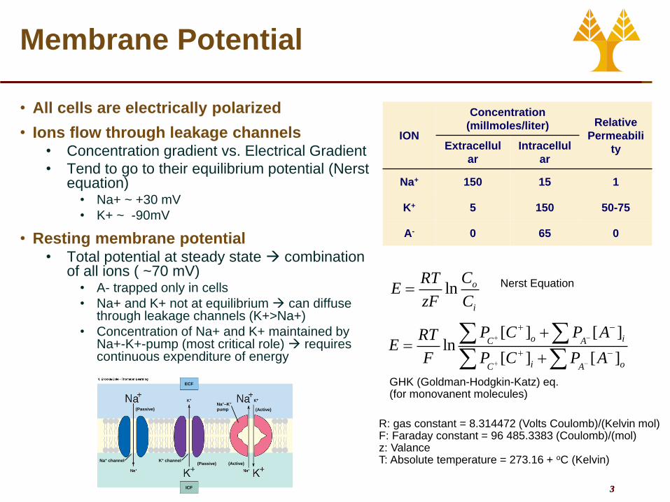
• All cells are electrically polarized
• Ions flow through leakage channels • Concentration gradient vs. Electrical Gradient
• Tend to go to their equilibrium potential (Nerst equation)
• Na+ ~ +30 mV
• K+ ~ -90mV
• Resting membrane potential • Total potential at steady state combination
of all ions ( ~70 mV) • A- trapped only in cells
• Na+ and K+ not at equilibrium can diffuse through leakage channels (K+>Na+)
• Concentration of Na+ and K+ maintained by Na+-K+-pump (most critical role) requires continuous expenditure of energy
ION
Concentration
(millmoles/liter) Relative
Permeabili
ty Extracellul
ar
Intracellul
ar
Na+ 150 15 1
K+ 5 150 50-75
A- 0 65 0
ln o
i
CRTE
zF C
[ ] [ ]ln
[ ] [ ]
o iC A
i oC A
P C P ARTE
F P C P A
Nerst Equation
GHK (Goldman-Hodgkin-Katz) eq. (for monovanent molecules)
R: gas constant = 8.314472 (Volts Coulomb)/(Kelvin mol) F: Faraday constant = 96 485.3383 (Coulomb)/(mol) z: Valance T: Absolute temperature = 273.16 + oC (Kelvin)
ICF
ECF
(Passive)Na+–K+
pump (Active)
(Active)(Passive)K+ channelNa+ channel

4 4
Excitable Tissues
• Changes in membrane
potential serve as signals
• Nerve and muscle are
excitable tissue
• Change their membrane
potential to produce electrical
signals
• Neurons messages
• Muscle contraction
Polarization
• When a potential (either + or -) exists across a
membrane
Depolarization
• Reduction of the magnitude of potential (e.g. -70
mV -50 mV)
Repolarization
• Return to resting potential
Hyperpolarization
• Increase in the magnitude of the potential (e.g. -
70 mV -90 mV)

5 5
Excitable Tissues
• Changes are triggered by
• Interaction of chemical messenger with receptors and channels
• Other stimulus (e.g. light, current, etc)
• Spontaneous change of potential by inherent ion leaks
• Changes are caused by movement of ions
• Leak channels (Open all the time)
• Gated channels (Closed but can be opened)
• Voltage, chemically, mechanically, or thermally gated
• Electrical signals
• Graded Potentials
• Action Potentials
6 6
Graded Potentials
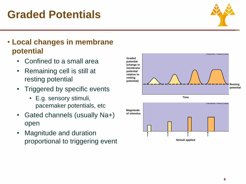
• Local changes in membrane
potential
• Confined to a small area
• Remaining cell is still at
resting potential
• Triggered by specific events
• E.g. sensory stimuli,
pacemaker potentials, etc
• Gated channels (usually Na+)
open
• Magnitude and duration
proportional to triggering event
Graded
potential
(change in
membrane
potential
relative to
resting
potential)
Magnitude
of stimulus
Resting
potential
Time
Stimuli applied

7 7
Graded Potentials
• Graded potentials die out over short distances
• Loss of charge
• Magnitude decreases as it moves away from the point of origin
• Completely disappear with a few mm
• ri inversly proportional to cross-
sectional area • ↑ diameter ↓ri
• ↑ rm better flow along the axis due to decrease loss of ions through the membrane
Portion of
excitable cellInitial site of
potential change
Loss of charge Loss of charge
Direction of current
flow from initial siteDirection of current
flow from initial site
* Numbers refer to the local potential in mV
at various points along the membrane.
i
m
r
r
x
eVV
0

8 8
Action Potentials
• Large (~100 mV) changes in the membrane potential
• Can be initiated by graded potentials
• Unlike graded potentials action potentials propagate
• Transmit information
• Changes during an action potential • Gradual depolarization to threshold
potential (-50 to -55 mV) • If not reached no action potential will occur
• Rapid depolarization (+30 mV) • Opening of voltage gated Na+ channels
• Rapid repolarization leading to hyperpolarization (-80 mV)
• Inactivation of Na+ channels, opening of voltage gated K+ channels
• Resting potential restored (-70 mV) • All voltage gated channels closed
• Constant duration and amplitude for given cell type (“all-or-none”)
• E.g. Nerves 1 msec
K+ equilibrium
potential
Na+ equilibrium
potential

9 9
Action Potentials
Time Event Potential
0 msec Resting state
All channels are closed Graded potential arrives
Begins depolarization
- 70 mV
2 msec Threshold reached
Activation gates of Na+ channels open
Activation gates of K+ channels begin to open slowly
Inactivation gates of Na+ channels begin to close slowly
- 50 mV
2.5 msec Peak potential reached
Inactivation gates of Na+ channels are now closed
Activation gates of K+ channels are now open
30 mV
3.75 msec Hyperpolarized state
Activation gates of K+ channels close - 80 mV
5 msec Resting state
Na+-K+-pump restores resting potential
Na+ channels are reset to close but active
-70 mV

10 10
Action Potentials
• Neuron structure • Input Zone
• Dendrites (up to 400 000)
• Cell Body
• Have receptors which receive chemical signals
• Conduction zone • Axon or nerve fiber (axon
hillock to axon terminals) <1 mm to >1m
• Output zone • Axon terminal
• Input • Graded Potentials
• Generated in the dendrites as a response to chemical signals
• Can trigger action potentials in the axon

11 11
Action Potentials
• AP Propagation
• APs initiated at the axon hilloc
• More voltage-gated channels lower threshold
• Once initiated the AP travels the entire axon
• Contiguous conduction
• Saltatory conduction
• Contiguous conduction
• Flow of ions depolarization of adjacent area to threshold
• As AP is initiated in adjacent area, the original AP is ending with repolarization
• The AP itself does not travel, it is regenerated at successive locations (like “wave” in a stadium)
12 12
Action Potentials
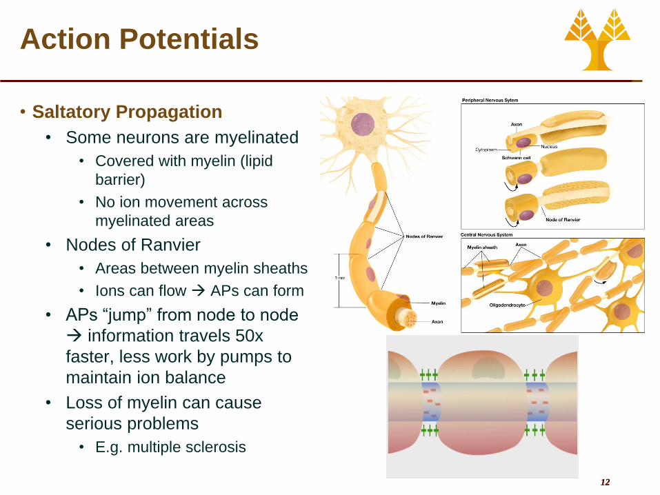
• Saltatory Propagation
• Some neurons are myelinated
• Covered with myelin (lipid
barrier)
• No ion movement across
myelinated areas
• Nodes of Ranvier
• Areas between myelin sheaths
• Ions can flow APs can form
• APs “jump” from node to node
information travels 50x
faster, less work by pumps to
maintain ion balance
• Loss of myelin can cause
serious problems
• E.g. multiple sclerosis

13 13
Action Potentials
• Refractory Period
• APs do not travel backwards
• Local currents do not regenerate an
AP in the previously-active-now-
inactive area
• Certain time must pass before a
second AP can be triggered
refractory period
• Absolute refractory period
• During an AP
• No APs can be triggered
• Relative refractory period
• Na+ channels are mostly inactive
• K+ channels are slow to close
• After an AP second AP can be
triggered only be exceedingly strong
signals
• Refractory period sets an upper limit
to the frequency of APs ~2.5 KHz
Previous active
area returned to
resting potential
New active area
at peak of action
potential
New adjacent inactive area
into which depolarization
is spreading; will soon reach
threshold
“Backward” current
flow does not re-excite
previously active area
because this area is
in its refractory period
“Forward” current flow excites new inactive area
Direction of propagation of
action potential
Absolute
refractory
period
Relative
refractory
period
Action potential
Na+ permeability
K+ permeability

14 14
Action Potentials
• Characteristics of APs • How does strength vary?
• Always the same! All-or-None Law
• Does not decrease during propagation
• How are stronger stimuli recognized? • Faster generation of APs
↑Frequency
• More neurons fire simultaneously
• What determines the speed of APs? • Myelination
• Neuron diameter (↑ diameter ↓ Resistance to local current ↑ Speed)
• Large myelinated fibers: 120 m/sec (432 km/hr) urgent information
• Small unmyelinated fiber: 0.7 m/sec (2.5 km/hr) slow-acting processes
• Without myelin the diameter would have to be huge! (50 x larger)
15 15
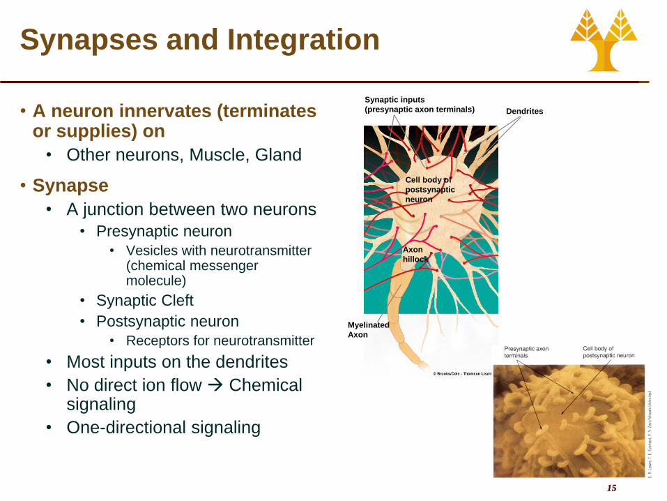
Synaptic inputs
(presynaptic axon terminals) Dendrites
Cell body of
postsynaptic
neuron
Axon
hillock
Myelinated
Axon
Synapses and Integration
• A neuron innervates (terminates or supplies) on
• Other neurons, Muscle, Gland
• Synapse
• A junction between two neurons
• Presynaptic neuron
• Vesicles with neurotransmitter (chemical messenger molecule)
• Synaptic Cleft
• Postsynaptic neuron
• Receptors for neurotransmitter
• Most inputs on the dendrites
• No direct ion flow Chemical signaling
• One-directional signaling

16 16
Synapses and Integration
• Synaptic Signaling • AP reaches the synaptic knob
• Voltage-gated Ca2+ channels open
• Ca2+ flows into the synapse from the ECF
• Ca2+ induces exocytosis of vesicles and release of neurotransmitter
• Neurotransmitter diffuses across the synaptic cleft to the post-synaptic neuron and binds to specific receptors
• Binding triggers opening of ion channels
• Cause permeability changes of different ions
• Can be • excitatory (cations)
depolarization, or
• inhibitory synapses (anions) hyperpolarization

17 17
Synapses and Integration
• Neurotransmitters and Receptors • Several neurotransmitters
• Each neurotransmitter can bind to a variety of receptors
• Each particular neuron releases one specific neurotransmitter and each synapse has one specific receptor
• Each neurotransmitter-receptor combination produces the same response
• Neurotransmitters combined with different receptors can produce different responses
• Neurotransmitter clearing • Removal or inactivation to stop the end the signal
• Inactivation by specific enzymes within the subsynaptic membrane
• Reuptake back in the axon recycling

18 18
Synapses and Integration
• Excitatory Synapses • Open non-specific cation channels
• More Na+ flows into the cell than K+ flows out
• Net result Excitatory Postsynaptic Potential (a small depolarization)
• Inhibitory Synapses • Different neurotransmitters
• Open either K+ or Cl- channels
• K+ efflux or Cl- influx Inhibitory Postsynaptic Potential (a small hyperpolarization)
• Usually one EPSP is not enough to trigger an AP
• Membrane is now more excitable
• Synaptic Delay • 0.5 to 1 msec
• Travel through more synapses ↑Total reaction time

19 19
Synapses and Integration
• Grand Postsynaptic Potential (GPSP)
• Summation of EPSPs and IPSPs
(graded potentials)
• About 50 EPSPs are required to
initiate AP
• Temporal Summation
• EPSPs occurring very close in time
can be summed
• E.g. repeated firing of pre-synaptic
neuron because of a persistent input
• Spatial Summation
• EPSPs from different but adjacent
synapses can be summed
• Concurrent EPSPs and IPSPs
• Cancel each other (more or less)
depending on amplitude and location

20 20
Synapses and Integration
• Post-synaptic Integration
• APs are initiated depending on a combination of inputs
• Neuron is a complex computational device
• Synapses = inputs
• Dendrites = processors
• Axons/APs = output
• Signaling and frequency of APs is a result of integration of information from different sources
• Information not significant enough is not passed at all
• Neurons are linked into complex networks (1011 neurons and 1014 synapses in the brain alone!)
• Converging
• Diverging
• Massively parallel processing
Presynaptic
inputs
Postsynaptic
neuron
Convergence of input
(one cell is influenced
by many others)
Presynaptic
inputs
Divergence of output
(one cell influences
many others)
Postsynaptic
neurons
Arrows indicate direction in which information is being conveyed.

21 21
Synapses and Integration
• Effects of drugs and diseases
• Drug actions may include
• Altering the synthesis, axonal transport, storage, or release of a neurotransmitter
• Modifying the neurotransmitter interaction with the postsynaptic receptor
• Influence neurotransmitter reuptake or destruction
• Replace a neurotransmitter with a substitute either more or less powerful
• Examples
• Cocaine blocks reuptake of neurotransmitter dopamine pleasure pathways remain “on”
• Tetanus toxin prevents release of inhibitory neurotransmitter GABA muscle excitation unchecked uncontrolled muscle spasms
• Strychnine blocks the receptor of inhibitory neurotransmitter glycine convulsions, muscle spasticity
22 22
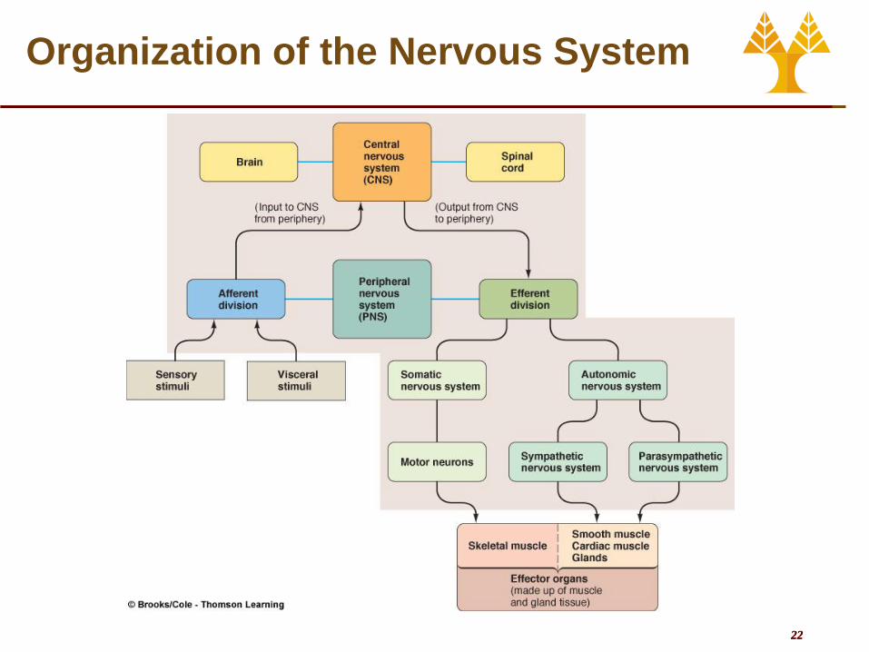
Organization of the Nervous System
23 23
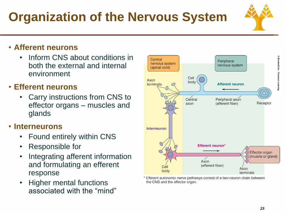
Organization of the Nervous System
• Afferent neurons
• Inform CNS about conditions in both the external and internal environment
• Efferent neurons
• Carry instructions from CNS to effector organs – muscles and glands
• Interneurons
• Found entirely within CNS
• Responsible for
• Integrating afferent information and formulating an efferent response
• Higher mental functions associated with the “mind”

24 24
Glial Cells
• 90 % of cells in the CNS (50 % of the volume)
• Communicate with chemical signals (no electrical impulses)
• Role • Support neurons physically
and metabolically
• Actively modulate synaptic function (major role in learning and memory)
• Provide immunologic protection
• Synthesize cerebrospinal fluid (CSF)
• Glial cells are the origin of most neural tumor (gliomas)
• Neurons can not divide
25 25
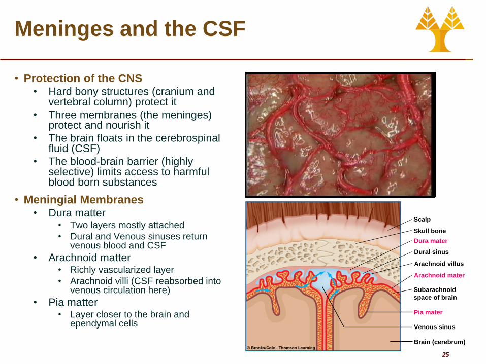
Meninges and the CSF
• Protection of the CNS • Hard bony structures (cranium and
vertebral column) protect it
• Three membranes (the meninges) protect and nourish it
• The brain floats in the cerebrospinal fluid (CSF)
• The blood-brain barrier (highly selective) limits access to harmful blood born substances
• Meningial Membranes • Dura matter
• Two layers mostly attached
• Dural and Venous sinuses return venous blood and CSF
• Arachnoid matter • Richly vascularized layer
• Arachnoid villi (CSF reabsorbed into venous circulation here)
• Pia matter • Layer closer to the brain and
ependymal cells
Subarachnoid
space of brain
Arachnoid villus
Dural sinus
Pia mater
Dura mater
Arachnoid mater
Scalp
Skull bone
Venous sinus
Brain (cerebrum)

26 26
Meninges and the CSF
• Cerebrospinal Fluid (CSF)
• Characteristics
• Same density as brain Brain floats in and is cushioned by the CSF
• CSF and interstitial fluid of the brain cells are free to exchange materials CSF composition must be carefully regulated
• Formed by choroid plexuses in the Flow around the brain and the spinal cord
• Pressure
• 10 mm Hg.
• Even small reduction (e.g. during spinal tabs) can lead to severe headaches
Subarachnoid space of brain
Arachnoid villus
Lateral ventricle
Dural sinus
Venous blood
Cerebrum
Vein
Choroid plexus
of lateral ventricle
Choroid plexus
of third ventricle
Third ventricle
Aperture
of fourth ventricle
Choroid plexus
of fourth ventricle
Central canal
Fourth ventricle

27 27
Meninges and the CSF
• Blood-Brain Barrier (BBB) • Tight junctions between endothelial cells of
brain capillaries (anatomical restriction)
• Few materials allowed to freely diffuse • Lipid soluble substances (O2, CO2, alcohol,
steroid hormones
• Water
• Careful and controlled exchange between blood and CSF for everything else
• Advantage • Brain shielded from changes in the ECF
and harmful blood borne materials
• Disadvantage • Limited types of drugs can pass through
BBB
• Brain Nourishment • Brain can only use glucose and can only
metabolize aerobically (O2 present)
• Highly dependent on blood supply
• Very sensitive to blood supply variations • Damage if O2 deprived for > 4-5 mins
Most capillaries in body
Cell
forming
capillary
wall
Pore passage
Lipid-soluble
substances
Transport
mechanisms
Water-lined pore
Lipid-soluble
substances
Transport
mechanisms
Astrocyte
processes
Tight junction (no pores)
Carrier-mediated transport
Brain capillaries

28 28
Overview of the CNS

29 29
Cerebral Cortex
• Cerebrum
• Left and right hemispheres
• Gyri and sulci
• Corpus callosum connects left
and right
• White matter (myelinated
axons)
• Inner most layer
• Interconnects
• Cerebral cortex or Gray
matter (cell bodies)
• Outermost layer
• Divided into four pairs of lobes

30 30
Cerebral Cortex
• Cerebral cortex lobes • Frontal
• Voluntary motor activity
• Language (speech production)
• Strategic planning (character?)
• Elaboration of thought
• Parietal
• Somatosensory processing
• Sensory Integration
• Higher visual processing
• Language (speech comprehension)
• Temporal
• Primary auditory and olfactory processing
• Emotion, Motivation
• Memory/learning
• Higher visual processing
• Occipital
• Primary visual processing

31 31
Cerebral Cortex

32 32
Cerebral Cortex
• Parietal Lobe – Primary Somatosensory Cortex
• Somesthetic sensation sensations from the surface of the body - touch, pain, pressure, heat and cold- and proprioception (awareness of body position)
• Projected to the somatosensory cortex (initial cortical processing and perception)
• Body regions are topographically mapped
• Different parts of the body are not equally represented
• Sensory Homonculus
• Proportional to precision and sensitivity
• Receives information from the opposite side of the body
• damage on right side results in sensory loss on left side)

33 33
Cerebral Cortex

34 34
Cerebral Cortex
• Frontal lobe – Primary Motor Cortex
• Voluntary control for muscle movement
• Motor cortex on each side controls muscles on the opposite side of the body
• Tracts originating in the cortex cross (at level of pyramids) before continuing down spinal cord to terminate muscle
• Body regions are topographically mapped
• Different parts of the body are not equally represented
• Motor Homonculus
• Proportional to precision and complexity of motor skills
• Controls the opposite side of the body
• Damage on right side results in motor deficit on left side
35 35
Cerebral Cortex
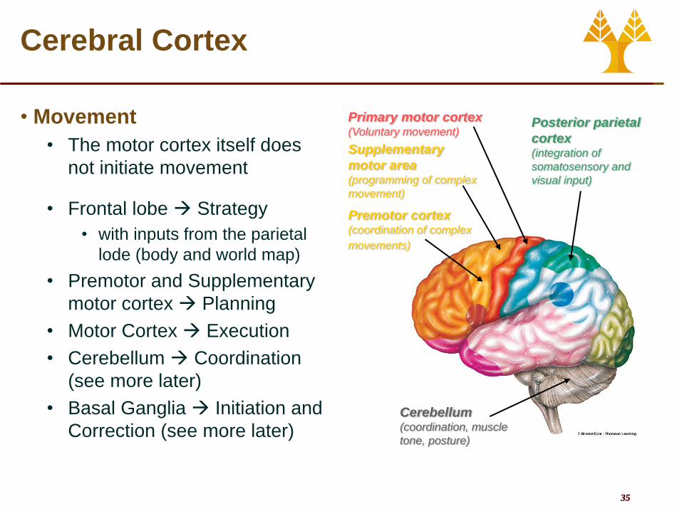
• Movement
• The motor cortex itself does
not initiate movement
• Frontal lobe Strategy
• with inputs from the parietal
lode (body and world map)
• Premotor and Supplementary
motor cortex Planning
• Motor Cortex Execution
• Cerebellum Coordination
(see more later)
• Basal Ganglia Initiation and
Correction (see more later)
Supplementary
motor area (programming of complex
movement)
Primary motor cortex (Voluntary movement)
Posterior parietal
cortex (integration of
somatosensory and
visual input)
Premotor cortex (coordination of complex
movements)
Cerebellum (coordination, muscle
tone, posture)

36 36
Cerebral Cortex
• Language • Areas responsible for language ability are found
in only 1 hemisphere (usually the left)
• Language involves the integration of 2 distinct capabilities
• Expression (speaking ability)
• Comprehension (understanding ability)
• Broca’s area • Responsible for speaking ability
• Frontal lobe - in association with the motor area that controls the muscles necessary for articulation
• Damage to Broca’s area Expressive aphasia • Failure of word formation
• The patient can still understand the spoken and written word
• Know what they want to say but cannot express it
• Wernicke’s area • Functions for language comprehension
• Parietal-temporal-occipital association cortex - critical role in understanding both written and spoken language
• Damage to Wernicke’s area receptive aphasia • Loss of understanding of words seen or heard
• Can speak fluently, but their words make no sense
• Cannot attach meaning to words nor choose appropriate words to express thoughts
Wernicke’s area (speech understanding)
Broca’s area (speech formation)

37 37
Cerebral Cortex
• Lateralization/dominance of the cerebral hemispheres
• Each hemisphere receives information from both sides of the body
• Connections via the corpus callosum
• Left hemisphere better at • logical, analytical, sequential, and verbal tasks
• Describing facial appearances
• Right hemisphere better at • Spatial perception
• Artistic and musical talents
• Recognizing faces
• Brain plasticity • Somatotopic maps
• Dynamic, not static • Use-dependent competition
• Plasticity • Functional remodeling of brain
• More pronounced in early developmental years
• Adults retain some plasticity
• Brain injuries • Other regions adapted to cover deficits

38 38
Basal Ganglia
• Masses of grey matter deep inside the white matter
• Act by modifying ongoing activity in motor pathways
• Inhibit muscle tone • Proper tone - balance of excitatory and
inhibitory inputs to motor neurons that innervate skeletal muscle
• Select and maintain purposeful motor activity while suppressing unwanted patterns of movement
• Monitor and coordinate slow and sustained contractions
• Especially those related to posture and support
• Parkinson’s disease • Damage to basal ganglia neurons and a
deficiency in dopamine • Increased muscle tone or rigidity
• Resting tremors (eg unwanted movements
• Slowness in initiating and carrying out motor behaviors
Caudate
Nucleus
Putamen Globus
Pallidus
39 39
Diencephalon
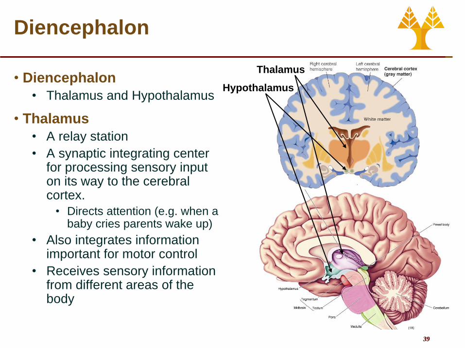
• Diencephalon
• Thalamus and Hypothalamus
• Thalamus
• A relay station
• A synaptic integrating center for processing sensory input on its way to the cerebral cortex.
• Directs attention (e.g. when a baby cries parents wake up)
• Also integrates information important for motor control
• Receives sensory information from different areas of the body
Hypothalamus
Thalamus
40 40
Diencephalon
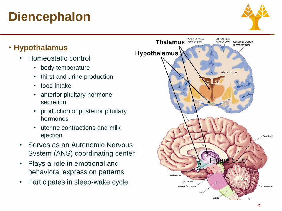
• Hypothalamus
• Homeostatic control
• body temperature
• thirst and urine production
• food intake
• anterior pituitary hormone
secretion
• production of posterior pituitary
hormones
• uterine contractions and milk
ejection
• Serves as an Autonomic Nervous
System (ANS) coordinating center
• Plays a role in emotional and
behavioral expression patterns
• Participates in sleep-wake cycle
Hypothalamus
Thalamus
Figure 5-16

41 41
Limbic System
• Several structures that function together
• Cortex (limbic association cortex)
• Cingulate gyrus
• Hippocampus
• Amygdala
• Basal Nuclei
• Thalamus
• Hypothalamus
• Plays a role in • Emotional state
• Basic behavioral patterns
• Reward and Punishment
• Motivation
• Learning and memory
Cingulate gyrus
Fornix
Thalamus
Hippocampus
Amygdala
Hypothalamus
42 42
Limbic System
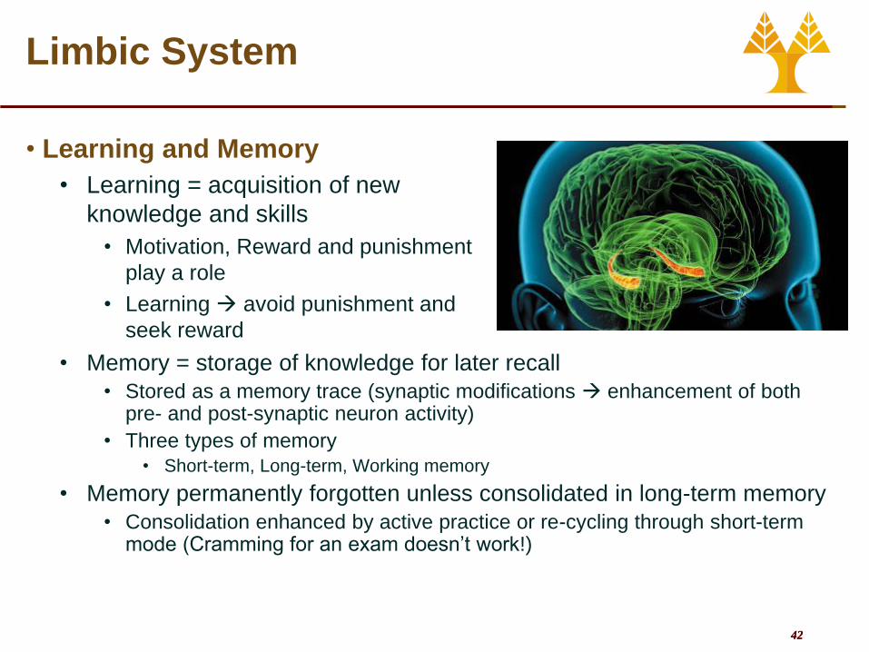
• Learning and Memory
• Learning = acquisition of new
knowledge and skills
• Motivation, Reward and punishment
play a role
• Learning avoid punishment and
seek reward
• Memory = storage of knowledge for later recall
• Stored as a memory trace (synaptic modifications enhancement of both pre- and post-synaptic neuron activity)
• Three types of memory
• Short-term, Long-term, Working memory
• Memory permanently forgotten unless consolidated in long-term memory
• Consolidation enhanced by active practice or re-cycling through short-term mode (Cramming for an exam doesn’t work!)

43 43
Cerebellum
• Highly folded, posterior, part of brain
• Important in • Balance
• Coordination of voluntary movement
• Procedural memories (e.g. motor skills gained through repetitive training
• Activities • Maintenance of balance, control of
eye movements
• Regulation of muscle tone (enhancement, opposite of basal nuclei), coordination of skilled voluntary movement
• Planning and initiation of voluntary activity
• Cerebellar disease • Intention tremor present only
during voluntary activity
44 44
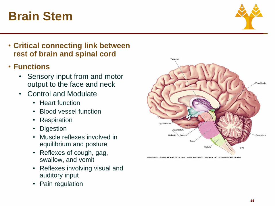
Brain Stem
• Critical connecting link between rest of brain and spinal cord
• Functions
• Sensory input from and motor output to the face and neck
• Control and Modulate
• Heart function
• Blood vessel function
• Respiration
• Digestion
• Muscle reflexes involved in equilibrium and posture
• Reflexes of cough, gag, swallow, and vomit
• Reflexes involving visual and auditory input
• Pain regulation

45 45
Spinal Cord
• Extends from brain stem through vertebral canal • Below L2 turns into a bundle of nerves
• Cauda equina
• Spinal tabs are taken below this point
• Two vital functions • Neuronal link between brain and PNS (bidirectional flow of information)
• Integrating center for spinal reflexes
Spinal cord
Dorsal root
ganglion
Spinal
nerve
Vertebra
Meninges
(protective
coverings)
Intervertebral
diskSympathetic
ganglion
chain

46 46
Spinal Cord
• 31 pairs of spinal nerves emerge from spinal cord through spaces formed between arches of adjacent vertebrae
• Named for region of vertebral column from which they emerge • 8 pairs cervical (neck) nerves
• 12 pairs thoracic (chest) nerves
• 5 pairs lumbar (abdominal) nerves
• 5 pairs sacral (pelvic) nerves
• 1 pair coccygeal (tailbone) nerves

47 47
Spinal Cord
• The spinal cord is an integrating center for many basic reflexes
• Reflex • Any response that occurs
automatically without conscious effort
• Two types of reflexes • Simple, or basic, reflexes
• Built-in, unlearned responses
• Acquired, or conditioned, reflexes • Result of practice and learning
• Reflex Arc • Neural pathway involved in
accomplishing reflex activity
• Five basic components • Receptor
• Afferent pathway
• Integrating center
• Efferent pathway
• Effector
Receptor
Aff
ere
nt
path
way
Integrating center
Effe
ren
t path
way
Effector

48 48
Spinal Cord
Stimulus
Biceps
(flexor)
contracts
Hand
withdrawn
Triceps
(extensor)
relaxes
Ascending pathway
to brain
Response
Integrating center
(spinal cord)
Thermalpain receptor
in finger
Efferent pathway
Effector
organs
= Inhibitory interneuron
= Excitatory interneuron
= Synapse
= Inhibits
= Stimulates
Afferent
Pathway

49 49
The Peripheral Nervous System
• Peripheral Nervous System • Afferent Division
• Sends information from the PNS to the CNS
• Efferent Division • Send information from the CNS to the PNS
• Afferent Division • Visceral afferents (subconscious input)
• Pressure, O2, temperature, etc.
• Sensory afferents (conscious input) • Somatic sensation
• Somesthetic sensation from skin
• Proprioception from muscle joints, skin and inner ear
• Special senses • Vision, hearing, taste and smell
• Efferent Division • Autonomic Nervous System
• Cardiac muscle, smooth muscle, most exocrine glands, some endocrine glands, adipose tissue
• Somatic Nervous system • Skeletal muscle
50 50
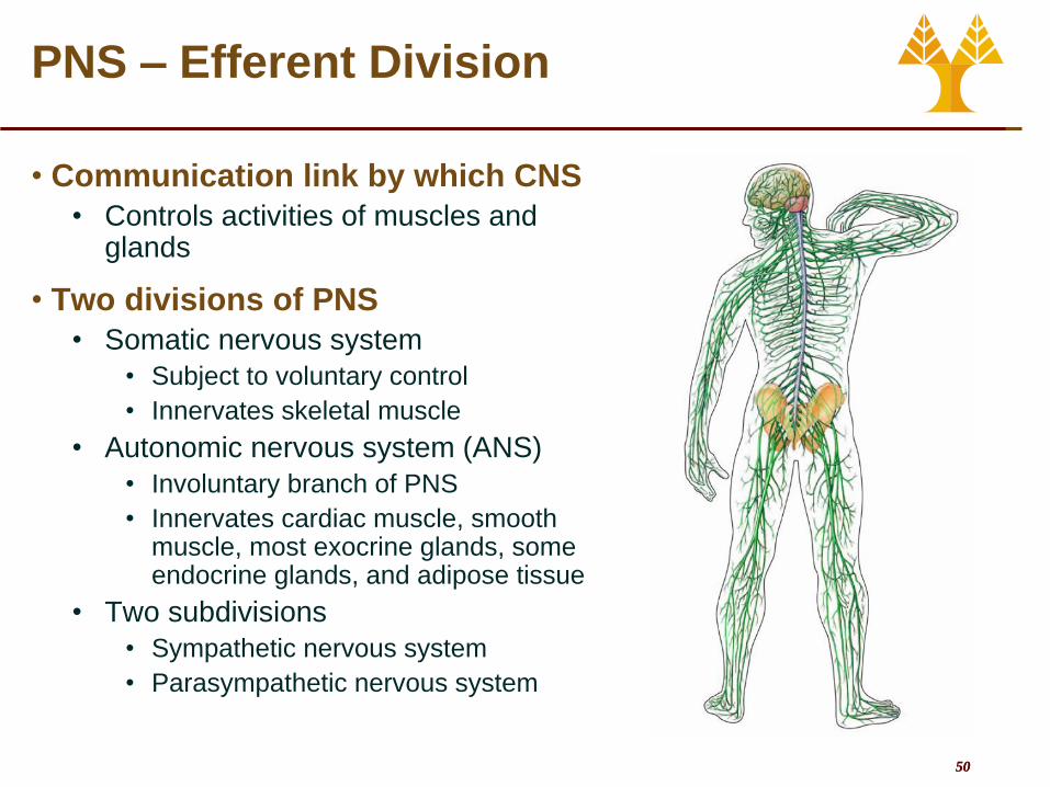
PNS – Efferent Division
• Communication link by which CNS
• Controls activities of muscles and glands
• Two divisions of PNS
• Somatic nervous system
• Subject to voluntary control
• Innervates skeletal muscle
• Autonomic nervous system (ANS)
• Involuntary branch of PNS
• Innervates cardiac muscle, smooth muscle, most exocrine glands, some endocrine glands, and adipose tissue
• Two subdivisions
• Sympathetic nervous system
• Parasympathetic nervous system

51 51
Autonomic Nervous System
• Most visceral organs innervated by both sympathetic and parasympathetic fibers
• In general produce opposite effects in a particular organ
• Dual innervation allows precise control of organ’s activity
• Coordinated by the hypothalamus and executed via the brain stem
• Sympathetic system dominates in emergency or stressful (“fight-or-flight”) situations
• Promotes responses that prepare body for strenuous physical activity
• Neurotransmitter is epinephrine (aka adrenaline)
• Parasympathetic system dominates in quiet, relaxed (“rest-and-digest”) situations
• Promotes body-maintenance activities such as digestion
• Neurotransmitter is Acetylcholine

52 52
Autonomic Nervous System
(except in muscle and lungs)
53 53
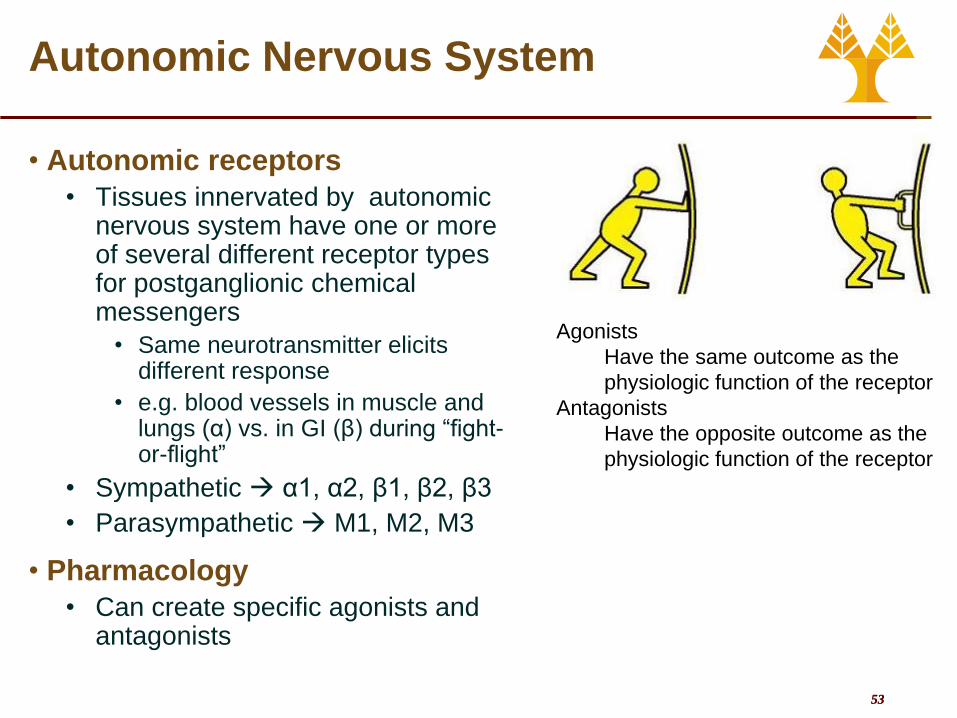
Autonomic Nervous System
• Autonomic receptors
• Tissues innervated by autonomic nervous system have one or more of several different receptor types for postganglionic chemical messengers
• Same neurotransmitter elicits different response
• e.g. blood vessels in muscle and lungs (α) vs. in GI (β) during “fight-or-flight”
• Sympathetic α1, α2, β1, β2, β3
• Parasympathetic M1, M2, M3
• Pharmacology
• Can create specific agonists and antagonists
Agonists
Have the same outcome as the
physiologic function of the receptor
Antagonists
Have the opposite outcome as the
physiologic function of the receptor
54 54
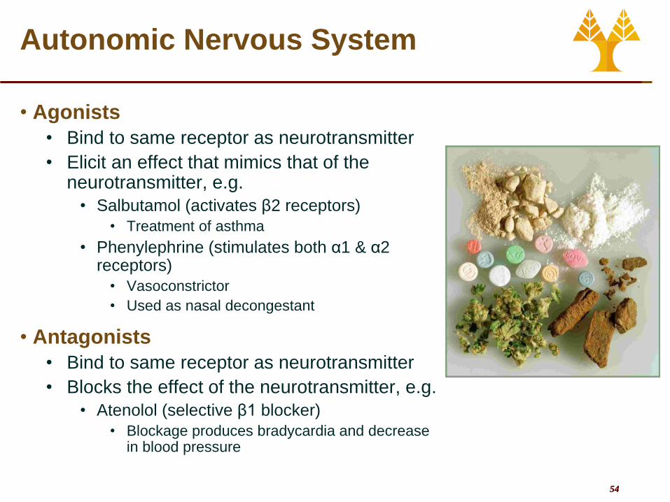
Autonomic Nervous System
• Agonists
• Bind to same receptor as neurotransmitter
• Elicit an effect that mimics that of the neurotransmitter, e.g.
• Salbutamol (activates β2 receptors)
• Treatment of asthma
• Phenylephrine (stimulates both α1 & α2 receptors)
• Vasoconstrictor
• Used as nasal decongestant
• Antagonists
• Bind to same receptor as neurotransmitter
• Blocks the effect of the neurotransmitter, e.g.
• Atenolol (selective β1 blocker)
• Blockage produces bradycardia and decrease in blood pressure


