Page 1
Edinburgh Research Explorer
Recombinant Ad35 adenoviral proteins as potent modulators ofhuman T cell activation
Citation for published version:Hay, J, Carter, D, Lieber, A & Astier, AL 2015, 'Recombinant Ad35 adenoviral proteins as potent modulatorsof human T cell activation' Immunology , vol. 144, no. 3, pp. 453-460. DOI: 10.1111/imm.12391
Digital Object Identifier (DOI):10.1111/imm.12391
Link:Link to publication record in Edinburgh Research Explorer
Document Version:Peer reviewed version
Published In:Immunology
Publisher Rights Statement:This article has been accepted for publication and undergone full peer review but has not been through thecopyediting, typesetting, pagination and proofreading process which may lead to differences between thisversion and the Version of Record.
General rightsCopyright for the publications made accessible via the Edinburgh Research Explorer is retained by the author(s)and / or other copyright owners and it is a condition of accessing these publications that users recognise andabide by the legal requirements associated with these rights.
Take down policyThe University of Edinburgh has made every reasonable effort to ensure that Edinburgh Research Explorercontent complies with UK legislation. If you believe that the public display of this file breaches copyright pleasecontact [email protected] providing details, and we will remove access to the work immediately andinvestigate your claim.
Download date: 01. Sep. 2018
Page 2
Acc
epte
d A
rtic
le
This article has been accepted for publication and undergone full peer review but has not been through the copyediting, typesetting, pagination and proofreading process which may lead to differences between this version and the Version of Record. Please cite this article as an 'Accepted Article', doi: 10.1111/imm.12391
This article is protected by copyright. All rights reserved.
Received Date : 10-Jun-2014 Revised Date : 03-Sep-2014 Accepted Date : 16-Sep-2014 Article type : Original Article Recombinant Ad35 adenoviral proteins as potent modulators of
human T cell activation
Joanne Hay1, Darrick Carter2,3, André Lieber4 and Anne L. Astier1,*
1 MRC Centre for Inflammation Research, University of Edinburgh, Queen’s Medical
Research Institute, Edinburgh EH16 4TJ, UK; 2 PAI Life Sciences Inc., Seattle, WA; 3
Compliment Corp., Seattle, WA; 4 Department of Medical Genetics, University of
Washington, Seattle, WA
Short title: recombinant adenoviral proteins modulate T cell activation
Keywords: CD46; human T cells; antagonist; agonist; activation; adenovirus 35.
Abbreviations; SCR: short-consensus repeat; Tr1: type 1 regulatory T cells
Page 3
Acc
epte
d A
rtic
le
This article is protected by copyright. All rights reserved.
* Address reprint requests and correspondence to Anne L Astier
Phone: +44 (0)131 242 6658; Fax: +44 (0)131 242 6578;
E-mail address: [email protected]
Summary
The protein CD46 protects cells from complement attack by regulating cleavage of C3b and
C3d. CD46 also regulates the adaptive immune response by controlling T cell activation and
differentiation. Co-engagement of the T cell receptor and CD46 notably drives T cell
differentiation by switching production of IFNγ to secretion of anti-inflammatory IL-10. This
regulatory pathway is altered in several chronic inflammatory diseases highlighting its key
role for immune homeostasis. The manipulation of the CD46 pathway may therefore provide
a powerful means to regulate immune responses. Herein, we investigated the effect of
recombinant proteins derived from the fiber knob of the adenovirus serotype 35 (Ad35) that
uses CD46 as its entry receptor, on human T cell activation. We compared the effects of
Ad35K++, engineered to exhibit enhanced affinity to CD46, and of Ad35K-, mutated in the
binding site for CD46. Ad35K++ profoundly affects T cell activation by decreasing the levels
of CD46 at the surface of primary T cells, and impairing T cell co-activation, shown by
decreased CD25 expression, reduced proliferation and lower secretion of IL-10 and IFNγ. In
contrast, Ad35K- acts a potent coactivator of T cells, enhancing T cell proliferation and
cytokine production. These data show that recombinant Ad35 proteins are potent modulators
of human T cell activation, and support their further development as potential drugs targeting
T cell responses.
Page 4
Acc
epte
d A
rtic
le
This article is protected by copyright. All rights reserved.
Introduction
The complement regulator CD46 is a type I transmembrane protein that protects cells from
autolysis by its regulation of the activating complement components C3b and C4b [1-3]. The
CD46 ectodomain consists of four short-consensus repeat domains (SCR1-4), which are
followed by a region rich in serine-threonine-proline (STP region), a transmembrane domain
and one of two short cytoplasmic tails produced by alternative splicing [4, 5]. Besides
binding to complement components, CD46 also acts as a cellular receptor for many
pathogens, and has been dubbed a ‘pathogen’s magnet’ [6]. Pathogens using CD46 as their
receptor include several serotypes of adenovirus such as Ad35 [7], the vaccinal strain of
measles virus, herpes virus 6 and some strains of bacteria such as Neisseria gonorrhoeae and
Neisseria meningitides, as well as group A Streptococcus. Moreover, CD46 is key in the
regulation of the adaptive immune response by controlling T cell activation, differentiation
and polarity [8-12]. Coligation of CD46 and the TCR provides a strong costimulatory signal
to T cells [8, 11], and expression of either the CD46-Cyt1 or the Cyt2 isoform in transgenic
mice exerted antagonist effects in vivo on T cell-mediated inflammation, which was
correlated with different effects in vitro on cytokine production and T cell proliferation [10].
CD46 expression at the surface of human T cells is tightly regulated, being shed by matrix
metalloproteases upon ligation, and CD46 cleavage is important for its functions [13, 14]. In
the presence of IL-2, CD46 drives differentiation of human CD4+ T cells towards a
regulatory T cell type I (Tr1), characterized by increased production of IL-10 and reduced
secretion of IFNγ [9, 15]. This regulatory pathway is altered in T cells from patients with
multiple sclerosis, rheumatoid arthritis and asthma, as IL-10 production upon CD46
costimulation is impaired [15-20]. The dysregulation of this pathway in several chronic
inflammatory diseases highlights its key role in regulating the homeostasis of the immune
response, and therefore makes it an attractive pathway to modulate in these pathologies.
Page 5
Acc
epte
d A
rtic
le
This article is protected by copyright. All rights reserved.
Moreover, most cancer cells upregulate CD46 to avoid complement lysis and inhibition of
CD46 on cancer cells may be a promising strategy to enhance antibody-dependent
cytotoxicity of therapeutic monoclonal antibodies [21-24].
Ad35K++ is a small recombinant protein derived from the fiber knob of the
adenovirus serotype 35 that has been modified to bind to CD46 with picomolar affinity [25].
Previous studies have shown that this CD46 antagonist could transiently remove CD46 from
the surface of several cancer cell lines, and as a consequence render them more sensitive to
antibody-dependent complement lysis. For example, Ad35K++ promotes killing of several
lymphoma cell lines by rituximab, a humanized IgG1 targeting CD20 [25]. Moreover, in vivo
administration of Ad35K++ to non-human primates, that ubiquitously express CD46, appears
to be safe and well tolerated [26]. Therefore, using recombinant proteins targeting CD46 may
be of potential use in clinical trials. Considering the key role of the CD46 pathway in
controlling human T cell activation, we investigated herein the effect of Ad35K++ on T cell
responses. We also assessed the effects of Ad35K- that has a point mutation (Arg279) that
affects binding to CD46 [25]. Our data show that Ad35K++ strongly affected CD46
expression at the surface of primary T cells, and importantly that it was able to significantly
impair T cell activation. T cells co-activated in presence of Ad35K++ had normal induction
of CD69, an early marker of T cell activation but failed to express high levels of CD25,
which was correlated with decreased proliferation and reduced cytokine production. In
contrast, Ad35K- surprisingly led to enhanced T cell activation, with increased proliferation
and cytokine production. These data emphasize the potency of recombinant adenoviral
proteins in modulating the CD46 pathway, and provide the rationale to further investigate
their effects on the immune response in order to maximize their therapeutic potential.
Page 6
Acc
epte
d A
rtic
le
This article is protected by copyright. All rights reserved.
Methods
Cell purification and activation: PBMC were isolated by Ficoll-Hypaque density gradient
centrifugation (Pharmacia LKB Biotechnology), from venous blood from healthy donors
obtained after informed consent. Naïve CD4+ T cells were negatively isolated using
magnetic beads (Stem Cells, purification > 95%), and cultured in RPMI 1640 with 10%
fetal calf serum at 0.5x10⁶ per well in 48-well plate pre-coated with α-CD3 (OKT3,
5µg/ml) or α-CD3/α-CD46 (OKT3, 5µg/ml, MC120.6, 10 µg/ml) (MC120.6 was kindly
provided by Dr. Chantal Rabourdin-Combe, France, and recognized the SCR1 domain of
CD46). Activated T cells also received rhIL-2 (Life Technologies - 10U/ml) to induce a
Tr1-like phenotype [9]. Ad35K++ and Ad35K- were added to the wells at the start of the
culture at the concentrations indicated. In some experiments, GM6001, a broad MMP
inhibitor, was also added to the culture (Sigma, 10μM) [14].
Ad35K++ and Ad35K-: Recombinant, synthetic proteins derived from the fiber knob
adenovirus Ad35 serotype. Both Ad35K++ and Ad35K- were selected using a display
library as previously described [27, 28], with the Ad35K++ version having greatly
increased affinity for the CD46 target. The coding sequences were cloned into a
prokaryotic expression vector and gene expression induced by the addition of IPTG to the
E. coli culture medium. Cells were lysed and the proteins purified using a combination of
nickel affinity chromatography and ion exchange chromatography. The final proteins had
high purity by SDS-PAGE and did not contain appreciable amounts of endotoxin that
could affect cellular readouts.
Cytokine detection: Cell culture supernatants from the 48-well plates (as described in the
cell activation section) were collected after five days of stimulation, and both IL-10 and
Page 7
Acc
epte
d A
rtic
le
This article is protected by copyright. All rights reserved.
IFNγ secretion was determined by ELISA specific for human IL-10 (BD Pharmingen,
San Diego, CA) and IFNγ (Endogen, Rockford, IL).
Flow cytometry. The expression level of CD46, CD69 and CD25 was assessed by flow
cytometry, by incubating the cells with the antibodies at 4°C for 20 min in FACS buffer
(PBS containing 1% fetal calf serum). We used different antibodies against CD46 as
described in the results section: anti-CD46-FITC (clone MEM-258 recognizing the SCR4
domain - Biolegend), anti-CD46-PE (clone 344519, R&D) or the MCI20.6 clone
recognizing the SCR1 domain followed by anti-IgG1-FITC antibodies. For activation
experiments, we used the following antibodies: anti-CD46-PE, anti-CD69-FITC
(Biolegend) and anti-CD25-APC (Biolegend). Samples were run with a FACSCalibur
and data analyzed using FlowJo. Relative expression to staining with the control was
calculated by calculating the DMFI (MFI obtained with antibody - MFI obtained with
isotype control). Staining for intracellular expression of CD46 was performed in FACS
buffer containing 0.1 % saponin (Sigma) for 30 min at room temperature, after fixation of
the cells. Proliferation was determined by pre-labeling purified T cells with eFluor 670
cell proliferation stain (eBioscience) before activation following the manufacturer’s
instructions, and assessing remaining fluorescence after 4 days.
Statistics. The groups were analyzed using Graphpad Prism software. Flow cytometry
data were analyzed using the Wilcoxon test, when assessing paired samples. ELISA data
are the average of duplicate wells, and the average obtained for the different donors were
analyzed using the Wilcoxon test. All p-values are two-tailed and with a 95% confidence
interval.
Page 8
Acc
epte
d A
rtic
le
This article is protected by copyright. All rights reserved.
Results
Effect of Ad35K++ and Ad35K- on CD46 expression on Jurkat cells
We first examined the effect of incubating Jurkat cells, a T leukemia cell line, with Ad35K++
and Ad35K- on CD46 surface expression. Jurkat cells were cultured in the presence or
absence of a fixed concentration of both proteins (20 μg/ml) for various periods of times, and
CD46 expression was then assessed by flow cytometry using an anti-CD46-FITC antibody.
Addition of Ad35K++ for 1hr led to a total lack of detection of CD46, and the lack of CD46
expression was still observed after 48hrs of culture (Fig 1A). A shorter kinetic experiment
showed that a total lack of detection of CD46 was also observed after a 5 min incubation
period (not shown). Incubation with Ad35K- led to a slight decrease in the level of CD46
detected after 24 and 48 hrs, suggesting a residual binding to CD46. We next incubated
Jurkat cells with various concentrations of Ad35K++ ranging from 1 pg/ml to 20 μg/ml, for 3
hrs. A dose dependent decrease in CD46 detection was observed. We observed a total effect
with 10 ng/ml while there was no more effect on CD46 expression at 1 pg/ml, with a partial
effect observed at 100 pg/ml (Fig 1B). These data suggested that Ad35K++ binding to CD46
could either mask the epitope in CD46 that is recognized by the antibody used for detection,
and/or induce its internalization or cleavage.
In order to determine whether Ad35K++ binding to CD46 was masking CD46
antibody binding sites, we performed some competition experiments using different anti-
CD46 antibodies recognizing distinct epitopes. We assessed the anti-CD46-FITC Ab used
above that recognizes the SCR4 domain; an anti-CD46-PE Ab whose binding site has not
been identified but that did not compete with the FITC Ab (Fig 2A); and the MCI20.6
monoclonal antibody that recognizes the SCR1 domain. Jurkat cells were first incubated with
Ad35K++ for 20 min on ice, washed and then stained with these different anti-CD46
Page 9
Acc
epte
d A
rtic
le
This article is protected by copyright. All rights reserved.
antibodies. Again, the use of the anti-CD46-FITC Ab led to a total lack of detection of CD46,
suggesting that binding of Ad35K++ to CD46 on Jurkat cells was masking the epitope of
CD46 recognized by this clone (Fig 2B). The use of the anti-CD46-PE antibody only led to a
partial decrease, suggesting a partial blockage. Importantly, there was no competition
between the MCI20.6 clone and Ad35K++, indicating that MCI20.6 could be used to activate
cells in the presence of Ad35K++.
Ad35K++ and Ad35K- affect the phenotype of primary human T cells
We next determined the effect of Ad35K++ and Ad35K- on expression of CD46 on primary
T cells. Purified naïve CD4+ T cells were cultured with Ad35K++ or Ad35K-(500 ng/ml),
and expression of CD46 assessed after 24hrs using the MCI20.6 antibody that does not
compete with Ad35K++. As binding of Ad35K++ to CD46 in several lymphoma cell lines
has been shown to induce CD46 internalization [25], we also assessed intracellular CD46
expression after cell permeabilization. Incubation of T cells with Ad35K++ for 24hrs resulted
in a reduced surface expression of CD46 on these cells (Fig. 3A, 3B) while Ad35K- had a
lesser effect. Incubation with Ad35K++ had no significant effect on intracellular CD46
expression. We also assessed whether downregulation of CD46 expression by Ad35K++
could be affected by lowering the temperature to 4°C. T cells were incubated with Ad35K++
or Ad35K- (500 ng/ml) at 4°C or 37°C, and CD46 expression assessed by flow cytometry.
Downregulation of CD46 expression was only observed at 37°C (Fig. 3C). These data
suggest that internalization and/or MMP-dependent shedding of CD46 was blocked [14]. We
next investigated whether MMP inhibition could modulate the effect of Ad35K++ on CD46
expression in T cells. Naïve CD4+ T cells were cultured in presence or absence of Ad35K++
(500 ng/ml) and/or GM6001, a broad MMP inhibitor, and CD46 expression was monitored
Page 10
Acc
epte
d A
rtic
le
This article is protected by copyright. All rights reserved.
by flow cytometry using the MCI20.6 antibody (Fig. 3D). Inhibition of MMP increased the
levels of surface CD46 in Ad35K++-treated T cells. These data suggest that ligation of
Ad35K++ to CD46 on primary T cells led to, at least, a partial shedding of CD46.
We next investigated whether activation of primary T cells was affected in the
presence of Ad35K++ or Ad35K-. Purified human naïve CD4+ T cells were left unstimulated
or were activated with pre-immobilized anti-CD3 or anti-CD3/anti-CD46 (using the MCI20.6
antibody that does not compete with Ad35K++), as previously described [9, 17], in the
presence or absence of Ad35K++ or Ad35K- (500 ng/ml). CD46 surface expression, as well
as expression of the activation markers CD69 and CD25, was examined by flow cytometry
after 2 and 5 days (Fig 4). As expected, CD3/CD46 costimulation of control cells induced a
strong downregulation of CD46 surface expression, and this was correlated with an increased
expression of CD69 and CD25, at both day 2 and day 5 [14]. In the presence of Ad35K++, a
marked decrease in CD46 surface expression was observed in all conditions of activation, an
effect that was still observed after 5 days of culture. The presence of Ad35K++ did not affect
the induction of CD69 expression in T cells activated with either anti-CD3 or anti-
CD3/CD46. In contrast, the presence of Ad35K++ significantly impaired CD25 induction in
CD3/CD46-costimulated T cells. As previously observed, a slight decrease in surface CD46
expression was noted with Ad35K-. Surprisingly, Ad35K- significantly enhanced CD69
induction on CD3-activated T cells compared to control cells, and a significant effect, albeit
more moderate, was also observed for CD25 expression. These data suggest that while
Ad35K++ blocks late CD46-mediated T cell costimulation, the residual binding of Ad35K-
to CD46 is sufficient to costimulate T cells activated by CD3.
Ad35K++ and Ad35K- modulate primary human T cell responses
Page 11
Acc
epte
d A
rtic
le
This article is protected by copyright. All rights reserved.
In order to determine whether these changes in phenotype would affect T cell functions, we
next examined T cell responses. Naive T cells were stained with eFluro670 before activation
with immobilized anti-CD3 or anti-CD3/anti-CD46, in the presence of Ad35K++ or Ad35K-,
and cell proliferation was determined after 4 days (Fig 5A). As expected, control cells
proliferated upon CD3 activation and the proliferation was further increased upon CD3/CD46
costimulation [8]. Cells incubated in presence of Ad35K++ proliferated in response to CD3
ligation but failed to show increased proliferation upon CD3/CD46 costimulation. In contrast,
cells cultured in the presence of Ad35K- showed increased proliferation in response to CD3
ligation compared to control cells, but no co-stimulation was observed following CD3/CD46
co-ligation.
Next, we determined cytokine production of cells activated in the presence of
Ad35K++ and Ad35K-. As CD46 costimulation controls production of IFNγ and IL-10 [15],
we assessed the levels of these 2 cytokines in the culture supernatants of primary T cells
activated for 5 days in the presence of Ad35K++ or Ad35K- (Fig 5B). Cells costimulated in
the presence of Ad35K++ secreted reduced amounts of cytokines compared to control cells.
In contrast, T cells costimulated in the presence of Ad35K- secreted increased levels of IL-10
when compared with control cells or cells incubated with Ad35K++.
Discussion
In the past decade, the complement regulator CD46 has been of increasing interest to
immunologists as several groups reported that its ligation on human T cells could profoundly
affect T cell functions. While the first studies highlighted the role of CD46 ligation on T cell
activation and proliferation [8, 11], further reports have demonstrated its key role in the
control of inflammation by regulating not only T cell activation but also its ability to
differentiate T cells towards an anti-inflammatory phenotype [9, 10, 12]. Strikingly, this
pathway is dysregulated in a number of pathologies (MS, RA and asthma), which underlines
Page 12
Acc
epte
d A
rtic
le
This article is protected by copyright. All rights reserved.
the key role of CD46 in the control of inflammation [15, 17-20]. Moreover, the role of CD46
as a cellular receptor for multiple pathogens suggests that this is a powerful pathway that can
be exploited to the pathogens’ advantage [29]. Therefore, specifically targeting CD46 may
have potential use in several clinical settings.
Ad35K++ is a small recombinant protein derived from the fiber knob of the
adenovirus serotype 35 (AD35) that uses CD46 as its receptor. It has been previously shown
that Ad35K++ could increase the complement-dependent lysis of tumor cells lines [25],
highlighting again the attractiveness of this drug to improve antibody-based cancer
treatments. This suggestion is further supported by the fact that injection of Ad35K++ to
monkeys, the only mammals that express CD46 in a pattern resembling humans, appears to
be safe and well tolerated, and indeed in vivo administration of Ad35K++ could increase B
cell depletion by rituximab [26].
Herein, we have focused on the effects of Ad35K++ and of a mutant Ad35K- on
human T cell responses. We have examined CD46 expression, and response of T cells
costimulated by CD46 in the presence of both recombinant proteins. Our data show that
Ad35K++ binds to CD46 at the surface of primary T cells, and this interaction appears to be
very stable as decreased CD46 expression was still detected after 5 days of culture. Of note,
intravenous administration of Ad35K++ to macaques results in a decreased expression of
CD46 on PBMCs that was still observed after 2 weeks post injection [26]. These data may be
explained by the picomolar affinity of Ad35K++ for CD46. Previous studies have shown
internalization of CD46 after binding to Ad35K++ in lymphoma cell lines [25]. Our results
from experiments to examine intracellular staining of CD46 in primary T cells do not
completely support internalization of CD46 in primary T cells, although downregulation of
CD46 was blocked at 4°C, and we cannot exclude CD46 internalization and rapid
Page 13
Acc
epte
d A
rtic
le
This article is protected by copyright. All rights reserved.
degradation. Our data also suggest that binding of Ad35K++ to CD46 leads to a partial
MMP-dependent cleavage as partially blocked by addition of a broad MMP inhibitor.
The structure of the CD46 ectodomain resembles the shape of a ‘hockey stick’ [30],
which likely explains the data obtained with the different clones of anti-CD46 antibodies
used in the competition experiments with Ad35K++. Binding of Ad35K++ to the SCR1/2
domains of CD46 may also cause changes in CD46 conformation, as it totally competes with
the anti-CD46-FITC Ab that recognizes the SCR4 domain [31]. Importantly, binding of
Ad35K++ to CD46 does not affect recognition by the MCI20.6 Ab that also recognizes the
SCR1 domain, allowing us to use this antibody to determine the effect of Ad35K
recombinant proteins on CD46 costimulation. Our data show that early T cell activation
appears to be normal in Ad35K++ treated cells, as we could observe a strong costimulation
effect on CD69 induction between CD3 and CD3/CD46 costimulated T cells. However, there
was no further increase in CD25 in CD46-costimulated T cells in presence of Ad35K++,
especially after longer activation periods, and CD46-mediated costimulation was strongly
impaired, as evidenced by reduced proliferation and cytokine production. Altogether, these
data indicate that, although early T cell co-activation may remain unchanged, Ad35K++
blocks late T cell costimulation. Interestingly, recombinant Ad35 fiber knob proteins could
inhibit CD3/CD28-mediated T cell activation, albeit inhibition could only be observed when
the Ad35 knob was immobilized [32, 33], suggesting that multimerisation was required to
suppress activation, possibly by hindering the spatial organization of the immune synapse.
Therefore, Ad35K++ is a potent protein that strongly impairs the CD46-mediated pathway, at
least in vitro.
Our data show a surprising effect of Ad35K- on T cell functions. This result was
unexpected as Ad35K- was designed as a negative control, having a point mutation supposed
to abrogate CD46 binding. However, incubation of primary T cells with Ad35K- led to a
Page 14
Acc
epte
d A
rtic
le
This article is protected by copyright. All rights reserved.
significant decrease in CD46 expression (see Fig 4B), indicating that there is a residual
binding of Ad35K- to CD46. Indeed, binding of Ad35K- to several human cells lines were
also observed (data not shown). Moreover, addition of Ad35K- was able to modulate T cell
responses, providing a costimulatory signal to CD3-activated T cells, and leading to
enhanced secretion of IL-10, suggesting its ability to promote Tr1 differentiation. Hence, in
contrast to Ad35K++, Ad35K- acts as a CD46 agonist, which may be useful to boost the
CD46 pathway in vivo.
In conclusion, both recombinant proteins differently modulate primary T cell
responses, at least in vitro. Therefore, caution should be exerted in targeting this pathway, as
ligation of CD46 can either promote or inhibit activation, likely depending on conformational
changes triggered by ligand binding and subsequent signaling cascades. Nevertheless, our
data emphasize the potency of recombinant proteins to modulate the CD46-mediated
pathway, which is key to control immune homeostasis. The analysis of the effects of these
two recombinant proteins on other immune cell types for which ligation of CD46 has been
shown to modulate their response, such as dendritic cells [34] and B cells [35] will further
support the development of these potential drugs into clinical trials.
Acknowledgements
We thank Dongqing Ma who performed some preliminary experiments during her
Master. JH has done the experiments, AB and DC have provided the recombinant proteins,
have performed experiments (data not shown) and discussed data; AA has written the
manuscript. We thank Fiona Rossi and Shonna Johnston for their help with flow cytometry.
We are grateful to Ian Dransfield for the critical reading of the manuscript. AA is an MRC
academic fellow and detached member of CNRS, France. JH is an MRC-PhD student. AL
and DC were supported by NIH grant 2R44CA162582-02.
Page 15
Acc
epte
d A
rtic
le
This article is protected by copyright. All rights reserved.
Competing Interests statement
The authors declare no competing interests.
References
1. Seya T, Turner JR, Atkinson JP. Purification and characterization of a membrane protein
(gp45-70) that is a cofactor for cleavage of C3b and C4b. J Exp Med 1986;
163(4):837-55.
2. Loveland B, Russell S, Purcell D, Johnstone R, Thorley B, Sparrow R, McKenzie I. The
molecular basis of CD46 polymorphic expression and demonstration of a protective
role against lysis by xenosera. Transplant Proc 1992; 24(2):685-6.
3. Russell SM, Johnstone RW, Wilton A, Sparrow RL, McKenzie IF, Purcell DF.
Molecular characterization of the polymorphic expression of CD46: a cell surface
molecule protecting cells from complement attack. Transplant Proc 1992; 24(1):211-
3.
4. Purcell DF, Russell SM, Deacon NJ, Brown MA, Hooker DJ, McKenzie IF.
Alternatively spliced RNAs encode several isoforms of CD46 (MCP), a regulator of
complement activation. Immunogenetics 1991; 33(5-6):335-44.
5. Seya T, Hirano A, Matsumoto M, Nomura M, Ueda S. Human membrane cofactor
protein (MCP, CD46): multiple isoforms and functions. Int J Biochem Cell Biol 1999;
31(11):1255-60.
6. Cattaneo R. Four viruses, two bacteria, and one receptor: membrane cofactor protein
(CD46) as pathogens' magnet. J Virol 2004; 78(9):4385-8.
7. Gaggar A, Shayakhmetov DM, Lieber A. CD46 is a cellular receptor for group B
adenoviruses. Nat Med 2003; 9(11):1408-12.
8. Astier A, Trescol-Biemont MC, Azocar O, Lamouille B, Rabourdin-Combe C. Cutting
edge: CD46, a new costimulatory molecule for T cells, that induces p120CBL and
LAT phosphorylation. J Immunol 2000; 164(12):6091-5.
9. Kemper C, Chan AC, Green JM, Brett KA, Murphy KM, Atkinson JP. Activation of
human CD4(+) cells with CD3 and CD46 induces a T-regulatory cell 1 phenotype.
Nature 2003; 421(6921):388-92.
Page 16
Acc
epte
d A
rtic
le
This article is protected by copyright. All rights reserved.
10. Marie JC, Astier AL, Rivailler P, Rabourdin-Combe C, Wild TF, Horvat B. Linking
innate and acquired immunity: divergent role of CD46 cytoplasmic domains in T cell
induced inflammation. Nat Immunol 2002; 3(7):659-66.
11. Zaffran Y, Destaing O, Roux A, Ory S, Nheu T, Jurdic P, Rabourdin-Combe C, Astier
AL. CD46/CD3 costimulation induces morphological changes of human T cells and
activation of Vav, Rac, and extracellular signal-regulated kinase mitogen-activated
protein kinase. J Immunol 2001; 167(12):6780-5.
12. Oliaro J, Pasam A, Waterhouse NJ, Browne KA, Ludford-Menting MJ, Trapani JA,
Russell SM. Ligation of the cell surface receptor, CD46, alters T cell polarity and
response to antigen presentation. Proc Natl Acad Sci U S A 2006; 103(49):18685-90.
13. Ni Choileain S, Astier AL. CD46 processing: a means of expression. Immunobiology
2012; 217(2):169-75.
14. Ni Choileain S, Weyand NJ, Neumann C, Thomas J, So M, Astier AL. The dynamic
processing of CD46 intracellular domains provides a molecular rheostat for T cell
activation. PLoS One 2011; 6(1):e16287.
15. Cardone J, Le Friec G, Vantourout P, et al. Complement regulator CD46 temporally
regulates cytokine production by conventional and unconventional T cells. Nat
Immunol 2010; 11(9):862-71.
16. Astier AL. T-cell regulation by CD46 and its relevance in multiple sclerosis.
Immunology 2008; 124(2):149-54.
17. Astier AL, Meiffren G, Freeman S, Hafler DA. Alterations in CD46-mediated Tr1
regulatory T cells in patients with multiple sclerosis. J Clin Invest 2006;
116(12):3252-7.
18. Ma A, Xiong Z, Hu Y, et al. Dysfunction of IL-10-producing type 1 regulatory T cells
and CD4+CD25+ regulatory T cells in a mimic model of human multiple sclerosis in
Cynomolgus monkeys. International Immunopharmacology 2009; 9(5):599-608.
19. Martinez-Forero I, Garcia-Munoz R, Martinez-Pasamar S, et al. IL-10 suppressor
activity and ex vivo Tr1 cell function are impaired in multiple sclerosis. Eur J
Immunol 2008; 38(2):576-86.
20. Xu YQ, Gao YD, Yang J, Guo W. A defect of CD4+CD25+ regulatory T cells in
inducing interleukin-10 production from CD4+ T cells under CD46 costimulation in
asthma patients. J Asthma 2010; 47(4):367-73.
21. Di Gaetano N, Cittera E, Nota R, et al. Complement activation determines the
therapeutic activity of rituximab in vivo. J Immunol 2003; 171(3):1581-7.
Page 17
Acc
epte
d A
rtic
le
This article is protected by copyright. All rights reserved.
22. Kolev M, Towner L, Donev R. Complement in cancer and cancer immunotherapy. Arch
Immunol Ther Exp (Warsz) 2011; 59(6):407-19.
23. Carter D, Lieber A. Protein engineering to target complement evasion in cancer. FEBS
letters 2014; 588(2):334-40.
24. Geis N, Zell S, Rutz R, Li W, Giese T, Mamidi S, Schultz S, Kirschfink M. Inhibition of
membrane complement inhibitor expression (CD46, CD55, CD59) by siRNA
sensitizes tumor cells to complement attack in vitro. Curr Cancer Drug Targets 2010;
10(8):922-31.
25. Wang H, Liu Y, Li ZY, Fan X, Hemminki A, Lieber A. A recombinant adenovirus type
35 fiber knob protein sensitizes lymphoma cells to rituximab therapy. Blood 2009;
115(3):592-600.
26. Beyer I, Cao H, Persson J, et al. Transient removal of CD46 is safe and increases B-cell
depletion by rituximab in CD46 transgenic mice and macaques. Mol Ther 2012;
21(2):291-9.
27. Wang H, Liu Y, Li Z, et al. In vitro and in vivo properties of adenovirus vectors with
increased affinity to CD46. J Virol 2008; 82(21):10567-79.
28. Wang H, Liu Y, Li ZY, Fan X, Hemminki A, Lieber A. A recombinant adenovirus type
35 fiber knob protein sensitizes lymphoma cells to rituximab therapy. Blood 2010;
115(3):592-600.
29. Russell S. CD46: a complement regulator and pathogen receptor that mediates links
between innate and acquired immune function. Tissue Antigens 2004; 64(2):111-8.
30. Persson BD, Schmitz NB, Santiago C, Zocher G, Larvie M, Scheu U, Casasnovas JM,
Stehle T. Structure of the extracellular portion of CD46 provides insights into its
interactions with complement proteins and pathogens. PLoS Pathog 2011; 6(9).
31. Fleischli C, Verhaagh S, Havenga M, Sirena D, Schaffner W, Cattaneo R, Greber UF,
Hemmi S. The distal short consensus repeats 1 and 2 of the membrane cofactor
protein CD46 and their distance from the cell membrane determine productive entry
of species B adenovirus serotype 35. J Virol 2005; 79(15):10013-22.
32. Adams WC, Gujer C, McInerney G, Gall JG, Petrovas C, Karlsson Hedestam GB, Koup
RA, Lore K. Adenovirus type-35 vectors block human CD4+ T-cell activation via
CD46 ligation. Proc Natl Acad Sci U S A 2011; 108(18):7499-504.
33. Adams WC, Berenson RJ, Karlsson Hedestam GB, Lieber A, Koup RA, Lore K.
Attenuation of CD4+ T-cell function by human adenovirus type 35 is mediated by the
knob protein. J Gen Virol 2012; 93(Pt 6):1339-44.
Page 18
Acc
epte
d A
rtic
le
This article is protected by copyright. All rights reserved.
34. Vaknin-Dembinsky A, Balashov K, Weiner HL. IL-23 is increased in dendritic cells in
multiple sclerosis and down-regulation of IL-23 by antisense oligos increases
dendritic cell IL-10 production. J Immunol 2006; 176(12):7768-74.
35. Jabara HH, Angelini F, Brodeur SR, Geha RS. Ligation of CD46 to CD40 inhibits CD40
signaling in B cells. Int Immunol 2011; 23(3):215-21.
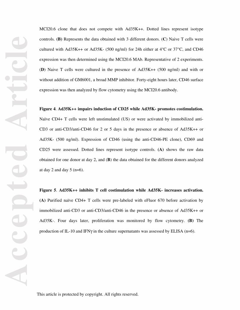
Figure Legends
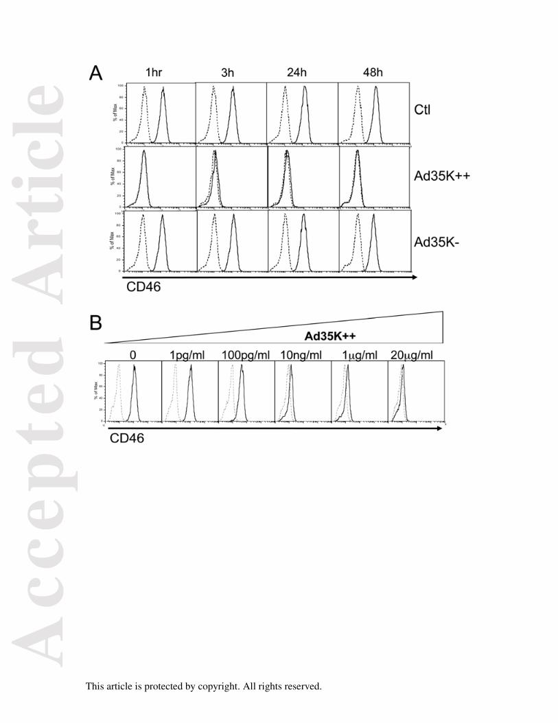
Figure 1. Ad35K++ affects the detection of CD46 expression on Jurkat cells. (A) Jurkat
cells were incubated with Ad35K++ or Ad35K- (20 μg/ml) for various lengths of time, as
indicated. CD46 surface expression was then monitored by flow cytometry using an anti-
CD46-FITC antibody. (B) Jurkat cells were incubated for 3h with various concentrations of
Ad35K++ as indicated and CD46 surface expression was monitored by flow cytometry
using the anti-CD46-FITC antibody. Dotted lines represent isotype controls.
Figure 2. Competition experiments between Ad35K++ and anti-CD46 antibodies. (A)
Jurkat cells were stained with anti-CD46-FITC or anti-CD46-PE, or with both antibodies
added in the different orders, as indicated, and CD46 expression was assessed by flow
cytometry. These two clones do not compete with each other. (B) Jurkat cells were first
incubated with Ad35K++ (500 ng/nl) for 20 min, washed and then stained with the anti-
CD46-FITC, anti-CD46-PE, or indirectly labeled with the MCI20.6 clone followed by anti-
mIgG1-FITC. CD46 expression was assessed by flow cytometry. Dotted lines represent
isotype controls.
Figure 3. Ad35K++ affects expression of surface CD46 on primary naïve CD4+ T cells.
(A) Naïve T cells were cultured with Ad35K++ or Ad35K- (500 ng/ml) for 24h, and CD46
expression was determined at the cell surface or after intracellular staining using the
Page 19
Acc
epte
d A
rtic
le
This article is protected by copyright. All rights reserved.
MCI20.6 clone that does not compete with Ad35K++. Dotted lines represent isotype
controls. (B) Represents the data obtained with 3 different donors. (C) Naive T cells were
cultured with Ad35K++ or Ad35K- (500 ng/ml) for 24h either at 4°C or 37°C, and CD46
expression was then determined using the MCI20.6 MAb. Representative of 2 experiments.
(D) Naive T cells were cultured in the presence of Ad35K++ (500 ng/ml) and with or
without addition of GM6001, a broad MMP inhibitor. Forty-eight hours later, CD46 surface
expression was then analyzed by flow cytometry using the MCI20.6 antibody.
Figure 4. Ad35K++ impairs induction of CD25 while Ad35K- promotes costimulation.
Naïve CD4+ T cells were left unstimulated (US) or were activated by immobilized anti-
CD3 or anti-CD3/anti-CD46 for 2 or 5 days in the presence or absence of Ad35K++ or
Ad35K- (500 ng/ml). Expression of CD46 (using the anti-CD46-PE clone), CD69 and
CD25 were assessed. Dotted lines represent isotype controls. (A) shows the raw data
obtained for one donor at day 2, and (B) the data obtained for the different donors analyzed
at day 2 and day 5 (n=6).
Figure 5. Ad35K++ inhibits T cell costimulation while Ad35K- increases activation.
(A) Purified naïve CD4+ T cells were pre-labeled with eFluor 670 before activation by
immobilized anti-CD3 or anti-CD3/anti-CD46 in the presence or absence of Ad35K++ or
Ad35K-. Four days later, proliferation was monitored by flow cytometry. (B) The
production of IL-10 and IFNγ in the culture supernatants was assessed by ELISA (n=6).
Page 20
Acc
epte
d A
rtic
le
This article is protected by copyright. All rights reserved.
Page 21
Acc
epte
d A
rtic
le
This article is protected by copyright. All rights reserved.
Page 22
Acc
epte
d A
rtic
le
This article is protected by copyright. All rights reserved.
Page 23
Acc
epte
d A
rtic
le
This article is protected by copyright. All rights reserved.