18 th NATIONAL CONFERENCE OF NEUROLOGY CHAPTER OF IAP NEUROPEDICON 2018 7 - 9 SEPTEMBER, 2018 GURUGRAM, DELHI / NCR EMERGING TRENDS & ADVANCES IN DIAGNOSTICS & THERAPEUTICS IN PEDIATRIC NEUROLOGY I N D I A N A C A D E M Y O F P E D I A T R I C S ABSTRACT AND SCIENTIFIC PROGRAM BOOK
Transcript
18th
NATIONAL CONFERENCE OF NEUROLOGY CHAPTER OF IAP
NEUROPEDICON
2018
7 - 9 SEPTEMBER, 2018GURUGRAM, DELHI / NCR
EMERGING TRENDS & ADVANCES IN DIAGNOSTICS & THERAPEUTICS
IN PEDIATRIC NEUROLOGY
IN
DIANACA
DEMY OF
PEDIATRIC
S
ABSTRACT AND SCIENTIFIC PROGRAM BOOK
NEUROPEDICON 2018
E-LEARNING MODULES
1
NEUROPEDICON 2018
INDEX
From the Desk of Organising Secretary 2
Office Bearers of Child Neurology Chapter 3
Delhi IAP Office Bearers 3
Organising Committee 4
Scientific Program 5
International Faculty 12
National Faculty 13
Supporting Agencies 18
E-Learning Modules and Development Tools 18
Symposium Support 18
Session Support 19
Exhibitors 20
Other Support 21
List of Abstracts 22
Full Abstracts 45
2
NEUROPEDICON 2018
FROM THE DESK OFORGANISING SECRETARY, NEUROPEDICON 2018
This conference is going to be unique as we are going to provide you access to the E-Learning Modules diligently prepared by a team of experts. These E-learning modules are in sync with the theme. They are in consonance with conference sessions in a simplified manner with clinical approach based relevant topics for Pediatricians with interest in Pediatric Neurology and Pediatric Neurologists.
6. Neuromuscular Disorders
On the behalf of the Organizing Committee of the Conference I would like to extend a warm welcome to all the Delegates, Eminent Speakers and Chairpersons who have spared their valuable time to be here and give us the opportunity to host this conference. I assure you that the organizers have left no stone unturned to ensure that the Scientific Content of this Conference is of Highest Academic Standards with Latest Concepts and at the same time Concise, Clinically Relevant and Useful in Day to Day Practice. I am certain that all of you will not only benefit from this academic feast but also enjoy it. I wish you all the best and hope you enjoy the hospitality as well.
This three day conference is a platform to share and learn from the knowledge and experiences of eminent Pediatric Neurologists from the country as well as across the globe. One of the most daunting tasks for Pediatricians is to keep abreast with the latest advances and guidelines in management of patients. Hence it is pertinent that the theme kept for the conference is “Emerging Trends and Advances in Diagnostics and Therapeutics in Pediatric Neurology”. With the ever expanding field of medicine, rapidly evolving concepts, advanced research and the rising expectations of patients owing to social media and internet boom it is imperative that we are updated at all times.
The Child Neurology Division, Center of Excellence and Advanced Research for Childhood Neurodevelopmental Disorders, Department of Pediatrics, All India Institute of Medical Sciences (AIIMS), New Delhi has the proud privilege to host the prestigious National Conference of Child Neurology Chapter of IAP, “Neuropedicon 2018”, at The Leela Ambience, Gurugram, NCR, New Delhi from 7-9 September 2018 in collaboration with Delhi IAP.
Greetings from New Delhi
The Conference deliberations are going to be in the following six domains:1. Neuroinfections2. Autoimmune Disorders3. Cerebral Palsy & Developmental Disorders4. Autism & Behavioral Disorders5. Epilepsy
Coordinator, DM Pediatric Neurology Programme
Prof. (Dr.) Sheffali GulatiOrganising Secretary, Neuropedicon 2018
Chief, Child Neurology Division
Faculty Incharge, Center of Excellence and Advanced Research for Childhood Neurodevelopmental Disorders, Department of Pediatrics, All India Institute of Medical Sciences, New Delhi
3
NEUROPEDICON 2018
OFFICE BEARERS OF CHILD NEUROLOGY CHAPTER, IAP
Chairperson Dr.Arun Agrawal
Chairperson Elect 2019 Dr. K. P. Sarbhai
Immediate Past Chairperson Dr. Anand Kesavan
Secretary Dr. Sanjeev Joshi (Yavatmal)
Treasurer Dr. Ashwani Agrawal (Raipur)
Executive Board Members Dr. Vijay JainDr. Ravishankar Dr. Pawan Ghanghoriya Dr. Lokesh LingappaDr. Jitendra SahuDr. Sheffali Gulati (OrganisingSecretary, Neuropedicon 2018)
National Cordinator Dr. Anoop Verma
National Advisory Board Dr. Santosh SoanDr. Uday Bodhankar Dr. G Kumaresan
Editor in Chief Paedneurobulletin Dr. Vasant Khalatkar
Executive Editor Dr. Amarjeet WaghDr. Vineet Wankhede
President Dr. G.P. KaushalSecretary Dr. Peeyush Khanna
DELHI IAP OFFICE BEARERS
4
NEUROPEDICON 2018Neuropedicon 2018
ORGANISING COMMITTEE
PatronsProf. V K PaulProf. Randeep GuleriaProf. Y K GuptaProf. V K BahlProf. Chitra SarkarProf. Anand PanditProf. M K C Nair Prof. A K Deorari
IAP Neurology ChapterDr. Arun AgrawalDr. Sanjeev JoshiProf. T M Ananda KesavanDr. Anoop VermaProf. P A M Kunju
IAP Delhi
Dr. G P KaushalDr. Peeyush Khanna
Organising Chairperson
Dr. Veena Kalra
Organising Secretary
Prof. Sheffali Gulati
Treasurer
Dr. Biswaroop Chakrabarty
Core Organising Committee
Dr. Prashant JauhariDr. Rachna DubeyDr. (Col.) Vishal SondhiDr. J S KaushikDr. Biswaroop ChakrabartyProf. Sheffali Gulati
Advisory Board
Dr. I C VermaProf. Bibek TalukdarDr. Pratibha SinghiDr. Jesson C UnniProf. Satinder AnejaProf. Man Mohan MehndirattaProf. Gagandeep SinghProf. Rashmi KumarDr. Vrajesh UdaniDr. Anaita HegdeDr. K S RanaDr. Rekha MittalProf. Manjari TripathiProf. K P VinayanProf. Anju Aggarwal
Scientific Committee
Prof. Mahesh KamateDr. Naveen SankhyanDr. Suvasini SharmaDr. Jitendra SahuDr. Vykunta RajuDr. Ramesh KonankiDr. Rachna SehgalDr. Akbar Mohamed
5
NEUROPEDICON 2018
SCIENTIFIC PROGRAM
DAY 1: 07th SEPTEMBER 2018, FRIDAY
Time Topic Speaker Chairperson
07:30-08:30 hrs Registration
NEUROINFECTIONS PART 1
08:30-08:45 hrs Neurological Manifestations of HIV
15:30-15:45 hrs Case Based Approach to Acute Febrile Encephalopathy
Satinder Aneja Subrata SihnaM V PadmaSujata Kanhere
15:45:16:15 hrs CNS Autoimmunity in Children: Neuroprotective, Neurodegenerative and Neuroregenerative
Lim Ming
16:15-16:35 hrs Autoimmune Encephalopathy: An Overview of the Spectrum and Clinical Features
Vrajesh Udani
16:35-17:05 hrs Autoimmune Encephalopathy: Treatment and Optimizing Outcome
Lim Ming Kameshwar PrasadV B GuptaHarish Pemde
17:05-17:35 hrs CNS Relapsing Demyelinating Disorders: The Increasing Role of Antibody Mediated Syndromes
Angela Vincent
17:35-19:00 hrsPractice Points
Ethics in Pediatrics (10 minutes)Algorithmic Approach to Myasthenic Syndromes (15 minutes)Algorithmic Approach to Demyelinating Disorders (15 minutes)Algorithmic approach to Autoimmune Encephalopathy (15 minutes)Case Based Approach to ANEC (10 minutes)Case Based Approach to OMA (10 minutes)1 Platform (7 minutes)
Naveen Sankhyan
Rachana Dubey
Mahesh Kamate
Sangeetha Yoganathan
Rajni Farmaniya
Naveen Sankhyan
Angela VincentLim MingVrajesh Udani
Tea will be served around middle of the afternoon session.
*Poster Tour Guides – Charles RJC Newton, Angela Vincent, Ashwani Sood, Rachna Dubey, Jyotindra Goswami
7
NEUROPEDICON 2018
Time Topic Speaker Chairperson
07:30-08:30 hrs Registration
CEREBRAL PALSY AND DEVELOPMENTAL DISORDERS
08:30-09:00 hrs Evaluation of Developmental Delay in the Clinic: Current Approaches
Michael Shevell Jeeson C UnniRekha MittalVinay GoyalRakesh Jain
09:00-09:20 hrs Approach to Movement Disorders
Anoop Verma
09:20-09:40 hrs Advances in Management of Childhood Dystonia
Biju Hameed
09:40-10:10 hrs Pathogenesis and Management of Cerebral Palsy: Emerging Insights and Strategies
Michael Shevell Arun Kumar AgrawalK S RanaSunanda Kolli
10:10-10:40 hrs Advances in Neurorehabilitation
Biju Hameed
10:40-11:00 hrs Tea/Coffee
11:00-11:20 hrs Role of Genetics in Child Neurology
I C Verma Anand PanditV K PaulRashmi Kumar11:20-11:40 hrs Neurodevelopmental
Follow Up of High Risk New Born (15 minutes)Early Stimulation from NICU (15 minutes)Holistic Evaluation of Cerebral Palsy (10 minutes)Botox: Current Status (15 minutes)Algorithmic Approach to Chronic Ataxia (15 minutes)Dental Challenges in Children with Special Needs (10 minutes)Craniophagus: Achieving the impossible (10 minutes)
Anil Israni
Asha Chitnis
K V N Raju
Ramesh Konanki
Prashant Jauhari
Vijay Mathur
Deepak Gupta
A K DeorariHarish ChellaniS SitaramanRajeswari R Moganty
13:30-14:00 hrs Lunch & Poster Tour*
13:40-14:00 hrs Quiz**
DAY 2: 08th SEPTEMBER 2018, SATURDAY
8
NEUROPEDICON 2018
Time Topic Speaker Chairperson
14:00-14:30 hrs Address by Shri Ashwini Kumar Choubey, Hon’ble Minister of State, Ministry of Health & Family Welfare, Government of India
AUTISM AND BEHAVIORAL DISORDERS
14:30-14:50 hrs Early Diagnosis of Autism Pratibha Singhi Michael ShevellBiju HameedSheffali Gulati14:50-15:10 hrs Diagnosis of Autism in
LMIC including Role of Genetic Testing
Charles RJC Newton
15:10-15:40 hrs Update on Neurobiology of Autism; Value of Studying Syndromic Models
Shruti Garg Charles RJC NewtonLim MingGauri Divan
15:40-16:00 hrs Autoimmunity and Neurodevelopmental/Neurobehavioral Disorders
16:20-16:40 hrs Learning Disability: Current Update
Nandini Mundkur
16:40-17:00 hrs Role of Stem Cells and Gene Therapy in Pediatric Neurology
Sujata Mohanty Y K GuptaAnanda KesavanN K AroraVinit Wankhede17:00-17:30 hrs Conducting Drug Trials:
Guide for CliniciansEthics in Clinical Practice/Trials
R AnandSameer Bakshi
17:30-19:00 hrsPractice Points
Autism Mimics (10 minutes)Management of ASD: Current Evidence (15 minutes)CAM (10 minutes)Holistic care of ASD (15 minutes)Sensory Integration: Current Concepts (15 minutes)Evidence Based Neuro-Nutrition (10 minutes)1 Platform (7minutes)
Tea will be served around middle of the afternoon session.*Poster Tour Guides – Pratibha Singhi, Biju Hameed, Chhaya Sambharya Prasad, KVN Raju, Anita Choudhary**Quiz Masters
Dr. Angela Vincent, Medically-Qualified Scientist and Emeritus Professor, Department of Neuroimmunology, Oxford University, UK
Dr. Biju Hameed, Research Associate, Bristol Medical School, University of Bristol, UK
Dr. Charles Newton, Kenya Medical Research Institute, Kilifi, Kenya. Cheryl and Reece Scott Professor of Psychiatry, University of Oxford, United Kingdom.
Professor Helen Cross, The Prince of Wales’s Chair of Childhood Epilepsy and Head of the Developmental Neuroscience Programme, UCL-Great Ormond Street Institute of Child Health; Honorary Consultant Paediatric Neurology, Great Ormond Street Hospital for Children; NHS Foundation Trust, London and Young Epilepsy, Lingfield, UK\
Dr. J. Andoni Urtizberea (MD, Msc), Certified Paediatrician and PMR (Physical Medicine and Rehabilitation), Paris University, France. Clinical Myologist in Hendaye, south of France (Hôpital Marin, APHP), Deputy Coordinator of the French Neuromuscular Network (FILNEMUS) in Marseilles, and board member of the TREAT-NMD Alliance
Dr. Malinee Thambyayah, Consultant Paediatrician, Child Neurologist & Developmental Paediatrician, Pantai Hospital, Kuala Lumpur, Malaysia
Dr. Michael Shevell, Inaugural Harvey Guyda Chair in Pediatrics, Chair of the Department of Pediatrics, McGill Faculty of Medicine and Pediatrician-in-Chief, Montreal Children’s Hospital of the McGill University Health Centre (MUHC), Canada
Dr. Ming Lim, Consultant and Reader in Paediatric Neurology, Evelina London Children’s Hospital, Kings Health Partners Academic Health Science Centre, UK
Dr. Shruti Garg, Clinical Senior lecturer in Translational Child Psychiatry at the University of Manchester and Honorary Consultant in Child & Adolescent Psychiatry at the Royal Manchester Children’s Hospital.
13
NEUROPEDICON 2018
NATIONAL FACULTY
1. A Nalini, Professor, Neurology, NIMHANS, Bangalore
2. Aakash Shrivastava, Joint Director, National Center for Disease Control
3. Achal Srivastava, Professor, Department of Neurology, AIIMS, New Delhi
4. Ajay Khera, Consultant, WHO (on deputation), Deputy Commissioner, Child Health, Ministry of Health and Family Welfare, Government of India, New Delhi
5. Ajay Kumar, Pediatric Neurologist, Child Neurology Centre, Patna
6. Akbar Mohamed, Pediatric Neurologist, Aster Child Health, Ernakulum
7. Alok Mathur, Additional DDG, Dte. CGHS, Ministry of Health and Family Welfare, Government of India, New Delhi
8. Anand Pandit, Professor Emeritus, Department of Pediatrics, KEM, PUNE
9. Ananda Kesavan, Pediatric Neurologist, Government. Medical College, Thrissur
10. Anil Israni, Pediatric Neurologist, Aldyer Hey Children Hospital, Liverpool, UK
11. Anita Choudhary, Assistant Professor, Department of Pediatrics, SMS Medical College Jaipur
12. Anita Sharma, Head, Child Neurology, SGT Medical College, Gurgaon
13. Anju Aggarwal, Professor, Department of Pediatrics, UCMS, Delhi
14. Anoop Verma, Consultant Pediatrician, Swapnil Institute of Child Health, Raipur
15. Arijit Chattopadhyay, Pediatric Neurologist, Apollo Hospital and National Neurosciences Centre Kolkata
16. Arun Kumar Agarwal, Director, Chandra Laxmi Group of Hospitals, Ghaziabad. Chairperson for Neurology Chapter of IAP 2018
17. Asha Chitnis, Pediatric Physiotherapist, Director of the Vedanta, Pediatric Centre, Mumbai
18. Ashok Deorari, Professor & Head, Department of Pediatrics, New Delhi
19. Ashok Dutta, Emeritus Consultant Pediatrics, Apollo Hospitals, New Delhi
20. Ashwani Sood, Professor & HOD, Department of Pediatrics, Indira Gandhi Medical College, Shimla
21. Atin Kumar, Professor, Department of Radiodiagnosis, AIIMS, New Delhi
22. Bibek Talukdar, Professor, Pediatrician, Chacha Nehru Bal Chikitshalaya, New Delhi
23. Biswaroop Chakrabarty, Assistant Professor, Child Neurology Division, AIIMS, New Delhi
24. Chhaya Sambharya Prasad, Developmental and Behavioral Pediatrician, Patron, Umeed (NGO for Job Placement of Differently Abled), Chandigarh
25. Chitra Sarkar, Dean Research, AIIMS, New Delhi
26. Deepak Gupta, Professor, Department of Neurosurgery, AIIMS, New Delhi
14
NEUROPEDICON 2018
27. Deepak Sachan, Department of Pediatrics, RML Hospital, New Delhi
28. G P Kaushal, President, IAP Delhi
29. Gagan Gupta, Health Specialist, UNICEF
30. Gagandeep Singh, Secretary, IAN, Professor and Head, Department of Neurology, Dayanand Medical College, Ludhiana
31. Gauri Divan, Consultant Developmental Pediatrician Sangath, New Delhi
32. Gouri Passi, Consultant Pediatrician, Choithram Hospital and Research Centre, Indore
33. Gurpreet Kochar, Senior Consultant, Department of Pediatric Neurology, SPS Hospital, Ludhiana
34. Harish Chellani, Professor, Department of Pediatrics, VMMC and Safdarjung Hospital, New Delhi
35. Harish Pemde, Professor of Paediatrics, Lady Hardinge Medical College
36. Harsh Patel, Pediatric Neurologist, Department of Pediatrics, Zydus Hospital, Ahmedabad
37. I C Verma, Senior Consultant, Institute of Medical Genetics & Genomics, Sir Ganga Ram Hospital, New Delhi
38. J S Kaushik, Associate Professor, Pediatric Neurologist, Department of Pediatrics, PGIMER, Rohtak, Haryana
39. Jatinder Goraya, Professor, Pediatric Neurologist, Department of Pediatrics, Dayanand Medical College & Hospital, Ludhiana
40. Jeeson C Unni, Chairperson, IAP Neurodevelopment Chapter,Senior Lead Consultant, Aster Medcity, Kochi
41. Jitender Sahu, Additional Professor, Pediatric Neurologist, Department of Pediatrics, PGIMER, Chandigarh
42. Jyotindra Narayan Goswami, Faculty, Department of Pediatrics, Army Hospital(R & R), New Delhi
43. K P Sarbhai, Consultant Pediatrician Raipur, Chattisgarh
44. K P Vinayan, Professor, Department of Neurolgy, School of Medicine, Kochi
45. K S Rana, Senior Consultant, Venkateshwar Hospital, Dwarka
46. Kalpana Datta, Professor, Pediatrics, Medical College, Kolkata
47. Kameshwar Prasad, Professor and Head, Department of Neurology, AIIMS, New Delhi
48. KVN Raju, Associate Professor of Pediatric Neurology, Indira Gandhi Institute of Child Health, Bangalore
49. Lakshminarayanan Kanan, Consultant Pediatric Neurologist, Fortis Malar hospital, Chennai
50. Lalit Dar, Professor, Department of Microbiology, AIIMS, New Delhi
51. Lokesh Lingappa, Consultant Pediatric Neurologist, Rainbow Children Hospital, Hyderabad
52. Lokesh Saini, Assistant Professor, Pediatric Neurologist, Department of Pediatrics, PGIMER, Chandigarh
53. M C Sharma, Professor, Department of Pathology, AIIMS, New Delhi
15
NEUROPEDICON 2018
54. M D Nair, Professor and Head, Department of Neurology, Sree ChitraTirunal Institute for Medical Sciences & Technology, Trivandrum
55. M Gourie Devi, Former Director, NIMHANS, Bangalore
56. M K C Nair, Vice Chancellor, Kerala University of Health Sciences, Thrissur
57. M M Mehndiratta, Director, Janakpuri Superspeciality Hospital, Delhi
58. M V Padma, Professor, Neurology, AIIMS, New Delhi
59. Madhulika Kabra, Professor, Division of Genetics, AIIMS, New Delhi
60. Madhuri Kulkarni, Consultant Pediatrician, Mumbai Port Trust Hospital
61. Mahesh Kamate, Professor, Child Neurology, JN Medical College, Belgavai, Karnataka
62. Manisha Jana, Assistant Professor, Department of Radiodiagnosis, AIIMS, New Delhi
63. Manjari Tripathi, Professor, Neurology, AIIMS, New Delhi
64. N K Arora, Executive Director INCLEN Trust International, New Delhi
86. Ranjith Manokaran, Assistant Professor, Pediatric Neurology, Ramchandra Medical College, Chennai
87. Rashmi Kumar, Professor & Head, Department of Pediatrics, KGMU, Lucknow
88. Ratna Puri, Senior Consultant, Medical Genetics, Sir Ganga Ram Hospital, New Delhi
89. Rekha Mittal, Pediatric Neurologist, Rainbow Hospital, New Delhi
90. S Pradhan, Neurologist, SGPGI, Lucknow
91. S Sitaraman, Senior Professor and Head, Department of Pediatrics, SMS Medical College Jaipur
92. Sameer Bakhshi, Professor, Department of Medical Oncology, AIIMS, New Delhi
93. Samir H Dalwai, Developmental Pediatrician, Honorary Consultant, LD Clinic, Lokmanya Tilak Municipal General (Sion) Hospital and Medical College
94. Sangeeta Sharma, Professor, Head of Department of Pediatrics, National Institute of Tuberculosis and Respiratory Diseases, New Delhi
95. Sangeetha Yoganathan, Assistant of Pediatric Neurology, CMC, Vellore
96. Sanjeev Joshi, Secretary, Neurology Chapter of IAP, Joshi Children Hospital and Chirayu Criticare, Maharashtra
97. Sanjeev Sinha, Professor, Department of Medicine, AIIMS, New Delhi
98. Sanjeev V Thomas, President, IAN, Professor of Neurology and Head, R. Madhavan Nayar Centre for Comprehensive Epilepsy Care, Department of Neurology, Sree Chitra Tirunal Institute for Medical Sciences and Technology, Trivandrum, India
100. Satish Jain, Director, Indian Epilepsy Centre, New Delhi
101. Satish Khadilkar, Neurologist, Breach Candy Trust Hospital, New Delhi
102. Satyabrata Routray, Director, Neglected Tropical Diseases and Malaria, PATH
103. Seema Kapur, Professor, Pediatrics, MAMC, New Delhi
104. Sharmila Mukherjee, Professor, Pediatrics, LHMC, New Delhi
105. Sheffali Gulati, Professor and Head, Child Neurology Division, Center of Excellence and Advanced Research for Childhood Neurodevelopmental Disorders, Department of Pediatrics, AIIMS, New Delhi
106. Shoba Srinath, Professor, Psychiatry, Bangalore
107. Subrata Sinha, Professor and Head, Department of Biochemistry, AIIMS, New Delhi.
108. Sujata Kanhere, Professor, K.J. Somaiya Medical College & Hospital, Mumbai
109. Sujata Mohanty, Professor, Stem Cell Facility, AIIMS, New Delhi
17
NEUROPEDICON 2018
110. Sujeet Kumar Singh, Director, National Center for Disease Control
111. Sunanda Kolli, Pediatrician, New Delhi
112. Suvasini Sharma, Pediatric Neurologist, Assistant Professor, Lady Hardinge Medical College, New Delhi
113. V B Gupta, Pediatric Neurologist, Sarita Vihar, New Delhi
114. V K Paul, Member NITI Aayog, New Delhi
115. Veena Kalra, Senior Pediatric Neurologist, Child Center, New Delhi
116. Venkataraman Viswanathan, Apollo Hospital, Chennai
122. Vinod Puri, Neurologist, Max Super Specialty Hospital, Saket, New Delhi
123. Vinod Saxena, Secretary General, Indian Epilepsy Association
124. Vishal Sondhi, Faculty, Department of Pediatrics, AFMC, Pune
125. Vrajesh Udani, P. D Hinduja Hospital, Veer Savarkar Marg, Mahim West, Mumbai
126. Y K Gupta, Rtda Dean Academics, Professor & Head, Department of Pharmacology, AIIMS, New Delhi
18
NEUROPEDICON 2018
SUPPORTING AGENCIES
E-LEARNING MODULES & DEVELOPMENT TOOLS
SYMPOSIUM SUPPORT
WHO COUNTRY OFFICE FOR INDIA(Neurodevelopmental Disorders)
(Acute Encephalitis Syndrome)
SUPPORTING AGENCIES
E-LEARNING MODULES & DEVELOPMENT TOOLS
SYMPOSIUM SUPPORT
WHO COUNTRY OFFICE FOR INDIA(Neurodevelopmental Disorders)
(Acute Encephalitis Syndrome)
SUPPORTING AGENCIES
E-LEARNING MODULES & DEVELOPMENT TOOLS
SYMPOSIUM SUPPORT
WHO COUNTRY OFFICE FOR INDIA(Neurodevelopmental Disorders)
(Acute Encephalitis Syndrome)
SUPPORTING AGENCIES
E-LEARNING MODULES & DEVELOPMENT TOOLS
SYMPOSIUM SUPPORT
WHO COUNTRY OFFICE FOR INDIA(Neurodevelopmental Disorders)
(Acute Encephalitis Syndrome)
SUPPORTING AGENCIES
E-LEARNING MODULES & DEVELOPMENT TOOLS
SYMPOSIUM SUPPORT
WHO COUNTRY OFFICE FOR INDIA(Neurodevelopmental Disorders)
(Acute Encephalitis Syndrome)
SUPPORTING AGENCIES
E-LEARNING MODULES & DEVELOPMENT TOOLS
SYMPOSIUM SUPPORT
WHO COUNTRY OFFICE FOR INDIA(Neurodevelopmental Disorders)
(Acute Encephalitis Syndrome)
(Acute Encephalitic Syndrome)
19
NEUROPEDICON 2018Neuropedicon 2018
SESSION SUPPORT
(Neuroinfections)
(Cerebral Palsy and Developmental Disorders)
(Autism and Behavioural Disorders)
(Autism and Behavioural Disorders)
(Epilepsy)
(Neuromuscular Disorders)
20
NEUROPEDICON 2018Neuropedicon 2018
EXHIBITORS
Neuropedicon 2018
EXHIBITORS
21
NEUROPEDICON 2018Neuropedicon 2018
EXHIBITORS
SANDOR
INDIAN JOURNALOF PEDIATRICS
OTHER SUPPORT
ROHANIKA ELECTRONICS & MEDICAL SYSTEMS
THE INDIAN JOURNAL OF PEDIATRICS
Neuropedicon 2018
EXHIBITORS
SANDOR
INDIAN JOURNALOF PEDIATRICS
OTHER SUPPORT
22
NEUROPEDICON 2018
LIST OF ABSTRACTS
NEUROINFECTIONS
Poster No. Name Affiliation Title
I01 Mahesh Kamate1
Mayank Detroja2
Atul Mundhra3
1-3Department of Pediatrics, JNMC, Belagavi
SSPE masquerading as Autoimmune encephalitis
I02 Indar Kumar Sharawat1
Naveen Sankhyan2
Arun Bansal3
Jitendra Kumar Sahu4
Kushaljit Singh Sodhi5
Mangat Ram Dogra6
1-6PGIMER, Chandigarh Optic Nerve Sheath Diameter as a Non-invasive tool for detecting Raised Intracranial Pressure in the Pediatric Intensive Care Unit: An Observer Blinded, Prospective Study
I03 Sumeet R Dhawan1 Jitendra Kumar Sahu2 Pratibha D Singhi3
Naveen Sankhyan4 Jayashree Muralidharan5
1Postgraduate Institute of Medical Education and Research, Chandigarh
2Postgraduate Institute of Medical Education and Research, Chandigarh
3Medanta, The Medicity, Gurgaon, Haryana
4Postgraduate Institute of Medical Education and Research, Chandigarh
5Postgraduate Institute of Medical Education and Research, Chandigarh
Comparison of 4-weeks versus 12-weeks Anti-convulsant therapy for Acute Symptomatic Seizures in Children with Acute Encephalitis Syndrome-An Open-Label, Randomized Controlled Trial
I04 Pradeep Kumar Sharma1 Nikhil Vinayak2
1-2 Pediatric Critical Care and Pulmonology, Sri Balaji Action Medical Institute, New Delhi
Neurological Manifestations of Chikungunya Fever in Children- A Single Centre Experience
I05 Dilip M Chowdhary1 Aditi Baruah2
1-2Assam Medical College & Hospital, Dibrugarh
Profile of Acute Encephalitic Syndrome in Children: A Retrospective Analysis
1-8Child Neurology Division, Center of Excellence and Advanced Research for Childhood Neurodevelopmental Disorders, Department of Pediatrics, AIIMS, New Delhi
9Department of Radio-diagnosis, AIIMS, New Delhi
Monophasic acquired Central Nervous System demyelinating syndromes in children: experience of a tertiary centre from North India
1Madhukar Rainbow Children\’s Hopsital and Sitaram Bhartia Institute of Science and Research 2Kalawati Saran Children\’s Hospital associated with Lady Hardinge Medical College 3Sir Ganga Ram Hospital
A Typical Case of MTHFR Mutation
CP21 Suresh N1 Suvasini Sharma2 Shridhar Joshi3
1-3Lady Hardinge Medical College and Associated Kalawati Saran Children Hospital
Case Report Of Dopa - Responsive Dystonia (Drd): A Life Changing Diagnosis
1,2,3Centre for Chronic Conditions and Injuries, Public Health Foundation of India 3,4,6,7,9Sangath, C-1/52, Safdarjung Development Area, New Delhi - 110016 5,8Sapien Labs, 2231 Crystal Drive #1000, Arlington VA 22202 9Harvard Medical School and the Harvard Chan School of Public Health; 641 Huntington Ave, Boston, MA 02115, USA
Developmental Assessment on an E-Platform (DEEP) – A Scalable Gamified Assessment of Cognitive Development in Preschool Children in Rural India
30
NEUROPEDICON 2018
Poster No. Name Affiliation Title
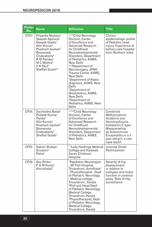
CP23 Priyanka Madaan1 Deepak Agarwal2 Deepak Gupta3 Atin Kumar4 Prashant Jauhari5 Biswaroop Chakrabarty6 R M Pandey7 M C Mishra8 V K Paul9 Sheffali Gulati10
1,5,10Child Neurology Division, Center of Excellence and Advanced Research for Childhood Neurodevelopmental Disorders, Department of Pediatrics, AIIMS, New Delhi 2,3,8Department of Neurosurgery, JPNA Trauma Center, AIIMS, New Delhi 4Department of Radio-diagnosis, AIIMS, New Delhi 7Department of Biostatistics, AIIMS, New Delhi 9Department of Pediatrics, AIIMS, New Delhi
Clinico- epidemiologic profile of Pediatric head injury: Experience of tertiary care hospital from Northern India
1-2,4-6Child Neurology Division, Center of Excellence and Advanced Research for Childhood Neurodevelopmental Disorders, Department of Pediatrics, AIIMS, New Delhi
Combined Methylmalonic Acidemia and Homocystinuria, Cobalamin C type: Masquerading as Autoimmune Encephalitis in a 5 year old girl: a rare case report
CP25 Sakshi Shakya1 Suvasini2 Patra3
1-3Lady Hardinge Medical College and Kalawati Saran Childrens Hospital
Juvenile Onset Parkinsonism
CP26 Gisi Shibu1 P A M Kunju2 Amruthalal3
1Paediatric Neurologist , SP Fort Hospital, Trivandrum, Amrithalal , Physiotherapist , Dept of Pediatric Neurology , Medical college, Trivandrum , Kerala 2Prof and Head Dept of Pediatric Neurology, Medical College, Trivandrum, Kerala 3Physiotherapist, Dept of Pediatric Neurology, Medical College, Trivandrum, Kerala
Severity of hip displacement in relation to subtypes and motor function in cerebral palsy- Role of Hip surveillance
1-3Center for Child Development and Disabilities, Bengaluru
Intervention training program for parents of children with autism spectrum disorder – EDITT program
A02 Jitendra Kumar Sahu1 Neeharika Sriram2
1-2Department of Pediatric Neurology, PGIMER
Evaluation of hyperandrogenism in children with autism spectrum disorder and age-sex matched controls
A03 Abhinayaa Janakiraman1 Udayakumar2
1-2Karthikeyan Child Development Unit, Sri Ramachandra Medical Centre, Chennai
Comparison of AIIMS Modified INCLEN Diagnostic Tool (Modified INDT-ASD) with Childhood Autism Rating Scale (CARS-2) in children with Autism Spectrum Disorder attending a Child Development Unit
1,2Autism Society West Bengal, Kolkata, India 2,3Manovikas Kendra, Kolkata, India
The impact of family-based early social responsiveness enhancement training on joint attention, engagement and participation of children with autism spectrum disorder
A05 Smita Awasthi Behavior Momentum India
Elimination of scratching behavior in a 4 year old girl with a diagnosis of atopic eczema and mild autism
A06 Smita Awasthi1 Shushma Vashist2
1-2Behavior Momentum India
Reduction of motor stereotypy in a 9-Year old boy with autism
32
NEUROPEDICON 2018
Poster No. Name Affiliation Title
A07 Hansashree Padmanabha1 Razia Adam Kadwa2 Pratibha Singhi3 Prabhjot Malhi4 Jitendra Kumar Sahu5 Naveen Sankhyan6 B R Mittal7 Rajinder8
1NIMHANS, Bengaluru 2Little Lily Hospital, Hyderabad 3Director, Pediatric Neurology and Neurodevelopment Medanta, The Medicity 4,5,6,7,8PGIMER, Chandigarh
18 F- FDG PET scan abnormalities at rest in children with Autism Spectrum Disorder
A08 Puja Kapoor CONTINUA KIDS Effect of Yoga therapy in behaviour problems of autistic spectrum disorder children
A09 Puja Kapoor1 Rajiv Chhabra2
1CONTINUA KIDS 2Artemis Hospital
Role of music therapy in improving social skills in Autism Spectrum Disorder children
A10 Kanwal Preet Kochhar
Cognitive Neurophysiology Lab, Department of Physiology, A.I.I.M.S, New Delhi, INDIA
1-4Department of Pediatrics, Vardhman Mahavir Medical College and Safdarjung Hospital, New Delhi
Prevalence of depression in primary caregivers of children with chronic neurologic ailments
A14 Jaai Joshi1 Sudha Chaudhari2
1Rehabilitation Officer,TDH Morris Child Development Center,KEM Hospital,Pune 2Consultant,Department of Pediatrics,KEM Hospital, Pune
Efficacy of training parents in improving parenting skills and reducing parent reported problem behaviours in hyperactive pre-scholer-A pilot study
33
NEUROPEDICON 2018
Poster No. Name Affiliation Title
A15 Shambhavi Seth1 Satinder Walia2 Zeba Parveen3
1,2Max Hospital, Gurgaon 1,3Bright Beginnings CDC
To study comparison of social emotional and communication scores on Development profile 3 (DP3) with Childhood Autism rating Scale (CARS) score in children fulfilling the DSM-V criteria for diagnosis of Autism Spectrum disorders
A16 S.V Aparna1 H M Rashmi2 S Gulati3 V K Batish4 S Grover5
1Assistant Professor, Department of Dairy Microbiology, College of Dairy Science and Technology, Kerala Veterinary and Animal Science University (KVASU) 2Scientist, Dairy Microbiology Division, ICAR- National Dairy Research Institute Karnal-132001, Haryana 3Chief, Child neurology Division, Department of Paediatrics, AIIMS, New Delhi 4Emeritus Scientist and Former Head, Dairy Microbiology Division, ICAR- National Dairy Research Institute, Karnal-132001, Haryana 5Principal Scientist and Head, Dairy Microbiology Division, ICAR-National Dairy Research Institute, Karnal-132001, Haryana
Comparative analysis of major gut microbiota of autistic and normal siblings in India by absolute PCR and metagenomic approach
34
NEUROPEDICON 2018
Poster No. Name Affiliation Title
A17 R Anand1 R Giuliani2 V Lucini3 E C Forrest4 S M Graham5 R D Hartman6
1 APC, AG, St. Moritz, Switzerland 2,3,4Newron Pharmaceuticals SpA, Bresso (MI), Italy 5Newron Pharmaceuticals US, Inc., Morristown, NJ USA 6NeurWrite LLC, Morristown, NJ USA
Sarizotan In The Treatment Of Respiratory Abnormalities In Patients With Rett Syndrome (Rtt): New Findings From An International, 6-Month, Randomized, Double-Blind, Placebo-Controlled, Phase Iii Trial (Stars)
1-5Child Neurology Division, Center of Excellence and Advanced Research for Childhood Neurodevelopmental Disorders, Department of Pediatrics, AIIMS, New Delhi
Clinical predictors of response to Applied Behavioral Analysis in children with Autism Spectrum Disorder: a prospective interventional study
1-7Child Neurology Division, Center of Excellence and Advanced Research for Childhood Neurodevelopmental Disorders, Department of Pediatrics, AIIMS, New Delhi
Clinical profile and management outcome of children with ADHD from a tertiary care center of North India: a retrospective cohort study
1-11Child Neurology Division, Center of Excellence and Advanced Research for Childhood Neurodevelopmental Disorders, Department of Pediatrics, AIIMS, New Delhi
Clinico-psychological profile and response to behavioral intervention of children with psychogenic headache from a tertiary care center in North India: a retrospective cohort study
1,3,13 Sangath, C-1/52, Safdarjung Development Area, New Delhi - 110016 1,2,4Centre for Chronic Conditions and Injuries, Public Health Foundation of India 5,9,10Birkbeck, University of London, Malet Street, Bloomsbury, London WC1E 7HX, UK 6,15School of Psychology and Clinical Language Sciences, University of Reading, Earley Gate, Reading RG6 6AL, UK 7The Com DEALL Trust, 224, 6th ‘A’ Main, 2nd block, HRBR Layout, Bangalore 560043, India 8,11Indian Institute of Technology-Bombay, Mumbai, Maharashtra 400076, India 12Harvard Medical School and the Harvard Chan School of Public Health; 641 Huntington Ave, Boston, MA 02115, USA 14All India Institute of Medical Sciences, Delhi, India
A Tablet Application for Screening Autism Risk in Community Settings
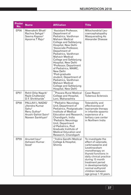
1Associate Professor, Department of Paediatrics, Vardhman Mahavir Medical College and Safdarjung Hospital, New Delhi 2Assistant Professor, Department of Paediatrics, Vardhman Mahavir Medical College and Safdarjung Hospital, New Delhi 3Medical Officer, Department of Paediatrics, Vardhman Mahavir Medical College and Safdarjung Hospital, New Delhi 4Post-graduate student, Department of Paediatrics, Vardhman Mahavir Medical College and Safdarjung Hospital, New Delhi
Diagnostic yield of electroencepha-logram (EEG) and patterns of EEG in children up to the age of 12 years: a retrospective study from a tertiary care hospital
1Assistant Professor, Department of Pediatrics, Vardhman Mahavir Medical College and Safdarjung Hospital, New Delhi 2Associate Professor, Department of Pediatrics, Vardhman Mahavir Medical College and Safdarjung Hospital, New Delhi 3Professor, Department of Pediatrics, MAMC, New Delhi 4Post-graduate student, Department of Pediatrics, Vardhman Mahavir Medical College and Safdarjung Hospital, New Delhi
Mitochondrial Leu-coencephalopathy Masquerading As Alexander Disease
1-5Pediatric Neurology Unit, Department of Pediatrics, Postgraduate Institute of Medical Education and Research, Chandigarh, India 2Pediatric Neurology Unit, Department of Pediatrics, Post Graduate Institute of Medical Education and Research, Chandigarh
Tolerability and effectiveness of topiramate therapy in infantile spasms- experience of a tertiary care center in Northern India
EP09 Anureet kaur1 Ashwani Kumar Sood2
1-2Indira Gandhi Medical College & Hospital, Shimla
To investigate the effect of valproate, carbmazepine and Levetiracetam monotherapy on thyroid functions in daily clinical practice during 15 month treatment period in developmentally normal euthyroid children between age group 1-18 years.
1-4,6-7Department of Pediatrics, Post Graduate Institute Of Medical Education and Research Chandigarh, India 5Department of Pharmacology, Post Graduate Institute Of Medical Education and Research Chandigarh, India 2Additional Professor, Pediatric Neurology Unit Post Graduate Institute Of Medical Education and Research Chandigarh, India
Effectiveness and Safety of high-dose, Oral Pyridoxine as an adjunct to high dose Adrenocorticotrophic hormone versus high dose Adrenocorticotrophic hormone alone for the treatment of West Syndrome: A Randomized open label Trial
EP11 N. BALAMURUGAN1 Vykuntraju K Gowda2 Asha Benakappa3
1-3Department of Pediatric Neurology, Indira Gandhi Institute of Child Health, Bangalore- 560029
Clinical profile of children with a treatable and a nutritionally preventable cause of West Syndrome at a Tertiary care referral centre from Southern India – A Descriptive study
1 Division of Pediatric Neurology,B L Kapur Super Speciality Hospital, New Delhi 2-6 Division of Pediatric Intensive care, Department of Pediatrics, B L Kapur Super Speciality Hospital, New Delhi
Experience of inhalational anesthetic agent in refractory status epilepticus
1Junior Resident, Department of Pediatric medicine, SMS Medical College, Jaipur 2Assistant Professor, Department of Pediatric medicine, SMS Medical College, Jaipur 3Senior Professor, Department of Pediatric medicine, SMS Medical College, Jaipur
Quality of life in children with idiopathic epilepsy
EP31 Ranjith Kumar Manokaran1 Biswaroop Chakrabarty2 Manjari Tripathi3 R M Pandey4 Sheffali Gulati5
1,2,5Department of Pediatrics 3Department of Neurology 4Department of Biostatistics Child Neurology Division AIIMS, New Delhi
Sleep Abnormalities And Polysomnographic Profile Among Children With Drug Resistant Epilepsy
1-11Child Neurology Division, Center of Excellence and Advanced Research for Childhood Neurodevelopmental Disorders, Department of Pediatrics, AIIMS, New Delhi
Epilepsy in children with cerebral palsy: experience from a tertiary care center in North India
1Post Graduate Trainee, Department of Paediatrics, Medical College and Hospital, Kolkata 2Professor, Department of Paediatrics, Medical College and Hospital, Kolkata 3RMO, Department of Paediatrics, Medical College and Hospital, Kolkata 4Professor, Department of Paediatrics, Medical College and Hospital, Kolkata 5RMO, Department of Paediatrics, Medical College and Hospital, Kolkata 6Assistant Professor, Department of Paediatrics, Medical College and Hospital, Kolkata
Assessment of behavioral problems in children with epilepsy
43
NEUROPEDICON 2018
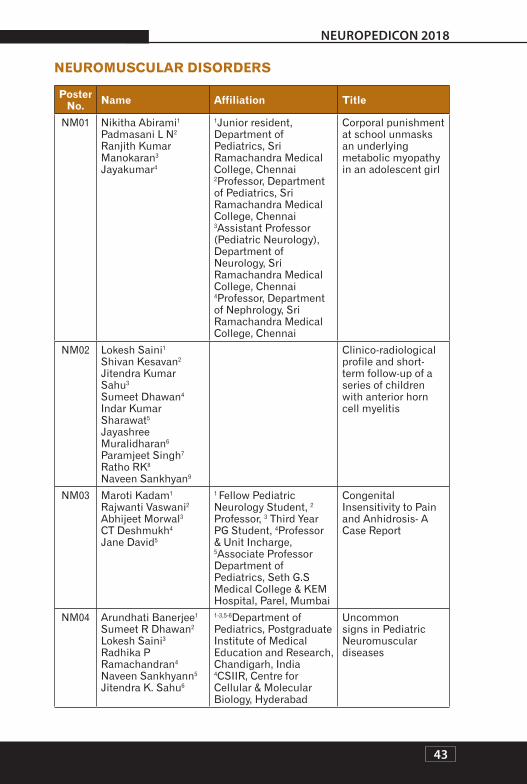
NEUROMUSCULAR DISORDERS
Poster No. Name Affiliation Title
NM01 Nikitha Abirami1 Padmasani L N2 Ranjith Kumar Manokaran3 Jayakumar4
1Junior resident, Department of Pediatrics, Sri Ramachandra Medical College, Chennai 2Professor, Department of Pediatrics, Sri Ramachandra Medical College, Chennai 3Assistant Professor (Pediatric Neurology), Department of Neurology, Sri Ramachandra Medical College, Chennai 4Professor, Department of Nephrology, Sri Ramachandra Medical College, Chennai
Corporal punishment at school unmasks an underlying metabolic myopathy in an adolescent girl
1 Fellow Pediatric Neurology Student, 2 Professor, 3 Third Year PG Student, 4Professor & Unit Incharge, 5Associate Professor Department of Pediatrics, Seth G.S Medical College & KEM Hospital, Parel, Mumbai
Congenital Insensitivity to Pain and Anhidrosis- A Case Report
NM04 Arundhati Banerjee1 Sumeet R Dhawan2 Lokesh Saini3 Radhika P Ramachandran4 Naveen Sankhyann5 Jitendra K. Sahu6
1-3,5-6Department of Pediatrics, Postgraduate Institute of Medical Education and Research, Chandigarh, India 4CSIIR, Centre for Cellular & Molecular Biology, Hyderabad
Uncommon signs in Pediatric Neuromuscular diseases
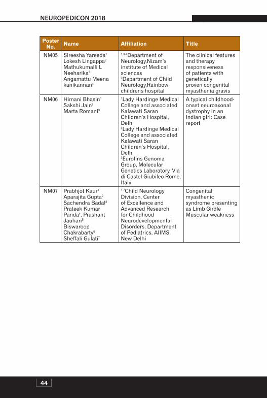
1,3-4Department of Neurology,Nizam’s institute of Medical sciences 2Department of Child Neurology,Rainbow childrens hospital
The clinical features and therapy responsiveness of patients with genetically proven congenital myasthenia gravis
NM06 Himani Bhasin1 Sakshi Jain2 Marta Romani3
1Lady Hardinge Medical College and associated Kalawati Saran Children’s Hospital, Delhi 2Lady Hardinge Medical College and associated Kalawati Saran Children’s Hospital, Delhi 3Eurofins Genoma Group, Molecular Genetics Laboratory, Via di Castel Giubileo Rome, Italy
A typical childhood-onset neuroaxonal dystrophy in an Indian girl: Case report
1-7Child Neurology Division, Center of Excellence and Advanced Research for Childhood Neurodevelopmental Disorders, Department of Pediatrics, AIIMS, New Delhi
Congenital myasthenic syndrome presenting as Limb Girdle Muscular weakness
NEU
ROIN
FEC
TIO
NS
45
NEUROPEDICON 2018
I03
COMPARISON OF 4-WEEKS VERSUS 12-WEEKS ANTI-CONVULSANT THERAPY FOR ACUTE SYMPTOMATIC SEIZURES IN CHILDREN WITH ACUTE ENCEPHALITIS SYNDROME-AN OPEN-LABEL, RANDOMIZED CONTROLLED TRIAL
1 2 3Dr Sumeet R Dhawan , Dr Jitendra Kumar Sahu , Prof Pratibha D Singhi , 4 5Dr Naveen Sankhyan , Prof Jayashree Murlidharan
1Postgraduate Institute of Medical Education and Research, Chandigarh2Postgraduate Institute of Medical Education and Research, Chandigarh3Medanta, The Medicity, Gurgaon, Haryana4Postgraduate Institute of Medical Education and Research, Chandigarh5Postgraduate Institute of Medical Education and Research, Chandigarh
Background: There exists poor evidence-base and conflicting literature regarding optimum duration of anti-epileptic drugs for acute symptomatic seizures in central nervous system infections. The study was designed to compare the effectiveness of 4-weeks versus 12-weeks anti-convulsant treatment in preventing seizure recurrences over a six-month period.
Methods: Children aged 3-months to 12-years having Acute Encephalitis Syndrome with acute symptomatic seizures receiving single anti-epileptic drug at 4-weeks of illness and without seizure recurrence from day 7- day 28 of illness were included in this comparative, parallel group assignment, open label, randomized control study. The exclusion criteria were included children with chronic meningitis, brain abscess, intracranial space occupying lesion, prior history of seizures, prior focal neurological deficit or any developmental delay, children suffering from HIV, chronic liver/kidney disease, acute hepatic encephalopathy, ≥2 anti-epileptic drugs and severely affected children were excluded. They were randomly allocated to receive anti-epileptic drugs either for 4-weeks or 12-weeks. The primary outcome was proportion of children developing seizure recurrence over 6-months follow up. The secondary outcome was to study factor(s) associated with seizure recurrence.
Results: Out of 232 children with Acute Encephalitis Syndrome, 60 children werefound to be eligible for randomization in two groups. Baseline demographics were comparable (except duration of illness) between the groups. None of the children developed any seizure recurrences in the follow up period. Although, 8 children had neurological deficits and 9 children had EEG abnormality, seizure recurrences were not seen in any of these children.
Conclusions: The present study suggests that a shorter duration (4-weeks) of anti-epileptic drug therapy is comparable with 12-weeks anti-epileptic drugs for preventing seizure recurrences over a six-month follow-up period in this cohort of children with Acute Encephalitis Syndrome.
The trial was registered with Clinical Trial Registry of India (CTRI/2017/06/008783) and Clinicaltrial.gov (NCT03181945).
Neuropedicon 2018
NE
UR
OIN
FEC
TIO
NS
NEU
ROIN
FEC
TIO
NS
46
NEUROPEDICON 2018
I02
OPTIC NERVE SHEATH DIAMETER AS A NON-INVASIVE TOOL FOR DETECTING RAISED INTRACRANIALPRESSURE IN THE PEDIATRIC INTENSIVE CARE UNIT: AN OBSERVER BLINDED, PROSPECTIVE STUDY
1 2 3Dr Indar Kumar Sharawat , Dr Naveen Sankhyan , Dr Arun Bansal , Dr Jitendra 4 5 6Kumar Sahu , Dr Kushaljit Singh Sodhi , Dr Mangat Ram Dogra
Background: The optic nerve sheath diameter (ONSD),measured by ultrasound, has been shown to increase within seconds of raised ICP. Hence, ONSD measurement can be potentially used to detect elevated ICP.
Method: A blinded, observational study of all children (2-12 years) admitted to PICU undergoing ICP monitoring using intra parenchymal catheterwas conducted November 2016 to December 2017. Healthy children of same age group were taken as healthy controls. The ONSD from eyes was measured using a 7.5 MHz ultrasound probe on closed eyelids. Horizontal and vertical diameters of both the optic nerves were measured and averages calculated. Repeated measurements were taken at least 3 hours apart. Observations with a parallel measured ICP ≥20 mm Hg were included as case-observations. Children with invasive ICP of <15 mmHg were taken as Neurological-control-observationsand healthy children served as healthy-control -observations. Twenty-two measurements of ONSD were assessed by two different observers in quick succession for interrater reliability.
Results: A total of 148 observations were performed in 30 children. Out of the 148 observations, 106 observations were case-observations (ICP ≥20), 38 observations were Neurological-control-observations (ICP<15mm Hg). An additional,66 observations werehealthy-control -observations. Themean binocular ONSD in cases was 5.71 ± 0.57mm, while in controls (all) it was 3.89 ± 0.51mm (p<0.001).An ONSD cut-off of 4.0 mm for detection of ICP ≥ 20 mm of Hg had an area under curve of 0.976, and sensitivity, specificity, PPV and NPV of 98%, 75%, 77% and 97% respectively. Interclass correlation coefficient for assessment of reliability of repeated measures for 22 paired observations for ONSD was 0.98.
Conclusions: ONSD has a good interrater reliability andwas accurate in identifying children with an ICP of ≥ 20 mmHg.
Neuropedicon 2018
NE
UR
OIN
FEC
TIO
NS
NEU
ROIN
FEC
TIO
NS
47
NEUROPEDICON 2018
NEUROPSYCHOLOGICAL AND SLEEP PROFILE OF HIV INFECTED CHILDREN: AN OBSERVATIONAL STUDY
1 2 3 4 5 6R Farmania , R Farmania , S K Kabra , B Chakrabarty , P Jauhari , S Sapra , 7 8 9A Kumar , R M Pandey , S Gulati
1 4 5 9 Child Neurology Division, Department of Pediatrics, All India Institute of Medical Sciences, New Delhi2 3 Pediatric Pulmonology division, Department of Pediatrics, All India Institute of Medical Sciences, New Delhi6Clinical Psychologist, Department of Pediatrics, All India Institute of Medical Sciences, New Delhi7Department of Radio diagnosis, JPNATC, All India Institute of Medical Sciences, New Delhi8Department of Biostatistics, All India Institute of Medical Sciences, New Delhi
Background: To determine the prevalence of neurologic syndromes, intellectual disability, abnormal behavior and sleep related problems in HIV infected children.
Method: HIV infected children aged 1-18 years registered in pediatric HIV clinic were randomly screened. Children with any acute illnesses, chronic comorbid chronic disorders were excluded. Neurological assessment was done by comprehensive neurological examination followed by targeted investigations such as MRI, electrophysiology and polysomnography. Intelligence quotient (IQ)was assessed by age appropriate scales; behavior by childhood behavior checklist, syndrome scale(CBCL) and sleep by childhood sleep habit Questionnaire (CSHQ). HIV infected children with IQ≥85 were compared with age and sex matched typically developing children (TDC) for behavioral and sleep problems.
Results: Hundred children (61 males), median age 11.42 years (1.67-17.5) were evaluated. 35% had at least one neurologic syndrome; cognitive dysfunction in 25%, seizure in 8%, pyramidal syndrome in 10%, extrapyramidal syndrome 8%, peripheral neuropathy in 3%. Etiology was unclear in 19/35 (53%); HIV encephalopathy was seen in 8 cases (23%). Nutritional status (BMI <3rd centile) was the only significant risk factor associated with any neurologic syndrome [OR 0.13 (95% CI 0.24-0.65)]. Fifty-six percent children had below average intelligence (IQ<90); 8% had intellectual disability (IQ<70). 24% children had behavioral problems;14% had sleep problems.HIV Associated Neurocognitive Disorder (HAND) criteria in age 6-16 years (69/100) was fulfilled in 39/69 (56.5%) as compared to HIVE in 5/69 (7.24%). HIV infected children as compared to TDC had significantly increased total sleep duration. Conclusion: Nutritional status is an important risk factor associated with presence of any neurologic syndrome. Neuropsychological dysfunction and sleep problems are seen in significant proportion of HIV infected children. HAND criteria devised for adults canidentify children with functional cognitive impairments who were otherwise not documented by HIVE criteria. Recognition of neurocognitive deficiencies is important to provide holistic care to these children.
1Department of Pediatrics, JNMC, Belagavi2Department of Pediatrics, JNMC, Belagavi3Department of Pediatrics, JNMC, Belagavi
Evaluation of a child with encephalitis is difficult due to the similarities in the clinical, imaging and laboratory findings of many forms of autoimmune and infectious encephalitis. Presentation of autoimmune encephalitis (AE) in childhood is often subacute, with varied clinical manifestation. However, as it takes time to get the results of autoimmune encephalitis antibody tests, many times immunosuppression is began with a presumed diagnosis of AE. Due to growing knowledge of AE, many primary-care physicians are diagnosing AE and starting immunomodulation, which may be detrimental at times. We here highlight the dark side of over-diagnoses of AE.
In past few months, 2 school-aged children presented to us in vegetative state. Both the children were diagnosed as AE based on their presentation with fever, behavioural changes and myoclonic jerks/ focal seizures. Pulse methylprednisolone was given to the children with presumed diagnosis of AE. There was no improvement on immunotherapy and children deteriorated to vegetative state in next 2-3 weeks. There was no history of measles in both children and they were vaccinated (one dose of measles vaccine at 9 months). During detailed evaluation, fundus examination showed hyperemic disc, large whitish subretinal patch over posterior-pole with satellite lesions, and MRI review showed subtle asymmetrical hyper-intensities in peri-ventricular white-matter. Based on these findings, Subacute sclerosingpanencephalitis (SSPE) was suspected and confirmed by enzyme-linked immunosorbent assay of CSF for measles virus IgG [The titre of IgG antibodies to measles in CSF was 1 in 625]. Both children died within 1 month.
It is better to withhold immunosuppression with methylprednisolone till we get the confirmation or use of IVIg instead of methylprednisolone. We intend to create awareness among primary physicians regarding judicious use of immunotherapy.
Neuropedicon 2018
NE
UR
OIN
FEC
TIO
NS
NEU
ROIN
FEC
TIO
NS
49
NEUROPEDICON 2018
I04
NEUROLOGICAL MANIFESTATIONS OF CHIKUNGUNYA FEVER IN CHILDREN - A SINGLE CENTRE EXPERIENCE
1 2Dr Pradeep Kumar Sharma , Dr Nikhil Vinayak
1Pediatric Critical Care and Pulmonology, Sri Balaji Action Medical Institute, New Delhi2Pediatric Critical Care and Pulmonology, Sri Balaji Action Medical Institute, New Delhi
Background: Chikungunya fever is usually regarded as a benign disease. Neurological manifestations of chikungunya fever in children have been previously reported however in view of recent chikungunya epidemics these manifestations assume greater significance.
Methodology: This study was conducted in the Pediatric Intensive Care Unit and High Dependency Unit of a tertiary care hospital in Delhi, India. Patients diagnosed with chikungunya infection by positive Real Time-Polymerase Chain Reaction (RT-PCR) assay from September to December 2016 were retrospectively analysed for neurological manifestations. The information recorded included demographic features, clinical features, laboratory parameters, course and hospital stay.
Results: Fourteen out of 49 children with chikungunya fever had neurological manifestations. Median age was 4 years and range was 1-12 years. 11 children hadseizures, 2 had encephalopathy without seizures and one had seizures and encephalitis. Nine patients had only one episode of seizure and three had multiple seizures. Eight children had seizure within 24 hours of fever. Cerebrospinal fluid assay done in two children revealed 5cells/mm3(all lymphocytes) with normal biochemistry. Magnetic resonance imaging was done in three children and was abnormal in one; showing flair hyperintensity along bilateral high parietal, frontal lobes, centrum semi-ovale regions and corpus callosum. In addition, SWAN image revealed multiple blooming foci in corpus callosum and bilateral basal ganglia. Three children underwent EEG and right focal discharges were seen in one. Nine children were given oral clobazam and were discharged without antiepileptics. One child was given valproic acid and another received levetiractetam. Both were discharged on oral antiepileptics. The child with encephalitis showed excellent neurological recovery. Mean (SD) hospital stay was 3.86 (2.87) days. There were no deaths.
Conclusion: This study shows that neurologic manifestation in children can be due to chikungunya fever during an outbreak.
Neuropedicon 2018
NE
UR
OIN
FEC
TIO
NS
NEU
ROIN
FEC
TIO
NS
50
NEUROPEDICON 2018
I05
PROFILE OF ACUTE ENCEPHALITIS SYNDROME IN CHILDREN: A RETROSPECTIVE ANALYSIS
1 2Dr Dilip M Chowdhary , Dr Aditi Baruah
1Assam Medical College & Hospital, Dibrugarh2Assam Medical College & Hospital, Dibrugarh
Background: Acute Encephalitis Syndrome(AES)is a group of clinically similar neurologic manifestation caused by several different viruses, bacteria, fungus, parasites and spirochetes. It predominately affects children less than 15 year of age. Japanese Encephalitis(JE) virus is endemic in Eastern part of India including Assam and it is the most common cause of AES.This study was conducted with an aim to analyse the profile and outcome of children admitted with AES with special reference to JE IgM positive cases.
Methodology: This is a retrospective recordbased hospital study. We collected data of the children admitted into the Department of Paediatrics, Assam Medical College & Hospital, Dibrugarh, Assam with the clinical diagnosis of AES from 1stJanuary,2017 to 31stDecember,2017.
Result: Most of cases 156(57.3%) were between 6 to 11 years of age. Male:Female ratio was 1.47:1. 255(93.7%)cases were Hindus, 12(4.4%) were Muslims, 3(1.1%) were Christians. Most of cases 123(45.2%) were reported during the month of June & July. Out of 272 AES cases, majority were due to Japanese encephalitis(JE) virus 94(34.5%) which were JE-positive.And JE-negative cases were 178(65.4%). Out of JE-negative cases, Dengue constitutes 11(6.17%) cases, Scrubtyphus 2(1.12%), Streptococcus pneumoniae 6(3.37%). No organism was detected in 159(58.4%). Out of 272 cases 42(15.4%) children were vaccinated with JE vaccine, of which 30 (71.4%)cases were JE-negative. Out of 94 JE-positive cases, 4(9.5%) were died, 37(88%) were discharged without sequelae, 1 was discharged against medical advice.
Conclusion: In our study JE-virus is the most common organism responsible for AES and commonest age was 6 to 12 years. Male:Female ratio was 1.47:1. Most of the cases reported during monsoon season. Number of JE cases decreased because of inclusion of JE-vaccine in routine NIS. So vaccination of children below 15yr is helpful in preventing JE.
Neuropedicon 2018
NE
UR
OIN
FEC
TIO
NS
NEU
ROIN
FEC
TIO
NS
51
NEUROPEDICON 2018
I06
RICKETTSIAL MENINGITIS - CASE SERIES (13)IN RMC LONI, MAHARASHTRA
Background: Rickettsial infections are increasingly detected in Ahmednagar district. We studied patients with ricketssial infections who developed neurological manifestations and their response to treatment.
Methodology: Total 13 Cases were studied prospectively from Pravara Rural Hospital, Loni, Ahmednagar. Weil felix test, CSF analysis, complete blood count was done. ELISA was used to confirm diagnosis.
Results: All had ELISA test positive. Median age was 4 yrs including one 27 dayoldneonate with meningeal signs and positive CSF study.8 were males and5 females, 9 were from rural areas(69%).All had history of exposure to ticks and 7 patients had history of tick removal(53%). All had fever with rashes involving palms and soles. Two patients had eschar(15%). Neurological manifestation admission were altered sensorium (drowsiness, confusion, semi comatose state)(85%), convulsions (23%), meningeal signs(46%). CSF fluid showed cellular reactions, predominantly lymphocytesin all cases except neutrophilic predominance in two cases. Most of the patients responded well and recovered to treatment with Doxycycline and Chloramphenicol. Two patients died (fatality rate 15%).
Conclusions: Failure of primary diagnosis could have led to significant morbidity and mortality. Due to high index of suspicion in this rural area, they were diagnosed and treated timely. Neonatal Rickettsial infection is seldom thought of. This also should be looked out for in high index suspicion areas. Response to early treatment is good and life saving.
Neuropedicon 2018
NE
UR
OIN
FEC
TIO
NS
NEU
ROIN
FEC
TIO
NS
52
NEUROPEDICON 2018
I07
PREDICTORS OF MECHANICAL VENTILATION IN ACUTE ENCEPHALITIS SYNDROME IN CHILDREN
1Senior Resident, King George Medical University, Lucknow, India2Senior Resident, King George Medical University, Lucknow, India3Professor, Department of Microbiology, King George Medical University, Lucknow, India4Professor, Department of Paediatrics, King George Medical University, Lucknow, India
Background: Incidence of acute encephalitis syndrome (AES) is high in children and is associated with high mortality and sequelae. Limited data is available about parameters predicting the need of mechanical ventilation in children of AES. The aim of the study was to identify predictors of need of mechanical ventilation.
Methods: This is a prospective cohort study, conducted at a tertiary care hospital of north India from 2017-2018. One hundred and forty two children in the age group of one month to 13 years presented with fever ≤ 2weeks duration and altered mental status lasting for more than 4 hours, were enrolled in the study. Variables present on admission which were associated with ventilation were delineated by univariate analysis. Those variables with p<0.05 were entered in a logistic regression model to identify independent predictors of need of ventilation, using SPSS version 16 package.
Results: Mean age of 142 enrolled children was 13±5.8 years with a male preponderance (62%). Median duration of fever at admission was 5 (4-7) days. Seizure was present in 117 (82.4%), GCS<7 in 36 (25.4%), shock in 19 (13.4%) and pupillary abnormalities in 21 (14.8%) children. Aetiology could be identified in 71 (53.8%). Overall mortality was 21.8%. Mechanical ventilation was required in 33 children (23.2%). After multivariate regression analysis, four parameters at admission emerged as independent predictors for mechanical ventilation - shock [OR 7.7 (95% CI; 1.9-31.2)], pneumonia [12.5 (2.2-69.9)], GCS<7 [3.7 (1.2-11.7)] and raised intracranial tension [4.4 (1.2-16.4)]. The proportion of mortality was higher among ventilated vs. non-ventilated children (92.7% vs. 30.3%) (p < 0.001).
Conclusions: Presence of pneumonia, shock, raised intracranial tension and GCS less than 7 at admission indicate high chances of requiring mechanical ventilation in children with AES. Mortality was very high in ventilated children, however.
Neuropedicon 2018
NE
UR
OIN
FEC
TIO
NS
NEU
ROIN
FEC
TIO
NS
53
NEUROPEDICON 2018
I08
SSPE MIMICKING ANTI-NMDA RECEPTOR ENCEPHALITIS – CASE REPORT
1 2 3Dr Himani Bhasin , Dr Shilpa Devamare , Dr Vikram Bhaskar , Dr Suvasini 4 5Sharma , Dr Manjari Tripathi
1Lady Hardinge Medical College and associated Kalawati Saran Children's Hospital2Lady Hardinge Medical College and associated Kalawati Saran Children's Hospital3Lady Hardinge Medical College and associated Kalawati Saran Children's Hospital4Lady Hardinge Medical College and associated Kalawati Saran Children's Hospital
Subacute sclerosing panencephalitis (SSPE) is a chronic progressive encephalitis of childhood and young adults. The atypical clinical presentations of SSPE are myriad leading to diagnostic dilemmas. This case report illustrates a 5 year old boy who presented with short history of cognitive decline and psychiatric symptoms, and movement disorders such as dystonia, orofacial dyskinesias mimicking N-methyl d- aspartate receptor encephalitis (NMDAR). Subsequently he was diagnosed to be a case of SSPE on the basis of EEG.
1 2 3Department of Pediatrics, LHMC & associated KSCH4AIIMS, New Delhi
Background: With free availability of anti-retroviral ART under national programme, increased numbers of HIV positive children are surviving into their adolescence and young adulthood. The current study was conducted to assess the impact of HIV status disclosure on mental health of children with HIV infection.
Method: A Cross sectional interview-based study was carried at tertiary care hospital from November 2015- March 2017. Caregivers (n=47) of 50 HIV infected children aged 8-18 years and the children themselves were sequentially interviewed. Information regarding disclosure of serostatus and circumstances of disclosure were assessed. Disclosed and non-disclosed children were interviewed with 2 different structured proformas to avoid inadvertent disclosure. Mental health indicators were assessed using Child Behaviour Checklist (CBCL) and Mini International Neuropsychiatric Interview for children and adolescents (M.I.N.I KID) respectively.
Results: Of 50 children (males, 62%; mean age 12.43± 2.89 years); 31 (62%) were infected perinatally. As per caregiver report, 19(38%) children were disclosed while only 17(34%) children accepted their serostatus. In 8 cases there was discordance between caregiver and child report regarding disclosure of serostatus.One disclosed (5.9%) and 3 non-disclosed (9.1%) children scored in clinical range for behavioural problems. Though proportion of children suffering from major depressive episodes (17.65% vs 9.09%; p=0.38) and generalized anxiety disorder (23.53% vs 9.09%; p=0.16) was more in disclosed children; the difference was not statistically significant. Both depression and anxiety were more prevalent in children with non-perinatally acquired HIV as compared to those with perinatal HIV (depression 31.6% and 0%; anxiety 26.3% and 6.5% in children with non-perinatal & perinatal transmission respectively).
Conclusions: There was no significant difference in mental health outcomes among children with HIV infection who were disclosed versus non- disclosed of their status. Caregivers should be assured that HIV status disclosure will not adversely affect the mental health of their children.
Neuropedicon 2018
NE
UR
OIN
FEC
TIO
NS
NEU
ROIN
FEC
TIO
NS
55
NEUROPEDICON 2018
I11
CLINICAL PROFILE AND AUDIOLOGICAL OUTCOME OF NEWBORNS WITH CONGENITAL CYTOMEGALOVIRUS INFECTION
1 2 3Dr. Khushboo Kanwal , Dr. Harsimran Singh , Dr. P.V. Nigwekar , Dr. D.Y. 4Shrikhande
1 2 3 4Rural Medical College, Pravara Institute of Medical Sciences, Loni
Background: Sensorineural hearing loss is a common sequelae of congenital cytomegalovirus (CMV) infection. Hearing loss due to congenital CMV infection can be present at birth or can manifest later.
Objectives: include estimating the prevalence of Cytomegalovirus (CMV) infection in newborns, to describe clinical profile and assess hearing outcomes.
Methods: This was a single-centre, observational, cross sectional study in newborns diagnosed with CMV infection at the Pravara Rural Hospital Loni in year 2018. Enzyme immunoassay for IgG and IgM antibodies against CMV was used to identify congenital CMV infection. BERA was used to assess audiological outcome.
Results: In the ongoing study, four newborns with CMV infection were identified; one died, 3 (75%) were symptomatic at birth, of whom 2 (50%) were neurologically symptomatic. Two (50%) presented with subsequent abnormal audiological outcomes.
Conclusions: All infants with subsequent SNHL or abnormal neurodevelopment were symptomatic at birth. Further studies are required to assess the magnitude of congenital CMV infections so that a routine screening program can be recommended and early intervention can be done for the resultant SNHL.
PNEUMOCEPHALUS - RARE COMPLICATION OF A COMMON DISEASE
Dr. T. M. Ananda Kesavan1, Dr. Tissa John2
1Dept of Pediatrics, Govt. Medical College, Thrissur, Kerala 2Dept of Pediatrics, Govt. Medical College, Thrissur, Kerala
Introduction: Meningitis one of the common infection of the nervous system leading to high morbidity and mortality. E coli meningitis is very rare in infants and complications like pneumocephalus still very rare. We are presenting a case of E coli meningitis with pneumocephalus
Key Words: Meningitis, E coli, Pneumocephalus
Case Report: A 6 months old child admitted with Fever for 5 days and Seizures – 3 episodes two days back. Also noted incessant cry and poor feedingBorn out of non-consanguinous marriage, uneventful antenatal and postnatal period, term, normal delivery of 2.650 KgImmunised up to age, developmentally normal On examination: Alert, GCS-14/15Vitals-stableHead to foot examination : AF tense, Pupils equal and reacting to lightNS: Cranial nerves-normal, Normal tone and power, DTR: exaggerated Plantar- bilaterally extensorOther systems: NormalWith this picture we kept a provisional diagnosis meningitis and investigated All blood routine investigations were normal. Screening for HIV-negative, Neurosonogram- normal LP done on D2 and thick pus drained. Gram stain showed Gram negative baciili . Initially treated with Ceftriaxone and later upgraded to Vancomycin and meropenem added on D3. On D3, she developed skew deviation of eyesCT Scan: Mild hydrocephalus, signs of meningeal inflammation and air collection (Fig 1)CSF C/S- E coli sensitive to imipenem,meropenem,cefaperazone sulbactam,piptaz,tigecycline. Blood C/S- E coli sensitive to imipenem, meropenem,cefaperazone sulbactam, piptaz, tigecycline,colistin. On D5 child developed seizures with refractory shock. Expired inspite of resuscitative measures.
Discussion: Pneumocephalus may develop following surgery, trauma , brain abscess and meningitis. Meningitis with gas forming organism like clostridium or a mixed aerobic and anerobic infection lead to localized pneumocephalus. Pneumocephalus due to E coli is very rare.
I12
AU
TOIM
MU
NE
DIS
ORD
ERS
57
NEUROPEDICON 2018
AI02
OMA AS A COMPLICATION OF DKA
1 2Mahesh Kamate , Preeti Gopal
1Department of Paediatrics, JNMC Belgavi2Department of Paediatrics, JNMC Belgavi
OMA syndrome (myoclonic encephalopathy of infancy or Kinsbourne syndrome) is a paraneoplastic syndrome associated with neuroblastoma in children. The ocular globes are in a state of continuous agitation with rapid involuntary irregular and conjugate eye movements, myoclonic jerking of the limbs, trunk, and hand.
Neuroblastoma is diagnosed by MIBG scan to identify neuroblastoma. The treatment is by excision of the tumor followed by immunomodualtion.
This is a case report of a 9month old male child who presented with acidotic breathing and rapid horizontal nystagmus. Further evaluation with serial ABG, urine ketone bodies, RBS monitoring, HBA1C and SLC19A2 genetic studies were done and he was diagnosed with DKA secondary to type 1 DM with thiamine responsive megaloblastic anemia.
VMA levels were determined to rule out neuroblastoma and BERA diagnosed the child to have severe SNHL.
OMA is characteristically seen as a paraneoplastic manifestation of neuroblastoma in children and breast cancer in adults.
OMA has never been reported as a complication of DKA and this case report hopes to shed further light on the matter.
Neuropedicon 2018
AU
TO
IMM
UN
E D
ISO
RD
ER
S
AU
TOIM
MU
NE
DIS
ORD
ERS
58
NEUROPEDICON 2018
AI05
CHILDHOOD ONSET POLYARTERITIS NODOSA AND DEFICIENCY OF ADENOSINE DEAMINASE 2 (DADA 2): NOVEL MUTATION IN CECR 1 GENE
1 2 3 1Dr Nikit Shah , Dr Rajkiran , Dr Chandra S Koyalakonda , Dr Lokesh Lingappa , 1Dr Ramesh Konanki
1Dept of Pediatric neurology, Rainbow Children's Hospital, Hyderebad2Hyderabad Rheumatology Center3Dept of pediatrics and intensive care, Rainbow Children's hospital, Hyderebad
7 years old boy presented with complains of on and offf abdominal pain since 6 months requiring medical attention and right internuclear ophthalmoplegia (INO) since 3 days before presentation. He was noted to have hypertension. On further investigation microaneurysms in branch renal artery and hepatic arteries were detected. He had thickened bowel walls with normal bowel mucosa (on GI scopy), mesentric artery aneurysms could not be demonstrated. His ANA & ANCA levels were negative. We started pulse methylprednisolone, cyclophosphamide and oral steroids. On 6 months followup he is completely normal with BP controlled with single medication. Six months into the treatment he had hypertensive intracranial bleed with left hemiparesis. He is now on Etanercept for disease control. Blood was sent for analysis of CECR 1 (Cat's eye syndrome chromosome region, candidate 1) gene. Analysis showed homozygous mutation in CECR 1 gene. Amino acid substitution of Arginine for Glycine at codon 47 in the adenosine/AMP deaminase N-terminal domain of the CECR1 protein was identified. Parents were heterozygous for the same mutation. ADA 2 Enzyme levels were done, were very low in patient and were in carrier range in parents as compared to non affected sibling (3 years younger to patient) and Indian adult control. This variation is not reported in English literature previously. To best of our knowledge this is 1st case report of genetically proved DADA2 deficiency from India.
NEUROMYELITIS OPTICA SPECTRUM DISORDER (NMOSD) PRESENTING ONLY AS BILATERAL INTERNUCLEAR OPTHALMOPLEGIA IN A 12 YEAR OLD GIRL CHILD: A RARE ENTITY
1 2 3Dr. Indira. V , Dr. Saji James , Dr. Ranjith Kumar Manokaran
1Junior resident, Department of Pediatrics, Sri Ramachandra Medical College2Professor of Pediatrics, Sri Ramachandra Medical College 3Assistant Professor (Pediatric Neurology), Department of Neurology, Sri Ramachandra Medical College
Background: Neuromyelitis Optica (NMO) is characterized by immune-mediated demyelination and axonal damage predominantly involving optic nerve and spinal cord. Aquaporin-4, the target antigen of NMO- IgG is a water channel protein highly concentrated in spinal cord gray matter. Some patients present with brain symptoms as their first manifestation and develop recurrent brain symptoms without optic neuritis or myelitis which are classified as NMOSD. Here we present a 12 year old girl child who developed bilateral internuclear opthalmoplegia as the initial presentation of NMOSD.
Method: A twelve year old premorbidly normal girl presented with headache and vomiting for two weeks, following which she developed diplopia. There was no fever. On examination, child had wall eyed bilateral internuclear ophthalmoplegia (WEBINO) and ataxic gait. Higher mental functions, other cranial nerves, sensory and motor system were intact at the time of presentation. There was no evidence of optic neuritis and myelitis clinically. MRI brain and spine showed focal demyelination of dorsal part of the midbrain. There was no radiological evidence of optic neuritis and myelitis. CSF analysis showed no pleocytosis, mild protein elevation and was positive for anti-aquaporin 4 antibody. A diagnosis of NMOSD was made and child was started on IV Methyl prednisone pulse therapy followed by oral steroids. On follow up, she has showed good neurological recovery.
Conclusion: Brain stem involvement in NMOSD are more common than previously thought, and rarely children even manifest brain symptoms as their first presentation. Presentation of NMOSD without myelitis and optic neuritis with only brain stem involvement is highly likely. High index of suspicion for NMOSD and identification of anti-aquaporin 4 sero-positivity can help in early institution of immunotherapy and planning for subsequent long term immunomodulation.
Neuropedicon 2018
AU
TO
IMM
UN
E D
ISO
RD
ER
S
AU
TOIM
MU
NE
DIS
ORD
ERS
60
NEUROPEDICON 2018
AI03
ANTI NMDA RECEPTOR ENCEPHALITIS PRESENTING AS ACUTE FLACCID PARALYSIS IN A YOUNG CHILD
1Division of Pediatric Neurology, B L Kapur Super Specialty Hospital, New Delhi2Division of Pediatric Intensive care, Department of Pediatrics, B L Kapur Super Specialty Hospital, New Delhi3Division of Pediatric Intensive care, Department of Pediatrics, B L Kapur Super Specialty Hospital, New Delhi4Division of Pediatric Intensive care, Department of Pediatrics, B L Kapur Super Specialty Hospital, New Delhi5Division of Pediatric Intensive care, Department of Pediatrics, B L Kapur Super Specialty Hospital, New Delhi
Background: The classical presentation of antiNMDAR encephalitis comprisesof psychotic behavior, abnormal movements and seizures. However,atypical manifestation may be seen in young children. We report a case of young child who presented as acute flaccid paralysis and later developed full blown features of anti NMDARencephalitis.
Method: A 16 month old boy premorbidly normal presented with progressive ascending paresis after 7 days of a viral prodrome of fever and loose motion. The nadir of weakness reached over next 3 days in form of neck flop with sparing of respiratory muscles. There was no mal alignment of eyes, facial deviation or nasal regurgitation. On examination,child was awake with gestrual communication to mother. There was no cranial nerve palsy. He had generalized hypotonia, axial more than appendicular with truncal ataxia. Power was 3-/5 in all muscle group across joints in both upper and lower limbs. Deep tendon reflexes were elicitable only in right knee with flexor plantar response. No signs of cerebellar dysfunction besides truncal ataxia was present. Possibility of Guillainbarre syndrome was considered, which was confirmed by NCS showing absent H reflex and F responses. MRI spine and brain with contrast and CSF analysis on day 14 of illness were normal. Child was given IVIG 2gm/kg over 2 days. Over next 4 days, he had become extremely irritable with rage attacks and sleeplessness. Concurrently abnormal movements in form of fragmentary myoclonus and oromotordyskinesias also appeared. Child was then investigated for autoimmune encephalitis and paraneoplastic syndromes along with tumorscreen. CSF NMDA antibody was positive confirming the diagnosis. Child was treated with pulse methylprednisolone.In view of poor response to steroids, later Plasmapharesis followed by Rituximab was given, to which child showed response.
Conclusion: Anti NMDAR encephalitis can present with acute flaccid paralysis in a young child.High index of suspicion is required in this age group to reach to diagnosis.
Neuropedicon 2018
AU
TO
IMM
UN
E D
ISO
RD
ER
S
AU
TOIM
MU
NE
DIS
ORD
ERS
61
NEUROPEDICON 2018
MONOPHASIC ACQUIRED CENTRAL NERVOUS SYSTEM DEMYELINATING SYNDROMES IN CHILDREN: EXPERIENCE OF A TERTIARY CENTRE FROM NORTH INDIA
1Child Neurology Division, Center of Excellence and Advanced Research for Childhood Neurodevelopmental Disorders, Department of Pediatrics, AIIMS, New Delhi2Department of Radio-diagnosis, AIIMS, New Delhi
Introduction: Acquired CNS demyelinating syndromes, a commonly recognized malady in children, include a spectrum of disorders- Acute disseminated encephalomyelitis (ADEM), clinically isolated syndrome (CIS), Multiple sclerosis (MS) and Neuromyelitis optica spectrum disorders (NMOSD). They can be monophasic or recurrent. Monophasic syndromes include ADEM, CIS and NMOSD (some cases).
Methods: Retrospective chart review of consecutive children with acquired demyelinating disorders presenting to a north Indian tertiary care hospital over 9 years (2009-2017)
Results: Out of 95 cases of ADS, 66 (~70% cases) were monophasic (19 ADEM and 47 CIS). Median age at presentation was 7years (Range:1-12years). Gender distribution for monophasic ADS was 41 boys to 25 girls. Among monophasic ADS, presenting features included fever (27), encephalopathy (19), seizures (16), paraparesis (26) and features of raised ICP (1).Among monophasic ADEM (19/66), all patients had alteration of sensorium, 2 had associated TM (cervicodorsal) and none of the patients had evidence of associated optic neuritis.Among CIS (47/66), 19 had isolated acute transverse myelitis like presentation, 5 had NMOSD like presentation (diagnosed after imaging) and rest presented as isolated optic neuritis, hemispheric syndromes or extrapyramidal syndromes. Steroids led to significant improvement in acute episodes of demyelination {PCPS score 3-6 in 9 children including 1 death with hemorrhagic ADEM}.
Conclusion: Acquired demyelinating syndromes are treatable disorders with a good prognosis in children with monophasic demyelination. Studies with larger population size are required to characterize features that predict future recurrences.
Neuropedicon 2018
AU
TO
IMM
UN
E D
ISO
RD
ER
S
AI04
AU
TOIM
MU
NE
DIS
ORD
ERS
62
NEUROPEDICON 2018
AI06
MYASTHENIC CRISIS IN A 7 YEAR OLD CHILD WITH AUTONOMIC.DYSFUNCTION
Dr Narendranadha Reddy K1, Dr Mahesh Kamate2, Dr Mayank Detroja3
1JNMC,Belagavi2JNMC, Belagavi3JNMC, Belagavi
Myasthenia gravis is an autoimmune disorder of peripheral nervous system, leading to fluctuating muscle weakness. It is caused by circulating antibodies that block acetyl choline receptors on post synaptic neuromuscular junction. Myasthenic crisis is defined as acute fatal condition, due to weakness from acquired myasthenia gravis and diagnosed by detection of acetyl choline antibodies in the body, respiratory failure.
This is a case report of 7 year old female child who presented with ptosis, dysphagia, upper and lower limb weakness leading to respiratory failure requiring mechanical ventilation. Further evaluation with detection of acetyl choline antibodies in the plasma, nerve conduction studies was done and diagnosed as Myasthenic crisis. Child was treated with plasmapheresis, IV Immunoglobulin, corticosteroids, pyridostigmine and ventilatory support. Child couldnot be revived because of autonomic dysfunction, where >50% mortality is present.
Given rarity of this condition, this case report is to stress prognosis in a Myasthenic crisis child, when he/she develops autonomic dysfunction, where mortality rates are high(>50%).
1All India Institute of Medical Sciences, New Delhi
Background: Craniopagus conjoined twins are children born with joint heads. It is rare of rarest disease noted in 1 in 2.5 million live births. Modern neurosurgery practice documents 15 odd cases since 1987 with handful of survivors reported so far (< 10).
Case Summary: JB conjoined twins from Odisha presented at age of 2 yr. with type 3 Vertical total craniopagus union (intertwine angle 120 degrees). Their DQ was 59 at admission, however, both kids when joint were active and playful. After detailed neuroradiological workup and preoperative planning and detailed consenting process, staged surgical separation was carried out in two stages. Their preoperative angiogram revealed shared circular sinus (Superior sagittal sinus) with directional flow of drainage from one child to another. First stage consisted of 180-210 degree circumferential craniotomy, creation of venous conduit to bypass part of shared venous sinus between two children using saphenous vein [multiple cortical veins were implanted on new conduit], partial hemispheric disconnection and placement of expanders (skin). This procedure lasted over 25 hours. Both children had stormy postoperative period with episode of pulseless ventricular tachycardia in first child and refractory seizures in second child. Eight weeks later, first child developed features of chronic cardiac overload due to one way fistula development between two children secondary to graft thrombosis. Thence, emergency separation surgery was planned to separate the kids on 25th Oct 2017 [lasted 22 hrs.]. Stormy postop course was well managed. 10 months postoperatively, children have survived and improved significantly. Present case is the first successful separation surgery of conjoined twins at head level wherein we have been able to save both children successfully. It was challenging case for the entire team, however, team work and adequate planning with intensive care back up helped in achieving successful outcome in this case.
Neuropedicon 2018
CE
RE
BR
AL
PA
LSY
& D
EV
ELO
PM
EN
TAL
DIS
OR
DE
RS
CER
EBRA
L PA
LSY
AN
D D
EVEL
OPM
ENTA
L D
ISO
RDER
S
64
NEUROPEDICON 2018
CP03
WHİCH İS THE MOST COMMON PHYSİOLOGİCAL TYPE OF CEREBRAL PALSY İN INDİA?
1 1Mahesh Kamate , Mayank Detroja
1KLE University's JN Medical college, Belgaum, Karnataka State, India
Objective/Background: Cerebral palsy is one of the common developmental disability in children. Spastic cerebral palsy is the commonest physiological type in literature and data comes mainly from the developed countries where prematuriy is a common cause for cerebral palsy. In developing countries like India the leading causes of cerebral palsy are birth asphyxia, infections and hyperbilirubinemia and hence the physiological type of CP is likely to be different. However the data from our country is scant.
Design/Methods: 103 consecutive cerebral palsy patients attending pediatric neurology OPD were evaluated in detail using an ojective tool Hypertonia assessment tool by two qualified pediatric neurologists. Based on the predominant tone, the cases were classified as spastic, dyskinetic, hypotonic and mixed. The type of cerebral palsy was correlated with neuroimaging findings.
Results: Out of 103 children, predominant dykinetic CP was seen in 54 (52.4%), predominant spastic CP in 30 (29.1%) and mixed (dyskinetic+spastic) CP in 19 (18.4%). The most common cause for predominant type of CP was perinatal asphyxia (61%) followed by neonatal jaundice/hypoglycemia (14.8%); for spastic CP was prematurity (56.7%) followed by perinatal asphyxia (30%); for mixed CP, the main cause was perinatal asphyxia (63.2%), followed by neuroinfection (15.8%). The main neuroimaging finding in predominant dyskinetic CP was basal ganglia involvement followed by parieto-occipital gliosis, where as in spastic CP it was periventricular leucomalacia. In mixed CP, there was multicystic encephalomalacia.
Conclusions: Dyskinetic cerebral palsy either as predominant type or along with spasticity is the most common physiogical type of cerebral palsy in India and is mostly due to birth asphyxia, hyperbilirubinemia, hypoglycemia and infections. The medications to be used, the physiotherapy techniques to be adopted and the prognosis varies for the dyskinetic CP.
Neuropedicon 2018
CE
RE
BR
AL
PA
LSY
& D
EV
ELO
PM
EN
TAL
DIS
OR
DE
RS
CER
EBRA
L PA
LSY
AN
D D
EVEL
OPM
ENTA
L D
ISO
RDER
S
65
NEUROPEDICON 2018
CP08
C L I N I C A L P R O F I L E O F C H I L D R E N W I T H AT R E ATA B L E NEURODEVELOPMENTAL DISORDER AT A TERTIARY CARE REFERRAL CENTRE IN SOUTHERN INDIA – A DESCRIPTIVE STUDY
1 1 1 1Balamurugan N , Sukanya V , Vykuntaraju K Gowda , Asha Benakappa
1Department of Paediatric Neurology, Indira Gandhi Institute of Child Health, Bangalore, Karnataka
Objective/Background: The clinical and radiological spectrum of biotinidase deficiency has been diverse in our observation.Only case reports of biotinidase deficiency are predominantly available in literature and there is paucity of case series in order to study the diverse clinical and radiological spectrum of this disorder.Hence, we performed this study with an objective to explore the clinical profile of children with biotinidase deficiency in our unit.
Methods: This is a retrospective chart review of all children attending a tertiary pediatric centre in southern India between January 2010 and April 2018. All children with enzyme proved biotinidase deficiency were included in the study which included one genetically confirmed case.The clinical, laboratory, radiological findings and the follow up data were collected and tabulated. Simple descriptive statistics were used to analyse the data in the form of frequencies with percentages and median with interquartile range as applicable.
Results: Totally 15 children with biotinidase deficiency were identified.12(80%) were male.Median age of onset of symptoms was 2 months (Interquartile range 4 - 2).Median age of presentationwas 4 months (Interquartile range 14-3). The observed clinical features include developmental delay in 14(93.3%) followed by seizures13(86.6%), dermatitis 8(53.3%), alopecia 12(80%), dystonia and involuntary movements 4(26.6%), ataxia 1(6.6%), optic atrophy 7(46.6%) and hearing impairment 6(40%). Four(26.6%) presented with infantile spasms. Metabolic acidosis and elevated serum ammonia levels were present in 13(86.6%).12(80%) had elevated serum lactate.TMS revealed elevated C5OH in all (100%).4(26.6%) had normal EEG .MRI was normal in 5(33.3%). 4(26.6%) had only cerebral atrophy,3(20%) had cystic leukoencephalopathy with predominantly subcortical cysts,2(13.3%) had cerebral atrophy with basal ganglia signal changes and 1(6.6%) had bilateral frontal and subcortical U fibre involvement.Response to biotin was excellent in all. All are showing improvement in hair growth, weight gain, increase in general activity and gaining new milestones in follow up. Onechild had hyperactivity without inattention at 7 years follow up. Genetic confirmation has been done in a child who has revealed a novel compound heterozygous mutation involving exon 4 of the BTD gene.
Conclusion: Biotinidasedeficiency has diverse clinical and radiological features apart from the classical description in literature .Biotinidase deficiency should be considered even in the absence of skin or hair changes. Recurrent encephalopathy can be a presenting manifestation. Cystic leukoencephalopathy is one of the radiological features.
Neuropedicon 2018
CE
RE
BR
AL
PA
LSY
& D
EV
ELO
PM
EN
TAL
DIS
OR
DE
RS
CER
EBRA
L PA
LSY
AN
D D
EVEL
OPM
ENTA
L D
ISO
RDER
S
66
NEUROPEDICON 2018
CP22
DEVELOPMENTAL ASSESSMENT ON AN E-PLATFORM (DEEP) – A SCALABLE GAMIFIED ASSESSMENT OF COGNITIVE DEVELOPMENT IN PRESCHOOL CHILDREN IN RURAL INDIA
1Centre for Chronic Conditions and Injuries, Public Health Foundation of India2Sangath, C-1/52, Safdarjung Development Area, New Delhi - 110016 3Sapien Labs, 2231 Crystal Drive #1000, Arlington VA 222024Harvard Medical School and the Harvard Chan School of Public Health; 641 Huntington Ave, Boston, MA 02115, USA
Background: Assessment of cognitive development is essential to identify children with faltering developmental attainment and monitor the impact of interventions. A key barrier to achieving these goals is the lack of standardised, scalable tools to assess cognitive abilities. This study aimed to develop a low-cost, portable, tablet-based gamified assessment of cognitive abilities of 3-year old children which can be administered by non-specialist field workers.
Design/Methods: A series of expert consultations, literature search for established paradigms of cognitive assessments and their gamification and rapid review of off-the-shelf mobile games for 3-year old children was done to conceptualise the games developed in this study. Formative household visits (N=20) informed the design and content of the games. Finally, a pilot study (N=25) was done to test the beta version of the game and assess if increasing levels of difficulty and the expected variability between children were evident in the game metrics.
Results: Six cognitive domains were identified as being integral to learning – divided attention, response inhibition, reasoning, visual form perception and integration and memory. The use of a narrative, musical soundtrack and positive reinforcement were found to impact gameplay and were thus incorporated into the tool. Child performance on alpha versions determined level timers and difficulty levels in each game. Pilot data indicates that children differ in their performance profile on the tool as measured by the number of difficulty levels played within a game and accuracy and completion time within game levels indicating that it might be possible to differentiate children based on these metrics.
Conclusion: A Developmental assessment on an E-Platform (DEEP) has been created comprising distinct games woven into a narrative, which assess six cognitive domains, and shows high levels of acceptability and generates metrics which may be used for validation against gold standard cognitive assessments.
Neuropedicon 2018
CE
RE
BR
AL
PA
LSY
& D
EV
ELO
PM
EN
TAL
DIS
OR
DE
RS
CER
EBRA
L PA
LSY
AN
D D
EVEL
OPM
ENTA
L D
ISO
RDER
S
67
NEUROPEDICON 2018
CP01
STRUCTURAL CHANGES IN BRAIN ON CRANIAL MAGNETIC RESONANCE IMAGING (MRI) IN SEVERELY MALNOURISHED CHILDREN
1 1Harshit Bhargava , Jyoti Singh
1Department of Pediatrics, GMH & S S Medical College, Rewa, MP
Objective: To study the structural changes in brain on cranial Magnetic Resonance Imaging (MRI) in Severely Malnourished Children.
Background: Malnutrition is a global health problem and nutrition plays a critical role in brain development. The brain of the child is one of the most vulnerable organs affected during growth with potential morphological changes, which can be detectable with neuroimaging technology.
Design/Methods: Prospective Cross-sectional study at Severe Malnutrition Treatment Unit (SMTU), Department of Paediatrics, Shyam Shah Medical College, Rewa, Madhya Pradesh.52 Severely Malnourished (SAM) Children in the age group of 0-5 years admitted in Severe Malnutrition Treatment Unit(SMTU) of Department of Paediatrics. The cases with neurological disease like epilepsy, cerebral palsy, meningitis, tumour, hydrocephalus, history of perinatal asphyxia or any sign and symptom suggestive of CNS involvement were excluded to detect CNS changes attributable to malnutrition only.MRI Brain without contrast was done in all 52 cases and was reported by experienced radiologist. The common MRI findings in our study include cerebral atrophy, dilated ventricles, periventricular white matter changes, widened cortical sulci, enlarged basal cisterns, widened interhemispheric fissure and cerebellar folia. Development assessment using DDST II was also performed to infer correlation with MRI findings.
Results: Out of 52 SAM children 10 cases had abnormal MRI findings. Thus 19.2% of cases had abnormal MRI. Development assessment revealed that out of 10 abnormal MRI cases 80% had delayed developmental milestones as well, whereas in normal MRI group only 42.8% had delayed development.
Conclusions: Our study reveals that malnutrition per se has a significant effect on structure of the developing brain and also affects the neurological development.
Neuropedicon 2018
CE
RE
BR
AL
PA
LSY
& D
EV
ELO
PM
EN
TAL
DIS
OR
DE
RS
CER
EBRA
L PA
LSY
AN
D D
EVEL
OPM
ENTA
L D
ISO
RDER
S
68
NEUROPEDICON 2018
CP02
AICARDI GOUTIÈRES SYNDROME PRESENTING WITH CONGENITAL GLAUCOMA AND LEUKOENCEPHALOPATHY IN SIBLINGS WITH RNASEH2C MUTATION
1 1 1Hemadri Vegda , Vykuntaraju K N , Asha Benakappa
1Department of Pediatric Neurology, Indira Gandhi Institute of Child Health, Bangalore
Objective: Report rare phenotype- Aicardi Goutières Syndrome presenting with congenital glaucoma in siblings.
Background: Aicardi Goutières Syndrome is characterized by microcephaly, dystonia, seizures, poor feeding, failure to thrive, regression of milestones, cerebral calcifications, white matter abnormalities cerebrospinal fluid lymphocytosis and liver involvement.
Design/Methods: We report genetically confirmed AGS with RNASEH2C mutation presenting as congenital glaucoma in siblings at Indira Gandhi Institute of Child Health, Bengaluru.Fifteen month old male child 2nd born of 3rd degree consanguineous marriage with normal birth history presented with no neck control yet and loss of social smile, mother recognition, eye to eye contact and large eyes at four months of age. At six month of age child was diagnosed to have congenital glaucoma. Child had microcephaly, not following and fixing lights, megalocornea, hypotonia, dystonia, and diminished deep tendon reflexes. CT brain was suggestive of bilateral basal ganglia calcification. MRI brain had diffuse atrophy. CSF study was normal. Family history of elder male sibling four year old with global developmental delay, congenital glaucoma and seizures. MRI of elder sibling has cystic changes. Diagnosis has been confirmed by clinical exome showing RNASEH2C gene mutation suggestive of type three Aicardi Goutières syndrome.
Conclusion: Any child presenting with global developmental delay with brain calcification and positive family history should be considered for Aicardi Goutières syndrome. Glaucoma and cystic leukoencephalopathy can be a manifestation of AGS.
Neuropedicon 2018
CE
RE
BR
AL
PA
LSY
& D
EV
ELO
PM
EN
TAL
DIS
OR
DE
RS
CER
EBRA
L PA
LSY
AN
D D
EVEL
OPM
ENTA
L D
ISO
RDER
S
69
NEUROPEDICON 2018
CP04
FUCOSIDOSIS IN SIBLINGS WITH MUTATION IN FUCA1GENE FROM INDIA–A REPORT OF FOUR CASES
1Department of Pediatric Neurology, Indira Gandhi Institute of Child Health, Bangalore
Objective: To report genetically confirmed siblings with Fucosidosis from India.
Background: Fucosidosis is a rare autosomal recessive lysosomal storage disorder. Fewer than 100 patients had been reported worldwide.
Design/Methods: We describe clinical, radiological and molecular features of 4 siblings from two families with fucosidosis.Family 1:Four yearfemale, first born of3rddegree consanguineous marriage,uneventful perinatalhistory, global developmental delay and regression of acquired milestones from 3yrs of age.Child had MPS phenotype. Weight and height <3SD with normal head circumference. She hadhypertonia, brisk DTR, and bilateral extensor planters. No organomegaly, joint contractures. Younger sibling1.5yearboy,had global developmental delay with mild coarse facies with thoracolumbar kyphosis, hepatomegaly present.X-ray revealed anterior beaking of vertebrae. Exon sequencingin both siblings identified homozygous variant of FUCA1 gene. Parents were carrier for same mutations. Elder child is advised hematopoietic stem cell transplantation.Family-2:Six year male born of 2nddegree consanguineous marriage, normal birth history, developmental delay and irrelevant behavior. Child has microcephaly(48.5cm), angiofibromas on the back, and MPS phenotype and pyramidal signs. MRI-brain showed hypomyelination with globus pallidus mineralization. Enzyme study showed absent alpha-fucosidase level. Genetics showed homozygous variant p.E353* on Exon6 of FUCA1 gene, novel variant, not reported yet. Parental analysis confirmed the carrier state of same mutation. Younger sibling 2.2yr male also had similar features. Genetic mutation was confirmed by Sanger sequencing.
Conclusion: Any child with developmental delay,MPS phenotype with basal ganglia signal changes Fucosidosis should be considered.
Neuropedicon 2018
CE
RE
BR
AL
PA
LSY
& D
EV
ELO
PM
EN
TAL
DIS
OR
DE
RS
CER
EBRA
L PA
LSY
AN
D D
EVEL
OPM
ENTA
L D
ISO
RDER
S
70
NEUROPEDICON 2018
CP05
LATE INFANTILE METACHROMATIC LEUKODYSTROPHY- EXPERIENCE FROM TERTIARY CARE CENTRE OF SOUTHERN INDIA
1 1 1 1Syed Shah Sarmast , Narmadham , Vykuntaraju K Gowda , Asha Benakappa
1Department of PaediatricNeurology, Indira Gandhi Institute of Child Health, Bangalore, Karnataka
Background/Objective: Metachromatic leukodystrophy (MLD) is inherited white matter disorder caused by autosomal recessive mutations in the ARSA gene encoding Arylsulfatase A. It is one of the most prevalent inherited white matter disorders.
Design/Methods: It was a retrospective chart review of 12 children from Jan 2015 till Apr 2018 with MLD confirmed by molecular genetic testing, nerve biopsy or Aryl sulfatase A level.
Results: Median age of presentation to center was 27 months;mean age of onset of disease was around 15months. 8Malesand 4females, eight were siblings among them. All were having consanguinity. All initially presented with neuroregressionfirst in motor aspect followed by cognition. 3cases presented with seizures (Myoclonic jerks).5 were having exaggerated startle response. All were having signs of pyramidaltract involvement with peripheral neuropathy. MRI brain revealed, white matter signal changes particularly involving periventricular area with sparing of sub arcuate u fibres in all, with involvement of posterior limb of internal capsule in 8cases. EEG performed in 3 and was abnormal. NCV revealed demyelinating neuropathy in all. Aryl sulfatase A level was low in 4cases. Nerve biopsy performed in cases of normal aryl sulfatase level showed metachromatic granules, positive for MLD.Genetic test performed in 5cases revealed mutation in ARSA and prenatal diagnostic testing prevented disease recurrence, in three families.
Conclusion: Metachromaticleukodystrophyshould be considered in any children who presents with developmental regression, pyramidal signs, peripheral neuropathy andMRI white matter signal changes. Normal Arylsulfatase A doesn't rule out MLD and performing prenatal diagnostic testing prevents disease recurrences.
Neuropedicon 2018
CE
RE
BR
AL
PA
LSY
& D
EV
ELO
PM
EN
TAL
DIS
OR
DE
RS
CER
EBRA
L PA
LSY
AN
D D
EVEL
OPM
ENTA
L D
ISO
RDER
S
71
NEUROPEDICON 2018
CP06
ETIOLOGICAL EVALUATION OF GLOBAL DEVELOPMENTAL DELAY IN CHILDREN
1Department of Pediatric Neurology, Indira Gandhi Institute of Child Health, Bangalore, Karnataka
Background: Developmental delay is one of the most common chronic disorders of childhood requiring referral of the child to a tertiary care centre and occurs in 2-3% of general population.
Objectives: Etiological evaluation of the Global Developmental Delay
Design/Methods: The present study is a descriptive cross sectional study of children aged between 6 months to 12 years with global developmental delay from January 2016-January 2017. Eighty two children with global developmental delay were enrolled. History and Clinical examination was done using a systematically designed proforma. Diagnostic workup into the unresolved causes of global developmental delay by specific laboratory investigations like Computed tomography(CT), Magnetic Resonance Imaging(MRI), Electroencephalogram(EEG), Arterial blood gas analysis(ABG), ammonia, lactate, blood Tandem mass spectrometry (TMS), urine Gas chromatography Mass spectrometry (GCMS), Karyotyping studies were done as and when indicated.
Results: In the present study most important aetiology for global developmental delay was genetic cause 41%(33) which included 3 sub categories- chromosomal anomalies 13%(11), inborn errors of metabolism 14%(11), neurocutaneous syndromes 14%(11). The second most important aetiology of GDD was perinatal insult 24%(20), followed by CNS malformations 9%(7), congenital infections 7%(6) and other causes 7%(6). The exact aetiology could not be identified in 12 %( 10) of children. The most common comorbid condition associated with GDD was convulsions which was found in 70 %( 57) of children and spasticity which was found in 51 %( 42) of children.
Conclusion: Genetic cause (41%) contributed to the most common aetiology of global developmental delay followed by perinatal insult (24%).
Neuropedicon 2018
CE
RE
BR
AL
PA
LSY
& D
EV
ELO
PM
EN
TAL
DIS
OR
DE
RS
CER
EBRA
L PA
LSY
AN
D D
EVEL
OPM
ENTA
L D
ISO
RDER
S
72
NEUROPEDICON 2018
CP07
COHORT OF CLINICAL, BIOCHEMICAL AND RADIOLOGICAL PROFILE OF MENKES DISEASE
1 1 1 1Kapil Jetha , Vykuntaraju K N Gowda , Sahana M Srinivas , Asha Benakappa
1Department of Pediatric Neurology, Indira Gandhi Institute of Child Health, Bangalore, Karnataka
Background: Menkes disease (MD) is an X-linked recessive neurodegenerative disorder caused by mutations in ATP7A gene and has an incidence of 1 case per 1-3lac people. MD is characterized by seizures, progressive cerebral degeneration with psychomotor deterioration, connective tissue alteration and hair abnormalities.
Design/Methods: This is a retrospective analysis of clinical profile, laboratory and radiological data of seven children with MD who presented to us from Jan 2014 to April 2018.
Results: All seven children had developmental delay; one had regression of milestones aspredominant complaint. Seizuresof different semiology (GTC in three and Myoclonic in two) were present in five children. Five out of 7 were product of consanguineous marriage with 2 of them having significant family history. Five children had sagging cheeks and loose skin over nape of neck. All had axial hypotonia with hair changes and low serum copper and ceruloplasmin levels. Recurrent respiratory tract infections were present in three and one had inguinal hernia. Microcephaly and Skeletal changes were present in two children. Imaging revealedcortical atrophy with subdural effusion in all. MR angiography done in three children showedelongated and tortuous vessels. Failure to thrive was present in five children. USG abdomen, Tandem mass spectroscopy and LFT were normal in all children.
Conclusion: Menkes disease should be considered in the first two years of age in any male child presenting with developmental delay, sparse hypopigmented hair, convulsion and neuroimaging showing corticalatrophy with subdural effusion. Microscopic ha i r examinat ion should be cons idered whenever sparse,curly;hypopigmented hair is seen in developmentally delayed child.
Neuropedicon 2018
CE
RE
BR
AL
PA
LSY
& D
EV
ELO
PM
EN
TAL
DIS
OR
DE
RS
CER
EBRA
L PA
LSY
AN
D D
EVEL
OPM
ENTA
L D
ISO
RDER
S
73
NEUROPEDICON 2018
CP09
NUTRITIONAL STATUS OF CHILDREN WITH NEUROMOTOR IMPAIRMENT
1 1Swarupa Shamrao Bansode , Vaishali Ghane
1ESI-PGIMSR, Mumbai
Objectives: We conducted this study to assess anthropometrically the nutritional status of children with neuromotor impairment.
Background: Prevalence of neuromotor impairment (NI) is 19-61/1000 worldwide. Cerebral palsy (CP) is the commonest neurological disorder of childhood associated with significant motor impairment. Poor caloric intake and feeding difficulty are factors attributed for malnutrition in them. Children with NI are malnourished as compared to normal children. Nutritional problems are common in children with NI and significantly impacts health and quality of life, yet are scarcely evaluated especially in developing countries.
Design/Methodology: The prospective clinical observational study included 84 children of neuromotor impairment attending outpatient and inpatient department of tertiary care institute in Mumbai. Out of 84 cases enrolled, maximum cases (67) were of CP and cases other than CP (17) were compiled as other group. Anthropometric assessment for underweight, wasting and stunting was calculated based on age, weight and height measurements.
Results: Underweight (W/A) was commonest malnutrition seen in 56(66.7%) followed by stunting (H/A) in 44(52.4%). No child was overweight. Wasting (W/H) was present in 18(49.9%) of underfive children. Severe stunting was present in 26(31.0). Malnutrition was common in children with neuromotor impairment. Common feeding problems were caloric deficit (90.5%), assistance required (66.7%), time required >20 minutes (52.4%), >6 feed required per day (35.7%), loss of food while eating (23.8%), drooling of saliva (22.6%), inadequate tongue lateralization (14.3%), regurgitation (13%) & choking (4.8%). Caloric deficit & assistance for feeding had significant correlation with underweight. General examination findings were microcephaly (73%), pallor (66.7%), contracture (25%), callosity (17.9%), dysmorphic features (16.7%), vitamin efficiency (10.7%), spine abnormality (7.1%) & bedsore (2.4%).
Conclusion: Underweight was more common followed by stunting and wasting.Caloric deficit & assistance for feeding had significant association with underweight.Adequate dietary calorie supplementation and more judicious clinical follow up for early detection of malnutrition is required especially for children dependent on caretaker for feeding.
Neuropedicon 2018
CE
RE
BR
AL
PA
LSY
& D
EV
ELO
PM
EN
TAL
DIS
OR
DE
RS
CER
EBRA
L PA
LSY
AN
D D
EVEL
OPM
ENTA
L D
ISO
RDER
S
74
NEUROPEDICON 2018
CP10
A STUDY OF MICRONUTRIENT (TRACE ELEMENTS) STATUS IN CHILDREN WITH INFANTILE TREMOR SYNDROME (ITS) AND PRE-ITS
1 1G Trinity Deepak , Naveen Sankhyan
1Pediatric Neurology and Neurodevelopment Unit, Department of Pediatrics, Post Graduate Institute of Medical Education & Research, Chandigarh
Background/Objective: Infantile tremor syndrome (ITS) is a clinical syndrome characterized by anemia, hyperpigmentation of skin, delayed developmental milestones and tremors. Studies have attributed it to nutritional deficiencies such as cobalamin deficiency and magnesium deficiency, viral infections, or some underlying CNS pathology etc.. The aim of this study was to study the micronutrient (Trace Elements) status in children with Infantile Tremor Syndrome (ITS) and Pre-ITS.
Design/Methodology: A cross-sectional observational study in a tertiary care hospital in North India from July 2016 to Dec 2017. Fifty-eight children were screened and 26 enrolled as cases. Their developmental history, anthropometry and complete blood count, serum Vitamin B12, homocysteine levels and Trace element levels were measured. The cases' mothers sample as well as age-matched controls with their mothers' samples were also taken. Trace elements were measured using ICP-MS.
Results: The proportion of MCV >95fL among the cases (17%) vs the controls (0) was significant(p<0.001).Sixty-eight percent cases enrolled in our study had low vitamin B12 levels as compared to five percent in the control group, which was statistically significant.Elevated homocysteine (>15) was seen in 96% (n=23) of cases. Ninety-two percent (n=12) cases of pre-ITS and 100% children with ITS had elevated homocysteine levels. This proportion was significant with a p value of <0.001.Mean S. Vitamin B12 level in the mothers of affected children was 294.4. Four percent had low vitamin B12 levels. Serum homocysteine was also elevated with statistically significant difference between the two groups. (P=0.008)The data showed higher prevalence of low ferritin levels in the control group than that of the cases.
Conclusion: The present study did not show any association between the studied micronutrients and ITS. Nevertheless, markers of vitamin B-12 deficiency were significantly more common in children with ITS and their mothers.
Neuropedicon 2018
CE
RE
BR
AL
PA
LSY
& D
EV
ELO
PM
EN
TAL
DIS
OR
DE
RS
CER
EBRA
L PA
LSY
AN
D D
EVEL
OPM
ENTA
L D
ISO
RDER
S
75
NEUROPEDICON 2018
CP11
A CROSS SECTIONAL STUDY DONE TO DETERMINE THE PREVALENCE AND RISK FACTORS OF EPILEPSY IN CEREBRAL PALSY CHILDREN
1 1 1 1Murugan T. P , Samuel P. Oommen , Sangeetha Yoganathan , Swathi T O , Susan 1 1 1Zachariah , SumanBhattacharji , Beena Koshy
1Developmental Paediatrics Unit, CMC, Vellore
Background: Epilepsy occurs in about 15-60% of children with Cerebral palsy (CP).
Objective: To determine the prevalence, risk factors and describe clinical features of epilepsy in children with CP.
Design/Methods: 439 children (303 boys, 136 girls) with CP aged between 1-15 yearswere recruited consecutively. EEG was done for children with epilepsy. Social quotient (SQ) was measured on the Vineland Adaptive Behaviour Scales. SQ <70 was considered significant developmental delay. Motor functions were assessed using Gross Motor Function score (GMFCS)
Results: 169 children had epilepsy so prevalence was 38.5% (33.9 to 43.2 95% CI).Factors associated with epilepsy in CP children compared to CP children without epilepsy were: Social quotient of <70 (p<0.001),neonatal seizures (p<0.001), perinatal asphyxia. (p<0.001), family history of seizures (p<0.001),inability to ambulate independently (GMFCS levels III-V)(p< 0.001). Generalized epilepsy (55%) was most common followed by myoclonic epilepsy (36.1%) and partial epilepsies (9%). Median age of onset was 6 months in myoclonic epilepsy, 12 months in generalized epilepsies and 24 months in partial epilepsy. EEG abnormalities: 72 children (46.2%) had generalized epileptiform discharges, 27 children (17.3%) focal discharges, 8 children (5.1%) hypsarrhythmia and normal in 49 children (31.5%).Seizures were uncontrolled in 27%. Factors associated with uncontrolled epilepsy were being non ambulant (GMFCS levels III-V) (p<0.001), on polytherapy (p<0.001), abnormal EEG (p=0.004), generalized epilepsy (p=0.09).
Conclusions: Epilepsy is common complication of CP and has an early onset. Epilepsy should be anticipated when there are neonatal seizures, family history of epilepsy, significant developmental delay and significant motor disability.
Neuropedicon 2018
CE
RE
BR
AL
PA
LSY
& D
EV
ELO
PM
EN
TAL
DIS
OR
DE
RS
CER
EBRA
L PA
LSY
AN
D D
EVEL
OPM
ENTA
L D
ISO
RDER
S
76
NEUROPEDICON 2018
CP12
PATTERN OF MRI BRAIN CHANGES IN NEONATES WITH HYPOXIC ISCHEMIC ENCEPHALOPATHY STAGE II AND STAGE III
Objective: To see the pattern of MRI brain changes in Neonates with Hypoxic ischemic encephalopathy stage II and stage III.
Background: The pattern of injury on MRI brain is related to the type and severity of the insult in an asphyxiated infant.
Design/Methods: The present study was carried out in the Department of Pediatrics in collaboration with Department of Radiology at a tertiary care hospital, Lucknow. A total of 50 neonates of HIE Stage II and stage III who fulfilled inclusion criteria were enrolled in the study after proper consent. Detailed antenatal, intra-natal,postnatal history was taken. MRI brain was done at the end of Ist week in term neonates and at 40 weeks of gestation in pre-terms and was read by an expert radiologist.
Results: 82% of total enrolled neonates had abnormal brain MRI findings. Main sites of brain involvement in HIE stage II were cortex (29%), basal ganglia/thalamus (22.5%), and ventricle (22.5%) while in HIE stage III white matter (57.8%), basal ganglia/thalamus (31.5%), cerebellum (26.3%) and brainstem (15.8%) mainly involved. Spectrum of findings in HIE IIincluded neuronal necrosis (41.9%), intra ventricular hemorrhage (16.1%) and cortical highlighting (16.1%), and in HIE III main findings were neuronal necrosis (42.8%), encephalomalacia (36.8%) and intraparenchymal hemorrhage(21.1%).In preterm, cortex (25%), periventricular white matter (16.7%) were commonly involved whereas term neonates had basal ganglia/thalamus(28.9%), cortex(28.9%), subcortical white matter (13.1%), cerebellum (10.5%) involvement.Spectrum of findings in pre-terms were neuronal necrosis (33.3%), IPH (25%), whereas term neonates had neuronal necrosis (44.7%), encephalomalacia (13.1%), cortical highlighting (10.5%) and SDH (10.5%).
Conclusion: Neuronal necrosis/ infarct was the predominant finding in both stages and encephalomalacia was seen only in HIE III. IPH and IVH were higher in pre-terms whereas neuronal necrosis is higher in term neonates.
Neuropedicon 2018
CE
RE
BR
AL
PA
LSY
& D
EV
ELO
PM
EN
TAL
DIS
OR
DE
RS
CER
EBRA
L PA
LSY
AN
D D
EVEL
OPM
ENTA
L D
ISO
RDER
S
77
NEUROPEDICON 2018
CP13
NEURODEVELOPMENTAL OUTCOME OF PRETERM BABIES OF GESTATION 32 –36 WEEKS
1 1 1Akshara E S , P.Gohiya , J.Srivastav
1Department of Paediatrics, Gandhi Medical College
Background/Objective: Across 184 countries the rate of preterm births ranges from 5% to 18% of babies born. Almost 1 million children die each year due to complications of preterm birth. And many of the survivors face lot of disabilities .In this study we have made attempts to observe the neurodevelopment of preterm babies over a period of 1 year and to study the various risk factors contributing to the delay. The primary objective was to observe the neurodevelopment of preterm(32 – 36 weeks) NICU graduates at 1 year of age and factors affecting it.
Methodology: This study includes 159 babies admitted and discharged from NICU of gestation age 32 – 36 weeks during the study period of March 2017 – June 2017 as the sample size. They were serially followed at 3, 6,9,12 months of age after discharge. Neurodevelopment was assessed using TDSC and DASII. The results obtained were analysed using IBM SPSS software version 20. Chi square test and ANOVA were used for assessment of level of significance.
Result: We categorised development quotient into motor and mental quotients. 60% children had a motor DQ of >75, 27.8% had DQ of 75 – 65, 8.2% had a DQ of 50 -65 and 3.2% were below 50%. 68.4% of children had a mental DQ of >75%, 20.4% had a mental DQ of 65 – 75% and 11.2 % had a DQ of 50 -65.The average gestation age observed was 33.92 weeks, average age at admission 6.84 hours, weight 1.54 kgs and average duration of stay is 6.36. Average motor DQ is 73.27 and mental DQ is 74.71.
Conclusion: Hereby we conclude that there is a neurodevelopment delay of 7% on average in the preterm babies taken into study. In further statistics we found that babies with Hypoxic ischemic encephalopathy and sepsis had a significant development delay.
Neuropedicon 2018
CE
RE
BR
AL
PA
LSY
& D
EV
ELO
PM
EN
TAL
DIS
OR
DE
RS
CER
EBRA
L PA
LSY
AN
D D
EVEL
OPM
ENTA
L D
ISO
RDER
S
78
NEUROPEDICON 2018
CP14
CLINICAL CORRELATION OF QUALITY OF LIFE IN A CHILD OF CEREBRAL PALSY BASED ON TYPE OF CEREBRAL PALSY AND GMFCS LEVEL
1Department of Pediatrics, King George's Medical University, Lucknow
Objective: Correlation between QoL and MRI findings and GMFCS level in CP patients.
Background: Much literature exists on quality of life (QoL) in children with cerebral palsy (CP). Few studies have focused on clinicalcorrelation of QoLwithGross Motor Functional Classification System (GMFCS).
Methods: Prospective observational study conducted in Pediatric Neurology Clinic at a public teaching hospital in northern India. First 3 new cases of CP diagnosed by a pediatric neurologist were enrolled. Functional mobility of CP was classified according to GMFCS. Mothers of CP children were administered the Quality of Life questionnaire (Caregiver's questionnaire).
Results: 138 cases of CP [mean age 2.71 (SD=1.91) years]; male 89 (64.5%)] were enrolled. Types of CP were spastic diplegia (39, 28.5%), spastic quadriplegia (66, 47.8%), spastic hemiplegia (16, 11.6%), extrapyramidal (9, 6.5%) and ataxic/hypotonic (8, 5.8%). GMFCS were level 1 - 18, (13%), level 2 - 10, (7.2%), level 3 – 6 (4.3%), level 4 – 15 (10.9%) and level 5 – 89 (64.5%). There was significant association between QoL scores in personal care, positioning /transferring, interaction & communication domains with types of cerebral palsy(p < 0.001). In personal care domain, positioning and transferring, spastic quadriplegia group had the most difficult level, closely followed by ataxic-hypotonic and extrapyramidal type. Similarly QoL was strongly associated with GMFCS in personal care , positioning and transferring, interaction and communication domains with GMFCS 5 having the highest mean scores (p<0.001).
Conclusion: QoL is strongly associated with GMFCS and type of CP, which is useful for prognostication.
Neuropedicon 2018
CE
RE
BR
AL
PA
LSY
& D
EV
ELO
PM
EN
TAL
DIS
OR
DE
RS
CER
EBRA
L PA
LSY
AN
D D
EVEL
OPM
ENTA
L D
ISO
RDER
S
79
NEUROPEDICON 2018
CP15
CLINICAL PROFILE OF NEURODEGENERATIVE DISORDERS IN CHILDREN ATTENDING THE NEUROLOGY OUTPATIENT DEPARTMENT
1 1Ayesha Mariam , V Vishwanathan
1KanchiKamakoti Child's Trust Hospital, Chennai
Background/Objective: Neurodegenerative disorders is an umbrella term for over 600 conditions characterized by varied clinical manifestations, complex molecular biology and protean investigations. The lack of a precise classification system and optimal diagnostic facilities causes these conditions to be underdiagnosed.Hence our study aims to assess the incidence, spectrum of clinical manifestations and use of the specific investigations in children with neurodegenerative disorders.
Design/Methodology: It is an observational retrospective study conducted for a duration of 1 year by Neurology department,KanchiKamakoti Child's Trust Hospital. Children agedbetween 6 months to 18 years, with loss of previously acquired milestones or family history of neurodegenerative disorders were included in the study. The data was collected from the medical records department from May 2017 to May 2018.
Results: The study included 53 children who satisfied the inclusion criteria. Children were classified based on their clinical presentation as white matter disorders (42%), grey matter disorders (28%),Ataxia associated disorders (17%), extrapyramidal syndromes (9%), mixed findings (4%). Based on organelle dysfunction patients were further classified as having lysosomal disorders (24%), peroxisomal disorders (11%), mitochondrial disorders (45%) and others (19%). Overall age of onset of illness was <2 years in 44%, 2-5 years in 33%, >5 years in 23% of cases. While 45% of patients had significant contributory ophthalmological findings, 70% of patients had MRI findings supporting the clinical diagnosis. The diagnosis of mitochondrial disorders was based on MR spectroscopy, lactate: pyruvate ratios and genetic studies. Enzyme assays, genetic mutation studies were done in 26 (49%) of cases and werepositive for 54 % of the cases.
Conclusion: The most common clinical presentation was regression of milestones with White matter involvement. Mitochondrial disorders were found to be the most common cause of Neurodegenerative disease in our study.
Neuropedicon 2018
CE
RE
BR
AL
PA
LSY
& D
EV
ELO
PM
EN
TAL
DIS
OR
DE
RS
CER
EBRA
L PA
LSY
AN
D D
EVEL
OPM
ENTA
L D
ISO
RDER
S
80
NEUROPEDICON 2018
CP16
A CASE SERIES OF PATIENTS WITH ATAXIA TELENGIECTASIA FROM A TERTIARY HOSPITAL FROM SOUTH INDIA
Background/Objective: Ataxia telengiectasia is a rare genetic disorder that affects the nervous system and the immune system and is associated with ataxia and telengiectasia. The current study is a series of cases of children with ataxia telengiectasia from a tertiary hospital from South India.
Methods: This is an observational study of children below 18 years of age presenting with ataxia telengiectasia. This studywas conducted between 2013-2018.Detailed demographical and family history, clinical symptoms and signs were documeneted. Episodes of respiratory or gastrointestinal infections, age of onset of first symptom and evolution of symptoms were thoroughly documented. Severity of the disease was quantifies with ataxia rating scales. Laboratory findings included alpha fetoprotein and immunoglobulin levels. Radiological findings such as presence of cerebellar atrophy was documented. Therapeutic agents used were studied. Presence of peripheral neuropathy was identified or confirmed with nerve conduction study. Few of these patients underwent genetic study.
Results: There were 36 patients with ataxia telengiectasia. Detailed statistical analysis and pattern of involvement under computation.
Conclusion: Patients with ataxia telengiectasia have a variable clinical spectrum and severity of the disease depending upon the genetic mutation .Peripheral neuropathy was seen in few patients.
Neuropedicon 2018
CE
RE
BR
AL
PA
LSY
& D
EV
ELO
PM
EN
TAL
DIS
OR
DE
RS
CER
EBRA
L PA
LSY
AN
D D
EVEL
OPM
ENTA
L D
ISO
RDER
S
81
NEUROPEDICON 2018
CP17
M Y O C A R D I A L D Y S F U N C T I O N A S A P R E D I C T O R O F NEURODEVELOPMENTAL OUTCOME IN SEVERELY ASPHYXIATED TERM NEONATES – A CASE CONTROL STUDY
1 1 1Himani Bhasin , Arvind Saili , Sushma Nangia
1Lady Hardinge Medical College and associated Kalawati Saran Children's Hospital, Delhi
Background/Objective: In perinatal asphyxia hypoxia is often responsible for myocardial ischaemia which may affect the neurodevelopmental outcome. This study was planned to evaluate myocardial dysfunction in neonates with severe perinatal asphyxia by cardiac enzymes (CK Total, CK-MB, Troponin T), electrocardiography (ECG) and echocardiography and to find out its relationship with neurodevelopmental outcome.
Methodology: Twenty five term neonates with severe perinatal asphyxia were enrolled and compared with 25 term babies without asphyxia. Myocardial involvement was assessed by ECG, echocardiography and CK total, CK MB and Troponin T measurements. Follow up at 3 months and 6 months was done for neurodevelopmental assessment using DASII.
Results: Twenty three (92%) cases had evidence of myocardial involvement as compared to one (4%) in control group. ECG was abnormal in 23 (92%) cases and 1 (4%) control. Serum levels of CK total, CK MB and Troponin T were raised in 23(92%), 23(92%) and 13(52%) cases respectively. Echocardiography was abnormal in 3 (12%) cases. ECG changes and enzymatic levels showed increasing abnormalities with severity of HIE in cases (p < 0.001). Cardiac enzymes, echocardiography (LVEF and RVEF) and ECG changes showed significant correlation with delayed neurodevelopmental outcome at 3 months and 6 months (p < 0.001).
Conclusion: Myocardial dysfunction may have an association with HIE severity and neurodevelopmental outcome.
Neuropedicon 2018
CE
RE
BR
AL
PA
LSY
& D
EV
ELO
PM
EN
TAL
DIS
OR
DE
RS
CER
EBRA
L PA
LSY
AN
D D
EVEL
OPM
ENTA
L D
ISO
RDER
S
82
NEUROPEDICON 2018
CP19
A CHILD LABELLED DYSKINETIC CEREBRAL PALSY : HISTORY SOLVES THE MYSTERY AND GIVES ALTERNATIVE HOPE FOR LIFE
1 1 1Shridhar Joshi , Anju Seth , Suvasini Sharma
1Lady Hardinge Medical College and Associated Kalawati Saran Children Hospital, New Delhi
Background: Most children with neurodevelopmental problems are often given a basket diagnosis of cerebral palsy. Current case highlights the significance of proper neurological history and examination in child with such problems.
Methods/Case summary: A 4.5 years old female child, first born male child of non-consanguinous marriage with uneventful birth and antenatal history was reffered to pediatric neurology OPD with increasing abnormal involuntary movements of distal extremities labelled dyskinetic cerebral palsy. Child had been admitted 1 month prior with fever, generalised vesicular rashes and multiple seizures with diagnosis – “ Varicella Meningoencephalitis”. Post discharge child slowly lost previously acquired milestones. Neurological examination revealed the child non-ambulatory with tremors dystonia and choreo-athetoid movements of upper and lower limbs. On further exploring history caregiver revealed that since 6 months of age child had recurrent episodes of weakness of one side of body associated with tremulousness.2-3 days and involved only one side of body at a time. Child was given multiple anti-epileptics ( phenytoin/ valproate/ levera/ clobazam) without effect. The child was apparently normal in between the episodes. Rest systemic examination was within normal limits. Neuroimaging revealed diffuse cerebral and cerebellar atrophy and EEG, TMS and GCMS were within normal limits.
Results: Based on history of unilateral weakness with onset in early infancy along with regression of milestones and abnormal involuntary movements , alternating hemiplegia of childhood was suspected and trial of flunarizine was given. Child responded dramatically within a month, became ambulatory and regained her lost milestones. Gradually anti epileptics were tappered and child currently is only on flunarizine.
Conclusions: A structured neurological history can identify treatable cause to neurodevelopmental problems and spare child of morbidity of polypharmacy and tag of being disabled for life with hope for better health related quality of life.
1Madhukar Rainbow Children's Hopsital and Sitaram Bhartia Institute of Science and Research2Kalawati Saran Children\'s Hospital associated with Lady Hardinge Medical College3Sir Ganga Ram Hospital
Background: 5,10-Methylene-tetrahydrofolate reductase (MTFHR) deficiency can have highly variable clinical presentations. We report a 17-year-old girl with mild intellectual disability, generalized epilepsy, progressive cognitive decline and episodic neuropsychological symptoms.
Case Summary: 17-year-old female second child from non-consanguineous parents with normal motor milestones but mildly delayed speech and cognitive milestones had first seizure at 8 years, in the form of fearfulness and vomiting followed by generalized convulsive seizure. EEG showed bilateral fronto-parietal discharges and neuroimaging was normal. Despite antiepileptic drugs, she continued to get generalized seizures (tonic, and tonic-clonic) every 2-4 months and cognitively declined. At 11 years had sudden onset difficulty in walking, ataxia, intention tremors, encephalopathy and abnormal behavior including echolalia. Tone and reflexes were normal, but power was reduced in bilateral lower limbs. EEG showed generalized discharges with background slowing. Encephalopathy and abnormal behavior resolved after intravenous methylprednisolone and other symptoms improved gradually over next 3-6 months. Had similar episode of sudden onset difficulty in walking with ataxia, encephalopathy and tremors which gradually improved over next few months at 13 years. A repeat episode at 16 years did not show any improvement. There was progressive cognitive decline, behavioral changes, along with difficulty in communicating. Cranial nerves, fundi, tone were normal, power reduced in bilateral lower limbs, and DTR not elicitable. Normal EEG, Nerve conduction suggestive of motor sensory axonal neuropathy, MRI brain normal, metabolic workup (Serum ammonia, TMS, GCMS, lactate) and thyroid function normal. Anti-TPO antibodies, CSF Anti measles ab and HIV ELISA were negative. Serum homocysteine levels were markedly elevated, and child started on folic acid, vitamin B12 and betaine, to which she is showing improvement. Genetic testing subsequently reveled a mutation in the MTFHR gene.
Conclusion: MTFHR deficiency should be considered in unexplained chronic neurological disorders and is easily detectable by elevated homocysteine levels.
Neuropedicon 2018
CE
RE
BR
AL
PA
LSY
& D
EV
ELO
PM
EN
TAL
DIS
OR
DE
RS
CER
EBRA
L PA
LSY
AN
D D
EVEL
OPM
ENTA
L D
ISO
RDER
S
84
NEUROPEDICON 2018
CP21
CASE REPORT OF DOPA - RESPONSIVE DYSTONIA (DRD) : A LIFE CHANGING
1 1 1Suresh N , Suvasini Sharma , Shridhar Joshi
1Lady Hardinge Medical College and Associated Kalawati Saran Children Hospital
Case Summary: Dopa-responsive dystonia (DRD) classically presents as limb-onset, diurnally fluctuating dystonia that has a clear and sustained response to levodopa. The most common cause is mutation in the GTP cyclohydrolase I gene (GCH1). However, due to the heterogeneity of conditions that underlie DRD, it is frequently misdiagnosed, which delays the appropriate treatment with Levodopa. In this report we present a 5-year old girl who was misdiagnosed to have dyskinetic Cerebral Palsy (CP) with atonic seizures and was started on multiple anti-epileptic drugs. She struggled in maintaining her gait during the day to a degree that she was crawling in the afternoon. She was obligated to attend a school for the special need. Once the proper diagnosis of DRD was reached and treatment was initiated, she showed a dramatic improvement and was able to return back to his former school. DRD is a rare, easily missed disease which should be considered when a child presents with cerebral palsy-like patterns, walking difficulties, spasticity or dystonia, with a characteristic diurnal variation, normal brain MRI scan, and in the absence of history of perinatal asphyxia. We present this incidence to emphasize on the importance of keeping DRD within our differential diagnosis when dealing with similar cases.
Neuropedicon 2018
CE
RE
BR
AL
PA
LSY
& D
EV
ELO
PM
EN
TAL
DIS
OR
DE
RS
CER
EBRA
L PA
LSY
AN
D D
EVEL
OPM
ENTA
L D
ISO
RDER
S
85
NEUROPEDICON 2018
CP23
CLINICO- EPIDEMIOLOGIC PROFILE OF PEDIATRIC HEAD INJURY: EXPERIENCE OF TERTIARY CARE HOSPITAL FROM NORTHERN INDIA
1Child Neurology Division, Center of Excellence and Advanced Research for Childhood Neurodevelopmental Disorders, Department of Pediatrics, AIIMS, New Delhi2Department of Neurosurgery, JPNA Trauma Center, AIIMS, New Delhi3Department of Radio-diagnosis, AIIMS, New Delhi4Department of Biostatistics, AIIMS, New Delhi5Department of Pediatrics, AIIMS, New Delhi
Purpose: To study clinic-epidemiological profile of children (≤16 years) with Traumatic Brain Injury (TBI) retrospectively
Methods: Retrospective record analysis of children aged ≤16yrs who presented to Jai Prakash Narayan Trauma Centre with traumatic brain injury (from January 2014 to October 2017).
Results: Among 15560 patients with suspected head injury, 4833 patients (22%) were ≤16 years old. Of 4833 patients, 1074 patients were admitted to inpatient department (IPD); 3147 patients were boys (65.1%) with mean age at presentation being 6.6 ± 4.9 years. Most patients (85%) had GCS of 13 to 15 at presentation while GCS of ≤8 and that ranging from 9-12 was seen in 10.4% and 4.6% patients respectively. Neuroimaging (CT) abnormalities were seen in 12% patients. Most common mode of injury was accidental falls (59%) followed by road traffic/ rail accidents (34%).Among the inpatients with head injury (aged ≤16 years), 69% were boys with a mean age at presentation of 7.6 years (± 5.01). Severity of TBI varied as mild (64%), moderate (11%) and severe (25%). Neuroimaging abnormalities were seen in 50% inpatients with TBI with most common abnormality being skull fracture (21.4%)
Conclusion: Pediatric head injuries are an important public health problem and constitute around 22% of all head injuries. They are more common in boys and most common modes of injury are accidental falls followed road traffic accidents.Key words: Pediatric, head injury, traumatic brain injury, India
Neuropedicon 2018
CE
RE
BR
AL
PA
LSY
& D
EV
ELO
PM
EN
TAL
DIS
OR
DE
RS
CER
EBRA
L PA
LSY
AN
D D
EVEL
OPM
ENTA
L D
ISO
RDER
S
86
NEUROPEDICON 2018
CP24
COMBINED METHYLMALONIC ACIDEMIA AND HOMOCYSTINURIA, COBALAMIN C TYPE: MASQUERADING AS AUTOIMMUNE ENCEPHALITIS IN A 5 YEAR OLD GIRL: A RARE CASE REPORT
1Child Neurology Division, Center of Excellence and Advanced Research for Childhood Neurodevelopmental Disorders, Department of Pediatrics, AIIMS, New Delhi
Introduction: Combined methylmalonic acidemia (MMA) and homocysteinemia are a group of autosomal recessive disorders which are caused by defects of cobalamin metabolism, which includes CblC, D, F, and J, and cblC being the most common subtype and has been ascribed to mutations in MMACHC gene. The clinical manifestations of combined MMA and homocysteinemia is varied which includes neurological, developmental, hematologic, pulmonary and renal abnormalities. CblC commonly presents in the neonatal period with neurological deterioration, failure to thrive, cytopenias, and multisystem involvement including renal and hepatic dysfunction.
The long-term outlook for patients with early-onset disease varies, with moderate to severe developmental delay occurring in many. Rarely, affected individuals present with milder phenotype, the late onset type which usually presents with cognitive abnormalities (encephalopathy, dementia, executive dysfunction, and other psychiatric disturbances) and myelopathy in adolescence or adulthood.
Case Summary: We report a case of combined Methylmalonic Acidemia and Homocystinemia CblC deficiency presenting with features suggestive of autoimmune encephalitis like behavioural abnormalities and poor cognition after a febrile prodromal illness and having a waxing and waning course prompting us to investigate for a metabolic cause and responded well to Hydroxycobalamine treatment. This case is unique in its uncommon presentation of a metabolic disorder which is amenable to treatment. It emphasizes the need to consider cobalamin defects in the differential diagnosis of developmentally normal children presenting with acute neuroregression and having a waxing and waning course.Conclusion: This case emphasizes the need of considering a metabolic workup before escalating or intensifying management in children with suspected autoimmune encephalitis with poor response or relapse after immunotherapy.
1Lady Hardinge Medical College and Kalawati Saran Childrens Hospital2Lady Hardinge Medical College and Kalawati Saran Childrens Hospital3Lady Hardinge Medical College and Kalawati Saran Childrens Hospital
Juvenile onset parkinsonism is defined as symptoms onset before age of 21 years.Case Studies have found there incidence more in males than females. The presence of this disease may complicate the course of dopamine responsive dystonia, if left untreated. A trial of levadopa-carbidopa as treatment is mostly given to these patients.Various genes like parkin, PARK7, PINK1 genes are involved which solely present with features of parkinsonism.
A 15 year old female Ishika presented to us in OPD with handwriting difficulties and unclear speech since 6 years of age. She was first born of non consanguineous mairrage with two normal younger siblings. She was a full term normal vaginally delivered baby with no perinatal problems. She was developmentaly normal. The symptoms were insidious in onset with difficulties in writing, holding pen, eating and problem with the grip. Speech became unclear and laboured, abnormal twisting of hands and drolling of saliva. Symptoms have remained static since 10-11 years with increasing incidence in period of stress, accompanied with hyperhydrosis and panic attacks. There was no difficulty in walking, can run but speed is less than her classmates. No history suggestive of cognitive impairment, seizures, jaundice.
On examination the child had no dysmorphism, KF ring, neurocutaneous markers, organomegaly, fundus, DYT1 mutation was negative. Tone, Power, reflxes were normal with flexor plantar response.
MRI brain was done at 7 years,10 years and 13 years of age were normal.Wilsons Workup (KF Rrings,S.cerruloplasmin,24 hour urinary protien) were normal. In Targeted gene sequencing park-2 gene came positive.
Thus target gene sequencing (selective capture and sequencing of protein coding genes) is a useful tool making diagnosis of rare disorders like juvenile Parkinsonism and also being cost effective.
CER
EBRA
L PA
LSY
AN
D D
EVEL
OPM
ENTA
L D
ISO
RDER
S
88
NEUROPEDICON 2018
CP26
SEVERITY OF HIP DISPLACEMENT IN RELATION TO SUBTYPES AND MOTOR FUNCTION IN CEREBRAL PALSY- ROLE OF HIP SURVEILLANCE
1 2 3Dr P A M Kunju , Dr Gisi Shibu , Amruthalal
1Prof and Head Dept of Pediatric Neurology, Medical College, Trivandrum, Kerala2Paediatric Neurologist , SP Fort Hospital, Trivandrum3Amrithalal, Physiotherapist , Dept of Pediatric Neurology , Medical college, Trivandrum , Kerala
Background: Hip dislocation in children with cerebral palsy (CP) is a common and often over looked problem by the treating pediatricians. Though it can be diagnosed early by using radiographs, knowledge about the standardized methodology and need for periodic surveillance is lacking among primary care pediatricians. Hip surveillance by X-ray pelvis can identify early hip dislocation and it is shown that
1early intervention may prevent the need for surgery .
Methods: The study was done in a tertiary care hospital as a onetime radiological evaluation of children with CP between the age group of 3-7 yrs. One hundred and one children with CP formed our study population. Clinical evaluation for details regarding CP type and assessment of motor ability by gross motor function classification system (GMFCS) was done. A hip X-ray was done for calculation of, migration index for establishing or ruling out hip displacement. Migration percentage (MP) in relation to CP subtypes and GMFCS grades were done.
Results: There were 48 boys and 53 girls (mean age 4.80 years). 12 children were Gross Motor Function Classification System (GMFCS) level 5, while 26 were GMFCS level 4. Out of 36 hemiplegic CP only one had MP > 40. Out of 5 children with spastic quadriplegia, 6 (83%) had MP > 40%. Spastic diplegic and choreoathetotic subtypes showed MP >40% in 9 out of 43 and 7 out of 16 respectively. According to the gross motor function classification system, GMFCS level I had no child with MP > 40%. Whereas 50% of children in GMFCS level IV and V had MP > 40% compared to only 4.76% in GMFCS I and II put together.
Conclusion: All the children in this study did not undergo a hip Xray prior to this study. 22 out of 101 children had severe degree of hip displacement. The maximum number of hip displacement was seen in children with spastic quadriplegia; Spastic diplegic and choreoathetotic subtypes showed intermediate risk of hip displacement and hemiplegia had very low risk. According to the gross motor function classification system, GMFCS level I had no child with MP > 40%. Whereas 50% of children in GMFCS level IV and V had MP > 40%. The study showed the relationship between the CP subtypes and the severity of the motor involvement. It also emphasized the need for early hip surveillance.
Neuropedicon 2018
CE
RE
BR
AL
PA
LSY
& D
EV
ELO
PM
EN
TAL
DIS
OR
DE
RS
AU
TISM
AN
D B
EHAV
IOU
RAL
DIS
ORD
ERS
89
NEUROPEDICON 2018
A17
SARIZOTAN IN THE TREATMENT OF RESPIRATORY ABNORMALITIES IN PATIENTS WITH RETT SYNDROME (RTT): NEW FINDINGS FROM AN INTERNATIONAL, 6-MONTH, RANDOMIZED, DOUBLE-BLIND, PLACEBO-CONTROLLED, PHASE III TRIAL (STARS)
1 2 2 2 3 4R Anand , R Giuliani , V Lucini , E.C Forrest , S.M Graham , R.D Hartman
1APC, AG, St. Moritz, Switzerland2Newron Pharmaceuticals SpA, Bresso (MI), Italy3Newron Pharmaceuticals US, Inc., Morristown, NJ USA4NeurWrite LLC, Morristown, NJ USA
Background: Rett Syndrome (RTT), a severe neurological disorder primarily affecting females with prevalence of 1 in 10,000, is characterized by normal development for the first 18 months of life, followed by loss of acquired skills, and development of neurological, cognitive and autonomic dysfunction. The most prominent symptoms are seizures (~60-70%), severe intellectual impairment and autistic behaviours (61%), stereotypies (>90%), other movement disorders (~30-50%) and respiratory abnormalities [hyperventilation (26%), apnoeas (32%) and breath-holds (60%)]. Respiratory dysregulation is prevalent across all stages of RTT with onset between 3 to 5 years of age and peaks between 5 to 15 years; the most common symptoms of hyperventilation and apnoeas are present during wakefulness and worsened by anxiety and agitation. Apnoeas can lead to cyanosis, loss of consciousness, and progress to cardiorespiratory uncoupling with subtle decreases in heart rate, followed by exaggerated increases. Respiratory abnormalities lead to prolonged QT syndromes, with sudden death, anxiety and fearfulness, gastrointestinal reflux, contribute to scoliosis, and may influence normal development of the brain in younger patients.
Methodology: The STARS (Sarizotan Treatment of Apnoeas in Rett Syndrome) study, a multi-national, double-blind, placebo-controlled, randomized trial, was designed to evaluate the effects of sarizotan on respiratory abnormalities in RTT patients. The BioRadioTM, was used in the home setting, after extensive training of the caregivers. Patients selected had a clinical diagnosis of RTT, were 4 years of age or older, and experienced at least 10 apnoeic episodes (≥10 seconds each) per hour, as assessed by home recordings of up to 6 hours daily, 3 days per week, repeating over a 3-week period if necessary.
Results and conclusions: Results from the first 100 patients, including demographics, concomitant illnesses and medications, and distribution of significant apnoea by age, will be presented, and suggest differences from commonly held views of evolution of respiratory abnormalities.
Neuropedicon 2018
AU
TIS
M &
BE
HA
VIO
RA
L D
ISO
RD
ER
S
AU
TISM
AN
D B
EHAV
IOU
RAL
DIS
ORD
ERS
90
NEUROPEDICON 2018
A07
18 F- FDG PET SCAN ABNORMALITIES AT REST IN CHILDREN WITH AUTISM SPECTRUM DISORDER
1 2 3Dr Hansashree Padmanabha , Dr Razia Adam Kadwa , Prof Pratibha Singhi , 4 4 4Prof Prahbhjot Malhi , Dr Jitendra Kumar Sahu , Dr Naveen Sankhyan , Prof B R
4 4Mittal , Dr Rajinder
1NIMHANS, Bengaluru2Little Lily Hospital, Hyderabad3Director, Pediatric Neurology and Neurodevelopment Medanta, The Medicity4PGIMER, Chandigarh
Background: The authors aimed to determine the functional status of brain at rest in children with ASD by using 18-FDG-PET.
Methods: Medical records and PET scans of all children aged between 2 – 12 years who were diagnosed as ASD (based on DSM-V criteria) and those who underwent 18 F-FDG PET scan were reviewed retrospectively during July 2014 – June 2016. Children with secondary causes of ASD such as tuberous sclerosis, neuro-metabolic disorders, or primary epilepsy with associated autism were excluded. Institute ethical clearance was obtained for the study (NK/3404/study575).
Results: A total of 68 children who fulfilled the eligibility criteria were included. The median age of children enrolled was 48 ± 22.9 months. Male children constituted 90% of study population (M:F =61:7); majority of the children had severe autism with CARS ≥37(81%). The most common abnormality noted was relatively reduced metabolism in left temporal lobe in 15(22.1%) followed by reduced metabolism in left parietal lobe in 6(8.8%). Increased metabolism was seen in both frontal lobes in 5 (7.4%), right posterior calcarine cortex in 5(7.4%), left post calcarine cortex in 2 (2.9%) and bilateral occipital lobe in 2(2.9%).
Conclusion: To conclude, around one-third of ASD children showed abnormalities at rest in 18 F-FDG PET scan. Majority showed reduced metabolism in left temporal and parietal lobe, which probably correlates with the speech abnormalities in children with ASD. Increased metabolism noted in frontal lobe, occipital lobe and calcarine cortex possibly show an association with hyperactivity, and visual inattentiveness seen in these children.
Neuropedicon 2018
AU
TIS
M &
BE
HA
VIO
RA
L D
ISO
RD
ER
S
AU
TISM
AN
D B
EHAV
IOU
RAL
DIS
ORD
ERS
91
NEUROPEDICON 2018
A16
COMPARATIVE ANALYSIS OF MAJOR GUT MICROBIOTA OF AUTISTIC AND NORMAL SIBLINGS IN INDIA BY ABSOLUTE PCR AND METAGENOMIC APPROACH
1Assistant Professor, Department of Dairy Microbiology, College of Dairy Science and Technology, Kerala Veterinary and Animal Science University (KVASU)2Scientist, Dairy Microbiology Division, ICAR- National Dairy Research Institute Karnal-132001, Haryana3Chief, Child neurology Division, Department of Paediatrics, AIIMS, New Delhi4Emeritus Scientist and Former Head, Dairy Microbiology Division, ICAR- National Dairy Research Institute, Karnal-132001, Haryana5 Pr i n c i p a l S c i e n t i s t a n d H e a d , D a i r y M i c r o b i o l o g y D i v i s i o n , ICAR-National Dairy Research Institute, Karnal-132001, Haryana
Background: Autism Spectrum Disorders (ASDs) have recently been recognized as one of the most important neurological diseases. There is a strong link between gastrointestinal (GI) symptoms and autism severity, which provides evidence for the microbiota-gut-brain link. This investigation was undertaken to study the comparative abundance of major gut microflora in normal and autistic children.
Methodology: A total of eighteen subjects that included six each of normal siblings, related and unrelated autistic children belonging to age group less than 15 years were selected after getting ethical approval from AIIMS, New Delhi. Relative and Absolute qPCR as well as 16S rRNA diversity using High throughput Sequencing (HTS) of metagenomic DNA analysis were carried out for studying comparative abundance of the targeted microbiota.
Results: Our results on gut microbial diversity by qPCR as well as MiSeq Illumina sequencing clearly indicated the decreased gut bacterial diversity of gut microflora of autistic children. The phylum Firmicutes significantly decreased whereas increased abundance of phylum Bacteroidetes in autistic children as compared to normal siblings was observed using both qPCR and HTS approach. A considerable increase in several bacterial species was observed including Clostridium perfringens in the gut microbiota of autistic children. Clostridiumis an important genus closely associated with the pathogenesis of Autism. Hence, the antagonistic activity of seven putative probiotic Lactobacillus strains along with one reference strain LGG against Clostridium perfringens was performed. Most of the strains demonstrated significant antibacterial activity against target pathogens in agar spot assay.
Conclusion: The data pertaining to gut microbiota profile of autistic children obtained from this study revealed that gut microbiota composition is indeed distinctive in Autistic children. However, more in depth and extensive clinical studies are required with a larger number of populations for better understanding of the role of gut microbes and their metabolites in pathogenesis of autism.
Neuropedicon 2018
AU
TIS
M &
BE
HA
VIO
RA
L D
ISO
RD
ER
S
AU
TISM
AN
D B
EHAV
IOU
RAL
DIS
ORD
ERS
92
NEUROPEDICON 2018
A01
INTERVENTION TRAINING PROGRAM FOR PARENTS OF CHILDREN WITH AUTISM SPECTRUM DISORDER – EDITT PROGRAM
1Center for Child Development and Disabilities, Bengaluru
Background: In India, >10 million children are affected by autism spectrum disorder (ASD). However, there continues to be a deficit of professionals in the field of autism intervention. Parent-implemented intervention has shown better outcomes for both children and parents compared to other interventions for ASD, and has extended the benefits of intervention to the home environment. We have developed programs to involve and support parents in the developmental intervention of children with ASD.
Methodology: We developed EDITT (EDucating parents on Interactive Teaching Techniques), a novel hands-on training program for parents of children with ASD, with home-based parent-delivered online developmental intervention using SCoPE (Social Communication Play and Emotional). Participants were children <4 years old with confirmed diagnosis of ASD according to DSM V criteria with no associated co morbidities, enrolled in SCoPE program between June - Nov 2017 and their parents. In the EDITT program, parents received group-training and individual sessions with their child. Before and after the sessions, parents were assessed for their mental status and interactive behaviors. Children were assessed for their skills in 4 domains using Rossetti infant-toddler language assessment scale and EDITT social engagement checklist. Children-parent dyads were followed up after 3 months and at 6 months. Paired t-tests and Spearman Rank correlation were used for statistical analyses.
Results: The mental status of parents in the EDITT program, practical knowledge, fidelity, and interactive behavior improved significantly post-training and was maintained even after 6 m (P<0.05). Children showed significant progress in the curriculum-based assessment, SCoPE levels, and emerging skills in Rossetti scale (P<0.05).
Conclusions: EDITT program helped parents to become more sensitive and responsive to their children with ASD and to facilitate socio-emotional, functional communicative and play development through theoretical and practical knowledge. It significantly improved their depression status and quality of life and provided a sense of empowerment.
Neuropedicon 2018
AU
TIS
M &
BE
HA
VIO
RA
L D
ISO
RD
ER
S
AU
TISM
AN
D B
EHAV
IOU
RAL
DIS
ORD
ERS
93
NEUROPEDICON 2018
A02
EVALUATION OF HYPERANDROGENISM IN CHILDREN WITH AUTISM SPECTRUM DISORDER AND AGE-SEX MATCHED CONTROLS
1 1Dr Jitendra Kumar Sahu , Dr Neeharika Sriram
1Department of Pediatric Neurology, PGIMER
Background: To evaluate for hyperandrogenism in children with Autism spectrum disorder(ASD) and to look for its correlation with severity of autism as measured by CARS score.
Methodology: 32 children with ASD diagnosed according to DSM 5 criteria in age range of 7-12 years (mean 8.5) and 23 controls of similar age and sex were enrolled. Quantitative measurement of serum testosterone and DHEAS was done by electrochemiluminescence immunoassay and serum androstenedione was analyzed by ELISA.CARS score and SMR weredone for all cases and controls.
Results:1. There were no significant differences in serum levels of testosterone, DHEAS and androstenedione in autism children compared to the control group. 2. Among children with autism, elevated serum levels of >95thcentile of lab reference range was seen for testosterone, DHEAS and androstenedione in 12.3 %, 6.2% and 9% respectively. However, these values were not statistically significant. 3. There was significant correlation in levels of DHEAS with both testosterone and androstenedione in autism group but there was no correlation between testosterone and androstenedione levels. 4. There was no statistically significant correlation between CARS score and serum levels of testosterone, DHEAS and androstenedione in autism children. However, in autism children with hyperandrogenism with androgen hormonal level >95thcentile of lab reference range, we found that 7 out of 9 children (78%) had severe autism with higher CARS scores.
Conclusion: In our study, we couldn't find significant hyperandrogenism in autism children when compared to normal children of same age, sex and Tanner's stage. Currently, we don't have sufficient evidence to screen children with autism spectrum disorder for hyperandrogenism in Indian children.
Neuropedicon 2018
AU
TIS
M &
BE
HA
VIO
RA
L D
ISO
RD
ER
S
AU
TISM
AN
D B
EHAV
IOU
RAL
DIS
ORD
ERS
94
NEUROPEDICON 2018
A03
COMPARISON OF AIIMS MODIFIED INCLEN DIAGNOSTIC TOOL (MODIFIED INDT-ASD) WITH CHILDHOOD AUTISM RATING SCALE (CARS-2) IN CHILDREN WITH AUTISM SPECTRUM DISORDER ATTENDING A CHILD DEVELOPMENT UNIT
1 1Dr Abhinayaa Janakiraman , Dr Udayakumar
1Karthikeyan Child Development Unit, Sri Ramachandra Medical Centre, Chennai
Background: AIIMS Modified INCLEN Diagnostic Tool (Modified INDT-ASD) is an indigenously developed tool for the assessment and diagnosis of Indian children with Autism spectrum disorder (ASD) and is being used in many parts of India. Studies in this regard in Tamilnadu are lacking. This study compares Modified INDT-ASD with Childhood Autism Rating Scale(CARS-2) for the diagnosis of ASD in our Child Development Unit(CDU).
Hence, this study will be useful in validating the reliability of Modified INDT-ASD in Tamilnadu and will be help reassure health professionals using the tool
Methodology: This Prospective study was done in a Tertiary CDU from September 2017 to April 2018 after IEC approval. Children (2-10years) with symptoms suggestive of ASD were enrolled in the study after obtaining informed consent from the parents. All these patients were assessed using CARS-2 and Modified INDT-ASD independently and results were kept blinded.CARS-2 was administered by Child Psychologist and INDT-ASD was administered by trainee in developmental pediatrics. DSM-V was taken as the gold standard to confirm the diagnosis. The accuracy of Modified INDT-ASD was compared with CARS.
Results: 40 children (36 boys, 4 girls) were enrolled in the study. CARS-2 was suggestive of ASD in 30 of the 40 children. Modified INDT-ASD was diagnostic of ASD in 28 of the 40 children. Out of the 40 children, 8 were diagnosed as Not ASD by both CARS-2 and Modified INDT-ASD. DSM-V was suggestive of ASD only in the 30 children diagnosed as ASD by CARS-2. 4 children diagnosed as ASD by CARS-2 were diagnosed as Not ASD by Modified INDT-ASD(false negatives). Similarly, 2 children diagnosed as ASD by Modified INDT-ASD were diagnosed as Not ASD by CARS-2(false positives). The sensitivity and specificity of Modified INDT-ASD against CARS-2 was 86.6% and 81.8% respectively. The positive and negative predictive value was 92.8% and 66.6% respectively.
Conclusion: Validity and effectiveness of Modified INDT-ASD in diagnosing ASD was demonstrated in a CDU in Tamilnadu.
Neuropedicon 2018
AU
TIS
M &
BE
HA
VIO
RA
L D
ISO
RD
ER
S
AU
TISM
AN
D B
EHAV
IOU
RAL
DIS
ORD
ERS
95
NEUROPEDICON 2018
A04
THE IMPACT OF FAMILY-BASED EARLY SOCIAL RESPONSIVENESS ENHANCEMENT TRAINING ON JOINT ATTENTION, ENGAGEMENT AND PARTICIPATION OF CHILDREN WITH AUTISM SPECTRUM DISORDER
1Autism Society West Bengal, Kolkata, India2Manovikas Kendra, Kolkata, India
Background: Autism Spectrum Disorder (ASD) is a neuro-developmental condition, core impairments being language, communication and reciprocal social interaction. The identification of the specific causes and underlying mechanisms still remains elusive. The main theories proposed disruptions in social cognition, sensory-motor processing and social motivation. Social motivation theory suggests, interacting with social object requires heightened attention to register and process the relevant information which guides later actions. The children with ASD show a lower level of this attention and inter-personal synchrony. An early dysfunction in this process leads to difficulties in building social and emotional bonds with caregivers, language, understanding and responding to social cues and developing joint attention. Recent studies on value learning show that children with ASD have disruptions in selectively attending to social stimuli, especially faces; it further suggests that they don't have a lack of perceptual bias for processing faces, but instead, that the underlying problem is one with attention. Based on this social motivation and pragmatic theories, the intervention technique which focuses on motivations, naturalistic interactions and social use of language are emerging. The present study focuses on the effectiveness of such intervention on the social cognition, language and building relationship of children with ASD.
Methodology: The play-based naturalistic intervention was conducted on 8 children with ASD (age-2-5years); for a 3 months period with 8 hours of intervention per week. The pre and post assessments of their social, communication, adaptation and motor skills were reviewed by the Vineland Adaptive Behavior Scale-2nd edition (VABS-II).
Results: A Wilcoxon-signed ranked test showed that the intervention did elicit a statistically significant improved condition in these children in the domains of Communication (z=-2.527, p=0.006), Daily living skills (z=-2.371, p=.009), Socialization (z=-2.527, p=.006) and Motor skills (z=-2.371, p= 0.009). The median score rating were higher in post-condition than the pre-condition.
Conclusions: The statistical analysis suggests that this 12-week play-based therapeutic intervention has a significant positive effect on the communication, socialization, daily living skills and motor skills of young children with ASD.
Neuropedicon 2018
AU
TIS
M &
BE
HA
VIO
RA
L D
ISO
RD
ER
S
AU
TISM
AN
D B
EHAV
IOU
RAL
DIS
ORD
ERS
96
NEUROPEDICON 2018
A05
ELIMINATION OF SCRATCHING BEHAVIOR IN A 4 YEAR OLD GIRL WITH A DIAGNOSIS OF ATOPIC ECZEMA AND MILD AUTISM
Dr. Smita Awasthi
Behavior Momentum India
Background: Atopic Eczema is a medical condition, characterized by inflammatory, chronically relapsing, non-contagious, and pruritic skin condition causing the desire to scratch by reflex. Reviewof behavioral literature suggests treatments for SIB (self-injurious behavior) maintained by attention and difficulties with transitioning. These include Visual schedules (Flannery & Homer, 1994); High probability request sequence (Davis et al, 1998); Differential Reinforcement of Alternative behaviors + Extinction (withholding of reinforcers that strengthen behaviors) + Blocking (McCord et al, 2001) and DRO (Differential reinforcement of other behaviors + Extinction (Waters et al, 2009).
Method: “AN” a girl with a diagnosis of mild Autism also suffered from Atopic Eczema since the age of 2 years and was under medical treatment in London. She would vigorously scratched her face and other parts of body leading to open wounds and bleeding which would take 7-8 hours to subside. This was accompanied with vocal stereotypy. A functional assessment at the age of 4 years, revealed the behavior occurred during transitioning from one setting to another as well as during presentation of new learning materials. It was also maintained by attention from family members.
Phase 1 of the intervention included implementation of Extinction by parents along with communication training. Frequency and duration data was recorded. Baseline data suggested “AN” scratched an average 55 minutes/day.
Results: Implementation of intervention led to reduction to 25 minutes/day average in 22 days. During a 3-week break with intervention, the conditions reverted to baseline. Baseline 2 showedan increase in SIB to average 65 min/day. In Phase 2 of intervention parent training in DRO added. Results showed the elimination of scratching behavior in 50 days leading to a clean skin. IOA on data was 94%.
Conclusions: The results suggest that behavioral interventions can be effective in addressing SIB when underlying conditions are medical.
Neuropedicon 2018
AU
TIS
M &
BE
HA
VIO
RA
L D
ISO
RD
ER
S
AU
TISM
AN
D B
EHAV
IOU
RAL
DIS
ORD
ERS
97
NEUROPEDICON 2018
A06
REDUCTION OF MOTOR STEREOTYPY IN A 9-YEAR OLD BOY WITH AUTISM
1 1Dr. Smita Awasthi , Shushma Vashist
1Behavior Momentum India
Background: Stereotypy is defined as repetitive, non-functional, specific movements (e.g. hand flapping, rocking), or rhythmic patterns of movements and classified as physiological or pathological (Harvey, 2009). They may occur in typically developing children but are higher in children with neurodevelopmental disorders such as autism (Smith & Van Houton, 1996) and should not be confused with tics, OCD and epileptic automatisms (Muthugovindan et al, 2009). The effectiveness of pharmacological agents in suppressing stereotypes have no evidence base. However behavior modification techniques such as habit reversal, differential reinforcement of other behaviors, and RIRD (Response interruption and redirection) have proven effective (Piazza, Adelinis, Hanley, Goh, & Delia, 2000; Rapp, 2006; Rincover, 1978). Research suggests stereotypies have a sensory function and are maintained by automatic reinforcement however they may also be maintained by operant conditioning (Cunningham & Schreibman, 2008). Stereotypies interfere with skill acquisition (Dunlap, Dyer, & Koegel, 1983; Rapp & Vollmer, 2005) simple discrimination learning (Koegel & Covert, 1972) and toy play (Koegel et al. 1974).
Method: PS a 9-year-old boy's motor stereotypy extended to shaking objects and teaching materials and interfered with skill acquisition and socialization. When the behavior was blocked it led to immediate crying. A functional assessment suggested that the behavior was maintained by automatic reinforcement. Baseline data showed that the boy shook objects 100% of duration across settings; table-top teaching, free play, natural environment and mealtime. The behavioral intervention included antecedent control procedures and Differential Reinforcement of Alternative behaviors.
Results: PS behavior reduced to 15% (a total of 3 minutes in 5 hours, IOA 98%) after 9 months of intervention. Follow up reports from home suggested reduction in stereotypy with the behavior under stimulus control and emergence of play skills.
Conclusion: Results suggest that behavioral interventions, systematically applied can be effective in reducing stereotypic behaviors.
Neuropedicon 2018
AU
TIS
M &
BE
HA
VIO
RA
L D
ISO
RD
ER
S
AU
TISM
AN
D B
EHAV
IOU
RAL
DIS
ORD
ERS
98
NEUROPEDICON 2018
A08
EFFECT OF YOGA THERAPY IN BEHAVIOUR PROBLEMS OF AUTISTIC SPECTRUM DISORDER CHILDREN
Dr Puja Kapoor
CONTINUA KIDS
Background: Autistic Spectrum Disorder (ASD) is a multifactorial disease, with unidentified etiologies and with an increasing prevalence rate. Many scientifically proven interventions are documented in alleviating ASD's core symptoms but as of now no single intervention has proved totally effective, thus giving way to other Complimentary and Alternative medicine (CAM).
Yoga therapy has proved effective in attenuating many core symptoms of ASD in many studies.But majority of these studies have inadequate number of cases or no case control or randomization.
The aim of this study was to find the effect of Yoga Therapy in behaviour problems of autistic spectrum disorder children.
Method: This is a case control study where twenty six children diagnosed with ASD practiced a specially formulated Integrated Yoga Therapy module for one hour, five days a week for 3 months, in addition to occupational therapy. Another set of twenty six ASD diagnosed children, age and severity matched, continued with occupational therapy only.
The Autism Treatment Evaluation Checklist (ATEC) was applied by the parents, before and after the completion of the therapeutic intervention in both the groups.
Results: Statistically significant changes were observed in the behaviour of ASD children who were given both Yoga Therapy along with occupational therapy as compared to those who were given occupational therapy only.
Conclusion: Thus this study consolidates the role of Yoga Therapy in alleviating behaviour concerns in children with ASD.
Neuropedicon 2018
AU
TIS
M &
BE
HA
VIO
RA
L D
ISO
RD
ER
S
AU
TISM
AN
D B
EHAV
IOU
RAL
DIS
ORD
ERS
99
NEUROPEDICON 2018
A09
ROLE OF MUSIC THERAPY IN IMPROVING SOCIAL SKILLS IN AUTISM SPECTRUM DISORDER CHILDREN
1 2Dr Puja Kapoor , Dr Rajiv Chhabra
1CONTINUA KIDS2Artemis Hospital
Background: As most of the approved conventional interventions and pharmacotherapy do not alleviate the core symptoms of Autism Spectrum Disorder (ASD) completely, more and more parents seek for Complementary and Alternative Medicine (CAM). Music Therapy (MT) is considered as non-biological CAM therapy in treating children with autism. The aim of the study is to identify the effectiveness of Music Therapy in improving social skills of children with autism.
Method: This is a case control study where twenty five children diagnosed with ASD on the basis of Diagnostic and Statistical Manual (DSM) V criterion, practiced MT for three days a week, 30-40 minutes a session, for 3 months, in addition to occupational therapy. Another set of twenty five ASD diagnosed children, age and severity matched, continued with occupational therapy only. Social skills level of both the groups was measured and recorded with the help of Social Responsiveness Scale 2 (SRS-2).
The data was analyzed with Statistic Package for Social Science (SPSS) software,t-test and analysis of covariance was used to compare groups.
Result: In post test, the results of covariance analysis showed a significant increase in social skills scores of the experiment group (P< 0.001). Also, results of the paired-sample t-test showed that the effectiveness of MT has been persistent up to the follow-up phase.
Conclusion: The study showed that MT is an effective method with deep and consistent effects on improving social skills of children with autism.
Neuropedicon 2018
AU
TIS
M &
BE
HA
VIO
RA
L D
ISO
RD
ER
S
AU
TISM
AN
D B
EHAV
IOU
RAL
DIS
ORD
ERS
100
NEUROPEDICON 2018
A10
TOOLS AND TECHNIQUES IN COGNITIVE NEUROSCIENCE
Prof. Kanwal Preet Kochhar
Cognitive Neurophysiology Lab, Department of Physiology, A.I.I.M.S, New Delhi, INDIA
Today with the rising prevalence of childhood neurodevelopmental disorders there is an emerging need to elicit their neurobiological correlates. Over the last six years Cognitive Neurophysiology Laboratory in the Department of Physiology, AIIMS, New Delhi has established SOPs to assess cognitive correlates of motor planning and neuroplasticity called Bereitschafts' potentials a consistent negative cortical potential that develops about 1500 to 1000 milliseconds prior to the onset of a self-paced movement including the study of brain areas and factors influencing motor planning and execution. Real-time clinical practice and non-invasive neuroimaging are used in functional near-infrared spectroscopy (fNIRS) as a tool to supplement the cognitive and affective assessment, social functioning, psychotherapy and rehabilitation. fNIRS is used to measure cortical activation by changes in cerebral hemodynamics, executive function and decision making during various tasks and maze navigation. Visual Tracking of movements of pupil and the latency direction bias and duration bias to standardized food and mood images are helpful in assessing effects of interventions such as Yoga, Meditation and Mindfulness practices or cognitive behaviour therapy, art, music and dance therapy and also in infantile nystagmus, Gaucher's disease . Simultaneous recording of physiologic variables like ECG, EEG, ENG, Respiratory rate, skin temperature, electrodermal activity, photo plethysmography can give us the Autonomic profile and Heart rate variability .In combination these devices are child friendly, high temporal resolution and can help us to establish databases and electrophysiologic profiles of paediatric patients and monitor the course of disease and effect of intervention and monitor cognitive and neuromotor functional outcomes.
Neuropedicon 2018
AU
TIS
M &
BE
HA
VIO
RA
L D
ISO
RD
ER
S
AU
TISM
AN
D B
EHAV
IOU
RAL
DIS
ORD
ERS
101
NEUROPEDICON 2018
A11
HOW CLOSE ARE WE IN DIAGNOSING CHILDREN WITH AUTISM SPECTRUM DISORDER, WHICH TOOL TO RELY UPON?
1 2 3Dr Himani Narula Khanna , Dr Rajiv Chhabra , Dr Puja Grover Kapoor
1Continua Kids Pvt. Ltd.2Artemis Health Institute3Continua Kids Pvt. Ltd.
Background: To compare the diagnostic accuracy of various tools used in making a diagnosis of autism spectrum disorder (ASD) as compared to DSM V criteria.
Methodology: Children aged 2 to 6 years being refereed with social and communication delays were included in the study. Those diagnosed with hearing impairment, cerebral palsy, neurological conditions mimic autism spectrum syndrome were excluded.
All eligible participants were first subjected to a comprehensive developmental assessment using a DP-3, all those who were detected to be below average or delayed in social and communication domains were subjected to DSM V criteria, INCLEN INDT ASD (modified Version as per DSM V criteria) tool, Indian scale for assessment of autism (ISAA) and Childhood autism rating scale-2 standard version (CARS-2 ST) by a trained expert.
Results: It was observed that those who did not fulfill the DSM V diagnostic criteria for ASD were diagnosed with mild ASD by ISAA especially in the age group less than 3years. Hence the diagnostic accuracy of ISAA in the children less than 3 years was found to be inadequate. On the contrary CARS2 -ST had finding closer to the DSM V based results and its diagnostic accuracy was better in younger age group.
Conclusion: We need a comprehensive single tool for diagnosis of autism in early childhood especially under 3 years to be able to make a more accurate diagnosis. ISAA can be used to simply measure the symptom severity rather than for diagnostic procedure in children less than 3 years. We may consider using other DSM V based tools for diagnostic purpose in this age group. However the limitation of the study is that the findings need to be replicated with a larger study groups.
Neuropedicon 2018
AU
TIS
M &
BE
HA
VIO
RA
L D
ISO
RD
ER
S
AU
TISM
AN
D B
EHAV
IOU
RAL
DIS
ORD
ERS
102
NEUROPEDICON 2018
A12
A RARE CASE REPORT ON RETT SYNDROME
1 1Dr Priyanka Vishnumoorthy Nayak , Dr. (Col.) D.Y. Shrikhande
1Dept of Pediatrics, PRH, Loni
Background: Rett syndrome is a neurodevelopmenal disorder that affects girls almost exclusively. It is characterized by normal early growth and development followed by a slowing of development, loss of purposeful use of the hands, distinctive hand movements, slowed brain and head growth, problems with walking, seizures, and intellectual disability.
The disorder was identified by Dr. Andreas Rett, an Austrian physician who first described it in a journal article in 1966.
Methodology: A 13yr old female child the first product of a third degree consanguineous union came to PRH, Loni with complaints of tonic clonic seizures involving 3 episodes in the past week each lasting approximately 5 minutes with post ictal drowsiness.
The child has history of neuroregression noticed at the age of 2yrs after being admitted for bronchopneumonia with no evidence of meningitis along with seizure activity lasting for a month 2-3 seizures per day following which the child was kept on antiseizure medication.
The child lost ability of speech and developed a habit of continuous wringing of hands and grinding of teeth with loss of ability to perform simple tasks which the child could perform earlier like eating meals with own hands.
Child has little direct eye contact and diminished interest in surroundings which the mother claims has slightly improved in recent years. Child can walk only with support and has minimal vocalization.
Results: The child displays most major criteria pointing towards Rett syndrome like a loss of acquired purposeful hand skills, loss of acquired spoken language, dyspraxia, stereotypic hand movements. Confirmation can be done by confirming the presence of the MECP2 mutation by blood tests.
Conclusion: There is no cure. Treatment for the disorder is symptomatic focusing on the management of symptoms and supportive, requiring a multidisciplinary approach. Medication may be needed for breathing irregularities and motor difficulties, and anticonvulsant drugs may be used to control seizures. Occupational therapy can help children develop skills needed for performing self-directed activities and nutritional programs to help them maintain adequate weight. Despite the difficulties with symptoms, many individuals with Rett syndrome continue to live well into middle age and beyond. Because the disorder is rare, very little is known about long-term prognosis and life expectancy.
Neuropedicon 2018
AU
TIS
M &
BE
HA
VIO
RA
L D
ISO
RD
ER
S
AU
TISM
AN
D B
EHAV
IOU
RAL
DIS
ORD
ERS
103
NEUROPEDICON 2018
A13
PREVALENCE OF DEPRESSION IN PRIMARY CAREGIVERS OF CHILDREN WITH CHRONIC NEUROLOGIC AILMENTS
1 1 1 1Dr Meenakshi Bhatt , Dr Rachna Sehgal , Dr Suad Akhtar , Dr Anirban
1Department of Pediatrics, Vardhman Mahavir Medical College and Safdarjung Hospital, New Delhi
Background: To determine the prevalence of depression in primary caregivers of children with chronic neurological ailments
Methods: A cross sectional descriptive study was done at neurology clinic of VMMC and Safdarjung hospital. Hundred consecutive primary caregivers of all children with chronic neurological ailments like epilepsy, neuromuscular disorders, neurodegenerative disorders, cerebral palsy, autism, chronic headache who gave written informed consent were included.
Patient health questionnaire (PHQ9) scoringwas applied for scoring of each caregiver. They were characterised as having mild /moderate / severe depression. Counselling for need of treatment and treatment options were discussed with the participants.
Results: There is a high prevalence of depression in primary caregivers of children with neurological ailments.
Most of the primary caregivers are mothers who are not employed and belong to the lower socio-economic strata. Despite being educated until high school, most of them are unable to take up jobs due to the demanding nature of the child's illness. This along with the long-term treatment requirements may also contribute to the low financial status.
There is no statistically significant association between the presence of depression and age/per capita income of the caregiver as well as the diagnosis of the child, duration of his illness and the number of drugs he is on.
Conclusions: This study highlights the burden of depression in caregivers of children with chronic neurological illnesses and emphasise the need for treatment of the family as a unit rather than just the patient.
Neuropedicon 2018
AU
TIS
M &
BE
HA
VIO
RA
L D
ISO
RD
ER
S
AU
TISM
AN
D B
EHAV
IOU
RAL
DIS
ORD
ERS
104
NEUROPEDICON 2018
A14
EFFICACY OF TRAINING PARENTS IN IMPROVING PARENTING SKILLS AND REDUCING PARENT REPORTED PROBLEM BEHAVIOURS IN HYPERACTIVE PRE-SCHOOLER-A PILOT STUDY
1 2Dr. Jaai Joshi , Dr.Sudha Chaudhari
1Rehabilitation Officer,TDH Morris Child Development Center,KEM Hospital,Pune2Consultant,Department of Pediatrics, KEM Hospital, Pune
Background: To study effect of training parents in improving their parenting skills and in reducing problem behaviours in hyperactive pre-schooler (as reported by parent) of age group 3-6 years by using a parenting model by Elizabeth Harvey.
Methodology: Children between age group 3-6 years who attended outpatient department with complaints of hyperactivity were enrolled in study.40 children who showed significant behavioural issues in domain of inattention and hyperactivity (T score>60) as assessed by 'Conners Early Childhood scale-parent report -Behaviour' were selected as study group. Parenting skills of mother were assessed by using Arnold scale of parenting. Parents were then trained using manual 'Parenting Hyperactive Preschooler' by Elizabeth Harvey. Post training assessment of parenting skills in mother and Behavioural issues in child was done using same measures. No medication of any kind was started during entire study period.
Results: Significant improvement in parenting skill (decrease in parenting score by 1 point) was observed in 30 mothers. All 40 mothers showed improvement in at least one domain of parenting-i.e. laxness, over reactivity and verbosity. 27 parents showed improvement in Laxness.30 parents improved on over reactivity scale while 25 parents showed significant improvement on verbosity scale. All children showed significant improvement in T scores (p<.10) in at least one domain of behaviour(inattention and hyperactivity, defiance and aggression, Social functioning and atypical behaviour, anxiety, mood and affect) as measured by Conners EC-Parent report for behaviour.
Conclusion: The study thus shows that training parents in appropriate parenting strategies improves parenting skills and also helps in reducing problem behaviours in hyperactive pre-schooler in Indian family settings.
Neuropedicon 2018
AU
TIS
M &
BE
HA
VIO
RA
L D
ISO
RD
ER
S
AU
TISM
AN
D B
EHAV
IOU
RAL
DIS
ORD
ERS
105
NEUROPEDICON 2018
A15
TO STUDY COMPARISON OF SOCIAL EMOTIONAL AND COMMUNICATION SCORES ON DEVELOPMENT PROFILE 3 (DP3) WITH CHILDHOOD AUTISM RATING SCALE (CARS) SCORE IN CHILDREN FULFILLING THE DSM-V CRITERIA FOR DIAGNOSIS OF AUTISM SPECTRUM DISORDERS
Background: ASD clinical criteria for diagnosis have been defined by Diagnostic and statistic manual (DSM). Childhood Autism Rating Scale (CARS) has been used as a assessment tool to categorize autism into mild- moderate (> 30), severe autism (scores >37) and <30 as non-autistic. Development Profile III is a test widely used for screening development in five different domains – physical, social emotional, cognitive, communication and adaptive behaviour. In our study we compared the social emotional and communication scores obtained on DP3 with CARS scale in all children who fulfilled the DSM V criteria for ASD.
Methodology: Total of 51 children that fulfilled the DSM V criteria of ASD, were assessed by a team of developmental paediatrician & child psychologist. For all CARS scale and DP3 tests were administered.
Results: Of the 30 children with CARS score <30, the social- emotional and communication delays (standard score<70 i.e. below 2 SD) were present in 66 % children & below average scores (1SD- 2SD, standard score 71-84) in 33 % children. None of the children had average score (standard score >85) for social emotional development & only 3 % (n=1) of children had an average communication development. Of the 21 children with CARS score > 30, 100 % had delays in social emotional development & 95% had delays in communication development.
Conclusion: CARS score <30, does not rule out Autism. Social emotional & communication scores in DP3 correlates well with social communication delays/deficits in children with ASD even if the CARS score was <30.
Neuropedicon 2018
AU
TIS
M &
BE
HA
VIO
RA
L D
ISO
RD
ER
S
AU
TISM
AN
D B
EHAV
IOU
RAL
DIS
ORD
ERS
106
NEUROPEDICON 2018
A18
CLINICAL PREDICTORS OF RESPONSE TO APPLIED BEHAVIOR ANALYSIS IN CHILDREN WITH AUTISM SPECTRUM DISORDER: A PROSPECTIVE INTERVENTIONAL STUDY
1Child Neurology Division, Center of Excellence and Advanced Research for Childhood Neurodevelopmental Disorders, Department of Pediatrics, AIIMS, New Delhi
Introduction: Applied behavior analysis (ABA) is the process of applying interventions that are based on the principles of learning derived from experimental psychology research to systematically change behavior. Currently, it is the most recommended behavioral intervention for children with Autism Spectrum Disorder (ASD).
Methods: Children with ASD (DSM V criteria) between ages 2-12 years who were provided ABA with the help of caregivers, along with various medications as and when indicated were retrospectively identified. They were reevaluated for symptom severity and behavioral co-morbidities by performing Childhood Autism Rating Scale (CARS) and Childhood Behavior Check List (CBCL), compared with the baseline values and various predictors of response to ABA were identified.
Results: Out of 103 children with ASD, there were 70 boys with median age 6 years, IQR 3-10 years and CARS score 37.2+/-2.8. 79/103 required antipsychotic medications (risperidone in 51/103, Aripiprazole in 27/103, Atomoxetine for associated hyperactivity in 3/103). 21/103 had history of fever triggered or unprovoked seizures and 15/103 were on Antiepileptic drugs (Valproate, Benzodiazepine and Levetiracetam). 27/103 children required melatonin for sleep disturbance (CSHQ>41).
Children with age of institution of ABA <4 years had better response to ABA as compared to older children (p=0.02). No difference was identified between both sexes. Parents with higher educational qualification (graduates and post-graduates) had better compliance and their children had better response to ABA (p=0.01). Children with duration of ABA > 6 months had more reduction in symptom severity, as compared to those on ABA <6 months (p=0.04) Children with hyperactivity or epilepsy had less favorable response as compared to their counterparts (p=0.03 and 0.04 respectively).
Conclusion: Younger age at institution of ABA, higher SES of parents, absence of co-morbidities like epilepsy or hyperactivity and longer duration of ABA are important predictors for favorable response to ABA in children with ASD.
Neuropedicon 2018
AU
TIS
M &
BE
HA
VIO
RA
L D
ISO
RD
ER
S
AU
TISM
AN
D B
EHAV
IOU
RAL
DIS
ORD
ERS
107
NEUROPEDICON 2018
A19
CLINICAL PROFILE AND MANAGEMENT OUTCOME OF CHILDREN WITH ADHD FROM A TERTIARY CARE CENTER OF NORTH INDIA: A RETROSPECTIVE COHORT STUDY
1 1 1 1Shobha Sharma , Ankita Pal , Prateek Kumar Panda , Sana Sayeed , Sachendra 1 1 1Badal , Mable Josey , Sheffali Gulati
1Child Neurology Division, Center of Excellence and Advanced Research for Childhood Neurodevelopmental Disorders, Department of Pediatrics, AIIMS, New Delhi
Introduction: Attention-deficit/hyperactivity disorder (ADHD) is a common neurobehavioral disorder affecting children causing significant limitations in functioning across different settings.
Methods: The clinical profile, socio-demographic, neuropsychological factors, co-morbidities and management outcome of children with ADHD in a tertiary care center in North India between November 2017 and July 2018 were analyzed. DSM V criteria, Conner's Comprehensive Behavior Rating Scale, Malin's Intelligence Scale for Indian Children (MISIC), Childhood Behavior Check List (CBCL) and Childhood Sleep Health Questionnaire (CSHQ) were utilized for diagnosing and evaluating these children.
Results: In 43 children (34 boys, 79%, median age 7 years, IQR 6-9 years), commonest was combined type (27/43), followed by hyperactive type (11/43) and inattentive type (5/43). Commonest co-morbidities were sleep disturbances (11/43, CSHQ>41), Specific Learning Disorders (6/43), Oppositional Defiant Disorder (ODD, 5/43) and conduct disorder (4/43). ODD and CD were more common among boys with combined and hyperactive subtype. 10/43 children had other emotional issues on CBCL. 3/43 children had history of fever triggered or unprovoked seizures and 6/43 children had history of isolated language milestone delay. Mean IQ of the group was 93+/- 4.
36/43 children required medication, with commonest drug used were Atomoxetine(31/43, median dose 10 mg), Methyl Phenidate(4/43, median dose 5 mg), melatonin(4/43)and Clonidine(3/43, median dose 100 mcg), along with behavioral intervention. At median follow up duration of 6 months, 40/43 children had shown significant improvement as measured by Conner's CBRS, with significant difference between Pre-and Post-intervention (p=0.02).
Conclusion: Timely identification with proper pharmacological and behavioral intervention is imperative for early remediation of ADHD symptoms.
Neuropedicon 2018
AU
TIS
M &
BE
HA
VIO
RA
L D
ISO
RD
ER
S
AU
TISM
AN
D B
EHAV
IOU
RAL
DIS
ORD
ERS
108
NEUROPEDICON 2018
A20
CLINICO-PSYCHOLOGICAL PROFILE AND RESPONSE TO BEHAVIORAL INTERVENTION OF CHILDREN WITH PSYCHOGENIC HEADACHE FROM A TERTIARY CARE CENTER IN NORTH INDIA: A RETROSPECTIVE COHORT STUDY
1Child Neurology Division, Center of Excellence and Advanced Research for Childhood Neurodevelopmental Disorders, Department of Pediatrics, AIIMS, New Delhi
Introduction: Apart from migraine, psychogenic headache also constitutes a significant proportion of cases presenting with chronic headache in school age group children.
Methods: Clinical features, sociodemographic profile and management outcome of children diagnosed with psychogenic headache between August 2017 and July 2018 in a tertiary care center were analyzed.
Results: Out of 387 children presenting with chronic headache syndrome, 18% (n=71, 48 girls, 67%, median age 10 years, IQR-8-14 years),) were found to have psychogenic headache. Median duration of headache was 4 months (IQR-1 -11 months) before definitive diagnosis was established. Chronic daily headache of mild to moderate severity, diffuse localization, nonspecific heaviness of head/nonthrobbing headache, absence of aura/nausea, no family history and presence of definite stressors were the factors favoring psychogenic etiology.
House Tree Person test done in all these children revealed definite stressors in 65/71 children (12 children had multiple stressors). Commonest stressors were school/education related (39/71) followed by family/sibling related stressors (17/71). On personality testing, 52/71 cases were introvert, 25/71 had anxiety, 14/71 had internal conflict and 17/71 were submissive. Other non-specific somatic complaints were present in 45/71 children, most common being non-specific multiple site body ache (29/71). 6/71 children had associated psychogenic nonepileptic seizures, as demonstrated by short term video EEG with induction and 8/71 children had associated preexisting migraine.35/71 children had borderline intellectual disability (ID), 6/71 had mild ID and 7/71 had specific learning disability, mainly dyslexic or combined type. Commonest behavioral abnormality in Childhood Behavioral checklist (CBCL) was somatic problems (45/71), followed by attention problems (20/71) and hyperactivity (5/71). 66/71 children had remarkable decrease in headache frequency and severity on Cognitive Behavioral Therapy (CBT) at median follow up duration of 5 months. Rest 5 children had some improvement in symptoms.
Conclusion: Inquiry for social/family stressors and other somatic symptoms help in establishing early diagnosis of psychogenic headache in children, which is imperative for timely institution of CBT and avoiding unnecessary diagnostic and therapeutic interventions.
Neuropedicon 2018
AU
TIS
M &
BE
HA
VIO
RA
L D
ISO
RD
ER
S
AU
TISM
AN
D B
EHAV
IOU
RAL
DIS
ORD
ERS
109
NEUROPEDICON 2018
A21
A TABLET APPLICATION FOR SCREENING AUTISM RISK IN COMMUNITY SETTINGS
1Sangath, C-1/52, Safdarjung Development Area, New Delhi - 1100162Centre for Chronic Conditions and Injuries, Public Health Foundation of India3Birkbeck, University of London, Malet Street, Bloomsbury, London WC1E 7HX, UK4School of Psychology and Clinical Language Sciences, University of Reading, Earley Gate, Reading RG6 6AL, UK5The Com DEALL Trust, 224, 6th ‘A’ Main, 2nd block, HRBR Layout, Bangalore 6Indian Institute of Technology-Bombay, Mumbai, Maharashtra 400076, India 7Harvard Medical School and the Harvard Chan School of Public Health; 641 Huntington Ave, Boston, MA 02115, USA 8All India Institute of Medical Sciences, Delhi, India
Background: Majority of children in low and middle income countries with autism spectrum disorders (ASD) do not receive timely interventions as they remain undiagnosed due to shortage of specialists. Developing scalable methods for community-based screening in could facilitate a referral pathway, thereby reducing this detection gap and promoting earlier intervention. The START project aims to design and validate an open-source tablet application, for ASD screening in community settings by minimally trained health workers.
Methods: This international consortium is a collaboration amongst neuroscientists, mental health professionals, public health researchers, computer scientists, app designers and developers. The project is divided into three phases 1) app development, 2) creation of a data transfer, storage and analysis pipeline and 3) field testing of the app in India. The app has been developed and validation phase is currently ongoing. Two health workers with no prior child assessment experience are field testing the app with ASD, ID and typically developing children in household settings. Feasibility of training health workers for START administration has been evaluated using qualitative methods.
Results: START is an android application which incorporates multiple tasks adapted from studies differentiating between typically developing and ASD children in laboratory settings (Anzulewicz, Sobota, & Delafield-Butt, 2016; Belmonte et al., 2016). Data captured on the platform includes preferential eye gaze using webcam eye-tracking, fine-motor parameters, social preference, sensory behaviors, questionnaire data and video recording of parent child play interaction. Following structured classroom training and supervised practice, health workers with no prior exposure to using tablets or child assessment, were comfortable administering START in household settings.
Conclusion: The START app has potential to capture data using a scalable platform which can be delivered in community settings by minimally trained health workers. This offers potential for scaling up as part of regular community health assessments.
EPIL
EPSY
110
NEUROPEDICON 2018
EP10
EFFECTIVENESS AND SAFETY OF HIGH-DOSE, ORAL PYRIDOXINE AS AN ADJUNCT TO HIGH DOSE ADRENOCORTICOTROPHIC HORMONE VERSUS HIGH DOSE ADRENOCORTICOTROPHIC HORMONE ALONE FOR THE TREATMENT OF WEST SYNDROME: AN OPEN-LABEL, RANDOMIZED CONTROL TRIAL
1Department of Pediatrics, Post Graduate Institute Of Medical Education and Research Chandigarh, India2Department of Pharmacology, Post Graduate Institute Of Medical Education and Research Chandigarh, India
Background: West Syndrome is a severe devastating form of epileptic encephalopathy with poor response to treatment. High dose (200-400 mg/kg/day) oral pyridoxine has been an age old modality tried in Japan as first line therapy, however the effectiveness of this relatively cheaper drug in our setting remains unexplored. This prompted us to undertake this trial to test the effectiveness and safety of high dose oral pyridoxine as an adjunct to standard practice of using high dose adrenocorticotrophic hormone (150 IU/m2). The primary objective was to compare and evaluate whether there was any significant difference in the proportion of children that achieved complete cessation of spasms at 6 weeks of therapy between the two groups, while the secondary objectives were to assess the safety and tolerability of the drug, and look for improvement in development scores (DASII) and EEG (By Jeavons Score) at 6 weeks and 3 months.
Methodology: In this open label randomized trial, 110 children were screened for eligibility and of which 80 were enrolled, who had a clinical diagnosis of epileptic spasms and a hypsarhythmic record in the past 7 days of enrolment. They were randomized (1:1) in two groups with block randomization of unequal block sizes. Parents and clinicians were not masked to therapy but the investigators assessing electroclinical outcome were masked to treatment allocation. Primary outcome was assessed at 6 weeks. Intention to treat analysis was used. The trial is registered with Clinical Trials Registry- India (CTRI ) no CTRI/2017/12/010742.
Results: Of 110 children screened, 80 were randomized in intervention (ACTH and pyridoxine) and control arm (ACTH). The proportion of cessation of spasms at 6 weeks was similar in the two groups (20 vs 24, p = 0.52). Overall high dose oral pyridoxine was tolerable.
Conclusion: There was no evidence to suggest the superiority of Combination in comparison with ACTH alone for treatment of epileptic spasms.
Neuropedicon 2018
EP
ILE
PS
Y
EPIL
EPSY
111
NEUROPEDICON 2018
EP04
DIAGNOSTIC YIELD OF ELECTROENCEPHALOGRAM (EEG) AND PATTERNS OF EEG IN CHILDREN UP TO THE AGE OF 12 YEARS: A RETROSPECTIVE STUDY FROM A TERTIARY CARE HOSPITAL
1 2 3 4Dr Rachna Sehgal , Dr Meenakshi Bhatt , Dr Shamsuddin Hassan , Dr Eesha
1Associate Professor, Department of Paediatrics, Vardhman Mahavir Medical College and Safdarjung Hospital, New Delhi2Assistant Professor, Department of Paediatrics, Vardhman Mahavir Medical College and Safdarjung Hospital, New Delhi3Medical Officer, Department of Paediatrics, Vardhman Mahavir Medical College and Safdarjung Hospital, New Delhi4Post-graduate student, Department of Paediatrics, Vardhman Mahavir Medical College and Safdarjung Hospital, New Delhi
Background: Electroencephalogram is commonly ordered in children with epilepsy. Whilst it is mandatory for diagnosing age-dependent benign and catastrophic epilepsy syndromes, it may add value in all cases of epilepsy. This study was planned to study the predominant patterns of EEG in childhood and diagnostic yield of EEG in the various indications in children upto 12 years of age.
Methods: In this hospital-based study, data over the course of a two-year period was retrospectively analysed after ethical approval. Inter-ictal EEG was done in all cases of epilepsy who attended Pediatric OPD and the Pediatric Neurology clinic. Data was collected on the age, gender and clinical diagnosis of the child. The EEG was recorded by a trained EEG technician using electrode application as per the standard 10-20 method for a minimum of 30 minutes. Both sleep and awake EEG were recorded and was classified as normal, generalized epilepsy, localization-related epilepsy, or other patterns (e.g. hypsarrthythmia).
Results: To the best of our knowledge this is one of the few studies where the types of seizures and the various EEG patterns in Indian children were analysed. In our study there were more boys than girls. The most common seizures with which patients presented were generalized seizures, followed by febrile or fever triggered seizures and focal seizures. Most of the EEG records were normal, followed by generalized epilepsy and localization-related epilepsy. The diagnostic yield was found to be about one in five.
Conclusions: Seeing the low yield of EEG in febrile seizures, EEG needn't be used as part of the assessment in resource-limited settings. In India, due to the high prevalence of neurocysticercosis, EEG should be done in cases of focal seizures only if the neuroimaging does not reveal a cause or if the focal lesion seen in imaging does not correlate with the seizure semiology.
Neuropedicon 2018
EP
ILE
PS
Y
EPIL
EPSY
112
NEUROPEDICON 2018
EP11
CLINICAL PROFILE OF CHILDREN WITH A TREATABLE AND A NUTRITIONALLY PREVENTABLE CAUSE OF WEST SYNDROME AT A TERTIARY CARE REFERRAL CENTRE FROM SOUTHERN INDIA – A DESCRIPTIVE STUDY
1 2 3Balamurugan N , Vykuntaraju K Gowda , Asha Benakappa
1Department of Pediatric Neurology, Indira Gandhi Institute of Child Health, Bangalore
Background: Infantile Tremor Syndrome (ITS) is a treatable and preventable cause of neuroregression. However, we observed that outcome of these children are sometimes complicated by development of West syndrome (WS). Literature search revealed no report of WS secondary to ITS. Hence, we performed this study with an objective to describe the clinical profile of children with WS secondary to ITS.
Methods: This is a retrospective chart review of all children with ITS presented to a tertiary pediatric centre in southern India between January 2013 and April 2018. All children with typical features of ITS who also had the features of typical WS, both clinically and electrographically were included in the study. Detailed evaluation which included TMS was performed in all to exclude other causes of WS including metabolic causes. The clinical, laboratory and imaging details were collected. They were reviewed and the follow up data were collected. Simple descriptive statistics were used to analyze the data in the form of frequencies with percentages and median with interquartile range as applicable
Results: Totally 6 children with WS secondary to ITS were identified. Four (66.7%) were males. The median age of diagnosis of ITS was 12.5 months (Interquartile range 14-8).The median age of diagnosis of WS was made at 12 months (Interquartile range 16-11). The median age of onset of infantile spasms was at 10.5 months (Interquartile range 12-8). 3(50%) had normal head circumference and 3(50%) had microcephaly. Tremors were present in 3(50%) and remaining 3(50%) were in pre-tremor phase of ITS. Developmental delay, pallor, hypopigmented hair, knuckle hyperpigmentation were present in all 6(100%). Two (33.3%) had pseudo regression. 5(83.3%) were vegetarians. Improper breastfeeding practice was present in 4(66.7%) and improper complementary feeding practice was present in all 6(100%). All 6(100%) had normal perinatal events, flexor spasms, normal fundus, normal TMS and hypsarrhythmia in EEG. Five (83.3%) had low serum B12 (< 200 pg/ml). All 6(100%) had macroovalocytes and hyperpigmented neutrophils in peripheral smear. Four (66.7%) had maternal B12 deficiency. MRI brain was normal in 4(66.7%) and 2(33.3%) had cerebral atrophy. Three (50%) responded well to immunosuppressive therapy, 1(16.6%) to vigabatrin, 1(16.6%) to nitrazepam and 1(16.6%) was refractory despite rationale polytherapy at 1 year follow up.
Conclusion: WS secondary to ITS should be considered in all children who present with infantile spasms and have /had typical features of ITS either at presentation or in the past. Early recognition is crucial as it is a treatable and nutritionally preventable cause of neuroregression in children.
Neuropedicon 2018
EP
ILE
PS
Y
EPIL
EPSY
113
NEUROPEDICON 2018
EP14
CLINICORADIOLOGICAL PROFILE OF CHILDREN WITH KRABBE DISEASE: A RETROSPECTIVE CASE SERIES
Background: Krabbe disease (KD) is an autosomal recessive disorder caused by the deficiency of galactocerebrosidase enzyme. Here, we describe the clinical and radiological findings of 10 patients with KD.
Methods: The clinical and radiological data of 10patients with a diagnosis KD during January 2015 to February 2017 were extracted from the hospital database and the data was analysed.
Results: Regression of milestones was observed in eight children and two patients had presented with vision loss. Developmental delay prior to the onset of neuroregression was observed in eight children. Consanguinity was observed in only two patients. Exaggerated startle was noted in 4 children and seven children had history of seizures. Excessive irritability was present in all infantile onset cases. Pyramidal signs were observed in nine patients. Hyporeflexia or areflexia was observed in three children. Cerebellar signs were detected in two children and extrapyramidal signs were seen in four children. CT brain findings observed in our cohort were thalamic hyperdensity and calcification along the corticospinal tract and centrum semiovale. MRI brain findings observed were T2W hyperintensity involving the various sites such as periventricular white matter, deep white matter, posterior limb of internal capsule, cerebellar white matter, splenium of corpus callosum, corticospinal tract, cerebellar white matter and dentate nuclei. Nerve conduction studies had revealed axonal polyneuropathy in five children. Galactocerebrosidase enzyme was deficient in nine patients. One patient alone had atypical KD due to saposin A deficiency. Five patients had infantile-onset disease and three patients had onset of disease in the childhood or late adolescence.
Conclusion: The clinical spectrum and imaging findings of patients with KD are highly variable depending upon the age at onset of symptoms. Since option of bone marrow transplantation is available for juvenile or adult-onset forms, early diagnosis is a crucial step in the management.
Neuropedicon 2018
EP
ILE
PS
Y
EPIL
EPSY
114
NEUROPEDICON 2018
EP31
SLEEP ABNORMALITIES AND POLYSOMNOGRAPHIC PROFILE AMONG CHILDREN WITH DRUG RESISTANT EPILEPSY
1Child Neurology Division, Department of Pediatrics, All India Institute of Medical Sciences, New Delhi2Department of Neurology, All India Institute of Medical Sciences, New Delhi3Department of Biostatistics, All India Institute of Medical Sciences, New Delhi
Introduction: Sleep related problems are one of the significant comorbidities among children with epilepsy. Altered neuronal networks, antiepileptic drugs and severity of epilepsy make an intricate interaction between sleep and epilepsy. We conducted a study at a tertiary care epilepsy referral centre in North India to compare sleep profile among children with drug resistant epilepsy (DRE), well controlled epilepsy (WCE) and age matched typically developing children (TDC).
Methods: A cross sectional study consisting of 40 children in each group (DRE, WCE and TDC) was conducted at AIIMS, New Delhi. Children sleep habits questionnaire (CSHQ) and modified pediatric Epworth daytime sleepiness scale (MPEDSS) were administered to all three groups. Thirty-five children each in DRE and WCE group and 17 controls underwent single night polysomnography (PSG).
Results: The prevalence of sleep abnormalities by administration of CSHQ in DRE group was 72.5% (95% C.I-58.7 to 86.3, mean score: 47.5+/- 7.1) compared to 32.5% (42.4+/- 6.2) and 15% (37.3+/-5) respectively in WCE and control groups (p=0.01). On MPEDSS, 52.5% children in DRE group had excessive daytime sleepiness compared to 12.5% in WCE and 5% in control groups (p=0.03).On overnight PSG, sleep efficiency and REM sleep duration were significantly reduced in DRE group. N2 duration, REM latency, arousal index and apnea-hypopnea index were significantly increased in DRE group compared to WCE and control groups.
Conclusion: Sleep related issues are significant co morbidity in epilepsy, particularly in DRE group. This unravels the research avenues in terms of understanding of pathophysiology of DRE, exploring novel therapeutics and unveiling novel prognostic biomarkers.
Neuropedicon 2018
EP
ILE
PS
Y
EPIL
EPSY
115
NEUROPEDICON 2018
EP01
LEVETIRACETAM VERSUS PHENYTOIN IN CHILDREN WITH STATUS EPILEPTICUS
1 2 3 4Dr Gowhar Wani , Dr Ayesha Imran , Dr Anumodan Gupta , Dr Neeraj Dhawan
Background: Benzodiazepine is the first-line therapy for status epilepticus. Phenytoin is recommended for second-line therapy; however Levetiracetam may also be effective. Herein, we compared the efficacy and safety of intravenous levetiracetam and phenytoin in status epilepticus. The objective was to compare seizure control for 24 hours in 2 groups, to evaluate the time taken to control seizure, to compare seizure recurrence in first hour and adverse effect in 2 groups.
Methodology: Prospective randomized controlled non-blinded study was conducted in children 1month-12year with active seizure and with status epilepticus, at GMSH-16 Chandigarh.
Total 104 children were randomly allocated to either Group 1 (Levetiracetam) and group 2 (Phenytoin) on the basis of computer generated random number tableChildren already on antiepileptic drugs, very sick children with shock, impending respiratory failure or head injury and having hypersensitivity to phenytoin or levetiracetam were excluded.
Data analysis was done by IBM SPSS statistics
Results: Mean age was 4.09 years with male preponderance with the commonest type of seizure being generalized type (74%). Seizures were controlled in all 104 patients initially within 40 minutes. Seizure control for 24 hours was significantly better in group 1(96%) as compared to group 2(59.6%) (P=0.0001). Mini-bolus of drug was given in 28.8% in group 1 and 46.2% in group 2 (P=0.068).The seizure reccurence in group 1 and 2 in 1st hour was 1.9% and 5.8% respectively (P=0.61) whereas the reccurence between 1 to 24 hour was significantly more in group 1(34.6%) as compared to group 2(3.8%) (P=0.0001). Mean time to control seizure was comparable between 2 groups (P=0.71). There was no significant adverse effect in both the groups.
Conclusion: Levetiracetam is more effective than phenytoin for seizure control for 24 hour in children with status epilepticus and it is safe and effective as second line therapy.
Neuropedicon 2018
EP
ILE
PS
Y
EPIL
EPSY
116
NEUROPEDICON 2018
EP02
A STUDY OF EPILEPSY OUTCOMES AND HIPPOCAMPAL VOLUMES IN CHILDHOOD MULTIPLE NEUROCYSTICERCOSIS (NCC)
Shrimanth YS, Pratibha Singhi, Naveen Sankhyan, Chirag Ahuja, N Khandelwal
Background: Multiple lesion NCC has a guarded prognosis as per the limited data available. Secondly, the relationship between NCC and hippocampal sclerosis is still controversial as to whether they bear a causal relationship or a dual pathology. Hence this study was designed to assess the epilepsy and radiological outcomes in multiple lesion NCC as compared to solitary lesion NCC; and also to study the hippocampal volumes in children with NCC.
Methods: Hospital based, cross-sectional study involving 229 consecutive children (up to 18 years of age), with NCC (single lesion – 150; multiple - 79) who had completed at least 2years follow up after their first symptom or had documented clearance of lesion or resolution of epilepsy before two years. Clinical and radiological outcomes were assessed. (proportion of children seizure free for at least 3 months after AED withdrawal). 153 children (single lesion – 103; multiple - 50) were assessed for radiological outcome of lesions. 41 children (single lesion- 19; multiple-22) were subjected to MRI for hippocampal volume assessment by Region of interest (ROI) based manual technique.
Results: Mean age of children was 10.5 + 3 years; median duration of follow up was 30months (range 12 – 132 months); and 223(97.4%) had received anti-helminthic therapy. Out of the 229 children assessed for clinical outcomes, 64 (43%) amongst 150 children with single lesion NCC were seizure free as against 29 (37%) amongst 79 children with multiple lesion NCC, (P= 0.383). Failure of AED withdrawal was seen in 35 (15.2%) children. Children who were off AEDs had higher resolution of lesions [23(26.4%) v/s 9(6.6%); P<0.001], lower rate of calcification [42(45%) v/s 80(59%); P 0.042] and lower AED withdrawal failure [7 (7.5%) v/s 28 (20.6%); 0.008]. Out of the 153 children (103 – single lesion; 50 – multiple lesion) who had a follow up imaging were assessed for radiological outcome and 79(51%) children had MRI as follow up imaging. Children with multiple lesion NCC as compared to children with single lesion, had lower rate of lesion resolution [28 (27%) v/s 4 (8%); P 0.006] and higher rate of calcification [52/79 (65.8%) v/s 70/103 (47%); P 0.008]. A total of 122 (53%) children had calcified lesions including the 58 (25.3%) children who had calcified lesions at diagnosis. Appearance of new lesions during follow up was seen in among 34 (22.2%) children. The mean hippocampal volume among children with multiple lesion NCC (right – 2.93 + 0.426 ml; left - 2.83 + 0.454ml; combined - 2.877 + 0.426ml) and single (right – 2.98 + 0.627ml; left - 2.94 + 0.633ml; combined - 2.959 + 0.618ml) lesion NCC showed no statistically significant difference. There was no statistically significant difference between those with calcified lesions(32children) (right – 2.944 + 0.52ml; left - 2.834 + 0.519ml; combined - 2.898 + 0.507ml) and non- calcified lesions(09 children) (right – 2.976 + 0.533ml; left – 3.036 + 0.615ml; combined – 3.006 + 0.578 ml).
Conclusion: Children with multiple lesion NCC have poor radiological outcome in comparison with children with single lesion, with lower rate of resolution of lesion and higher rate of calcification. We did not find any association between Hippocampal volumes and the number of NCC or the presence of calcified lesions.
Neuropedicon 2018
EP
ILE
PS
Y
EPIL
EPSY
117
NEUROPEDICON 2018
EP03
PSYCHOGENIC NON-EPILEPTIC SEIZURE OR EPILEPTIC SEIZURE: A DIAGNOSTIC DILEMMA
1 2 3 4Dipti Kapoor , Aman Elwadhi , Suvasini Sharma , B. Patra
1Department of Paediatrics, Kalawati Saran Children's Hospital and LHMC, New Delhi2Department of Paediatrics, Kalawati Saran Children's Hospital and LHMC, New Delhi3Department of Paediatrics, Kalawati Saran Children's Hospital and LHMC, New Delhi4Department of Paediatrics, Kalawati Saran Children's Hospital and LHMC, New Delhi
Background: Psychogenic Non-Epileptic Seizure (PNES) closely mimics an Epileptic Seizure (ES) and co-exist in about one-third of the patients with epilepsy. Careful history taking and video-EEG recording during the event often helps in differentiating the two. We present a case who was earlier misdiagnosed as PNES but was later confirmed to have epilepsy.
Case: A 10 year old developmentally normal male child presented with abnormal body movements since last 6 months.
The episodes were preceded by aura in the form of feeling of fear, anxiety and abdominal pain. It was followed by bizarre whole body movements associated with looking around, agitation, oral and verbal automatisms. The child used to hold his abdomen and talk irrelevantly in form of phrases like “save me save me” and “she will kill me” in response to visual hallucinations of a recently expired relative.
The episodes were occurring 8-10 times a day, mostly during awake state. There was no history of loss of consciousness, bladder-bowel incontinence, deterioration in school performance or neuro-regression. The neurological system examination was normal.
On the basis of semiology of episodes and inconclusive EEG and CT brain, the child was diagnosed of having PNES in a government hospital. Counselling of the child and parents along with behavioural therapy was initiated. However, there was no relief in symptoms.
The parents brought the child to us in view of persistent symptoms. We performed vEEG during the episode which revealed ictal discharges from right frontal, central and temporal leads. MRI brain suggested presence of multifocal cortical dysplasia in right basitemporal lobe. The diagnosis was revised to that of epilepsy and the patient was started on carbamazepine. He responded promptly and became seizure free within few days.
Conclusion: PNES and ES closely resemble each other and often pose a diagnostic dilemma. However, timely diagnosis and prompt initiation of specific therapy often results in good outcome.
Neuropedicon 2018
EP
ILE
PS
Y
EPIL
EPSY
118
NEUROPEDICON 2018
CLINICAL SPECTRUM AND TREATMENT OUTCOME OF CHILDREN WITH WEST SYNDROME: A RETROSPECTIVE CHART REVIEW
1Department of Pediatrics, Pt B D Sharma Postgraduate Institute of Medical Sciences, Rohtak, Haryana
Background: This study was intended to document the clinical profile and treatment outcome of West syndrome in children attending a tertiary care center in Northern India.
Methods: Data were collected by a retrospective chart review of children diagnosed with West syndrome between January 2017 to January 2018. Information was recorded pertaining to the age at onset and presentation, etiology, and associated co-morbidities; results of electroencephalography (EEG) and neuroimaging; treatment given; and final outcome. The following drugs were used for treatment: ACTH (n=7), prednisolone (n=17), vigabatrin (n=25), sodium valproate (n=28), clonzepam (n=30), and levetiracetam (n=13) and modified atkins diet (n=7). The response was categorized as spasm cessation, partial improvement (>50% improvement), or no improvement.
Results: Records of 30 children (21 boys) were analyzed. The mean (SD) age at onset was 4 (3, 6.5). The median lag time to treatment was 5 (2,14) months. Eight (26%) were premature, 2 (7%) were SGA, birth asphyxia in 56%, neonatal encephalopathy in 62%. EEG findings were compatible with hypsarrhythmia in 13(43.3%) children and 9 (30%) had modified hypsarrythmia. MRI finding was periventricular leucomalacia (54.1%), cystic encephalomalacia (13.8%), normal MRI (20.7%) and 1 had arrested hydrocephalus. There was no improvement with valproate (93%), clonazepam (89%), levetiracetam (78%). Cessation of spasm was achieved with vigabatrin (28 %), prednisolone (38.2%), ACTH (42.8%). Hypsarrhythmia resolved with presence of background and other epileptiform abnormalities in 34 children.
Conclusion: The present research highlights limited role of sodium valporate, levetiracetam and clonazepam in initial management of children with west syndrome. However, further studies with larger sample size are required before concluding its limited role.
Neuropedicon 2018
EP
ILE
PS
Y
EP05
EPIL
EPSY
119
NEUROPEDICON 2018
EP06
MITOCHONDRIAL LEUCOENCEPHALOPATHY MASQUERADING AS ALEXANDER DISEASE
1 2 3 4Dr Rachna Sehgal , Dr Meenakshi Bhatt , Dr Seema Kapoor , Dr Apoorva T Raju
1Associate Professor, Department of Pediatrics, Vardhman Mahavir Medical College and Safdarjung Hospital, New Delhi2Assistant Professor, Department of Pediatrics, Vardhman Mahavir Medical College and Safdarjung Hospital, New Delhi3Professor, Department of Pediatrics, MAMC, New Delhi4Post-graduate student, Department of Pediatrics, Vardhman Mahavir Medical College and Safdarjung Hospital, New Delhi
Background: Mitochondrial encephalopathy is known to have highly varied presentations. We present a rare case with initial MRI features suggestive of Alexander disease.
MethodsCase presentation:A one-year old boy was brought by his parents with regression of motor milestones which they noticed at seven months of age. His parents had a non-consanguineous marriage and he had an uneventful birth history. He was sitting with support and speaking monosyllables by 7 months of age. However, after this he had a progressive loss of motor milestones followed by generalised seizures. On examination, the child was found to have a normal head size, spasticity of all four limbs, exaggerated deep tendon jerks and extensor plantars. He did not have any dysmorphism, neurocutaneous markers, cherry red spot or organomegaly. His MRI showed anterior predominant pattern of white matter hyperintensities on T2/FLAIR sequences. A provisional diagnosis of Alexander disease was made.
However, the child went on to have a waxing and waning course of regression subsequently with worsening precipitated by minor intercurrent illness.Investigations: Serial MRI (a, b: initial; c: follow up)showed development of cystic changes. A possibility of a mitochondrial leucoencephalopathy was kept which was further supported by an elevated arterial and CSF lactate. Visual and auditory evoked potentials were normal.
Mitochondrial DNA mutation analysis showed a Homozygous mutation in NDUFV1 gene at chr11.67379449(C>T) suggestive of mitochondrial complex I deficiency (NADH: ubiquinone oxidoreductase core subunit V1). Both parents were carrier.
Neuropedicon 2018
EP
ILE
PS
Y
EPIL
EPSY
120
NEUROPEDICON 2018
EP07
CASE REPORT-TUBEROUS SCLEROSIS
Dr. Rohit Dilip Nagrik1, Prof. Dr. Rajib Chatterjee2, Prof. Dr. (Col.) D.Y. Shrikhande3
1Pravara Rural Medical College and Hospital, Loni, Maharashtra2Pravara Rural Medical College and Hospital, Loni, Maharashtra3Pravara Rural Medical College and Hospital, Loni, Maharashtra
Introduction: Tuberous sclerosis complex is inherited as an autosomal dominant disorder. Prevalence of 1 in 6000 newborns. Molecular genetic studies have identified 2 foci for TSC. TSC 1 gene is located on chromosome 9q34, TSC 2 gene is on chromosome 16p13 encodes Hamartin and Tuberin respectively, which are tumor suppressor genes. Clinically presented with CNS, skin and other organ involvement.
Case: 3 month male child presented with episode of generalised tonic clonic seizure multiple episodes in last 15 days. Multiple hypopigmented patch over body since birth.On examination-He had hypopigmented patches over upper back and lumbar region ash leaf macule.A clinical diagnosis of tuberous sclerosis made based on the presence of hypopigmente patches and epilepsy,
Investigation:MRI Brain - Shows cortical and subcortical tubersCT Brain - Multiple subependymal and cortical calcified nodulesEchocardiography - Rhabdomyoma of 14/14 mm in right atrium.
Treatment: Patient treated with sodium valproate, seizure frequency reduced with duration.
Discussion: Tuberous sclerosis is hereditary disease in which the abnormal gene has been localised to one of the two sites,the long arm of chromosome 9(9q34) designated as TSC1( encoding hemartin) and short arm of chromosome 16 (16p13.3) designated as TSC 2 (encoding tuberin).One should look for neurocutaneous marker in patien of epilepsy.
EPIL
EPSY
121
NEUROPEDICON 2018
EP08
TOLERABILITY AND EFFECTIVENESS OF TOPIRAMATE THERAPY IN INFANTILE SPASMS- EXPERIENCE OF A TERTIARY CARE CENTER IN NORTHERN INDIA
1 2 3 4 5Pediatric Neurology Unit, Department of Pediatrics, Postgraduate Institute of Medical Education and Research, Chandigarh, India
Introduction: Infantile Spasms is a peculiar, age-dependent, infantile onset, epileptic encephalopathy, usually characterized by clustered epileptic spasms and hypsarrhythmia in electoencephalogram. Cessation of epileptic spasms is associated with better neurodevelopmental outcome. Adrenocorticotropic hormone, oral steroids and vigabatrin are initial preferred treatment choices. However, there are limited therapeutic options after failure of hormonal or vigabatrin therapy. There is conflicting evidence on effectiveness of Topiramate, a broad spectrum anti-epileptic drug, in Infantile Spasms.
Method: The present study was designed as a prospective study to evaluate the safety, tolerability, and effectiveness of oral topiramate therapy in children with Infantile Spasms who failed to hormonal therapy. The study was approved by Institute Ethics Committee.
Results: Data on 39 children with Infantile Spasms was analyzed. The study participants had a long treatment lag to hormonal therapy, preponderance of male sex (66%) and structural etiology (87%). A quarter of children (23%) had cessation of epileptic spasms at a median dose of 3.5mg/kg/day. However, there was high number of relapses (77%) among responders on topiramate therapy. There were no significant group differences between responders and non-responders. Overall, topiramate was well tolerated. Somnolence and lethargy with decreased oral intake were commonly observed adverse effects.
Conclusion: The study observed a poor efficacy of topiramate therapy, which is probably due to a long treatment lag and a high proportion of structural etiology. Key words: Infantile Spasms; Topiramate; West Syndrome
Neuropedicon 2018
EP
ILE
PS
Y
EPIL
EPSY
122
NEUROPEDICON 2018
EP09
TO INVESTIGATE THE EFFECT OF VALPROATE, CARBMAZEPINE AND LEVETIRACETAM MONOTHERAPY ON THYROID FUNCTIONS IN DAILY CLINICAL PRACTICE DURING 15 MONTH TREATMENT PERIOD IN DEVELOPMENTALLY NORMAL EUTHYROID CHILDREN BETWEEN AGE GROUP 1-18 YEARS
1 2Dr Anureet Kaur , Dr Ashwani Kumar Sood
1Indira Gandhi Medical College & Hospital, Shimla2Indira Gandhi Medical College & Hospital, Shimla
Background: Patients with epilepsy are frequently required to take antiepileptic drugs for a long period of time. Many studies have shown that antiepileptic drugs have a negative influence on endocrine function including the thyroid gland. This study aims to determine the potential risk factors of low thyroid function in patients with epilepsy.
Objective: To investigate the effect of valproate, carbmazepine and Levetiracetam monotherapy on thyroid functions in daily clinical practice during 15 month treatment period in developmentally normal euthyroid children between age group 1-18 years.
Method: A total of 50 children (females n=20 and males n=30) with new onset epilepsy and control led on monotherapy with valproate (n=25), carbamazepine(n=13), levetiracetam(n=12) were enrolled for the study. Serum freeT3 (fT3), feeT4(fT4) and Thyroid stimulating hormone (TSH) levels were measured at enrollment, 6 months and 9 months of therapy on follow up.
Results: At baseline average ft4 and TSH concentration were not different between the different drug groups. However repeat ft4 and TSH levels at 6 months showed 8 children having high TSH and low Ft4 levels . Valproate treated patients had decreased ft4 (n=5) and increased TSH (n=7) levels at 6 months. Carbamazepine treated patients had decreased fT4(n=3) and increased TSH (n=1) levels at 6 months. Overall frequency of overt hypothyroidism is 6% and subclinical hypothyroidism is 10%. Levetiracetam treated patients showed no significant change of fT4 and TSH at 6 months follow-up.
Conclusion: Our data suggests that all the antiepileptic drugs studied except levetiracetam had varying degrees of deleterious effects on thyroid function.
Neuropedicon 2018
EP
ILE
PS
Y
EPIL
EPSY
123
NEUROPEDICON 2018
EP12
A CASE OF LENNOX GESTAUT SYNDROME IN A SIX YEAR CHILD WITH MOYAMOYA DISEASE
1Department of Paediatrics, Pt B D Sharma Postgraduate Institute of Medical Sciences, Rohtak, Haryana 2Department of Paediatrics, Pt B D Sharma Postgraduate Institute of Medical Sciences, Rohtak, Haryana3Department of Paediatrics, Pt B D Sharma Postgraduate Institute of Medical Sciences, Rohtak, Haryana4Department of Paediatrics, Pt B D Sharma Postgraduate Institute of Medical Sciences, Rohtak, Haryana
Background: Lennox Gestaut syndrome (LGS) is an age dependent epileptic encephalopathy that may result from hypoxic ischemic encephalopathy, Meningoencephalitis, cortical malformation or neurocutaneous syndromes. Seizure control and cognitive functions are often disappointing in children with LGS.
Case Report: We report a 6 year-old-boy who presented with complaints of the paucity of movement of the right side of the body since 2 ½ years of age and multiple episodes of convulsions since 3 years of age. He was apparently well till 2 ½ years of age when he developed insidious onset of weakness involving right upper and lower limb. At the age of 3 years, he developed a cluster of flexor spasms on awakening from sleep. This was followed by loss of attained language and cognitive skills. By the age of 6 years, he developed multiple types of daily seizure including nocturnal tonic seizure, tonic drop attacks, and head drops. Examination revealed microcephaly, increased tone in right upper limb and lower limb, power of less than 3/5 MRC, brisk deep tendon reflexes and extensor plantar response. Magnetic resonance imaging (MRI) Brain reveals bilateral asymmetric chronic infarcts with contrast scan showing a bunch of collaterals in a basal cistern. MR angiography revealed occlusion of the bilateral distal internal carotid artery with preserved posterior circulation and dense collaterals suggestive of Moyamoya disease (MMD)
Conclusion: This case highlights surgical revascularization as a possible surgical option for LGS. The present case report adds epileptic encephalopathy as a first clinical presentation of MMD. Moyamoya disease may be considered as one of possible reversal cause of LGS.
Neuropedicon 2018
EP
ILE
PS
Y
EPIL
EPSY
124
NEUROPEDICON 2018
EP13
DRUG RESISTANT FOCAL EPILEPSY ASSOCIATED WITH COMPOUND HETEROZYGOUS ZNF 335 GENE MUTATION: A CASE REPORT
1 2 3 4Department of Pediatrics, Pt B D Sharma Postgraduate Institute of Medical Sciences, Rohtak, Haryana
Background: ZNF 335 homozygous and compound heterozygous gene mutation have been associated with microcephaly, epilepsy, developmental delay, and spasticity. Most of the reported cases of ZNF 335 have well controlled epilepsy.
Case Report: Seven year old girl was hospitalized with near continuous twitching movement of the right angle of mouth with the flickering movement of right hand suggestive of epilepsia partialis continua. She was operated for congenital cataract at 10 months of her age. Subsequently, she developed flexor spasms at the age of 2 years that evolved to multifocal and focal seizures. There was a global developmental delay. Examination revealed microcephaly, hypotonia, and subtle facial dysmorphism. Magnetic resonance imaging revealed bilateral hippocampal atrophy, patchy areas of altered signal in the left thalmoganglionic region and bilateral frontoparietal cortex with gross cerebral and cerebellar atrophy. Electroencephalography (EEG) revealed poorly organized background with abundant interictal epileptiform discharges from left hemispheric leads. Tandem mass spectrometry and urine gas chromatography mass spectrometry was normal. Next generation sequencing revealed compound heterozygous c.2572G>G/A, p. (Glu858Lys) and c.2653A >A/ G, p.(Thr885Ala) variants in exon 18 of ZNF 335 gene. Segregation analysis could not be performed owing to cost logistics.
Conclusion: The present case highlights compound heterozygous ZNF 335 mutation presenting with intellectual disability, microcephaly, drug resistant focal epilepsy, and congenital cataract in a seven year old girl.
Neuropedicon 2018
EP
ILE
PS
Y
EPIL
EPSY
125
NEUROPEDICON 2018
EP15
CLINICAL PROFILE AND OUTCOME OF PATIENTS WITH MANGANESE TRANSPORTER DEFICIENCY: A RETROSPECTIVE CASE SERIES
Objective: To describe the clinical profile, management and outcome in children with manganese transporter deficiency.
Background: Manganese is an essential trace metal vital for biological pathways. Manganese transporter deficiency may occur due to pathogenic variants in SLC30A10 or SLC39A14. These disorders are characterized by increased serum manganese, extrapyramidal symptoms, spasticity, bulbar dysfunction and extraneurological manifestations such as polycythaemia and hepatic dysfunction.Methods: The clinical profile, laboratory and radiological data, and follow-up of four children with manganese transporter deficiency were collected and analysed.
Results: All 4 patients presented with predominant extrapyramidal symptoms. The diagnosis was considered based on the MRI brain findings such as hyperintensity in T1W and hypointensity in T2W images involving bilateral globus pallidi and other variable sites of involvement are substantia nigra, pons, dentate nuclei, cerebellar peduncles and pituitary. Pathogenic variant in SLC39A14 was identified in only one patient. The clinical exome study report is awaited in one patient. History of consanguinity was present in two cases. Seizures were present in 2 patients but were well controlled on treatment. Cognition was preserved in all cases. All children were treated with supportive measures and monthly infusion of Sodium Calcium Edetate. One patient was lost to follow-up after 8 months while other three children under follow-up had stabilization of disease in terms of non-progression of dystonia.
Conclusion: Early diagnosis and management are essential to have a better clinical outcome. More research is needed to explore and devise new therapies in the management of patients with manganese transporter deficiency.
Neuropedicon 2018
EP
ILE
PS
Y
EPIL
EPSY
126
NEUROPEDICON 2018
EP16
THE EFFECTS OF SODIUM VALPROATE, LEVETIRACETAM AND PHENYTOIN THERAPY ON EVOKED POTENTIALS IN CHILDREN WITH EPILEPSY
1 2 3Department of Pediatrics and Neurology#, Pt B D Sharma Postgraduate Institute of Medical Sciences, Rohtak, Haryana
Background: There is emerging concern of unfavorable effect of commonly prescribed antiepileptic drugs (AED) including sodium valporate (VPA), levitiracetam (LEV) and phenytoin (PHT) on visual and auditory potentials of adult epileptic patients. However, there is paucity of literature on its effect among children with epilepsy. The present study was designed to explore the effect of monotherapy with any of the three antiepileptic drugs (VPA, LEV, PHT) on visual evoked potentials (VEP) and brainstem evoked auditory potentials (BEAP) in children with epilepsy.
Methods: A descriptive cross sectional, case control study included 21 children aged 5-14 years with epilepsy on any of the three antiepileptic drugs [VPA (n=9), LEV (n=2), PHT (n=10)]. Healthy children (n=23) attending outpatient unit for minor respiratory illness served as controls. All children underwent flash VEP and BEAP as per the standard protocol. The outcome parameters including P100 latency, VEP amplitude, peak latencies of waves I-III-V and interpeak intervals I-III and I-V were compared between cases and controls. E CHESS scores were determined for grading severity of epilepsy.
Results: Median (IQR) duration of AED among enrolled children was 24 (12, 24) months. P 100 latency on both the sides were comparable among cases and controls [right side: 102.5 (20.5) Vs 103.6 (16.7); p=0.84; left side: 101.1 (19.9) Vs 103.5 (15.1); p=0.65]. There was no significant difference in VEP amplitude in cases and controls [right side: 11.1 (5.5) Vs 10.1 (5.1); p=0.53; left side: 11.5 (4.7) Vs 8.9 (4.5); p=0.09]. A positive correlation was found between E-CHESS scores and P100 latency [Pearson correlation coefficient r= 0.702 (p<0.01) right; r=0.663(p=0.001) left]. Peak latencies of waves I, wave III, wave V and interpeak intervals I-III and I-V were comparable between the two groups.
Conclusion: Our study with limited sample size demonstrates that for children with epilepsy patients treated with either of sodium valproate, phenytoin or levetiracetam does not result in electrophysiologic dysfunction of visual and auditory sensory pathways. However, P100 latencies were prolonged with increasing severity of epilepsy.
Neuropedicon 2018
EP
ILE
PS
Y
EPIL
EPSY
127
NEUROPEDICON 2018
EP17
SLEEP DISTURBANCES IN CHILDREN WITH WEST SYNDROME AND ITS IMPACT ON SLEEP, FATIGUE AND ANXIETY LEVELS OF THEIR MOTHERS: A CROSS SECTIONAL STUDY
1 2 3Department of Pediatrics and Neurology#, Pt B D Sharma Postgraduate Institute of Medical Sciences, Rohtak, Haryana
Background: There is a high prevalence of disturbed sleep among children with epilepsy. There is paucity of literature on prevalence of sleep disturbances among children with West syndrome. The purpose of this study were to explore the prevalence of sleep disturbances in children aged 6 months- 5 years with West syndrome and its impact on sleep, fatigue and anxiety level of their mothers.
Methods: A cross sectional, questionnaire based, case control study included 62 children with west syndrome and 59 children with well controlled epilepsy. The children's sleep habits questionnaire (CSHQ) and brief infant sleep questionnaire (BISQ) were used as assessment tool. Mothers were also subjected to Pittsburg Sleep Quality Index (PSQI), Iowa Fatigue Scale (IFS) and Hamilton Anxiety Scale (HAS) to determine their sleep, fatigue and anxiety levels.
Results: Total CSHQ scores were comparable between children with west syndrome and those with well controlled epilepsy [49.8 (6.4) versus 49.5 (6.6); p=0.84]. Similarly, PSQI scores were comparable in mothers of cases and controls [4.4 (3.2) and 3.8 (3.7); p=0.35]. However, mothers of children with west syndrome had significantly higher anxiety [HAM score: 6.8 (6.2) and 2.7 (3.7); p<0.001] and fatigue level [IFS score 22.0 (9.3) and 18.1 (6.2); p<0.001].
Conclusion: We conclude that children with west syndrome had similar sleep characteristics to those with well controlled epilepsy. Mothers of children with west syndrome have higher anxiety and fatigue level, although their sleep disturbances were comparable to mothers of children with well controlled epilepsy. Measures to decrease the anxiety and fatigue must be suggested to mothers of children with west syndrome along with appropriate counselling.
Neuropedicon 2018
EP
ILE
PS
Y
EPIL
EPSY
128
NEUROPEDICON 2018
EP18
THE BEHAVIORAL MODEL OF EPILEPSY AND A REVIEW OF BEHAVIORAL INTERVENTIONS
1 2Smita Awasthi , Sridhar Aravamudhan
1 2Behavior Momentum India
Introduction: Epileptic seizures can be viewed as a chain of behaviors and environmental variables both internal and external can affect the probability of seizure occurrence (Lundgren, Dahl, Yardi & Melin, 2008).Seizures can also be viewed as the terminal behavior in a chain. Interrupting and modifying reliable precursor behaviors ( Zlutnik, Mayville & Moffat, 1975) can lead to effective reduction in seizures. Seizures have also been treated using second order conditioning. Efron (1957) used smelling of jasmine scent contingent upon a precursor to treat an internationally known Jazz singer who had seizures on stage. Forster, Paulsen & Baughman (1969) used continuous EEG recording to demonstrate that seizures can be completely extinguished using desensitization and competing responses. More recently, Lundgren et al., used yoga and ACT (Acceptance and Commitment Therapy based on behavioral principles) to reduce seizure indices in a study with 18 participants. This paper will review literature on management of epilepsy using principles of operant conditioning and enumerate the types of behavior change technologies and their success rates.
Method: The study involved a survey of scientific studies from peer reviewed journals. In each study, the participant characteristics, the type of behavioral intervention used (example – Response interruption, ACT), evaluation of the degree of experimental control demonstrated and the results achieved were recorded and analyzed.
Results and Conclusions: The studies reviewed suggest that behavioral science based on radical behaviorism (Skinner,1932) and operant conditioning can be beneficial in creating new approaches to Seizure management. If these are conducted in collaboration with neuro scientists with records of EEG and other devices that can monitor brain activity, new frontiers in treatment of this complex disorder can be opened up.
Neuropedicon 2018
EP
ILE
PS
Y
EPIL
EPSY
129
NEUROPEDICON 2018
EP19
A RETROSPECTIVE ANALYSIS OF EFFECT OF ACTH THERAPY VERSUS ACTH AND VIGABATRIN COMBINATION THERAPY ON CLINICAL OUTCOMES IN CHILDREN WITH WEST SYNDROME
1 2 3Dr Devaraja Sethi , Dr Kavita Srivastava , Dr Surekha Rajadhyaksha
1 2 3Department of Pediatrics , Bharati Vidyapeeth Medical College Hospital and Research Centre
Background: West syndrome is an epileptic encephalopathy of infancy, we wanted to study whether ACTH and vigabatrin combination therapy is superior to ACTH alone in improving electro-clinical outcome.
Methodology: This was a retrospective study to evaluate the spasm reduction and EEG improvement in 115 children of West syndrome treated at Bharati Hospital, Pune between 2009 to 2017 (9 years) .
Results: Out of 115 patients of West syndrome, 70% were below 1 year of age at disease onset, commonest etiology was perinatal asphyxia (44%) ,88% had global developmental delay and most common EEG pattern was modified hypsarrythmia (72%). 65 patients records were available on follow up and rest 50 patients were lost to follow up after initial therapy. In 25 infants who received ACTH alone: complete remission seen in 40%,partial response seen in 32% and no response in 24%. 23 patients who were on combined therapy of ACTH and vigabatrin showed complete remission in 52.2%, Partial response 34.8% and No response in 13% .Response to ACTH and vigabatrin combination therapy is superior and statistically significant(p value < 0.05) from ACTH alone.The remaining 17 patients received various combinations and were excluded.
Conclusion: Although this study reconfirms better electro-clinical outcomes in children when treated with combination therapy (ACTH and vigabatrin),as previously suggested by ICISS trial. We need larger randomized controlled trial to validate our findings in Indian scenario.
Neuropedicon 2018
EP
ILE
PS
Y
EPIL
EPSY
130
NEUROPEDICON 2018
EP20
SEMIOLOGY AND PSYCHOLOGICAL PROFILE OF CHILDREN WITH PNEE
Background: Psychogenic non-epileptic events (PNEE) are paroxysmal events with absence of clinical or electrophysiological evidence of epilepsy. Up to one fourth of patients attending Pediatric neurology department with refractory epilepsy were later diagnosed to have PNEE. This study aimed to identify phenomenology and psychological factors associated with PNEE.
Methods: Consecutive patients attending pediatric Neurology OPD in KKCTH was included in the study. Diagnosis of PNEE was made, based on clinical assessment, co-morbid psychiatric diagnosis and psychological factors leading to PNEE and statistical analysis was performed with the help of SPSS Version 17.
Results: In the sample of 30 children and adolescents with PNEE, there was mild preponderance of girls (56.66%). Mean age of presentation was 11.73 years. 63.33% of them had co-morbid developmental disorder, with most common being Specific learning disability. 80% of the children had comorbid emotional and behavioral disorder with most common one being anxiety disorder. 93.33% of them had atypical motor movements like turning head from side to side, pelvic thrusting, cycling movements of leg. 66.66% of them have eyes tightly closed during episode. 70% of them had episodes of hyperventilation during episodes. Average duration of the episodes were 25.56 min. None of them had post-ictal confusion or deficits. None of them had localizing signs during neurological assessment. 33.33% of them had co-morbid true seizures in the past. EEG was abnormal in mere 6.66% of the cases. 93.33% of them were on AEDs and 50% of them were on more than 2 AEDs at the time of presentation. 83.33% of them had difficult temperament and evolving maladaptive personality traits on psychological assessment. 23.33% of them had acute psychological meltdown before the start of the PNEE.
Conclusion: Our study shows, that there is a distinct phenomenological profile associated with PNEE compared with true seizures and requires high index of clinical suspicion and expertise for the diagnosis and early referral
Neuropedicon 2018
EP
ILE
PS
Y
EPIL
EPSY
131
NEUROPEDICON 2018
EP21
CEREBRAL VENOUS THROMBOSIS AND IT`S ASSOCIATION WITH HOMOZYGOUS C677T MTHFR GENE MUTATION
1 2 3Dr Deepika P , Dr Ranjith Kumar Manokaran , Dr Udayakumar
Background: Cerebral vein and dural sinus thrombosis is less common than most other types of stroke but can be more challenging to diagnose, with high mortality rate, potential to cause disability and the tendency to recur. Hyperhomocystinemia is a risk factor for venous thrombosis resulting from interaction between genetic and acquired determinants such as cobalamin deficiency which are involved in metabolic pathways of homocysteine. Methylene tetrahydrofolate reductase (MTHFR) mutation leads to 50% reduction in the activity of the enzyme causing hyperhomocystinemia predisposing to thrombosis. Here we present a 15-year-old girl presenting with cortical vein thrombosis who had elevated homocysteine levels and MTHFR mutation.
Case Report: A 15-year-old female presented with headache and vomiting for two days following which she developed seizures. She was the first born to non-consanguineous parents, delivered at term with no significant perinatal problems. Development milestones were appropriate for age. On examination there were no focal neurological deficits. MRI Brain and MR venogram showed thrombosis of anterior superior sagittal sinus thrombosis and bilateral cortical vein thrombosis. Baseline investigations showed macrocytic anemia. Iron and Vitamin B12 levels done were reduced. Pro - Coagulant workup (Protein C, Protein S, Antithrombin III, Lupus anticoagulant, Factor II/V mutation ) was negative. Homocysteine done was elevated (31) Child was started on low molecular weight heparin, vitamin B12, iron and folate supplements. Targeted gene testing for MTHFR mutation showed homozygous C677T missense variation.
Conclusion: We recommend extensive thrombophilia workup be carried out with patients with CSVT, especially the young. MTHFR mutation and elevated homocysteine levels patients are associated with increased risk of venous thrombosis.
Neuropedicon 2018
EP
ILE
PS
Y
EPIL
EPSY
132
NEUROPEDICON 2018
EP22
LAMOTRIGINE IN REFRACTORY JUVENILE ABSENCE EPILEPSY
1 2Dr. Dhana Rathna Moorthy , Dr. Ranjith Kumar Monakaran , Dr. Ramachandran 3P
Background: Absence seizures in juvenile absence epilepsy are severe, frequent, and the main seizure type. The onset is often after the age of 10 years, generalized tonic clonic seizures and random myoclonic jerks can occur. Treatment may be lifelong. Ethosuximide and sodium valproate are equally effective as monotherapy in controlling the absences in more than 80% of children. Ethosuximide is not available in India. Here we report cases who responded to lamotrigine as monotherapy or as an add on in the management of juvenile absence epilepsy.
Methodology: A retrospective study was conducted in our paediatric epilepsy clinic which is a tertiary care referral centre from January 2015 to March 2018. Forty children with juvenile absence epilepsy were identified out of which 36 responded to first line AEDs while 4 had refractory juvenile absence epilepsy.
Results:Representative case: Fifteen years old girl with onset of absence seizures at 11 years of age which was initially 3-4 episodes/day and frequency increased upto 10-15 episodes/day. She was started on Sodium Valproate. Dose was hiked upto 1500mg/day. As the seizures were poorly controlled, clobazam (upto 30mg/day) and leviteracetam (upto 1500mg/day) were added in succession. Then she developed 3 episodes of GTCS and even the possibility of psychogenic non-epileptiform activity was considered. Video EEG showed 3 Hz/sec spike wave pattern. She was diagnosed to have refractory juvenile absence epilepsy and was started on lamotrigine (0.5mg/kg/day) and titrated upto 200mg/day gradually. Other AEDs were tapered and stopped gradually in succession. Now she is on lamotrigine monotherapy and seizure free for >1year. Similarly 2 more cases are on lamotrigine monotherapy and one case is on lamotrigine as an add on after failing the first line AEDs and are seizure free.
Conclusion: Lamotrigine monotherapy was effective and well tolerated for the treatment of juvenile absence epilepsy. Sodium valproate may have unacceptable side effects particularly in adolescent girls. Lamotrigine may be an alternative for these patients. Nearly 80% of children respond to sodium valproate monotherapy, while about 20 to 30% may need lamotrigine for seizure control.
Neuropedicon 2018
EP
ILE
PS
Y
EPIL
EPSY
133
NEUROPEDICON 2018
EP23
EXPERIENCE WITH VERY HIGH DOSE (8MG/KG/DAY MAXIMUM 60MG/DAY) ORAL PREDNISOLONE FOR INFANTILE WEST SYNDROME IN A RESOURCE LIMITED SETTING
1 2 3Dr Sunil Malik , Dr Ashok Kumar , Prof Saurabh Chopra
1Subharti Medical College, Meerut2Subharti Medical College, Meerut3BLK Hospital, Delhi
Background: To assess efficacy of very high dose (8mg/kg/day, maximum 60mg/day) prednisolone in patients with West syndrome.
Methodology: This was an observational study conducted at a tertiary level hospital from August 2015 to August 2016. Children, aged 2 to 23 months, presenting with infantile spasms with hypsarrhythmia or its EEG variants, were enrolled. Study participants were started on very high dose prednisolone (8 mg/kg/day, maximum 60mg/day). The primary outcome measure was complete cessation of spasms and clearance of hypsarrhythmia on EEG after 2 weeks of prednisolone treatment. The study was approved by the institutional ethical committee.
Results: Forty children were started on very high dose prednisolone (8 mg/kg/day, maximum 60mg/day) of whom four did not come for the 2 week follow up and were excluded. Response rate in the remaining patients was 55.6% (20/36) after 2 weeks of steroid therapy. Sixteen patients who did not respond to prednisolone were advised ACTH. Of the 7 who consented for ACTH (7/16) only one (14.3%) responded. The 15 non-responders to prednisolone and ACTH were started on Vigabatrin. After initially successful treatment with prednisolone, three patients had relapse of spasms. Side effects seen during hormone therapy included increased appetite in 29 (80.6%) patients, irritability in 27 (75.0%) patients and weight gain in 26 (72.2%) patients.
Conclusion: Our 55.6% response rate is supportive better outcome with high dose prednisolone. It is comparable to 63% & 51.6% response rate from previous studies (4-8mg/kg/day) and the 31% pooled response to traditional dose (2mg/kg/day) prednisolone. Even patients who did not achieve full response to steroid showed more than 75% reduction in spasms frequency.
Neuropedicon 2018
EP
ILE
PS
Y
EPIL
EPSY
134
NEUROPEDICON 2018
EP24
MALIGNANT MIGRATING PARTIAL SEIZURES OF INFANCY
1 2 3Siddharth Khanna , Suvasini Sharma , B. Patra
1Kalawati Saran Children's Hospital, Lady Hardinge Medical College, Delhi2Kalawati Saran Children's Hospital, Lady Hardinge Medical College, Delhi3Kalawati Saran Children's Hospital, Lady Hardinge Medical College, Delhi
10 months old female child presents with complaints of multiple episodes of seizures since day 10 of life. Patient was apparently will till day 10 of life when the father noticed that the child had multiple episode of vacant stare and tonic movement of right upper limb and lower limb which lasted for a few seconds and self aborted. The patient was prescribed syp. Phenobarbitione which did not bring relief in symptoms.The seizures then started to involve the left side of the body and were of similar semiology. After a few days the parents noticed that the seizures involved both upper and lower limbs and now had tonic-clonic component also. Along with this complaint, parents also complained of delayed attainment of milestones.
2 EEG were done for this child. The 1st EEG was suggestive of generalized epileptiformm discharges and the 2nd EEG was suggestive of localization related epilepsy( B/L frontocentral).
On this basis, a diagnosis of malignant migrating partial seizure of infancy was kept.Samples were sent for KCNT1 gene mutation which turned out to be positive, thus confirming the diagnosis.The birth history is uneventful and the child has global developmental delay. There is no organomegaly.
Currently the patient is on syp. Levetiracetam (@35mkd), syp. Valproate (@30mkd), syp. Phenobarbitone (@3mkd) and tab. Clobazam (@0.7mkd). However, the seizures are still not well controlled.
Neuropedicon 2018
EP
ILE
PS
Y
EPIL
EPSY
135
NEUROPEDICON 2018
EP25
EXPERIENCE OF INHALATIONAL ANESTHETIC AGENT IN REFRACTORY STATUS EPILEPTICUS
1Division of Pediatric Neurology,B L Kapur Super Speciality Hospital, New Delhi2Division of Pediatric Intensive care, Department of Pediatrics, B L Kapur Super Speciality Hospital, New Delhi3Division of Pediatric Intensive care, Department of Pediatrics, B L Kapur Super Speciality Hospital, New Delhi4Division of Pediatric Intensive care, Department of Pediatrics, B L Kapur Super Speciality Hospital, New Delhi5Division of Pediatric Intensive care, Department of Pediatrics, B L Kapur Super Speciality Hospital, New Delhi6Division of Pediatric Intensive care, Department of Pediatrics, B L Kapur Super Speciality Hospital, New Delhi
Background: Febrile infection related status epilepticus (FIRES) is a challenging epilepsy of childhood to manage. Though, antiepileptics and immunosuppressive agents play the major part in management, each case reacts differently. We present a case of FIRES in which non convulsive status epilepticus (NCSE) responded optimally to Isoflurane.
Method: A 6-year-olddevelopmentally normal girl presented with fever for 3 days, generalized seizures and altered sensorium. Child quickly progressed to super-refractory status epilepticus despite optimizing first line anticonvulsants. Workup for viral encephalitis and acute demyelination disorder (CSF and MRI brain) was normal. Possibility of autoimmune encephalitis was considered and methylprednisone was started followed by IVIG.Continuous EEG monitoring revealed persistent NCSE which was refractory to thiopentone, ketamine, propofolinfusions, keto diet. CSF for autoimmune antibodies, antineuronal antibodies, serum GAD, TPO was also negative.Septic markers were negative. Inhalational anaesthetic agent Isoflurane was started and titratedto MAC of maximum 1.2 for a total duration of 14 days. Seizures were controlled but reappeared on tapering isoflurane so plasmapheresis was done suspecting seronegative autoimmune encephalitis followed by repeat pulse steroid and IVIG. Nosignificant adverse effect of Isoflurane was noted. Child was extubated after 4 weeks of PICU stay, but continued to have focal seizures though less than before. Repeat MRI was done suggestive of bilateral hippocampal changes. Rituximab was then started and seizure frequency reduced considerably. Child was discharged after 65 days of hospital stay. Currently after 4 months of follow up she is seizure free and started gaining milestones.
Conclusion: FIRES is an unpredictable disease with variable treatment response to different antiepileptic and immunosuppressive agents. Inhalational anesthetic agents can be used safely and should be used as a preferred choice for inducing coma in such cases.
Neuropedicon 2018
EP
ILE
PS
Y
EPIL
EPSY
136
NEUROPEDICON 2018
EP26
NEUROCUTANEOUS MEL ANOSIS WITH GIANT CONGENITAL MELANOCYTIC NEVI
1 2 3 4Dr. Shriganesh Patil , Dr.Lekha Mishra , Dr. Arpita Thakker , Dr. Smita Patil
1Fellow Pediatric Neurology Student, Department of Pediatrics, LTMMC & GH , Sion , Mumbai2Fellow Pediatric Neurology Student, Department of Pediatrics, LTMMC & GH , Sion , Mumbai, 3AssociateProfessor, Department of Pediatrics, LTMMC & GH , Sion , Mumbai4Assistant Professor, Department of Pediatrics, LTMMC & GH , Sion , Mumbai
Introduction: A congenital melanocytic nevus is defined as a melanocytic nevus present at birth or one which appears within first few months of life. GCMN is an uncommon birthmark occurring in approximately 1 in 20,000 live births. We discuss a case of multiple GCMNs in whom symptomatic central nervous system (CNS) melanosis was detected at 12 yrs of age.
Case Description: A 12-year-old male child was reported with multiple episodes left focal seizures since 11 year of age and weakness of left side. Physical examination revealed a giant, “cape-like”hairy nevus. Laboratory testing revealed normal parameters. Contrast-enhanced magnetic resonance imaging (MRI) of the head showed diffuse leptomeningeal thickening in left parasaggital region. Skin biopsy from nevus showed increase pigmentation in the dermis along with proliferation of melanocytic cells in deeper dermis. CSF examination shows presence of atypical naevus cells. Diagnosis of Neurocutaneous melanosis made. Patient was transferred to medical oncology unit for further management.
Conclusion: NCM is a rare disease , In children with congenital giant nevus, regular periodic surveys of the central nervous system (brain and spinal cord) with magnetic resonance imaging or cerebrospinal fluid analysis should be performed to diagnose NCM. Active treatment should be undertaken to improve the prognosis
Neuropedicon 2018
EP
ILE
PS
Y
EPIL
EPSY
137
NEUROPEDICON 2018
EP27
BETA KETOTHIOLASE DEFICIENCY MASQUERADING AS DIABETIC KETOACIDOSIS
1 2 3Dr. Manikantan. A. R , Dr. Arpita Thakker Adhikari , Dr. Vidya Manjeri , Dr. Mona 4Gajre
1Division of Paediatric Neurology and Epilepsy, Lokmanya Tilak Municipal Medical College, Mumbai2Division of Paediatric Neurology and Epilepsy, Lokmanya Tilak Municipal Medical College, Mumbai3Division of Paediatric Neurology and Epilepsy, Lokmanya Tilak Municipal Medical College, Mumbai4Division of Paediatric Neurology and Epilepsy, Lokmanya Tilak Municipal Medical College, Mumbai
Introduction: ß-ketothiolase deficiency (BKD) is an autosomal recessive disorder characterized by impaired metabolism of ketones and isoleucine. It occurs in less than 1 in 1,00,000 births. Till date around 100 cases were reported from over 25 countries. Mutation in the ACAT1 gene causes ßKD. It is characterized by an increased plasma glycine levels, metabolic acidosis and ketosis. Individuals with ß-KD are at risk of metabolic crisis, particularly after fasting, illness/infection or high protein intake. Patients usually present with lethargy, seizures, feeding difficulty, fever, diarrhea and sometimes coma or death.
Case Report: 6 months male child born of 3rd degree consanginuty, presented with acute gastroenteritis with severe dehydration with altered sensorium and seizures. On examination, child had stuporous, hypotonic, pale and had acidotic breathing. Respiratory and abdomen system examination was normal. Blood investigations was suggestive of high anion gap severe metabolic acidosis,deranged RFTS(BUN-65mg/dl and creatinine – 1.2mg/dl), high blood Sugars(356mg/dl) and ketonuria. Child was put on IVF, antibiotics, AEDS(Phenobarbitone iv) and treated as per DKA protocols inspite of which, acidosis persisted. On high suspicion of IEM, urine GCMS done, which showed increased levels of 2methylacetone, 2- methyl3-OHbutyrate and triglycine levels indicating ß-KD. Treatment for ß-KD was started which include low protein high calorie diet, levocarnitine and vitaminB complex. Child improved intermittently, but repeatedly had metabolic crisis and succumbed to death.
Conclusion: An infant with acute gastroenteritis with severe dehydration, presenting features of DKA,not responding to initial management and having persistent ketosis with altered sensorium could be possible IEM. Early diagnosis would result in improved outcome with best possible intervention. It is also useful in prenatal diagnosis.
Neuropedicon 2018
EP
ILE
PS
Y
EPIL
EPSY
138
NEUROPEDICON 2018
EP28
EFFECTIVENESS OF 2ND LINE ANTIEPILEPTIC DRUGS IN TREATMENT OF BENZODIAZEPINE-RESISTANT CONVULSIVE STATUS EPILEPTICUS
1 2 3Dr. Harsimran Singh , Dr. Khushboo Kanwal , Dr. Prabhat Kumar , Dr. D.Y. 4Shrikhande
1Rural Medical College, Pravara Institute of Medical Sciences, Loni2Rural Medical College, Pravara Institute of Medical Sciences, Loni3Rural Medical College, Pravara Institute of Medical Sciences, Loni4Rural Medical College, Pravara Institute of Medical Sciences, Loni
Background: To evaluate the safety and efficacy of other available benzodiazepine drugs: Sodium Valproate , Levetiracetam , Phenobarbital and Phenytoin when given parenterally in the control of acute seizure.
Methods: To identify all the available literature related to the use of the three anti-epileptic drugs in benzodiazepine-resistant status epilepticus, to analyze the extracted data to quantify the relative efficacy of these drugs, and to provide recommendations for the use of the latter in patients with benzodiazepine-resistant status epilepticus. All patients with convulsive status epilepticus, of any type, and who had failed to respond to benzodiazepine therapy and were thus given one of the five study drugs as second-line therapy were included, regardless of age or other clinical variable. Fifteen children of either sex in the age group 1 month to 12 years brought convulsing to the pediatric emergency services, and whose seizures didn't subside with any of the benzodiazepines were enrolled in the study. These were randomised to three equal groups of 5 patients each; Group A-received Sodium valproate, Group B-received Levetiracetam, Group C-received Phenytoin. End of seizure episode (clinically) was defined as cessation of visible convulsion episode after drug administration. The primary outcome was the time to seizure cessation.
Results: Efficacy of levetiracetam was 68.5% (95% CI: 56.2–78.7%), phenobarbital 73.6% (95% CI: 58.3–84.8%), phenytoin 50.2% (95% CI: 34.2–66.1%) and valproate 75.7% (95% CI: 63.7–84.8%).
Conclusions: Valproate, levetiracetam and phenobarbital can all be used as first line therapy in benzodiazepine-resistant status epilepticus. The evidence does not support the first-line use of phenytoin.Valpraote was better tolerated due to fewer side effects.
SUPER-REFRACTORY STATUS EPILEPTICUS(SRSE) IN CHILDREN: A TERTIARY CARE INTENSIVE CARE UNIT EXPERIENCE
1 1 1Dr. Eshita Bhowmik , Dr. Mihir Sarkar , Dr. Satyabrata Roychowdhuri , Prof. Dr. 1Kalpana Datta
1Department of Pediatric Medicine. Medical College, Kolkata
Objectives: To determine the etiology, clinical features, and predictors of outcome of Super refractory status epilepticus.
Methods: A prospective observational study was done in the pediatric intensive care unit of a tertiary care hospital over a period of 18 months. Children aged 3m-12y, meeting the case definition of SRSE were included. Children of age <3m or >12y, with major congenital anomalies, pre-existing neurological impairment were excluded. Data on age, sex, presence and type of prodromal symptoms, presence and type of seizures and SE before admission, consciousness level on admission, duration of SE, duration of hospital and intensive care unit (ICU) stay, and ultimate control of SE were recorded. Information on number and type of anti seizure medications, anesthetics, immune therapies received, ventilator support, ionotropes support, mean arterial pressure, blood glucose and calcium level, PRISM Score on PICU admission, common co morbidities, complications, degree of disability, functional outcome, morbidity and mortality at ICU using a semi-structured pre-designed proforma were recorded. Continuous EEG (CEEG) monitoring was done. Functional outcome at 6 months post discharge was graded according to the Glasgow outcome scale Extended Pediatric Revision (GOS-E Peds) , and classified as good (GOS 4 and 5) and poor (GOS 1, 2 and 3) outcome groups.
Results: Fourteen children were diagnosed with SRSE, comprising 12.7% of all children admitted with SE. Encephalitis was the commonest etiology (6/14) followed by Nonparaneoplastic Autoimmune encephalitis (4/14), seizure disorder (2/14), acute disseminated encephalomyelitis (1/14) and Febrile infection related epilepsy syndrome (FIRES) (1/14). A median of 5(range 4–7) anticonvulsant drugs were used. Immune therapies were used in 64%. The median GOS-E Peds at 6 months post-discharge was 4 (range 1-7). In multivariate analyses, higher PRISM III score at admission (P=0.013), longer duration of intensive care unit stay (P=0.041) and requirement of general anesthesia >48h (P=0.029) were associated with worse outcomes. Half of the survivors remaining on antiseizure medications.
Conclusion: Infective encephalitis was the most common cause of SRSE, followed by autoimmune encephalopathy. Outcome at discharge is poor but improves during follow-up. The role of anesthetics and immune therapies warrants further investigation.
Neuropedicon 2018
EP
ILE
PS
Y
EPIL
EPSY
140
NEUROPEDICON 2018
EP30
QUALITY OF LIFE IN CHILDREN WITH IDIOPATHIC EPILEPSY
1Junior Resident, Department of Pediatric medicine, SMS Medical College, Jaipur2Assistant Professor, Department of Pediatric medicine, SMS Medical College, Jaipur3Senior Professor, Department of Pediatric medicine, SMS Medical College, Jaipur
Background: Epilepsy is one of the most common chronic neurological disease in children. It has been observed that epilepsy has influence on child´s cognition, emotional, social and physical development, thus affecting their quality of life. The aim of the study was to assess quality of life in children with idiopathic epilepsy (aged 6 to 15 years) using quality of life in children with epilepsy (QOLCE) 55 questionnaire.
Methodology: This Hospital based observational study was carried out on 100 consecutive children with idiopathic epilepsy in the age group of 6 to 15 years attending OPD, Department of paediatrics, SMS medical college, Jaipur, using QOLCE 55 questionnaire.
Results: 100 children were enrolled in this study, out of which 66 were males and 33 were females. Mean age of study population was 9.79 years. Physical quality of life was the most affected domain in children with epilepsy. Mean QOLIE 55 scores with respect to cognition domain was 71.11 ±17.5, emotional domain was 66.48±14.08, social domain was 69.09±19.33, and physical domain was 57.54±19.05. Boys had low mean QOL scores (65.21±17.89) compared to girls (67.63±16.78). Children age group 13 to 15 years had low mean QOL scores (62.93±17.97) compared to 6 to 9 years (66.68±17.34) and 9 to 13 years age group(66.81±17.38) .
Conclusion: Epilepsy impairs all aspects of quality of life, although at different degree, both in children and adolescent. Physical activity of life is the most affected domain.
Neuropedicon 2018
EP
ILE
PS
Y
EPIL
EPSY
141
NEUROPEDICON 2018
EP32
EPILEPSY IN CHILDREN WITH CEREBRAL PALSY: EXPERIENCE FROM A TERTIARY CARE CENTER IN NORTH INDIA
1Child Neurology Division, Center of Excellence and Advanced Research for Childhood Neurodevelopmental Disorders, Department of Pediatrics, AIIMS, New Delhi
Introduction: Epilepsy is one of the predominant co-morbidities seen in children with cerebral palsy (CP) with overall prevalence of about 60%.Methods: Clinical, radiological and electrographic profile of all children with CP between Jan 2015 and Dec 2017 were retrospectively analyzed, for the presence of epilepsy and its associated variables.
Results: In 261 children with cerebral palsy (61% boys, median age at presentation to our center 2.5 years, IQR-1-4.5 years, median duration of follow up-12 months, 28% mixed CP, 24% spastic diplegia, 21% spastic quadriparesis, 17% hemiparetic CP and 14% extrapyramidal CP), 7%, 36%, 32% and 25% were in GMFCS stage II, III, IV and V respectively. Most probable etiology was: perinatal asphyxia (48%, including preterm brain injury and birth asphyxia in term baby), neonatal hyperbilirubinemia (14%), neonatal hypoglycemia (10%), encephalopathy due to neonatal sepsis and other causes(11%), TORCH infection(7%), cortical malformation(6%) and indeterminate(4%). Active epilepsy was present in 54% of children at presentation (median age of seizure onset 4 months, 37% had history of seizure during neonatal period). Focal seizures were present in 17%, out of which 6% had tonic, 7% had clonic and 4% had both tonic and clonic seizures. Similarly, 41% had generalized seizures, out of which 29% had motor onset seizures (11% had tonic seizure, 13% had epileptic spasm, 10% had myoclonic seizures) and 12% had both motor and nonmotor onset seizures. Electroclinical diagnosis was established in 21% children (n=55) (13% West syndrome, 8% LGS and 2 children had CSWS). Commonest EEG abnormality was multifocal discharges (21%), classic/modified hypsarrhythmia(12%), focal discharges(11%), generalized discharges(8%). Out of the 33 children with West syndrome, 16/33 responded to oral steroid, 7/33 responded to ACTH, 6/33 responded to Vigabatrin, 4 children developed drug resistant epilepsy and 7/33 later evolved to Lennox Gastut syndrome). In 20 children with LGS, Valproate, Levetiracetam, Benzodiazepine, Zonisamide, Topiramate and Lamotrigine were Antiepileptics used, 5/20 had pharmacoresistant epilepsy. Corticosteroid was administered in 2 children with CSWS. Valproate and Benzodiazepines were the most common AEDs used in other children. Monotherapy and polytherapy is required in 7% and 47 % cases respectively. Overall 4.5% children had pharmacoresistant epilepsy and required dietary therapy. Factors associated with development of epilepsy in CP were found to be spastic quadriparesis (OR 3.63), history of neonatal seizure (OR 1.38) and GMFCS IV or V (OR 3.80).
Conclusion: Generalized and focal onset motor seizures are predominant seizure semiology in children with cerebral palsy. West syndrome and LGS were commonest electroclinical syndromes. Children with GMFCS stage IV and V, spastic quadriparesis and history of neonatal seizure have more risk of developing epilepsy.
Neuropedicon 2018
EP
ILE
PS
Y
EPIL
EPSY
142
NEUROPEDICON 2018
EP33
ASSESSMENT OF BEHAVIORAL PROBLEMS IN CHILDREN WITH EPILEPSY
Dr. Debapriya Roy1, Dr. Kalpana Datta2, Dr. Eshita Bhowmik3, Dr. Malay Ghosal4, Dr. Rudra Acharya5, Dr. Pramit Ghosh6
1Post Graduate Trainee, Department of Paediatrics, Medical College and Hospital, Kolkata2Professor, Department of Paediatrics, Medical College and Hospital, Kolkata3RMO, Department of Paediatrics, Medical College and Hospital, Kolkata4Professor, Department of Paediatrics, Medical College and Hospital, Kolkata5RMO, Department of Paediatrics, Medical College and Hospital, Kolkata6Assistant Professor, Department of Paediatrics, Medical College and Hospital, Kolkata
Objective: To assess prevalence of behavioral problems in children with epilepsy; To assess the factors behind the occurrence of behavioral problems in children with epilepsy; To estimate the burden imposed on caregivers by the presence of epilepsy in children.
Methodology: This is a cross sectional case control observational single centre tertiary hospital based study of children aged 4 to 17 years with idiopathic epilepsy and normal age and sex matched controls, enrolled between January 2018 to July 2018. They were administered SDQ English, Hindi, Bengali version. Detailed information regarding seizure onset, type, frequency, duration, symptom free duration, treatment details, compliance control and family history were recorded. Caregivers of the cases were administered Burden Assessment Schedule, developed by R. Thara et al. 1998.
Results: 40 children with epilepsy (40 controls) were enrolled. Out of 40 cases, 15 were female and 25 were male. Mean values of behavioral scores in patients with epilepsy are significantly higher than control in SDQ scales of peer problem score ( p. value= 0.04), Internalization score (p.value=0.046), Hyperactivity score ( p. value= 0.045), Externalization score (p. value= 0.031). Total difficulty score (p. value= 0.010) and Impact score (p. value= 0.040). Among the cases Mean (SD) of total difficulty score is significantly higher in patients who delayed treatment: 12.22 (6.942) compared to patients who were immediately treated: 5.94 (5.240) ( p. value= 0.006) and who received anti – epileptic drug polytherapy: 11.41 (7.675) compared to receiving monotherapy: 5.31 (4.047) ( p. value= 0.003). No significant differences were observed on total difficulty score by seizure onset, duration, frequency and type. Significant differences on Burden Assessment Score (BAS) Mean (SD) were seen in children with delayed treatment: 77.56 ( 33.934 ) compared to immediate treatment: 53.29 (25.000) ( p. value= 0.023) and those receiving polytherapy: 73.14(33.830) compared to monotherapy: 53.29(25.000) ( p. value= 0.023). No impact on BAS scores by age of seizure onset, gender, frequency, duration and type. 40% of cases receiving polytherapy were non-complaint compared to 4% receiving monotherapy.
Conclusion: Behavioral disorders are more prevalent in children with epilepsy than healthy children. Epileptic children with behavioral disorders had delay in treatment and received increased number of anti-epileptic medications. These two factors also associated with increased burden on caregivers. Increasing number of medications lead to a decrease in compliance.
NEU
ROM
USC
ULA
R D
ISO
RDER
S
143
NEUROPEDICON 2018
NM05
THE CLINICAL FEATURES AND THERAPY RESPONSIVENESS OF PATIENTS WITH GENETICALLY PROVEN CONGENITAL MYASTHENIA GRAVIS
1 2 1Dr Sireesha Yareeda , Dr Lokesh Lingappa , Dr Mathukumalli L Neeharika , Dr 1Angamattu Meena Kanikannan
1Department of Neurology, Nizam's Institute of Medical Sciences2Department of Child Neurology, Rainbow Children’s Hospital
Background: Congenital myasthenia syndrome(CMS) encompasses a heterogenous entity with failure of transmission of electrical impulse at the neuromuscular junction. The current aim is to study the phenotypic and genotypic characters of patients with CMS and to assess the therapeutic response of genetically confirmed patients.
Methodology: Around 15 children aged below 18 years with genetically confirmed diagnosis of congenital myasthenia syndrome (CMS)were included. Detailed demographic and clinical history pertaining to the ophthalmological, bulbar, respiratory or limb involvement, presence of diurnal variability and fatiguability were documented. Examination quantified the MRC score of proximal and distal muscles and presence of contractures or skeletal abnormalities. Ice pack test, neostigmine challenge test and repetitive nerve stimulation was obtained in these patients. The genetic mutations were studied and patients were subtyped. Therapy response to pyridostimine, salbutamol, fluoxetine based on the genetic sub type and follow up was studied.
Results: There were 15 patients with CMS with equal male –female ratio. The mean age was 7 years and consanguinity was seen in 70 %.Ptosis was the most common complaint .56% had history of diurnal variability, while fatiguability was present in all(100%).The most common mutation was CHRNE followed by COL Q and DOK 7.GFPT 1 mutation was associated with shoulder girdle weakness and retinitis pigmentosa.
Conclusions: Markers for clinical suspicion and indicators for mutational analysis are chronicity of the disease , fatiguabile weakness, decremental response on RNS .The definite genetic confirmation enables the clinician a targeted therapy and is an invaluable tool in the current era of precision medicine. The response rates to therapy are variable ,depending on the genetic subtype and indicate a prognostic marker.
Neuropedicon 2018
NE
UR
OM
US
CU
LAR
DIS
OR
DE
RS
NEU
ROM
USC
ULA
R D
ISO
RDER
S
144
NEUROPEDICON 2018
NM04
UNCOMMON SIGNS IN PEDIATRIC NEUROMUSCULAR DISEASES
1 1 1Arundhati Banerjee , Sumeet R Dhawan , Lokesh Saini , Radhika P 2 1 1Ramachandran , Naveen Sankhyan , Jitendra K. Sahu
1Department of Pediatrics, Postgraduate Institute of Medical Education and Research, Chandigarh, India2CSIIR, Centre for Cellular & Molecular Biology, Hyderabad
We present two children with uncommon clinical signs in two children with hereditary motor sensory neuropathy and spinal muscular atrophy. The first is a four-year old boy presented with delay in motor milestones and progressive difficulty in walking. His antenatal and perinatal period were uneventful. On examination, he had proximal and distal weakness, Gower's sign, calf and deltoid hypertrophy and hyporeflexia. A clinical diagnosis of muscle disease (congenital myopathy and muscular dystrophy) was considered. His Father's and Grandfather's examination revealed flat foot, distal peripheral sensory loss and areflexia NCS of child, father and grandfather showed demyelinating polyneuropathy. Analysis of PMP22 showed heterozygous deletion in exon 1-5 confirming the diagnosis of Charcot Marie Tooth disease 1 (CMT1/HMSN1). The second child is a a three-year-old girl presented with stagnation of motor milestones since the age of six months. Her language, cognitive and social development was normal. She had velvety hands, neck flexor weakness, facial weakness, hyperlaxity, and knee and ankle contractures and generalized areflexia. A clinical diagnosis of Collagen-VI associated myopathy and SMA were considered. MLPA analysis of the SMN1 gene showed deletion of exons 7 and 8 confirming the diagnosis of SMA.
The first case highlight the poor correlation of clinical and signs of neuropathy in the index child. All the clinical signs like hyporeflexia, calf hypertrophy and pxoximal weakness suggested a myopathic process. However, rarely muscle hypertrophy can also be present in HMSN. In the second child, SMA should also be considered as a differential in any child with neuromuscular weakness and hyperlaxity, especially when there is associated areflexia.
Neuropedicon 2018
NE
UR
OM
US
CU
LAR
DIS
OR
DE
RS
NEU
ROM
USC
ULA
R D
ISO
RDER
S
145
NEUROPEDICON 2018
NM01
CORPORAL PUNISHMENT AT SCHOOL UNMASKS AN UNDERLYING METABOLIC MYOPATHY IN AN ADOLESCENT GIRL
1 2Dr. Nikitha Abirami MD , Dr. Padmasani. L. N MD, Dr. Ranjith Kumar 3 4Manokaran MD DM, Dr. Jayakumar MD DM
1Junior resident, Department of Pediatrics, Sri Ramachandra Medical College, Chennai2Professor, Department of Pediatrics, Sri Ramachandra Medical College, Chennai3Assistant Professor (Pediatric Neurology), Department of Neurology, Sri Ramachandra Medical College, Chennai4Professor, Department of Nephrology, Sri Ramachandra Medical College, Chennai
Introduction: McArdle disease is an autosomal recessive disorder of glycogen metabolism. They usually suffer from fatigability, cramps and/or exercise intolerance. Deficiency of myophosphorylase enzyme results in inability to degrade glycogen stores, causing glycogen accumulation in muscle tissue and energy deficit. Evolution with rhabdomyolysis may occur and can be complicated with acute kidney injury. Rhabdomyolsis as the initial disease manifestation is rare. Here, we present a rare and interesting case wherein the disease was unmasked in a 15 yr old previously asymptomatic adolescent after a corporal punishment in school.
Case Summary: 15 year old girl who has previously asymptomatic was given corporal punishment at school in the form of 15 sit-ups. Five days later child presented with abdominal pain, vomiting , swelling of lower limbs and hematuria. Investigations revealed elevated CPK levels of 2971 units/litre with high creatinine levels of 7.2 mg/dl. Child was treated with 2 cycles of hemodialysis. Sepsis work up was negative. USG abdomen done showed bilateral kidney size of 11cm with increased echoes and normal cortico-medullary differentiation. Urine for myoglobin was positive. In view of elevated CPK levels even after creatinine levels returning back to normal , metabolic myopathy was suspected and forearm ischemic stress test using isometric exercise was done which showed increasing ammonia levels with exercise and no elevation of lactate levels. Diagnosis of metabolic myopathy was made and clinical exome sequencing revealed variation in intron 19 of PYGM gene, diagnostic of McArdle disease.
Conclusion: This case report underlines the importance of considering metabolic myopathy in patients with acute kidney injury and rhabdomyolysis after strenuous exercise. This report also emphasises that unrestricted corporal punishment at school may have serious medical consequences in children.
Neuropedicon 2018
NE
UR
OM
US
CU
LAR
DIS
OR
DE
RS
NEU
ROM
USC
ULA
R D
ISO
RDER
S
146
NEUROPEDICON 2018
NM02
CLINICO-RADIOLOGICAL PROFILE AND SHORT-TERM FOLLOW-UP OF A SERIES OF CHILDREN WITH ANTERIOR HORN CELL MYELITIS
Background: Anterior horn cell myelitis is a relatively uncommon cause of acute flaccid paralysis in children, while acute transverse myelitis and Guillain-Barre Syndrome constitute the predominant disorders.
Methodology: A retrospective/prospective observational study of all children (up to 12 years) admitted to the Pediatric Neurology Unit from July-September 2017 with a final clinico-radiological diagnosis of acute anterior horn cell myelitis was conducted. A pre-determined proforma was used by the investigators to collect clinical information. They were followed up for a variable period from the time of discharge, and clinical outcomes were recorded.
Results: During this period, a cluster of 10 cases of long-segment myelitis were admitted. Asymmetric quadriparesis was the most common pattern of weakness (5/10), and a descending pattern was seen in 4 of these patients. Symmetric paraparesis (2/10) was the next most common pattern followed by asymmetric paraparesis (1/10). Two patients presented with isolated monoparesis of the upper limb. Sensory loss and bladder/bowel involvement were seen in none. Pain/paresthesias requiring medication was seen in 4 patients. A viral prodrome (<1 week) was present in 6/10 of patients. Initial MRI of the spine revealed long-segment myelitis in all patients. All patients showed no to minimal improvement in power with persistent areflexia, flaccidity, and wasting of the affected limbs during a median follow up six months post-discharge.
Conclusions: Neuroimaging findings are similar to acute transverse myelitis in the acute stage. The prognosis of this disorder as regards to the return of motor function appears to be uniformly poor. Clinical features including patchy motor involvement and sparing of the bladder and sensory functions, along with a high degree of suspicion based on a seasonal clustering of cases, may be useful in early identification and prognostication as well as avoidance of unnecessary therapy.
1Fellow Pediatric Neurology Student, Department of Pediatrics, Seth G.S Medical College & KEM Hospital, Parel, Mumbai2Professor, Department of Pediatrics, Seth G.S Medical College & KEM Hospital, Parel, Mumbai3Third Year PG Student, Department of Pediatrics, Seth G.S Medical College & KEM Hospital, Parel, Mumbai4Professor & Unit Incharge, Department of Pediatrics, Seth G.S Medical College & KEM Hospital, Parel, Mumbai5Associate Professor, Department of Pediatrics, Seth G.S Medical College & KEM Hospital, Parel, Mumbai
Introduction: Congenital insensitivity to pain and anhidrosis (CIPA) or hereditary sensory autonomic neuropathies (HSAN) type IV is an extremely rare autosomal recessive condition that presents in infancy with anhidrosis, absence of pain sensation and self mutilation. We report an 11 months old boy with clinical features of CIPA.
Case Description: An 11 months old boy presented with injury to lower lip followed by non healing ulcer, self mutilation behavior. On enquiry got history of absence of crying in response to painful stimuli & delayed achievement of developmental milestones. No history of episodic fever, alacrimia, feeding difficulty, repeated vomiting, recurrent pneumonia. No other affected family member. On general examination found ulcer over lower lip, dryness of skin, bilateral karatitis, normal blood pressure. Neurological examination found absent corneal, deep & superficial reflexes, absent sensation to pain and temperature whereas preserved sensation to touch. On NCV study found sensory & autonomic neuropathy with absent sympathetic skin response. Report of genetic mutation panel for HSAN is awaited. Child was managed conservatively and a constant vigilance was undertaken to avoid any injuries.
Discussion: HSAN occur much less frequently than hereditary motor sensory neuropathies. Only few case reports of HSAN are available from India. Clinical suspicion arises when symptoms begin early in infancy, which includes loss of pain sensitivity leading to injuries, self-mutilation. Developmental delay, episodic hyperthermia and anhidrosis are some of the prominent features. Our case had these prominent features with electrophysiological studies supporting diagnosis of HSAN. We are waiting for report of genetic mutation panel. Early death from hyperpyrexia occurs in up to 20% of cases. As the disorder is incurable only supportive treatment can be offered.
Conclusion: Early recognition of CIPA children, prevention of accidental injuries, regular eye follow up could be useful in reducing frequency and severity of complications in this disorder.
Neuropedicon 2018
NE
UR
OM
US
CU
LAR
DIS
OR
DE
RS
NEU
ROM
USC
ULA
R D
ISO
RDER
S
148
NEUROPEDICON 2018
NM06
ATYPICAL CHILDHOOD-ONSET NEUROAXONAL DYSTROPHY IN AN INDIAN GIRL: CASE REPORT
1 2 3Dr Himani Bhasin , Dr Sakshi Jain , Dr Marta Romani
1Lady Hardinge Medical College and associated Kalawati Saran Children's Hospital, Delhi2Lady Hardinge Medical College and associated Kalawati Saran Children's Hospital, Delhi3Eurofins Genoma Group, Molecular Genetics Laboratory, Via di Castel Giubileo Rome, Italy
A seven-year old girl presented with progressive walking difficulties, spasticity and cognitive decline with onset at 3 years of age. There were no seizures, vision or hearing impairment. The MRI of the brain revealed cerebellar atrophy and evidence of iron deposition in the globi pallidi and substantia nigra. The clinico-radiological profile was suggestive of atypical childhood onset neuroaxonal dystrophy. The patient was found to have compound heterozygous mutations in the PLA2G6 gene confirming the diagnosis.
1Child Neurology Division, Center of Excellence and Advanced Research for Childhood Neurodevelopmental Disorders, Department of Pediatrics, AIIMS, New Delhi
Introduction: Congenital Myasthenic syndromes (CMS) are uncommon disorders presenting in early life with hypotonia and delayed motor development.However, rarely Congenital Myasthenic Syndromes can also present with limb girdle weakness. Here we present a case of Congenital Myasthenic Syndrome presenting as limb girdle weakness.
Case Report: A 5year old boy presented with complaints of motor predominant development delay and history of frequent falls since the time he attained independent walking. He was unable to run, required support for getting up from squatting position, climbing upstairs and getting up from supine position. It was noticed that he had episodes of worsening of weakness after a minor febrile illness which then improved over the next 2-3 weeks; however, diurnal variation in weakness was absent. Examination revealed a thin built child with presence of myopathic facies, generalized hypotonia, proximal predominant symmetrical weakness in upper and lower limbs, hyporeflexia, presence of Gower's sign and waddling gait. There was no ptosis, facial or bulbar weakness, focal atrophy or hypertrophy of muscles, scapular winging, dysarthria, diplopia, extraocular movement restriction, fasciculations, polyminimyoclonus or contractures. Clinical possibilities of metabolic myopathy, congenital myasthenia (in view of episodic worsening) and congenital myopathy/muscular dystrophy were considered.
Investigations revealed CPK of 86 IU/L, normal nerve conduction study and myopathic pattern on electromyography. Repetitive nerve conduction (RNS) study showed a decremental response suggestive of post synaptic neuromuscular junction disorder. Later Next Generation Sequencing was done which showed pathogenic compound heterozygous mutation in gene DPAGT1 consistent with a diagnosis of Congenital Myasthenic syndrome. This was also confirmed by Sanger sequencing. He was started on oral Salbutamol with significant improvement noticed within next 2 months.
Conclusion: While eliciting history in children with inherited muscular weakness, history of short term or long term fluctuation should always be elicited and RNST in addition to NCS should be done to rule out congenital myasthenic syndrome in such cases, as it has therapeutic and prognostic implications.
Neuropedicon 2018
NE
UR
OM
US
CU
LAR
DIS
OR
DE
RS
150
NEUROPEDICON 2018
Child Neurology Division, Departm
ent of Pediatrics AIIM
S, New
Delhi, India
Funded by
AIIMS G
rant in aid (supported by Ministry of Health and Fam
ily Welfare)
&
India Infrastructure Finance Company Lim
ited (IIFCL) (as a part of its Corporate Social Responsibility (CSR))
Signing of Mem
orandum of U
nderstanding
between
AIIMS and IIFCL
Date and Time: Dec 1
st, 3.30 PM to 4.00 PM
Venue: Director’s Comm
ittee Room, AIIM
S
Center of Excellence & Advanced Research
for Childhood N
eurodevelopmental Disorders
151
NEUROPEDICON 2018
Child
Neu
rolo
gy D
ivisi
on, C
ente
r of E
xcel
lenc
e &
Adv
ance
d Re
sear
ch o
n
Child
hood
Neu
rode
velo
pmen
tal D
isord
ers,
Depa
rtm
ent o
f Ped
iatr
ics,
Al
l Ind
ia In
stitu
te o
f Med
ical
Sci
ence
s, N
ew D
elhi
12
th A
pril
2018
Lau
nch
of
24x7
Tol
l Fre
e N
ati
ona
l C
hild
Neu
rolo
gy
Tele
-Hel
plin
e S
ervi
ces
An
AII
MS-
IIF
CL
Join
t Ini
tiativ
e*
To
ll fr
ee N
umbe
r : 1
800-
11-7
776
•Fun
ded
by A
IIMS
Gra
nt in
aid
(sup
port
ed b
y M
inis
try
of H
ealth
and
Fam
ily W
elfa
re) a
nd In
dia
Infr
astr
uctu
re
Fin
ance
Com
pany
Lim
ited
(IIFC
L) a
s a p
art o
f its
Cor
pora
te S
ocia
l Res
pons
ibili
ty (C
SR)
Laun
ched
by
Smt.
Anup
riya
Pate
l H
on’b
le M
inis
ter o
f Sta
te
Min
istr
y of
Hea
lth &
Fam
ily W
elfa
re
152
NEUROPEDICON 2018
153
NEUROPEDICON 2018
Notes
154
NEUROPEDICON 2018
Cultural Evening with N
rityarupa: A M
osaic of Indian Dance on 8
th September 7.30 pm
onwards
by Sangeet Natak A
cademy, N
ew D
elhifollow
ed by Gala D
inner
NRITYA
RUPA
: A M
OSA
IC OF IN
DIA
N D
AN
CE
Nrityarupa, presented in the Festival of India,
encapsulates for a new audience the experience of
Indian dance as it has evolved in various parts of
the country. In so doing, it offers a glimpse of the
great mosaic of cultures that constitutes the Indian
nation, and demonstrates in a creative, kinetic,
form their dynam
ics in relation to each other.
Six dance forms representing the diversity of
India’s culture have been chosen: Bharatanatyam
of Tamil N
adu (and the rest of southern India);
Kathak, preeminently the dance of northern India;
Odissi, from
Odisha in eastern India; M
anipuri
from the north-eastern State of India; Kathakali of
Kerala at the southern tip of the Indian peninsula;
an inherent symbolism
. First, it exhibits before
the audience the unique yet complem
entary
character of each dance; the individual identity
of the separate dance forms is then established;
finally, in the celebratory tarana, the entire mosaic
comes together, each part uniting w
ith the other.
This could be said to be characteristic of the Indian