Functional analysis of B1-type cyclins in Arabidopsis thaliana Inaugural-Dissertation zur Erlangung des Doktorgrades der Mathematisch-Naturwissenschaftlichen Fakultät der Universität zu Köln vorgelegt von Farshad Roodbarkelari aus IRAN Köln 2007
Transcript
Functional analysis of B1-type cyclins in Arabidopsis thaliana
Inaugural-Dissertation
zur Erlangung des Doktorgrades
der Mathematisch-Naturwissenschaftlichen Fakultät der Universität zu Köln
vorgelegt von
Farshad Roodbarkelari
aus IRAN
Köln 2007
II
Berichterstatter: Prof. Dr. Martin Hülskamp
Prof. Dr. Wolfgang Werr
Prüfungsvorsitzender: Prof. Dr. Siegfried Roth Tag der mündlichen Prüfung: 02. November 2007
III
Acknowledgment
It is a heart warming and rewarding experience to pay tribute to the people whose
invaluable contributions helped me through out my time as a PhD researcher at max-
Planck institute, Köln.
My sincere and profound gratitude goes to Prof. Martin Hülskamp how gave me a
chance to be a member of Botanical Institute III and Dr. Arp Schnittger for giving me
an opportunity to join his group and for the trust that he put into me. It was a unique
experience to work with him. His excellent scientific guidance helped me expanded my
capabilities.
My special thanks go to my thesis Committee, Prof. Dr. Martin Hülskamp, Prof. Dr.
Wolfgang Werr and Prof. Dr. Siegfried Roth.
My special thanks go to the past members of Uni-group; Christina, Suzanne, Sebastian,
Oliver and Doris and present members, Marc, Moritz, Nico, Stefan, Alex, Manoj and
other new members.
Special thanks go to Gardeners of Max-Planck Institute, Frank, Anderias and Tomas
who prepared excellent plants for my research.
And many thanks go to Elmon Schmelzer for his help on Confocal Microscopy and to
Rolf-Dieter Hirtz for his Scanning Electron Microscopy helps.
I appreciated from Deutscher Akademischer Austausch Dienst (DAAD) for their
financial support during my study.
Finally appreciation and praise is due to my family, my wife, Mojgan my little daughter
Dorsa, my father, mother, brothers, sisters and Mehrdad whom helped me to stay out of
Iran during my study.
IV
CONTENTS Contents ................................................................................................................. IV Zusammenfassung................................................................................................. VII Abstract .................................................................................................................. IX Abbreviations and gene names.............................................................................. XI Figure and table index............................................................................................ XIII 1. INTRODUCTION.................................................................................... 1
1.1. The basic cell cycle machinery········································································ 1 1.2. The cell cycle control ........................................................................................3
Cyclins in plants ··························································································· 7 Factors that regulate mitotic B-type cyclin genes in higher plants ·················· 9
1.3. Regulation of the cell cycle by APC/C-type ubiquitin ligases ························· 10 1.4. Model systems to study the function of cell cycle regulators································ 12 1.5. Aim of this work ·································································································· 14
2. RESULTS
2.1. .....Studying CYCB1 function: loss of function approach································· 15 2.1.1. Characterization of B1-type cyclins ·························································· 15 2.1.2. B1-type cyclins mutants analysis ······························································ 18 2.1.3. Transcription of cycb1s knock out genes ·················································· 18 2.1.4. Characterization of b1-type mutants·························································· 19
Phenotypic description of cycb1 mutants······················································ 19 Plant development can be regulated by B1-type cyclins ······························· 19 Root growth analysis of b1-type cyclins ······················································· 22 Rosette leaf growth analysis ········································································· 22 Flowering time······························································································ 23
2.1.5. Redundancy within B1-type cyclins·························································· 24 2.1.6. Leaf growth analysis of cycb1;1-/-cycb1;2-/+ and cycb1;1-/+cycb1;2-/- ········25 2.1.7. Expression analysis of upstream region of B1-type cyclins······················· 26 2.1.8. Rescue cycb1;1-/- cycb1;2-/+ phenotype····················································· 27 2.1.9. Phenotype of cycb1;1 and cycb1;2 double mutant ···································· 27 2.1.10. Loss of CYCB1;1 and CYCB1;2 induce male and female development defects····························································································· 27
2.2. Gain of function analysis of B1-type cyclins ·················································30 2.2.1. Misexpression of B1-type cyclins in endoreplicating cells························ 30 2.2.2. Misexpression of CYCB1;1 and CYCB1;2 destruction box mutation in
endoreplicating cells ····················································································· 32 2.2.3. Different functions of destruction box in endoreplicating and dividing
trichome cells································································································ 34 Misexpression of ProGL2:GUS and ProGL2:CYCB1;11-112:GUS in wild type ··································································································· 34
V
ProGL2:CYCB1;11-112:GUS in ProGL2:CYCD3;1 misexpression line and siamese mutant ····························································································· 35
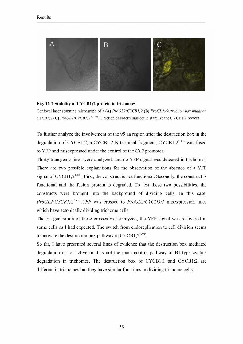
2.2.4. Novel degradation motifs in CYCB1;2 .......................................................36 2.2.5. Stability of the CYCB1;2 full length, the destruction box mutation
CYCB1;2 and the CYCB1;2∆1-135 in trichomes ..............................................37 2.2.6. Analysis of B1-type cyclins in dividing cells..............................................39 2.2.7. Misexpression of CYCB1;1, CYCB1;2 and CYCB1;3 in dividing
epidermal cells ................................................................................................39 2.2.8. Misexpression of the CYCB1;1 and CYCB1;2 destruction Box mutation
in stomata lineage............................................................................................40 2.2.9. Misexpression of the CYCB1;2∆1-135 in cells of the stomata lineage as a
model for dividing cell ...................................................................................41 2.2.10. Localization of CYCB1;2 variants in dividing cells ...................................41 2.2.11. Misexpression of ProTMM: CYCB1;21-135:YFP in dividing cells..............42 2.2.12. Search for a novel degradation motif in CYCB1;2 .....................................43 2.2.13. Barbie Box is a novel degradation box in plant cyclins ..............................44 2.2.14. Misexpression of CYCB1;21-135 containing mutations in the Barbie Box
in trichomes.....................................................................................................44 I60R or I60D exchange in CYCB1;21-135 Barbie box..............................................44 Q67T or Q67D exchange in Barbie box of CYCB1;21-135 .....................................44 Expression of CYCB1;21-135YFP with I60R and Q67T exchanges in trichomes....45 Expression of the CYCB1;21-135:YFP with the Barbie Box analogous region of CYCB1;1 in trichomes ............................................................................................45 Ectopic expression of CYCB1;2I60R, CYCB1;2Q67T and CYCB1;2I60R, Q67T in trichomes .............................................................................................................46 2.2.15. Misexpression of CYCB1;2 without Barbie box in trichomes and stomata
lineage cells .....................................................................................................46 2.2.16. Misexpression of CYCB1;2∆57-75 in the siamese mutant .............................47 2.2.17. Rescue of cycb1;1-/-cycb1;2-/+ with ProCYCB1;2:CYCB1;2∆57-7 ...............48 2.2.18. CYCB1;2∆57-75 and CYCB1;2∆1-135 induced multicellular trichomes in ccs52a1 mutant .......................................................................................................48
2.3. APC/C dependent degradation in trichomes ................................................50 2.3.1. Misexpression of APC11 RNAi did not induce any phenotype in trichomes ................................................................................................................50 2.3.2. YFP:APC11 over expression in Arabidopsis thaliana.................................50 2.3.3. Expression of APC11 RNAi in YFP:APC11 over expression line .............51 2.3.4. Expression of APC11 RNAi in ProGL2:YFP:APC11 line .........................52 2.3.5. ProGL2:APC11 RNAi in siamese mutant....................................................53 2.3.6. Presence of Cdh1/Fizzy related, activator of APC/C in trichomes .............54
3. Discussion
3.1. B1-type cyclins in Arabidopsis thaliana ·························································· 56 3.2. A Regulatory Role of B1-type cyclins in Arabidopsis thaliana ······················· 56 3.3. Redundancy of B1-type cyclins······································································· 57 3.4. Distinct roles for CYCB1;1, CYCB1;2 with CYCB1;4··································· 59 3.5. Complementation of cycb1;1-/- cycb1;2+/- ······················································· 59 3.6. Function of B1-type cyclins in endoreplicating and dividing cells ·················· 60 3.7. Expression of CYCB1;1 and CYCB1;2 in dividing cells ································ 61 3.8. The Barbie box: a novel degradation motive in plant cyclins ························· 62
3.8.1. Barbie Box function in dividing and endoreplicating cells ························ 63
VI
3.8.2. CYCB1;2∆57-75 without Barbie box and the truncation CYCB1;2∆1-135 induce cell death in siamese mutant······························································ 63
3.8.3. Barbie box mediates degradation of CYCB1;2 independent from FIZZY related (CCS52A1) ······················································································· 64
Plant growth conditions...........................................................................71 Crossing of plants....................................................................................71 Plant transformation ................................................................................72 Seed surface sterilization.........................................................................72 Selection of transformants.......................................................................72
4.2.2. Microscopy and cytological methods ..........................................................72 Microscopy..............................................................................................72 LR-White embedding and semi-thin sectioning of seeds .......................73 Whole-Mount preparation of seeds.........................................................73 GUS staining ...........................................................................................73 Pollen preparation for fluorescence analysis...........................................73 Pollen viability assay...............................................................................73
4.3. Molecular-biological methods...........................................................................74 4.3.1. Genomic DNA preparation from plant tissue I ...........................................74 4.3.2. Genomic DNA preparation from plant tissue II ..........................................74 4.3.3. Plasmid DNA preparation from bacteria .....................................................75 4.3.4. DNA-manipulation ......................................................................................75 4.3.5. Cloning of complementation and reporter constructs..................................75 4.3.6. RNA isolation, reverse transcription and RT-PCR .....................................75 4.3.7. Identification of B1-type cyclins mutants by PCR.......................................76
Zykline spielen eine entscheidende Rolle bei den Durchtrittskontrollen des
eukaryontischen Zellzyklus. Für jeden Zellzyklusübergang werden spezifische Zykline
benötigt, die durch Untereinheitenbindung ihren katalytischen Partner aus der Familie
der Zyklinabhängigen Kinasen aktivieren. Der Fokus dieser Arbeit liegt auf den
Zyklinen des B1-Types, welche während der G2- und M-Phase exprimiert werden und
dadurch den Eintritt in die Mitose regulieren. Zykline der B1-Familie werden in
Arabidopsis thaliana durch die vier Mitglieder CYCB1;1, CYCB1;2, CYCB1;3 and
CYCB1;4 vertreten, deren Mutanten in der zugrunde liegenden Arbeit untersucht
werden. Morphologische Analysen des Wachstums ergeben lediglich geringfügige
Unterschiede im einfach mutanten Hintergrund. Während die Anzahl der Rosettblätter
in der cycb1;2-Mutante abnimmt, steigt sie für cycb1;4 an. Die Anzahl der Seiten- und
Nebentriebe wird nicht durch Mutation von B1-Zyklinen beeinträchtigt. Die Mutanten
cycb1;1 und cycb1;2 zeigen ein verstärktes, das Fehlen von CYCB1;4 hingegen ein
gehemmtes Wachstum der Rosettblätter. Wachstumsanalysen der Wurzel erwiesen, dass
es sich bei CYCB1;4 um den wichtigsten Vertreter der B1-Zykline handelt, dessen
Aufgabe die Regulation des Wurzelwachstums ist. Demzufolge ist dieses in der
cycb1;4-Mutante deutlich reduziert. Sämtliche Mutanten für die vier Zykline des B1-
Types zeigen eine verlängerte Wachstumsphase und eine verspätete Blühinduktion. Die
nur geringfügigen Effekte, die in den einfachen Mutanten beobachtet werden, lassen
eine starke Redundanz der B1-Zykline untereinander vermuten. Entsprechend ist die
cycb1;1-/-;cycb1;2-/--Doppelmutante letal: die Embryonen sterben zwischen Herz- und
Torpedostadium. Während die Expression des CYCB1;4 unter Kontrolle des CYCB1;1-
Promoters die Doppelmutante cycb1;1-/-;cycb1;2-/- nicht retten kann, wird die
wildtypische Morphologie durch die Expression von CYCB1;1, CYCB1;2 sowie
CYCB1;3 auch in der Doppelmutante wiederhergestellt. Die Doppelmutanten cycb1;1-/-
;cycb1;4-/- und cycb1;2-/-;cycb1;4-/- führen zu keinem deutlich vom Wildtyp
abweichenden Phänotypen. Doppelmutanten von cycb1;3 mit anderen zyklinen des b1-
typs werden in dieser Arbeit nicht beleuchtet. Untersuchungen an Zyklinen des B1-
Types bestätigen sowohl eine starke Redundanz zwischen CYCB1;1, CYCB1;2 und
CYCB1;3 aber auch, dass sich CYCB1;4 unabhängig von den anderen
Familienmitgliedern entwickelt hat.
VIII
In allen Eukaryonten, deren Zellteilung größtenteils auf dem Wechselspiel zwischen
Zyklinabhängigen Kinasen und den Zyklinen beruht, ist der „Destruction box“-
abhängige Abbau der Zykline des B-Typs durch den APC/C-Komplex reguliert.
Während die Degradation von CYCB1;1 und CYCB1;2 in der Abstammungslinie der
Spaltöffnungen von Arabidopsis thaliana ein Abbaumotiv erfordert, ist dieses in
endoreplizierenden Blatthaaren nicht essentiell. In dieser Arbeit wird das neuartige
Motiv der „Barbie box“ beschrieben, dass sich für den Abbau von Zyklinen in
Blatthaaren verantwortlich zeigt. Die „Barbie box” ist ein für Pflanzen spezifisches
Abbaumotiv, welches nur in einigen pflanzlichen Zyklinen des B-Types gefunden
werden konnte. Die Abbaubox scheint nur eine untergeordnete Rolle im Zyklinabbau in
Blatthaaren zu spielen. Anhaltspunkte für diese Annahme kommen von Markerlinien,
deren Reporter GUS an eine Abbaubox fusioniert wurde. Darüber hinaus wurde ein
RNAi-Konstrukt blatthaarspezifisch gegen APC11 – einer zentralen Komponente von
APC/C – gerichtet und zeigte dabei keine Abweichung vom wildtypischen
Blatthaarphänotyp.
IX
ABSTRACT
Cyclins play a vital role in controlling progress through the eukaryotic cell cycle.
Specific cyclins are required at each cell cycle transition to activate their partner cyclin-
dependent kinase. The focus of this study were the B1-type cyclins that are expressed in
G2/M phase and control entry into mitosis. B1-type cyclins are represented by four
members in Arabidopsis thaliana and in this study mutant lines for all family members,
CYCB1;1, CYCB1;2, CYCB1;3 and CYCB1;4, were analyzed. Morphological analyses
revealed only minor growth alterations of the single mutant plants. While the number of
rosette leaves decreased in the cycb1;2 mutant, cycb1;4 increased the number of rosette
leaf. Side and auxiliary shoots numbers did not affected by mutation in B1-type cyclins.
cycb1;1 and cycb1;2 mutants increased the rosette leaf growth while knock out of
CYCB1;4 reduced the growth of rosette leaf. Root growth analysis revealed that
CYCB1;4 is the main B1-type cyclins in root growth and root growth significantly
decreased in cycb1;4 mutant. All B1-type cyclin mutants prolonged the vegetative phase
and flowering was delayed. The minor effects seen in the single mutants suggested a
high level of redundancy among the B1-type cyclins. Consequently, a cycb1;1-/-
cycb1;2-/- was lethal and embryos died in the middle of heart and torpedo stage. While
the expression of CYCB1;4 under the CYCB1;1 promotor could not rescue cycb1;1-/-
cycb1;2-/- mutants, the expression of CYCB1;1, CYCB1;2 and CYCB1;3 could restore
wild type morphology in the double mutant. The cycb1;1-/- cycb1;4-/- or cycb1;2-/-
cycb1;4-/- double mutant did not induce any severe phenotype. Double mutants of
cycb1;3 with other b1-type cyclins were not analyzed. Analysis of B1-type cyclins
shows that there is high level of redundancy between CYCB1;1, CYCB1;2 and
CYCB1;3 but theCYCB1;4 developed independent from other B1-type cyclins.
In dividing cells of all organisms, the destruction box dependent degradation of B-type
cyclins is mediated by the APC/C complex. While the degradation of CYCB1;1 and
CYCB1;2 in the stomata lineage of Arabidopsis thaliana is required a destruction box,
it is interestingly not required in endoreplicating trichomes. In this study, a new motif
was identified that mediates cyclin degradation in trichomes, the motif was designated
Barbie box. The Barbie box is a plant specific degradation motif which was found only
in some plant B-type cyclins. The Destruction box appeared to be not of primary
importance for degradation of cyclins in trichomes. Evidence for this hypothesis came
from the analysis of GUS marker lines that were fused to a destruction box. Moreover,
X
an RNAi construct directed trichome-specifically against ACP11, a central component
of the APC/C, .resulted in no deviation from wild-type trichome phenotype.
XI
Abbreviations and gene names % percent °C degree Celsius 3' three prime end of a DNA fragment 35S 35S promotor from the Cauliflower Mosaic virus 5' five prime end of a DNA fragment ATP adenosinetriphosphate Bp base pair cDNA complementary DNA CDS coding sequence CAK CDK ACTIVATING KINASE CDK CYCLIN DEPENDENT KINASE CKI CYCLIN DEPENDENT KINASE INHIBITOR CKS1 CDC KINASE SUBUNIT 1 CLF CURLY LEAF CYC CYCLIN CYCB CYCLIN B E2F ADENOVIRUS E2 PROMOTOR BINDING FACTOR DP DIMERIZATION PARTNER APC/C anaphase-promoting complex/cyclosome CAK CDK ACTIVATING KINASE CaMV Cauliflower Mosaic Virus CCS52 CELL-CYCLE SWITCH 52 CDC6 CELL DIVISION CYCLE DEFECTIVE 6 CDC25 CELL DIVISION CYCLE DEFECTIVE 25 CDK CYCLIN DEPENDENT KINASE CLSM confocal laser scanning microscopy CPC CAPRICE CUL1 CULLIN 1 DEL DP-E2F LIKE EF1 ELONGATION FACTOR 1 E2F ADENOVIRUS E2 PROMOTOR BINDING FACTOR FZR FIZZY-RELATED FZY FIZZY GL2 GLABRA2 GL3 GLABRA3 ICK INTERACTOR/INHIBITOR OF CDKs KRP KIP RELATED PROTEIN Rb RETINOBLASTOMA RBX1 RING BOX PROTEIN1 Col Arabidopsis thaliana Columbia accession sim siamese mutant d.a.g. days after germination DAPI 4',6'-diamidino-2-phenylindole DMSO dimethylsulfoxide DNA desoxyribonucleic acid
XII
EDTA ethylenediaminetetraacetic acid e.g. exempli gratia [Lat.] for example et al. et alii / et aliae [Lat.] and others F1, F2, F3 first, second, third... filial generation after a cross FDA fluorescein diacetate Fig. Figure G1 Gap phase between M phase and S phase G2 Gap phase between S phase and M phase gene-/- homozygous mutant of a gene gene+/- heterozygous mutant of a gene YFP Yellow fluorescent protein GUS beta-glucuronidase i.e. id est [Lat.] that is aa amino acid CDS coding sequence Kb kilo bp N number NLS nuclear localization signal/sequence PCR polymerase chain reaction RNAi RNA-interference Rpm rounds per minute RT room temperature RT-PCR reverse transcription PCR SCF Skp1; Cdc53 (cullin); F-box protein SD standard deviation SEM scanning electron microscopy SIM SIAMESE T-DNA transferred DNA TIS trichome initiation site UTR untranslated region WT wild type All gene and mutant names are written in italics. WT-genes are written in capital letters. Proteins are written in non-italic letters.
XIII
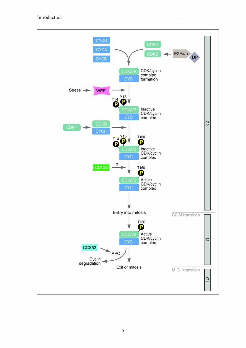
Figure and table index Figures Fig. 1-1 The cell cycle ······································································································ 2 Fig. 2-1 Different cell cycle modes··················································································· 2 Fig. 3-1 Representation of cyclin kinase activity during G2 to M phase transition in plants············································································································ 5 Fig. 4-1 Relative expression data of expressed cyclin genes during cell cycle·················· 8 Fig. 5-1 APC/C subunits ··································································································· 11 Fig. 6-1 Expression of ProTMM:TMM and ProGL2:GUS in epidermal cells··················· 13 Fig. 1-2 Alignment and phylogenetic tree of Arabidopsis thaliana B1-type cyclins········· 16 Fig. 2-2 The B1-type cyclins mutant················································································· 18 Fig. 3-2 Description of single mutant················································································ 20 Fig. 4-2 Root growth analyses of cycb1; 1, cycb1; 2, cycb1; 4 and Col Plants ················· 22 Fig. 5-2 Growth analysis of rosette leaves of cycb1; 1, cycb1; 2, cycb1; 4 and Col Plants ························································································································ 23 Fig. 6-2 Flowering time ···································································································· 24 Fig. 7-2 Embryo development of cycb1;1-/-cycb1;2-/+ or cycb1;1-/+cycb1;2-/- ··················· 25 Fig. 8-2 Rosette leaves Growth analysis of cycb1;1-/- cycb1;2-/+ and cycb1;1-/+ cycb1;2-/- ·········································································································· 26
Fig. 9-2 Histochemical Analysis of CYCB1;1, CYCB1;2 and CYCB1;4 Promoters Activity····························································································································· 27 Fig. 10-2 FDA staining of pollen of wild type and cycb1;1-/- cycb1;2-/-···························· 28 Fig. 11-2 Ovule development in wild type and cycb1;1-/- cycb1;2-/- double mutant ·········· 29 Fig. 12-2 Schematics of the CYCB1;1, the CYCB1;2 and their fusions ··························· 31 Fig. 13-2 Morphological analysis of multicellular trichome ············································· 33 Fig. 14-2 Schematic of CYCB1; 2 truncations·································································· 33 Fig. 15-2 GUS analysis of CYCB1;11-112 in endoreplicating and dividing trichomes ······· 35 Fig. 16-2 Stability of CYCB1;2 protein in trichomes························································ 38 Fig.17-2 Light micrograph of CYCB1;2 and CYCB1;2 destruction box mutation in stomata lineage ················································································································· 40 Fig. 18-2 Localization of CYCB1;2 and its variants in dividing cells······························· 42 Fig. 19-2 Alignment of CYCB1;1 and CYCB1;2 N-termini ············································· 43 Fig. 20-2 Mutation in I 60 and Q67 able to restore YFP signal of CYCB1;21-135:YFP ···· 45 Fig. 21-2 Morphological analyses of ProTMM:CYCB1;2 and ProTMM:CYCB1;2∆57-74 Plants ································································································································ 47 Fig. 22-2 Scanning electron micrograph of ProGL2:CYCB1;2∆57-75 and ProGL2:CYCB1;2∆1-135 in sim mutant ·············································································· 48 Fig. 23-2 Alignment of APC11 and RBX1 ······································································· 50 Fig. 24-2 Analysis of the APC11 RNAi function in trichomes ········································ 52 Fig. 25-2 Analysis of trichomes of wild type, sim mutant and ProGL2:APC11 RNAi in sim mutant ························································································································ 53 Fig. 26-2 Ethanol inducible CCS52B RNAi in Arabidopsis thaliana································· 54 Fig. 1-3 Expression profile of B1-type cyclins during embryogenesis······························ 58 Fig. 2-3 Model of function of Destruction and Barbie box in trichomes··························· 66 Tables Table 1-2. T-DNA or Transposone lines of B1-Type cyclins············································ 17 Table 2-2 Morphological analysis of b1-type cyclin mutants··········································· 21
XIV
Table 3-2 Trichome phenotype upon misexpression of CYCB1;2 and CYCB1;2 truncations ········································································································ 32 Table 4-2 RNAi constructs to knock out CCS52B ··························································· 54 Table 1-4 Primers of T-DNA and transposone ································································ 77
Fig4-1. Relative expression data of expressed cyclin genes were plotted against the different time points during cell cycle re-entry (left column) and further cell cycle progression (right column) as indicated. For clarity, CYCD, CYCA and B, and the novel CYCB1;5 and CYCB2;5 are shown in separate panels. (taken from Menges et al.2005) D-type cyclins have a large sequence divergence and were originally identified by
functional complementation of a yeast strain deficient for G1 cyclins (Dahl et al.,
1995; Soni et al., 1995; Inze and De Veylder, 2006).
In Arabidopsis, the 10 CYCDs are classified into seven groups, designated CYCD1 to
CYCD7, with the CYCD3 and CYCD4 groups consisting of three and two members,
respectively (Vandepoele et al., 2002). The large number of cyclins might reflect the
high developmental plasticity of sessile plants to respond to both intrinsic
developmental signals and extrinsic environmental cues. Possibly, the complex cell
cycle machinery is the trade-off for the tremendous plasticity and robustness of plant
growth, which requires the presence of flexible regulatory networks (Inze and De
Veylder, 2006). The large number of cyclins might possess a wide range of expression
patterns and confer different substrate specificities. There is probably an extensive
functional redundancy among D type cyclins, because the genome-wide insertional
mutagenesis surveys have yet to report severe phenotypes for D-cyclin knockouts
the promoter retained G2/M-specific promoter activity in BY2 cells. Further analysis
of the 80 bp fragment by inducing mutations resulted in identification of a 9bp
sequence that has been identified as a critical fragment for cell cycle-regulated
promoter activation. (Ito et al., 2000). The full-length CYM promoter contains three
other sequences similar to the 9 bp element. These 9 bp sequences, in a heterologous
context, could direct G2/M-specific expression of a reporter gene called MSA (M-
phase-specific activator), can function in an orientation-independent fashion. Multiple
MSA elements are present in the promoter of B1 and B2 classes of cyclin genes from
various plant species (Ito et al., 1998; Ito, 2000)
1.3. Regulation of the cell cycle by APC/C-type ubiquitin ligases The degradation of cell cycle regulator proteins is necessary to maintain the periodic
fluctuations in protein levels during the cell cycle, and serves as a means of cell cycle
control. The highly regulated proteolysis of B-type cyclins involved in sister
chromatid separation is required at the onset of anaphase. Proteins subjected to
degradation are marked with ubiquitin tags and subsequently are targeted for
degradation by the 26S proteasome (Hochstrasser, 1995). The ubiquitin/26S
proteasome proteolytic pathway is highly conserved in eukaryotes and is involved in
many other important cellular functions aside from cell cycle progression
(Hochstrasser, 1995; Genschik et al., 1998; Nakayama and Nakayama, 2006).
Degradation via this pathway is a two-step process: the protein is first tagged by the
covalent attachment of ubiquitin; subsequently, it is degraded by a multicatalytic
protease complex called the 26S proteasome. Conjugation of ubiquitin to the protein
involves a cascade of three enzymes: E1, E2, and E3. The E1 (ubiquitin-activating)
enzyme forms a high-energy bond with ubiquitin, which is then transesterified to a
ubiquitin-conjugating enzyme (E2). E3 ubiquitin ligase activity is then used for
transfer of the ubiquitin to the target protein substrate (Genschik et al., 1998; Castro et
al., 2005).Transfer of ubiquitin to the target protein substrate requires specificity and
versatility, which are provided by the existence of 500–1,000 different E3 ligases.
RING-finger-type E3s are thought to be the largest family and are further divided into
subfamilies; the cullin-based E3 subfamily, is one of the largest single classes of E3.
There are seven cullin-based E3s, including the SKP1–CUL1–F-box-protein (SCF)
complex (Zachariae and Nasmyth, 1999) and the anaphase-promoting
complex/cyclosome (APC/C) (Zachariae and Nasmyth, 1999; Buschhorn and Peters,
scaffold protein) and at least 9 other Components (Fig5-1) (Nakayama and
Nakayama, 2006)
Fig. 5-1. APC contains a Cullin, and a Ring-H2 finger protein, designated here as Apc2 and Apc11 respectively. The cullin and Ring-H2 finger proteins are required to bind E2 and catalyze the ubiquitination of APC/C substrates. (Adapted from Castro et al. 2005) APC regulates exit from mitosis and events in G1 (Fang et al., 1998{Peters, 2006
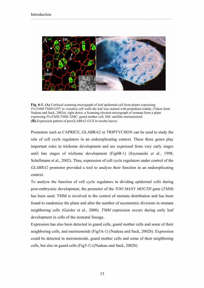
Fig. 6-1. (A) Confocal scanning micrograph of leaf epidermal cell from plants expressing ProTMM:TMM:GFP; to visualize cell walls the leaf was stained with propidium iodide; (Taken form Nadeau and Sack, 2002a). right down, a Scanning electron micrograph of stomata from a plant expressing ProTMM:TMM. GMC: guard mother cell, SM: satellite merisetemoid. (B).Expression pattern of proGLABRA2:GUS in rosette leaves.
Promotors such as CAPRICE, GLABRA2 or TRIPTYCHON can be used to study the
role of cell cycle regulators in an endoreplicating context. These three genes play
important roles in trichome development and are expressed from very early stages
until late stages of trichome development (Fig6B-1) (Szymanski et al., 1998;
Schellmann et al., 2002). Thus, expression of cell cycle regulators under control of the
GLABRA2 promotor provided a tool to analyze their function in an endoreplicating
context.
To analyze the function of cell cycle regulators in dividing epidermal cells during
post-embryonic development, the promotor of the TOO MANY MOUTH gene (TMM)
has been used. TMM is involved in the control of stomata distribution and has been
found to randomize the plane and alter the number of asymmetric divisions in stomata
neighboring cells (Geisler et al., 2000). TMM expression occurs during early leaf
development in cells of the stomatal lineage.
Expression has also been detected in guard cells, guard mother cells and some of their
neighboring cells, and meristemoids (Fig5A-1) (Nadeau and Sack, 2002b). Expression
could be detected in meristemoids, guard mother cells and some of their neighboring
cells, but also in guard cells (Fig5-1) ((Nadeau and Sack, 2002b)
2.1. Studying CYCB1 function: loss of function approach
Plant cell cycle regulators constitute much larger families than animal regulators. For
example in Drosophila there are two B and one A type cyclins whereas in Arabidopsis
11 B-type cyclins and 10 A type cyclins have been identified (Pines, 1995)(Wang et
al., 2004). The specific function of the different members is not understood. To
analyze whether the different B1-type cyclins (Fig.1A and B) have specific roles in
development or whether these genes have a solely redundant function, I analyzed
CYCB1 knock out lines.
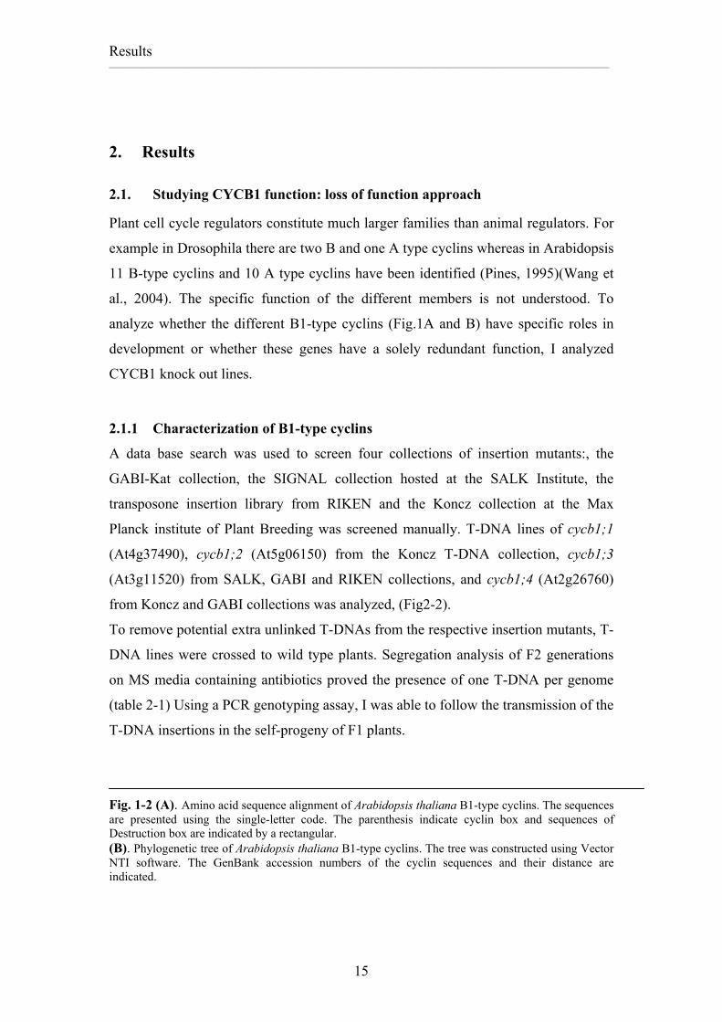
2.1.1 Characterization of B1-type cyclins
A data base search was used to screen four collections of insertion mutants:, the
GABI-Kat collection, the SIGNAL collection hosted at the SALK Institute, the
transposone insertion library from RIKEN and the Koncz collection at the Max
Planck institute of Plant Breeding was screened manually. T-DNA lines of cycb1;1
(At4g37490), cycb1;2 (At5g06150) from the Koncz T-DNA collection, cycb1;3
(At3g11520) from SALK, GABI and RIKEN collections, and cycb1;4 (At2g26760)
from Koncz and GABI collections was analyzed, (Fig2-2).
To remove potential extra unlinked T-DNAs from the respective insertion mutants, T-
DNA lines were crossed to wild type plants. Segregation analysis of F2 generations
on MS media containing antibiotics proved the presence of one T-DNA per genome
(table 2-1) Using a PCR genotyping assay, I was able to follow the transmission of the
T-DNA insertions in the self-progeny of F1 plants.
Fig. 1-2 (A). Amino acid sequence alignment of Arabidopsis thaliana B1-type cyclins. The sequences are presented using the single-letter code. The parenthesis indicate cyclin box and sequences of Destruction box are indicated by a rectangular. (B). Phylogenetic tree of Arabidopsis thaliana B1-type cyclins. The tree was constructed using Vector NTI software. The GenBank accession numbers of the cyclin sequences and their distance are indicated.
2.1.3. Transcription of B1-type cyclins knock out genes To test whether the insertion resulted in a knock-out or knock-down of B1-type cyclin
function, RT-PCR analyses were performed. No transcript could be detected in the
homozygous mutants using primer combinations which annealed downstream of the
T-DNA insertion and spanned the coding sequence of: cycb1;1 and cycb1;2 from the
Koncz collection, cycb1;3 from RIKEN and cycb1;4 from the GABI collection. Thus,
these lines represent null alleles of the respective genes.
However, in cycb1;3 from GABI and Salk collections and cycb1;4 from the GABI
collection the respective RNA was expressed more strongly than in wild type control
plants. This could be because the T-DNA contains the 35S promoter which might
drive the expression of CYCB1;3 or CYCB1;4. It can not be ruled out that the C-
terminal transcript of CYCB1;3 and CYCB1;4 was properly translated and no further
experiments were performed with these mutant lines.
Fig 2-2. The B1-type cyclins mutants: Schematic drawing of the B1-type cyclins genes showing the T-DNA or Transposone insertion in the B1-type cyclins. Green box represents the Exon, colorless rectangular shows Intron.
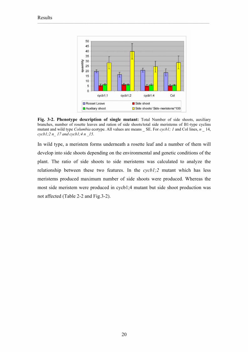
Fig. 3-2. Phenotype description of single mutant: Total Number of side shoots, auxiliary branches, number of rosette leaves and ration of side shoots/total side meristems of B1-type cyclins mutant and wild type Columbia ecotype. All values are means _ SE. For cycb1; 1 and Col lines, n _ 14, cycb1;2 n_ 17 and cycb1;4 n _15. In wild type, a meristem forms underneath a rosette leaf and a number of them will
develop into side shoots depending on the environmental and genetic conditions of the
plant. The ratio of side shoots to side meristems was calculated to analyze the
relationship between these two features. In the cycb1;2 mutant which has less
meristems produced maximum number of side shoots were produced. Whereas the
most side meristem were produced in cycb1;4 mutant but side shoot production was
not affected (Table 2-2 and Fig.3-2).
Mean and standard deviation of auxiliary shoot, side shoot rosette leave and out growing buds/meristems of cycb1;1, cycb1;2, cycb1;4, cycb1;1-/+
cycb1;2-/-, cycb1;1-/- cycb1;2+/- and Col wild type. M: Mean, SD: Standard deviation and n= number of plants in each experiment. Significant differences of Rosette leave and outgrowing bud/ meristem*100 between wild type and cycb1;1, cycb1;2, cycb1;4, cycb1;1-/-cyc1; 2-/+ and cycb1;1-/+ cycb1;2-/- mutants are designed. *P<0,05, **P<0,01 and ***P<0,001. P values are determined by the Student T-test
Table 2-2: Morphological analysis of b1-type cyclin mutants Auxiliary shoot
Root growth analysis of b1-type cyclins B1-type cyclin mutants and wild type control plants were grown on vertical plates in a
growth chamber with controlled growth conditions. On odd days the plates were
scanned and the length of the roots measured using Image J software. The data was
analyzed using SPSS soft ware.
Growth analysis of root development revealed that knock outs of CYCB1;1 and
CYCB1;2 have slightly decreased root growth but it is not significantly different from
wild type root growth. While cycb1;4 mutant reduced root growth (Fig.4-2).
Fig. 4-2. Root growth analysis of cycb1; 1, cycb1; 2, cycb1; 4 and Col Plants. CYCb1;4 mutant and col clustered in Class a and Class b respectively while cycb1;1 and cycb1;2 grouped in a intermediated class a,b.
Rosette leaf growth analysis
Measurements of leaf area of single mutants showed that cycb1;1 and cycb1;2
mutants have a larger leaf area than wild type leaves. The number of rosette leaves
was equal in cycb1;1 and Columbia. And cycb1;2 mutant produced the minimum leaf
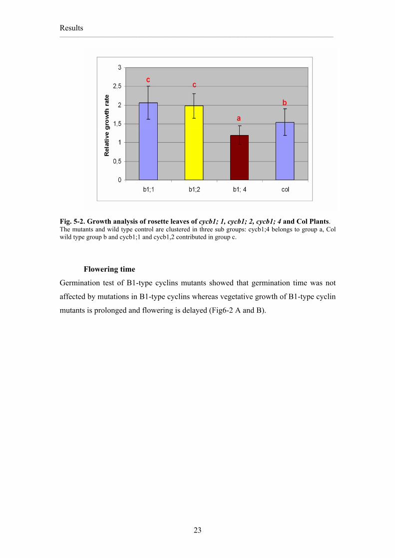
Fig. 5-2. Growth analysis of rosette leaves of cycb1; 1, cycb1; 2, cycb1; 4 and Col Plants. The mutants and wild type control are clustered in three sub groups: cycb1;4 belongs to group a, Col wild type group b and cycb1;1 and cycb1,2 contributed in group c.
Flowering time
Germination test of B1-type cyclins mutants showed that germination time was not
affected by mutations in B1-type cyclins whereas vegetative growth of B1-type cyclin
mutants is prolonged and flowering is delayed (Fig6-2 A and B).
Fig. 6-2. Flowering time: Frequency distribution of flowering time of b1-type cyclin mutants in long day condition (16 hours light/ 8 hours dark). A. flowering time of cycb1; 1, cycb1; 2, cycb1; 4 and wild type Columbia ecotype. Distribution of cycb1; 3 in Nössen background and wild type Nössen were shown separately.
2.1.5. Redundancy within B1-type cyclins
No severe phenotype from wild-type was found in single mutant cyclins. One
possibility is that the B1-type cyclins act redundantly. Therefore, Double mutants of
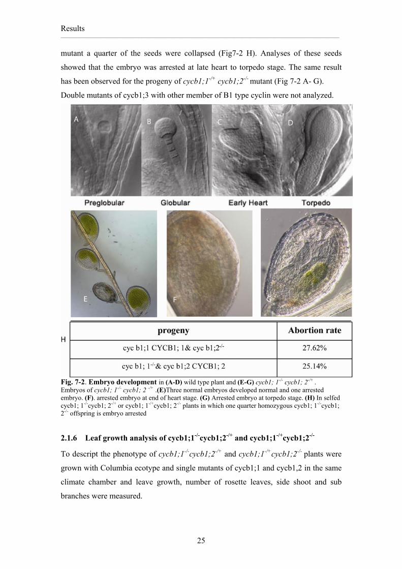
mutant a quarter of the seeds were collapsed (Fig7-2 H). Analyses of these seeds
showed that the embryo was arrested at late heart to torpedo stage. The same result
has been observed for the progeny of cycb1;1-/+ cycb1;2-/- mutant (Fig 7-2 A- G).
Double mutants of cycb1;3 with other member of B1 type cyclin were not analyzed.
Fig. 7-2. Embryo development in (A-D) wild type plant and (E-G) cycb1; 1-/- cycb1; 2-/+ . Embryos of cycb1; 1-/- cycb1; 2 -/+ .(E)Three normal embryos developed normal and one arrested embryo. (F). arrested embryo at end of heart stage. (G) Arrested embryo at torpedo stage. (H) In selfed cycb1; 1-/-cycb1; 2-/+ or cycb1; 1-/+cycb1; 2-/- plants in which one quarter homozygous cycb1; 1-/-cycb1; 2-/- offspring is embryo arrested
2.1.6 Leaf growth analysis of cycb1;1-/-cycb1;2-/+ and cycb1;1-/+cycb1;2-/-
To descript the phenotype of cycb1;1-/-cycb1;2-/+ and cycb1;1-/+cycb1;2-/- plants were
grown with Columbia ecotype and single mutants of cycb1;1 and cycb1,2 in the same
climate chamber and leave growth, number of rosette leaves, side shoot and sub
Fig.12-2 Schematics of the CYCB1;1, the CYCB1;2 and their fusions (A). CYCB1;1, its destruction box (light yellow color) and the analogue fragment of the CYCB1;2 Barbie box in the CYCB1;1 gene (light green) (B) Schematic representation of the position of the destruction box, (Yellow color), Barbie box (green) cyclin boxes, CYCN (red color) and CYCC (blue color). (C) and (D). Double fusion of the N-terminus of CYCB1;1 and C-terminus of CYCB1;2 and of the N-terminus of CYCB1;2 and C-terminus of CYCB1;1 respectively. (E) and (F) Triple fusion of CYCB1;1-CYCB1;2-CYCB1;2 and CYCB1;2-CYCB1;1-CYCB1;2 respectively. (G) and (H) Destruction box mutation of CYCB1;1 and CYCB1;2. Mutations in CYCB1;1 and CYCB1;2 destruction box and the exchanged amino acids are shown in red. 1 mm represents 4 amino
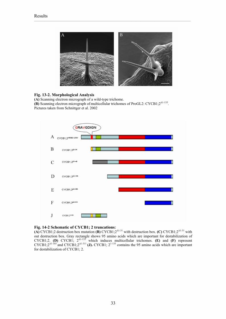
Fig. 13-2. Morphological Analysis (A) Scanning electron micrograph of a wild-type trichome. (B) Scanning electron micrograph of multicellular trichomes of ProGL2: CYCB1;2∆1-135. Pictures taken from Schnittger et al. 2002
Fig. 14-2 Schematic of CYCB1; 2 truncations: (A) CYCB1;2 destruction box mutation (B) CYCB1;2∆1-31 with destruction box. (C) CYCB1;2∆1-31 with out destruction box. Gray rectangle shows 95 amino acids which are important for destabilization of CYCB1;2. (D) CYCB1; 2∆1-135 which induces multicellular trichomes. (E) and (F) represent CYCB1;2∆1-185 and CYCB1;2∆1-311 (J). CYCB1; 21-135 contains the 95 amino acids which are important for destabilization of CYCB1; 2.
Fig.15-2. GUS analysis of CYCB1;11-112 in endoreplicating and dividing trichomes. (A) and (B) Light micrograph of ProGL2:GUS and ProGL2: CYCB1;11-112:GUS in wild type. (C) and (D) Light micrograph of ProGL2:GUS and ProGL2:CYCB1;11-112: GUS in ProGL2:CYCD3;1 misexpression line which has multicellular trichomes. Mitotic regulators could activate the destruction box pathway in dividing trichome cells and patchy pattern of GUS activity is produced. Ectopic expression of the destruction box mutated version of CYCB1;1 did not
produce any phenotype in trichome cells but mutations in the destruction box of
CYCB1;2 produced a mild phenotype (table2-2). My data suggests that the
destruction box is not important or is backuped by some other degradation signals.
ProGL2:CYCB1;11-112:GUS in ProGL2:CYCD3;1 misexpression line and siamese
mutant
To analyze the destruction box function for the degradation of B1-type cyclins in
dividing cells, GUS and the GUS fusion with CYCB1;11-112 were misexpressed in
Fig.17-2. Light micrograph of CYCB1;2 and CYCB1;2 destruction box mutation in stomata lineage. (A) and (B) Cytokinesis defects and cluster of stomata in ProTMM:CYCB1;2 misexpression lines (C) and (D) phenotype of ProTMM:CYCB1;2 destruction box mutation which induced Cytokinesis defect and more clusters of stomata. Cytokinesis defects are shown with black arrows and Stomata clusters with red arrows.
Misexpression of the CYCB1;1 and CYCB1;2 destruction Box mutation in
stomata lineage
To define functionality of the CYCB1;1 and the CYCB1;2 destruction box mutations
they were transformed under control of the TMM promoter.
Analysis of transgenic lines revealed enlargement of epidermal cell size, cytokinesis
defects, fewer stomata, and clusters of stomata. It was shown that the destruction box
mutated CYCB1;1 or CYCB1;2 induced a stronger phenotype in comparison to
CYCB1;1 or CYCB1;2 full length and induced cytokinesis defects, prevented cell
wall formation, increased endoreplication in epidermal cells, and decreased the
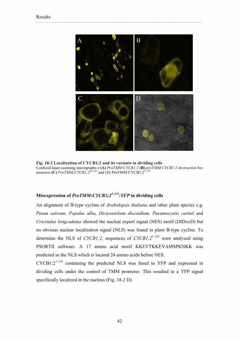
Fig. 18-2 Localization of CYCB1;2 and its variants in dividing cells Confocal-laser-scanning micrographs a (A) ProTMM:CYCB1;2 (B) proTMM:CYCB1;2 destruction box mutation (C) ProTMM:CYCB1;2∆1-135 and (D) ProTMM:CYCB1;21-135
Misexpression of ProTMM:CYCB1;21-135:YFP in dividing cells
An alignment of B-type cyclins of Arabidopsis thaliana and other plant species e.g.
Pisum sativum, Populus alba, Dictyostelium discoidium, Pneumocystis carinii and
Cricetulus longcadatus showed the nuclear export signal (NES) motif (DIDxxD) but
no obvious nuclear localization signal (NLS) was found in plant B-type cyclins. To
determine the NLS of CYCB1;2, sequences of CYCB1;21-135 were analyzed using
PSORTII software. A 17 amino acid motif KKEVTKKEVAMSPKNKK was
predicted as the NLS which is located 24 amino acids before NES.
CYCB1;21-135 containing the predicted NLS was fused to YFP and expressed in
dividing cells under the control of TMM promoter. This resulted in a YFP signal
specifically localized in the nucleus (Fig. 18-2 D).
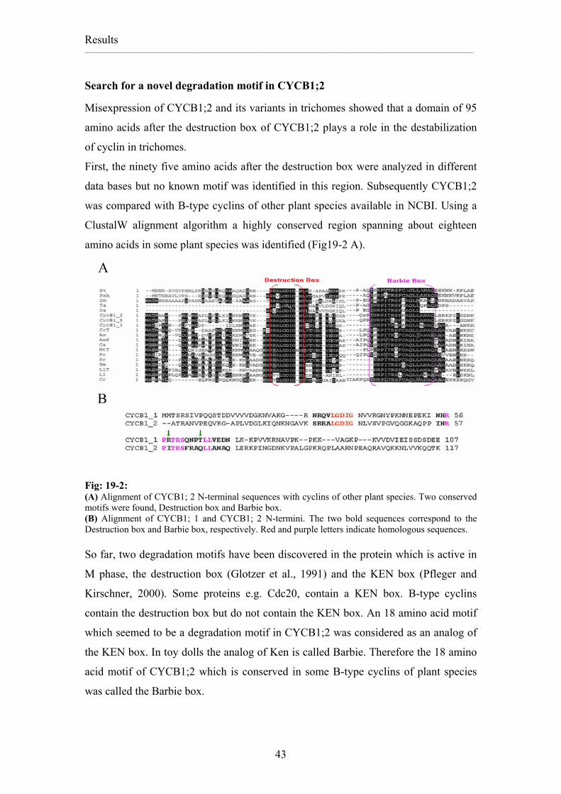
Misexpression of CYCB1;2 and its variants in trichomes showed that a domain of 95
amino acids after the destruction box of CYCB1;2 plays a role in the destabilization
of cyclin in trichomes.
First, the ninety five amino acids after the destruction box were analyzed in different
data bases but no known motif was identified in this region. Subsequently CYCB1;2
was compared with B-type cyclins of other plant species available in NCBI. Using a
ClustalW alignment algorithm a highly conserved region spanning about eighteen
amino acids in some plant species was identified (Fig19-2 A).
Fig: 19-2: (A) Alignment of CYCB1; 2 N-terminal sequences with cyclins of other plant species. Two conserved motifs were found, Destruction box and Barbie box. (B) Alignment of CYCB1; 1 and CYCB1; 2 N-termini. The two bold sequences correspond to the Destruction box and Barbie box, respectively. Red and purple letters indicate homologous sequences. So far, two degradation motifs have been discovered in the protein which is active in
M phase, the destruction box (Glotzer et al., 1991) and the KEN box (Pfleger and
Kirschner, 2000). Some proteins e.g. Cdc20, contain a KEN box. B-type cyclins
contain the destruction box but do not contain the KEN box. An 18 amino acid motif
which seemed to be a degradation motif in CYCB1;2 was considered as an analog of
the KEN box. In toy dolls the analog of Ken is called Barbie. Therefore the 18 amino
acid motif of CYCB1;2 which is conserved in some B-type cyclins of plant species
Fig. 20-2. Mutation in I 60 and Q67 able to restore YFP signal of CYCB1;21-135:YFP Confocal laser scanning micrograph of (A) ProGL2:CYCB1;21-135,I60R (B) ProGL2:CYCB1;21-135,Q67T
(C) ProGL2:CYCB1;11-135,I60R,Q67 and (D) ProGL2:CYCB1;21-135Barbie box analogous region of CYCB1;1
Expression of CYCB1;21-135YFP with I60R and Q67T exchanges in trichomes
Because the single mutation of I60R or Q67T showed stabilization of the N-terminus
of the CYCB1;2, a I60R and Q67T mutant was created to study the function of double
mutations. Confocal laser scanning microscopy data showed that double mutations
stabilized the N-terminus of the CYCB1;2 protein more than a single mutation of
I60R or Q67T (Fig. 20-2 C). This experiment revealed that two amino acids have
additive effect on stabilization of the N-terminus of CYCB1;2.
Expression of the CYCB1;21-135:YFP with the Barbie Box analogous region of
CYCB1;1 in trichomes
Isolucine 60 and Glutamine 67 are key amino acids for the destabilization of
CYCB1;21-135. A region in CYCB1;1 was identified as an analog to the Barbie Box in
CYCB1;2. To find out the role of the analogous region in stabilization of the
Fig 21-2. Morphological analyses of ProTMM:CYCB1;2 and ProTMM:CYCB1;2∆57-74 Plants. Scanning electron micrograph of a (A) ProTMM:CYCB1;2 and (B) ProTMM:CYCB1;2∆57-74 misexpression lines
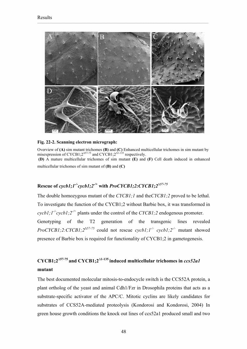
Misexpression of CYCB1;2∆57-75 in the siamese mutant
Triple fusion of CYCB1;1 and CYCB1;2 did not change the sim phenotype whereas
CYCB1;1-CYCB1;2 and CYCB1;2-CYCB1;1 double fusions increased the sim
phenotype faintly. However, CYCB1;2 without the Barbie box gave rise to slightly
stronger multicellular trichome phenotype than CYCB1;1 and CYCB1;2 fusions.
Nevertheless misexpression of CYCB1;2∆57-75 induced cell death in multicellular
trichomes of the sim mutant plants (Fig. 22-2 A, B, D and E).
Analysis of the misexpression lines of the proGL2:CYCB1;2∆1-135 in the sim mutant
showed that CYCB1;2∆1-135 increased the multicellular phenotype of the sim mutant
more than CYCB1;2∆57-75 misexpression, and at the end induced cell death in
multicellular trichomes of the sim mutant lines (Fig. 22-2 C and F).
It was shown that that overexpression of cyclin E enhances cytokine mediated
apoptosis in breast cancer cells (Dhillon and Mudryj, 2003) Or ectopic expression of
cyclin B3 in mouse testa induces abnormal round spermatids and increased apoptosis
in the testa (Refik-Rogers et al., 2006) All these data show that deregulation of cyclins
induce more cell divisions and finally led to cell death in mitotic cells. Taken
together, these data suggest that the CYCB1;2∆1-135 or CYCB1;2∆57-75 stabilizes
CYCB1;2 protein and deregulate the cell cycle in sim mutants. Even though, the
CYCB1;2∆1-135 induced more cell divisions than CYCB1;2∆57-75, both induced cell
Fig. 22-2. Scanning electron micrograph: Overview of (A) sim mutant trichomes (B) and (C) Enhanced multicellular trichomes in sim mutant by misexpression of CYCB1;2∆57-75 and CYCB1;2∆1-135 respectively. (D) A mature multicellular trichomes of sim mutant (E) and (F) Cell death induced in enhanced
multicellular trichomes of sim mutant of (B) and (C)
Rescue of cycb1;1-/-cycb1;2-/+ with ProCYCB1;2:CYCB1;2∆57-75
The double homozygous mutant of the CYCB1;1 and theCYCB1;2 proved to be lethal.
To investigate the function of the CYCB1;2 without Barbie box, it was transformed in
cycb1;1-/-cycb1;2-/+ plants under the control of the CYCB1;2 endogenous promoter.
Genotyping of the T2 generation of the transgenic lines revealed
ProCYCB1;2:CYCB1;2∆57-75 could not rescue cycb1;1-/- cycb1;2-/- mutant showed
presence of Barbie box is required for functionality of CYCB1;2 in gametogenesis.
CYCB1;2∆57-75 and CYCB1;2∆1-135 induced multicellular trichomes in ccs52a1
mutant
The best documented molecular mitosis-to-endocycle switch is the CCS52A protein, a
plant ortholog of the yeast and animal Cdh1/Fzr in Drosophila proteins that acts as a
substrate-specific activator of the APC/C. Mitotic cyclins are likely candidates for
substrates of CCS52A-mediated proteolysis (Kondorosi and Kondorosi, 2004) In
green house growth conditions the knock out lines of ccs52a1 produced small and two
The Destruction Box appeared not to be involved in CYCB1;1 degradation in
trichomes and it has a redundant role with the Barbie box for the removal of
CYCB1;2 in trichomes. This raises the question of what role APC/C dependent
degradation plays during trichome development. To assess the functionality of the
APC/C dependent degradation of B1-type cyclins, an artificial RNAi was designed
against the ANAPHASE PROMOTING COMPLEX11 (APC11) subunit of the APC/C.
The APC11 is a RING-H2 finger protein and functions as the catalytic core of the
APC/C complex by mediating the transfer of ubiquitin from an ubiquitin-conjugating
enzyme (E2) to the substrate (Chang et al., 2004).
The APC11 is structurally related to the RBX1 component of another E3 ligase class,
called the Skp1, CDC53/Cullin, F-box (SCF) complex. (Gmachl et al., 2000 Capron,
2003 #947) (Fig. 23-2).
To analyze the effect of APC11 loss of function in trichomes, an attempt was made to
silence APC11 activity by expressing RNAi constructs against it.
Fig. 23-2. Alignment of APC11 (AT3G05870) and RBX1 (AT3G42830 and AT5G20570). APC11 showed 37.3% similarity and 26.3% identity with RBX1 (AT3G42830) and 36.4% similarity and 26.4% identity with RBX1-2 (AT5G20570) . Alignment was done using ClustalW software.
Misexpression of APC11 RNAi did not induce any phenotype in trichomes
APC11 RNAi was designed against the whole APC11 ORF and expressed under the
control of the GLABRA2 promoter in trichomes. No deviation of trichome
development was observed in APC11 RNAi transgenic lines.
YFP:APC11 over expression in Arabidopsis thaliana
The absence of phenotypic alterations in the APC11 RNAi lines can be explained by
two reasons: either the RNAi construct was not functional or APC11 is not important
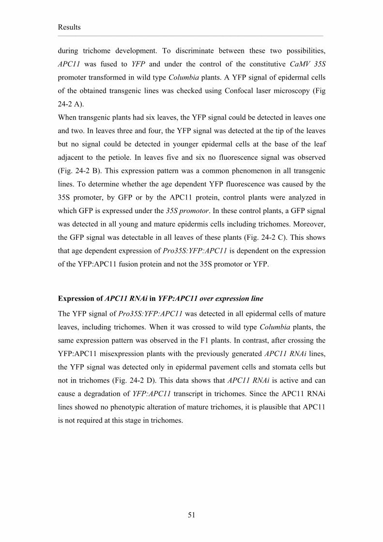
Fig. 24-2 Analysis of the APC11 RNAi function in trichomes. (A), (C), (D), (E)and (F) Confocal laser scanning micrographs of (A) expression of Pro35S:YFP:APC11 in mature trichome and epidermal cells. (B) Schematic of age dependent expression pattern of Pro35S:YFP:APC11 in wild type Col plants.YFPAPC11 was detected in old leaves but it was observed in the apex of younger leaves. Newly developed leaves did not show any YFP signal. (C) Expression of GFP under the control of the 35S promoter. GFP signal was detected in all leaves showing that age dependent expression of Pro35S:YFP:APC11 is dependent on APC11 protein not promoter or YFP. (D) proGL2:APC11 RNAi in Pro35S:YFP:APC11 plants silenced the YFPAPC11 in trichomes. Other cells showed the YFPAPC11 signal. (E) YFP expression pattern of ProGL2:YFPAPC11 in a young trichome. (F) APC11 RNAi in ProGL2:YFPAPC11 silenced the YFPAPC11.
Expression of APC11 RNAi in ProGL2:YFP:APC11 line
To determine activity of APC11 RNAi in young trichomes, the YFP:APC11 fusion
protein was expressed under the control of the GLABRA2 promoter. Analysis of
transgenic lines revealed that the YFP:APC11 fusion protein accumulates in both,
young and mature trichomes (Fig. 24-2 E). Next, ProGL2:YFP:APC11 was crossed to
wild type Columbia and ProGL2:APC11RNAi.
In control crosses, a YFP signal was seen on trichomes of ProGL2:YFP:APC11
crossed to wild type Columbia. However, no YFP fluorescent signal was detected in
trichomes in the progeny of the ProGL2:APC11 RNAi crossed to the
ProGL2:YFP:APC11 (Fig. 24-2 E and F).
Hence APC11RNAi is active not only in mature trichomes but also in young trichomes
and can remove YFP:APC11 RNA from the trichomes. Consequently, APC11 and
likely the APC/C are not necessary for protein degradation during trichome
development.
ProGL2:APC11 RNAi in siamese mutant
Silencing of APC11 using RNAi has revealed that APC11 is not essential for trichome
development. To analyze the function of APC11 in mitotic cells, APC11 RNAi, was
transformed in the sim mutant which displays multicellular trichomes (Walker et al.,
2000). (Fig. 25-2 B)
The analysis of transgenic lines showed that APC11 RNAi could reduce the number of
cells in sim trichomes almost restoring a wild-type phenotype (Fig A, B and C).
Fig. 25-2 Analysis of trichomes of wild type, sim mutant and ProGL2:APC11 RNAi in sim mutant. (A) Scanning electron micrographs showing a wild type trichome. taken from Schnittger et al. 2002. (B) Trichome of sim mutant. (C) Scanning electron micrographs showing the proGL2:APC11 RNAi blocked multicellular trichomes induction of sim mutant. Presence of APC/C complex is required for cell division of sim mutant
Presence of Cdh1/Fizzy related, activator of APC/C in trichomes
APC11 appears not to be essential during trichome development. If the APC/C is not
active in trichomes, its activators should also not be required for trichome
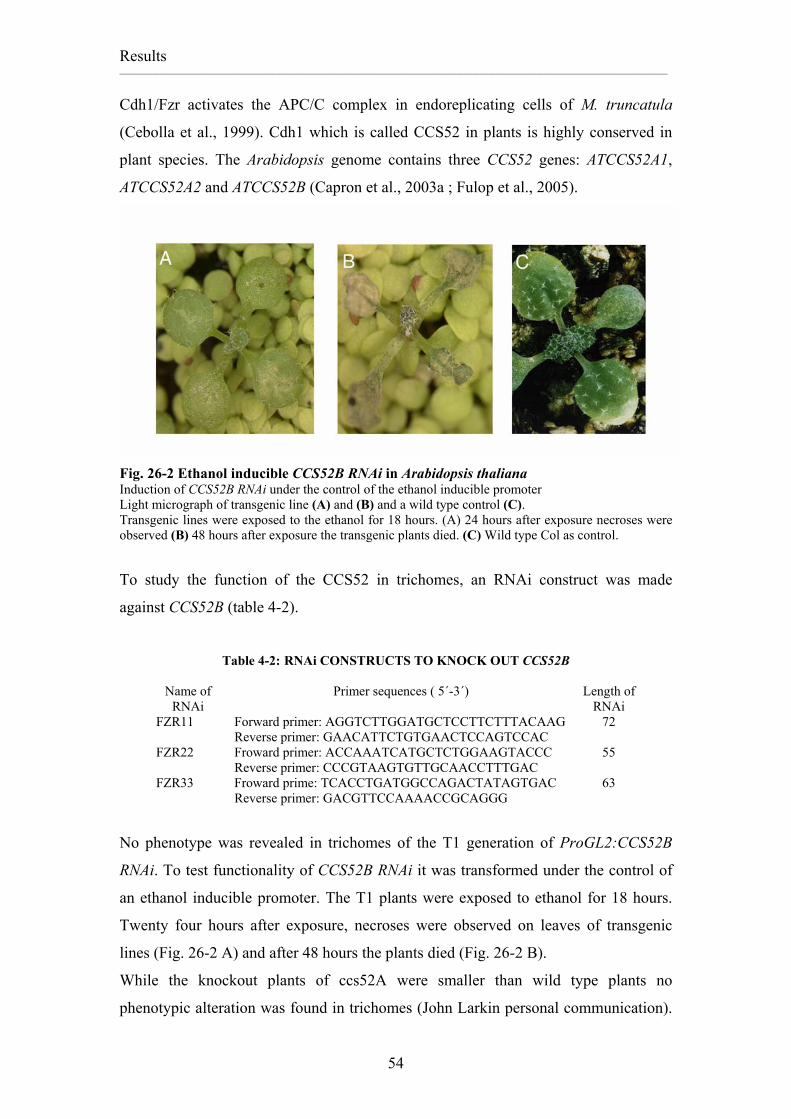
Cdh1/Fzr activates the APC/C complex in endoreplicating cells of M. truncatula
(Cebolla et al., 1999). Cdh1 which is called CCS52 in plants is highly conserved in
plant species. The Arabidopsis genome contains three CCS52 genes: ATCCS52A1,
ATCCS52A2 and ATCCS52B (Capron et al., 2003a ; Fulop et al., 2005).
Fig. 26-2 Ethanol inducible CCS52B RNAi in Arabidopsis thaliana Induction of CCS52B RNAi under the control of the ethanol inducible promoter Light micrograph of transgenic line (A) and (B) and a wild type control (C). Transgenic lines were exposed to the ethanol for 18 hours. (A) 24 hours after exposure necroses were observed (B) 48 hours after exposure the transgenic plants died. (C) Wild type Col as control.
To study the function of the CCS52 in trichomes, an RNAi construct was made
Fig. 3-1 Expression profile of B1-type cyclins during embryogenesis. Data was extracted from genevestigator – B1;2 is not on the affy chip and the probes used for B1;5 also recognize B1;2, thus the depicted B1;5 might reflect a mixture of B1;2 and B1;5 or even only B1;2 if B1;5 is a pseudogene.
Leaf growth analysis has shown that cycb1;1 and cycb1;2 single mutants as well as
cycb1;1-/-cycb1;2-/+ and cycb1;1-/+ cycb1;2-/- double mutants have the same leaf area.
This shows that other cyclins have redundancy with CYCB1;1 and CYCB1;2.
A lack of both CYCB1;1 and CYCB1;2 causes much more severe mitotic defects., A
double homozygous mutants appeared in the progeny of cycb1;1-/-cycb1;2-/+ plants
with a frequency of about 0, 05% (expected 25%) and displayed severe phenotypes.
Male and female developmental defects of cycb1;1 cycb1;2 mutant reveal a high level
of redundancy function between CYCB1;1 and CYCB1;2 in gametogenesis.
It may be that the amount of CYCB1;1 or CYCB1;2 distributed from sporophytic
tissues is sufficient to drive cell division or other cyclins partially compensate for
CYCB1;1 and CYCB1;2 function that allowed for double mutant viable pollen to
develop into mature pollen in the one plant identified as a double mutant. 10% of
pollen was FDA positive and DAPI staining of pollen revealed trinucleated pollen but
formation of trinucleated pollen does not guarantee fertility of pollen and pollination
References Ach, R.A., Taranto, P., and Gruissem, W. (1997). A conserved family of WD-40
proteins binds to the retinoblastoma protein in both plants and animals. Plant Cell 9, 1595-1606.
Amon, A., Irniger, S., and Nasmyth, K. (1994). Closing the cell cycle circle in yeast: G2 cyclin proteolysis initiated at mitosis persists until the activation of G1 cyclins in the next cycle. Cell 77, 1037-1050.
Andrews, B., and Measday, V. (1998). The cyclin family of budding yeast: abundant use of a good idea. Trends Genet 14, 66-72.
Boudolf, V., Barroco, R., Engler Jde, A., Verkest, A., Beeckman, T., Naudts, M., Inze, D., and De Veylder, L. (2004). B1-type cyclin-dependent kinases are essential for the formation of stomatal complexes in Arabidopsis thaliana. Plant Cell 16, 945-955.
Brandeis, M., and Hunt, T. (1996). The proteolysis of mitotic cyclins in mammalian cells persists from the end of mitosis until the onset of S phase. Embo J 15, 5280-5289.
Buschhorn, B.A., and Peters, J.M. (2006). How APC/C orders destruction. Nat Cell Biol 8, 209-211.
Campisi, L., Yang, Y., Yi, Y., Heilig, E., Herman, B., Cassista, A.J., Allen, D.W., Xiang, H., and Jack, T. (1999). Generation of enhancer trap lines in Arabidopsis and characterization of expression patterns in the inflorescence. Plant J 17, 699-707.
Capron, A., Okresz, L., and Genschik, P. (2003a). First glance at the plant APC/C, a highly conserved ubiquitin-protein ligase. Trends Plant Sci 8, 83-89.
Capron, A., Serralbo, O., Fulop, K., Frugier, F., Parmentier, Y., Dong, A., Lecureuil, A., Guerche, P., Kondorosi, E., Scheres, B., and Genschik, P. (2003b). The Arabidopsis anaphase-promoting complex or cyclosome: molecular and genetic characterization of the APC2 subunit. Plant Cell 15, 2370-2382.
Castro, A., Bernis, C., Vigneron, S., Labbe, J.C., and Lorca, T. (2005). The anaphase-promoting complex: a key factor in the regulation of cell cycle. Oncogene 24, 314-325.
Cebolla, A., Vinardell, J.M., Kiss, E., Olah, B., Roudier, F., Kondorosi, A., and Kondorosi, E. (1999). The mitotic inhibitor ccs52 is required for endoreduplication and ploidy-dependent cell enlargement in plants. Embo J 18, 4476-4484.
Chang, T.S., Jeong, W., Lee, D.Y., Cho, C.S., and Rhee, S.G. (2004). The RING-H2-finger protein APC11 as a target of hydrogen peroxide. Free Radic Biol Med 37, 521-530.
Colon-Carmona, A., You, R., Haimovitch-Gal, T., and Doerner, P. (1999). Technical advance: spatio-temporal analysis of mitotic activity with a labile cyclin-GUS fusion protein. Plant J 20, 503-508.
Criqui, M.C., Parmentier, Y., Derevier, A., Shen, W.H., Dong, A., and Genschik, P. (2000). Cell cycle-dependent proteolysis and ectopic overexpression of cyclin B1 in tobacco BY2 cells. Plant J 24, 763-773.
Criqui, M.C., Weingartner, M., Capron, A., Parmentier, Y., Shen, W.H., Heberle-Bors, E., Bogre, L., and Genschik, P. (2001). Sub-cellular localisation of GFP-tagged tobacco mitotic cyclins during the cell cycle and after spindle checkpoint activation. Plant J 28, 569-581.
Dahl, M., Meskiene, I., Bogre, L., Ha, D.T., Swoboda, I., Hubmann, R., Hirt, H., and Heberle-Bors, E. (1995). The D-type alfalfa cyclin gene cycMs4 complements G1 cyclin-deficient yeast and is induced in the G1 phase of the cell cycle. Plant Cell 7, 1847-1857.
De Veylder, L., Beeckman, T., Beemster, G.T., de Almeida Engler, J., Ormenese, S., Maes, S., Naudts, M., Van Der Schueren, E., Jacqmard, A., Engler, G., and Inze, D. (2002). Control of proliferation, endoreduplication and differentiation by the Arabidopsis E2Fa-DPa transcription factor. Embo J 21, 1360-1368.
Desdouets, C., Sobczak-Thepot, J., Murphy, M., and Brechot, C. (1995). Cyclin A: function and expression during cell proliferation. Prog Cell Cycle Res 1, 115-123.
Dhillon, N.K., and Mudryj, M. (2003). Cyclin E overexpression enhances cytokine-mediated apoptosis in MCF7 breast cancer cells. Genes Immun 4, 336-342.
Edgar, B.A., Britton, J., de la Cruz, A.F., Johnston, L.A., Lehman, D., Martin-Castellanos, C., and Prober, D. (2001). Pattern- and growth-linked cell cycles in Drosophila development. Novartis Found Symp 237, 3-12; discussion 12-18, 36-42.
Eloy, N.B., Coppens, F., Beemster, G.T., Hemerly, A.S., and Ferreira, P.C. (2006). The Arabidopsis anaphase promoting complex (APC): regulation through subunit availability in plant tissues. Cell Cycle 5, 1957-1965.
Evans, T., Rosenthal, E.T., Youngblom, J., Distel, D., and Hunt, T. (1983). Cyclin: a protein specified by maternal mRNA in sea urchin eggs that is destroyed at each cleavage division. Cell 33, 389-396.
Fang, G., Yu, H., and Kirschner, M.W. (1998). Direct binding of CDC20 protein family members activates the anaphase-promoting complex in mitosis and G1. Mol Cell 2, 163-171.
Fry, A.M., and Yamano, H. (2006). APC/C-mediated degradation in early mitosis: how to avoid spindle assembly checkpoint inhibition. Cell Cycle 5, 1487-1491.
Fulop, K., Pettko-Szandtner, A., Magyar, Z., Miskolczi, P., Kondorosi, E., Dudits, D., and Bako, L. (2005). The Medicago CDKC;1-CYCLINT;1 kinase complex phosphorylates the carboxy-terminal domain of RNA polymerase II and promotes transcription. Plant J 42, 810-820.
Furuno, N., den Elzen, N., and Pines, J. (1999). Human cyclin A is required for mitosis until mid prophase. J Cell Biol 147, 295-306.
Gallant, P., and Nigg, E.A. (1992). Cyclin B2 undergoes cell cycle-dependent nuclear translocation and, when expressed as a non-destructible mutant, causes mitotic arrest in HeLa cells. J Cell Biol 117, 213-224.
Geisler, M.D., Nadeau, J.A., and Sack, F.D. (2000). Oriented asymmetric divisions that generate the stomatal spacing pattern in Arabidopsis are disrupted by the too many mouths mutation. Plant Cell 12, 2075-2086.
Genschik, P., Criqui, M.C., Parmentier, Y., Derevier, A., and Fleck, J. (1998). Cell cycle -dependent proteolysis in plants. Identification Of the destruction box pathway and metaphase arrest produced by the proteasome inhibitor mg132. Plant Cell 10, 2063-2076.
Glotzer, M., Murray, A.W., and Kirschner, M.W. (1991). Cyclin is degraded by the ubiquitin pathway. Nature 349, 132-138.
Gmachl, M., Gieffers, C., Podtelejnikov, A.V., Mann, M., and Peters, J.M. (2000). The RING-H2 finger protein APC11 and the E2 enzyme UBC4 are
sufficient to ubiquitinate substrates of the anaphase-promoting complex. Proc Natl Acad Sci U S A 97, 8973-8978.
Gu, L., Zheng, H., Murray, S.A., Ying, H., and Jim Xiao, Z.X. (2003). Deregulation of Cdc2 kinase induces caspase-3 activation and apoptosis. Biochem Biophys Res Commun 302, 384-391.
Hanks, M., Wurst, W., Anson-Cartwright, L., Auerbach, A.B., and Joyner, A.L. (1995). Rescue of the En-1 mutant phenotype by replacement of En-1 with En-2. Science 269, 679-682.
Harper, J.W., Burton, J.L., and Solomon, M.J. (2002). The anaphase-promoting complex: it's not just for mitosis any more. Genes Dev 16, 2179-2206.
Hemerly, A., Engler, J., Bergounioux, C., Van Montagu, M., Engler, G., Inze, D., and Ferreira, P. (1995). Dominant negative mutants of the Cdc2 kinase uncouple cell division from iterative plant development. Embo J 14, 3925-3936.
Hirayama, T., Imajuku, Y., Anai, T., Matsui, M., and Oka, A. (1991). Identification of two cell-cycle-controlling cdc2 gene homologs in Arabidopsis thaliana. Gene 105, 159-165.
Hochstrasser, M. (1995). Ubiquitin, proteasomes, and the regulation of intracellular protein degradation. Curr Opin Cell Biol 7, 215-223.
Horvath, D.P., Schaffer, R., West, M., and Wisman, E. (2003). Arabidopsis microarrays identify conserved and differentially expressed genes involved in shoot growth and development from distantly related plant species. Plant J 34, 125-134.
Hulskamp, M. (2000). How plants split hairs. Curr Biol 10, R308-310. Hulskamp, M., Schnittger, A., and Folkers, U. (1999). Pattern formation and cell
differentiation: trichomes in Arabidopsis as a genetic model system. Int Rev Cytol 186, 147-178.
Imai, K.K., Ohashi, Y., Tsuge, T., Yoshizumi, T., Matsui, M., Oka, A., and Aoyama, T. (2006). The A-type cyclin CYCA2;3 is a key regulator of ploidy levels in Arabidopsis endoreduplication. Plant Cell 18, 382-396.
Inze, D., and De Veylder, L. (2006). Cell cycle regulation in plant development. Annu Rev Genet 40, 77-105.
Irniger, S. (2002). Cyclin destruction in mitosis: a crucial task of Cdc20. FEBS Lett 532, 7-11.
Irniger, S., and Nasmyth, K. (1997). The anaphase-promoting complex is required in G1 arrested yeast cells to inhibit B-type cyclin accumulation and to prevent uncontrolled entry into S-phase. J Cell Sci 110 ( Pt 13), 1523-1531.
Ito, M. (2000). Factors controlling cyclin B expression. Plant Mol Biol 43, 677-690. Ito, M., Iwase, M., Kodama, H., Lavisse, P., Komamine, A., Nishihama, R.,
Machida, Y., and Watanabe, A. (1998). A novel cis-acting element in promoters of plant B-type cyclin genes activates M phase-specific transcription. Plant Cell 10, 331-341.
Ito, T., Kim, G.T., and Shinozaki, K. (2000). Disruption of an Arabidopsis cytoplasmic ribosomal protein S13- homologous gene by transposon-mediated mutagenesis causes aberrant growth and development. Plant J 22, 257-264.
Iwakawa, H., Shinmyo, A., and Sekine, M. (2006). Arabidopsis CDKA;1, a cdc2 homologue, controls proliferation of generative cells in male gametogenesis. Plant J 45, 819-831.
Joubes, J., De Schutter, K., Verkest, A., Inze, D., and De Veylder, L. (2004). Conditional, recombinase-mediated expression of genes in plant cell cultures. Plant J 37, 889-896.
Joubes, J., Chevalier, C., Dudits, D., Heberle-Bors, E., Inze, D., Umeda, M., and Renaudi, J.P. (2000). CDK-related protein kinases in plants. Plant Mol Biol 43, 607-620.
Kondorosi, E., and Kondorosi, A. (2004). Endoreduplication and activation of the anaphase-promoting complex during symbiotic cell development. FEBS Lett 567, 152-157.
Kondorosi, E., Roudier, F., and Gendreau, E. (2000). Plant cell-size control: growing by ploidy? Curr Opin Plant Biol 3, 488-492.
Koroleva, O.A., Tomlinson, M., Parinyapong, P., Sakvarelidze, L., Leader, D., Shaw, P., and Doonan, J.H. (2004). CycD1, a Putative G1 Cyclin from Antirrhinum majus, Accelerates the Cell Cycle in Cultured Tobacco BY-2 Cells by Enhancing Both G1/S Entry and Progression through S and G2 Phases. Plant Cell 16, 2364-2379.
Kowles, R.V., and Phillips, R.L. (1985). DNA amplification patterns in maize endosperm nuclei during kernel development. Proc Natl Acad Sci U S A 82, 7010-7014.
Landrieu, I., Hassan, S., Sauty, M., Dewitte, F., Wieruszeski, J.M., Inze, D., De Veylder, L., and Lippens, G. (2004a). Characterization of the Arabidopsis thaliana Arath;CDC25 dual-specificity tyrosine phosphatase. Biochem Biophys Res Commun 322, 734-739.
Landrieu, I., da Costa, M., De Veylder, L., Dewitte, F., Vandepoele, K., Hassan, S., Wieruszeski, J.M., Corellou, F., Faure, J.D., Van Montagu, M., Inze, D., and Lippens, G. (2004b). A small CDC25 dual-specificity tyrosine-phosphatase isoform in Arabidopsis thaliana. Proc Natl Acad Sci U S A 101, 13380-13385.
Larkins, B.A., Dilkes, B.P., Dante, R.A., Coelho, C.M., Woo, Y.M., and Liu, Y. (2001). Investigating the hows and whys of DNA endoreduplication. J Exp Bot 52, 183-192.
Laronne, A., Rotkopf, S., Hellman, A., Gruenbaum, Y., Porter, A.C., and Brandeis, M. (2003). Synchronization of interphase events depends neither on mitosis nor on cdk1. Mol Biol Cell 14, 3730-3740.
Lees, E.M., and Harlow, E. (1993). Sequences within the conserved cyclin box of human cyclin A are sufficient for binding to and activation of cdc2 kinase. Mol Cell Biol 13, 1194-1201.
Leverson, J.D., Joazeiro, C.A., Page, A.M., Huang, H., Hieter, P., and Hunter, T. (2000). The APC11 RING-H2 finger mediates E2-dependent ubiquitination. Mol Biol Cell 11, 2315-2325.
Magyar, Z., Meszaros, T., Miskolczi, P., Deak, M., Feher, A., Brown, S., Kondorosi, E., Athanasiadis, A., Pongor, S., Bilgin, M., Bako, L., Koncz, C., and Dudits, D. (1997). Cell cycle phase specificity of putative cyclin-dependent kinase variants in synchronized alfalfa cells. Plant Cell 9, 223-235.
Marks, M.D. (1997). Molecular Genetic Analysis of Trichome Development in Arabidopsis. Annu. Rev. Plant Physiol. Plant Mol. Biol. 48, 137-163.
Menges, M., de Jager, S.M., Gruissem, W., and Murray, J.A. (2005). Global analysis of the core cell cycle regulators of Arabidopsis identifies novel genes, reveals multiple and highly specific profiles of expression and provides a coherent model for plant cell cycle control. Plant J 41, 546-566.
Minshull, J., Pines, J., Golsteyn, R., Standart, N., Mackie, S., Colman, A., Blow, J., Ruderman, J.V., Wu, M., and Hunt, T. (1989). The role of cyclin synthesis, modification and destruction in the control of cell division. J Cell Sci Suppl 12, 77-97.
Mironov, V.V., De Veylder, L., Van Montagu, M., and Inze, D. (1999). Cyclin-dependent kinases and cell division in plants- the nexus. Plant Cell 11, 509-522.
Moore, R., and Boyd, L. (2004). Analysis of RING finger genes required for embryogenesis in C. elegans. Genesis 38, 1-12.
Morgan, D.O. (2006). The Cell Cycle: Principles of Control. (oxford university press).
Muller, R. (1995). Transcriptional regulation during the mammalian cell cycle. Trends Genet 11, 173-178.
Nadeau, J.A., and Sack, F.D. (2002a). Stomatal development in Arabidopsis. In The Arabidopsis book, C.R. Somerville and E.M. Meyerowitz, eds (American Society Plant Biologists.
Nadeau, J.A., and Sack, F.D. (2002b). Control of stomatal distribution on the Arabidopsis leaf surface. Science 296, 1697-1700.
Nakagami, H., Kawamura, K., Sugisaka, K., Sekine, M., and Shinmyo, A. (2002). Phosphorylation of retinoblastoma-related protein by the cyclin D/cyclin-dependent kinase complex is activated at the G1/S-phase transition in tobacco. Plant Cell 14, 1847-1857.
Nakayama, K.I., and Nakayama, K. (2006). Ubiquitin ligases: cell-cycle control and cancer. Nat Rev Cancer 6, 369-381.
Nowack, M.K., Grini, P.E., Jakoby, M.J., Lafos, M., Koncz, C., and Schnittger, A. (2006). A positive signal from the fertilization of the egg cell sets off endosperm proliferation in angiosperm embryogenesis. Nat Genet 38, 63-67.
Passmore, L.A., McCormack, E.A., Au, S.W., Paul, A., Willison, K.R., Harper, J.W., and Barford, D. (2003). Doc1 mediates the activity of the anaphase-promoting complex by contributing to substrate recognition. Embo J 22, 786-796.
Peters, J.M. (2002). The anaphase-promoting complex: proteolysis in mitosis and beyond. Mol Cell 9, 931-943.
Peters, J.M. (2006). The anaphase promoting complex/cyclosome: a machine designed to destroy. Nat Rev Mol Cell Biol 7, 644-656.
Pfleger, C.M., and Kirschner, M.W. (2000). The KEN box: an APC recognition signal distinct from the D box targeted by Cdh1. Genes Dev 14, 655-665.
Piaggio, G., Farina, A., Perrotti, D., Manni, I., Fuschi, P., Sacchi, A., and Gaetano, C. (1995). Structure and growth-dependent regulation of the human cyclin B1 promoter. Exp Cell Res 216, 396-402.
Pines, J. (1995). Cyclins and cyclin-dependent kinases: a biochemical view. Biochem J 308, 697-711.
Porceddu, A., De Veylder, L., Hayles, J., Van Montagu, M., Inze, D., and Mironov, V. (1999). Mutational analysis of two Arabidopsis thaliana cyclin-dependent kinases in fission yeast. FEBS Lett 446, 182-188.
Porceddu, A., Stals, H., Reichheld, J.P., Segers, G., De Veylder, L., Barroco, R.P., Casteels, P., Van Montagu, M., Inze, D., and Mironov, V. (2001). A plant-specific cyclin-dependent kinase is involved in the control of G2/M progression in plants. J Biol Chem 276, 36354-36360.
Potuschak, T., and Doerner, P. (2001). Cell cycle controls: genome-wide analysis in Arabidopsis. Curr Opin Plant Biol 4, 501-506.
Prinz, S., Hwang, E.S., Visintin, R., and Amon, A. (1998). The regulation of Cdc20 proteolysis reveals a role for APC components Cdc23 and Cdc27 during S phase and early mitosis. Curr Biol 8, 750-760.
Refik-Rogers, J., Manova, K., and Koff, A. (2006). Misexpression of cyclin B3 leads to aberrant spermatogenesis. Cell Cycle 5, 1966-1973.
Rimmington, G., Dalby, B., and Glover, D.M. (1994). Expression of N-terminally truncated cyclin B in the Drosophila larval brain leads to mitotic delay at late anaphase. J Cell Sci 107 ( Pt 10), 2729-2738.
Roudier, F., Fedorova, E., Gyorgyey, J., Feher, A., Brown, S., Kondorosi, A., and Kondorosi, E. (2000). Cell cycle function of a Medicago sativa A2-type cyclin interacting with a PSTAIRE-type cyclin-dependent kinase and a retinoblastoma protein. Plant J 23, 73-83.
Schellmann, S., Schnittger, A., Kirik, V., Wada, T., Okada, K., Beermann, A., Thumfahrt, J., Jurgens, G., and Hulskamp, M. (2002). TRIPTYCHON and CAPRICE mediate lateral inhibition during trichome and root hair patterning in Arabidopsis. Embo J 21, 5036-5046.
Schnittger, A., Schobinger, U., Stierhof, Y.D., and Hulskamp, M. (2002a). Ectopic B-type cyclin expression induces mitotic cycles in endoreduplicating Arabidopsis trichomes. Curr Biol 12, 415-420.
Schnittger, A., Schobinger, U., Bouyer, D., Weinl, C., Stierhof, Y.D., and Hulskamp, M. (2002b). Ectopic D-type cyclin expression induces not only DNA replication but also cell division in Arabidopsis trichomes. Proc Natl Acad Sci U S A 99, 6410-6415.
Schwab, M., Lutum, A.S., and Seufert, W. (1997). Yeast Hct1 is a regulator of Clb2 cyclin proteolysis. Cell 90, 683-693.
Shaul, O., Mironov, V., Burssens, S., Van Montagu, M., and Inze, D. (1996). Two Arabidopsis cyclin promoters mediate distinctive transcriptional oscillation in synchronized tobacco BY-2 cells. Proc Natl Acad Sci U S A 93, 4868-4872.
Sigrist, S., Jacobs, H., Stratmann, R., and Lehner, C.F. (1995). Exit from mitosis is regulated by Drosophila fizzy and the sequential destruction of cyclins A, B and B3. Embo J 14, 4827-4838.
Soni, R., Carmichael, J.P., Shah, Z.H., and Murray, J.A. (1995). A family of cyclin D homologs from plants differentially controlled by growth regulators and containing the conserved retinoblastoma protein interaction motif. Plant Cell 7, 85-103.
Sorrell, D.A., Menges, M., Healy, J.M., Deveaux, Y., Amano, C., Su, Y., Nakagami, H., Shinmyo, A., Doonan, J.H., Sekine, M., and Murray, J.A. (2001). Cell cycle regulation of cyclin-dependent kinases in tobacco cultivar Bright Yellow-2 cells. Plant Physiol 126, 1214-1223.
Stals, H., Casteels, P., Van Montagu, M., and Inze, D. (2000). Regulation of cyclin-dependent kinases in Arabidopsis thaliana. Plant Mol Biol 43, 583-593.
Stewart, E., Kobayashi, H., Harrison, D., and Hunt, T. (1994). Destruction of Xenopus cyclins A and B2, but not B1, requires binding to p34cdc2. Embo J 13, 584-594.
Su, T.T., Sprenger, F., DiGregorio, P.J., Campbell, S.D., and O'Farrell, P.H. (1998). Exit from mitosis in Drosophila syncytial embryos requires proteolysis and cyclin degradation, and is associated with localized dephosphorylation. Genes Dev 12, 1495-1503.
Sudakin, V., Ganoth, D., Dahan, A., Heller, H., Hershko, J., Luca, F.C., Ruderman, J.V., and Hershko, A. (1995). The cyclosome, a large complex containing cyclin-selective ubiquitin ligase activity, targets cyclins for destruction at the end of mitosis. Mol Biol Cell 6, 185-197.
Sugimoto-Shirasu, K., and Roberts, K. (2003). "Big it up": endoreduplication and cell-size control in plants. Curr Opin Plant Biol 6, 544-553.
Surana, U., Amon, A., Dowzer, C., McGrew, J., Byers, B., and Nasmyth, K. (1993). Destruction of the CDC28/CLB mitotic kinase is not required for the metaphase to anaphase transition in budding yeast. Embo J 12, 1969-1978.
Swaminathan, K., Yang, Y., Grotz, N., Campisi, L., and Jack, T. (2000). An enhancer trap line associated with a D-class cyclin gene in Arabidopsis. Plant Physiol 124, 1658-1667.
Szymanski, D.B., Jilk, R.A., Pollock, S.M., and Marks, M.D. (1998). Control of GL2 expression in Arabidopsis leaves and trichomes. Development 125, 1161-1171.
Tantikanjana, T., Yong, J.W., Letham, D.S., Griffith, M., Hussain, M., Ljung, K., Sandberg, G., and Sundaresan, V. (2001). Control of axillary bud initiation and shoot architecture in Arabidopsis through the SUPERSHOOT gene. Genes Dev 15, 1577-1588.
Trehin, C., Ahn, I.O., Perennes, C., Couteau, F., Lalanne, E., and Bergounioux, C. (1997). Cloning of upstream sequences responsible for cell cycle regulation of the Nicotiana sylvestris CycB1;1 gene. Plant Mol Biol 35, 667-672.
Vandepoele, K., Raes, J., De Veylder, L., Rouze, P., Rombauts, S., and Inze, D. (2002). Genome-wide analysis of core cell cycle genes in Arabidopsis. Plant Cell 14, 903-916.
Vinardell, J.M., Fedorova, E., Cebolla, A., Kevei, Z., Horvath, G., Kelemen, Z., Tarayre, S., Roudier, F., Mergaert, P., Kondorosi, A., and Kondorosi, E. (2003). Endoreduplication mediated by the anaphase-promoting complex activator CCS52A is required for symbiotic cell differentiation in Medicago truncatula nodules. Plant Cell 15, 2093-2105.
Visintin, R., Prinz, S., and Amon, A. (1997). CDC20 and CDH1: a family of substrate-specific activators of APC-dependent proteolysis. Science 278, 460-463.
Walker, J.D., Oppenheimer, D.G., Concienne, J., and Larkin, J.C. (2000). SIAMESE, a gene controlling the endoreduplication cell cycle in Arabidopsis thaliana trichomes. Development 127, 3931-3940.
Wang, Y., Magnard, J.L., McCormick, S., and Yang, M. (2004). Progression through meiosis I and meiosis II in Arabidopsis anthers is regulated by an A-type cyclin predominately expressed in prophase I. Plant Physiol 136, 4127-4135.
Weingartner, M., Pelayo, H.R., Binarova, P., Zwerger, K., Melikant, B., de la Torre, C., Heberle-Bors, E., and Bögre, L. (2003). A plant cyclin B2 is degraded early in mitosis and its ectopic expression shortens G2-phase and alleviates the DNA-damage checkpoint. J Cell Sci 116, 487-498.
Weingartner, M., Criqui, M.C., Meszaros, T., Binarova, P., Schmit, A.C., Helfer, A., Derevier, A., Erhardt, M., Bogre, L., and Genschik, P. (2004). Expression of a nondegradable cyclin B1 affects plant development and leads
to endomitosis by inhibiting the formation of a phragmoplast. Plant Cell 16, 643-657.
Weinl, C., Marquardt, S., Kuijt, S.J., Nowack, M.K., Jakoby, M.J., Hulskamp, M., and Schnittger, A. (2005). Novel Functions of Plant Cyclin-Dependent Kinase Inhibitors, ICK1/KRP1, Can Act Non-Cell-Autonomously and Inhibit Entry into Mitosis. Plant Cell 17, 1704-1722.
Weinstein, J. (1997). Cell cycle-regulated expression, phosphorylation, and degradation of p55Cdc. A mammalian homolog of CDC20/Fizzy/slp1. J Biol Chem 272, 28501-28511.
Yamano, H., Gannon, J., and Hunt, T. (1996). The role of proteolysis in cell cycle progression in Schizosaccharomyces pombe. Embo J 15, 5268-5279.
Yamano, H., Tsurumi, C., Gannon, J., and Hunt, T. (1998). The role of the destruction box and its neighbouring lysine residues in cyclin B for anaphase ubiquitin-dependent proteolysis in fission yeast: defining the D-box receptor. Embo J 17, 5670-5678.
Yoshitome, S., Furuno, N., Hashimoto, E., and Sagata, N. (2003). The C-terminal seven amino acids in the cytoplasmic retention signal region of cyclin B2 are required for normal bipolar spindle formation in Xenopus oocytes and embryos. Mol Cancer Res 1, 589-597.
Yu, Y., Steinmetz, A., Meyer, D., Brown, S., and Shen, W.H. (2003). The tobacco A-type cyclin, Nicta;CYCA3;2, at the nexus of cell division and differentiation. Plant Cell 15, 2763-2777.
Zachariae, W., and Nasmyth, K. (1999). Whose end is destruction: cell division and the anaphase-promoting complex. Genes Dev 13, 2039-2058.
Zur, A., and Brandeis, M. (2002). Timing of APC/C substrate degradation is determined by fzy/fzr specificity of destruction boxes. Embo J 21, 4500-4510.
Name of Constructs Name of Construct Cloning/Sources Resistance B1;1 in pBuescript from Arp Amp B1;2 in pBuescript from Arp Amp B1;1 pDONR201 Kan B1;2 pDONR201 Kan B1;1-B1;2 pDONR201 Kan B1;2-TF pDONR201 Kan B1;1TF pDONR201 Kan B1;1DB pDONR201 Kan B1;2 DB pDONR201 Kan B1;4 pDONR201 Kan B1;3 pDONR201 Kan B1;2HA pDONR201 Kan B1;1HA pDONR201 Kan Truncation B1;2 with UTR pDONR201 Kan Truncation B1;2 with out UTR pDONR201 Kan pGl2:B1;1 pGL2-pAM-pAT-GW Amp pGl2:B1;2 pGL2-pAM-pAT-GW Amp pGl2:B1;1-B1;2 pGL2-pAM-pAT-GW Amp pGl2:B1;2-B1;1 pGL2-pAM-pAT-GW Amp pGl2:B1;1TF pGL2-pAM-pAT-GW Amp pGl2:B1;2TF pGL2-pAM-pAT-GW Amp pGl2:B1;1DB pGL2-pAM-pAT-GW Amp pGl2:B1;2DB pGL2-pAM-pAT-GW Amp pGl2:B1;1 HA pGL2-pAM-pAT-GW Amp pGl2:B1;2 HA pGL2-pAM-pAT-GW Amp pTRY:B1;3 pTRY-pAM-PAT-GW Amp B2;1 pDONR201 Kan pTRY:B12 Tra-UTR pTRY-pAM-PAT-GW Amp pTRY:B12 Tra+UTR pTRY-pAM-PAT-GW Amp pGl2:B12 Tra+UTR pTRY-pAM-PAT-GW Amp pGl2:B12 Tra-UTR pTRY-pAM-PAT-GW Amp pTRY:B1;1 pTRY-pAM-PAT-GW Amp pTRY:B1;2 pTRY-pAM-PAT-GW Amp pTRY:B1;1-B1;2 pTRY-pAM-PAT-GW Amp pTRY:B1;2-B1;1 pTRY-pAM-PAT-GW Amp pTRY:B1;1TF pTRY-pAM-PAT-GW Amp pTRY:B1;2TF pTRY-pAM-PAT-GW Amp pTRY:B1;1DB pTRY-pAM-PAT-GW Amp pTRY:B1;2DB pTRY-pAM-PAT-GW Amp pTRY:B1;1 HA pTRY-pAM-PAT-GW Amp pTRY:B1;2 HA pTRY-pAM-PAT-GW Amp YFP in PDONR201from Christina weinl Kan CFP in pDONR201from Christina weinl Kan pAMPAT GW from Arp Amp+Chl pAMPAT(2x 35S) from Arp Amp+Chl pAMPAT pGl2 from Arp Amp+Chl
ERKLARUNG Ich versichere, dass ich die von mir vorgelegte Dissertation selbständig angefertigt, die benutzten Quellen und Hilfsmittel vollständig angegeben und die Stellen der Arbeit - einschließlich Tabellen, Karten und Abbildungen - , die anderen Werken im Wortlaut oder dem Sinn nach entnommen sind, in jedem Einzelfall als Entlehnung kenntlich gemacht habe; dass diese Dissertation noch keiner anderen Fakultät oder Universität zur Prüfung vorgelegen hat; dass sie – abgesehen von unten angegebenen Teilpublikationen – noch nicht veröffentlicht worden ist sowie, dass ich eine solche Veröffentlichung vor Abschluss des Promotionsverfahrens nicht vornehmen werde. Die von mir vorgelegte Dissertation ist von Prof. Dr. Martin Hülskamp betreut worden. Farshad Roodbarkelari