+ All Categories
Home > Documents > High-resolution tomographic imaging of a human cerebellum: comparison of absorption and...

High-resolution tomographic imaging of a human cerebellum: comparison of absorption and...

Date post: 31-Dec-2015
Category:
Upload: mitchell-miller
View: 216 times
Download: 1 times
Share this document with a friend
8
High-resolution tomographic imaging of a human cerebellum: comparison of absorption and grating-based phase contrast by Georg Schulz, Timm Weitkamp, Irene Zanette, Franz Pfeiffer, Felix Beckmann, Christian David, Simon Rutishauser, Elena Reznikova, and Bert Müller Interface Volume 7(53):1665-1676 December 6, 2010 ©2010 by The Royal Society
Transcript

High-resolution tomographic imaging of a human cerebellum: comparison of absorption and grating-based

phase contrast

by Georg Schulz, Timm Weitkamp, Irene Zanette, Franz Pfeiffer, Felix Beckmann, Christian David, Simon Rutishauser, Elena Reznikova, and Bert Müller

InterfaceVolume 7(53):1665-1676

December 6, 2010

©2010 by The Royal Society

Top view of the experimental set-up consisting of the detector and the grating interferometer composed of a beam-splitter and an analyser grating.

Georg Schulz et al. J. R. Soc. Interface 2010;7:1665-1676

©2010 by The Royal Society

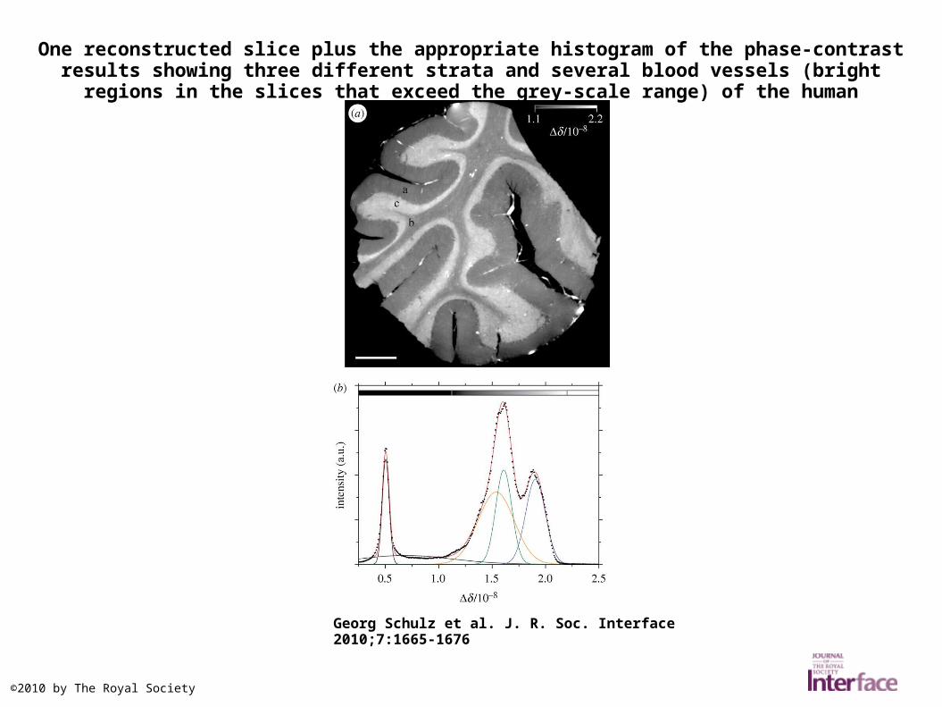
One reconstructed slice plus the appropriate histogram of the phase-contrast results showing three different strata and several blood vessels (bright regions in the slices that exceed the grey-

scale range) of the human cerebellum.

Georg Schulz et al. J. R. Soc. Interface 2010;7:1665-1676

©2010 by The Royal Society

Grating interferometry phase-contrast reconstruction with a grey-scale range corresponding to 34 standard deviations of the formalin peak (a) compared with accordant BW2 absorption-

contrast reconstruction with a grey-scale range corresponding to 34 (c) and ...

Georg Schulz et al. J. R. Soc. Interface 2010;7:1665-1676

©2010 by The Royal Society

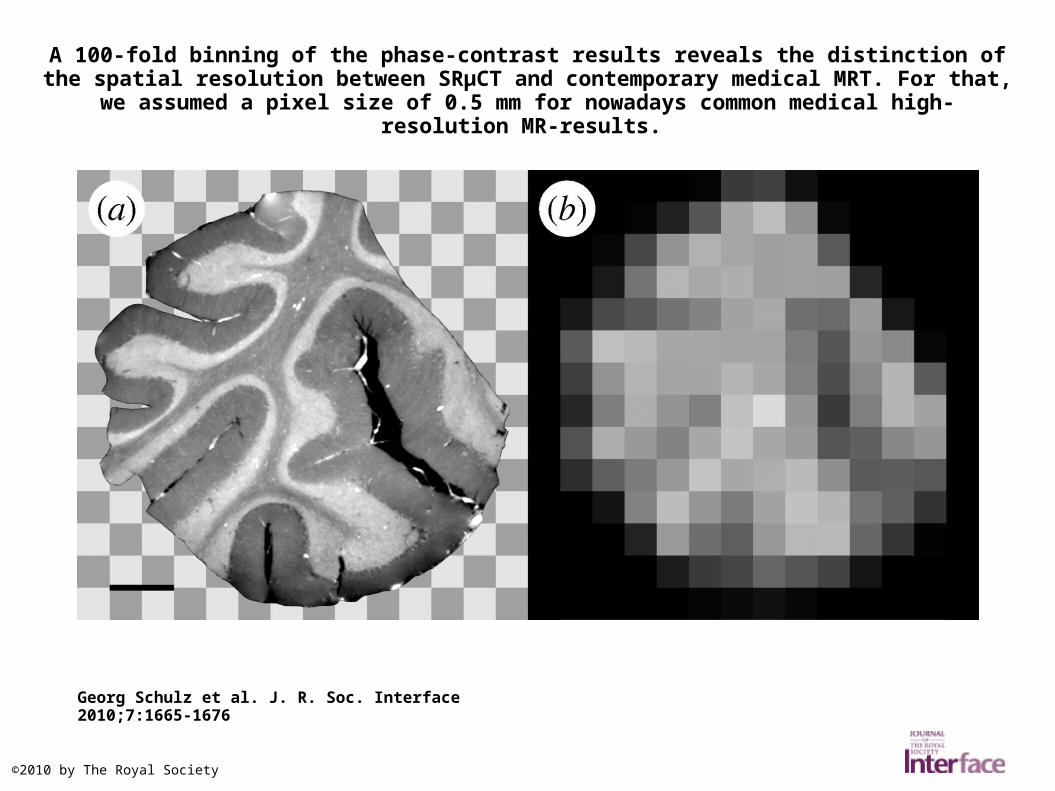
A 100-fold binning of the phase-contrast results reveals the distinction of the spatial resolution between SRµCT and contemporary medical MRT. For that, we assumed a pixel size of 0.5 mm for

nowadays common medical high-resolution MR-results.

Georg Schulz et al. J. R. Soc. Interface 2010;7:1665-1676

©2010 by The Royal Society

For the calculation of the spatial resolution, the ratio between rSPstruc of a tomogram ROI with a fine structure and rSPback of a tomogram ROI with background (water) was plotted over spatial

frequency.

Georg Schulz et al. J. R. Soc. Interface 2010;7:1665-1676

©2010 by The Royal Society

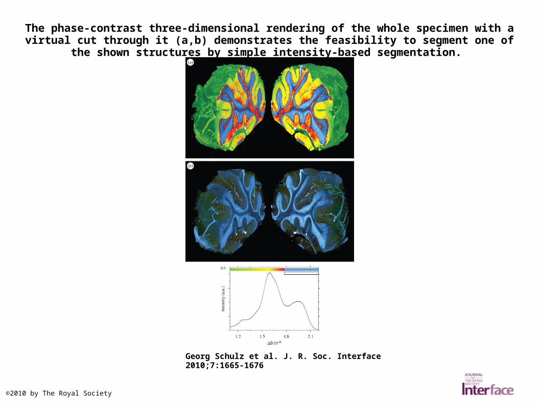
The phase-contrast three-dimensional rendering of the whole specimen with a virtual cut through it (a,b) demonstrates the feasibility to segment one of the shown structures by simple intensity-

based segmentation.

Georg Schulz et al. J. R. Soc. Interface 2010;7:1665-1676

©2010 by The Royal Society

Another phase-contrast three-dimensional rendering of the specimen presumably shows the so-called Purkinje cells.

Georg Schulz et al. J. R. Soc. Interface 2010;7:1665-1676

©2010 by The Royal Society


Recommended