Page 1
Immunohistochemistry
A cost effective approach to lymphoma diagnosis
ISIMM Tata Conference on Immunohistochemistry.
Kolkata, India, January 2018
Clive R. Taylor, M.D., Ph.D., Department of Pathology, Keck School of Medicine,
University of Southern California
Disclosures; CRT –has consulting arrangements with for Philips, Agilent, PerkinElmer, Optra
Page 2
1. Lymphoma VS Anaplastic tumor
2. Reactive VS Malignant
3. Sub - classification, B VS T, HL VS Non-HL& specific types, CD1, ALK etc
4. Prognostic markers & predictive markers Inc PDL1
5. Micrometastases in nodes and marrow
Page 3
ANAPLASTICTUMOUR
discussed elsewhere
Carcinoma??Lymphoma??
Melanoma??Sarcoma??
IHC stains
Page 4
carcinoma sarcoma lymphoma melanoma
keratin
vimentin
CD 45
S100
Basic screening panelCRT. USC.
Page 5
The DifferentTypes ofNHL and HL
Stem cellfetal liver/marrow
FetusPost - natal life
Thymus / T cells
Bursa /marrow /B cells
Lymphocytedifferentiation*gene
rearrangement*diversification*tolerance
Immune response in peripheral lymphoid tissues
Lymph nodesspleenresp tractGuttonsilAg processing
cells
Humoral immunity*antibodies
Cell mediated immunity* sens T cells
Plasmacells
Igs
‘killer’T cells
Resting B cell
Resting T cell
Transformation/amplification
Transformation/amplification
T
B
Single cell lineage
Multiple morphologies
Page 6
Virchows Arch (2013) 463:353–365
Beginning with Hodgkin in 1832 there have been numerous classifications of lymphoma.
Page 7
THE FIRST “LYMPHOMA”
7 autopsyCases
No microscopy
1825- 1837Inspector of the DeadCurator of the Museum,
Guys Hospital, London.
Page 8
Reed, D. (1902)On the pathological changes in Hodgkin's disease, with especial reference to its relation to tuberculosis.Johns Hopkins Hospital Reports 10, 133-196
Sternberg, C. (1898) Uber eine eigenartige unter dem Bilde der Pseudoleukamic verlaufende Tuberculose des lymphatischen Apparates. Ztchr Heilk, 19, 21-90
60 years later – the microscope makes its mark.
Page 9
With advent of microscopy -many other ‘lymphomas’were described – based upon morphology
Lympho-sarkomatosis - 1903
Giant Lymph Follicle Hyperplasia - 1927
By 1950 more than 50 different lymphomas had been described;
and almost as many differentclassifications
Page 10
For 100+ years Pattern Recognition
Panel 7 pathologists - ‘experts’.Reviewed 105 follicular lymphomas
Diagnosis - small cellconsensus 39 cases, range 24 - 65 among the 7
Diagnosis - mixed cellconsensus 40 cases; all 7 unanimous in only ONE
In 37% of cases both small & large cell werediagnosed by different members of the 7.
Metter et al. J Clin Oncol 3, 25, ‘85
Page 11
Diagnosis by Pattern Recognition
Accuracy versus consensus Dx.
Average panelist 71 %Best panelist 81 %Image Analysis* 89 %
*Using a continuous class approach,based upon SD cell/nuclear size, & measurements of high and lowfrequency diversity
vol 29, 2061, 1996.
Page 12
Then from 1960-1990 we hadthe struggle to change classification basis from morphology alone --
Rappaport – histiocytic / lymphocytic
Bob Collins, Karl Lennert, Bob LukesTo Immune based
The beginning of the WHO consensus process
To Immune based
Page 13
TDT
TDT
TDT
Lymphoid neoplasms related to normal Lymphoid Development
follicle
Stemcell
precursor
precursor
Stem cellfetal l iver/marrow
FetusPost - natal life
Thymus / T cells
Bursa /marrow /B cells
Lymphocytedifferentiation*gene rearrangement*diversification*tolerance
Immune response in peripheral lymphoid tissues
Lymph nodesspleenresp tractGuttonsilAg processing
cells
Humoral immunity*antibodies
Cell mediated immunity * sens T cells
Plasmacells Igs
‘killer’T cells
Resting B cell
Resting T cell
Transformation/amplification
Transformation/amplification
T
B
Morphology
Phenotype
CD2 CD3 CD4 CD5 CD8
CD10 CD19 CD20 CD79
IMMUNE BASED
Page 14
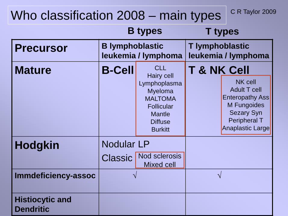
Who classification 2008.
Precursor Lymphoid Neoplasms
Mature B-Cell Neoplasms
Mature T-Cell & NK-Cell Neoplasms
Hodgkin lymphoma (Hodgkin disease)
Immunodeficiency-associated lymphoproliferative disorders
Histiocytic and Dendritic Cell Neoplasms
C R Taylor 2009
6 main groups
72 + types
Page 15
THE POINT-- 3 common types - rest uncommon (ADULTS).
Page 16
Who classification 2008 – main types
Precursor B lymphoblastic leukemia / lymphoma
T lymphoblastic leukemia / lymphoma
Mature B-Cell T & NK Cell
Hodgkin Nodular LPClassic
Immdeficiency-assoc √ √
Histiocytic and Dendritic
B types T types
CLLHairy cell
LymphoplasmaMyeloma
MALTOMAFollicularMantleDiffuseBurkitt
NK cellAdult T cell
Enteropathy AssM FungoidesSezary SynPeripheral T
Anaplastic Large
Nod sclerosisMixed cell
C R Taylor 2009
Page 17
the basis Who classification 2008
C R Taylor 2009
Morphology Phenotype
Small v large cell
Follicular v diffuse
IHC B v T80+ antibodies
molecular Hodgkin v NHL
6 main groups
72 + types
+ fine criteria
TranslocationsGene rearrangements
Page 18
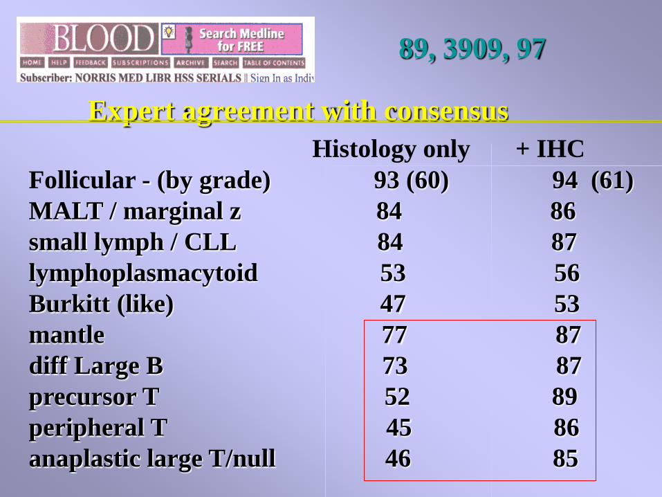
89, 3909, 97
Expert agreement with consensusHistology only + IHC
Follicular - (by grade) 93 (60) 94 (61)MALT / marginal z 84 86small lymph / CLL 84 87lymphoplasmacytoid 53 56Burkitt (like) 47 53mantle 77 87diff Large B 73 87precursor T 52 89peripheral T 45 86anaplastic large T/null 46 85
Page 19
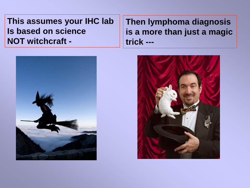
This assumes your IHC lab Is based on scienceNOT witchcraft -
Then lymphoma diagnosis is a more than just a magic trick ---
Page 20
CD 45 - leucocyte common antigen
CD20,CD79a,CD10,CD75, bcl 6, MUM1CD138, myc
cyclinD1,K,L
CD19, PAX5CD22,CD23
(bcl 2)(Annexin-A1 HCL)
(cd5,cd43)
CD3CD5,CD43CD4,CD8,
CD7CD56TIA-1
Gran BTdT
[ALK]
CD68CD163CD11c
lysozyme
(CD45)(EMA)
CD30,CD15
BLA36,Fascin
clusterinPax5CD40LMP
B T HL H
Assuming that most of these keyleucocyte markers are validated in your lab
Page 21
IHC and the WHO Classification of LymphomasCost Effective Immunohistochemistry Using aDeductive Reasoning ‘‘Decision Tree’’ Approach
Clive R. Taylor, MD, DPhil
(Appl Immunohistochem Mol Morphol 2009;17:366–374)
The WHO Classification of Lymphomas: Cost-effective Immunohistochemistry Using a Deductive Reasoning‘‘Decision Tree’’ Approach Part II: Diffuse Patterns of Proliferation in Lymph Nodes
Clive R. Taylor, MA, MD, DPhil
(Appl Immunohistochem Mol Morphol 2009;17:470–482)
Page 22
Diagnosisoflymphoma
DECISIONSDECISIONS
*Reactive (‘benign’) vs malignant
*Lymphoma (leukemia) vs metastasis
*Hodgkin vs Non-Hodgkin
*Sub-type, classification, B/T etc
Morphology –gold standard
Phenotype – flowIHC
Gene RX - Ig / TCR
Genotype – t(8;14), t(14;18)
4 decisions
4 methods
Page 23
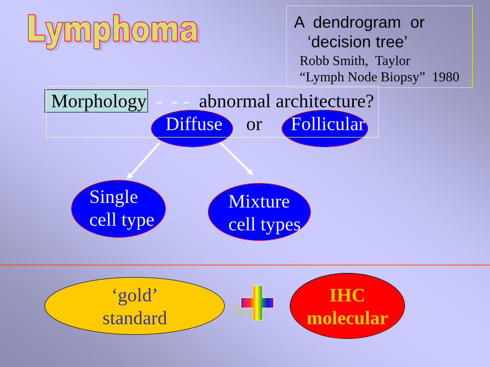
Single cell type
Morphology - - - abnormal architecture?Diffuse or Follicular
Mixture cell types
Robb Smith, Taylor“Lymph Node Biopsy” 1980
‘gold’standard
IHCmolecular
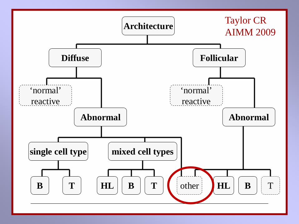
A dendrogram or ‘decision tree’
Page 24
Architecture
‘normal’reactive
FollicularDiffuse
AbnormalAbnormal
‘normal’reactive
single cell type mixed cell types
HLT otherB B T HL B T
Taylor CR AIMM 2009
Page 25
B B or T
Immunohistochemistry (flow cytometry)
B -redT -brown
Morphology Phenotype
Page 26
Architecture
‘normal’reactive
FollicularDiffuse
AbnormalAbnormal ‘normal’reactive
HL B T
Hodgkin lymphomaLymphocyte
PredominantNodular Sclerosis
FCC lymphomaBurkitt LMantle cell LMarginal zone LSmall lymphocytic L
Other
Lympho-blastic
Taylor CR AIMM 2009
Page 27
Marginal Zone:small lymphocytes
Pan B +SIgM +
Mantle Zone:small lymphocytes
Pan B +SIgD+, SIgM+
Reactive/germinal center:larger cells
Pale zone – centrocytesDark Zone – centroblasts
LymphocytesPan B+CD10+BCL6+
MUM1+/-Ki67++
BCL2 neg
Reactive Follicle
Pan B +
Variable T cellsCD3+, CD5+ :
usually fewIn mantle
Dendritic cells
CD21+CD35+
HistiocytesCD68+
CD163+
Morphology
Phenotype
Page 28
CD 20 BCL6 CD 21 IgD
Ki67 CD10 CD23 CD5
Page 29
R. Miller 2003 – Propath.
Page 30
Bcl-2 - the protein productInhibits apoptosis
Reactive Follicles –
negative
NeoplasticFollicles –
positive
T(14;18)
Page 31
18 bcl-2
14 H chain
Immunestimulation
t 14;18
lymphoma
B
**
Bcl-2 +++ Inhibits apoptosis
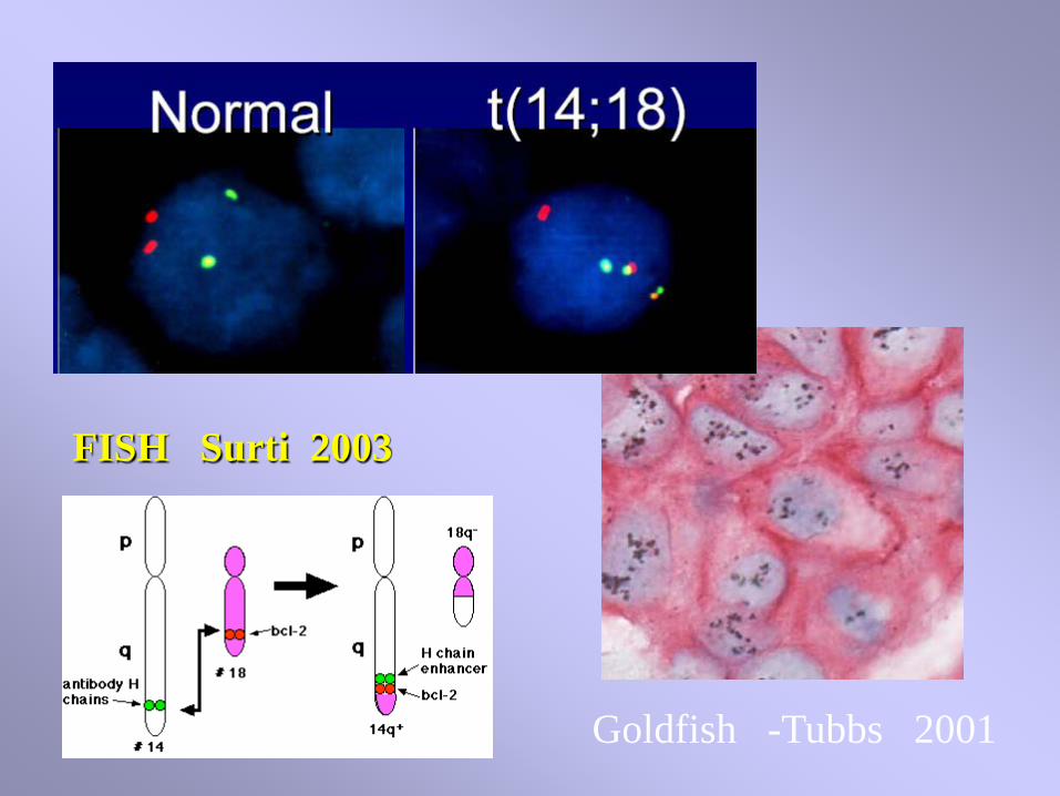
Page 32
FISH Surti 2003
Goldfish -Tubbs 2001
Page 33
ReactiveFollicle‘center’
NeoplasticFollicleFCC lymphoma
Bcl6Red
Bcl2brown
Shows ‘red’ FCC cells express BCL2
Shows ‘red’ FCC cells do notexpress BCL2
Page 34
CD 10 FCC Lymphoma
CD10 REACTIVE
CD10
Helpful with
loss polarity
and
extra-follicular Diffuse areas
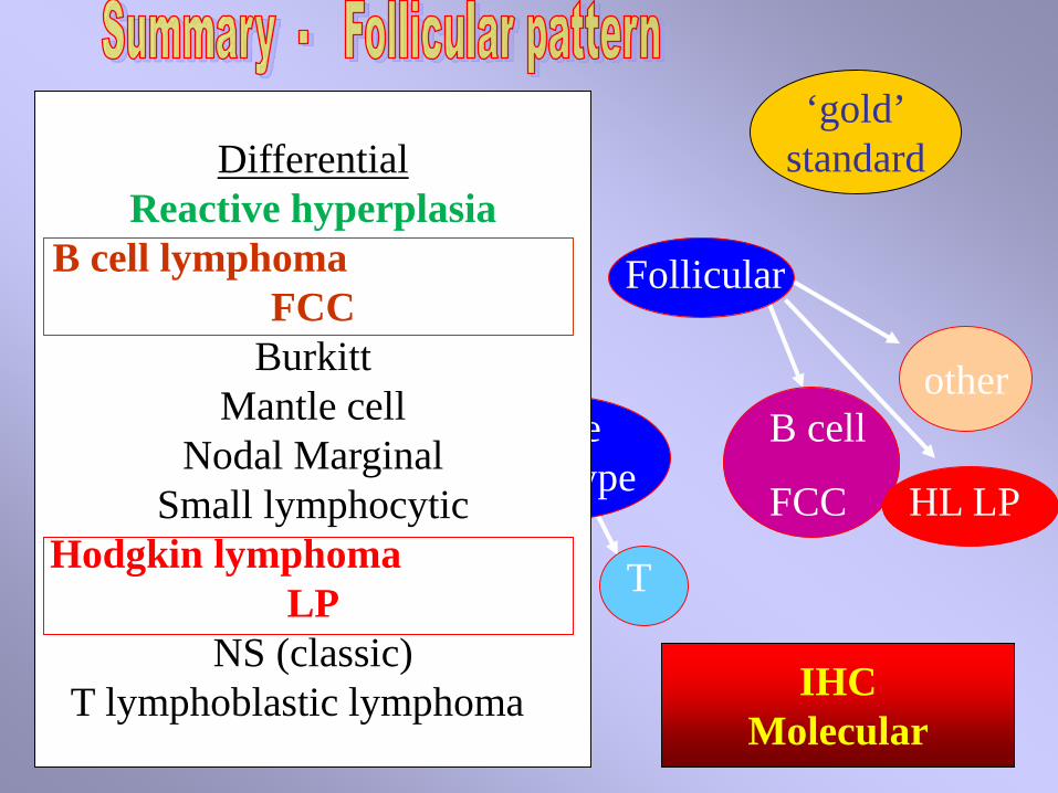
Page 35
IHCMolecular
Single cell type
B cell
FCC HL LP
MorphologyDiffuse or Follicular
Mixture cell types
HL B T B T
‘gold’standard
other
DifferentialReactive hyperplasia
B cell lymphoma FCC
BurkittMantle cell
Nodal MarginalSmall lymphocytic
Hodgkin lymphoma LP
NS (classic)T lymphoblastic lymphoma
Page 36
CD19,CD20Pax5,CD79aCD10, bcl 6,
(bcl 2)cyclinD1,
K,LCD23
(cd5,cd43)PD-1
Follicular
Nod LP FCCCD45 + RS +CD20 + +CD79a + +bcl6 + +Pax5 + +EMA + -bcl2 +/- (+)CD3 ( ++) (+/-)K/L clonal
(+) (+)
FCCHL LP
+ mantle and marginal – under diffuse
Role of IHC
Page 37
‘B’
‘B’‘B’‘B’‘B’‘B’
‘B’-
T
CD10, Bcl6CD10, Bcl6, -/+43Cyclin D1,CD5,4321, -/+43
CD23, 5, 43
EMACD15, 30CDc3,7,4+8, Tdt
reactive*
BCL2+
reactive*reactive* ‘pseudo-’
reactive*reactive*
Reactive hyperplasiaB cell lymphoma
FCCBurkittMantle cellMarginal ‘Nodal’Small lymphocytic
Hodgkin lymphomaLPNS (classic)
T lymphoblastic L
‘follicles’Lymphoma markersFOLLICULAR
‘B’ CD10, BCL6,-/+43 -----------
T CDc3,7,4+8,Tdt --------------
‘B’ CD10, BCL6 ------ BCL2+
Pay attention to the ‘follicles’
Page 38
Architecture
‘normal’reactive
FollicularDiffuse
AbnormalAbnormal
‘normal’reactive
single cell type mixed cell types
HLT otherB B T HL B T
Taylor CR AIMM 2009
70% of allLymphomas fit here
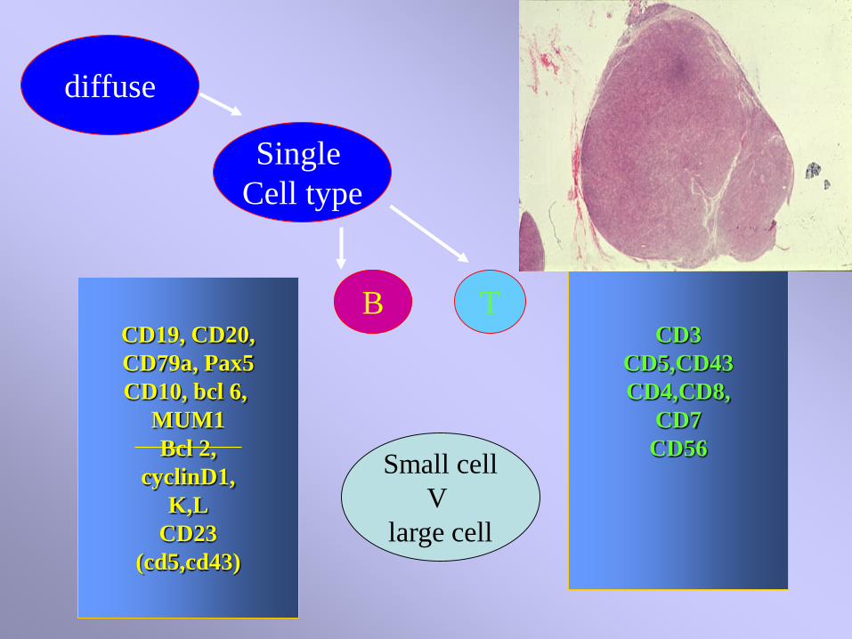
Page 39
CD19, CD20,CD79a, Pax5CD10, bcl 6,
MUM1Bcl 2,
cyclinD1,K,L
CD23(cd5,cd43)
CD3CD5,CD43CD4,CD8,
CD7CD56
diffuse
Single Cell type
B T
Small cellV
large cell
Page 40
B cell -Diffuse lymphoma PhenotypesCD s B* 10 21 23 5 43 bcl6 other*
B SL lymph/CLL + + + + cd11cL’pcytoid + CIg,cd38FCC + + (+/-) -/+ + (bcl2)mantle + + + D1marginal + [+/-] -/+ OCT,BOB
Diff large cell + +/- -/+ +/- +/-MUM1Burkitt + + -/+ + cd38
T -most + + are CD2+,3 +
Adapted fr Taylor et al. Immunomicroscopy and Molecular Morphology Elsevier/Saunders. 2005.
*CD19, 20, 22, 79a, Pax5[ ] FDC network
Page 41
DLBCL
CD 20
Bcl 6
MUM 1
Post or nonGC case –
PoorerPrognosis?
Myc/BCL2 both positive also indicates poor prognosisThreshold >40% myc+
Page 42
DLBCL..Also….Bcl2+ and survivin + = poorer prognosis
Germinal Center GC - and Post GC - DLBCLGC = CD10+, or BCL6+ and MUM -
Chang et al 2004
Page 43
CYCLIN D1 / PRAD 1 : MANTLE C L.
T 11;14, translocates BCL-1
MCL 90% +
Other ML -: rare immcytoma; marginal ML
Nuclear stain; requires AR ++*
Page 44
FISH Surti 2003
Goldfish -Tubbs 2001
Page 45
Bcl-1
Bcl-2
Seen in ‘mature’B lymphomas’ but break
at early B stage‘cancer stem cell’
Mantle cell
Follicular center cell
Tsai et al. USC
Page 46
Bcl-1 - t(14;11) – mantle cell –B lymphoma
Bcl-2 - t(14;18) – follicular center cell –B lymphoma
Break points cluster around cpg sites (red)
Tsai et al
Translocation events common in MLBecause of gene rearrangement
Page 47
Lymphomas - remarkable fit between ‘old morphologic typesAnd IHC and molecular classification
t (14;18) FCC bcl-2t (11;14) mantle bcl-1;cycD1t (2;5) ALCL npm/alkt(11;18) marginal(malt) API2/MALT1t(9;14) lymphoplasma pax5t (8;14) burkitt myct (3;14)/n diff large cell bcl-6
12+ CLL
Page 48
T cell - Diffuse lymphoma Phenotypes
*+CD99,34# cytoplasmic CD3 only
CD s 2,3 4 8 5 7 25 30 56 otherB -/+
T.NK +# -/+ + + GrB,Fas
Adult T + + + + -/+ FOXP3
Entero Ass + -/+ + -/+ -/+ -/+ GrB, 103
M fungoides + + -/+ + Peripheral + + -/+ -/+ -/+ -/+Anaplastic -/+ -/+ -/+ -/+ -/+ + + GrB,EMA
Lymphoblastic* + -/+ -/+ -/+ -/+ cd10
CRT 2017
Page 49
89, 3909, 97
Expert agreement with consensusHistology + IHCFollicular - (by grade) 93 (60) 94 (61)
MALT / marginal z 84 86small lymph / CLL 84 87lymphoplasmacytoid 53 56Burkitt (like) 47 53mantle 77 87diff Large B 73 87precursor T 52 89peripheral T 45 86anaplastic large T/null 46 85
Page 50
Architecture
‘normal’reactive
FollicularDiffuse
AbnormalAbnormal
‘normal’reactive
single cell type mixed cell types
HLT otherB B T HL B T
Taylor CR AIMM 2009
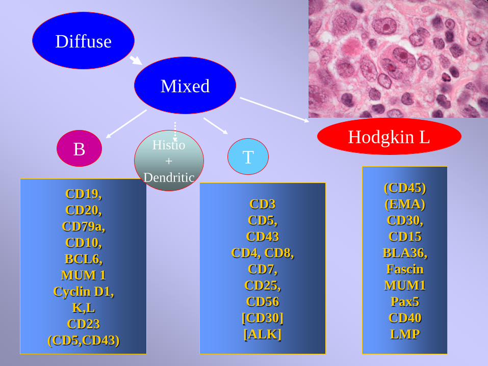
Page 51
Diffuse
Mixed
B T
CD19,CD20,
CD79a,CD10,BCL6,
MUM 1Cyclin D1,
K,LCD23
(CD5,CD43)
CD3CD5,CD43
CD4, CD8,CD7,
CD25,CD56
[CD30][ALK]
(CD45)(EMA)CD30,CD15
BLA36,FascinMUM1Pax5CD40LMP
Hodgkin LHistio+
Dendritic
Page 52
sclerosis
lymphs R-S cells
other
LP - +++++ +
ClassicalMCLR
-/+ +++ +++Histio,Plasma c
NS ++++ +++ +++ Fib bands
LD -/+ + ++++
Page 53
CD 15
CD 30ALK1
EMA
PAX5Classic HL
diffuse
vs
ALCL +
vs
HL LP+
Page 54
HDLP - CD 20, CD45
HD classical CD 15, CD30
Taylor, Riley, AIMM 9,187,2001
Markers useful for subclass HL and other lymphomas
Taylor CRHuman Path 36, 1-4, 2005.
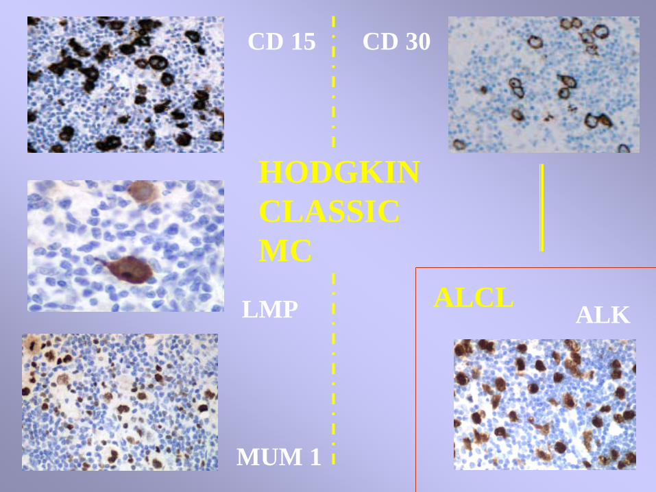
Page 55
HODGKIN CLASSICMC
CD 15 CD 30
LMP
MUM 1
ALCL ALK
Page 56
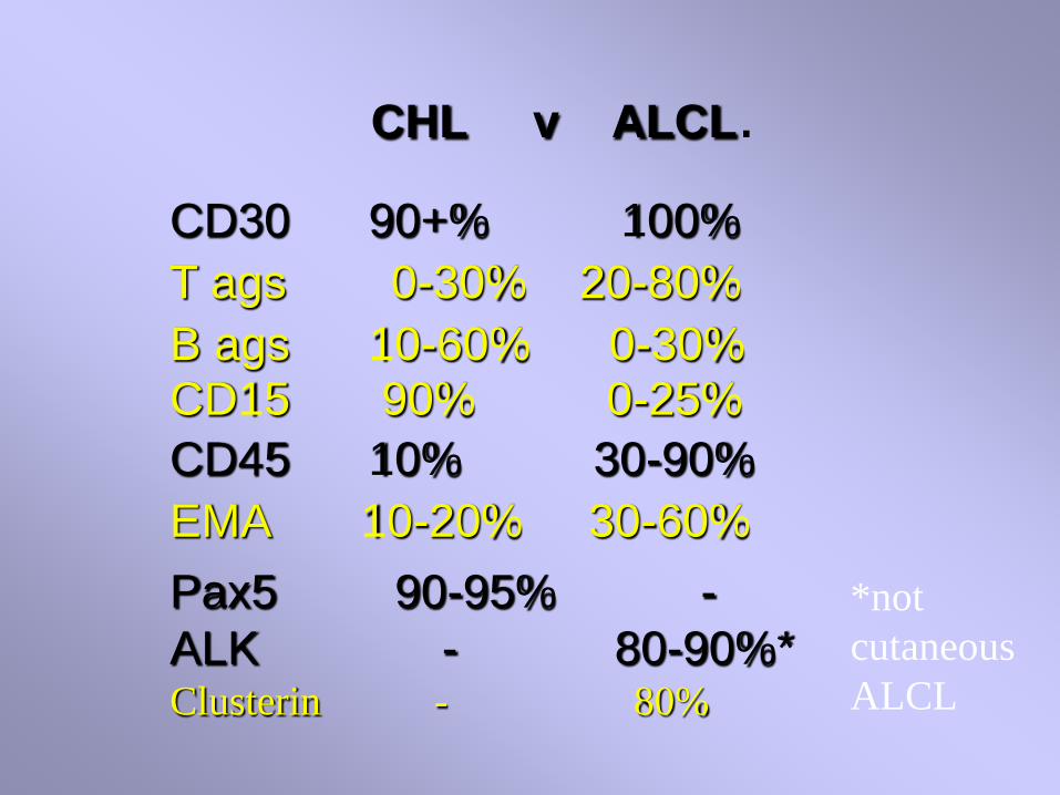
CHL v ALCL.CD30 90+% 100%T ags 0-30% 20-80%B ags 10-60% 0-30%CD15 90% 0-25%CD45 10% 30-90%EMA 10-20% 30-60% Pax5 90-95% -ALK - 80-90%*Clusterin - 80%
*notcutaneousALCL
Page 57
Weshi et al Leuk Lymphoma 48, 1764, 2007HL vs T cell (histiocyte) rich B cell lymphoma
CD20, CD79a, CD15, Fascin, EMA
Page 58
PANEL Nod LPRS cell
ClassicRS cell
LRBcellLarge cell
ALCLLarge cell
cd45 + + -/+cd30 + +cd15 +cd20,79a + -/+ +MUM1 + -/+Bcl6 + -/+ +Pax5 + + +OCT2 + +EMA +/- + -/+ +/-lymphocytes manyB+T Vary B+T Many B few
IHC
Large
Cells
Page 59
Added to which is the PD-1 PD L-1 story, which now isbecoming important for lymphomas-----With all of the challenges of scoring etc.
Page 61
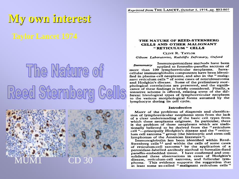
Taylor Lancet 1974
My own interest
MUM1 CD 30
Page 62
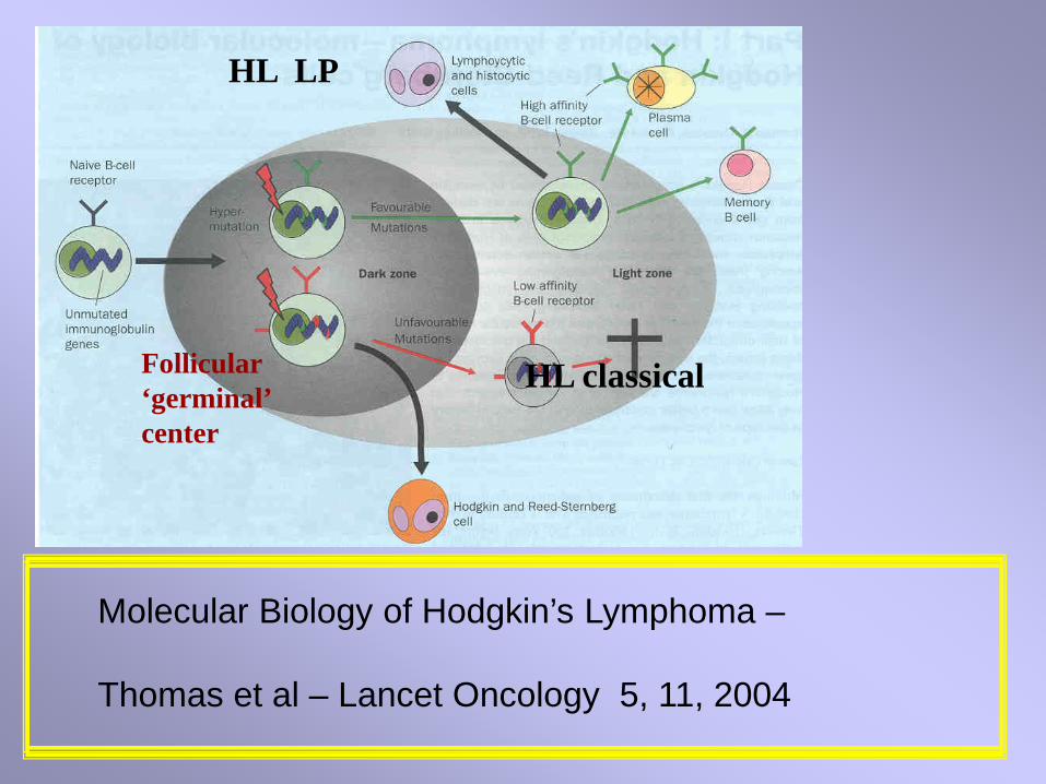
Molecular Biology of Hodgkin’s Lymphoma –
Thomas et al – Lancet Oncology 5, 11, 2004
HL LP
HL classicalFollicular‘germinal’center
Page 63
Architecture
‘normal’reactive
FollicularDiffuse
AbnormalAbnormal
‘normal’reactive
single cell type mixed cell types
HLT otherB B T HL B T
Taylor CR AIMM 2009
Page 64
Taylor CR. Hodgkin’s disease is a Non-Hodgkin Lymphoma. Hum Pathol. 36, 1-4, 2005.
Taylor CR. The WHO Classification of Lymphomas: Cost Effective Immunohistochemistry using a Deductive Reasoning ‘Decision Tree’ Approach. Part I. Appl. Immunohistochem & Mol Morphol, 17 :366-374, 2009. Part II. Appl. Immunohistochem & Mol Morphol, 17 :470-482, 2009.
Taylor CR Hartsock RJ. Classifications of Lymphoma; Reflections of Time andTechnology. Virchow Archiv. 458: 637-648. 2011.
Geller SJ, Taylor CR. Hodgkin; the Man and his Disease. Virchow Arch 460.DOI 10.1007/s00428-013-1442-0. 2013
Taylor CR and Riley CR. Molecular Morphology of Hodgkin Lymphoma. AIMM, 9(3):187-202, 2001.
Relevant personal bibliography
Van den Tweel, J, Gu, J, Taylor CR. From Magic to Molecules: An IllustratedHistory of Disease. Beijing University Press, 2016. Amazon.com