183
Education Guide Immunohistochemical Staining Methods Fourth Edition PATHOLOGY

Dako provides cancer diagnostic
products for leading reference
laboratories, hospitals and other
clinical and research settings.
Our instrumentation portfolio is
complemented by a full line of
antibodies, pharmDx™ assays,
detection systems and ancillaries.
Consider Dako for all your
laboratory needs.
Education Guide Immunohistochemical Staining Methods Fourth Edition
PATHOLOGY

�
Immunohistochemical Staining Methods, Fourth Edition
EditorMarc Key, Ph.D.Key B�omed�cal Serv�ces ° Oja�, CA, USA
ContributorsKaren Atwood, B.S. MT (ASCP) CLSDako ° Carp�nter�a, CA, USA
Kirsten Bisgaard, B.S.Dako ° Glostrup, Denmark
Kenneth J. Bloom, M.D.Clar�ent ° Al�so V�ejo, CA, USA
Thomas Boenisch, M.S.Dako ° Carp�nter�a, CA, USA
Nanna K. Christensen, M.S., Ph.D.Dako ° Glostrup, Denmark
A.J. Farmilo, Ph.D.Dako ° M�ss�ssauga, Ontar�o, Canada
Richard Harvey, Ph.D.Un�vers�ty of New Mex�co School of Med�c�ne ° Albuquerque, NM, USA
Jim Hudson, Ph.D.Dako ° Carp�nter�a, CA, USA
Mehrdad Nadji, M.D.Un�vers�ty of M�am� School of Med�c�ne ° M�am�, FL, USA
W. Roy Overton, Ph.D.GCAT Inc. ° Fort Coll�ns, CO, USA
Gale E. Pace, B.S. MT (ASCP) IDako ° Carp�nter�a CA, USA
Ole Feldballe Rasmussen, M.S., Ph.D.Dako ° Glostrup, Denmark
Andreas Schønau, M.S., EBADako ° Glostrup, Denmark
Helle Grann Wendelboe, M.S.Dako ° Glostrup, Denmark
Lars Winther, M.S., Ph.D.Dako ° Glostrup, Denmark
Ron Zeheb, Ph.D.The Lahey Cl�n�c ° Burl�ngton, MA, USA
© Copyright 2006 Dako, Carpinteria, California. All rights reserved. No part of this book may be reproduced, copied or transmitted without written permission. US $50
Immunoh�stochem�cal Sta�n�ng Methods
��
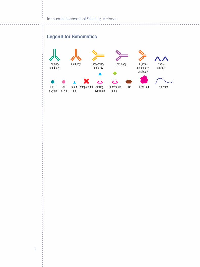
Legend for Schematics
primaryantibody
antibody secondaryantibody
antibody F(ab1)2 secondaryantibody
tissue antigen
HRPenzyme
AP enzyme
biotin label
streptavidin biotinyl tyramide
fluorescein label
DBA Fast Red polymer
Immunoh�stochem�cal Sta�n�ng Methods
���
Table of Contents
Preface VI
ParT I: Theory
Chapter 1 ° antibodies Thomas Boenisch
Introduction 1, Immunoglobulins 1, IgG 2, IgM 3, Polyclonal Antibodies 5, Monoclonal Antibodies 6, Antibody Affinity 7, Antibody Cross-Reactivity 9, Antibody Reaction Rates 10, Antibody Stability 10, Handling of Antibodies 12
Chapter 2 ° Basic Immunochemistry Thomas Boenisch
Introduction 15, Antibody Titer 15, Antibody Dilution 15, Antibody Incubation 17
Chapter 3 ° Basic Enzymology Thomas Boenisch
Introduction 19, Enzymes 19, Substrates and Chromogens 22, Suggested Procedures for Substrate-Cromogen Reagents 23
ParT II: Processing
Chapter 4 ° Fixation and Processing A.J. Farmilo
Introduction 27, Fixation 27, Tissue Handling 29, Specialized Tissue Preparations 31
Chapter 5 ° Molecular-Friendly Tissue Processing Mehrdad Nadji
Why Molecular Pathology 35, Complete Molecular-Friendly Histology Platforms 35, Fixation 35, Processing 36, Validation 37, Summary 37,Conclusion 39
Chapter 6 ° antigen retrieval Marc Key and Thomas Boenisch
Introduction 41, Short History of Antigen Retrieval 41, Principle and Technique 42, Mechanism of Action 43, Cytology 43, Target Retrieval for In Situ Hybridization 44, Antigen Retrieval and Its Use in Double Staining 44, Conclusion 44
Immunoh�stochem�cal Sta�n�ng Methods
�v
ParT III: Methodologies
Chapter 7 ° Immunohistochemistry Staining Methods Marc Key
Introduction 47, Immunohistochemistry 47, Avidin-Biotin Immunohistochemistry 48, Polymer-Based Immunohistochemistry 50, Tyramide Amplification 51, Cycled Tramide Amplification 52, Fluorescyl-Tyramide Amplification 52, Rolling Circle Amplification 52, Conclusion 53
Chapter 8 ° Immunofluorescence W. Roy Overton, Revised by Jim Hudson and Karen Atwood
Introduction 55, Fading, Quenching and Photobleaching 57, Fluorescein 57
Chapter 9 ° Multi-Staining Immunohistochemistry Nanna K. Christensen and Lars Winther
Introduction 61, Advantages of Multiple Staining 61, Technical Challenges 62, Pretreatment 62, Staining Method Selection 63, Dye Selection 65, Other Labels 67, Automated Image Acquisition and Analysis 67, Conclusion 67
Chapter 10 ° ancillary Methods in Immunohistochemistry Gale E. Pace
Introduction 71, Endogenous Enzyme Blocks 74, Antibody Diluents 79, Wash Buffers 82, Chromogen Enhancers 86
Chapter 11 ° In Situ Hybridization Richard Harvey, Revised by Andreas Schønau
Introduction 89, Types of Probes 89, Types of Samples 91, Detection of High-Risk HPV Infections in a Cervical Smear 93, Conclusion 93
Chapter 12 ° Methods of Immunocytology for Slide-Based Cellular analysis Marc Key
Introduction 95, Sample Preparation 97, Fixation 98, Immunostaining Methods 99, Controls 100
Chapter 13 ° automating Immunohistochemistry Ron Zeheb
Introduction 103, Choosing the Right IHC Stainer 103, Maintaining IHC Stainers 104, Economics of IHC Stainers 104, Flexibility and Ease of Use 104, Workflow and Data Management 105
Immunoh�stochem�cal Sta�n�ng Methods
v
Chapter 14 ° Virtual Microscopy and Image analysis Kenneth J. Bloom
History 107, Scanning Slides 108, Viewing the Virtual Slide 109, Capturing Images for Image Analysis 110
Chapter 15 ° Controls Ole Feldballe Rasmussen
Introduction 113, Reagent Controls 113, Negative Controls 114, Tissue Controls 114, Cell Line Controls 116, Control Programs 117, Future Aspects 118
Chapter 16 ° Background Helle Grann Wendelboe and Kirsten Bisgaard
Introduction 119, Background Associated with Detection Methods 119, Double Staining 121, General Factors 123, Natural and Contaminating Antibodies 125, General Aspects 129
Chapter 17 ° Troubleshooting Karen Atwood and Dako Technical Support Group
Introduction 131, Section One: Inadequate Staining 132, General Background 137, Limited Background 140, Undesired Specific Staining 144, Miscellaneous 145, Section Two: Background Staining 146, Section Three: Tissue Specimen 150, Section Four: Using a Typical Specification Sheet for an IVD Antibody 152
Glossary 157
Index 161
Immunoh�stochem�cal Sta�n�ng Methods
v�
Preface to the Fourth Edition of the Dako Educational Guide to Immunohistochemical Staining Methods
It �s my pleasure to �ntroduce th�s fourth ed�t�on of Dako’s Gu�debook to Immunoh�stochem�cal Sta�n�ng Methods. Th�s un�que reference �s prov�ded by Dako to academ�c research �nvest�gators, patholog�sts, h�stopatholog�sts and students from med�cal and sc�ent�fic d�sc�pl�nes around the world as part of the�r cont�nu�ng comm�tment to foster excellence �n the fields of �mmunology and h�stopathology.
In order to prov�de the most up-to-date �nformat�on �n these fields, Dako per�od�cally rev�ses th�s book’s content to reflect advancements �n these d�sc�pl�nes. For example, th�s ed�t�on, l�ke prev�ous ones, conta�ns relevant �nformat�on on the well-establ�shed theoret�cal bas�s and methodology that �s employed for these techn�ques. Of equal �mportance, contemporary approaches �n automat�on, �mage analys�s, molecular d�agnost�cs and mult�-sta�n�ng have been updated and expanded �n th�s ed�t�on. F�nally, of great pract�cal �mportance, knowledgeable pract�t�oners have prov�ded very helpful �nformat�on related to �nterpretat�on of sta�n�ng results and troubleshoot�ng to resolve unexpected problems that can ar�se.
On behalf of Dako, I would l�ke to thank all the contr�butors to th�s fourth ed�t�on. Most of the authors are Dako employees d�rectly act�ve �n the development of these methods prov�d�ng un�que �ns�ghts �n th�s h�ghly spec�al�zed field. The other authors, Thomas Boen�sch, Marc Key, Mehrdad Nadj�, Kenneth Bloom, W. Roy Overton and Ron Zeheb are recogn�zed experts �n the�r fields and greatly respected by all of us here at Dako. We value the�r �nd�v�dual contr�but�ons h�ghly and apprec�ate the fact that they would part�c�pate so fully �n th�s endeavor.
All ava�lable cop�es of prev�ous ed�t�ons of th�s handbook have been d�str�buted. It �s our goal to cont�nue th�s pract�ce �n order to expand knowledge �n th�s area and serve �nterested sc�ent�sts around the world. We hope you find th�s ed�t�on to be as useful as the pr�or ones and we ask that you share your learn�ng w�th your colleagues.
Denn�s E. Chenoweth, Ph.D., M.D.
Corporate V�ce Pres�dent, Bus�ness Development
�
Chapter 1 ° antibodies
Thomas Boenisch
IntroductionThe p�votal reagent common to all �mmunoh�stochem�cal* techn�ques �s the ant�body. The ava�lab�l�ty of new ant�sera, the�r �mmunoglobul�n fract�ons and monoclonal ant�bod�es to an ever-�ncreas�ng number of cl�n�cally useful t�ssue ant�gens has expanded the quant�ty and qual�ty of the �mmunoh�stolog�c reperto�re enormously. To better comprehend the potent�al of �mmunoh�stochem�cal sta�n�ng methods as well as assoc�ated problems, �t �s necessary to have a bas�c knowledge of ant�bod�es, the�r potent�als and the�r l�m�tat�ons.
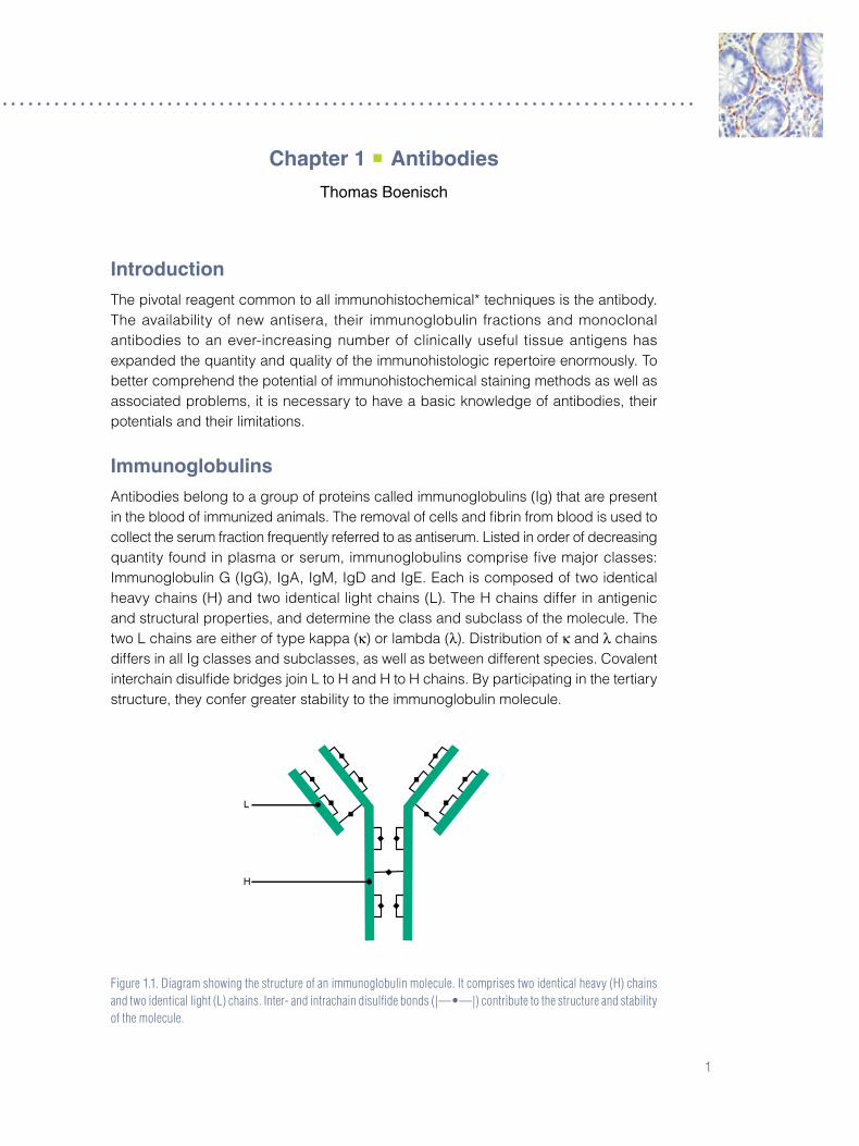
ImmunoglobulinsAnt�bod�es belong to a group of prote�ns called �mmunoglobul�ns (Ig) that are present �n the blood of �mmun�zed an�mals. The removal of cells and fibr�n from blood �s used to collect the serum fract�on frequently referred to as ant�serum. L�sted �n order of decreas�ng quant�ty found �n plasma or serum, �mmunoglobul�ns compr�se five major classes: Immunoglobul�n G (IgG), IgA, IgM, IgD and IgE. Each �s composed of two �dent�cal heavy cha�ns (H) and two �dent�cal l�ght cha�ns (L). The H cha�ns d�ffer �n ant�gen�c and structural propert�es, and determ�ne the class and subclass of the molecule. The two L cha�ns are e�ther of type kappa (κ) or lambda (λ). D�str�but�on of κ and λ cha�ns d�ffers �n all Ig classes and subclasses, as well as between d�fferent spec�es. Covalent �ntercha�n d�sulfide br�dges jo�n L to H and H to H cha�ns. By part�c�pat�ng �n the tert�ary structure, they confer greater stab�l�ty to the �mmunoglobul�n molecule.
Figure 1.1. Diagram showing the structure of an immunoglobulin molecule. It comprises two identical heavy (H) chains and two identical light (L) chains. Inter- and intrachain disulfide bonds (|—•—|) contribute to the structure and stability of the molecule.
Immunoh�stochem�cal Sta�n�ng Methods
�
Of the five classes of �mmunoglobul�ns, IgG and IgM w�ll be cons�dered �n more deta�l here, as these ant�bod�es are ut�l�zed by far the most frequently �n �mmunoh�stochem�stry. Unless otherw�se noted, most of what �s descr�bed of the IgG structure �n th�s text was learned from stud�es w�th human IgG of subclass IgG�.
IgG
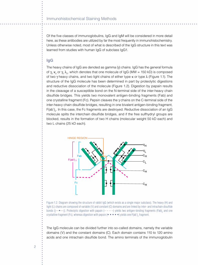
The heavy cha�ns of IgG are denoted as gamma (γ) cha�ns. IgG has the general formula of γ� κ� or γ� λ�, wh�ch denotes that one molecule of IgG (MW = �50 kD) �s composed of two γ heavy cha�ns, and two l�ght cha�ns of e�ther type κ or type λ (F�gure �.�). The structure of the IgG molecule has been determ�ned �n part by proteolyt�c d�gest�ons and reduct�ve d�ssoc�at�on of the molecule (F�gure �.�). D�gest�on by papa�n results �n the cleavage of a suscept�ble bond on the N-term�nal s�de of the �nter-heavy cha�n d�sulfide br�dges. Th�s y�elds two monovalent ant�gen-b�nd�ng fragments (Fab) and one crystall�ne fragment (Fc). Peps�n cleaves the γ cha�ns on the C-term�nal s�de of the �nter-heavy cha�n d�sulfide br�dges, result�ng �n one b�valent ant�gen-b�nd�ng fragment, F(ab’)�. In th�s case, the Fc fragments are destroyed. Reduct�ve d�ssoc�at�on of an IgG molecule spl�ts the �ntercha�n d�sulfide br�dges, and �f the free sulfhydryl groups are blocked, results �n the format�on of two H cha�ns (molecular we�ght 50 kD each) and two L cha�ns (�5 kD each).
Figure 1.2. Diagram showing the structure of rabbit IgG (which exists as a single major subclass). The heavy (H) and light (L) chains are composed of variable (V) and constant (C) domains and are linked by inter- and intrachain disulfide bonds (|—•—|). Proteolytic digestion with papain (– – – –) yields two antigen-binding fragments (Fab)2 and one crystalline fragment (Fc), whereas digestion with pepsin (• • • • •) yields one F(ab’)2 fragment.
The IgG molecule can be d�v�ded further �nto so-called doma�ns, namely the var�able doma�ns (V) and the constant doma�ns (C). Each doma�n conta�ns ��0 to ��0 am�no ac�ds and one �ntracha�n d�sulfide bond. The am�no term�nals of the �mmunoglobul�n
Ant�bod�es
�
molecule are located on the var�able doma�n of the l�ght cha�n (VL), and on the var�able doma�n of the heavy cha�n (VH). Together, VL and VH form the ant�gen-comb�n�ng s�te. Several hypervar�able (HV) reg�ons are located w�th�n the VL and VH doma�ns of the ant�body. Dur�ng the�r react�on w�th ant�gens, HV reg�ons are brought �nto close prox�m�ty to the ant�gen�c determ�nant (ep�tope). The d�stance between the ant�gen and HV reg�ons of the ant�body �s approx�mately 0.� to 0.� nm.
Un�que structural spec�fic�t�es called �d�otyp�c determ�nants are located �n th�s reg�on. Each ant�body clone expresses �ts own �d�otype. Each L cha�n also has one constant doma�n (CL) �n add�t�on to the VL doma�n. The H cha�n also has three constant doma�ns (CH
�, CH� and CH
�) and carr�es the carboxyl term�nal port�on of the �mmunoglobul�n. Located on the CH
� doma�n �s the carbohydrate mo�ety of the IgG molecule and several strongly hydrophob�c neutral aromat�c am�no ac�ds. The h�nge reg�ons are located between the CH
� and CH� doma�ns of the H cha�ns. M�nor d�fferences w�th�n these
h�nge reg�ons contr�bute to the subclass spec�fic�ty of �mmunoglobul�n G. The same are des�gnated by subscr�pts as �n IgG�, IgG�a, IgG�b, IgG� and IgG4. Whereas �n human IgG the overall rat�o of κ to λ �s �:�, �n the subclasses IgG� and IgG4, for example, the rat�os are �:� and 8:�, respect�vely. M�ce have approx�mately 95 percent κ cha�ns, and therefore most monoclonal IgG ant�bod�es from th�s spec�es have κ cha�ns. The number of d�sulfide br�dges l�nk�ng the heavy cha�ns also var�es among the IgG subclasses. IgG� and IgG4 each have two, wh�le IgG� and IgG� have four and five, respect�vely. Because of the flex�b�l�ty of the h�nge reg�on, the angle that both Fab fragments form can vary to accommodate a range of d�stances between �dent�cal ant�gen�c determ�nants.
IgM
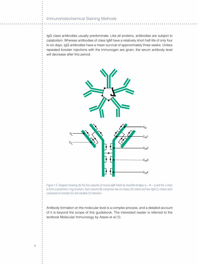
IgM �s a pentamer (MW approx�mately 900 kD) cons�st�ng of f�ve subun�ts of approx�mately �80 kD each (F�gure �.�). The general formula can be expressed as (μ� κ �) or (μ � λ �)
5. Each subun�t �s l�nked by a sulfhydryl-r�ch pept�de, the J cha�n (�5 kD), and cons�sts of two heavy cha�ns μ and two l�ght cha�ns of type κ or λ. The J-cha�ns contr�bute to the �ntegr�ty and stab�l�ty of the pentamer. As w�th IgG, IgM subun�ts can be fragmented by enzymat�c and reduct�ve cleavage �nto F(ab’)�, Fab and Fc port�ons, as well as heavy and l�ght cha�ns, respect�vely. The Fc fragment of IgM �s a cycl�c pentamer (molecular we�ght approx�mately �40 kD). Treatment of pentamer�c IgM w�th 0.� percent mercaptoethanol cleaves the d�sulfide br�dges between the subun�ts to y�eld five monomers. Subclasses of IgM� and IgM� have been reported.
Whereas IgG �s the most abundant ant�body �n the hyper�mmun�zed host, �n the newly �mmun�zed an�mal, IgM �s the first humoral ant�body detectable. The pr�mary ant�body format�on proceeds �n several major stages. Injected �mmunogen first reaches equ�l�br�um between extra- and �ntravascular spaces, then undergoes catabol�sm result�ng �n smaller fragments, and finally �s el�m�nated from the �ntravascular spaces by the newly formed ant�bod�es. The per�od from the �ntroduct�on of an �mmunogen unt�l the first appearance of humoral IgM ant�bod�es �s called the latent per�od and may last approx�mately one week. W�th�n two weeks, or �n response to a second �nject�on,
Immunoh�stochem�cal Sta�n�ng Methods
4
IgG class ant�bod�es usually predom�nate. L�ke all prote�ns, ant�bod�es are subject to catabol�sm. Whereas ant�bod�es of class IgM have a relat�vely short half-l�fe of only four to s�x days, IgG ant�bod�es have a mean surv�val of approx�mately three weeks. Unless repeated booster �nject�ons w�th the �mmunogen are g�ven, the serum ant�body level w�ll decrease after th�s per�od.
Figure 1.3. Diagram showing (A) the five subunits of mouse IgM linked by disulfide bridges (|—•—|) and the J chain to form a pentameric ring structure. Each subunit (B) comprises two mu heavy (H) chains and two light (L) chains each composed of constant (C) and variable (V) domains.
Ant�body format�on on the molecular level �s a complex process, and a deta�led account of �t �s beyond the scope of th�s gu�debook. The �nterested reader �s referred to the textbook Molecular Immunology by Atass� et al (�).

Ant�bod�es
5
Polyclonal antibodies
Polyclonal ant�bod�es are produced by d�fferent cells, and �n consequence, are �mmunochem�cally d�ss�m�lar. They react w�th var�ous ep�topes on the ant�gen aga�nst wh�ch they are ra�sed (F�gure �.4). By far, the most frequently used an�mal for the product�on of polyclonal ant�bod�es �s the rabb�t, followed by goat, p�g, sheep, horse, gu�nea p�g and others. The popular�ty of rabb�ts for the product�on of polyclonal ant�bod�es �s attr�buted pr�mar�ly to the�r easy ma�ntenance. An add�t�onal advantage �s that human ant�bod�es to rabb�t prote�ns are much more rare than to prote�ns from rum�nants, such as goat. In add�t�on, rabb�t ant�bod�es prec�p�tate human prote�ns over a w�der range of e�ther ant�gen or ant�body excess, and pools of ant�bod�es made from many rabb�ts are less l�kely to result �n major batch-to-batch var�at�ons than pools made from only a few, larger an�mals. Many years of select�ve breed�ng for favorable �mmun�zat�on response has made the New Zealand Wh�te rabb�t the most frequently used an�mal for the product�on of polyclonal ant�bod�es (�).
Figure 1.4. Schematic diagram of polyclonal antibodies binding to various epitopes on an antigen.
Depend�ng on the �mmunogen�c�ty of the ant�gen, doses of from �0 µg to �00 µg are trad�t�onally adm�n�stered to generate an �mmune response �n an�mals. The ant�gen �s �njected most often �ntradermally or subcutaneously, but �nject�ons �nto the footpad muscle or per�toneal cav�ty are used also. In rabb�ts, volumes of 0.�–0.5 mL are g�ven usually �ntradermally and d�str�buted over several s�tes; the ant�gen �s suspended �n an equal volume of adjuvant, such as Complete or Incomplete Freund’s Adjuvant. Booster
Immunoh�stochem�cal Sta�n�ng Methods
�
shots, repeated once a month or when decreas�ng t�ters are noted, are �ntended to ma�nta�n or �ncrease ant�body levels. Blood �s collected most often from the ear (rabb�ts), the jugular ve�n (larger an�mals) or from the heart, somet�mes by sacr�fic�ng the an�mal. After the removal of cells from the blood, polyclonal ant�bod�es can be obta�ned e�ther �n the form of stab�l�zed ant�sera or as pur�fied �mmunoglobul�n fract�ons. For the latter, prec�p�tat�on by salts, followed by �on exchange chromatography, serves to remove the bulk of other serum prote�ns. Affin�ty chromatography can be used to �solate the ant�gen-spec�fic ant�bod�es and thereby free them of nonspec�fic ant�bod�es.
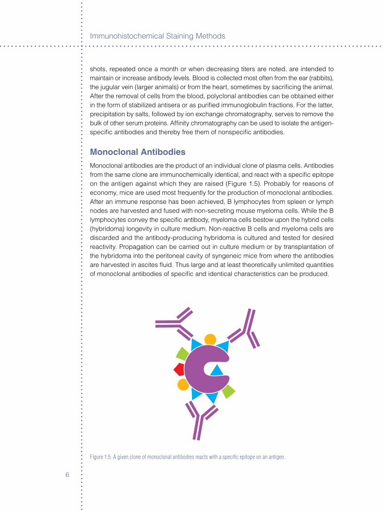
Monoclonal antibodiesMonoclonal ant�bod�es are the product of an �nd�v�dual clone of plasma cells. Ant�bod�es from the same clone are �mmunochem�cally �dent�cal, and react w�th a spec�fic ep�tope on the ant�gen aga�nst wh�ch they are ra�sed (F�gure �.5). Probably for reasons of economy, m�ce are used most frequently for the product�on of monoclonal ant�bod�es. After an �mmune response has been ach�eved, B lymphocytes from spleen or lymph nodes are harvested and fused w�th non-secret�ng mouse myeloma cells. Wh�le the B lymphocytes convey the spec�fic ant�body, myeloma cells bestow upon the hybr�d cells (hybr�doma) longev�ty �n culture med�um. Non-react�ve B cells and myeloma cells are d�scarded and the ant�body-produc�ng hybr�doma �s cultured and tested for des�red react�v�ty. Propagat�on can be carr�ed out �n culture med�um or by transplantat�on of the hybr�doma �nto the per�toneal cav�ty of syngene�c m�ce from where the ant�bod�es are harvested �n asc�tes flu�d. Thus large and at least theoret�cally unl�m�ted quant�t�es of monoclonal ant�bod�es of spec�fic and �dent�cal character�st�cs can be produced.
Figure 1.5. A given clone of monoclonal antibodies reacts with a specific epitope on an antigen.
Ant�bod�es
�
In �mmunoh�stochem�stry, monoclonal ant�bod�es have certa�n advantages over the�r polyclonal counterparts. These �nclude h�gh homogene�ty, absence of nonspec�fic ant�bod�es, ease of character�zat�on and m�n�mal batch-to-batch or lot-to-lot var�ab�l�ty.
Some p�tfalls �n the use of monoclonal ant�bod�es should be noted. Test methods for select�on of useful clones and for qual�ty control must be �dent�cal to the methods for wh�ch they ult�mately w�ll be used. For example, monoclonal ant�bod�es must be character�zed on formal�n-fixed t�ssues and not on frozen t�ssue, �f they ult�mately are �ntended for use on formal�n-fixed spec�mens.
S�m�larly, results from test�ng react�v�ty of a new ant�body on opt�mally fixed t�ssue must not be rel�ed upon to pred�ct �ts react�v�ty on sub-opt�mally fixed t�ssue, such as t�ssue fixed for a prolonged or �ncons�stent length of t�me. Also, as �mproved ant�gen retr�eval procedures are be�ng publ�shed cont�nuously, �t �s �mperat�ve that the screen�ng of new ant�bod�es cons�der these add�t�onal var�ables (see Ant�gen Retr�eval, Chapter �).
Targeted ep�topes also must be un�que to a g�ven ant�gen. Spec�fic�ty, one of the greatest benefits of monoclonal ant�bod�es �s lost �f the ant�body �s d�rected aga�nst an ep�tope shared by two or more d�fferent ant�gens (see Ant�body Cross-React�v�ty). Wh�le cross-react�v�ty of a polyclonal ant�body can be removed usually by absorpt�on, that �s not poss�ble w�th a monoclonal ant�body.
Screen�ng methods also should cons�der that monoclonal ant�bod�es, unl�ke polyclonal ant�bod�es, depend more on env�ronmental factors such as pH and solute for opt�mum performance (�).
antibody affinityAnt�bod�es from hyper�mmun�zed an�mals not only d�ffer w�th regard to the determ�nants they recogn�ze on mult�valent ant�gens, but also d�ffer �n the�r affin�t�es for the same. The term “affin�ty” has been used to descr�be both �ntr�ns�c and funct�onal affin�t�es (4).
The �ntr�ns�c affin�ty of an ant�body res�des �n the HV reg�on and �s determ�ned by the same sequence of am�no ac�ds that determ�nes spec�fic�ty. Pr�mar�ly �on�c (electrostat�c) �nteract�ons, but also hydrogen bond�ng and van der Waals forces are the major contr�butors to the �ntr�ns�c affin�ty between the paratope on the ant�body and the ep�tope on the ant�gen. Hydrophob�c�ty forms last and has a stab�l�z�ng effect on the cult�vated �mmune complex, and, w�th soluble reactants, usually leads to �ts prec�p�tat�on. Covalent b�nd�ng between ant�body and ant�gen does not occur. The assoc�at�on constant (Ka) of the b�nd�ng between an ant�body and �ts ant�gen�c determ�nant �s a measure of the ant�body’s affin�ty. It can range from �0� to �0�0 l�ters per mole and �s the rec�procal of concentrat�on �n moles per l�ter. The h�gher the �ntr�ns�c affin�ty of the ant�body, the lower the concentrat�on of the ant�gen needed for the ava�lable b�nd�ng s�tes of the ant�body to become saturated (reach equ�l�br�um). Just as the quant�ty (t�ter) of an ant�body �ncreases w�th t�me dur�ng �mmun�zat�on, so does �ts qual�ty (affin�ty). Th�s has been called “affin�ty maturat�on” (5). Lower doses of �mmunogen �ncrease the rate of affin�ty maturat�on, but may result �n lower t�ters of ant�body, and v�ce versa.
Immunoh�stochem�cal Sta�n�ng Methods
8
In �mmunoh�stochem�stry, the funct�onal affin�ty of an ant�body or an ant�serum can be defined very loosely by the t�me requ�red to reach equ�l�br�um w�th the t�ssue ant�gen. If equal al�quots of two ant�bod�es or ant�sera of �dent�cal t�ter are �ncubated for �ncreas�ng per�ods of t�me w�th the ant�gen on the t�ssue, the ant�body that reaches a plateau of max�mum sta�n�ng �ntens�ty first �s of a h�gher funct�onal affin�ty. The term “av�d�ty” has been used synonymously to descr�be funct�onal affin�ty (5), but also has been used to denote the strength of the b�nd�ng reached between ant�body and �ts ant�gen (�). The term av�d�ty also has been used to descr�be the sum total of all �ntr�ns�c affin�t�es found �n a polyclonal ant�body populat�on.
Because ant�gen-ant�body react�ons are revers�ble, the s�mple �mmune complexes formed on t�ssue occas�onally may d�ssoc�ate dur�ng the wash�ng cycles used �n �mmunoh�stochem�stry. The ease and extent of th�s d�ssoc�at�on vary from ant�body to ant�body, and low salt concentrat�ons as well as low temperatures w�ll reduce the l�kel�hood of weak sta�n�ng due to d�ssoc�at�on of an already-formed �mmune complex. Thus, h�gh-affin�ty ant�bod�es are des�rable and have the advantage that d�ssoc�at�on �s less l�kely to occur than w�th low-affin�ty ant�bod�es dur�ng wash�ng. As ment�oned before, a polyclonal populat�on of ant�bod�es conta�ns a more or less cont�nuous spectrum of low to h�gh affin�t�es aga�nst several ep�topes on a g�ven ant�gen. Therefore after �ncubat�on w�th a pr�mary ant�body of th�s type, excess�ve wash�ng �s unl�kely to result �n any apprec�able loss of sta�n�ng.
On the other hand, monoclonal ant�bod�es are of un�form affin�ty and, �f the same �s low, loss of sta�n�ng may be due to the d�ssoc�at�on of the ant�body from �ts ep�tope. Therefore, �f poss�ble, monoclonal ant�bod�es of h�gh affin�ty should be selected. As �nd�cated above, factors that weaken the ant�gen-ant�body bond such as h�gh salt concentrat�ons, h�gh temperature and very low pH dur�ng the wash�ng of the spec�mens should be avo�ded. Exper�ence �n the handl�ng of ant�bod�es �n �mmunoh�stochem�stry has shown that the wash�ng and �ncubat�on �n buffer baths can be reduced safely and that gentle ag�tat�on helps to reduce background sta�n�ng (�).
Affin�ty of ant�bod�es also �s related to the�r capac�ty to form �nsoluble �mmune complexes. Generally, the h�gher the affin�ty of an ant�body, the greater �ts tendency to form a prec�p�tate. Prec�p�tat�on proceeds through a rap�d stage �n wh�ch soluble ant�gen-ant�body complexes form, followed by slower aggregat�on and, eventually, prec�p�tat�on. Non-prec�p�tat�ng ant�bod�es are mostly of lower affin�ty and are �ncapable of form�ng the latt�ce requ�red for prec�p�tat�on to occur.
Monoclonal ant�bod�es, regardless of whether they are of h�gh or low affin�ty, do not form a latt�ce w�th ant�gen, and, hence only rarely form �nsoluble prec�p�tates. However, �n �mmunoh�stochem�stry, the capab�l�ty of a pr�mary ant�body to form prec�p�tat�ng �mmune complexes �s of l�ttle �mportance because react�on w�th �mmob�l�zed t�ssue ant�gen enta�ls ant�body capture onto t�ssue rather than prec�p�tat�on.
Prozone �s a property that was first noted �n ant�body-�nduced agglut�nat�ons. It �s the observat�on that some ant�bod�es, when �nsuffic�ently d�luted, fa�l to agglut�nate cells
Ant�bod�es
9
even though h�gher d�lut�ons w�ll do so. Wh�le prozone also can be observed �n prec�p�t�n react�ons, �n �mmunoh�stochem�stry, �t �s a rare event (�).
As most ant�bod�es carry a net pos�t�ve electrostat�c charge, the strength of the ant�body’s affin�ty for the targeted t�ssue ant�gen also depends on the ava�lab�l�ty and abundance of the net negat�ve electrostat�c charges present on the latter. Excess�ve formal�n-fixat�on t�mes of many t�ssues were held largely respons�ble for alterat�on of these charges, and as a consequence, for the unpred�ctably errat�c �mmune react�v�ty w�th the pr�mary ant�body. Lost affin�t�es, however, were restored largely by the rout�ne use of heat-�nduced retr�eval for all ant�gens (8).
antibody Cross-reactivityThe term “cross-react�v�ty” denotes an �mmunochem�cal act�v�ty that can occur e�ther between an ant�body and two or more ant�gens or v�ce versa, when an ant�gen reacts w�th several d�fferent ant�bod�es. Typ�cal examples are when ant�-λ (or -κ) cha�n ant�bod�es �nteract w�th all five Ig classes or when carc�noembryon�c ant�gen (CEA) reacts w�th ant�bod�es aga�nst CEA, blood group ant�gens and normal t�ssue prote�ns, respect�vely. The common denom�nator �n each case �s the shar�ng of at least one common ep�tope between several ant�gens.
Another val�d use of the term cross-react�v�ty denotes the exper�mentally-or acc�dentally- �nduced changes w�th�n one or several ep�topes, through ant�gen retr�eval (9), lead�ng to a poss�ble loss of spec�fic�ty by a g�ven monoclonal ant�body for th�s ant�gen. The term cross-react�v�ty also descr�bes the �nteract�on of an ant�body w�th s�m�lar or d�ss�m�lar ep�topes on unrelated ant�gens. Th�s latter phenomenon however �s frequently a property of low-affin�ty ant�bod�es, and usually �s subject to change because of affin�ty maturat�on dur�ng �mmun�zat�on.
Cross-react�v�ty of ant�bod�es to human ant�gens w�th �dent�cal or s�m�lar ant�gens of other spec�es, or “cross-spec�es cross-react�v�ty,” can be of �nterest to the researcher and veter�nar�an because of the scarc�ty of an�mal-spec�fic ant�bod�es. In an effort to overcome th�s, two groups publ�shed reports on the results of cross-spec�es react�v�ty stud�es us�ng commerc�ally ava�lable ant�human polyclonal and monoclonal ant�bod�es (�0, ��). It was demonstrated that the major�ty of an�mal ant�gens selected showed strong react�v�ty w�th ant�human ant�bod�es. However, for more techn�cal deta�l on the use of a g�ven mouse pr�mary ant�body on an�mal t�ssues, the reader �s referred to an�mal research k�t products.
The term�nology of cross-react�v�ty however �s m�splaced when descr�b�ng any observed sta�n�ng by the same ant�body of d�fferent cells or t�ssue components, regardless of whether or not they conta�n common ant�gens, as th�s would d�stort the str�ct �mmunochem�cal defin�t�on of the term.
Immunoh�stochem�cal Sta�n�ng Methods
�0
antibody reaction ratesAlthough under �deal cond�t�ons ant�bod�es react w�th the�r ant�gens very rap�dly, �n �mmunoh�stochem�stry the cond�t�ons are rarely �deal. Depend�ng on length of t�ssue fixat�on, ant�body concentrat�on, amb�ent temperature and other var�ables, pr�mary ant�body �ncubat�on t�mes of up to 48 hours may be requ�red for max�mum react�v�ty (��). It �s not surpr�s�ng therefore, that as �mmunoh�stochem�cal procedures have become �ncreas�ngly useful �n surg�cal pathology, the need for shortened process�ng t�mes also has been vo�ced. Very short �ncubat�on per�ods are made feas�ble by the relat�vely rap�d react�on rates that occur when h�gher concentrat�ons of h�gh-affin�ty pr�mary and l�nk ant�bod�es are used.
In these s�tuat�ons equ�l�br�um between ant�gen-bound and free ant�body rarely �s ach�eved. To ach�eve equ�l�br�um, very long �ncubat�on per�ods w�th more d�lute ant�body preparat�ons are requ�red. It �s not known whether shorter �ncubat�ons w�th more concentrated ant�body preparat�ons would establ�sh equ�l�br�um sooner, because as a rule nonspec�fic background sta�n�ng may result under these cond�t�ons, prevent�ng unamb�guous �nterpretat�on. Incubates of pr�mary ant�body have been salvaged exper�mentally after the�r first use by asp�rat�on from one sect�on, and transferred to add�t�onal sect�ons (�). W�th some ant�bod�es, up to seven �dent�cal t�ssue spec�mens could be sta�ned w�th equal qual�ty when the pr�mary ant�body was used �n concentrat�ons requ�red for rout�ne �0-m�nute �ncubat�ons. Th�s suggests that only a very small fract�on of the ava�lable ant�body actually �s ut�l�zed dur�ng these relat�vely short �ncubat�on t�mes. Needless to say, once an �ncubat�on t�me has been selected, �t must be ma�nta�ned un�formly, or sta�n�ng w�ll not be cons�stently reproduc�ble.
Generally, the s�ze and shape of the ant�body molecule and �ts conjugates or complexes appear to be of l�ttle consequence �n �mmunoh�stochem�stry. Insuffic�ent t�ssue penetrat�on, even when sta�n�ng �ntranuclear or cytoplasm�c ant�gens, has never been observed, regardless of whether pr�mary ant�bod�es of class IgM (900 kD), large complexes l�ke PAP (400–4�0 kD) or APAAP (approx�mately 5�0 kD) or dextran-l�nked reagents were used (see Immunoh�stochem�stry Sta�n�ng Methods, Chapter �). However, �t �s reasonable to assume that gross overfixat�on of t�ssue may make penetrat�on more d�fficult for ant�bod�es and the�r complexes.
antibody StabilityPolyclonal ant�bod�es, when stored unfrozen and used subsequently �n �mmunoh�stochem�stry, are somewhat less stable as �mmunoglobul�n fract�on compared to whole ant�serum (�). However, th�s reduced stab�l�ty was found to depend largely on the method of pur�ficat�on and storage as well as on the method of appl�cat�on. Exposure of ant�bod�es to extreme pH, as well as h�gh or very low concentrat�ons of salts dur�ng pur�ficat�on tends to decrease the�r stab�l�ty more than does exposure to m�ld cond�t�ons such as �on exchange chromatography. Format�on of soluble aggregates, and subsequently prec�p�tated polymers are the most frequent changes noted after prolonged storage. These changes are probably the result of hydrophob�c �nteract�on
Ant�bod�es
��
between the IgG molecules �n solut�on. Wh�le the presence of soluble aggregates may enhance the�r performance as prec�p�tat�ng ant�bod�es, the�r �ncreased hydrophob�c�ty has been shown to cause �ncreased nonspec�fic b�nd�ng �n �mmunoh�stochem�stry (see Chapter ��, Background) (�). Removal of these aggregates and polymers from IgG fract�ons �s therefore prudent pr�or to the�r appl�cat�on for �mmunoh�stochem�stry.
Just as storage of pur�fied ant�bod�es may augment the�r hydrophob�c�ty due to aggregat�on and polymer�zat�on, so may the�r conjugat�on to other molecules (��). Conjugat�on w�th glutaraldehyde �nvolves the eps�lon-am�no groups of lys�ne and alpha-am�no groups of the N-term�nal am�no ac�ds result�ng �n the�r cross-l�nk�ng. Because there are many glutaraldehyde-react�ve s�tes �n IgG molecules, the hydrophob�c�ty of the conjugated ant�bod�es may �ncrease s�gn�ficantly, result�ng �n augmented attract�on to hydrophob�c s�tes �n the fixed t�ssue and �ncreased background.
Monoclonal ant�bod�es also have been shown to be �nfluenced �n the�r performance by methods of pur�ficat�on and storage; 4� percent of monoclonal ant�bod�es �nvest�gated by Underwood and Bean showed changes �n spec�fic�ty, affin�ty and cross-react�v�ty (�4). Ant�bod�es of class IgM and subclass IgG�b were espec�ally sens�t�ve.
It must be noted that actual-t�me test�ng of prote�naceous reagents �s not feas�ble. Wh�le commonly pract�ced �n the pharmaceut�cal field (�5, ��), h�gh-temperature accelerated degradat�on test�ng when appl�ed to �mmunochem�cals such as ant�sera and ant�bod�es, can be �rrelevant or even m�slead�ng (��, �8).
Ant�body stab�l�ty �n commerc�ally produced reagents �s determ�ned best by real-t�me and real-temperature test�ng by each manufacturer. Most manufacturers demonstrate stab�l�ty by test�ng dur�ng a pre-determ�ned per�od of t�me, �e, the “shelf l�fe.” Wh�le many ant�bod�es may reta�n act�v�ty longer, the only regulatory requ�rement for the manufacturer �s to cert�fy the per�od of t�me that the ant�body has been tested. There �s no requ�rement to cont�nue test�ng unt�l the ant�body loses act�v�ty.
In add�t�on, �t �s th�s wr�ter’s exper�ence that the cond�t�ons for the storage of reagents �n the user’s laboratory are frequently not �dent�cal to those that preva�led dur�ng the manufacturer’s shelf-l�fe stud�es. Because of the poss�b�l�ty of adverse storage cond�t�ons after the purchase of the product, the manufacturer can offer only a l�m�ted l�ab�l�ty �nstead of pred�ct�ng the actual dem�se of a reagent.
The only poss�ble corollary to these requ�rements �s to allow laborator�es to document the act�v�ty of the product unt�l the loss of the same. Alternat�vely, laborator�es may al�quot and freeze und�luted ant�body at –�0 °C for later use. At th�s t�me, laborator�es must confirm act�v�ty pr�or to the use of the ant�body �n any test.
F�nally, exp�rat�on dat�ng as pract�ced today also serves the purpose of conform�ng to regulatory requ�rements. Regulatory gu�del�nes �n place �n the Un�ted States for cl�n�cal laborator�es have been mandated by the Cl�n�cal Laboratory Improvement Act of �988 and by the College of Amer�can Patholog�sts. These regulat�ons mandate that exp�red reagents cannot be used �n the cl�n�cal d�agnost�c laboratory on human t�ssue.

Immunoh�stochem�cal Sta�n�ng Methods
��
Handling of antibodiesIn order to ach�eve opt�mal performance from reagents used �n �mmunoh�stochem�stry, �t �s �mperat�ve to observe bas�c rules for the�r handl�ng and storage. If properly ma�nta�ned, most reagents w�ll rema�n stable for months or even years. Recommendat�ons g�ven by the manufacturer on spec�ficat�on sheets and on v�al labels always should be heeded.
receiving
Although many commerc�ally produced �mmunochem�cals are guaranteed to be stable for up to several years, ready-to-use (RTU) ant�bod�es have a shorter shelf l�fe (see Ant�body Stab�l�ty). Upon rece�pt, �mmunochem�cals should be stored promptly accord�ng to the manufacturer’s recommendat�ons. Log reagents by enter�ng the manufacturer’s lot numbers, exp�rat�on date, date of rece�pt and �nvo�ce number. These entr�es prov�de valuable �nformat�on for the user, espec�ally �f later reclamat�ons should become necessary.
Storage
Perhaps the two most �mportant cons�derat�ons when stor�ng ant�bod�es are the storage conta�ner and the temperature.
Storage Containers
Ideally, preferred mater�als for storage conta�ners of prote�n solut�ons should have negl�g�ble prote�n adsorpt�v�ty. Polypropylene, polycarbonate or boros�l�cate glass are recommended and are used w�dely. Solut�ons conta�n�ng very low concentrat�ons of prote�n (�e, less than �0–�00 µg/ml), should rece�ve an add�t�on of �mmunochem�cally �nert prote�n. Generally, 0.� percent to �.0 percent bov�ne album�n �s used to reduce loss through polymer�zat�on and adsorpt�on onto the conta�ner. Conta�ners made of clear and colorless mater�als are preferred, as these w�ll allow ready �nspect�on of contents. Conta�ner labels also should allow access for �nspect�on.
Storage Temperature
Probably more than any other factor, observe proper storage temperature as recommended by the manufacturer. Mon�tor refr�gerators and freezers used for storage of �mmunochem�cals for accurate and cons�stent temperatures. Store valuable or large quant�t�es of �mmunochem�cal reagents �n equ�pment w�th temperature alarm and emergency back-up power systems.
Store most RTU ant�bod�es and the�r conjugates solut�ons at �–8 °C, because freez�ng and thaw�ng �s known to have a deleter�ous effect on the�r performance. Th�s also appl�es to ent�re k�ts that conta�n ready-to-use reagents, �nclud�ng monoclonal ant�bod�es. Store concentrated prote�n solut�ons such as ant�sera and �mmunoglobul�n fract�ons �n al�quots and frozen at –�0 °C or below, �n order to prevent cycles of repeated freez�ng
Ant�bod�es
��
and thaw�ng. Br�ng frozen prote�n solut�ons to room temperature slowly, and avo�d temperatures above �5 °C.
Use and Care
Proper reagent care can reduce problems stemm�ng from contam�nat�on, heat or excess�ve l�ght exposure. Reagent contam�nat�on can be avo�ded by the use of clean p�pet t�ps. Prompt return of reagents to proper storage cond�t�ons w�ll prolong the�r shelf l�fe.
The appearance of �mmunochem�cal reagents, part�cularly und�luted ant�sera, �s not always �nd�cat�ve of the�r performance. Although beta-l�poprote�ns have a strong hydrophob�c property, ne�ther l�pem�a nor l�polys�s �n ant�sera has been stud�ed systemat�cally for �nterference w�th �mmunoh�stochem�cal sta�n�ng. Where obv�ous l�pem�a �s encountered �n an ant�serum and thought to be the cause of �nterference w�th successful sta�n�ng, removal of the l�p�ds by use of dextran sulfate and calc�um (�9), or by extract�on w�th organ�c solvents �s recommended. Alternat�vely, the add�t�on of � g Aeros�l (Degussa, NY) to �00 mL ant�serum followed by �ncubat�on for four hours at �� °C has proven useful.
M�ld to moderate hemolys�s �n ant�serum result�ng from subopt�mal bleed�ng techn�ques probably does not �nterfere w�th most �mmunoh�stochem�cal sta�n�ng procedures, but excess�ve hemolys�s should be avo�ded. If excess�ve hemolys�s or l�pem�a �s encountered, �solat�on of the �mmunoglobul�n fract�on from the ant�serum may be necessary. Such �solates usually w�ll appear colorless and clear. D�scard all �mmunochem�cals, �nclud�ng ant�sera and normal non-�mmune sera contam�nated w�th bacter�al growth. The�r use �n �mmunoh�stochem�cal procedures most l�kely w�ll �ntroduce art�facts and nonspec�fic sta�n�ng.
Fam�l�ar�ty w�th the nature of ant�bod�es, the�r capab�l�t�es and l�m�tat�ons, w�ll allow the user to better ut�l�ze these reagents and to more effic�ently solve problems, �f they occur. The follow�ng chapters w�ll further contr�bute to the understand�ng of ant�bod�es and also prov�de deta�led �nformat�on about the anc�llary reagents and procedures used �n �mmunoh�stochem�stry.
references
�. Atass� MZ et al. Molecular Immunology. Marcel Decker, Inc. New York, �984. �. Harboe NMG and Ing�ld A. Scand J Immunol �98�;��:�45-5�. �. Boen�sch T. Appl. Immunoh�stochem �999;�(4):�00-�. 4. Horn�ck CL and Karush F. Immunochem�stry �9�9;9:��5-40. 5. Steward MW and Steensgaard J. Ant�body Affin�ty: Thermodynam�c Aspects and
B�olog�cal S�gn�ficance. Boca Raton: CRC Press, �98�. �. Herschow�tz HID Immunophys�ology: Cell funct�on and cellular �nteract�ons �n ant�body
format�on. In Bellant� JA. Immunology III. Ph�ladelph�a: Saunders, �985. �. Boen�sch T. Appl Immunoh�stochem �00�;9(�):���-9. 8. Boen�sch T. Appl Immunoh�stochem �005;��(�):�8�-�.
Immunoh�stochem�cal Sta�n�ng Methods
�4
9. Alexander J and Dayal Y. Appl Immunoh�stochem �99�;5(4):�5�-�. �0. Sm�th RA. J H�stotech �990;��(4):�55-�9. ��. Mart�n CA and Badrán AF. Appl Immunoh�stochem �998;�(�):84-8. ��. Boen�sch T. Appl Immunoh�stochem �00�;�0(4):���-�. ��. Sternberger LA. Immunocytochem�stry (�nd ed). New York: W�ley, �9�9. �4. Underwood PA and Bean PA. J Immunol Meth �985;80:89-9�. �5. K�rkwood TBL et al. B�ometr�cs �9��;�:�8-48. ��. Tydeman MS and K�rkwood TBL. J B�ol Standard �984;��:�95-�0�. ��. Jackson CM. IVD Technology �99�;�:�8-48. �8. van Erp R et al. J B�otech �99�;�0:�49-��. �9. K�m YC and N�sh�da TJ. B�ol Chem �9�9;�54:9���-��.
Footnote
*It should be understood that the term “�mmunoh�stochem�stry,” as used �n th�s chapter, denotes and �ncludes the term “�mmunocytochem�stry.”
�5
Chapter 2 ° Basic Immunochemistry
Thomas Boenisch
IntroductionIn �mmunoh�stochem�stry (IHC), ant�body t�ter and d�lut�ons as well as �ncubat�on t�me and temperature are t�ghtly �nterwoven �n the�r effect on sta�n�ng qual�ty. These factors can be changed �ndependently, or as �s more often the case, �n complementary fash�on to br�ng about pos�t�ve d�fferences. Generally, when mak�ng changes the overr�d�ng goal should be to ach�eve opt�mal spec�fic sta�n�ng accompan�ed by m�n�mal �nterference from background sta�n�ng. Th�s chapter w�ll h�ghl�ght these var�ables.
antibody TiterOpt�mum ant�body t�ter may be defined as the h�ghest d�lut�on of an ant�serum (or monoclonal ant�body) that results �n max�mum spec�fic sta�n�ng w�th the least amount of background under spec�fic test cond�t�ons. Th�s h�ghest d�lut�on �s determ�ned pr�mar�ly by the absolute amount of spec�fic ant�bod�es present.
W�th polyclonal ant�sera, ant�body t�ters have been expressed trad�t�onally as m�crograms of ant�gen prec�p�tated per m�ll�l�ter of ant�serum. Wh�le th�s �s of �nterest, �t �s not necessary �nformat�on to the �mmunoh�stochem�st. Augment�ng polyclonal ant�sera t�ters by �solat�ng and enr�ch�ng �mmunoglobul�n fract�ons produces l�ttle benefit for �mmunoh�stochem�cal appl�cat�ons, because nonspec�fic ant�bod�es and soluble aggregates - frequent sources of nonspec�fic background - also become enr�ched (see Background, Chapter ��). For monoclonal ant�body preparat�ons, the absolute concentrat�on of spec�fic ant�bod�es can be determ�ned read�ly, and frequently forms the bas�s for mak�ng requ�red d�lut�ons.
An opt�mal ant�body d�lut�on also �s governed by the �ntr�ns�c affin�ty of an ant�body. If the t�ter �s held constant, a h�gh-affin�ty ant�body �s l�kely to react faster w�th the t�ssue ant�gen and g�ve more �ntense sta�n�ng w�th�n the same �ncubat�on per�od than an ant�body of low affin�ty.
In more pract�cal terms, t�ters may vary from �:�00 to �:�000 for polyclonal ant�sera, from �:�0 to �:�,000 for monoclonal ant�bod�es �n cell culture supernatants, and up to �:�,000,000 for monoclonal ant�bod�es �n asc�tes flu�d. These d�lut�ons may be exceeded �n the future due to ever-�ncreas�ng sens�t�v�t�es of newer detect�on methods, �nclud�ng the use of an appropr�ate ant�gen retr�eval procedure.
antibody DilutionCorrect d�lut�ons w�ll contr�bute to the qual�ty of sta�n�ng �f they are prepared accurately and cons�stently. Often a manufacturer offers ready-to-use (RTU) reagents, or
Immunoh�stochem�cal Sta�n�ng Methods
��
recommends d�lut�on ranges compat�ble w�th other var�ables such as method, �ncubat�on t�me and temperature. If th�s �nformat�on �s not prov�ded, opt�mal work�ng d�lut�ons of �mmunochem�cal reagents must be determ�ned by t�trat�on. Correct d�lut�ons are determ�ned best by first select�ng a fixed �ncubat�on t�me and then by mak�ng small volumes of a ser�es of exper�mental d�lut�ons. Depend�ng on spec�men s�ze, appl�cat�ons of 0.�-0.4 mL of solut�on per sect�on �s generally adequate. It should be noted that at least on paraffin sect�ons opt�mal d�lut�ons of pr�mary ant�bod�es are not only s�gnaled by a peak �n sta�n�ng �ntens�ty, but also by the presence of m�n�mal background (max�mal s�gnal-to-no�se rat�os). Once the opt�mal work�ng d�lut�on has been found, larger volumes can be prepared accord�ng to need and stab�l�ty.
The extent to wh�ch monoclonal ant�bod�es can be d�luted �s subject to add�t�onal cr�ter�a. Because of the�r restr�cted molecular conformat�on and well-defined pI, monoclonal ant�bod�es are more sens�t�ve to the pH and �ons of the d�luent buffer (�). Indeed, �t has been demonstrated that w�th the except�on of the relat�vely rare IgG� �sotype, all monoclonal ant�bod�es could be d�luted h�gher and sta�ned more �ntensely at pH �.0, espec�ally after the use of heat-�nduced ep�tope retr�eval (HIER) (�). IgG� �sotype ant�bod�es reta�ned a preference for a more alkal�ne pH both before and after HIER. Almost all monoclonal ant�bod�es sta�ned more �ntensely �n the absence of NaCl. Of several d�luents used �n th�s �nvest�gat�on, phosphate buffered sal�ne (PBS), although st�ll w�dely used as a d�luent for pr�mary ant�bod�es, was found to suppress the react�v�ty of all monoclonal ant�bod�es tested (�). D�fferences �n the net negat�ve electrostat�c charges of the target ant�gen are l�kely the explanat�on for these pH- and �on-related observat�ons (�).
D�lut�ons usually are expressed as the rat�o of the more concentrated stock solut�on to the total volume of the des�red d�lut�on. For example, a �:�0 d�lut�on �s made by m�x�ng one part of stock solut�on w�th n�ne parts d�luent. Two-fold ser�al d�lut�ons are made by success�ve �:� d�lut�ons of the prev�ous d�lut�on. In order to make a very small volume of a h�ghly d�luted solut�on, �t may be necessary to make �t �n two steps. For example, to prepare �.0 mL of a �:�000 d�lut�on, first make �00 µl of a �:�0 d�lut�on (�0 µl + 90 µl), and then �000 µl of a �:�00 d�lut�on us�ng �0 µl of the �ntermed�ate d�lut�on (�0 µl + 990 µl).
The use of adjustable p�pets for prepar�ng d�lut�ons allows for greater flex�b�l�ty and more accurate del�very. To measure volumes �n excess of �.0 mL, serolog�cal or volumetr�c p�pets can be used. Table �.� �nd�cates the volumes of stock reagents and d�luents necessary to obta�n d�lut�ons rang�ng from �:50 to �:�00. Checkerboard t�trat�ons are used to determ�ne the opt�mal d�lut�on of more than one reagent s�multaneously. In the follow�ng example of a checkerboard t�trat�on, the opt�mal d�lut�ons of the pr�mary ant�body and the streptav�d�n-HRP reagent are found, wh�le the d�lut�on of the b�ot�nylated l�nk ant�body �s held constant. N�ne t�ssue sect�ons are requ�red for test�ng three d�lut�ons.
Bas�c Immunochem�stry
��
Table 2.1. Volumes of stock reagents and diluents.
Streptavidin-Peroxidase Primary Antibody Dilutions
1:50 1:50 1:100 1:200
1:100 1:50 1:100 1:200
1:200 1:50 1:100 1:200
If results ach�eved by use of several d�fferent d�lut�ons are �dent�cal or s�m�lar, reagent costs may become an add�t�onal factor �n select�ng opt�mal d�lut�ons.
Prec�se defin�t�on of the opt�mal s�gnal-to-no�se rat�o as a funct�on of the pr�mary ant�body d�lut�on �s l�kely to be more cr�t�cal w�th some methods. For example, �t has been found to be more restr�cted w�th the use of unlabeled enzyme-ant�enzyme complexes (PAP, APAAP), than w�th methods ut�l�z�ng the streptav�d�n-b�ot�n technology (4). Th�s �s probably cons�stent w�th the observat�on that as opposed to the PAP method, the av�d�n-b�ot�n method cannot d�st�ngu�sh between h�gh and low concentrat�ons of t�ssue ant�gens (5). For add�t�onal �nformat�on on �mmunoh�stochem�stry sta�n�ng methods the reader �s referred to Immunoh�stochem�stry Sta�n�ng Methods, Chapter �.
antibody IncubationAs ment�oned above, �ncubat�on t�me, temperature and ant�body t�ters are �nterdependent. A change �n one factor w�ll affect the others.
Incubation Time
There �s an �nverse relat�onsh�p between �ncubat�on t�me and ant�body t�ter: The h�gher the ant�body t�ter, the shorter the �ncubat�on t�me requ�red for opt�mal results. In pract�ce however, �t �s exped�ent to first set a su�table �ncubat�on t�me before determ�n�ng the opt�mal ant�body d�lut�on.
Incubat�on t�mes for the pr�mary ant�body may vary w�th�n up to �4 hours, w�th �0-�0 m�nutes probably be�ng the most w�dely used �ncubat�on t�me. For an ant�body to react suffic�ently strongly w�th the bound ant�gen �n a short per�od of t�me, �t must be of h�gh affin�ty and concentrat�on, as well as have the opt�mal react�on m�l�eu (pH and d�luent �ons). Var�ables bel�eved to contr�bute to �ncreased nonspec�fic background sta�n�ng should be kept to a m�n�mum (see Background, Chapter ��). Pr�mary ant�body �ncubat�ons w�th a �4-hour durat�on allow for greater economy, because h�gher d�lut�ons of the same may be used. Low affin�ty and/or low t�ter ant�bod�es must be �ncubated for long per�ods �n order to reach equ�l�br�um*. But noth�ng can be ga�ned by prolong�ng pr�mary ant�body �ncubat�on beyond the t�me at wh�ch the t�ssue ant�gen �s saturated w�th ant�body.
Equ�l�br�um �s usually not reached dur�ng pr�mary ant�body �ncubat�ons of less than �0 m�nutes. Cons�stent t�m�ng of th�s step �s therefore �mportant. Incons�stent �ncubat�on
Immunoh�stochem�cal Sta�n�ng Methods
�8
t�mes can cause var�at�ons �n overall sta�n qual�ty and �ntens�ty, and may lead to �ncorrect �nterpretat�on of results. These cr�ter�a are part�cularly essent�al �n efforts that attempt to assess the degree of tumor d�fferent�at�on.
Incubation Temperature
Because ant�gen-ant�body react�ons reach equ�l�br�um more qu�ckly at �� °C compared to room temperature, some workers prefer to �ncubate at the h�gher temperature. However, wh�le �ncreases �n �ncubat�on temperature allow for greater d�lut�on of the ant�body and/or a shortened �ncubat�on t�me, cons�stency �n �ncubat�on t�me becomes even more cr�t�cal. It �s not known whether an �ncreased temperature promotes the ant�gen-ant�body react�on select�vely, rather than the var�ous react�ons that g�ve r�se to background.
A temperature of 4 °C �s used frequently �n comb�nat�on w�th overn�ght or longer �ncubat�ons. Sl�des �ncubated for extended per�ods, or at �� °C should be placed �n a hum�d�ty chamber to prevent evaporat�on and dry�ng of t�ssue sect�ons. S�m�larly, t�ssue �ncubated at room temperature �n a very dry or drafty env�ronment w�ll requ�re the use of a hum�d�ty chamber.
references
�. Larsson L-I. Immunocytochem�stry: Theory and Pract�ce. CRC Press, Inc. Boca Raton, FL.
�. Boen�sch T. Appl Immunoh�stochem & Mol Morph �999;�(4):�00-�.�. Boen�sch T. Appl Immunoh�stochem & Mol Morph �00�;�0(4):���-�.4. Boen�sch T. Personal observat�ons.5. Sternberger LA and Sternberger NH. J H�stochem Cytochem �98�;�4:599-�05.
Footnote
*The term “equ�l�br�um” here denotes saturat�on of ant�gen w�th ant�body.

�9
Chapter 3 ° Basic Enzymology
Thomas Boenisch
IntroductionImmunoenzymat�c sta�n�ng methods ut�l�ze enzyme-substrate react�ons to convert colorless chromogens �nto colored end products. Of the enzymes used �n these appl�cat�ons, only horserad�sh perox�dase and calf �ntest�ne alkal�ne phosphatase w�ll be cons�dered �n some deta�l. Because of �ts low sens�t�v�ty, glucose ox�dase (Asperg�llus n�ger) �s used only rarely today.
Th�s chapter also w�ll d�scuss the var�ous chromogens and substrates that can be used �n conjunct�on w�th perox�dase and phosphatase, together w�th suggested procedures for the preparat�on of some substrate solut�ons.
EnzymesEnzymes are prote�naceous catalysts pecul�ar to l�v�ng matter. Hundreds have been obta�ned �n pur�fied and crystall�ne form. The�r catalyt�c effic�ency �s extremely h�gh – one mole of a pure enzyme may catalyze the transformat�on of as many as �0,000 to �,000,000 moles of substrate per m�nute. Wh�le some enzymes are h�ghly spec�fic for only one substrate, others can attack many related substrates. A very broad class�ficat�on of enzymes would �nclude hydrolyt�c enzymes (esterases, proteases), phosphorylases, ox�doreduct�ve enzymes (dehydrogenases, ox�dases, perox�dases), transferr�ng enzymes, decarboxylases and others.
Enzymat�c act�v�ty �s dependent upon several var�ables, such as enzyme and substrate concentrat�ons, pH, salt concentrat�on of the buffer m�l�eu, temperature and l�ght. Many enzymes also possess non-prote�naceous chem�cal port�ons termed prosthet�c groups. Typ�cal prosthet�c groups are the �ron-protoporphyr�n of perox�dase, and b�ot�n of C0� transferases. In add�t�on, many enzymes requ�re the presence of metal �ons such as Mg++, Mn++, and Zn++, wh�ch funct�on as electroph�l�c (electron-attract�ng) agents.
The general formula, wh�ch descr�bes the react�ons of an enzyme w�th �ts substrate, may be wr�tten as follows:
1. Enzyme (E) + Substrate (S) = ES complex2. ES ‡ E + Products (P)
Thus before format�on of the product, a trans�ent enzyme-substrate complex �s formed at the “act�ve s�te” (prosthet�c group) of the enzyme.
Substances that �nterfere w�th the spec�fic b�nd�ng of the substrate to the prosthet�c group are “spec�fic �nh�b�tors,” and d�ffer s�gn�ficantly from agents, wh�ch cause nonspec�fic denaturat�on of an enzyme (or any prote�n). Two bas�c types of �nh�b�t�ons
Immunoh�stochem�cal Sta�n�ng Methods
�0
are recogn�zed: Compet�t�ve �nh�b�t�on and noncompet�t�ve �nh�b�t�on. Compet�t�ve �nh�b�t�on �s the result of a revers�ble format�on of an enzyme-�nh�b�tor complex (EI):
E + Inhibitor (1) + S = EI + S
The format�on of the complex EI can be reversed by a change �n the concentrat�on of e�ther the substrate or the �nh�b�tor, unless the affin�ty of I for E �s greater than of S for E. The act�on of carbon monox�de or az�des on the heavy metals of resp�ratory enzymes �s a typ�cal example of compet�t�ve �nh�b�t�on.
In noncompet�t�ve �nh�b�t�on, the �nh�b�t�on depends solely on the concentrat�on of the �nh�b�tor and generally �s not revers�ble. Noncompet�t�ve �nh�b�t�on may or may not �nvolve the prosthet�c group of the enzyme, and man�fests �tself by slow�ng down or halt�ng the veloc�ty of the enzyme’s react�on upon the substrate:
E + I + S ‡ E I S
Select�ng the enzyme most su�table for a part�cular �mmunoh�stochem�cal appl�cat�on depends on a number of cr�ter�a:
�. The enzyme should be ava�lable �n h�ghly pur�fied form and be relat�vely �nexpens�ve.
�. Conjugat�on (covalent b�nd�ng to ant�body or av�d�n, for example) or noncovalent b�nd�ng should not abol�sh enzyme act�v�ty, although �t may d�m�n�sh �t.
�. The bound enzyme should be stable �n solut�on.
4. Endogenous enzyme act�v�ty should �nterfere only m�n�mally w�th spec�fic ant�gen-related sta�n�ng.
5. Products of the enzyme react�ons should be read�ly detectable and stable.
Horserad�sh perox�dase and calf �ntest�ne alkal�ne phosphatase meet most of these cr�ter�a, and the follow�ng w�ll l�st the�r propert�es �n more deta�l.
Horseradish Peroxidase (HrP)
Th�s enzyme (molecular we�ght 40 kD) �s �solated from the root of the horserad�sh plant (Cochlearia armoracia). HRP has an �ron-conta�n�ng heme group (hemat�n) as �ts act�ve s�te, and �n solut�on �s colored brown. The hemat�n of HRP first forms a complex w�th hydrogen perox�de (H�0�), and then causes �t to decompose, result�ng �n water and atom�c oxygen. HRP ox�d�zes several substances, two of wh�ch are polyphenols and n�trates. It should be noted that s�m�lar to many other enzymes, HRP and some HRP-l�ke act�v�t�es can be �nh�b�ted by excess substrate. The complex formed between HRP and excess hydrogen perox�de �s catalyt�cally �nact�ve, and �n the absence of an electron donor (eg, chromogen�c substance), �s revers�bly �nh�b�ted. It �s the excess hydrogen perox�de and the absence of an electron donor that br�ngs about quench�ng of endogenous perox�dase act�v�t�es. Cyan�de and az�de are two other strong (revers�ble) �nh�b�tors of perox�dase.
Bas�c Enzymology
��
HRP can be attached to other prote�ns e�ther covalently or noncovalently. Covalent b�nd�ng of HRP to other prote�ns can be performed us�ng e�ther one-step or two-step procedures and glutaraldehyde. The chem�cal 4,4’-d�fluoro-�,�’-d�n�trophenyI sulfone (FNPS) �s used less commonly for th�s purpose. In all cases, the eps�lon-am�no groups of lys�ne and N-term�nal am�no groups of both prote�ns are �nvolved �n th�s react�on. The two-step conjugat�on procedure �s preferred, because relat�ve to the ant�body molecule the HRP molecule has a pauc�ty of react�ve groups. As a consequence, add�ng glutaraldehyde to a solut�on conta�n�ng an adm�xture of HRP and ant�body w�ll result �n more ant�body molecules be�ng conjugated to each other, than to the enzyme. In the two-step procedure, HRP reacts w�th the b�funct�onal reagents first. In the second stage, only act�vated HRP �s adm�xed w�th the ant�body, result�ng �n much more effic�ent label�ng and no polymer�zat�on. The subsequent conjugates are predom�nantly of �00,000 - �40,000 kD.
HRP also �s conjugated to (strept)av�d�n us�ng the two-step glutaraldehyde procedure and �s used �n th�s form �n the Labeled Streptav�d�n B�ot�n (LSAB) procedure for example. Conjugat�on w�th b�ot�n also �nvolves two steps, as b�ot�n must first be der�vat�zed to the b�ot�nyl-N-hydroxysucc�n�m�de ester or to b�ot�n hydraz�de before �t can be reacted w�th the eps�lonam�no groups of the enzyme.
Noncovalent b�nd�ng of HRP to ant�body, also known as unlabeled ant�body b�nd�ng, �s descr�bed �n great deta�l by Sternberger (�). Instead of the use of b�funct�onal reagents, IgG-class ant�bod�es to HRP are used to form a soluble sem�cycl�c �mmune complex cons�st�ng of two ant�body and three enzyme molecules. The molecular we�ght of the perox�dase-ant�perox�dase, “PAP” complex �s 400 - 4�0 kD.
Calf Intestine alkaline Phosphatase (aP)
Calf �ntest�ne alkal�ne phosphatase (molecular we�ght �00 kD) removes (by hydrolys�s) and transfers phosphate groups from organ�c esters by break�ng the P-0 bond; an �ntermed�ate enzyme-substrate bond �s formed br�efly. The ch�ef metal act�vators for AP are Mg++, Mn++ and Ca++.
AP had not been used extens�vely �n �mmunoh�stochem�stry unt�l publ�cat�on of the unlabeled alkal�ne phosphatase-ant�alkal�ne phosphatase (APAAP) procedure (�, �). The soluble �mmune complexes ut�l�zed �n th�s procedure have molecular we�ghts of approx�mately 5�0 kD. The major advantage of the APAAP procedure compared to the earl�er perox�dase techn�ques was the lack of �nterference posed by endogenous perox�dase act�v�ty. Because of the potent�al d�stract�on of endogenous perox�dase act�v�ty, the alkal�ne phosphatase techn�ques were recommended part�cularly for use on blood and bone marrow smears. Endogenous alkal�ne phosphatase act�v�ty from bone, k�dney, l�ver and some wh�te cells can be �nh�b�ted by the add�t�on of one mM levam�sole to the substrate solut�on (4), although five mM has been found to be more effect�ve (5). Intest�nal alkal�ne phosphatases are not adequately �nh�b�ted by levam�sole.
Immunoh�stochem�cal Sta�n�ng Methods
��
Substrates and Chromogens
Peroxidase
As descr�bed above, HRP act�v�ty �n the presence of an electron donor first results �n the format�on of an enzyme-substrate complex, and then �n the ox�dat�on of the electron donor. The electron donor prov�des the dr�v�ng force �n the cont�nu�ng catalys�s of H�0�, wh�le �ts absence effect�vely stops the react�on.
There are several electron donors, wh�ch upon be�ng ox�d�zed, become colored products and therefore are called chromogens. Th�s along w�th the property of becom�ng �nsoluble upon ox�dat�on, make such electron donors useful �n �mmunoh�stochem�stry.
3,3’‑diaminobenzidinetrahydrochloride (DAB)
Th�s produces a brown end product that �s h�ghly �nsoluble �n alcohol and other organ�c solvents. Ox�dat�on of DAB also causes polymer�zat�on, result�ng �n the ab�l�ty to react w�th osm�um tetrox�de, and thus �ncreas�ng �ts sta�n�ng �ntens�ty and electron dens�ty. Of the several metals and methods used to �ntens�fy the opt�cal dens�ty of polymer�zed DAB, gold chlor�de �n comb�nat�on w�th s�lver sulfide appears to be the most successful (�). DAB has been class�fied as a potent�al carc�nogen and therefore should be handled and d�sposed of w�th appropr�ate care.
3‑amino‑9‑ethylcarbazole (AEC)
Upon ox�dat�on, AEC forms a rose-red end product, wh�ch �s alcohol soluble. Therefore, spec�mens processed w�th AEC must not be �mmersed �n alcohol or alcohol�c solut�ons (for example, Harr�s’ hematoxyl�n). Instead, an aqueous countersta�n and mount�ng med�um should be used. AEC �s unfortunately suscept�ble to further ox�dat�on and, when exposed to excess�ve l�ght, w�ll fade �n �ntens�ty. Storage �n the dark therefore �s recommended.
4‑chloro‑11 ‑naphthol (CN)
CN prec�p�tates as a blue end product. Because �t �s soluble �n alcohol and other organ�c solvents, the spec�men must not be dehydrated, exposed to alcohol�c countersta�ns, or coversl�pped w�th mount�ng med�a conta�n�ng organ�c solvents. Unl�ke DAB, CN tends to d�ffuse from the s�te of prec�p�tat�on.
p‑phenylenediamine dihydrochloride/pyrocatechol (Hanker‑Yates reagent)
Th�s g�ves a blue-black react�on product, wh�ch �s �nsoluble �n alcohol and other organ�c solvents. L�ke polymer�zed DAB, th�s react�on product can be osm�cated. Vary�ng results have been ach�eved w�th Hanker-Yates reagent �n �mmunoperox�dase techn�ques.

Bas�c Enzymology
��
alkaline Phosphatase
In the �mmunoalkal�ne phosphatase sta�n�ng method, the enzyme hydrolyzes naphthol phosphate esters (substrate) to phenol�c compounds and phosphates.
The phenols couple to colorless d�azon�um salts (chromogen) to produce �nsoluble, colored azo dyes. Several d�fferent comb�nat�ons of substrates and chromogens have been used successfully.
Naphthol AS‑MX Phosphate
Th�s can be used �n �ts ac�d form or as the sod�um salt. The chromogens Fast Red TR and Fast Blue BB produce a br�ght red or blue end product, respect�vely. Both are soluble �n alcohol�c and other organ�c solvents, so aqueous mount�ng med�a must be used. Fast Red TR �s preferred when sta�n�ng cell smears.
New Fuchsin
Th�s also g�ves a red end product. Unl�ke Fast Red TR and Fast Blue BB, the color produced by New Fuchs�n �s �nsoluble �n alcohol and other organ�c solvents, allow�ng for the spec�mens to be dehydrated before coversl�pp�ng. The sta�n�ng �ntens�ty obta�ned by use of New Fuchs�n �s greater than that obta�ned w�th Fast Red TR or Fast Blue BB.
Add�t�onal substrates �nclude naphthol AS-BI phosphate, naphthol AS-TR phsophate and 5-bromo-4-chloro-�-�ndoxyl phosphate (BCIP). Other poss�ble chromogens �nclude Fast Red LB, Fast Garnet GBC, N�tro Blue Tetrazol�um (NBT) and �odon�trotertrazol�um V�olet (INT).
Deta�led descr�pt�ons and �nformat�on for the preparat�on of the most commonly used substrate-chromogen m�xtures for HRP (�) and AP (8), as well as the�r appropr�ate use and advantages or d�sadvantages are ava�lable (9-��).
Suggested Procedures for Substrate-Chromogen reagents
Peroxidase
aEC Substrate Solution (recommended for cell smears)
�. D�ssolve 4 mg AEC �n � mL N,N-d�methylformam�de.
�. Add �4 mL 0.� M acetate buffer, pH 5.� and 0.�5 mL �% hydrogen perox�de.
�. M�x, and filter �f prec�p�tate forms.
4. Add solut�on to t�ssue and �ncubate for five to �5 m�nutes at room temperature.
5. R�nse w�th d�st�lled water.
�. Countersta�n and coversl�p w�th aqueous-based med�um.
Immunoh�stochem�cal Sta�n�ng Methods
�4
DaB Substrate Solution
�. D�ssolve � mg DAB �n �0 mL 0.05 M Tr�s buffer, pH �.�.
�. Add 0.� mL �% hydrogen perox�de. M�x, and filter �f prec�p�tate forms. (Solut�on �s stable for one hour at room temperature.)
�. Add solut�on to t�ssue and �ncubate for three to �0 m�nutes at room temperature.
4. R�nse w�th d�st�lled water.
5. Countersta�n and coversl�p w�th e�ther organ�c- or aqueous-based med�um.
alkaline Phosphatase
Fast red Substrate Solution (recommended for cell smears)
�. D�ssolve � mg naphthol AS-MX phosphate, free ac�d (S�gma N 48�5) �n 0.� mL N,N-d�methylformam�de �n a glass tube.
�. Add 9.8 mL 0.� M Tr�s buffer, pH 8.�.
�. Add 0.0� mL of � M levam�sole (S�gma L 9�5�) to block endogenous alkal�ne phosphatase. (Solut�on can be stored at 4° C for several weeks, or longer at -�0’C.)
4. Immed�ately before sta�n�ng, d�ssolve �0 mg Fast Red TR salt (S�gma F �500) �n above solut�on and filter onto sl�des.
5. Incubate for �0-�0 m�nutes at room temperature.
�. R�nse w�th d�st�lled water.
�. Countersta�n and coversl�p w�th aqueous-based med�um.
New Fuchsin Substrate Solution (recommended for tissue sections)
�. Solut�on A: M�x �8 mL of 0.� M �-am�no-�-methyl-�, � propaned�ol (Merck 80�4�4) w�th 50 mL 0.05 M Tr�s buffer, pH 9.� and �00 mg sod�um chlor�de. Add �8 mg levam�sole (S�gma L 9�5�).
�. Solut�on B: D�ssolve �5 mg naphthol AS-BI phosphate (S�gma N ��50) �n 0.4� mL N,N-d�methylformam�de.
�. Solut�on C: Under fume hood, m�x 0.�4 mL 5% New Fuchs�n (S�gma N 0��8, 5 g �n �00 mL � N HCI) w�th 0.�5 mL of freshly prepared 4% sod�um n�tr�te (S�gma S ��5�, 40 mg �n � mL d�st�lled water). St�r for �0 seconds.
4. M�x Solut�ons A and B, then add Solut�on C; adjust to pH 8.� w�th HCI. M�x well and filter onto sl�des.
5. Incubate for �0-�0 m�nutes at room temperature.
�. R�nse w�th d�st�lled water.
�. Countersta�n and coversl�p w�th e�ther organ�c- or aqueous-based med�um.
Bas�c Enzymology
�5
New Fuchsin Substrate Solution (alternative procedure)
�. Solut�on A: In fume hood add 0.� mL of 5% New Fuchs�n (Merck 404�, �n � N HCI) to 0.5 mL of fresh 4% sod�um n�tr�te. Ag�tate for �0-�0 seconds. Add �00 mL of 0.05 M Tr�s buffer, pH 8.�, and �00 ~Ll of � M levam�sole to block endogenous alkal�ne phosphatase
�. Solut�on B: D�ssolve 50 mg naphthol AS-BI phosphate (S�gma N ��50) �n 0.� mL N,N-d�methylformam�de.
�. Add Solut�on B to Solut�on A and m�x well. F�lter d�rectly onto sl�des.
4. Incubate for �0-�0 m�nutes at room temperature.
5. R�nse w�th d�st�lled water.
�. Countersta�n and coversl�p w�th e�ther organ�c- or aqueous-based med�um.
references
�. Stemberger LA. Immunocytochem�stry (�nd ed). New York: W�ley, �9�9. �. Mason DY et al. J Cancer Res Cl�n Oncol �98�;�0�:��-��. �. Cordell JL et al. J H�stochem Cytochem �984;��:��9-�9. 4. Ponder BA and W�lk�nson MM J. H�stochem Cytochem �98�;�9:98�-4. 5. Gown AM In DeLell�s RA (ed) Advances �n Immunoh�stochem�stry. New York: Raven
Press, �988, pp ��-45. �. Newman GR et al. J M�croscopy �98�;���:RPl-�. �. Dako Spec�ficat�ons, numbers K059�, K0598, K0599, K0��4 and K0�98. 8. Dako Spec�ficat�ons numbers K�4��, K�4�5 and K�4��. 9. Newman GR et al. J M�croscopy �98�;���:RP�-�. �0. Clark CA et al. J. J H�stochem Cytochem �98�;�0:��-�4. ��. Gay et al. J H�stochem Cytochem �984;��:44�-5�. ��. Bond� A et al. H�stochem�stry �98�;��:�5�-8.

Immunoh�stochem�cal Sta�n�ng Methods
��
��
Chapter 4 ° Fixation and Processing
A. J. Farmilo and Ronald H. Stead, Revised by A. J. Farmilo
IntroductionImmunoh�stochem�stry (IHC) has advanced cons�derably s�nce the first ed�t�on of th�s handbook was publ�shed �n �98� (�), and the dr�v�ng force beh�nd that change has been the need for standard�zat�on. If t�ssue sta�n�ng �s to prov�de cons�stent, reproduc�ble d�agnost�c �nformat�on, �t must cont�nue to evolve from an “art form” to a sc�ence. That evolut�on demands quant�tat�on and reproduc�b�l�ty of methodology and, extend�ng from that, cons�stency of results.
One of the last of these IHC “art forms” �s t�ssue fixat�on and process�ng. Laboratory profess�onals are l�ttle closer to un�form�ty �n th�s part of the process, and ach�ev�ng that un�form�ty, or “standard�zat�on,” rema�ns one of the true unknowns �n d�agnost�c �nterpretat�on.
FixationPart of the challenge �s the fin�te amount of ant�gen �n each t�ssue sample, and the fact that most steps �n the IHC process destroy some of th�s ant�gen. Th�s �s espec�ally problemat�c at the cr�t�cal step of t�ssue fixat�on, because �t �s at th�s step that we �ntent�onally try to change prote�n structure �n order to preserve them from elut�on, degradat�on, or other mod�ficat�ons that occur �n normal, unfixed t�ssue samples.
In add�t�on to prevent�ng ant�gen elut�on or degradat�on, fixat�on also should preserve the pos�t�on of the ant�gen, whether nuclear, cytoplasm�c or membrane-bound, and preserve as much ant�gen�c secondary and tert�ary structure as poss�ble, to prov�de a target for ant�bod�es that w�ll be used to detect the ant�gen.
As a result of poor or �nadequate fixat�on, many examples ex�st of s�tuat�ons that have led to �ncorrect �nterpretat�on of sta�n�ng patterns. One example �s elut�on of estrogen receptor prote�n from nucleus to cytoplasm. In th�s s�tuat�on, the ant�gen �s detected �n the cytoplasm and therefore the cell sta�ns “pos�t�ve.” But �n fact the ant�gen should be pr�mar�ly local�zed �n the nucleus, and therefore d�agnost�cally the sta�n �s useless.
The same ant�gen can be used to demonstrate the �mportance of fixat�on and ant�body-ant�gen react�ons. F�xat�on �n neutral buffered formal�n w�ll result �n the destruct�on of an ep�tope aga�nst wh�ch some monoclonal ant�bod�es react. Use of those ant�bod�es would �nd�cate a “negat�ve” react�on for estrogen receptor, wh�le the use of ant�bod�es for a d�fferent ep�tope, one that �s not destroyed by the fixat�on, would �nd�cate a “pos�t�ve” react�on.
What �s the solut�on to th�s complex �ssue? Standard�zat�on of fixat�ve and fixat�on protocols would be an �deal start. Many fixat�ves have been developed over the years and at least two fa�rly recent ones have been promoted as poss�ble “standards.” But
Immunoh�stochem�cal Sta�n�ng Methods
�8
so far no s�ngle fixat�ve has proven �deal for all markers, ant�bod�es and appl�cat�ons. Therefore standard�zat�on and val�dat�on w�ll have to focus on part�cular ant�bod�es and the�r correspond�ng sta�n�ng protocols.
The acceptance of a common procedure for fixat�on �s also extremely �mportant and essent�al to ach�ev�ng reproduc�ble results. Th�s means that reagent preparat�on must be done exactly the same way each t�me a part�cular sta�n�ng protocol �s performed. Reagents and protocols need val�dat�on, wh�ch would �nclude determ�n�ng the l�m�ts of the reagent’s shelf l�fe, opt�mal fixat�on t�me and cond�t�ons such as temperature and hum�d�ty. Many fixat�on reagents are concoct�ons of react�ve and moderately tox�c chem�cals, and often l�ttle �s known about the exact react�ons that occur w�th�n them. For example, formal�n preparat�ons vary greatly, and concentrat�ons of aldehydes, ac�ds, and other by-products �n each preparat�on may change w�th t�me and storage, and those changes w�ll vary from product to product.
Val�dat�on �s an �n�t�al step for two reasons: F�rst, to ensure that a certa�n standard�zed procedure w�ll g�ve cons�stent and d�agnost�cally useful results. Second, to test the l�m�ts of changes �n the procedure that w�ll cont�nue to prov�de those results. For example, users can val�date fixat�on t�me by runn�ng a ser�es of tests us�ng fixat�on t�mes of zero, four, e�ght, ��, �4 and �� hours; plus t�mes of five, �5 and �0 days. For a g�ven ant�gen and ant�body comb�nat�on, users m�ght find that the zero-, four- and e�ght-hour fixat�ons gave sub-opt�mal results, perhaps because the ant�gen was not fixed completely, and d�ffused through the cell or t�ssue. They then m�ght determ�ne that a range of �� hours to five days �s opt�mal, and that the �5- and �0-day results are sub-opt�mal due to over-fixat�on. They therefore have val�dated the�r procedure w�th respect to fixat�on t�me, and now know that the t�ssue requ�res a m�n�mum fixat�on of �� hours and a max�mum fixat�on of five days. Pract�cally speak�ng, that would mean that overn�ght fixat�on would be requ�red, that weekend fixat�on would be OK, but a longer fixat�on t�me would not be useful. W�th th�s �nformat�on, users would be able to evaluate the results obta�ned from t�ssues rece�ved from outs�de sources by compar�ng fixat�on procedures.
For smaller laborator�es, the work �nvolved �n val�dat�on �s often d�fficult, but there are two alternat�ves. Users can choose a system w�th an ex�st�ng standard�zed and val�dated protocol and val�dated �nterpretat�on system. Commerc�ally ava�lable k�ts generally prov�de these, and when ut�l�zed exactly as descr�bed �n the k�t �nsert, are guaranteed to prov�de d�agnost�cally useful results. A second opt�on would be to use one of the more common “standard” systems of fixat�ves w�th known ant�bod�es, �n wh�ch publ�cat�on data has prov�ded some ev�dence of funct�onal�ty. As an example, a laboratory could use a �0 percent neutral buffered formal�n fixat�on w�th a standard protocol, followed by a b�ot�n-streptav�d�n HRP system, us�ng a monoclonal ant�body comb�nat�on called AE�/AE�. Th�s has been proven to be a rel�able measure of cytokerat�n �n t�ssue sect�ons.
�9
F�xat�on and Process�ng
Tissue HandlingThe computer-related adage, “garbage-�n, garbage-out” can apply to IHC as well, because the first steps of t�ssue handl�ng arguably d�ctate the qual�ty of results, more than do any steps that follow. Therefore a good foundat�on �s to remember that the “first steps” start the very moment that t�ssue becomes a sample. Necrot�c degradat�on beg�ns �mmed�ately once the t�ssue �s separated from �ts source of nutr�ents, so the t�me to process�ng �s qu�te often cr�t�cal.
For most IHC procedures, �t �s �mperat�ve that t�ssue not dry out. Collect�on from the surg�cal arena should be onto mo�st absorbent paper, �n a covered conta�ner, followed by rap�d del�very to the pathology lab for process�ng.
T�ssue then should be tr�mmed and cut for fixat�on. The area of �nterest should be cut �nto blocks no more than two cm square by four mm th�ck. Th�ckness �s �mportant. The fixat�ve must penetrate t�ssue �n order to be effect�ve. Fast penetrat�on �s des�rable – the th�nner the t�ssue, the faster fixat�on can beg�n. The most common formal�n fixat�ves penetrate qu�ckly, then fix t�ssue slowly.
The most frequently used fixat�ve �s a solut�on of �0 percent neutral buffered formal�n. Due to �ts cross-l�nk�ng character�st�c, �t �s an espec�ally good fixat�ve for small molecules such as hormones (�). Opt�mum fixat�on t�me �s cr�t�cal and w�ll vary from one ant�gen-ant�body comb�nat�on to another. Generally, s�x to �� hours �s acceptable, but longer fixat�on �s needed occas�onally. Over-fixat�on can pose problems, �n that the cross-l�nk�ng can mask ep�topes needed to react w�th the ant�body. A frequently used method of repa�r�ng th�s damage �nvolves heat�ng the fixed t�ssue �n d�st�lled water to a temperature of 95 degrees for �5 to �0 m�nutes. Th�s w�ll be d�scussed later �n th�s chapter as part of the overall sta�n�ng procedure.
Many other fixat�ves are ava�lable and a cons�derable body of l�terature ex�sts that descr�bes s�tuat�ons �n wh�ch one of these performs better than others. Some of these fixat�ves w�ll be d�scussed later �n th�s chapter. Other spec�fic appl�cat�ons ex�st �n wh�ch t�ssue �s frozen and cut, rather than fixed.
Formal�n always should be fresh (see above reference to formaldehyde and form�c ac�d format�on w�th t�me), and buffered to a pH of �.0-�.�. As th�s �s a slow react�ng fixat�ve, ac�d�c m�xtures may �nduce structural or ant�gen�c changes result�ng �n poor morphology and low detect�on.
Table 4.1. Ten percent neutral buffered formalin, pH 7 (10 percent NBF).
Formalin (40 percent formaldehyde) 100 mL
Dibasic sodium phosphate, anhydrous 6.5 g
Monobasic sodium phosphate, monohydrate 4.0 g
Distilled water 900 mL
Immunoh�stochem�cal Sta�n�ng Methods
�0
There are other aldehyde-based fixat�ves, such as those us�ng glutaraldehyde as a base, but they all act s�m�larly to �0 percent NBF and are used much less frequently.
Another class of fixat�ves used s�gn�ficantly �n the past �s mercur�c-chlor�de fixat�ves. These do not �n�t�ate aldehyde l�nkages, but react w�th a number of am�no ac�d res�dues such as th�ols, am�no groups, �m�dazole, phosphate and hydroxyl groups. On the pos�t�ve s�de, fixat�on t�mes are short, �n the order of five to e�ght hours. On the negat�ve s�de, �t should be noted that mercur�c chlor�de �s h�ghly tox�c, and spec�al d�sposal procedures are requ�red. For these reasons and because of the prevalence of v�able alternat�ves, these fixat�ves are used less and less �n laborator�es today.
Table 4.2. B5 Fixative
Reagent A:
Mercuric chloride 60 g
Sodium acetate 12.5 g
Distilled water 1000 mL
Reagent B: 10 percent neutral buffered formalin
Working solution is 90 mL of Reagent A with 10 mL of Reagent B
Table 4.3. Zenker’s Fixative
Distilled water 900 mL
Potassium dichromate 25 g
Mercuric chloride 50 g
Glacial acetic acid 50 g
NOTE: Fixation times are four to 24 hours, with an overnight wash or removal of mercuric chloride crystals required. This can be accomplished by one wash in 0.5 percent iodine in 70 percent ethanol, and a second wash in five percent sodium thiosuphate in water.
alcoholic Fixatives
Th�s class �ncludes Carnoy’s, Methacarn and others. They have been used for IHC purposes pr�mar�ly to avo�d the loss of ant�gen�c�ty caused by excess�ve formal�n fixat�on, or for monoclonal ant�bod�es that reacted aga�nst an ep�tope destroyed by formal�n. These fixat�ves typ�cally found most of the appl�cat�on �n look�ng at lymphocytes us�ng CD-spec�fic markers, and �n look�ng for �mmunoglobul�ns such as IgG, A, and M.
Spec�alty fixat�ves, such as Osm�um tetrox�de, wh�ch �s used pr�mar�ly �n electron m�crography and acetone, �s used �n fixat�on of frozen sect�ons. Others used for research
��
F�xat�on and Process�ng
purposes on spec�fic t�ssue, organs or even whole organ�sms are not d�scussed �n th�s publ�cat�on.
The final fixat�ve class that �s becom�ng more s�gn�ficant �s the “comb�nat�on fixat�ve.” These often comb�ne alcohol w�th formal�n, calc�um or other heavy metals, and also w�th some k�nd of buffer�ng m�xture. Many are commerc�al, and as such the�r exact formulat�ons are typ�cally not d�sclosed by the�r manufacturers. Most are des�gned to address the search for a un�versal fixat�ve that can standard�ze th�s element of IHC. To be truly un�versal, most of these fixat�ves also address RNA and DNA fixat�on, for genet�c stud�es �n fixed t�ssue (�, 4, 5).
Many of these fixat�ves are used w�dely, �nclud�ng Omn�fix (AnCon Genet�cs, Melv�lle, New York, USA), T�ssufix #� (Chaptec, Montreal, Quebec, Canada), UMFIX (Sakura F�netek USA Inc., Torrance, Cal�forn�a, USA) and H�stoCho�ce (AMRESCO, Solon, Oh�o, USA). So far none have become accepted un�versally as the standard fixat�ve.
Specialized Tissue PreparationsNo d�scuss�on of fixat�on would be complete w�thout ment�on�ng a few spec�al�zed t�ssue preparat�ons that have been popular �n the past. Frozen sect�ons used to be requ�red for some stud�es, part�cularly for lymph node. The�r use was due pr�mar�ly to ant�gen destruct�on caused by formal�n fixat�on, or for example, to the need to exam�ne a th�cker (�0 m�cron) sect�on to study axons �n nerve t�ssue. The �ntroduct�on of ant�gen unmask�ng methods us�ng heated water has reduced the need for frozen sect�ons.
The pr�mary rema�n�ng mot�ve for us�ng frozen sect�ons �n rout�ne pract�ce �s the need for a qu�ck exam�nat�on that el�m�nates the t�me requ�red for fixat�on, process�ng and de-wax�ng. Frozen t�ssue sect�ons also are used when d�rect or �nd�rect �mmunofluorescence �s the detect�on method, �n wh�ch case formal�n fixat�on can produce weaker results. Frozen sect�ons should be fixed w�th acetone (room temperature, five seconds) before stor�ng. They are then re-processed �n acetone (4 °C, �0 m�nutes) and then re-hydrated �n buffer for five m�nutes before �mmunosta�n�ng.
Blood smears, t�ssue �mpr�nts, cell cultures and pur�fied cells may be exam�ned as fresh t�ssue or as fixed t�ssue. These cells can be centr�fuged to make a pellet that �s then fixed just as �n t�ssue fixat�on. Alternat�vely, a fresh smear may be made on the sl�de, and the cells fixed e�ther w�th acetone or �0 percent NBF for �0 m�nutes. It �s �mportant to �ncubate the sl�de w�th an endogenous perox�dase block�ng solut�on pr�or to sta�n�ng �f there are a large number of erythrocytes present, as these w�ll sta�n due to endogenous perox�dase.
F�nally, many of the newer fixat�on methods �ncorporate m�crowave treatments, e�ther for the fixat�on �tself or to speed fixat�on of other reagents (�). D�rect m�crowave fixat�on �s probably fixat�on due to heat, and �s pr�mar�ly a coagulat�on of the prote�ns. In conjunct�on w�th fixat�ves, m�crowav�ng probably speeds the react�on by heat�ng the solut�on. It also perhaps speeds the penetrat�on of the solut�on due to the relax�ng of the cell structure.
Immunoh�stochem�cal Sta�n�ng Methods
��
Tissue and Slide Processing
Once the t�ssue �s well-fixed, subsequent steps seem to have l�ttle effect on ant�gen detect�on. Var�at�ons �n xylol process�ng, alcohol re-hydrat�on, wax temperature, t�me or formulat�on, �nstrumentat�on used etc., prov�de sat�sfactory results �n most cases. Some bas�c process�ng pr�nc�ples are:
° No processes should ra�se t�ssue temperature to h�gher than �0 °C, as th�s w�ll cause severe loss of ant�gen�c�ty that may not be recoverable.
° T�ssue fixat�on med�um must be replaced by wax, generally done through a ser�es of �ncubat�ons �n �ncreas�ng alcohol concentrat�ons to �00 percent, followed by xylene and then hot wax. Th�s �s to prov�de stab�l�ty of the t�ssue (wax) �n order to make cutt�ng the sect�ons eas�er.
° The t�ssue sect�ons should be cut at three or four m�crons or so �n th�ckness, and certa�nly no th�cker than five m�crons. Th�ck sect�ons have mult�ple layers of cells, and make �nterpretat�on extremely d�fficult.
° When cut, sect�ons are floated on water and p�cked up on sl�des that are coated w�th some adherent mater�al. Some commerc�ally ava�lable sl�des come w�th a pos�t�ve charge that attracts the negat�ve charges of t�ssue prote�ns. Sl�des can also be bought or prepared w�th a coat�ng of album�n or lys�ne, e�ther of wh�ch w�ll prov�de a st�cky surface for creat�ng flat, adherent sect�ons. Sect�ons that are not flat and that have non-adherent r�dges l�kely w�ll be d�gested or torn off of the sl�de dur�ng �mmunosta�n�ng.
° Once on the sl�de, wax must be removed completely, �n order that the aqueous ant�body solut�on can adhere properly to and penetrate the t�ssue. Th�s usually �s done by heat�ng the sl�des to about �0 °C to soften the wax, and then revers�ng the procedure descr�bed �n Deta�led De-Wax�ng Protocol, A, below. The sl�de �s �mmersed �n xylene, �00 percent alcohol and then d�m�n�sh�ng concentrat�ons of alcohol unt�l the final buffer �s fully aqueous. Note that 50 sl�des per �50 mL of xylene �s the l�m�t before the xylene �s no longer effect�ve, and res�dual wax beg�ns caus�ng art�facts �n the final sta�ned t�ssue.
Detailed De-Waxing Protocol
A. C�rcle and label the spec�men w�th a d�amond penc�l.
B. Place �n �0 °C oven for �0 m�nutes.
C. Transfer �mmed�ately to a fresh xylene bath for three m�nutes.
D. Repeat step C above w�th a second xylene bath.
E. Place �n a fresh bath of absolute alcohol for three m�nutes.
F. Repeat step E above w�th a second bath of absolute alcohol.
��
F�xat�on and Process�ng
G. Place �n a bath w�th 95 percent ethanol for three m�nutes.
H. Repeat step G w�th a second 95 percent ethanol bath.
I. R�nse under gently runn�ng water.
J. Do not let dry; store �n buffer; beg�n requ�red ant�gen treatment (see prev�ous sect�on) or �mmunosta�n�ng.
references
�. Immunochem�cal Sta�n�ng Methods, �rd Ed�t�on, Boen�sch, T. ed�tor, Dako Corporat�on, Carp�nter�a, �00�.
�. M�es C, Molecular b�olog�cal analys�s of paraffin-embedded t�ssues; Hum Pathol. �994;�5:555-�0.
�. G�llesp�e JW, et al. Evaluat�on of Non-Formal�n T�ssue F�xat�on for Molecular Profil�ng Stud�es, Am J. Pathol. �00�;�0:449-5�.
4. Herman GE, Chl�pala E, Bochensk� G, Sab�n L, Elfont E. Z�nc formal�n fixat�ve for automated t�ssue process�ng. J H�stotechnol �988;��:85-9.
5. Morales AR, et al. Cont�nuous-spec�men-flow, h�gh-throughput, �-hour t�ssue process�ng. A system for rap�d d�agnost�c t�ssue preparat�on. Arch Pathol lab Med �00�;���(5):58�-90.
�. N� C, Chang TC, Searl SS, Coughl�n-W�lk�nson E, Albert DM. Rap�d paraffin fixat�on for use �n h�stolog�cal exam�nat�on. Ophthalmology (Rochester) �98�;88:����-4.
Bibliography
Pearse AGE. H�stochem�stry. Theory and appl�ed. 4th ed. Ed�nburgh: Church�ll L�v�ngstone, �980.Leong A. Extract from Woods and Ell�s, Laboratory H�stopathology: A Complete Reference, �994, Church�ll L�v�ngstone.

Immunoh�stochem�cal Sta�n�ng Methods
�4
�5
Chapter 5 ° Molecular-Friendly Tissue Processing
Mehrdad Nadji, MD
Why Molecular Histopathology?Most pathology laborator�es use fixat�on and process�ng technolog�es that are more than �00 years old. These trad�t�onal methods cont�nue to be used w�dely because of the�r excellent qual�t�es for rout�ne morpholog�cal exam�nat�on and the�r cost-effect�veness (�). However, at the molecular level the�r �mpact on b�omolecule preservat�on �s var�able and unpred�ctable. Because of the rap�dly advanc�ng era of molecular med�c�ne, �t �s not unreasonable to pred�ct that molecular methods eventually w�ll e�ther replace ex�st�ng morpholog�c approaches, or more l�kely w�ll be used �n conjunct�on w�th them. Most of today’s d�agnost�c and therapeut�c dec�s�ons are based on the evaluat�on of small t�ssue b�ops�es, and laborator�es are expected to use the same prec�ous small volume of t�ssues for h�stology and molecular tests. Pathology laborator�es therefore should dev�se molecular-fr�endly t�ssue handl�ng systems that allow for morpholog�cal d�agnos�s wh�le render�ng the same arch�val t�ssue su�table for advanced molecular test�ng (�).
What Is a Complete Molecular-Friendly Histology Platform?A complete molecular-fr�endly h�stopathology platform requ�res that spec�mens are properly handled from the moment they are removed from a pat�ent to the t�me they are arch�ved. W�th the �ntroduct�on of fixat�ves and process�ng systems that protect molecular propert�es of t�ssue, two major steps toward th�s goal have been taken. It should be remembered, however, that a molecular-fr�endly fixat�ve alone or a s�mple formal�n-free process�ng system by �tself does not guarantee that the final product �s su�table for advanced molecular stud�es. Molecularly compat�ble fixat�ves and t�ssue process�ng are merely two components of a complete molecular h�stopathology platform. No matter how effic�ent the fixat�on and process�ng systems, �f the t�me �nterval between surg�cal exc�s�on and fixat�on �s long, there w�ll be cons�derable degradat�on of b�omolecules. Therefore, an overall “molecularly consc�ous” laboratory ph�losophy w�th prov�s�ons for proper pre-fixat�on and post-process�ng handl�ng of t�ssue �s somet�mes more �mportant than the fixat�ve or the processor alone.
FixationIt has been suggested that the best fixat�ve for preservat�on of t�ssue macromolecules �s “no” fixat�ve. Th�s �s because past exper�ences have shown that all chem�cal fixat�ves, �n one way or another, mod�fy, degrade or destroy nucle�c ac�ds and prote�ns. For th�s reason fresh or fresh-frozen t�ssues have been used for most molecular stud�es. But these have l�m�ted value for assessment of h�stomorphology or for the performance of rout�ne anc�llary tests such as h�stochem�stry (HC) and �mmunoh�stochem�stry
Immunoh�stochem�cal Sta�n�ng Methods
��
(IHC). In add�t�on, transportat�on and long-term storage of frozen t�ssue creates log�st�cal problems that renders �t �mpract�cal for rout�ne use �n most d�agnost�c h�stopathology laborator�es.
Formal�n-fixed, paraffin-embedded h�stolog�c sect�ons, however, are qu�te su�table for most HC and IHC tests, although they y�eld degraded nucle�c ac�ds su�table only for sl�de-based �n s�tu hybr�d�zat�on as well as PCR ampl�ficat�on of short ampl�cons. An example �s less than 500 bp for DNA and less than �00 for RNA. The mater�al therefore �s of l�m�ted value for perform�ng molecular assays that requ�re �ntact molecules, such as h�gh-molecular-we�ght RNA and b�olog�cally v�able prote�ns. For th�s reason a search for alternat�ve non-formaldehyde molecular fixat�ves has been ongo�ng the past several years (�, 4).
Chem�cal solut�ons that potent�ally can protect macromolecules �n t�ssue can be grouped �nto two general classes. One class protects nucle�c ac�ds well, but the same t�ssue �s unsu�table for h�stomorpholog�c evaluat�on. These “molecular preservat�ves” are therefore excellent alternat�ves to freez�ng the t�ssue, but of l�m�ted value as pract�cal h�stology fixat�ves. In other words they must be used �n add�t�on to formal�n, and that l�m�ts the�r ut�l�zat�on for small b�ops�es.
The second class could be regarded as true “molecular fixat�ves,” because they not only protect macromolecules but preserve acceptable h�stomorphology. They therefore can be used as a s�ngle un�versal reagent to fix t�ssue and to preserve �ts molecular propert�es at the same t�me. Such complete molecular fixat�ves have not been ava�lable unt�l recently.
In add�t�on to protect�ng �ntact b�omolecules and preserv�ng m�croanatomy, an �deal molecular fixat�ve preferably should be non-tox�c, non-volat�le, act�ve at amb�ent temperatures, econom�cally pr�ced and cost-effect�ve. A molecular fixat�ve recently �ntroduced by V�ncek et al for the most part meets these requ�rements (5). That fixat�ve - a m�xture of methanol and polyethylene glycol - �s non-volat�le and act�ve at room temperature. It protects t�ssue DNA, RNA and prote�ns along w�th h�stomorphology. In add�t�on, long exposure of t�ssue to the fixat�ve, up to s�x months at room temperature, does not s�gn�ficantly alter �ts molecular propert�es or h�stomorphology.
ProcessingThe value of harvest�ng m�crowave energy for h�stopathology �n general and for t�ssue process�ng �n part�cular, has been well establ�shed (�, �). In most publ�shed works convent�onal m�crowave ovens are used, �nclud�ng some that are adapted for h�stology purposes. In �00�, Morales and coworkers reported the development of a m�crowave- ass�sted, cont�nuous-spec�men-flow, one-hour t�ssue-process�ng method (8). Th�s manual rap�d-t�ssue-process�ng (RTP) system ut�l�zed m�crowave energy along w�th vacuum and a comb�nat�on of common h�stolog�c reagents – m�nus formal�n and xylene. It perm�tted preparat�on of paraffin blocks from e�ther fresh or prefixed t�ssue �n about one hour. The system ut�l�zed a spec�ally des�gned cyl�ndr�cal, low-energy m�crowave
��
Molecular-Fr�endly T�ssue Process�ng
that d�str�buted energy un�formly throughout the chamber, thus avo�d�ng the creat�on of hot-cold spots commonly observed �n convent�onal m�crowave process�ng. The manual procedure �s automated now �nto a rap�d-t�ssue-process�ng system, complete w�th robot�cs, �nternal reagent conta�ners, and user-fr�endly operat�ng software. As a corollary, �t has been observed that t�ssue samples processed by th�s methodology show �mproved RNA preservat�on, part�cularly when they are not prefixed �n formal�n. Th�s unexpected but h�ghly des�rable “molecular-fr�endl�ness” led to a search for a fixat�ve that s�m�larly could protect t�ssue b�omolecules. The result was development of the molecular fixat�ve by V�ncek et al, referred to above.
ValidationMolecular f�xat�ve and the RTP system must be used together to preserve macromolecules. Use of molecular fixat�ve w�th convent�onal process�ng, or the use of RTP w�thout a molecular fixat�ve, w�ll result �n degradat�on of t�ssue nucle�c ac�ds and prote�ns. S�nce the complete system was �ntended for use as a molecular-fr�endly alternat�ve to formal�n fixat�on and convent�onal process�ng, a deta�l morpholog�c, �mmunoh�stochem�cal and molecular b�olog�c evaluat�on of processed t�ssue was carr�ed out as summar�zed �n the follow�ng:
The Processing System
The h�stolog�c qual�ty of the RTP system for formal�n-fixed t�ssues was val�dated through comprehens�ve parallel stud�es, �nclud�ng a bl�nded rev�ew of sl�des by an expert external panel (9). In add�t�on to �ntegr�ty of h�stomorphology, qual�ty of h�stochem�cal and �mmunoh�stochem�cal propert�es of processed t�ssues was establ�shed through extens�ve test�ng and rev�ew�ng. Altogether results confirmed that when formal�n-fixed t�ssues were processed �n the RTP system, no mod�ficat�on of protocols was necessary for Hematoxyl�n and Eos�n (H&E) sta�n�ng, h�stochem�stry, �mmunoh�stochem�stry, and �n s�tu hybr�d�zat�on.
Combined Molecular Fixation and Processing Platform (MP)
When molecular fixat�ve was used as an alternat�ve to formal�n, the RTP-processed t�ssues were subjected to new sets of val�dat�on stud�es. These exper�ments used parallel sl�ces from surg�cally removed spec�mens. One sl�ce was fixed �n formal�n and the other �n molecular fixat�ve; they both then were processed by the RTP system.
Summary of results
Histomorphology
As safe alternat�ves to formal�n, alcohol-conta�n�ng fixat�ves have been �n use for many years and most patholog�sts are fam�l�ar w�th the�r h�stolog�c propert�es. As w�th other alcohol-based fixat�ves, the molecular fixat�ve produces a h�stomorphology that �s
Immunoh�stochem�cal Sta�n�ng Methods
�8
s�m�lar, but not �dent�cal to formal�n. For example, the molecular fixat�ve-exposed t�ssues show an overall br�ghter, sh�ny appearance �n hematoxyl�n and eos�n-sta�ned sl�des. The m�nor morpholog�c d�fferences, however, �n no way �nterfere w�th establ�sh�ng the correct d�agnos�s.
Immunohistochemistry
IHC of MP spec�mens �s d�fferent from that of formal�n-exposed t�ssue, and therefore mod�ficat�on of sta�n�ng protocols may be necessary. A comparat�ve study showed that for most rout�nely-used ant�bod�es the sens�t�v�ty of �mmunoh�stochem�stry performed on molecular-fixed t�ssue �s e�ther comparable or super�or to formal�n-fixed spec�mens. Th�s �s not surpr�s�ng, because the super�or�ty of alcohol-conta�n�ng fixat�ve for preservat�on of certa�n t�ssue ant�gens �n general and �ntermed�ate filaments �n part�cular has been observed before. A word of caut�on, however: S�nce a number of stand-alone �mmunoh�stochem�cal tests are des�gned solely for use on formal�n-fixed t�ssue, one must adjust the ant�body concentrat�on or remove the ant�gen retr�eval step to ach�eve comparable sens�t�v�ty �n MP spec�mens.
Molecular Properties
As a general rule, any molecular test that can be performed on formal�n-fixed t�ssue could be done on spec�mens processed �n the molecular system. Th�s �ncludes PCR ampl�ficat�on of small segments of DNA and RNA, as well as �n s�tu hybr�d�zat�on. The latter requ�res shorter pred�gest�on t�me, usually about one-th�rd of the t�me requ�red to d�gest formal�n-fixed t�ssues. Th�s �s because the formal�n-�nduced cross-l�nk�ng of t�ssue prote�ns �s not a problem �n a formal�n-free system. Tests that requ�re �ntact b�omolecules cannot be performed on formal�n-fixed paraffin-embedded spec�mens. The same tests, however, are feas�ble on arch�val t�ssue prepared by the molecular platform.
The follow�ng summar�zes val�dat�on of some of these tests as they apply to the preservat�on of �ntact RNA and prote�ns.
rNa Preservation
T�ssues processed by the molecular platform y�eld an �ntact RNA comparable to that of fresh t�ssue, whereas �n formal�n-fixed spec�mens, RNA �s degraded s�gn�ficantly as ev�denced by the absence of �8S and �8S r�bosomal bands. S�m�larly, �n quant�tat�ve real-t�me PCR, the copy number of templates of molecular system �s s�m�lar to fresh t�ssue, and s�gn�ficantly h�gher than that of formal�n-fixed samples. In add�t�on, extracted RNA from molecular and fresh t�ssue y�elds s�m�lar cDNA m�croarray profiles. F�nally, �t has been demonstrated that h�gh–molecular-we�ght RNA can be extracted successfully by laser capture m�crod�ssect�on from H&E sect�ons of paraffin blocks processed by the molecular platform (�0). It should be remembered, however, that val�dat�on of t�ssue RNA �ntegr�ty has to be carr�ed out under well-controlled RNase-free cond�t�ons. Th�s �ncludes ma�ntenance of an RNase-free laboratory env�ronment, mean�ng RNase-free
�9
Molecular-Fr�endly T�ssue Process�ng
�nstruments, glassware and reagents. Even more �mportant �s to ensure that endogenous RNase act�v�ty of t�ssue �s blocked �mmed�ately and effic�ently. To that end one has to establ�sh str�ct operat�ng room protocols that requ�re �mmed�ate fixat�on of small b�ops�es, and t�mely del�very of larger spec�mens to the laboratory for proper handl�ng.
Protein Preservation
Prote�n extracts from MP blocks show d�st�nct spot patterns on �D-gel electrophores�s s�m�lar to that of fresh t�ssue. Th�s �s �n contrast to formal�n-fixed spec�mens that produce a small number of d�st�ngu�shable spots. On Western blots, MP samples also reveal d�st�nct bands w�th most ant�bod�es tested, �nclud�ng some ant�bod�es aga�nst phosphorylated prote�ns. Only a few ant�bod�es react w�th prote�n extracts of formal�n-fixed t�ssues, and the �ntens�ty of bands �s usually weaker and less d�st�nct. Recent stud�es also have shown that prote�ns �solated from MP blocks are su�table for surface enhanced laser desorpt�on and �on�zat�on spectrometry (SELDI-TOF), y�eld�ng h�gh-resolut�on prote�n-profile patterns. No such pattern �s observed w�th formal�n-fixed spec�mens.
ConclusionAt th�s wr�t�ng, several molecular preservat�ves/fixat�ves and formal�n-free, t�ssue-process�ng systems are ava�lable. Wh�le an all-�nclus�ve, comb�ned system s�m�lar to MP has yet to be marketed and val�dated, such systems hopefully one day w�ll be commonplace. The most form�dable obstacle to th�s m�ght be ex�st�ng m�nd-sets, but the benefits of establ�sh�ng a complete molecular h�stopathology laboratory as a key component of “personal�zed” d�agnos�s and treatment outwe�gh all challenges.
references
�. Gr�zzle WE. The use of fixat�ve �n d�agnost�c pathology. J H�stotechnol �00�;�4:�5�-�. �. Morales AR, Essenfeld H, Essenfeld E, Duboue MC, V�ncek V, Nadj� M. Cont�nuous-
spec�men-flow, h�gh-throughput,�-hour t�ssue process�ng. A system for rap�d d�agnost�c t�ssue preparat�on. Arch Pathol Lab Med �00�;���:58�-90.
�. Bostw�ck DC, Al Anneuf N, Cho� C. Establ�shment of the formal�n-free surg�cal pathology laboratory. Ut�l�ty of an alcohol-based fixat�ve. Arch Pathol Lab Med �994;��8:�98-�0�.
4. Lew�s F, Maughan NJ, Sm�th V, H�llan K, Qu�rke P. Unlock�ng the arch�ve gene express�on �n paraffin-embedded t�ssue. J Pathol �00�;�95:��-��.
5. V�ncek V, Nass�r� M. Nadj� M, Morales AR. A t�ssue fixat�ve that protects macromolecules (DNA, RNA, and prote�n) and h�stomorphology �n cl�n�cal samples. Lab Invest �00�;8�:�4��-�5.
�. Kok LP, Boon ME. Ultrarap�d vacuum-m�crowave h�stoprocess�ng. H�stochem J �995;��:4��-9.
�. Leong AS-Y. M�crowave �rrad�at�on �n h�stopathology. Pathol Annu �998;�:���-��. 8. Morales AR, Nass�r� M, Kanhoush R, V�ncek V, Nadj� M. Exper�ence w�th an automated
m�crowave-ass�sted rap�d t�ssue process�ng method. Val�dat�on of h�stolog�c qual�ty and �mpact on the t�mel�ness of d�agnost�c surg�cal pathology. Am J Cl�n Pathol �004;���:5�8-��.

Immunoh�stochem�cal Sta�n�ng Methods
40
9. Nadj� M, Nass�r� M, V�ncek V, Kanhoush R, Morales AR. Immunoh�stochem�stry of t�ssue prepared by a molecular-fr�endly fixat�on and process�ng system. Appl Immunoh�stochem Mol Morphol �005;��:���-8�.
�0. V�ncek V, Nass�r� M, Block N, Welsh CF, Nadj� M, Morales AR. Methodology for preservat�on of h�gh molecular-we�ght RNA �n paraffin-embedded t�ssue. Appl�cat�on for laser-capture m�crod�ssect�on. D�ag Mol Pathol �005;�4:���-��.

4�
Chapter 6 ° antigen retrieval
Marc Key and Tom Boenisch
IntroductionBecause of the super�or preservat�on of morphology, formal�n-fixed paraffin-embedded (FFPE) t�ssue rema�ns the med�um of cho�ce for most cl�n�cal and research stud�es. However, the loss of �mmunoreact�v�ty by many ant�gens as a result of fixat�on �n formal�n has �ntroduced challenges. To more fully apprec�ate the chem�cal complex�ty of fix�ng t�ssue �n formal�n, the reader �s encouraged to consult two rev�ews on th�s top�c (�, �).
Incons�stent use of formal�n fixat�on between laborator�es, espec�ally as �t perta�ns to the var�ables of concentrat�on, pH and exposure t�me, has contr�buted to th�s complex�ty, as these factors �nfluence �mmunoh�stochem�stry (IHC) sta�n results. Phys�olog�cal and patholog�cal changes �n t�ssue compos�t�on, �nclud�ng the juxtapos�t�on of t�ssue prote�ns and the�r ant�gen�c s�tes (ep�topes), make fixat�on outcomes unpred�ctable. Each ant�gen may conta�n from one to many ep�topes, and each may be composed of five or more am�no ac�ds. These �n turn may be l�nked cont�nuously �n sequence, or be spat�ally arranged �n three-d�mens�onal prox�m�ty as a result of �ntermolecular fold�ng. Formal�n fixat�on w�ll allow some ep�topes to emerge unchanged. Those are cons�dered “formal�n-res�stant” ep�topes. Others that undergo substant�al changes are cons�dered “formal�n-sens�t�ve” ep�topes. In th�s process, cross-l�nk�ng unrelated prote�ns to target ant�gens �s also poss�ble, result�ng �n the ant�gen’s part�al or complete loss of �mmunoreact�v�ty. Such loss of �mmunoreact�v�ty may be �rrevers�ble or revers�ble. If �t �s revers�ble, the ep�tope frequently �s referred to as “masked.”
a Short History of antigen retrieval*The first attempt to �mprove formal�n-fixed t�ssue ant�gens’ �mmunoreact�v�ty used trypt�c d�gest�on pr�or to �mmunofluorescent sta�n�ng (�). Proteolyt�c d�gest�on compensates for the �mpermeable nature of non-coagulant fixat�ves by “etch�ng” t�ssue, and expos�ng h�dden determ�nants. Today other proteolyt�c enzymes, �nclud�ng bromela�n, chymotryps�n, fic�n, peps�n, pronase and other proteases have been reported to restore �mmunoreact�v�ty to t�ssue ant�gens w�th vary�ng success. Enzyme use may, however, also enta�l the r�sk of destroy�ng some ep�topes. Formal�n fixat�on �n conjunct�on w�th d�gest�on procedures needs to be opt�m�zed and then adhered to firmly (4).
An ent�rely new approach for �mmunoreact�v�ty restorat�on �n FFPE t�ssue sect�ons was reported by Sh� et al �99� (5). Th�s technology used solut�ons conta�n�ng var�ous metals and m�crowave heat�ng for restorat�on, and appl�ed the term “ant�gen retr�eval” (AR) for the first t�me.
The concept of recover�ng lost �mmunoreact�v�ty through exposure to heat near water’s bo�l�ng po�nt was met at first w�th skept�c�sm, because �t went aga�nst the tenet of
Immunoh�stochem�cal Sta�n�ng Methods
4�
protect�ng prote�ns from the denatur�ng effect of heat. However, Cattorett� et al developed another major step forward �n the use of heat by employ�ng a c�trate buffer of pH �.0 �nstead of the or�g�nal metal solut�on for the first successful demonstrat�on �n FFPE t�ssue of the prol�ferat�on marker K�-�� (�). Shortly thereafter, Gown (�) and Leong (8) were able to apply the�r mod�ficat�ons of AR methods to a w�de var�ety of add�t�onal markers. The�r mod�ficat�ons �mproved sta�n�ng of many t�ssue markers, but more �mportantly they showed that a whole new class of ant�gens, prev�ously found to be non-react�ve �n FFPE t�ssue, could be demonstrated successfully for the first t�me. These �ncluded add�t�onal prol�ferat�on markers, hormone receptors (ER and PR), growth factor receptors (HER�/neu), CD markers and others. Ant�gen retr�eval �s now a w�dely accepted method for heat-ass�sted retr�eval of ant�gens �n FFPE t�ssues pr�or to IHC sta�n�ng (9, �0).
More recently, comb�nat�ons of enzymat�c d�gest�on and heat-�nduced ant�gen retr�eval have been reported. Iczkowsk� et al (��) comb�ned steam heat w�th protease d�gest�on, an EDTA buffer of pH 8.0, and obta�ned sta�n�ng w�th monoclonal ant�-kerat�n ant�body �4ßE��. Th�s sta�n�ng was found to be super�or to that obta�ned when only one of these measures was appl�ed. Deta�led �nformat�on on opt�mal methods of ant�gen retr�eval are usually ava�lable �n the manufacturer’s product spec�ficat�on sheet.
Principle and TechniqueAnt�gen retr�eval rel�es on appl�cat�on of heat to FFPE t�ssue sect�ons �n an aqueous med�um. After deparaffin�z�ng and rehydrat�ng t�ssue sect�ons, sl�des are �mmersed �n an aqueous solut�on commonly referred to as a “retr�eval solut�on.” Although many d�fferent chem�cals have been proposed, most retr�eval solut�ons share a pH near two, seven or �0. Recent systemat�c compar�sons of several retr�eval solut�ons showed that 0.0� M TRIS-HCl, pH � or �0, was sl�ghtly super�or to c�trate buffer of pH �.0 and gave the best overall results (��).
Follow�ng �mmers�on �n the preheated retr�eval solut�on, conta�ners hold�ng the sl�des are exposed to heat. Th�s step �s cr�t�cal and the degree to wh�ch �mmunoreact�v�ty can be restored �s related d�rectly to the durat�on of �ncubat�on and the atta�ned temperature. The most commonly used heat�ng methods �nclude m�crowave ovens, autoclaves, steamers, pressure cookers and water baths (�, 8, ��-��). The�r advantages and d�sadvantages, however, are subject to ongo�ng exper�mentat�ons whose prel�m�nary results have been summar�zed by Batt�fora et al (��). Although an opt�mal temperature has not been establ�shed, most AR methods apply temperatures near the bo�l�ng po�nt of water. Opt�mal length of exposure to heat may vary from �0 m�nutes to �0 m�nutes and depends, to some extent, on the length of formal�n fixat�on. Twenty m�nutes appears to be the most sat�sfactory for most ant�gens and fixat�on protocols. Cool�ng usually �s allowed to take place slowly, requ�r�ng another �0 m�nutes to �0 m�nutes.
At h�gher elevat�ons (above 4,500 feet or �,�00 meters), bo�l�ng of the AR solut�on may occur pr�or to ach�ev�ng the des�red opt�mal temperature. In such s�tuat�ons, a recommended alternat�ve procedure �s to heat the sl�des at the max�mum ach�evable temperature and to extend the �ncubat�on t�me of the sl�des �n the AR solut�on unt�l
4�
Ant�gen Retr�eval
the des�red sta�n�ng �ntens�ty �s ach�eved (�8). An add�t�onal poss�ble solut�on �s to use a closed pressure system, such as a pressure cooker or autoclave to ach�eve temperatures of at least 95 °C. However, each laboratory must determ�ne the best method and ant�gen retr�eval t�me for the�r part�cular c�rcumstances.
Several stud�es have now confirmed that pressur�zed systems support�ng h�gher temperatures (up to about ��0 °C) have y�elded super�or results �n terms of �ntens�ty and number of s�tes sta�ned, compared to non-pressur�zed systems operat�ng at lower temperatures (�9-��).
Mechanism of actionThe prec�se mechan�sm of act�on of AR has long fasc�nated researchers and fueled numerous �nvest�gat�ons �nto th�s mystery (��-��). However, �n sp�te of these efforts the exact mechan�sm st�ll rema�ns largely unknown today. In v�ew of the complex�ty of ant�gens, th�s observat�on �s not surpr�s�ng. Heat �s obv�ously of great �mportance �n revers�ng the damage caused by fixat�on w�th formal�n and embedd�ng �n paraffin. Whatever the mechan�sm, some of the cross-l�nk�ng �nduced by formal�n must rema�n �ntact, as w�thout th�s stab�l�z�ng structure, prote�ns would be denatured �rrevers�bly by the heat used dur�ng AR. Th�s seem�ngly contrad�ctory observat�on can be expla�ned only by the fact that some cross-l�nks are revers�ble (Sch�ff bases), thus restor�ng the �mmunochem�cal �ntegr�ty of the prote�n, wh�le others are not (methylene br�dges).
Although much rema�ns to be learned, our pr�mary concern �s that AR works. Future stud�es almost certa�nly w�ll prov�de new �ns�ghts and help us to understand what we can presently only accept.
CytologyMethods of AR also have been used successfully for some cytology spec�mens. It has been shown that by certa�n mod�ficat�ons, AR procedures can be used successfully for the recovery of estrogen receptor, K�-��, LCA, HER�/neu and cytokerat�n. In contrast to FFPE mater�al, the success of th�s method �s not so much related to the mode of fixat�on, as �t �s read�ly appl�cable to aldehyde- and alcohol-based fixat�ves as well. It was proposed that �mmunoreact�v�ty was fac�l�tated by an �ncrease �n cell membranes’ permeab�l�ty, thus prov�d�ng access to prev�ously masked cell and nuclear ant�gens. The mod�ficat�on �ncludes the �ncorporat�on �nto the retr�eval solut�on of a small amount of detergent. For alcohol-fixed spec�mens �t was also necessary to reduce the temperature to �� °C �n order to ma�nta�n morphology. However by �nclud�ng formal�n �n the fixat�ve, no mod�ficat�ons were necessary, and standard h�gh-temperature AR y�elded opt�mal results w�thout comprom�s�ng morphology (see Methods of Immunocytology for Sl�de-Based Cellular Analys�s, Chapter ��, for further deta�ls).
Immunoh�stochem�cal Sta�n�ng Methods
44
Target retrieval for In Situ HybridizationSoon after the d�scovery of AR for �mmunoh�stochem�stry, �nvest�gators appl�ed s�m�lar approaches for the recovery of nucle�c ac�d targets �n FFPE. Today many retr�eval methods opt�m�zed for nucle�c ac�ds comb�ne proteolyt�c d�gest�on w�th h�gh-temperature target retr�eval. Th�s comb�ned protocol prov�des better overall results than e�ther method alone. For greater deta�l, see In S�tu Hybr�d�zat�on, Chapter ��.
antigen retrieval and Its Use in DoublestainingOne of the prerequ�s�tes for the successful sequent�al sta�n�ng of several ant�gens �n the same t�ssue sect�on �s the removal of all reactants pr�or to apply�ng the subsequent pr�mary ant�body. Dako’s EnV�s�on™ Doublesta�n System accompl�shes th�s us�ng of an ac�d-elut�on step, leav�ng beh�nd only the converted chromogen of the first cycle. However, depend�ng on the affin�ty of the first ant�body, th�s method occas�onally left some pr�mary ant�body st�ll bound to the t�ssue. An alternat�ve method that we have found to be super�or �s the �nterven�ng use of ant�gen retr�eval pr�or to the appl�cat�on of the subsequent pr�mary ant�body. Apparently h�gh temperature results �n nearly complete “removal” of bound ant�bod�es, because these prote�ns have not been fixed, wh�le the fixed prote�ns of the t�ssue rema�n �ntact.
The act�on of the ant�gen retr�eval reagent �s to e�ther phys�cally remove the reactants and/or to alter them suffic�ently so they are no longer �mmunoreact�ve. Th�s bas�c method can be extended to accommodate mult�ple sta�n�ng w�th�n the same t�ssue spec�men, prov�ded d�fferent chromogens are employed. The follow�ng chromogens were used for s�multaneous sta�n�ng: DAB (brown), Fuchs�n (red), Fast Red (red), BCIP/NBT (purple) and n�ckel-DAB (gray).
ConclusionAs �mmunoh�stochem�cal techn�ques cont�nue to be refined, the�r appl�cat�on �n rout�ne and research pathology �s becom�ng �ncreas�ngly useful. Ant�gen retr�eval has made a s�gn�ficant contr�but�on �n th�s endeavor, as many markers prev�ously bel�eved to be lost to the process of FFPE now can be demonstrated rout�nely. The benefits are espec�ally obv�ous w�th such �mportant d�agnost�c markers as estrogen and progesterone receptors, K�-�� and HER�/neu. The greater sens�t�v�ty �n the�r demonstrat�on ga�ned through AR may, however, requ�re reevaluat�ng sta�n�ng results and cl�n�cal �nterpretat�on (��).
As many recent publ�cat�ons have born out, heat-�nduced AR has been dec�dedly more successful than the use of proteolyt�c enzymes, and therefore has profoundly affected the pract�ce of �mmunoh�stochem�stry. However, because of the ongo�ng prol�ferat�on of alternat�ve AR methods, �nclud�ng new and better retr�eval solut�ons for d�fferent ant�gens, some bew�lderment ex�sts today among patholog�sts and h�stolog�sts. In the future therefore, greater attent�on w�ll have to be d�rected to the standard�zat�on of fixat�on �n conjunct�on w�th, ant�gen retr�eval (4, ��), and very l�kely opt�m�zed for each separate ant�gen (�8).
45
Ant�gen Retr�eval
references
�. Fox CH et al. J H�stochem Cytochem �985;��:845-5�. �. Puchtler H and Meloan SN. H�stochem �985;8�:�0�-4. �. Huang SN et al. Lab Invest �9��;�5:�8�-90. 4. Taylor CR et al. Appl Immunoh�stochem �99�;4:�44-�� 5. Sh� S-R et al. J H�stotech Cytochem �99�;�9:�4�-8. �. Cattorett� G and Suurme�jer AJH. Adv Anatom�c Pathol �994;�:�-9. �. Gown AM et al. Appl Immunoh�stochem �99�;�:�5�-��. 8. Leong AS-Y and M�l�os J Appl Immunoh�stochem �99�;�:���-�4. 9. K�m SH, Kook MC, Sh�n YK, Park SH, Song HGJ. Mol H�stol �004;�5:409-��. �0. Gown AM. Am J Cl�n Pathol �004;���:���-4. ��. Iczkowsk� et al. Mod Pathol �999;��(�):�-4. ��. Sh� S-R et al. Appl Immunoh�stochem �998;�:89-9�. ��. Sh� S-R et al. J H�stochem Cytochem �995;4�:�9�-�0�. �4. Bankfalv� A et al. J Pathol �994;��4:���-8. �5. M�ller RT and Estran C. Appl Immunoh�stochem �995;�:�90-�. ��. Port�ansky EL and G�meno J Appl Immunoh�stochem �9�;4:�08-�4. ��. Batt�fora H et al. Adv Pathol Lab Med �000;8:�-�9. �8. Koopal SA, Coma MI, T�ebosch ATMG, and Suurme�jer AJH. Appl Immunoh�stochem
�998;�:��8-��. �9. Bankalv� A, Navab� H, B�er B, et al. J Pathol �994;��4:���-8. �0. M�ller K, Auld J, Jessup E, et al. Adv Anat Pathol �995;�:�0-4. ��. M�ller RT, Estran C. Appl Immunoh�stochem �995;�:�90-�. ��. Leong AS, Lee ES, Y�n H, et al. Appl Immunoh�stochem �00�;�0:���-8. ��. Ra�t, VK, O’Leary TJ, Mason JT. Laboratory Invest�gat�on �004;84:�9�-9. �4. Ra�t, VK, Xu L, O’Leary TJ, Mason JT. Laboratory Invest�gat�on �004;84:�00-�. �5. Sompuram SR, Van� K, Messana E, Bogen SA. Am J Cl�n Pathol �004;���:�90-9. ��. Sompouram SR, Van� K, Bogen SA. Anatom�c Pathology �00�;��5:9�-8. ��. Sompouram SR, Van� K, Hafer LJ, Bogen SA. Anatom�c Pathology �00�;��5:8�-90. �8. Boen�sch T. Appl Immunoh�stochem �00�;9(�):���-9.
Footnote
*Alternate term�nology for “ant�gen retr�eval” �ncludes ep�tope retr�eval, heat-�nduced ep�tope retr�eval (HIER), target retr�eval and target unmask�ng. The latter two vers�ons have a more gener�c appeal and also have been appl�ed to the retr�eval of nucle�c ac�d targets for �n s�tu hybr�d�zat�on.

Immunoh�stochem�cal Sta�n�ng Methods
4�
4�
Chapter 7 ° Immunohistochemistry Staining Methods
Marc Key
IntroductionImmunoh�stochem�stry has emerged as a powerful �nvest�gat�ve tool that can prov�de supplemental �nformat�on to the rout�ne morpholog�cal assessment of t�ssues. The use of �mmunoh�stochem�stry to study cellular markers that define spec�fic phenotypes has prov�ded �mportant d�agnost�c, prognost�c, and pred�ct�ve �nformat�on relat�ve to d�sease status and b�ology. The appl�cat�on of ant�bod�es to the molecular study of t�ssue pathology has requ�red adaptat�on and refinement of �mmunoh�stochem�cal techn�ques, part�cularly for use �n fixed t�ssues. In contrast to solut�on-based �mmunoassays that detect relat�vely abundant nat�ve prote�ns, �n fixed t�ssues the preservat�on of ant�gen �s var�able and unpred�ctable. Thus, the h�story of �mmunoh�stochem�stry has been a constant effort to �mprove sens�t�v�ty for detect�on of rare surv�v�ng ant�gen�c targets w�th the ult�mate goal of �ntegrat�ng t�ssue-based analys�s w�th proteom�c �nformat�on.
Immunohistochemistry: In the BeginningBecause of the super�or morphology prov�ded by formal�n-fixed paraffin-embedded t�ssues, th�s has become the med�um of cho�ce for most cl�n�cal and research stud�es. The perox�dase-labeled ant�body method, �ntroduced �n �9�8, was the first pract�cal appl�cat�on of ant�bod�es to paraffin-embedded t�ssues and overcame some of the l�m�tat�ons of earl�er fluorescence ant�body methods (�). These p�oneer�ng stud�es us�ng enzyme labels �nstead of fluorescent dyes opened the door to the development of modern methods of �mmuoh�stochem�stry.
The successful appl�cat�on of �mmunoh�stochem�cal methods to formal�n-fixed surg�cal pathology spec�mens st�mulated rap�d progress �n th�s newly emerg�ng field, and �n qu�ck success�on came the �ntroduct�on of the �mmunoperox�dase br�dge method (�) and the perox�dase ant�-perox�dase (PAP) complex method (�).
Antigen
Step One Step Two
1° Antibody
2° Antibody
Enzymes
Dextran Backbone
Antigen
1° Antibody
2° Antibody
Enzyme (HRP or AP)
Dextran Backbone
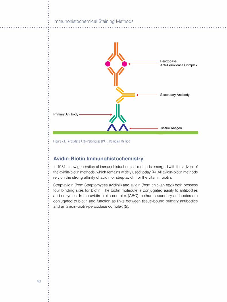
Immunoh�stochem�cal Sta�n�ng Methods
48
Peroxidase Anti-Peroxidase Complex
Secondary Antibody
Tissue Antigen
Primary Antibody
Figure 7.1. Peroxidase Anti-Peroxidase (PAP) Complex Method
avidin-Biotin ImmunohistochemistryIn �98� a new generat�on of �mmunoh�stochem�cal methods emerged w�th the advent of the av�d�n-b�ot�n methods, wh�ch rema�ns w�dely used today (4). All av�d�n-b�ot�n methods rely on the strong affin�ty of av�d�n or streptav�d�n for the v�tam�n b�ot�n.
Streptav�d�n (from Streptomyces av�d�n��) and av�d�n (from ch�cken egg) both possess four b�nd�ng s�tes for b�ot�n. The b�ot�n molecule �s conjugated eas�ly to ant�bod�es and enzymes. In the av�d�n-b�ot�n complex (ABC) method secondary ant�bod�es are conjugated to b�ot�n and funct�on as l�nks between t�ssue-bound pr�mary ant�bod�es and an av�d�n-b�ot�n-perox�dase complex (5).

49
Immunoh�stochem�stry Sta�n�ng Methods
Antigen
Step One Step Two
1° Antibody
2° Antibody
Enzymes
Dextran Backbone
Antigen
1° Antibody
2° Antibody
Enzyme (HRP or AP)
Dextran Backbone
Tissue Antigen
Primary Antibody
Avidin-Biotin Complex
Must be prepared 30minutes prior to use
BiotinylatedSecondary
Antibody
Figure 7.2. Avidin-Biotin Complex (ABC) Method
In a s�m�lar method, the labeled streptav�d�n-b�ot�n (LSAB) method also ut�l�zes a b�ot�nylated secondary ant�body that l�nks pr�mary ant�bod�es to a streptav�d�n-perox�dase conjugate (�). In both methods a s�ngle pr�mary ant�body subsequently �s assoc�ated w�th mult�ple perox�dase molecules, and because of the large enzyme-to-ant�body rat�o, a cons�derable �ncrease �n sens�t�v�ty �s ach�eved compared to d�rect perox�dase-conjugate methods.
Tissue Antigen
Primary Antibody
StreptavidinEnzyme Complex
BiotinylatedSecondary
AntibodyMouse/Rabbit
Figure 7.3. Labeled Streptavidin-Biotin (LSAB) Method
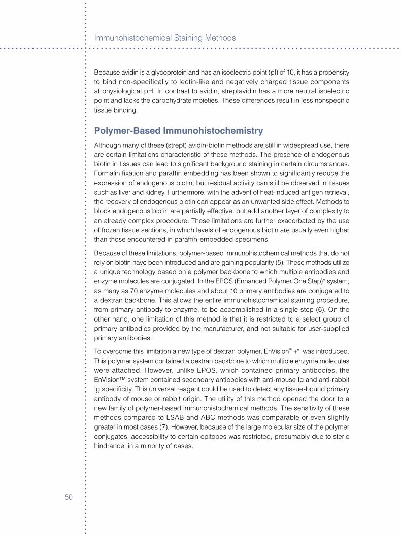
Immunoh�stochem�cal Sta�n�ng Methods
50
Because av�d�n �s a glycoprote�n and has an �soelectr�c po�nt (pI) of �0, �t has a propens�ty to b�nd non-spec�fically to lect�n-l�ke and negat�vely charged t�ssue components at phys�olog�cal pH. In contrast to av�d�n, streptav�d�n has a more neutral �soelectr�c po�nt and lacks the carbohydrate mo�et�es. These d�fferences result �n less nonspec�fic t�ssue b�nd�ng.
Polymer-Based ImmunohistochemistryAlthough many of these (strept) av�d�n-b�ot�n methods are st�ll �n w�despread use, there are certa�n l�m�tat�ons character�st�c of these methods. The presence of endogenous b�ot�n �n t�ssues can lead to s�gn�ficant background sta�n�ng �n certa�n c�rcumstances. Formal�n fixat�on and paraffin embedd�ng has been shown to s�gn�ficantly reduce the express�on of endogenous b�ot�n, but res�dual act�v�ty can st�ll be observed �n t�ssues such as l�ver and k�dney. Furthermore, w�th the advent of heat-�nduced ant�gen retr�eval, the recovery of endogenous b�ot�n can appear as an unwanted s�de effect. Methods to block endogenous b�ot�n are part�ally effect�ve, but add another layer of complex�ty to an already complex procedure. These l�m�tat�ons are further exacerbated by the use of frozen t�ssue sect�ons, �n wh�ch levels of endogenous b�ot�n are usually even h�gher than those encountered �n paraffin-embedded spec�mens.
Because of these l�m�tat�ons, polymer-based �mmunoh�stochem�cal methods that do not rely on b�ot�n have been �ntroduced and are ga�n�ng popular�ty (5). These methods ut�l�ze a un�que technology based on a polymer backbone to wh�ch mult�ple ant�bod�es and enzyme molecules are conjugated. In the EPOS (Enhanced Polymer One Step)* system, as many as �0 enzyme molecules and about �0 pr�mary ant�bod�es are conjugated to a dextran backbone. Th�s allows the ent�re �mmunoh�stochem�cal sta�n�ng procedure, from pr�mary ant�body to enzyme, to be accompl�shed �n a s�ngle step (�). On the other hand, one l�m�tat�on of th�s method �s that �t �s restr�cted to a select group of pr�mary ant�bod�es prov�ded by the manufacturer, and not su�table for user-suppl�ed pr�mary ant�bod�es.
To overcome th�s l�m�tat�on a new type of dextran polymer, EnV�s�on™ +*, was �ntroduced. Th�s polymer system conta�ned a dextran backbone to wh�ch mult�ple enzyme molecules were attached. However, unl�ke EPOS, wh�ch conta�ned pr�mary ant�bod�es, the EnV�s�on™ system conta�ned secondary ant�bod�es w�th ant�-mouse Ig and ant�-rabb�t Ig spec�fic�ty. Th�s un�versal reagent could be used to detect any t�ssue-bound pr�mary ant�body of mouse or rabb�t or�g�n. The ut�l�ty of th�s method opened the door to a new fam�ly of polymer-based �mmunoh�stochem�cal methods. The sens�t�v�ty of these methods compared to LSAB and ABC methods was comparable or even sl�ghtly greater �n most cases (�). However, because of the large molecular s�ze of the polymer conjugates, access�b�l�ty to certa�n ep�topes was restr�cted, presumably due to ster�c h�ndrance, �n a m�nor�ty of cases.
5�
Immunoh�stochem�stry Sta�n�ng Methods
Antigen
Step One Step Two
1° Antibody
2° Antibody
Enzymes
Dextran Backbone
Antigen
1° Antibody
2° Antibody
Enzyme (HRP or AP)
Dextran Backbone
Antigen
Step One Step Two
1° Antibody
2° Antibody
Enzymes
Dextran Backbone
Antigen
1° Antibody
2° Antibody
Enzyme (HRP or AP)
Dextran Backbone
Figure 7.4. Two-Step Polymer Method (EnvisionTM)
Tyramide amplificationThe tyram�de ampl�ficat�on techn�que �s based on the ab�l�ty of phenol�c compounds to become ox�d�zed to h�ghly react�ve and unstable �ntermed�ates (8). When b�ot�nyl tyram�de �s ox�d�zed, d�mer�zat�on w�th electron-r�ch aromat�c compounds, such as those found �n prote�n molecules, occurs (9). Th�s react�on can be harnessed �n �mmunoh�stochem�stry to generate h�ghly react�ve b�ot�nyl-tyram�de �ntermed�ates that b�nd rap�dly to prote�n molecules �n the �mmed�ate v�c�n�ty of perox�dase enzymes. Th�s react�on results �n the depos�t�on of numerous b�ot�n s�gnals. In a typ�cal �mmunoh�stochem�stry procedure, perox�dase enzymes are assoc�ated first w�th pr�mary ant�bod�es by any of the standard �mmunoh�stochem�cal methods, for example by the ABC or LSAB methods. B�ot�nyl tyram�de and hydrogen perox�de are appl�ed as a substrate to generate numerous b�ot�n (b�ot�nyl tyram�de) s�gnals. These b�ot�n molecules then can be used to capture subsequent streptav�d�n-perox�dase enzymes that are converted to a chromogen�c endpo�nt v�a d�am�nobenz�d�ne or s�m�lar chromogen�c substrates (�0).
Immunoh�stochem�cal Sta�n�ng Methods
5�
Cycled Tyramide amplificationThe sequence of streptav�d�n-perox�dase and b�ot�nyl-tyram�nde can be appl�ed alternately to perform a cycled tyram�de ampl�ficat�on procedure. In pract�cal�ty, however, cycl�ng usually cannot exceed two or three cycles before background sta�n�ng l�m�ts the ut�l�ty of th�s approach. Commerc�al tyram�de ampl�ficat�on products are ava�lable and �nclude Tyram�de S�gnal Ampl�ficat�on (TSA, DuPont NEN L�fe Sc�ences, Boston, MA) and Catalyzed S�gnal Ampl�ficat�on (CSA)*.
Fluorescyl-Tyramide amplificationIn keep�ng w�th current trends �n �mmunoh�stochem�stry to develop alternat�ves to b�ot�n-streptav�d�n detect�on methods, a fluorescyl-tyram�de ampl�ficat�on system has been �ntroduced recently (FT-CSA)*. In th�s procedure perox�dase �s assoc�ated w�th a t�ssue-bound pr�mary ant�body by appl�cat�on of a secondary ant�-mouse Ig ant�body to wh�ch perox�dase has been conjugated. The perox�dase catalyzes the convers�on and depos�t�on of fluorescyl-tyram�de onto the t�ssue sect�on. At th�s po�nt the react�on can be term�nated and v�ewed by fluorescence m�croscopy, or the s�gnal can be converted to a color�metr�c react�on by the sequent�al appl�cat�on of an ant�-fluorsece�n ant�body conjugated to perox�dase followed by a d�am�nobenz�d�ne-hydrogen perox�de substrate.
In compar�son to standard IHC methods, tyram�de ampl�ficat�on methods typ�cally have �ncreased sens�t�v�ty by at least 50 fold or greater (��). As w�th any ampl�ficat�on method, background tends to �ncrease along w�th s�gnal. Therefore �t �s essent�al to run appropr�ate pos�t�ve and negat�ve controls and �nterpret any pos�t�ve sta�n�ng w�th�n the context of the negat�ve control.
rolling Circle amplificationRoll�ng C�rcle Ampl�ficat�on (RCA) �s a novel s�gnal ampl�ficat�on system that generates a local s�gnal v�a extens�on and ampl�ficat�on of an ol�gonucleot�de ta�l. Although �n�t�ally developed for nucle�c ac�d detect�on, th�s method also can be appl�ed to �mmunoh�stochem�stry. RCA-med�ated �mmunoh�stochem�stry has been appl�ed successfully to the detect�on of a var�ety of cell surface and �ntracellular molecules (��). The method ut�l�zes a short ol�gonucleot�de sequence coupled to a pr�mary or secondary ant�body molecule. After b�nd�ng to the t�ssue, a c�rcular�zed nucle�c ac�d probe w�th a complementary sequence �s hybr�d�zed to the ol�gonucleot�de. The ol�gonucleot�de then acts as a pr�mer and �s extended l�nearly us�ng a DNA polymerase and the roll�ng c�rcle. Then the extended DNA �s hybr�d�zed w�th labeled ol�gonucleot�de probes. These labels may �nclude, for example, b�ot�n, wh�ch then can be v�sual�zed by any one of the many av�d�n-b�ot�n detect�on methods. RCA der�ves �ts spec�fic�ty from an ant�gen-ant�body react�on and �ts sens�t�v�ty from nucle�c ac�d synthes�s. RCA has been reported to generate a �05-fold �ncrease �n s�gnal (��).
5�
Immunoh�stochem�stry Sta�n�ng Methods
Antigen
Step One Step Two
1° Antibody
2° Antibody
Enzymes
Dextran Backbone
Antigen
1° Antibody
2° Antibody
Enzyme (HRP or AP)
Dextran Backbone
ConclusionAs �mmunoh�stochem�cal techn�ques cont�nue to evolve, the�r appl�cat�on to surg�cal and research pathology �s becom�ng �ncreas�ngly valuable. Var�ous ampl�ficat�on methods have made s�gn�ficant �mprovements to th�s technology such that many ant�gens, prev�ously bel�eved to have been lost to the process of fixat�on and embedd�ng, now can be demonstrated rout�nely. However, as the sens�t�v�ty of �mmunoh�stochem�stry cont�nues to �ncrease, accepted sta�n�ng cr�ter�a and cl�n�cal �nterpretat�on may requ�re re-evaluat�on.
New s�gnal ampl�ficat�on methods cont�nue to be developed, each w�th the�r own un�que strengths and weaknesses, and th�s can present a bew�lder�ng assortment of cho�ces that profoundly �nfluence the pract�ce of �mmunoh�stochem�stry to the �nvest�gator or cl�n�c�an. As technology marches forward, new arrays of t�ssue markers are emerg�ng that are prov�d�ng the tools to generate �mportant new d�scover�es. As new markers are added to th�s l�st, our knowledge of the underly�ng b�ology and pathogenes�s of d�sease �s �ncreased. The full �mpact �s st�ll many years away.
references
�. Nakene PK. S�multaneous local�zat�on of mult�ple t�ssue ant�gens us�ng the perox�dase labeled ant�body method: A study of p�tu�tary glands of the rat. J H�stochem Cytochem �9�8;��:55�-�0.
�. Mason TE, Ph�fer RF, Sp�cer SS. An �mmunoglobul�n-enzyme br�dge method for local�z�ng t�ssue ant�gens. J H�stochem Cytochem �9�9;��:5��-9.
�. Sternberger LA, Hardy PH Jr., Cucul�s JJ, Meyer HG. The unlabeled ant�body-enzyme method of �mmunoh�stochem�stry. Preparat�on and propert�es of soluble ant�gen-ant�body complex (horserad�sh perox�dase-ant�horse-rad�sh perox�dase) and �ts use �n �dent�ficat�on of sp�rochetes. J H�stochem Cytochem �9�0;�8:��5.
4. Hsu SM, Ra�ne L, and Fanger H. Use of av�d�n-b�ot�n perox�dase complex (ABC) �n �mmunoperox�dase techn�ques: A compar�son between ABC and unlabeled ant�body (PAP) procedures. J H�stochem Cytochem �98�;�9:5��-80.
5. Heras A, Roach CM, Key ME. Enhanced polymer detect�on system for �mmunoh�stochem�stry. Lab Invest �995;��:��5 (Abstract).
�. Ch�los� M, Lestan� M, Pedron S, Montagna L, Benedett� A, P�zzolo G, Menestr�na F. A rap�d �mmunosta�n�ng method for frozen sect�ons. B�otech H�stochem �994;�9:��5.
�. Sabatt�n� E, B�sgaard K, Ascan� S, Pogg� S, P�cc�ol� M, Ceccarell� C. The EnV�s�on™ system: a new �mmunoh�stochem�cal method for d�agnost�cs and research. Cr�t�cal compar�son w�th the APAAP, ChemMateTM, CSA, LABC, and SABC techn�ques. J Cl�n Pathol �998;5�:50�-��.
8. Gross AJ, S�zer IW. The ox�dat�on of tyram�ne, tyros�ne, and related compounds by perox�dase. J B�ol Chem �959;��4:����-�4.
9. Bobrow MN, Harr�s TD, Shaughnessy KJ, L�tt GJ. Catalyzed reporter depos�t�on, a novel method of s�gnal ampl�ficat�on. Appl�cat�on to �mmunoassays. J Immunol Methods �989;��5:��9-85.
�0. Adams JC. B�ot�n ampl�ficat�on of b�ot�n and horserad�sh perox�dase s�gnals �n h�stochem�cal sta�ns. J H�stochem Cytochem �99�;40:�45�-��.
Immunoh�stochem�cal Sta�n�ng Methods
54
��. Merz H, Mal�s�us R, Mann-We�ler S, Zhjow R, Hartmann W, Orscheschek K, Moubayed P, Feller AC. Methods �n laboratory �nvest�gat�on �mmunoMax. A max�m�zed �mmunoh�stochem�cal method for the retr�eval and enhancement of h�dden ant�gens. Lab Invest �995;��:�49-5�.
��. Gusev Y, Sparkowsk� J, Raghunathan J, Ferguson H, Montano J, Bogdan N, Schwe�tzer B, W�ltsh�re S, K�ngsmore SF, Maltzman W. Weeler V. Roll�ng c�rcle ampl�ficat�on. A new approach to �ncrease sens�t�v�ty for �mmunoh�stochem�stry and flow cytometry. Am J Pathol �00�;�59:��-�5.
��. W�ltsh�re S, O’Malley S, Lambert J, Kukansh�s K, Edgar D, K�kngsmore S, Schwe�tzer B. Detect�on of mult�ple allergen-spec�fic IgE on m�croarrays by �mmunoassay w�th roll�ng c�rcle ampl�ficat�on. Cl�n Chem �000;4�:�990-�.
Footnote
* A propr�etary methodology developed by Dako.
55
Chapter 8 ° Immunofluorescence
W. Roy Overton, Revised by Jim Hudson and Karen Atwood
IntroductionFluorescent dyes were developed first �n the late �9th century. But �t wasn’t unt�l �950 that ant�bod�es conjugated w�th fluoresce�n �soth�ocyanate were ut�l�zed to produce spec�fic b�olog�cal sta�n�ng to generate fluorescence �n a spec�men. These sta�ned t�ssues and cells could be exam�ned read�ly by fluorescence m�croscopy. Today, attach�ng fluorescent compounds, or “fluorochromes,” to ant�bod�es enables sc�ent�sts and cl�n�c�ans to ga�n tremendous �nformat�on about the b�ology and pathology of cells from humans, an�mals, plants and m�crobes. The follow�ng �s a d�scuss�on of these compounds.
Among the propert�es of many compounds �s the ab�l�ty to absorb one color of l�ght and then em�t a d�fferent color of l�ght. Th�s causes the compound to appear to be glow�ng, or fluoresc�ng, and the compound �s referred to as a fluorochrome. A “fluorophore” �s a component of a molecule that causes a molecule to fluoresce. It �s a funct�onal group �n a molecule that absorbs energy of a spec�fic wavelength and re-em�ts energy at a d�fferent wavelength. Fluoresce�n �soth�ocyanate �s an example of a fluorophore that can be attached chem�cally to a d�fferent, non-fluorescent molecule to create a new and fluorescent molecule.
To better understand how a fluorochrome works, �t �s necessary to exam�ne th�s process at the subatom�c level. Electrons normally sp�n around the nucleus of an atom at a d�stance that �s referred to as the electron’s “ground state” or “ground level.” If the atom �s h�t by photons of l�ght that can exc�te the electron, such as ultrav�olet rad�at�on, then the electron w�ll move up to a h�gher energy state that �s farther from the nucleus. The electron �s unable to ma�nta�n that d�stance from the nucleus due to the electron’s magnet�c attract�on to the protons �n the nucleus, so qu�ckly drops to a sl�ghtly closer d�stance, called the “lowest s�nglet exc�ted state.” Mov�ng to the lowest s�nglet exc�ted state causes the electron to release a l�ttle of the energy that �t got from the l�ght, but �t releases energy as heat. The electron then returns to �ts or�g�nal ground state, releas�ng the rest of the energy that �t absorbed from the l�ght. Th�s energy �s released as l�ght, but s�nce some energy was released as heat �nto the latt�ce of the molecule, there �s less energy �n that l�ght. Thus, the em�tted l�ght appears to be a d�fferent color than the l�ght that exc�ted the atom.
L�ght travels �n waves that determ�ne the color of the l�ght. If the l�ght has a lot of energy, �ts waves are shorter than a wave of l�ght w�th less energy. The wavelength of the l�ght determ�nes the color of the l�ght. Very h�gh-energy l�ght such as ultrav�olet (UV) l�ght has very short wavelengths. The wavelength of UV l�ght �s less than 400 nm. Because the wavelength �s so short, the human eye �s unable to see UV l�ght, but electron�c
Immunoh�stochem�cal Sta�n�ng Methods
5�
photodetectors are able to detect and measure th�s �nv�s�ble l�ght. V�s�ble l�ght has wavelengths from 400 nm to �00 nm, that produce the colors v�olet, blue, green, yellow, orange and red. Above �00 nm �s the �nfrared range, wh�ch �s also �nv�s�ble to the human eye, but can be felt as heat or detected by electron�c photodetectors.
In �mmunofluorescent sta�n�ng, mercury-vapor, xenon or halogen lamps �n the fluorescent m�croscope usually are used to exc�te the fluorochromes. The chem�cal propert�es of the fluorochrome determ�ne whether �ts electrons can be exc�ted to the h�gher energy state by a spec�fic wavelength. If the electrons can be exc�ted to the h�gher energy state, the chem�cal propert�es of the fluorochrome also w�ll determ�ne the amount of energy lost as heat when the electrons drop back down to the lowest s�nglet exc�ted state and the wavelength of l�ght produced when the electrons return to the�r ground state. The d�fference �n the wavelength of the l�ght that exc�tes the electrons and the l�ght that �s em�tted �s called the “Stokes sh�ft,” and �s determ�ned by the amount of energy lost as heat. The em�tted wavelength �s always longer (�f s�ngle photons are absorbed) or equal to the �nc�dent wavelength, due to energy conservat�on.
Some fluorochromes have a small Stokes sh�ft, and the exc�tat�on and em�ss�on wavelengths have almost the same wavelengths. But other fluorescent compounds have large Stokes sh�fts. For example, the fluorochrome, fluoresce�n, can be exc�ted by blue-green l�ght, and �ts Stokes sh�ft �s only about �5 nm, wh�ch means that the l�ght em�tted �s green. Th�s contrasts w�th another fluorochrome, phycoerythr�n, wh�ch also can be exc�ted by blue-green l�ght, but has a large Stokes sh�ft. Thus, the l�ght em�tted �s yellow-orange. In �mmunofluorescence, a s�ngle wavelength can be used to exc�te several fluorochromes w�th d�fferent Stokes sh�fts and, thereby, produce a var�ety of fluorescent colors.
The electrons of a fluorochrome can be exc�ted by a range of wavelengths of l�ght. For example, the fluorochrome, fluoresce�n, w�ll fluoresce when h�t by l�ght w�th a wavelength between 4�0 nm and 5�0 nm. However, the closer the exc�tat�on wavelength �s to 495 nm, the more fluorescence w�ll be produced. Th�s opt�mal wavelength �s called the exc�tat�on peak. S�m�larly, the l�ght produced by fluorochromes has a range of wavelengths. The em�ss�on of l�ght from fluoresce�n ranges from 490 nm to ��0 nm, and the em�ss�on peak �s approx�mately 5�0 nm.
Know�ng the exc�tat�on and em�ss�on propert�es of fluorescent compounds makes �t poss�ble to select comb�nat�ons of fluorochromes that w�ll work together. However, for a fluorochrome to be useful �n a b�olog�cal appl�cat�on, �t must attach to or be conta�ned w�th�n a part�cle of b�olog�cal s�gn�ficance. Many fluorochromes can be attached to ant�bod�es, wh�ch then w�ll b�nd to spec�fic chem�cal structures on or �ns�de of cells.
There are many other chem�cal and phys�cal propert�es of fluorochromes that determ�ne where and when these dyes are useful �n var�ous b�olog�cal assays. For example, some of the fluorochromes that b�nd to DNA, such as Hoechst ��4�, can get �nto l�v�ng cells, but most DNA-b�nd�ng fluorochromes cannot get past the cell membrane. The fluorescent dyes that cannot get past a v�able cell membrane, such as prop�d�um �od�de, often are used to d�st�ngu�sh l�ve from dead or dy�ng cells.
5�
Immunofluoresc�ence
Fading, Quenching and PhotobleachingFluorescence detect�on can be affected adversely by “fad�ng.” Th�s �s a reduct�on of fluorescent em�ss�on �ntens�ty caused by photobleach�ng or quench�ng.
Photobleach�ng �s an �rrevers�ble degradat�on of the act�vated or exc�ted fluorochrome as a result of �ts �nteract�on w�th molecular oxygen (�). Illum�nat�on of the fluorochromes by l�ght of the appropr�ate wavelength can result �n an exc�ted state that renders �t more chem�cally react�ve. It then may react �rrevers�bly w�th any ava�lable oxygen molecules. Th�s �nteract�on may result �n decompos�t�on, polymer�zat�on, or ox�dat�on and subsequent react�on w�th another molecule.
Photobleach�ng can be m�n�m�zed by us�ng the lowest �llum�nat�on poss�ble. To m�n�m�ze the effects of photobleach�ng, fluorescence m�croscopy can be comb�ned w�th other techn�ques that are non-destruct�ve to fluorochromes, such as d�fferent�al �nterference contrast (DIC), Hoffman modulat�on contrast (HMC), and phase contrast. The operator locates the area of �nterest on the sl�de us�ng a non-destruct�ve low level l�ght before sw�tch�ng on the fluorescence exc�tat�on l�ght.
Quench�ng of an exc�ted fluorochrome may occur due to a non-rad�at�ve or rad�at�ve energy loss and �s med�ated through compounds �n the m�cro-env�ronment. Non-rad�at�ve energy loss and reduct�on �n fluorescence �ntens�ty can be caused by the presence of ox�d�z�ng agents, salts, heavy metals or halogen compounds. In some cases, quench�ng results from rad�at�ve energy loss or the transfer of energy from the fluorochrome (donor molecule) to another molecule (acceptor) that res�des phys�cally close to the exc�ted fluorochrome. Th�s �s called fluorescence resonance energy transfer (FRET).
There are several commerc�ally ava�lable ant�-fad�ng mount�ng med�a ava�lable that s�gn�ficantly reduce th�s phenomenon (�).
FluoresceinPur�fied ant�bod�es are conjugated w�th fluoresce�n �soth�ocyanate �somer (FITC). After conjugat�on, unreacted FITC �s removed completely by gel filtrat�on on Sephadex G-�5. Further pur�ficat�on �s carr�ed out by �on exchange chromatography. Th�s process removes unconjugated ant�body molecules and ant�body molecules to wh�ch more than four molecules of FITC are attached. These conjugates cons�st of opt�mally labeled ant�body molecules for �mmunofluorescent use. Then fluoresce�n/prote�n rat�o measured as the absorbance rat�o measured as the absorbance rat�on A495nm/A��8nm �s 0.�5, correspond�ng to a molar FITC/prote�n rat�o of �.�-�.5. FITC conjugates w�th a h�gher F/P value, such as those prepared for flow cytometr�c analys�s, are not su�table for �mmunofluorescence, as they w�ll produce h�gh background, false pos�t�ve sta�n�ng and poss�ble self-quench�ng of the FITC molecule.
Immunoh�stochem�cal Sta�n�ng Methods
58
The FITC-labeled �mmunofluorescent sta�n does not requ�re strong ultra-v�olet rad�at�on for exc�tat�on. W�th a peak absorpt�on of 495, l�ght generated by a quartz-halogen or xenon lamp �s suffic�ent.
Procedure for Formalin-Fixed Paraffin-Embedded Tissue° Cut four m�cron sect�ons onto charged or s�lan�zed sl�des.
° Allow sect�ons to dry at �� °C for four hours.
° De-wax and hydrate t�ssue sect�ons.
° R�nse �n de�on�zed (DI) water.
° Wash �n PBS for �0 m�nutes at 4 °C.
° Enzyme d�gest t�ssue sect�ons �f proteolyt�c treatment �s requ�red.
° Stop enzyme d�gest�on by �ncubat�on �n TBS for �0 m�nutes at 4 °C.
° Wash �n PBS for �0 m�nutes at 4 °C.
° An opt�onal serum block may be used at th�s po�nt to block Fc receptors. Tap off excess.
° Incubate w�th FITC conjugated ant�bod�es, d�luted �n PBS and one percent bov�ne serum album�n, �n a mo�st chamber for �0 m�nutes at room temperature.
° R�nse sl�des �n PBS to remove excess ant�serum.
° Wash �n PBS for �0 m�nutes at 4 °C (�x).
° Mount w�th fluorescent mount�ng med�um .
° Seal coversl�p edges w�th clear na�l pol�sh and store at �-8 °C �n the dark.
Procedure for Frozen Sections° Cut four m�cron cryostat sect�ons.
° F�xat�on:
ß F�x �n acetone for �0 m�nutes at 4 °C, a�r dry for �0 m�nutes, post fix �n acetone for 90 seconds at 4 °C;
ß A�r dry for �-�� hours, fix �n acetone for �0 m�nutes at 4 °C.
° Wash �n PBS for one to five m�nutes at 4 °C.
° An opt�onal serum block may be used at th�s po�nt to block Fc receptors
° Tap off excess.
° Incubate w�th FITC conjugated ant�bod�es, d�luted �n PBS and one percent bov�ne serum album�n, �n a mo�st chamber for �0 m�nutes at room temperature.
° R�nse sl�des �n PBS to remove excess ant�serum.
° Wash �n PBS for �0 m�nutes at 4 °C (�x).
° Mount w�th fluorescent mount�ng med�um.
° Seal coversl�p edges w�th clear na�l pol�sh and store at �-8 °C �n the dark.
59
Immunofluoresc�ence
Buffers:
° Phosphate buffered sal�ne: pH �.�:
ß ��.99 g NaCl;
ß 9.�58 g Na�HP4�H�O;
ß �.�5 g KH�OPO4.
ß Br�ng to five l�ters w�th DI water.
° Phosphate buffered sal�ne: pH �.0;
ß Dako Code S�0�4.
° Tr�s-Buffered Sal�ne:
ß Dako Code S�9�8 or S�00�.
° Pronase:
ß Dako Code S�0��.
Table 8.1. Antibody dilution chart for direct immunofluorescence on frozen sections.
Code Dako FITC Conjugated Antibody Dilution in PBS
F0117 Albumin 1:20-1:40
F0254 C1q Complement 1:10-1:20
F0201 C3c Complement 1:75-1:100
F0111 Fibrinogen 1:50-1:70
F0204 IgA 1:20-1:40
F0202 IgG 1:20-1:40
F0203 IgM 1:20-1:40
F0198 Kappa Light Chains 1:20-1:40
F0199 Lambda Light Chains 1:20-1:40
X0929 Negative Reagent Control See NOTE
NOTE: The negative reagent control is used on an additional tissue section. To determine the dilution factor, calculate the total protein (TP) of each antibody to be used in the test run (TP of primary antibody/optimized dilution factor). Select the highest calculated TP and divide it into the TP of the negative reagent control (TP negative reagent control/highest calculated TP). This is the dilution factor of the negative reagent control for the run.
Immunoh�stochem�cal Sta�n�ng Methods
�0
Pos�t�ve and negat�ve controls are requ�red now by the Cl�n�cal Laboratory Improvement Amendments of �988 (CLIA) for each sta�n�ng run. Pos�t�ve and negat�ve t�ssues are sta�ned w�th the ant�body.
An unsta�ned control sl�de may be used to determ�ne �f autofluorescence �s present. Pr�mary or autofluorescence �s seen when a spec�men has not been labeled w�th a fluoresc�ng dye, but st�ll man�fests fluorescence dur�ng exposure to short wavelength energy. T�ssue autofluoresc�ng substances �nclude: Elast�c fibers, l�pofusc�ns, collagen, porphyr�ns, v�tam�n A, cart�lage, kerat�n, card�ac muscle, fat, waxes, hormones, calc�um and powdered crude drugs. Autofluorescence also may be caused by free aldehydes from fixat�on.
A number of “home-brew” procedures have been shown to quench autofluorescence. Those �nclude pronase d�gest�on; �00 mM glyc�ne �n PBS (pH �.4) for �0-�0 m�nutes; 50 mM ammon�um chlor�de �n PBS for �0 m�nutes; and one percent sod�um borohydr�de �n PBS for �0-�0 m�nutes.
Other factors that can contr�bute to unexpected or undes�red sta�n�ng �nclude heat-�nduced ep�tope retr�eval (HIER) l�pofus�on art�facts; reagent contam�nat�on; sub-opt�mal temperature and durat�on at wh�ch the reagents are �ncubated; dead or necrot�c cells and Tween �0 added to the wash buffer.
In add�t�on, due to the s�ze of some fluorochromes such as RPE, some conjugated ant�bod�es may become trapped �n an �ntact cell dur�ng analys�s for �ntracellular ant�gens.
references
�. Song L, Henn�nk EJ, Young IT, Tanke HJ. Photobleach�ng k�net�cs of fluoresce�n �n quant�tat�ve fluorescence m�croscopy. B�ophys J �995;�8:�588-�00, PN�98��.
�. Ono M, Murakam� T, Kudo A, Issh�k� M, Sawada H, and Segawa A. Quant�tat�ve Compar�son of Ant�-Fad�ng Mount�ng Med�a for Confocal Laser Scann�ng M�croscopy. J H�stochem Cytochem �00�;49:�05-��.

��
Chapter 9 ° Multi-Staining Immunohistochemistry
Nanna K. Christensen and Lars Winther
IntroductionImmunoh�stochem�stry (IHC) has become establ�shed as an �mportant tool for both research and d�agnost�c purposes. However, �n some cases there �s a need for knowledge about the relat�ve local�zat�ons of targets, wh�ch can be obta�ned only by v�sual�z�ng all relevant targets on one sl�de. Th�s chapter descr�bes the advantages of mult�ple sta�n�ng, as well as the cons�derat�ons that have to be made to ensure successful sta�n�ng. Th�s art�cle w�ll d�scuss the cho�ce of appropr�ate protocols as well as the cho�ce of v�sual�zat�on systems.
advantages of Multiple StainingMult�ple sta�n�ng can be defined as the detect�on of two or more targets on one sl�de, thus �ncreas�ng the �nformat�on obta�ned from each sl�de. Hands-on t�me per sl�de depends on the method used. Sequent�al sta�n�ng does not reduce hands-on t�me compared to comb�n�ng s�ngle sta�n�ng; whereas s�multaneous sta�n�ng does reduce turn-around t�me (see below). W�th �ncreas�ng demand for reduced turn-around-t�me, mult�ple sta�n�ng may offer at least part of the solut�on. Furthermore, there �s a demand for less �nvas�ve sampl�ng techn�ques g�v�ng smaller and fewer spec�mens and ava�lable sl�des. In such cases mult�ple sta�n�ng also may be a great advantage.
Equally �mportant, mult�ple sta�n�ng makes �t poss�ble to assess the topograph�c relat�onsh�p of the targets, for example, to determ�ne whether targets are present �n d�fferent cells, �n the same cell or even �n the same cellular compartment. Informat�on also can be obta�ned on poss�ble cell-to-cell spat�al contacts of d�fferent cell types. Some of th�s �nformat�on also can be obta�ned us�ng s�ngle sta�n�ng on ser�al sect�ons. However, th�s �s labor�ous and t�me consum�ng and the sect�ons must be very th�n to ensure all structures or cells are present �n the ent�re ser�es of sect�ons. Mult�ple sta�n�ng allows the comb�nat�on of �n s�tu hybr�d�zat�on (ISH) and IHC, g�v�ng �nformat�on about a part�cular target both at prote�n level and DNA/mRNA level.
The d�agnos�s of prostat�c ep�thel�al neoplas�a (PIN) �s just one example of the cl�n�cal �mportance of mult�ple sta�n�ng. Prostat�c needle b�opsy �s the preferred method for d�agnos�ng early prostate cancer. But �n some cases an amb�guous d�agnos�s �s made due to the fact that the b�opsy has �dent�fied only a few mal�gnant glands or several h�stolog�cal ben�gn m�m�cs of cancer (�). S�nce basal cells are present �n the ben�gn cancer m�m�cs but absent �n the mal�gnant glands, these cells can be used to d�st�ngu�sh between the two cases. Basal cells are labeled us�ng h�gh-molecular-we�ght cytokerat�n, cytokerat�n 5/� or p�� �mmunosta�n�ng. In add�t�on, the gene product of p504s, alpha-methylacyl-CoA-racemase �s expressed �n a h�gh percentage of prostate carc�nomas, but �s negat�ve or only weakly expressed �n ben�gn prostate t�ssue. Thus �t �s used as

Immunoh�stochem�cal Sta�n�ng Methods
��
a pos�t�ve cancer marker. In cases of small foc�, amb�guous les�ons may d�sappear when us�ng ser�al sect�ons, caus�ng suspected mal�gnanc�es to rema�n und�agnosed. A mult�ple sta�n�ng protocol reduces the percentage of res�dual amb�guous les�ons and the need for add�t�onal b�ops�es.
Mult�ple sta�n�ng �s well known from flow cytometry, where sta�n�ng of three to four d�fferent targets on the same cell �s rout�ne. A s�ngle sample el�c�ts cons�derable �nformat�on, allow�ng unamb�guous separat�on of d�fferent cell-types and �dent�ficat�on of abnormal�t�es. ISH rout�nely uses mult�ple sta�n�ng on sl�des to determ�ne gene ampl�ficat�on from the rat�o of the s�gnals from the gene probe of �nterest to a reference probe.
ISH also can be used �n mult�ple sta�n�ng to detect chromosome translocat�ons us�ng spl�t-s�gnal FISH. Probes d�rected towards stretches �mmed�ately upstream and downstream of the breakpo�nt are labeled green and red, respect�vely. Thus when probes are co-local�zed, the m�xture of green and red results �n a yellow s�gnal, but when the chromosome breaks, the s�gnals separate and �nd�v�dual green and red s�gnals can be seen. In th�s case, s�ngle-target sta�n�ng would not g�ve the des�red �nformat�on. Mult�ple sta�n�ng �s well establ�shed �n ISH, and there are obv�ous advantages to extend�ng th�s to an IHC format to ga�n the benefit of the add�t�onal �nformat�on.
Technical ChallengesUsers exper�enced w�th the challenges of s�ngle-target sta�n�ng w�ll find the demands for mult�ple sta�n�ng are s�m�lar but more complex.
Before embark�ng on a mult�-sta�n�ng project, some �mportant �ssues should be cons�dered:
° To avo�d target or spec�es cross-react�v�ty, complex protocols may be necessary.
° Spectral d�fferent�at�on of sta�n colors may be d�fficult, espec�ally �f the targets are co-local�zed. Rare targets that are co-local�zed w�th more abundant targets may not show, and �f colors are m�xed, results may be d�fficult to separate from s�ngle colors.
° Even �f targets are not co-local�zed �t �s d�fficult to balance s�gnals enabl�ng rare targets to be v�s�ble �n the same sl�de as h�ghly abundant targets. Th�s �s due to the narrow dynam�c range of IHC. An adjustment �n concentrat�on of the pr�mary ant�bod�es may solve th�s problem.
° If d�fferent targets are v�ewed under d�fferent magn�ficat�ons, �t may not be poss�ble to get the topograph�c �nformat�on des�red.
PretreatmentMult�ple sta�n�ng, l�ke s�ngle sta�n�ng, can be performed on both formal�n-fixed, paraffin-embedded t�ssue, cryosect�ons, cell smears and cytosp�n preparat�ons. Mult�ple sta�n�ng �s constra�ned by the fact that �t may not be poss�ble to find one t�ssue pre-treatment
��
Mult�-Sta�n�ng Immunoh�stochem�stry
protocol that �s opt�mal for all targets. Often protocols opt�m�zed for �nd�v�dual sta�n�ng d�ffer from one target to the other; for example, d�fferent target retr�eval methods are used. If that �s the case, �t �s necessary to determ�ne a method that allows all targets to be sta�ned, although the method may be sub-opt�mal for some targets.
In cases where targets of d�fferent abundances are to be sta�ned, a method must be selected that g�ves the best balanc�ng of the s�gnals. Comb�n�ng ISH and IHC on one sl�de �s challeng�ng, part�cularly because targets requ�re very d�fferent pre-treatment protocols. S�nce ISH processes such as DNA denatur�ng are not compat�ble w�th the presence of the ant�bod�es for IHC, the ISH protocol normally �s performed first.
Staining Method SelectionTo ensure success, IHC sta�n�ng must be planned carefully. Th�s �s even more �mportant w�th mult� sta�n�ng. If pr�mary ant�bod�es are commerc�ally ava�lable both d�rectly labeled and unlabeled and from d�fferent host-spec�es, there are several d�fferent sta�n�ng methods to choose from; however, very often the cho�ce �s l�m�ted by the ava�lable reagents (�). Care must be taken to avo�d cross-react�v�ty between reagents. A flow chart or s�m�lar a�d m�ght prove useful �n select�ng the best method.
In general, sta�n�ng methods can be d�v�ded �n the follow�ng classes:
Sequential Staining
An �nd�rect techn�que us�ng unlabeled pr�mary ant�bod�es where the sta�n�ng of one target �s completed �nclud�ng the appl�cat�on of the chromogen�c dye, before the appl�cat�on of the next.
The pr�mary and secondary ant�bod�es from the first sta�n�ng are eluted before sta�n�ng the next target (for an example, see F�gure 9.�). Th�s avo�ds cross-react�v�ty-related problems; however, elut�on may be d�fficult w�th some h�gh-affin�ty pr�mary ant�bod�es, lead�ng to spur�ous double sta�ned structures. Th�s techn�que therefore �s not recommended for evaluat�on of m�xed colors at s�tes of co-local�zat�on. Elut�on also r�sks denatur�ng ep�topes of ant�gens that are to be subsequently v�sual�zed. Furthermore, for some chromogens there �s a r�sk that the first chromogen (DAB �n part�cular) sh�elds other targets. Not all react�on products are capable of surv�v�ng the r�gorous wash�ng requ�red to remove the ant�bod�es. Thus �n order to avo�d blurry sta�n�ng results the most robust dyes should be appl�ed first.
Simultaneous Staining
A d�rect method w�th d�rectly labeled pr�mary ant�bod�es, or an �nd�rect method based on unlabeled pr�mary ant�bod�es ra�sed �n d�fferent host spec�es, or of d�fferent Ig �sotype or IgG subclass (�).
A s�mple example of th�s method �s when the pr�mary ant�bod�es are fluorescently or enzyme labeled to allow d�rect v�sual�zat�on. Th�s avo�ds cross-react�v�ty but �s rarely
Immunoh�stochem�cal Sta�n�ng Methods
�4
pract�cal s�nce some form of ampl�ficat�on �s necessary to get suffic�ent s�gnal. In that case, pr�mary ant�bod�es are conjugated d�rectly w�th enzymes, b�ot�n, haptens or fluorochromes, subsequently employ�ng the correspond�ng ant�body or streptav�d�n reagent as the second layer. Th�s �s less t�me-consum�ng than the sequent�al method, s�nce pr�mary and secondary ant�bod�es can be m�xed together �n two �ncubat�on steps. However, �t requ�res avo�d�ng all cross-react�v�ty.
W�th the �nd�rect method �t �s also poss�ble to apply t�mesav�ng ant�body cockta�ls. Generally �t �s advantageous to use secondary ant�bod�es ra�sed �n the same host �n order to prevent any unexpected �nter-spec�es cross-react�v�ty.
Multi-Step Technique
An �nd�rect/d�rect method comb�n�ng unlabeled pr�mary ant�bod�es w�th ant�bod�es that are d�rectly conjugated. The method starts w�th the sta�n�ng of the unlabeled ant�body/ant�bod�es.
Sequent�al sta�n�ng avo�ds the problem of cross-react�v�ty but cannot be used for co-local�zed targets. The techn�que often leads to a long sta�n�ng protocol and carr�es an �nherent r�sk of �ncorrect double sta�n�ng due to �nsuffic�ent elut�on of one set of reagents before appl�cat�on of the next. S�multaneous sta�n�ng �s less t�me-consum�ng s�nce the reagents of each layer can be m�xed together. However, the techn�que can be used only �f su�table pr�mary ant�bod�es are ava�lable. Mult�-step sta�n�ng can be used when the select�on of pr�mary ant�bod�es �s l�m�ted. However, when us�ng th�s method, �t �s not poss�ble to m�x reagents.
Users often w�ll find that the cho�ce of sta�n�ng method �s l�m�ted by the ava�lab�l�ty of the pr�mary ant�bod�es w�th respect to spec�es or�g�n or label.
When targets are known or suspected to be co-local�zed and the only ava�lable pr�mary ant�bod�es are unlabeled monoclonal mouse ant�bod�es of the same IgG subclass, none of the techn�ques descr�bed above are appl�cable.
One solut�on �s the Dako An�mal Research K�t (ARKTM) Perox�dase, wh�ch conta�ns reagents for label�ng mouse pr�mary ant�bod�es w�th a b�ot�nylated ant�-mouse Fab fragment, followed by block�ng of the rema�n�ng reagent w�th normal mouse serum. Th�s can be appl�ed to the t�ssue as part of the mult�-step techn�que (4). The k�t g�ves a non-covalently labeled ant�body, thus avo�d�ng the r�sk of reduc�ng the affin�ty. In add�t�on, only small amounts of pr�mary ant�body are needed and the k�t does not requ�re t�me-consum�ng pur�ficat�on steps.
Another solut�on �s Zenon Technology (Inv�trogen) developed for flow cytometry. It uses essent�ally the same techn�que, and offers label�ng k�ts for mouse pr�mary ant�bod�es ava�lable as enzyme conjugates or conjugated to one of a w�de var�ety of fluorescent dyes.
V�sual�zat�on systems w�th dual recogn�t�on such as the EnV�s�onTM+ Dual L�nk system do not d�scr�m�nate between spec�es, and are thus only su�table for mult�ple sta�n�ng when
�5
Mult�-Sta�n�ng Immunoh�stochem�stry
us�ng the sequent�al method. V�sual�zat�on k�ts w�th ampl�ficat�on layers that are not well spec�fied should be avo�ded, s�nce poss�ble cross-react�v�ty cannot be pred�cted.
Selection of DyesThe pr�mary cho�ce to make when dec�d�ng how to make the targets v�s�ble �s whether to use �mmunoenzyme sta�n�ng or fluorescence. Both have advantages and d�sadvantages and �n the end, dec�s�ons should be made based on cond�t�ons of the �nd�v�dual exper�ment.
Chromogenic Dyes
When select�ng color comb�nat�ons for mult�ple sta�n�ng w�th chromogen�c dyes, �t �s adv�sable to choose oppos�ng colors �n the color spectrum, to fac�l�tate spectral d�fferent�at�on. If us�ng a countersta�n, th�s also must be �ncluded �n the cons�derat�ons. When work�ng w�th co-local�zed targets, dyes must be chosen so that �t �s poss�ble to d�st�ngu�sh the m�xed color from the �nd�v�dual colors. Double sta�n�ng us�ng chromogen�c dyes �s well establ�shed, but �t �s demand�ng �f the targets are co-local�zed. For tr�ple sta�n�ng, �t �s more d�fficult to get colors that can be unamb�guously d�fferent�ated and �t �s very d�fficult �f targets are co-local�zed.
A narrow dynam�c range �s a hand�cap for �mmunoenzymat�c sta�n�ng. The prec�p�tat�on process, wh�ch �s cruc�al for th�s method, �s only tr�ggered at a certa�n threshold concentrat�on of substrate/product. On the other hand, at h�gh concentrat�ons the prec�p�tated product may �nh�b�t further react�on. Therefore �t �s d�fficult to v�sual�ze rare targets and h�ghly abundant targets �n the same sl�de. To reduce th�s problem us�ng the strongest dye to sta�n the most rarely expressed target �s advantageous. Or �f poss�ble use extra ampl�ficat�on l�ke the Catalyzed S�gnal Ampl�ficat�on (CSA) System to br�ng rare targets w�th�n the same dynam�c range as h�ghly expressed targets.
There �s a l�m�ted range of chromogen�c dyes to choose from. These are examples of enzyme/chromogen pa�rs su�table for tr�ple sta�n�ng:
° GAL/XGAL/Turquo�se, AP/Fast blue, HRP/AEC/Red;
° HRP/DAP/Brown, GAL/XGAL/Turquo�se, AP/Fast red;
° HRP/DAP/Brown, AP/New Fucs�n/Red, HRP/TMB/Green.
In conclus�on, chromogen�c dyes can be used successfully for double sta�n�ng; however, �dent�fy�ng co-local�zed targets may be a problem. Tr�ple sta�n�ng �s also poss�ble, but great care must be used �n select�ng dyes.
Immunoh�stochem�cal Sta�n�ng Methods
��
Fluorescent Dyes
Double �mmunofluorescence label�ng �s also qu�te well establ�shed (5). Some of the same cons�derat�ons as w�th chromogen�c dyes apply when work�ng w�th �mmunofluorescence. It �s equally necessary to select dyes w�th d�st�ngu�shable spectral propert�es. However, there are more colors ava�lable and the em�ss�ons spectra of the fluorescent molecules are narrower than the spectra of the chromogen�c dyes. DAB �n part�cular has a very broad spectrum. The use of mult�ple fluorescent colors �s also already well establ�shed �n FISH and flow cytometry, where d�chro�c filters and bandpass filters are employed to separate d�fferent fluorescent s�gnals. The spectral separat�on can be a�ded by d�g�tal compensat�on for overlapp�ng em�ss�on spectra.
When sta�n�ng targets that are co-local�zed fluorescent dyes, allow separate �dent�ficat�on of targets. Th�s makes �t poss�ble to d�scern targets even �n w�dely d�fferent concentrat�ons, whereas subtly m�xed colors may pass unnot�ced eas�ly w�th �mmunoenzyme sta�n�ng.
Immunofluorescence potent�ally has a w�der dynam�c range than �mmunoenzyme sta�n�ng. Us�ng th�s method, there �s no enzymat�c ampl�ficat�on �nvolved and thus the dynam�c range �s determ�ned solely by the sens�t�v�ty of the detectors.
On the other hand, there are some �nherent problems w�th the use of �mmunofluorescence:
° Fluorescence s�gnal �s quenched when the fluorochromes are �n close prox�m�ty.
° Dyes undergo photo bleach�ng when subjected to l�ght and w�ll thus only fluoresce for a l�m�ted t�me unless stored �n the dark.
° Even when stored protected from l�ght, some fluorochromes w�ll deter�orate slowly at room temperature.
° The morphology v�ewed �n sl�des �s d�fferent from what �s observed �n �mmunoenzyme sta�n�ng w�th countersta�ns.
° Increased background due to autofluorescence can pose a problem when work�ng w�th some formal�n-fixed t�ssues.
° Leach�ng of stored ant�body conjugates may pose a problem.
In sp�te of these drawbacks, �mmunofluorescence g�ves clear, sharp local�zat�on of targets and has advantages over chromogen�c dyes when work�ng w�th co- local�zed targets.
Some chromogen�c dyes fluoresce as well, such as Fast Red, an AP-substrate that �s br�ghter �n fluorescence m�croscopy than �n br�ght field m�croscopy.
��
Mult�-Sta�n�ng Immunoh�stochem�stry
Other LabelsCollo�dal gold-labeled ant�bod�es were developed or�g�nally for electron m�croscopy, but w�th s�lver-enhancement they are v�s�ble w�th normal l�ght m�croscopy.
Another example of �norgan�c sta�ns �s Quantum dots (Q-dots). These are fluorescent nanopart�cles w�th vary�ng em�ss�on wavelengths, depend�ng on the s�ze of the part�cle. They can be l�nked to ant�bod�es or streptav�d�n as an alternat�ve to fluorochromes (�). However, the s�ze of the�r conjugates may pose d�ffus�on problems.
automated Image acquisition and analysisD�g�tal �mage analys�s w�ll �ncrease the number of usable dyes s�nce �t does not rely on the human eye for detect�on and d�fferent�at�on. A d�g�tal �mage �s acqu�red at exc�tat�on wavelengths relevant for the dyes appl�ed, and separate detectors record �nd�v�dual colors. So, for example, d�g�tal �mage analys�s w�ll allow the comb�nat�on fluorescent and �mmunoenzyme dyes.
Detectors, however, have b�ased color v�s�on. They ampl�fy colors d�fferently than does the human eye. Therefore dyes used on �mage analys�s should be opt�m�zed for the best poss�ble fit w�th the detector’s filter propert�es.
Image analys�s systems conta�n algor�thms that allow compensat�on for overlapp�ng em�ss�on spectra comparable to flow cytometry. They also allow s�gnal gat�ng w�th�n an �nterest�ng range of wavelengths, enabl�ng users to see only s�gnals w�th�n the des�red range. V�sual�z�ng a comb�nat�on of several gates w�th color selected �ndependently of the dyes used for sta�n�ng may clar�fy p�ctures and make conclus�ons eas�er to reach. Th�s also makes �t poss�ble to set a threshold on s�gnal �ntens�ty to exclude unspec�fic sta�n�ng or background from final �mages.
Another advantage of d�g�tal �mage analys�s �s that �t allows s�gnal quant�tat�on. Through software man�pulat�on users, can count how many s�gnal clusters exceed a certa�n level of �ntens�ty, and, potent�ally, calculate the rat�o of d�fferent cell-types. For example, an �mage analys�s algor�thm can calculate the percentage of cells that sta�n pos�t�ve for a certa�n target, comb�ne that percentage w�th �nformat�on of other sta�ned targets and, based on th�s, h�ghl�ght d�agnos�s.
ConclusionMult�ple-target sta�n�ng w�ll one day be as rout�ne as s�ngle-target sta�n�ng �s today.
Use of the techn�que w�ll expand, s�nce �t offers reduced turn-around t�me and �nformat�on not obta�nable from s�ngle-target sta�n�ng. Ava�lab�l�ty of labeled pr�mary ant�bod�es, ant�bod�es ra�sed �n d�fferent host spec�es and mult�ple sta�n�ng k�ts also �s l�kely to �ncrease.
Software for automated �mage acqu�s�t�on and analys�s w�ll play a key role �n th�s evolut�on s�nce the l�m�t to how many colors the human eye can d�st�ngu�sh w�ll be
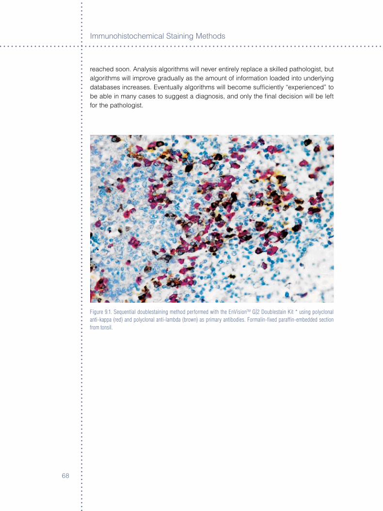
Immunoh�stochem�cal Sta�n�ng Methods
�8
reached soon. Analys�s algor�thms w�ll never ent�rely replace a sk�lled patholog�st, but algor�thms w�ll �mprove gradually as the amount of �nformat�on loaded �nto underly�ng databases �ncreases. Eventually algor�thms w�ll become suffic�ently “exper�enced” to be able �n many cases to suggest a d�agnos�s, and only the final dec�s�on w�ll be left for the patholog�st.
Figure 9.1. Sequential doublestaining method performed with the EnVisionTM G|2 Doublestain Kit * using polyclonal anti-kappa (red) and polyclonal anti-lambda (brown) as primary antibodies. Formalin-fixed paraffin-embedded section from tonsil.
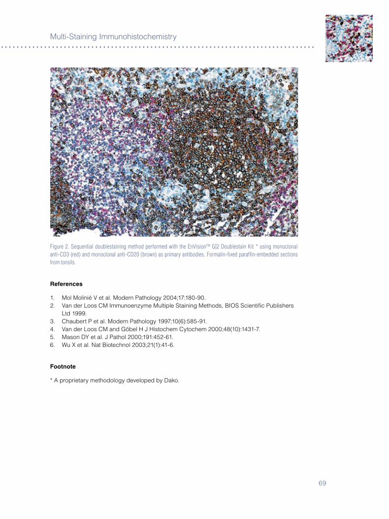
�9
Mult�-Sta�n�ng Immunoh�stochem�stry
Figure 2. Sequential doublestaining method performed with the EnVisionTM G|2 Doublestain Kit * using monoclonal anti-CD3 (red) and monoclonal anti-CD20 (brown) as primary antibodies. Formalin-fixed paraffin-embedded sections from tonsils.
references
�. Mol Mol�n�é V et al. Modern Pathology �004;��:�80-90.�. Van der Loos CM Immunoenzyme Mult�ple Sta�n�ng Methods, BIOS Sc�ent�fic Publ�shers
Ltd �999.�. Chaubert P et al. Modern Pathology �99�;�0(�):585-9�.4. Van der Loos CM and Göbel H J H�stochem Cytochem �000;48(�0):�4��-�.5. Mason DY et al. J Pathol �000;�9�:45�-��.�. Wu X et al. Nat B�otechnol �00�;��(�):4�-�.
Footnote
* A propr�etary methodology developed by Dako.

Immunoh�stochem�cal Sta�n�ng Methods
�0

��
Chapter 10 ° ancillary Methods in Immunohistochemistry
Gale E. Pace
IntroductionAnc�llary reagents �n �mmunoh�stochem�stry are supplemental components – per�pheral elements that max�m�ze qual�ty and rel�ab�l�ty. Th�s chapter covers these components, overlapp�ng top�cs from other chapters, but address�ng them from a d�fferent perspect�ve.
These reagents are subord�nate to the ma�n react�on of an ant�body w�th �ts target ep�tope. They �nclude some of the follow�ng components: Prote�n blocks, endogenous enzyme blocks, av�d�n/b�ot�n reduc�ng agents, chromogen enhancers, ant�body d�luents, enzymat�c ep�tope retr�eval solut�ons, wash buffers and water. Understand�ng the effects of each type of reagent when used �ndependently or �n conjunct�on w�th other anc�llary reagents �s cruc�al to obta�n�ng the des�red results.
Enzymat�c ep�tope retr�eval �s defined as a method used to relax the r�g�d�ty of the prote�n structure that results from the cross l�nkages of formal�n fixat�on. Proteolyt�c enzymes are used �n an attempt to restore the �mmunodom�nant structure �n the ep�tope of �nterest. Th�s method makes an ep�tope ava�lable to assoc�ate w�th �ts ant�body. As noted �n the ant�gen retr�eval chapter, the retr�evable ep�topes may be assoc�ated w�th Sch�ff bases, wh�le the methylene br�dges are cons�dered �rrevers�ble to ma�nta�n structural �ntegr�ty. Proteolyt�c enzymes are thought to cleave prote�ns at spec�fic locat�ons depend�ng on the spec�fic�ty of the enzyme. If cleavage po�nts are �n prox�m�ty to a cross-l�nk, then the result�ng effect �s a relaxat�on of the r�g�d prote�n structure fac�l�tat�ng contact between the pr�mary ant�body and the correspond�ng ant�gen�c determ�nant.
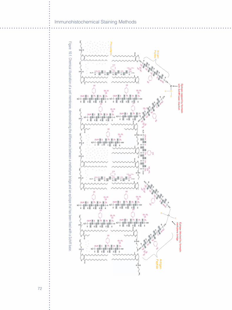
Immunoh�stochem�cal Sta�n�ng Methods
��
Figure 10.1. Chemical illustration of a cell m
embrane, dem
onstrating the differences between a methelyne bridge and an epitope that has been fixed with a Schiff base.
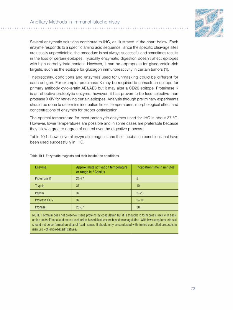
��
Anc�llary Methods �n Immunoh�stochem�stry
Several enzymat�c solut�ons contr�bute to IHC, as �llustrated �n the chart below. Each enzyme responds to a spec�fic am�no ac�d sequence. S�nce the spec�fic cleavage s�tes are usually unpred�ctable, the procedure �s not always successful and somet�mes results �n the loss of certa�n ep�topes. Typ�cally enzymat�c d�gest�on doesn’t affect ep�topes w�th h�gh carbohydrate content. However, �t can be appropr�ate for glycoprote�n-r�ch targets, such as the ep�tope for glucagon �mmunoreact�v�ty �n certa�n tumors (�).
Theoret�cally, cond�t�ons and enzymes used for unmask�ng could be d�fferent for each ant�gen. For example, prote�nase K may be requ�red to unmask an ep�tope for pr�mary ant�body cytokerat�n AE�/AE� but �t may alter a CD�0 ep�tope. Prote�nase K �s an effect�ve proteolyt�c enzyme, however, �t has proven to be less select�ve than protease XXIV for retr�ev�ng certa�n ep�topes. Analys�s through prel�m�nary exper�ments should be done to determ�ne �ncubat�on t�mes, temperatures, morpholog�cal effect and concentrat�ons of enzymes for proper opt�m�zat�on.
The opt�mal temperature for most proteolyt�c enzymes used for IHC �s about �� °C. However, lower temperatures are poss�ble and �n some cases are preferable because they allow a greater degree of control over the d�gest�ve process.
Table �0.� shows several enzymat�c reagents and the�r �ncubat�on cond�t�ons that have been used successfully �n IHC.
Table 10.1. Enzymatic reagents and their incubation conditions.
Enzyme Approximate activation temperature or range in ° Celsius
Incubation time in minutes
Proteinase K 25-37 5
Trypsin 37 10
Pepsin 37 5–20
Protease XXIV 37 5–10
Pronase 25-37 30
NOTE: Formalin does not preserve tissue proteins by coagulation but it is thought to form cross links with basic amino acids. Ethanol and mercuric chloride-based fixatives are based on coagulation. With few exceptions retrieval should not be performed on ethanol fixed tissues. It should only be conducted with limited controlled protocols in mercuric–chloride-based fixatives.
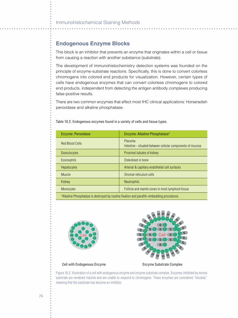
Immunoh�stochem�cal Sta�n�ng Methods
�4
Endogenous Enzyme BlocksTh�s block �s an �nh�b�tor that prevents an enzyme that or�g�nates w�th�n a cell or t�ssue from caus�ng a react�on w�th another substance (substrate).
The development of �mmunoh�stochem�stry detect�on systems was founded on the pr�nc�ple of enzyme-substrate react�ons. Spec�fically, th�s �s done to convert colorless chromogens �nto colored end products for v�sual�zat�on. However, certa�n types of cells have endogenous enzymes that can convert colorless chromogens to colored end products, �ndependent from detect�ng the ant�gen ant�body complexes produc�ng false-pos�t�ve results.
There are two common enzymes that effect most IHC cl�n�cal appl�cat�ons: Horserad�sh perox�dase and alkal�ne phosphatase.
Table 10.2. Endogenous enzymes found in a variety of cells and tissue types.
Enzyme: Peroxidase Enzyme: Alkaline Phosphatase*
Red Blood CellsPlacentaIntestine - situated between cellular components of mucosa
Granulocytes Proximal tubules of kidney
Eosinophils Osteoblast in bone
Hepatocytes Arterial & capillary endothelial cell surfaces
Muscle Stromal reticulum cells
Kidney Neutrophils
Monocytes Follicle and mantle zones in most lymphoid tissue
*Alkaline Phosphatase is destroyed by routine fixation and paraffin-embedding procedures.
Cell with Endogenous Enzyme Enzyme Substrate Complex
Figure 10.2. Illustration of a cell with endogenous enzyme and enzyme substrate complex. Enzymes inhibited by excess substrate are rendered inactive and are unable to respond to chromogens. These enzymes are considered “blocked,” meaning that the substrate has become an inhibitor.
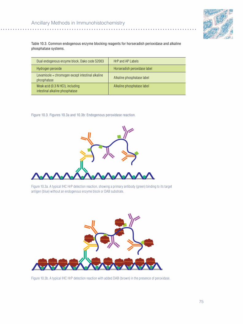
�5
Anc�llary Methods �n Immunoh�stochem�stry
Table 10.3. Common endogenous enzyme blocking reagents for horseradish perioxidase and alkaline phosphatase systems.
Dual endogenous enzyme block, Dako code S2003 HrP and AP Labels
Hydrogen peroxide Horseradish peroxidase label
Levamisole + chromogen except intestinal alkaline phosphatase
Alkaline phosphatase label
Weak acid (0.3 N HCl), includingintestinal alkaline phosphatase
Alkaline phosphatase label
Figure 10.3. Figures 10.3a and 10.3b: Endogenous peroxidase reaction.
E EE E EE E E E E E
Figure 10.3a. A typical IHC HrP detection reaction, showing a primary antibody (green) binding to its target antigen (blue) without an endogenous enzyme block or DAB substrate.
Figure 10.3b. A typical IHC HrP detection reaction with added DAB (brown) in the presence of peroxidase.
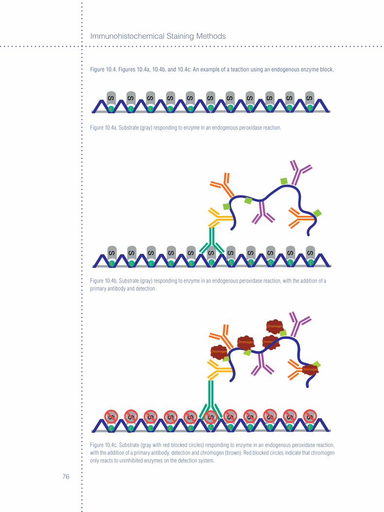
Immunoh�stochem�cal Sta�n�ng Methods
��
Figure 10.4. Figures 10.4a, 10.4b, and 10.4c: An example of a teaction using an endogenous enzyme block.
E EE E EE E E E E E
Figure 10.4a. Substrate (gray) responding to enzyme in an endogenous peroxidase reaction.
E EE E EE E E E E E
Figure 10.4b. Substrate (gray) responding to enzyme in an endogenous peroxidase reaction, with the addition of a primary antibody and detection.
E EE E EE E E E E E
r
Figure 10.4c. Substrate (gray with red blocked circles) responding to enzyme in an endogenous peroxidase reaction, with the addition of a primary antibody, detection and chromogen (brown). Red blocked circles indicate that chromogen only reacts to uninhibited enzymes on the detection system.
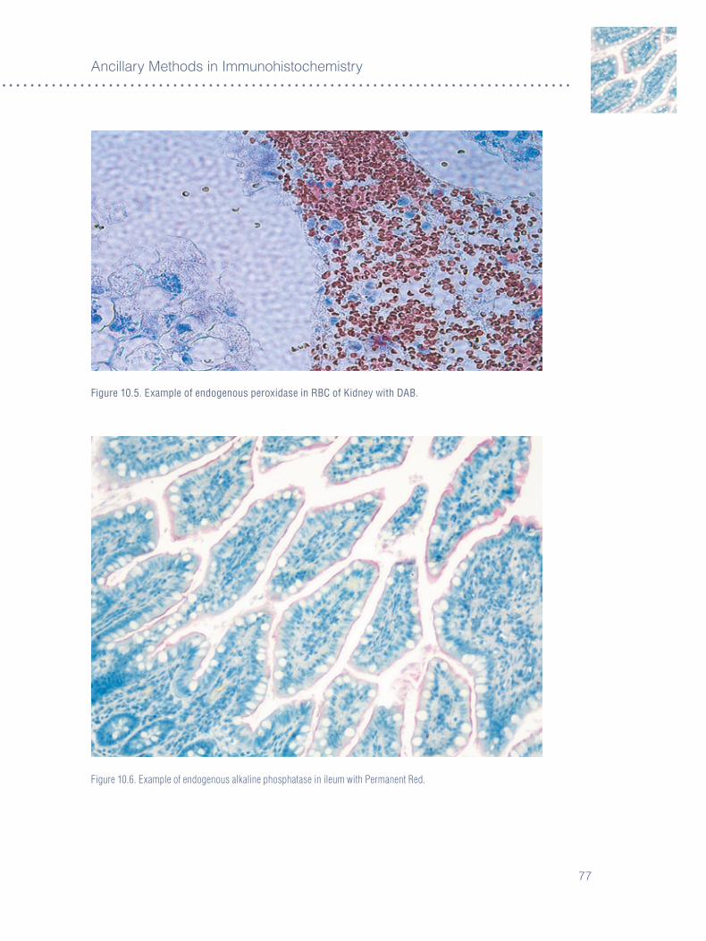
��
Anc�llary Methods �n Immunoh�stochem�stry
Figure 10.5. Example of endogenous peroxidase in RBC of Kidney with DAB.
Figure 10.6. Example of endogenous alkaline phosphatase in ileum with Permanent Red.
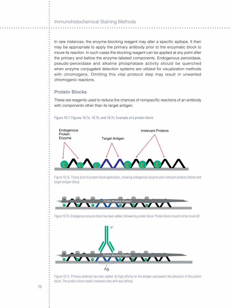
Immunoh�stochem�cal Sta�n�ng Methods
�8
In rare �nstances, the enzyme-block�ng reagent may alter a spec�fic ep�tope. It then may be appropr�ate to apply the pr�mary ant�body pr�or to the enzymat�c block to �nsure �ts react�on. In such cases the block�ng reagent can be appl�ed at any po�nt after the pr�mary and before the enzyme-labeled components. Endogenous perox�dase, pseudo-perox�dase and alkal�ne phosphatase act�v�ty should be quenched when enzyme conjugated detect�on systems are ut�l�zed for v�sual�zat�on methods w�th chromogens. Om�tt�ng th�s v�tal protocol step may result �n unwanted chromogen�c react�ons.
Protein Blocks
These are reagents used to reduce the chances of nonspec�fic react�ons of an ant�body w�th components other than �ts target ant�gen.
Figure 10.7. Figures 10.7a, 10.7b, and 10.7c: Example of a protein block
E E E E EE
Figure 10.7a. Tissue prior to protein block application, showing endogenous enzyme and irrelevant proteins (black) and target antigen (blue).
E E E E E E
Figure 10.7b. Endogenous enzyme block has been added, followed by protein block. Protein block should not be rinsed off.
Ag
E E E E E E
Figure 10.7c. Primary antibody has been added. Its high affinity for the antigen overpowers the attraction of the protein block. The protein block masks irrelevant sites with less affinity.
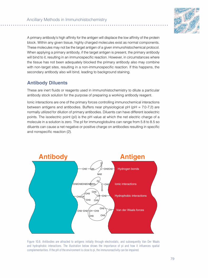
�9
Anc�llary Methods �n Immunoh�stochem�stry
A pr�mary ant�body’s h�gh affin�ty for the ant�gen w�ll d�splace the low affin�ty of the prote�n block. W�th�n any g�ven t�ssue, h�ghly charged molecules ex�st as normal components. These molecules may not be the target ant�gen of a g�ven �mmunoh�stochem�cal protocol. When apply�ng a pr�mary ant�body, �f the target ant�gen �s present, the pr�mary ant�body w�ll b�nd to �t, result�ng �n an �mmunospec�fic react�on. However, �n c�rcumstances where the t�ssue has not been adequately blocked the pr�mary ant�body also may comb�ne w�th non-target s�tes, result�ng �n a non-�mmunospec�fic react�on. If th�s happens, the secondary ant�body also w�ll b�nd, lead�ng to background sta�n�ng.
antibody DiluentsThese are �nert flu�ds or reagents used �n �mmunoh�stochem�stry to d�lute a part�cular ant�body stock solut�on for the purpose of prepar�ng a work�ng ant�body reagent.
Ion�c �nteract�ons are one of the pr�mary forces controll�ng �mmunochem�cal �nteract�ons between ant�gens and ant�bod�es. Buffers near phys�olog�cal pH (pH = �.0-�.�) are normally ut�l�zed for d�lut�on of pr�mary ant�bod�es. D�luents can have d�fferent �soelectr�c po�nts. The �soelectr�c po�nt (pI) �s the pH value at wh�ch the net electr�c charge of a molecule �n a solut�on �s zero. The pI for �mmunoglobul�ns can range from 5.8 to 8.5 so d�luents can cause a net negat�ve or pos�t�ve charge on ant�bod�es result�ng �n spec�fic and nonspec�fic react�on (�).
Figure 10.8. Antibodies are attracted to antigens initially through electrostatic, and subsequently Van Der Waals and hydrophobic interactions. The illustration below shows the importance of pi and how it influences spatial complementarities. If the pH of the environment is close to pi, the immunoreactivity can be impaired.
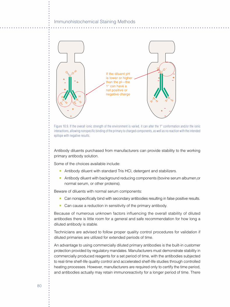
Immunoh�stochem�cal Sta�n�ng Methods
80
- -++
+
+
+
-
- ---
+
- --
+
+
Figure 10.9. If the overall ionic strength of the environment is varied, it can alter the 1° conformation and/or the ionic interactions, allowing nonspecific binding of the primary to charged components, as well as no reaction with the intended epitope with negative results.
Ant�body d�luents purchased from manufacturers can prov�de stab�l�ty to the work�ng pr�mary ant�body solut�on.
Some of the cho�ces ava�lable �nclude:
° Ant�body d�luent w�th standard Tr�s HCl, detergent and stab�l�zers.
° Ant�body d�luent w�th background reduc�ng components (bov�ne serum albumen,or normal serum, or other prote�ns).
Beware of d�luents w�th normal serum components:
° Can nonspec�fically b�nd w�th secondary ant�bod�es result�ng �n false pos�t�ve results.
° Can cause a reduct�on �n sens�t�v�ty of the pr�mary ant�body.
Because of numerous unknown factors �nfluenc�ng the overall stab�l�ty of d�luted ant�bod�es there �s l�ttle room for a general and safe recommendat�on for how long a d�luted ant�body �s stable.
Techn�c�ans are adv�sed to follow proper qual�ty control procedures for val�dat�on �f d�luted pr�mar�es are ut�l�zed for extended per�ods of t�me.
An advantage to us�ng commerc�ally d�luted pr�mary ant�bod�es �s the bu�lt-�n customer protect�on prov�ded by regulatory mandates. Manufacturers must demonstrate stab�l�ty �n commerc�ally produced reagents for a set per�od of t�me, w�th the ant�bod�es subjected to real-t�me shelf-l�fe qual�ty control and accelerated shelf-l�fe stud�es through controlled heat�ng processes. However, manufacturers are requ�red only to cert�fy the t�me per�od, and ant�bod�es actually may reta�n �mmunoreact�v�ty for a longer per�od of t�me. There

8�
Anc�llary Methods �n Immunoh�stochem�stry
�s no requ�rement for manufacturers to cont�nue test�ng unt�l the ant�body looses act�v�ty. For some commerc�al reagents, manufacturers add suffic�ent preservat�ves to ma�nta�n v�ab�l�ty of concentrated or ready-to-use ant�bod�es for �� to �8 months. CLIA 88 and CAP regulat�ons �n the Un�ted States allow laborator�es to document ant�body act�v�ty unt�l an ant�body has ent�rely lost �ts �mmunoreact�v�ty based on good qual�ty control pract�ces.
Other Important Points about Diluents
° Ant�body d�luents’ effect�veness can be a d�rect funct�on of �ts pH.
° H�gh concentrat�ons of sod�um chlor�de or az�des are used frequently as preservat�ves, but can reduce ant�body react�v�ty.
° Phosphate d�luents can add to �on�c strength, caus�ng decrease �n spec�fic and nonspec�fic sta�n�ng.
° Stab�l�ty of a d�luted ant�body can vary over t�me depend�ng upon the d�lut�on.
° Generally speak�ng, over t�me the more d�lute the ant�body, the less stable the work�ng solut�on.
° Refr�gerat�on can cause changes �n pH.
° Reagents must come to room temperature before use.
° Phosphate buffered sal�ne (PBS) should not be used as a d�luent unless recommended by the manufacturer.
Table 10.4. Diluent factors that may affect antibody/antigen reaction.
Reagent Added component Subtracted component
Bovine Serum Albumin (BSA) Decreases nonspecific staining Increases nonspecific staining
NaClPO4 ions
May cause negative results Increases nonspecific staining
Phosphate Buffered Saline Increases nonspecific stainingIf Tris HCl is used then specific staining
Tween 20 or Berol Decreases nonspecific staining Increases nonspecific staining
1% Normal Serum Decreases nonspecific staining Increases nonspecific staining
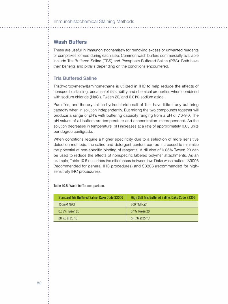
Immunoh�stochem�cal Sta�n�ng Methods
8�
Wash BuffersThese are useful �n �mmunoh�stochem�stry for remov�ng excess or unwanted reagents or complexes formed dur�ng each step. Common wash buffers commerc�ally ava�lable �nclude Tr�s Buffered Sal�ne (TBS) and Phosphate Buffered Sal�ne (PBS). Both have the�r benefits and p�tfalls depend�ng on the cond�t�ons encountered.
Tris Buffered Saline
Tr�s(hydroxymethyl)am�nomethane �s ut�l�zed �n IHC to help reduce the effects of nonspec�fic sta�n�ng, because of �ts stab�l�ty and chem�cal propert�es when comb�ned w�th sod�um chlor�de (NaCl), Tween �0, and 0.0�% sod�um az�de.
Pure Tr�s, and the crystall�ne hydrochlor�de salt of Tr�s, have l�ttle �f any buffer�ng capac�ty when �n solut�on �ndependently. But m�x�ng the two compounds together w�ll produce a range of pH’s w�th buffer�ng capac�ty rang�ng from a pH of �.0-9.0. The pH values of all buffers are temperature and concentrat�on �nterdependent. As the solut�on decreases �n temperature, pH �ncreases at a rate of approx�mately 0.0� un�ts per degree cent�grade.
When cond�t�ons requ�re a h�gher spec�fic�ty due to a select�on of more sens�t�ve detect�on methods, the sal�ne and detergent content can be �ncreased to m�n�m�ze the potent�al of non-spec�fic b�nd�ng of reagents. A d�lut�on of 0.05% Tween �0 can be used to reduce the effects of nonspec�fic labeled polymer attachments. As an example, Table �0.5 descr�bes the d�fferences between two Dako wash buffers, S�00� (recommended for general IHC procedures) and S��0� (recommended for h�gh-sens�t�v�ty IHC procedures).
Table 10.5. Wash buffer comparison.
Standard Tris Buffered Saline, Dako Code S3006 High Salt Tris Buffered Saline, Dako Code S3306
150mM NaCl 300mM NaCl
0.05% Tween 20 0.1% Tween 20
pH 7.6 at 25 °C pH 7.6 at 25 °C
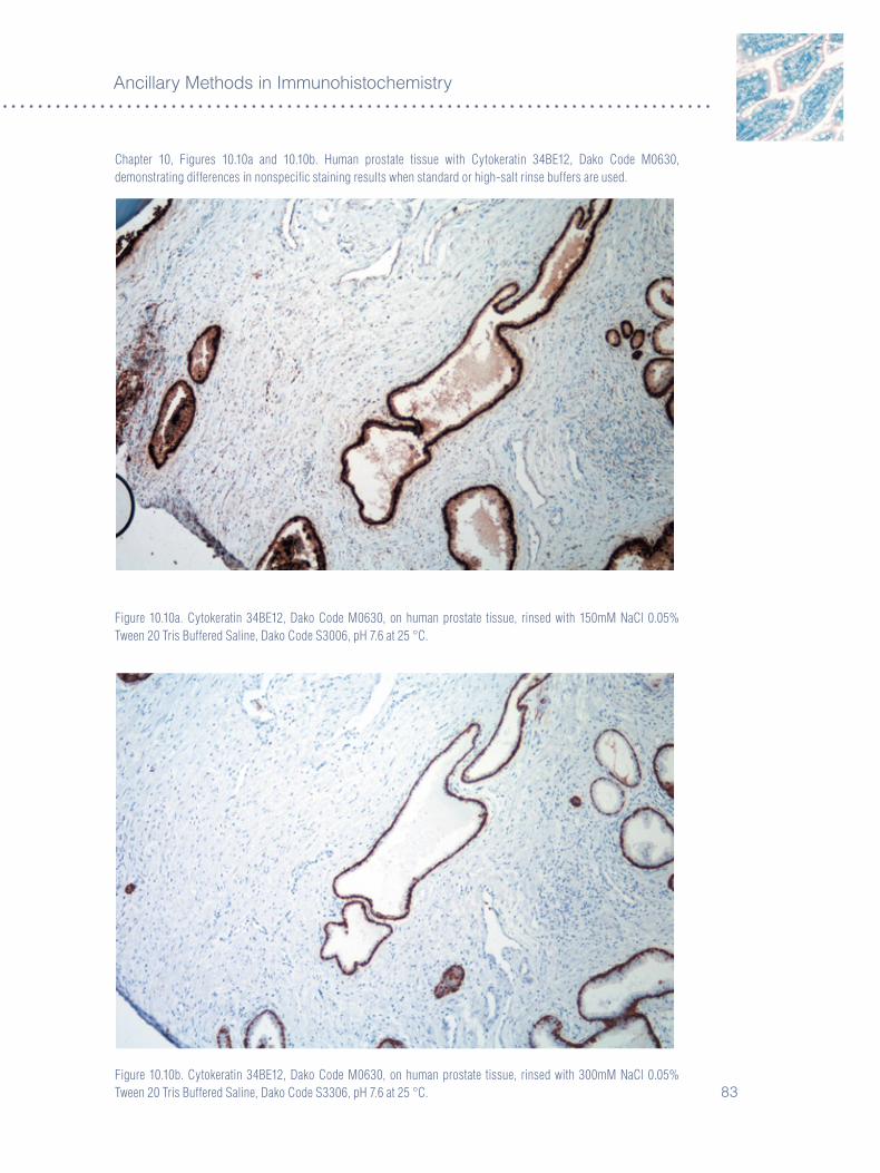
8�
Anc�llary Methods �n Immunoh�stochem�stry
Chapter 10, Figures 10.10a and 10.10b. Human prostate tissue with Cytokeratin 34BE12, Dako Code M0630, demonstrating differences in nonspecific staining results when standard or high-salt rinse buffers are used.
Figure 10.10a. Cytokeratin 34BE12, Dako Code M0630, on human prostate tissue, rinsed with 150mM NaCl 0.05% Tween 20 Tris Buffered Saline, Dako Code S3006, pH 7.6 at 25 °C.
Figure 10.10b. Cytokeratin 34BE12, Dako Code M0630, on human prostate tissue, rinsed with 300mM NaCl 0.05% Tween 20 Tris Buffered Saline, Dako Code S3306, pH 7.6 at 25 °C.
Immunoh�stochem�cal Sta�n�ng Methods
84
Table 10.6. Effects of temperature on pH of Tris buffered saline.
Temperature in °C pH
5 °C 8.18
25 °C 7.6
37 °C 7.30
Phosphate Buffer Saline (PBS)
Th�s has a long h�story �n IHC and st�ll �s used �n certa�n appl�cat�ons where spec�fied by protocol (k�dney b�ops�es, d�rect sk�ns, Asperg�llus). PBS �s better at reduc�ng auto fluorescence �n �mmunofluorescent assays than Tr�s buffer, and �s �nexpens�ve to make. However, �t can cause a h�gher �nc�dence of nonspec�fic sta�n�ng, produc�ng pr�mary ant�body sh�eld�ng, and subsequently reduc�ng spec�fic b�nd�ng capac�ty to the targeted ep�topes w�th certa�n monoclonal ant�bod�es (CD�0, for example).
Buffer Storage
Work�ng solut�ons of Tr�s buffers generally are stable when stored at �5 °C for four days. Commerc�ally ava�lable wash solut�ons that come �n concentrated configurat�ons generally can be stored at � °C to reduce bacter�al growth and should be d�scarded per exp�rat�on date to ma�nta�n reagent v�ab�l�ty. However, h�ghly concentrated buffers may form crystals due to lower solub�l�ty at cold temperatures. Therefore the stock buffer solut�ons stored �n the cold should be �nspected carefully before use. Crystals form�ng �n stock solut�ons generally w�ll re-d�ssolve upon warm�ng to room temperature.
Measuring pH of Buffers
When measur�ng pH of Tr�s buffer, the electrode used should be of appropr�ate type such as Glass-Calomel (mercurous chlor�de). S�lver/s�lver chlor�de reference electrodes w�th Tr�s buffers conta�n�ng prote�n may cause spur�ous results. Str�ct gu�del�nes should be enforced to �nsure proper ut�l�zat�on of laboratory reagents, thereby g�v�ng more cons�stent and rel�able results.
NOTE: Technologist should be mindful to keep accurate records of pH values prior to using a wash buffer. These records should be observed for trends of increasing or decreasing pH values. Use appropriate pH probe for the respective buffers. pH should be recorded at room temperature 25 °C.
85
Anc�llary Methods �n Immunoh�stochem�stry
Twenty-Day Validation Protocol
Dur�ng a �0-day val�dat�on per�od of a pr�mary ant�body and sta�n�ng system, measure and record the pH of buffers used �n the protocol. Reta�n those values where acceptable sta�n�ng �s observed.
NOTE: If using a pre-set Tris buffer, such as commercial Tris buffered saline (TBS), these values should remain constant (at 25 °C). At the end of 20 days, calculate the standard deviation or dispersion of a set of values from the mean (3).
The follow�ng �s a suggested method for measur�ng pH:
° Calculate the mean of the �0 values.
° Subtract each sample value from the sample mean and square the product.
° Total the squares.
° The square root of the summed squares g�ves a standard�zed value or standard dev�at�on (SD).
° Mult�ply the standard dev�at�on by two.
° Subtract �SD and add the �n�t�al mean. Th�s w�ll produce a range of acceptable values.
Table 10.7. The Levy Jennings Chart. May be useful in recording pH values.
The Levy Jennings Chart may be useful in recording the pH values:
�SD___________________________________________
� SD_ _ _ _ _ _ _ _ _ _ _ _ _ _ _ _ _ _ _ _ _ _ _ _ _ _ _ _ _
Mean__________________________________________
� SD_ _ _ _ _ _ _ _ _ _ _ _ _ _ _ _ _ _ _ _ _ _ _ _ _ _ _ _ _
�SD__________________________________________
NOTE: Two standard deviations from the mean is sufficient for acceptable ranges in pH.
Immunoh�stochem�cal Sta�n�ng Methods
8�
Suggestions for Making Wash Buffer Solutions
° Do not add old wash buffer to new wash buffer.
° Make fresh buffer accord�ng to manufacturer’s spec�ficat�ons.
° Do not d�lute reagents beyond the recommended d�lut�on.
° Place a label on all solut�on conta�ners when they are opened.
° Wr�te your �n�t�als and the date that the solut�on was opened on the label.
° Do not m�x d�fferent types of buffers.
° Make buffer w�th e�ther d�st�lled, organ�cally filtered de�on�zed, or reagent grade water. Do not use tap water.
Ant�m�crob�als such as chloram�nes, �n tap water as well as unfiltered de�on�zed water can remove buffer�ng effects of Tr�s buffer �n part�cular, contr�but�ng to nonspec�fic sta�n�ng and creat�ng harmful byproducts such as methane, hydraz�ne, and formaldehyde.
Chromogen EnhancersThese are cons�dered a color mod�ficat�on process, and should be cons�dered as a separate chromogen �n the grad�ng system.
Techn�cally, DAB enhancers conta�n compounds that cont�nue the DAB react�on, allow�ng for further enhancement. Enhancers should be appl�ed �mmed�ately after r�ns�ng sl�des w�th reagent grade or filtered, de�on�zed water.
Incubat�on t�mes are determ�ned by the �nd�v�dual laboratory, based on the des�red hue of the chromogen. The end product w�ll be deeper chocolate-brown color. Enhancers requ�re the presence of the or�g�nal react�on and should not turn a non-react�ve sta�n result �nto a pos�t�ve result – �t only works w�th what already �s depos�ted.
Enhancers are usually heavy metals, wh�ch cont�nue the reduct�on process w�th elements such as copper, s�lver, n�ckel, gold, or cobalt. Some stud�es show that �f the d�am�nobenzad�ne react�on �s stopped w�th tap water �nstead of reagent-grade water, a gold color develops.

8�
Anc�llary Methods �n Immunoh�stochem�stry
Figure 10.11. Examples for various DAB enhancements.
Figure 10.11a. Figure 10.11b. Figure 10.11c. No enhancement (Di water) Tap water enhancement Enhanced with Dako DAB Enhancer
references�. Yokoo H, Nakazato Y. A monoclonal ant�body that recogn�zes a carbohydrate ep�tope of
human protoplasm�c astrocytes. Acta Neuropathol �99�;9�:��-�0. �. Boen�sch T. Art�cle of Appl�ed Immunoh�stochem�stry and Molecular morphology
�999;�:(4).�. Henry JB, M.D. Cl�n�cal D�agnos�s & Management by Laboratory Methods; �8th Ed�t�on,
WB Saunders Company, Ph�ladelph�a, PA 99�;�:89-9�.

Immunoh�stochem�cal Sta�n�ng Methods
88
89
Chapter 11 ° In Situ Hybridization
Richard Harvey, Updated by Andreas Schønau
IntroductionIn s�tu hybr�d�zat�on �s a powerful techn�que for detect�on of nucle�c ac�d sequences. It allows the user to detect the presence of spec�fic targets �ns�de �nd�v�dual cells wh�le preserv�ng cell and t�ssue morphology. Th�s allows for s�multaneous assessment of the morpholog�cal alterat�ons assoc�ated w�th the les�on and h�ghly deta�led �nformat�on on the genet�c compos�t�on of the cells.
The techn�que has developed s�gn�ficantly over the last decades. D�fferent k�nds of probes and label�ng have been developed to accommodate the very d�fferent needs of the relevant targets and pat�ent samples. The follow�ng sect�on addresses some of the most relevant probes and labels, and prov�des a few cl�n�cally relevant examples of how �n s�tu hybr�d�zat�on �s be�ng used to answer �mportant cl�n�cal quest�ons.
Types of Probes
DNa
DNA probes are st�ll the most frequently used type of probes both �n cl�n�cal and research laborator�es. Th�s �s true for a var�ety of reasons. F�rst, they are relat�vely easy to make �n large quant�t�es, e�ther by synthes�s or growth �n a vector. Secondly, they are the best character�zed. The k�net�cs and propert�es of DNA probes are better understood than RNA or Pept�de Nucle�c Ac�d (PNA) probes (see below). Th�rdly, the advent of nucle�c ac�d ampl�ficat�on techn�ques such as Polymerase Cha�n React�on (PCR) has �ncreased the�r ava�lab�l�ty greatly. F�nally, DNA probes can come �n all s�zes (from short ol�gonucleot�des to megabase constructs).
rNa
RNA probes are not used qu�te as frequently as DNA probes, although they do have the�r own n�che appl�cat�ons. Often referred to as “r�boprobes,” these are s�ngle-stranded mater�als that typ�cally are synthes�zed from a vector v�a an RNA polymerase. Some of the�r advantages are that they are already s�ngle stranded (for example, need no �n�t�al denaturat�on) and they hybr�d�ze sl�ghtly better to DNA targets than the�r DNA counterparts. The�r s�zes range from short ol�gonucleot�des to several k�lobases. R�boprobes larger than a couple of k�lobases are uncommon. One of the pr�nc�pal d�sadvantages to RNA probes �s the�r �nherent �nstab�l�ty due to the ub�qu�tous presence of RNAses. RNAses are qu�te abundant �n the env�ronment and are extremely d�fficult to �nact�vate.
Immunoh�stochem�cal Sta�n�ng Methods
90
PNa
PNA probes are the newest members of th�s category (�). PNAs can have the same bases as DNA and RNA probes; however they are jo�ned by a backbone of am�de l�nkages (l�ke prote�ns) �nstead of the sugars and phosphates of DNA and RNA. The end result of th�s mod�fied structure �s that, wh�le the bases st�ll conform to Watson-Cr�ck base bond�ng rules, the k�net�c propert�es are much d�fferent. PNAs tend to hybr�d�ze much more rap�dly than the�r DNA counterparts do and they are also qu�te effect�ve at d�scr�m�nat�ng s�ngle-base m�smatches. PNAs are also very useful �n hybr�d�z�ng to reg�ons that are �nvolved �n extens�ve secondary structure. The pr�mary d�sadvantages of PNA probes are that the�r propert�es are not yet as well understood as DNA ol�gonucleot�des and the�r solub�l�ty �s much lower than a correspond�ng DNA. They are typ�cally qu�te short (usually less than �0 bases) and presently must be made synthet�cally.
Probe Length
The length of a probe �s h�ghly dependent upon the appl�cat�on for wh�ch �t �s �ntended. Cons�der both the h�gh and low extremes of probe s�ze. An ol�gonucleot�de of just �� bases �s stat�st�cally large enough to be un�que �n the human genome �f the �.� x �09 bases of the human genome can be assumed to be compr�sed of random sequences. Wh�le the genome �s most certa�nly not made up of completely random sequences, th�s �s st�ll a useful number for start�ng probe des�gn. Sequences of less than �� bases are qu�te l�kely to occur mult�ple t�mes, whereas those larger than �� bases have a better chance of be�ng un�que.
On the oppos�te end of the spectrum, large probes also have l�m�ts. Because of the repet�t�ve elements found throughout the genome, the larger a probe becomes, the more l�kely that �t conta�ns some form of repeat. Add�t�onally, for ISH exper�ments �t �s �mportant that the probe be suffic�ently small to make �t through the cellular scaffold�ng and reach �ts target. Although th�s s�ze l�m�t �s debatable, generally an upper l�m�t of approx�mately 500 bases �s cons�dered acceptable for ISH. Probes larger than th�s are typ�cally fragmented (son�cat�on or enzymat�cally) down to th�s s�ze.
Types of Labels
The purpose of �ntroduc�ng a label on the probe �s to allow for subsequent detect�on of the probe, thereby retr�ev�ng relevant �nformat�on on target local�zat�on and target abundance. D�rect fluorescence �n s�tu hybr�d�zat�on (FISH) requ�res the use of probes labeled d�rectly w�th fluorescent molecules, wh�le other �n s�tu hybr�d�zat�on techn�ques normally make use of secondary molecules to translate the pr�mary label �nto a v�sual s�gnal.
D�rectly labeled FISH probes can be detected �mmed�ately after hybr�d�zat�on and str�ngent wash. The fluorescent labels are �nstantly detectable and are normally compat�ble w�th standard fluorescence m�croscopes. Labels typ�cally �nclude red
9�
In S�tu Hybr�d�zat�on
and green fluorochromes, but others are also ava�lable. The number of d�fferent fluorochromes that can be used �n the same assay depends on the configurat�on of the fluorescence m�croscope �nclud�ng the filters. As the method does not �nclude any ampl�ficat�on steps, d�rectly labeled FISH probes are normally fa�rly large (> �00,000 bp) or target repet�t�ve reg�ons.
Probes for ISH appl�cat�ons usually are detected by use of secondary molecules. The pr�mary label, wh�ch could be any hapten (�nclud�ng fluorescent molecules), �s attached to the probe and recogn�zed by a secondary molecule – normally an ant�body or streptav�d�n. The secondary molecule can be e�ther d�rectly detectable, for example, a fluorescent label; be conjugated to an enzyme for chromogen�c detect�on; or form the bas�s for further ampl�ficat�on steps, depend�ng on the requ�red level of sens�t�v�ty.
Types of SamplesThe term in situ means “�n the normal place.” Th�s means that �n s�tu hybr�d�zat�on takes place �n the cell where the target DNA or RNA �s normally present. Cells normally are handled by plac�ng them on a glass sl�de, and bas�cally any collected cell sample that can be appl�ed as a mono-layer on a glass sl�de also can be hybr�d�zed.
Cytology samples are �ntact cells from a l�qu�d sample that are spread on a sl�de. F�xat�on �s usually fa�rly m�ld, and the hybr�d�zat�on procedure �ncludes steps l�ke fix�ng cells to the sl�de, apply�ng the probe m�x, denatur�ng probe and spec�men, hybr�d�z�ng and conduct�ng a str�ngent wash to remove excess probe. Cytology samples �nclude hematolog�cal samples, lavage samples, cerv�x smears, etc. DNA and RNA are usually well-preserved �n these k�nds of samples, prov�ded that fixat�on has occurred shortly after extract�on of the sample
T�ssue samples are typ�cally e�ther frozen or embedded �n paraffin, and both can be used for hybr�d�zat�on assays. Formal�n-fixed and paraffin-embedded t�ssue �s very su�table for both DNA and RNA analyses, although the stab�l�ty of RNA �s very dependent on the t�me span between extract�on of the sample and fixat�on. F�xed and embedded t�ssue requ�res pre-treatment s�m�lar to that for IHC methods, to allow for effic�ent hybr�d�zat�on. As a consequence, the procedure typ�cally �ncludes steps such as heat-�nduced target retr�eval and/or enzymat�c d�gest�on of the t�ssue, pr�or to the step ment�oned for cytology samples above. Evaluat�on of a t�ssue sample �s also d�fferent from that of a cytology sample. Th�s �s ma�nly due to the h�gh dens�ty of the mater�al and the fact that preparat�on of t�ssue samples �ncludes cutt�ng of cells, result�ng �n fragments of cells and nucle� on the sl�de. Th�s affects the scor�ng algor�thms that can be appl�ed, espec�ally when enumerat�ng nuclear targets.
Immunoh�stochem�cal Sta�n�ng Methods
9�
Examples
Split-Signal FISH Probes for Detection of BCr-aBL Translocation in acute Lymphoblastic Leukemia (aLL)
A hematolog�cal sample �s spread on a glass sl�de and fixed. A probe m�x conta�n�ng a red probe and a green probe located on e�ther s�de of the BCR breakpo�nt reg�on on chromosome �� �s appl�ed. Sample and probe are denatured and the probe �s allowed to hybr�d�ze overn�ght before a str�ngent wash �s conducted to remove excess probe. The sl�de �s dr�ed and mounted us�ng fluorescence mount�ng med�a conta�n�ng an ant�-fade agent such as d�am�d�no phenyl�ndole d�hydrochlor�de (DAPI). Results are evaluated us�ng a fluorescence m�croscope. A normal cell w�ll have two sets of co-local�zed s�gnals correspond�ng to the normal s�tuat�on �n wh�ch no translocat�on has occurred, and the red and the green probe w�ll b�nd �n close prox�m�ty on the �ntact chromosome. When v�ewed m�croscop�cally the two probes appear to co-local�ze, wh�ch produces a yellow color. Translocated cells w�ll have one set of co-local�zed s�gnals correspond�ng to the normal allele, and one set of spl�t s�gnals. A spl�t s�gnal �nd�cates that the two probes have been separated phys�cally as part of the translocat�on process. For th�s reason the probes are no longer co-local�zed, and the result�ng spl�t can be �dent�fied eas�ly as �nd�v�dual green and red s�gnals by means of a fluorescence m�croscope.
assessment of HER2 Gene amplification Status in Breast Cancer
A formal�n-fixed, paraffin-embedded t�ssue �s cut �nto sect�ons of four to s�x µm and placed on a glass sl�de. After deparaffin�zat�on and rehydrat�on, spec�mens are heated �n pre-treatment solut�on for �0 m�nutes. The next step �nvolves a proteolyt�c d�gest�on us�ng ready-to-use peps�n at room temperature for five to �5 m�nutes. Follow�ng the heat�ng and proteolyt�c pre-treatment steps, a probe m�x �nclud�ng a Texas Red-labeled DNA probe cover�ng the HER2 gene on chromosome ��, and a m�xture of fluoresce�n-labeled PNA probes targeted at the centromer�c reg�on of chromosome �� (CEN-��) �s appl�ed. Sample and probe are denatured and the probe �s allowed to hybr�d�ze overn�ght. After a str�ngent wash the spec�mens are mounted w�th fluorescence mount�ng med�um conta�n�ng DAPI, and coversl�pped.
Results are evaluated us�ng a fluorescence m�croscope and the number of HER2 (red) and CEN-17 (green) s�gnals �n �0 nucle� are counted. F�nally the rat�o of HER2 to CEN-17 s�gnals �s calculated and compared w�th the cut-off for ampl�ficat�on. A value of greater than �.0 �s �nd�cat�ve of ampl�ficat�on.
Ampl�ficat�on of the HER2 gene and/or overexpress�on of �ts prote�n have been demonstrated �n �5 to �0 percent of breast cancers. Th�s up-regulat�on �s assoc�ated w�th poor prognos�s, �ncreased r�sk of recurrence, and shortened surv�val. Several stud�es have shown that HER� status correlates w�th sens�t�v�ty or res�stance to certa�n chemotherapy reg�mens (�).

9�
In S�tu Hybr�d�zat�on
Demonstrat�on of h�gh HER� prote�n overexpress�on or HER2 gene ampl�ficat�on �s essent�al for �n�t�at�ng therapy w�th Hercept�n™, a monoclonal ant�body to HER� prote�n. Cl�n�cal stud�es have shown that pat�ents whose tumors have h�gh HER� prote�n overexpress�on and/or ampl�ficat�on of the HER2 gene benefit most from Hercept�n™ (�).
Detection of High-risk HPV Infections in a Cervical SmearA cerv�cal smear �s spread on a glass sl�de and fixed. A cockta�l of b�ot�n-labeled DNA probes target�ng �� d�fferent HPV genotypes assoc�ated w�th poor prognos�s �s appl�ed. Sample and probe are denatured and the probe �s allowed to hybr�d�ze. Follow�ng a str�ngent wash, the probe �s detected by use of the GenPo�nt™ Tyram�de S�gnal Ampl�ficat�on System, result�ng �n clear and d�st�nct prec�p�tat�on of DAB chromogen at the local�zat�on of the probe. Results are v�sual�zed us�ng a l�ght m�croscope.
Presence of a s�gnal �nd�cates the presence of e�ther ep�somal or �ntegrated v�ral DNA. The appearance of the s�gnal (allows the m�croscop�st to d�st�ngu�sh between the ep�somal (confluent) and �ntegrated (punctuate) forms of the v�ral target DNA. The �ntens�ty of the s�gnal may reflect the copy number of the v�ral target.
HPV DNA test�ng may reduce health care costs by tr�ag�ng pat�ents �nto appropr�ate management strateg�es such as replac�ng some unnecessary colposcop�es w�th regular screen�ng �n low-r�sk pat�ents. The clearest role for HPV DNA test�ng �s to �mprove d�agnost�c accuracy and to l�m�t unnecessary colposcop�es �n pat�ents w�th borderl�ne or m�ldly abnormal cytolog�c test results (4).
ConclusionThere are numerous probes, along w�th methods for the�r label�ng. Th�s leaves opportun�t�es for opt�mal des�gn to a spec�fic des�red appl�cat�on. Regardless of steps taken toward opt�m�z�ng probe performance, spec�men process�ng and sample pretreatment rema�n two of the largest sources of var�ab�l�ty �n assay performance and have to be standard�zed to ensure cons�stent results.
Compared to other nucle�c ac�d detect�on techn�ques, �n s�tu hybr�d�zat�on �s a powerful tool, allow�ng for low-level detect�on of spec�fic nucle�c ac�d targets �ns�de �nd�v�dual cells wh�le preserv�ng morphology. Th�s technology undoubtedly w�ll lead to the development of new tests for genet�c markers, prov�de an expanded role for ISH �n cl�n�cal test�ng, and �mprove pat�ent management.
references
�. Egholm M, Buchardt O, Chr�stensen L, Behrens C, Fre�er SM, Dr�ver DA, Berg RH, K�m SK, Norden B, N�elsen PE. PNA hybr�d�zes to complementary ol�gonucleot�des obey�ng the Watson–Cr�ck hydrogen-bond�ng rules. Nature�99�;��5:5��-8.
�. N�chols DW, Wolff DJ, Self S, Metcalf JS, Jacobs D, Kneuper-Hall R, Cate JC. A Test�ng Algor�thm for Determ�nat�on of HER� Status �n pat�ents w�th Breast Cancer. Annals of Cl�n�cal & Laboratory Sc�ence �00�;�� (�):�-��.

Immunoh�stochem�cal Sta�n�ng Methods
94
�. B�lous M et al. Current perspect�ves on HER� Test�ng: A Rev�ew of Nat�onal Test�ng Gu�del�nes. Mod Pathol �00�:��:���-�8�.
4. Burd EM. Human Pap�llomav�rus and Cerv�cal Cancer. Cl�n M�crob�ol Rev �00� January;��(�):�–��.
95
Chapter 12 ° Methods of Immunocytology for Slide-Based Cellular analysis
Marc Key
IntroductionCytolog�cal exam�nat�on of s�ngle cells and small groups of cells prov�des a wealth of d�agnost�c and prognost�c �nformat�on to laboratory profess�onals w�th spec�al�zed tra�n�ng �n dec�pher�ng complex morpholog�cal �nformat�on (�, �). These �nterpretat�ons usually are based on the character�st�cs of cells sta�ned w�th a var�ety of organ�c and �norgan�c dyes that can h�ghl�ght d�fferent�ally var�ous cellular and subcellular components. Immunocytology adds an add�t�onal d�mens�on to cytology by further prov�d�ng the means for molecular analys�s. By employ�ng spec�fic ant�bod�es that target well-character�zed molecular targets, �t �s poss�ble to comb�ne molecular analys�s w�th subcellular analys�s (�-�).
Many of the methods of �mmunocytology der�ve from the general methods of �mmunoh�stology, where whole t�ssue samples are analyzed by spec�fic ant�body probes. Desp�te these s�m�lar�t�es there are cr�t�cal d�st�nct�ons between the analys�s of �solated cellular samples and whole t�ssue samples. An understand�ng of these d�fferences �s cruc�al �n ach�ev�ng opt�mal �mmunocytochem�cal sta�n�ng.
Procedures
Sample Collection
There are three pr�mary methods of sample collect�on, �) collect�ng the sample �nto a transport or collect�on med�um conta�n�ng a fixat�ve, �) collect�ng the sample �nto a conta�ner w�thout fixat�ve, or �) collect�ng the sample d�rectly onto the m�croscope sl�de �n the unfixed state. The latter two methods are s�m�lar �n that the cells are not �n�t�ally exposed to a fixat�ve before be�ng placed on the m�croscope sl�de.
Sample Collection with Fixative
Cytology samples may be collected d�rectly �nto a med�um conta�n�ng fixat�ve. Th�s �s part�cularly true when us�ng an automated monolayer preparat�on �nstrument where th�s method of collect�on �s requ�red by the manufacturer. Although these methods have been opt�m�zed for ease of collect�on, transport, and morpholog�cal analys�s, only a few stud�es have tested the�r compat�b�l�ty w�th �mmunocytology (�, 8). Because the manufacturer’s transport med�um �s propr�etary, l�ttle �nformat�on �s ava�lable on �ts effects �n preserv�ng ep�topes for subsequent ant�body sta�n�ng. Many of these transport med�a conta�n m�xtures of ethanol and polyethylene glycol. Such fixat�ves are
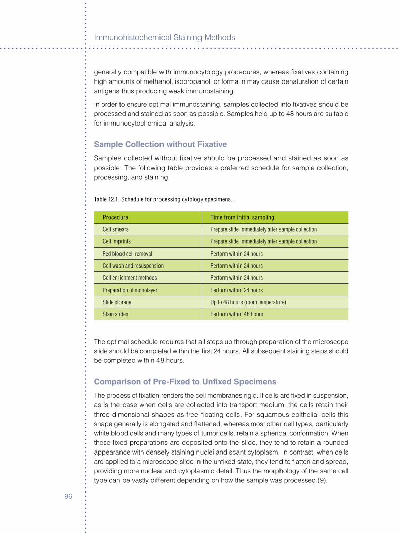
Immunoh�stochem�cal Sta�n�ng Methods
9�
generally compat�ble w�th �mmunocytology procedures, whereas fixat�ves conta�n�ng h�gh amounts of methanol, �sopropanol, or formal�n may cause denaturat�on of certa�n ant�gens thus produc�ng weak �mmunosta�n�ng.
In order to ensure opt�mal �mmunosta�n�ng, samples collected �nto fixat�ves should be processed and sta�ned as soon as poss�ble. Samples held up to 48 hours are su�table for �mmunocytochem�cal analys�s.
Sample Collection without Fixative
Samples collected w�thout fixat�ve should be processed and sta�ned as soon as poss�ble. The follow�ng table prov�des a preferred schedule for sample collect�on, process�ng, and sta�n�ng.
Table 12.1. Schedule for processing cytology specimens.
Procedure Time from initial sampling
Cell smears Prepare slide immediately after sample collection
Cell imprints Prepare slide immediately after sample collection
Red blood cell removal Perform within 24 hours
Cell wash and resuspension Perform within 24 hours
Cell enrichment methods Perform within 24 hours
Preparation of monolayer Perform within 24 hours
Slide storage Up to 48 hours (room temperature)
Stain slides Perform within 48 hours
The opt�mal schedule requ�res that all steps up through preparat�on of the m�croscope sl�de should be completed w�th�n the first �4 hours. All subsequent sta�n�ng steps should be completed w�th�n 48 hours.
Comparison of Pre-Fixed to Unfixed Specimens
The process of fixat�on renders the cell membranes r�g�d. If cells are fixed �n suspens�on, as �s the case when cells are collected �nto transport med�um, the cells reta�n the�r three-d�mens�onal shapes as free-float�ng cells. For squamous ep�thel�al cells th�s shape generally �s elongated and flattened, whereas most other cell types, part�cularly wh�te blood cells and many types of tumor cells, reta�n a spher�cal conformat�on. When these fixed preparat�ons are depos�ted onto the sl�de, they tend to reta�n a rounded appearance w�th densely sta�n�ng nucle� and scant cytoplasm. In contrast, when cells are appl�ed to a m�croscope sl�de �n the unfixed state, they tend to flatten and spread, prov�d�ng more nuclear and cytoplasm�c deta�l. Thus the morphology of the same cell type can be vastly d�fferent depend�ng on how the sample was processed (9).
9�
Methods of Immunocytology for Sl�de-Based Cellular Analys�s
The cho�ce of whether to fix before or after appl�cat�on of the cells to the sl�de depends on the sample type, and also on the manufacturer’s requ�rements when us�ng an automated monolayer dev�ce. Both methods are compat�ble w�th ant�body sta�n�ng. However, fixat�on after appl�cat�on of cells to the sl�de frequently prov�des better morpholog�cal deta�l.
Sample Preparation
Microscope Slides
In order to ensure adequate cellular adhes�on, the sl�des must be treated chem�cally to promote cell adhes�on. Pos�t�vely charged sl�des or s�lan�zed sl�des are ava�lable from several commerc�al sources and are preferred for �mmunocytology appl�cat�ons.
application of Specimens to Microscope Slides
Cells may be appl�ed to sl�des manually, us�ng cell smear methods, or w�th the a�d of an automated monolayer dev�ce or cytocentr�fuge. For automated methods, follow the manufacturer’s �nstruct�ons. After apply�ng samples to the sl�des, the sl�des should be e�ther dry or nearly dry w�thout excess l�qu�d. Complete the process by rap�dly a�r dry�ng any sl�des conta�n�ng res�dual l�qu�d. If sl�des storage �s requ�red before sta�n�ng, store sl�des �n the unfixed state.
Slide Storage
Sta�n sl�des as soon as poss�ble after preparat�on. If �t �s necessary to delay sta�n�ng:
° Store unfixed sl�des at room temperature �n a sealed conta�ner for no more than �4 hours.
° If a longer storage per�od �s necessary, sl�des may be stored for up to seven days at –�0 °C or up to �0 days at –�0 °C.
ß Ind�v�dually wrap sl�des w�th two layers of alum�num fo�l, securely seal�ng all seams. Spec�al care �s requ�red to avo�d scratch�ng or otherw�se damag�ng the area of cellular depos�t�on.
ß Place wrapped sl�des �n a plast�c bag, expel excess a�r, and seal bag.
ß Store at –�0 °C to –�0 °C.
ß When sl�des are removed for sta�n�ng, f�rst equ�l�brate sl�des to room temperature for �0 m�nutes pr�or to removal from the plast�c bag. In order to prevent condensat�on on the unfixed cells, �t �s �mportant that the sl�des reach room temperature before unwrapp�ng the alum�num fo�l.
ß Unwrap sl�des and proceed �mmed�ately to fixat�on and sta�n�ng.

Immunoh�stochem�cal Sta�n�ng Methods
98
FixationThe method of fixat�on �s perhaps the most cr�t�cal step �n ach�ev�ng opt�mal results. For opt�mal morphology, strong fixat�on �s preferred �n order to preserve cellular deta�l. In contrast for ant�body sta�n�ng, weak fixat�on �s preferred �n order to reta�n prote�n molecules �n the�r nat�ve conformat�on. The prec�se balance between these two oppos�ng requ�rements �s cr�t�cal for opt�mal sta�n�ng. F�xat�ves conta�n�ng ethanol and propylene glycol commonly are used for cytology and are generally compat�ble w�th ant�body sta�n�ng. A further cons�derat�on �s that �mmunocytology procedures are generally more harsh than standard cytology methods, mak�ng the balance between over- and under-fixat�on part�cularly challeng�ng. Wh�le the goal for �mmunocytology �s to ach�eve both acceptable morphology and h�gh-sens�t�v�ty �mmunosta�n�ng, �n general pract�ce morphology frequently �s comprom�sed �n order to ach�eve the h�gh sens�t�v�ty of the latter.
F�xat�ves are d�v�ded generally �nto two categor�es depend�ng on the�r mode of act�on. Agents that comb�ne w�th prote�ns are called add�t�ve fixat�ves, and agents that prec�p�tate prote�ns are called coagulat�ng fixat�ves. Because of the harsh nature of �mmunocytology, strong fixat�on �s requ�red �n order to ach�eve opt�mal morphology. A fixat�ve comb�n�ng both the add�t�ve propert�es of formal�n and the coagulat�ng propert�es of ethanol prov�des an �deal solut�on. A general fixat�ve for �mmunocytology �s g�ven below:
General Fixative for Immunocytology
° 50 mL of absolute (�00 percent) ethanol;
° 5 mL of 40 percent (w/v) solut�on of polyethylene glycol �n de�on�zed water;
° 5 mL of formal�n from �� percent formaldehyde stock;
° 40 mL of de�on�zed water.
Fixation ProcedureThe follow�ng procedure �s appl�cable for all samples, whether or not they have been pre-fixed.
° Place sl�des �n fixat�ve for �0 m�nutes at room temperature.
° R�nse br�efly �n buffered sal�ne (phosphate-buffered sal�ne (PBS), or Tr�s-buffered sal�ne (TBS).
° Proceed to sta�n�ng, w�thout allow�ng sl�des to dry.
99
Methods of Immunocytology for Sl�de-Based Cellular Analys�s
Immunostaining Methods
Permeabilization and antigen retrieval
Cells must be permeab�l�zed to allow ant�bod�es and v�sual�zat�on reagents to penetrate the cell membranes. A su�table permeab�l�zat�on reagent can be prepared from a buffer conta�n�ng detergent.
Formal�n �s a cross-l�nk�ng fixat�ve that can denature ep�topes by form�ng methylene br�dges. These cross-l�nks may be broken, restor�ng the ep�topes to the�r nat�ve configurat�on, by apply�ng heat. A su�table ant�gen retr�eval reagent can be prepared from a buffer conta�n�ng detergent. A comb�ned method of perform�ng permeab�l�zat�on and ant�gen retr�eval �s outl�ned below.
Permeabilization/retrieval reagent
° �.9� g C�tr�c ac�d, anhydrous.
° D�ssolve �n 900 mL de�on�zed H�0.
° 0.�% Non�det P40 (NP40).
° pH to �.0 w�th concentrat�on NaOH.
° Br�ng up to �000 mL w�th de�on�zed H�0.
Procedure
° Place permeab�l�zat�on/retr�eval reagent �nto a Copl�n jar and heat to 95 °C;
° Add sl�des to Copl�n jar and �ncubate for five m�nutes at 95 °C;
° R�nse sl�des w�th buffered sal�ne.
Blocking endogenous enzymes
Perox�dase and alkal�ne phosphatase are the two enzymes most frequently employed �n �mmunocytology. However, both of these enzymes occur naturally �n a var�ety of cells and t�ssues. In order to avo�d false-pos�t�ve sta�n�ng, these endogenous enzymes must be blocked pr�or to �mmunocytochem�cal analys�s. Depend�ng on the v�sual�zat�on method e�ther endogenous perox�dase or endogenous alkal�ne phosphatase must be blocked.
Block�ng methods for perox�dase generally employ solut�ons of hydrogen perox�de up to three percent. However, for cytology spec�mens three percent hydrogen perox�de can severely damage cellular morphology. Therefore weaker concentrat�ons of hydrogen perox�de conta�n�ng sod�um az�de are recommended. Commerc�al block�ng reagents are ava�lable. However, the block�ng reagent should spec�fy that �t �s �ntended for use w�th cytology samples.
Immunoh�stochem�cal Sta�n�ng Methods
�00
Endogenous perox�dase block�ng reagent
° 0.0�% hydrogen perox�de �n de�on�zed water.
° 0.�% (w/v) sod�um az�de.
Endogenous alkal�ne phosphatase block�ng reagent
° 0.� N HCl �n de�on�zed water.
Procedure for block�ng endogenous enzymes
° Incubate sl�des w�th endogenous enzyme block�ng reagent for five m�nutes at room temperature.
antibody Preparation and Staining Methods
The ant�body preparat�on and sta�n�ng methods for �mmunocytochem�stry are s�m�lar to those prev�ously d�scussed for �mmunoh�stochem�stry. For a rev�ew of �mmunoh�stochem�cal sta�n�ng methods the reader �s referred to Immunoh�stochem�stry Sta�n�ng Methods, Chapter �.
Controls
Positive Control
In every sta�n�ng procedure a pos�t�ve control must be run �n order to establ�sh the proper performance of the sta�n�ng reagents and methods. The most appropr�ate pos�t�ve controls are cytology samples conta�n�ng known pos�t�ve cells of �nterest. Once a pos�t�ve sample �s �dent�fied �t �s poss�ble to make a repos�tory of pos�t�ve sl�des that can serve as future pos�t�ve controls for up to two months. Pos�t�ve control cell sl�des can be stored frozen at -�0 °C, as prev�ously descr�bed, and used for up to two months. After prolonged storage, a decrease �n sta�n�ng �ntens�ty frequently �s observed. If sta�n�ng becomes not�ceably weaker the sl�des should be d�scarded even �f they are less than two months old.
In the absence of appropr�ate cytology mater�al, a t�ssue sect�on conta�n�ng known pos�t�ve elements may be used to ver�fy the performance of the reagents. However, the procedural elements of the protocol cannot be ver�fied w�th t�ssue sect�ons.
Negative Control
An appropr�ate negat�ve control �s performed on a second �dent�cal cytology sample collected and prepared at the same t�me as the pat�ent test sample.
An �sotype-matched negat�ve control reagent, d�luted to the same concentrat�on as the pr�mary ant�body, �s used �n place of the pr�mary ant�body.
�0�
Methods of Immunocytology for Sl�de-Based Cellular Analys�s
references
�. Nasut� JF, Tam D, Gupta PK. D�agnost�c value of l�qu�d-based (Th�nPrep) preparat�ons �n nongynecolog�c cases. D�agn Cytopathol �00�;�4:���-4�.
�. Leung CS, Ch�u B, Bell V. Compar�son of Th�nPrep and convent�onal preparat�ons: Nongynecolog�c cytology evaluat�on. D�agn Cytopathol �99�;��:��8-��.
�. Bedard YC, Pollett AF, Leung SW, O’Malley FP. Assessment of th�n-layer breast asp�rates for �mmunocytochem�cal evaluat�on of HER� status. Acta Cytol �00�;�:9�9-84.
4. Ch�eng DC, Ko EC, Yee HT, Shultz JJ, Dorvault CC, Eltoum IA. Mal�gnant pleural effus�ons due to small-cell lung carc�noma: A cytolog�c and �mmunocytochem�cal study. D�agn Cytopathol �00�;�5:�5�-�0.
5. Dabbs DJ, Abendroth CS, Grenko RT, Wang X, Radcl�ffe GE. Immunochytochem�stry on the Th�nPrep Processor. D�agn Cytopathol �99�;��:�88-9�.
�. Fetsch PA, Abat� A. Immunocytochem�stry �n effus�on cytology: A contemporary rev�ew. Cancer (Cancer Cytopathol) �00�;9�:�9�-�08.
�. Kaplan MA, Segura AM, Wang HH, Schn�t SJ, Upton MP. Evaluat�on of CytoLyt and PreservCyt as preservat�ves for �mmunocytochem�stry for cytokerat�n �n fine needle asp�rat�on. Appl�ed Immuhoh�stochem �998;�:��-9.
8. Leung SW, Bedard YC: Immunocytochem�cal sta�n�ng on Th�nPrep processed smears. Mod Pathol �99�;9:�0�-�.
9. Fetsch PA, S�ms�r A, Brosky K, Abat� A. Compar�son of three commonly used cytolog�c preparat�ons �n effus�on �mmunocytochem�stry. D�agn Cytopathol �00�;��:��-�.

Immunoh�stochem�cal Sta�n�ng Methods
�0�
�0�
Chapter 13 ° automating Immunohistochemistry
Ron Zeheb
IntroductionIn today’s busy h�stology lab, an �nstrument to automate �mmunoh�stochem�cal (IHC) sta�ns has become almost as essent�al as a Hematoxyl�n and Eos�n (H&E) sta�ner or an automated coversl�pper. Sk�lled h�stotechnolog�st t�me �s too valuable to be spent on hand p�pett�ng, egg t�mers and ensur�ng mo�st �ncubat�on chambers, when effect�ve alternat�ves are read�ly ava�lable. IHC automat�on leverages technolog�st t�me, by hav�ng an �nstrument perform the many reagent add�t�ons, �ncubat�ons and washes. Wh�le the �nstrument �s runn�ng, the h�stotech who would otherw�se tend the IHC, could help keep up w�th the day’s rout�ne workload by embedd�ng, sect�on�ng, and label�ng sl�des.
Choosing the right IHC StainerSo what exactly �s automated IHC, and how do you know �f �t’s r�ght for your lab? In essence, an automated IHC sta�ner �s s�mply a robot that follows pre-programmed �nstruct�ons that m�rror the steps that a sk�lled h�stotech would use when sta�n�ng a sl�de. L�ke any other cr�t�cal p�ece of equ�pment that’s brought �nto the lab, the automated IHC sta�ner should: �) produce cons�stent, h�gh-qual�ty sl�des, �) be robust and rel�able, �) be cost effect�ve, 4) be easy to use and ma�nta�n and 5) fit �n well w�th the lab’s workflow and report�ng requ�rements.
A properly funct�on�ng and ma�nta�ned �nstrument w�ll perform �ts pre-programmed �nstruct�ons exactly the same way aga�n and aga�n every t�me �t �s run. Th�s �s one of the great advantages of an automated system, and the pr�nc�pal reason why an �nstrument’s sl�de sta�n�ng cons�stency �s typ�cally super�or to manually sta�ned sl�des. The operator of the �nstrument and the reagent manufacturers share respons�b�l�ty for ensur�ng that the pr�mary ant�bod�es, detect�on (v�sual�zat�on) reagents, and any washes or buffers are made correctly and pos�t�oned on the �nstrument; are contam�nat�on-free; and are used w�th�n appropr�ate exp�rat�on dates.
Because t�ssue handl�ng and process�ng may vary w�dely from �nst�tut�on to �nst�tut�on, pr�or to the rout�ne �mplementat�on of any new IHC sta�n for cl�n�cal d�agnost�c use, the user of an automated IHC sta�ner should ver�fy that the sta�n�ng results are appropr�ate. Ant�body t�ter and react�on cond�t�ons should be adjusted to ach�eve the des�red sta�n�ng results, and the �nstrument should lend �tself to mak�ng such mod�ficat�ons when needed. W�th these s�mple prerequ�s�tes, the user of an automated IHC sta�ner can expect cons�stent, h�gh-qual�ty sl�des every t�me. Some �nstruments are des�gned to perm�t the user cons�derable flex�b�l�ty �n cho�ce of reagents and sta�n�ng protocol des�gn. That �s, the user can choose the reagents they want, and program the reagent add�t�ons and wash steps to produce the sta�n of the�r choos�ng. Th�s approach �s well

Immunoh�stochem�cal Sta�n�ng Methods
�04
su�ted for a lab that �s knowledgeable and comfortable w�th the IHC sta�n�ng procedure and des�res a h�gh degree of flex�b�l�ty. Other �nstruments perm�t a more l�m�ted degree of user-mod�ficat�on to the sta�n�ng protocol, and typ�cally l�ttle or no flex�b�l�ty (other than cho�ce of pr�mary ant�body) to use other than �nstrument-matched detect�on reagents. Th�s may su�t labs that prefer a more hands-off approach.
Maintaining Your IHC StainerW�th proper ma�ntenance, most automated �nstruments can be expected to prov�de many years of rel�able serv�ce. You probably w�ll be runn�ng your IHC sta�ner at least once every day and �n many cases two or three t�mes per day depend�ng on case-load and work sh�fts. Once �nstalled and operat�ng, �t’s easy to develop a dependence on the �nstrument. Any unexpected �nterrupt�on �n the ab�l�ty to use the �nstrument can throw the ent�re lab’s schedule �nto turmo�l. The best way to keep your sta�ner humm�ng along �s to read, understand and follow the d�rect�ons �n the manufacturer’s user’s manual. Make sure you pay attent�on to and follow recommended clean�ng and ma�ntenance schedules. If someth�ng doesn’t look r�ght or sound r�ght, call the manufacturer’s customer serv�ce department. An act�ve approach m�ght make the d�fference between a s�mple, rap�d fix and a complex, lengthy one. When shopp�ng around for a new IHC sta�ner, one of the cr�ter�a you may w�sh to cons�der when mak�ng your select�on �s the manufacturer’s proven h�story of �nstrument rel�ab�l�ty as well as �ts comm�tment to customer support and serv�ce.
Economics of IHC StainersThe cost to acqu�re and operate an automated IHC sta�ner var�es from manufacturer to manufacturer. In add�t�on to the outr�ght purchase of a new �nstrument, most compan�es make ava�lable numerous purchase opt�ons, �nclud�ng the purchase of a refurb�shed �nstrument at a reduced pr�ce and var�ous leases, rentals and reagent acqu�s�t�on plans. The opt�ons should be rev�ewed to determ�ne wh�ch works best for your part�cular needs, t�me of year, fund�ng cycle, etc. Of course, once the �nstrument �s acqu�red, you st�ll have the da�ly operat�ng expenses �ncurred through the purchase of ant�bod�es, detect�on reagents, buffer solut�ons and other anc�llar�es. Est�mat�ng these operat�ng expenses can be done w�th the help of the �nstrument company’s representat�ves and w�ll help you plan your budget. In many cases the �nstrument’s operat�ng expenses may be offset part�ally by free�ng h�stotechnolog�sts’ t�me. The add�t�onal labor freed by use of the �nstrument may help reduce the need for expens�ve overt�me or �ncreased personnel. The cons�stent, h�gh-qual�ty sta�n�ng that �nstruments prov�de can also help save money by reduc�ng the number of re-sta�ns and by mak�ng �t eas�er and faster for a patholog�st to render a d�agnos�s.
Flexibility and Ease of UseIn some respects, a more flex�ble platform �s by necess�ty more complex. However, well-des�gned software and a clean, �ntu�t�ve user �nterface goes a long way toward
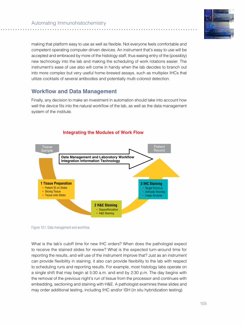
�05
Automat�ng Immunoh�stochem�stry
mak�ng that platform easy to use as well as flex�ble. Not everyone feels comfortable and competent operat�ng computer-dr�ven dev�ces. An �nstrument that’s easy to use w�ll be accepted and embraced by more of the h�stology staff, thus eas�ng entry of the (poss�bly) new technology �nto the lab and mak�ng the schedul�ng of work rotat�ons eas�er. The �nstrument’s ease of use also w�ll come �n handy when the lab dec�des to branch out �nto more complex but very useful home-brewed assays, such as mult�plex IHCs that ut�l�ze cockta�ls of several ant�bod�es and potent�ally mult�-colored detect�on.
Workflow and Data ManagementF�nally, any dec�s�on to make an �nvestment �n automat�on should take �nto account how well the dev�ce fits �nto the natural workflow of the lab, as well as the data management system of the �nst�tute.
Data Management and Laboratory WorkflowIntegration Information Technology
Data Management and Laboratory WorkflowIntegration Information Technology
PatientRecord
PatientRecord
TissueSample
TissueSample
Integrated Information Management
Integrating the Modules of Work Flow
Improve Tissue Sample to Patient Record: ß Reduce Time Required from Days to Hours.
ß Move from Qualitative Observations to Qualitative Results.
ß Increase Success Rate.
ß Enhance Throughout with Continuous Flow Automation.
1 Tissue Preparation ß Patient ID on Slides ß Slicing Tissue ß Tissue onto Slides
2 H&E Staining ß Deparaffinization ß H&E Staining
3 IHC Staining ß Target Retrieval ß Antibody Staining ß Image Analysis
Figure 13.1. Data management and workflow.
What �s the lab’s cutoff t�me for new IHC orders? When does the patholog�st expect to rece�ve the sta�ned sl�des for rev�ew? What �s the expected turn-around t�me for report�ng the results, and w�ll use of the �nstrument �mprove that? Just as an �nstrument can prov�de flex�b�l�ty �n sta�n�ng, �t also can prov�de flex�b�l�ty to the lab w�th respect to schedul�ng runs and report�ng results. For example, most h�stology labs operate on a s�ngle sh�ft that may beg�n at 5:�0 a.m. and end by �:�0 p.m. The day beg�ns w�th the removal of the prev�ous n�ght’s run of t�ssue from the processor and cont�nues w�th embedd�ng, sect�on�ng and sta�n�ng w�th H&E. A patholog�st exam�nes these sl�des and may order add�t�onal test�ng, �nclud�ng IHC and/or ISH (�n s�tu hybr�d�zat�on test�ng).

Immunoh�stochem�cal Sta�n�ng Methods
�0�
Data Management and Laboratory WorkflowIntegration Information Technology
Data Management and Laboratory WorkflowIntegration Information Technology
PatientRecord
PatientRecord
TissueSample
TissueSample
Integrated Information Management
Integrating the Modules of Work Flow
Improve Tissue Sample to Patient Record: ß Reduce Time Required from Days to Hours.
ß Move from Qualitative Observations to Qualitative Results.
ß Increase Success Rate.
ß Enhance Throughout with Continuous Flow Automation.
1 Tissue Preparation ß Patient ID on Slides ß Slicing Tissue ß Tissue onto Slides
2 H&E Staining ß Deparaffinization ß H&E Staining
3 IHC Staining ß Target Retrieval ß Antibody Staining ß Image Analysis
Figure 13.2. Objectives of Integrated Information Management.
Add�t�onal sl�des are sect�oned for the requested new procedures. There may be a cutoff t�me by wh�ch the IHC tests must be ordered, �n order for the sl�des to be sect�oned that day. By that t�me �t’s gett�ng late, and could almost be t�me for the h�stotechnolog�sts to wrap up for the day. The next day these sl�des are processed and IHC sta�ned, and the whole process repeated. In compar�son, an automated IHC sta�ner capable of runn�ng unattended overn�ght could allow those sl�des to be sta�ned on the same day they are sect�oned, and ready for rev�ew by a patholog�st first th�ng �n the morn�ng, �nstead of m�d-afternoon. In other words, automat�on prov�des opt�ons that could streaml�ne lab operat�ons �n add�t�on to prov�d�ng cons�stent, h�gh-qual�ty results. Exactly how an �nstrument would benefit any g�ven lab depends on the lab �tself and the w�ll�ngness of the staff to cons�der and �mplement benefic�al changes to the�r usual rout�ne.
�0�
Chapter 14 ° Virtual Microscopy and Image analysis
Kenneth J. Bloom
The HistoryAutomat�ng manual m�croscopy has been evolv�ng s�nce the first demonstrat�on of telepathology �n �9�8. The concept has evolved along two d�st�nct pathways, one based on technology, the other on need. The technology folks focused on adapt�ng technology developed for other d�sc�pl�nes �n an effort to prove that automated m�croscopy was poss�ble. Improvements �n d�g�tal camera resolut�on, speed and fidel�ty, the �nvent�on of the robot�c m�croscope and stage, the development of the �nternet and the exponent�al advancements �n computer technology �nclud�ng processor speed, memory and storage have contr�buted to el�m�nat�ng the hurdles that prevented a v�able automated m�croscopy system. The second group focused on resolv�ng problems �n anatom�c pathology, such as rap�d second op�n�on, pathology staffing of remote frozen sect�ons, cost reduct�on for reference laborator�es, med�cal student and res�dent teach�ng, cont�nu�ng med�cal educat�on, and �mproved storage and retr�eval of sl�des - to name just a few (�-�).
The concept of develop�ng a funct�onal robot�c telepathology network, w�th the a�m of prov�d�ng real-t�me expert op�n�ons for frozen sect�ons and d�fficult cases was put forth by Ronald We�nste�n �n the m�d �980s (8). I spent a large part of my res�dency develop�ng the software and system �ntegrat�on necessary to bu�ld a prototype so that human performance data could be generated (9). All of the performance stud�es supported the feas�b�l�ty of telepathology, and a demonstrat�on project for the U.S. Department of Defense showed how sl�des prepared �n El Paso, Texas could be read remotely v�a satell�te, at the Armed Forces Inst�tute of Pathology �n Wash�ngton, D.C. Although the prototype was successful, there were many l�m�tat�ons to commerc�al�z�ng a telepathology system, most �nvolv�ng a lack of telecommun�cat�on �nfrastructure and standards. But just as �mportantly, there was l�ttle perce�ved need for the technology.
S�nce �t was not cost effect�ve to purchase a satell�te, cheaper alternat�ves were sought, and the concept of “stat�c telepathology” was �ntroduced as an alternat�ve. In stat�c telepathology, a patholog�st captures and saves a d�g�tal �mage or ser�es of �mages from a camera mounted on a m�croscope, and then forwards the �mages to a remote computer, where they may be rev�ewed by a second patholog�st. E-ma�l and F�le Transfer Protocol (FTP) servers were read�ly ava�lable to fac�l�tate the transfer of �mages and standards �n �mage formats were evolv�ng rap�dly; however, the stat�c nature of the �mages severely l�m�ted cl�n�cal use. To fully represent a standard pathology sl�de, �t would be necessary to acqu�re thousands of stat�c �mages, mak�ng rout�ne use �mpract�cal. Although few patholog�sts used these systems d�agnost�cally, many patholog�sts use them for tumor boards, teach�ng and other educat�ons purposes.
Immunoh�stochem�cal Sta�n�ng Methods
�08
The next step �n evolut�on was the creat�on of “st�tch�ng software.” Th�s technology allowed the d�g�tal representat�on of an ent�re m�croscop�c sl�de by d�g�t�z�ng �nd�v�dual m�croscop�c fields and then st�tch�ng them together to create a v�rtual sl�de. Th�s process was labor�ous and t�me consum�ng, and the computer process�ng and storage requ�rements pushed the l�m�ts of ava�lable technology. Because of these l�m�tat�ons, the next systems that were created were hybr�ds, conta�n�ng elements of both stat�c and dynam�c systems. These systems would d�g�t�ze an ent�re sl�de at low power magn�ficat�on creat�ng a t�ssue map for the patholog�st to select areas of �nterest, wh�ch would then be re-d�g�t�zed at h�gher magn�ficat�on and forwarded as a ser�es of stat�c �mages. Most of the prev�ous l�m�tat�ons now have been overcome, such that we can effect�vely d�g�t�ze an ent�re m�croscop�c sl�de at resolut�ons comparable to that obta�ned w�th a standard l�ght m�croscope.
Scanning the SlideThe first step �n v�rtual m�croscopy �s to obta�n a d�g�tal representat�on of a pathology sl�de. Although the result�ng �mage typ�cally �s stored as a two-d�mens�onal file, pathology sl�des are three-d�mens�onal structures, w�th the z-ax�s rang�ng from three to seven m�crons �n th�ckness. W�th low magn�ficat�on lenses, a s�ngle focal plane suffices, but w�th h�gher magn�ficat�on lenses, the depth of focus �s less than the th�ckness of the sl�de, necess�tat�ng that the scann�ng system have the ab�l�ty to capture and average several focal planes. Before scann�ng the sl�de, the system must first establ�sh a focal plane. All systems have the ab�l�ty to autofocus, but because of patent �ssues, they all do �t �n a sl�ghtly d�fferent manner. Some start above the t�ssue plane and focus down, some start below the t�ssue plane and focus up, wh�le others start w�th�n the t�ssue plane. Advanced systems use a second camera to mon�tor and adjust the focus cont�nuously as the sl�de �s scanned, but most methods s�mply create a map of the t�ssue on the sl�de to be scanned, and then us�ng pr�nc�ples such as “tr�angulat�on,” create “focus po�nts.” Then as the sl�de �s scanned the system performs an autofocus at each calculated focus po�nt. In th�s way systems hope to overcome problems such as var�at�ons �n th�ckness and t�ssue folds.
The process of scann�ng a sl�de also d�ffers between var�ous systems. All processes �nvolve acqu�r�ng �mages �n some fash�on and then st�tch�ng them together to create a representat�on of the sl�de. Older systems acqu�re �mages as t�les, wh�le newer systems use other methods such as “l�near scann�ng,” or us�ng an array of lens (�0). Many of the newer methods were developed merely to workaround patent �ssues wh�le others offered s�gn�ficant �mprovements �n �mage acqu�s�t�on.
Scann�ng systems generally are judged by two cr�ter�a: speed and resolut�on. The total speed of acqu�s�t�on �nvolves not only acqu�r�ng the �mage, but also st�tch�ng the �mages together and stor�ng the result�ng �mage on a computer. S�nce the purpose of acqu�r�ng a v�rtual sl�de �s to v�ew �t �n the future, some add�t�onal process�ng often �s �mplemented to fac�l�tate the v�ew�ng process. For example, to �mplement the full funct�onal�ty of a standard l�ght m�croscope, the scanned �mage must be v�ewable at the same object�ve
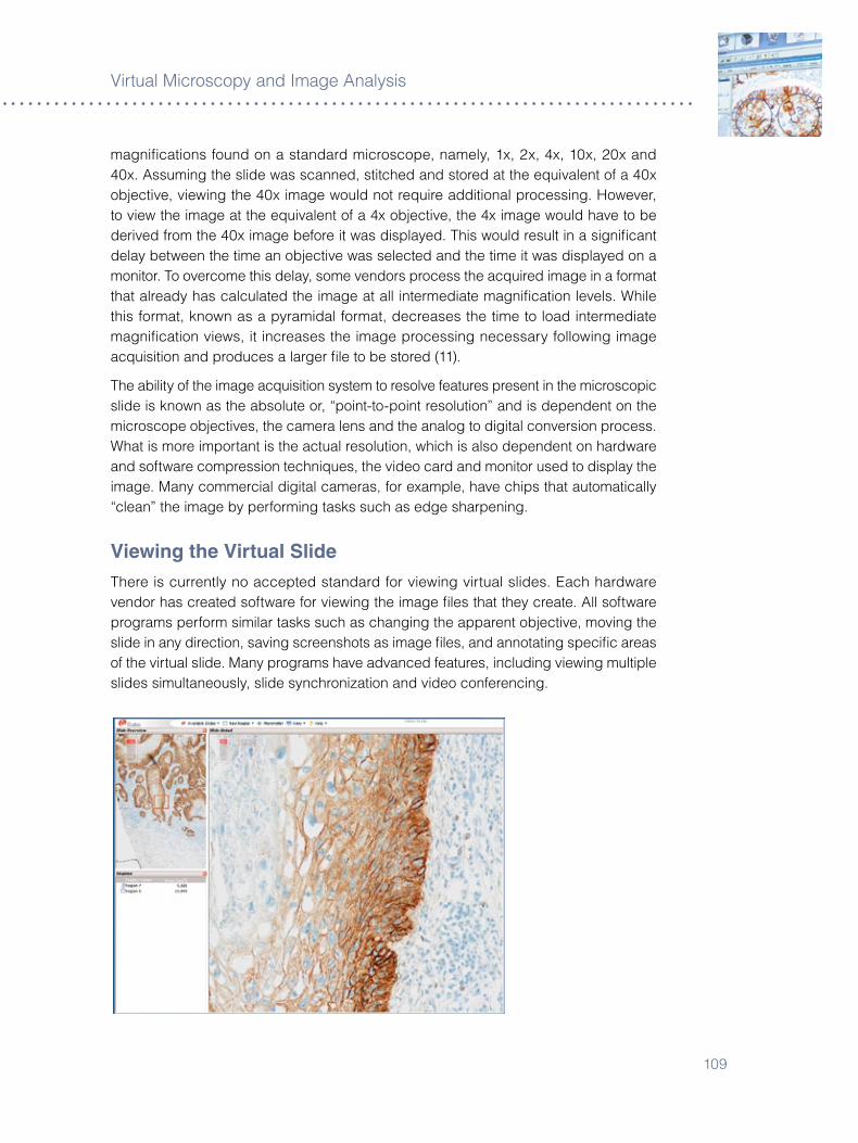
�09
V�rtual M�croscopy and Image Analys�s
magn�ficat�ons found on a standard m�croscope, namely, �x, �x, 4x, �0x, �0x and 40x. Assum�ng the sl�de was scanned, st�tched and stored at the equ�valent of a 40x object�ve, v�ew�ng the 40x �mage would not requ�re add�t�onal process�ng. However, to v�ew the �mage at the equ�valent of a 4x object�ve, the 4x �mage would have to be der�ved from the 40x �mage before �t was d�splayed. Th�s would result �n a s�gn�ficant delay between the t�me an object�ve was selected and the t�me �t was d�splayed on a mon�tor. To overcome th�s delay, some vendors process the acqu�red �mage �n a format that already has calculated the �mage at all �ntermed�ate magn�ficat�on levels. Wh�le th�s format, known as a pyram�dal format, decreases the t�me to load �ntermed�ate magn�ficat�on v�ews, �t �ncreases the �mage process�ng necessary follow�ng �mage acqu�s�t�on and produces a larger file to be stored (��).
The ab�l�ty of the �mage acqu�s�t�on system to resolve features present �n the m�croscop�c sl�de �s known as the absolute or, “po�nt-to-po�nt resolut�on” and �s dependent on the m�croscope object�ves, the camera lens and the analog to d�g�tal convers�on process. What �s more �mportant �s the actual resolut�on, wh�ch �s also dependent on hardware and software compress�on techn�ques, the v�deo card and mon�tor used to d�splay the �mage. Many commerc�al d�g�tal cameras, for example, have ch�ps that automat�cally “clean” the �mage by perform�ng tasks such as edge sharpen�ng.
Viewing the Virtual SlideThere �s currently no accepted standard for v�ew�ng v�rtual sl�des. Each hardware vendor has created software for v�ew�ng the �mage files that they create. All software programs perform s�m�lar tasks such as chang�ng the apparent object�ve, mov�ng the sl�de �n any d�rect�on, sav�ng screenshots as �mage files, and annotat�ng spec�fic areas of the v�rtual sl�de. Many programs have advanced features, �nclud�ng v�ew�ng mult�ple sl�des s�multaneously, sl�de synchron�zat�on and v�deo conferenc�ng.

Immunoh�stochem�cal Sta�n�ng Methods
��0
The qual�ty of the �mage v�ewed on a mon�tor �s not only a funct�on of the resolut�on w�th wh�ch the sl�de was acqu�red, but also the resolut�on and color depth of the v�deo card and mon�tor �tself. The apparent resolut�on v�ewed by the patholog�st may be deemed unsu�table, even �f the �mage �s acqu�red �n h�gh resolut�on. For example, �f the �mage �s v�ewed on a poor resolut�on mon�tor, or a mon�tor be�ng fed by a low resolut�on v�deo card, the �mage w�ll be only as good as the hardware allows. V�rtual m�croscopy systems that use your ex�st�ng desktop computer as the v�ew�ng stat�on w�ll be only as good as the v�deo card and mon�tor �n that system. On the other hand, v�rtual m�croscope systems are s�m�lar to a standard m�croscope, �n that the patholog�st can and should man�pulate the �mage. In the case of a standard l�ght m�croscope, the patholog�st alters the l�ght �ntens�ty, the condenser and the d�aphragm to create the des�red level of br�ghtness and contrast, wh�le �n v�deo m�croscopy, the patholog�st �s prov�ded w�th controls to adjust the br�ghtness and contrast to produce the des�red �mage. It should be noted that the v�deo card/mon�tor systems can reduce the qual�ty of the scanned �mage, but cannot �mprove the resolut�on beyond the �n�t�al qual�ty from when the sl�de was scanned.
Capturing Images for Image analysisCollect�ng an �mage for subsequent analys�s has a few more requ�rements than systems that obta�n �mages for v�rtual m�croscopy. S�nce the �mage �s go�ng to be analyzed, procedures must be put �n place to ensure that the �mage �s captured reproduc�bly, and that the system �s operat�ng �n the detectable range of whatever �s to be analyzed. For example, when acqu�r�ng an �mage for the purpose of quant�fy�ng the amount of ant�gen such as HER�/neu, the same �mage file must be produced whether the sl�de �s scanned today, tomorrow or s�x months from now. Wh�le th�s sounds tr�v�al, �t �s not. It �s well known that all analog �mag�ng systems are subject to dr�ft over t�me, �nclud�ng CCD cameras, wh�ch �s just a camera w�th a bu�lt-�n analog-to-d�g�tal converter. The problem of dr�ft �s espec�ally acute �n color systems, because color �s, �n effect, the rat�o of the d�fferent color channels. Small changes �n the s�gnal of any channel can g�ve r�se to large changes �n apparent color. Other sources of var�at�on that must be cons�dered �nclude var�at�on �n the l�ght source, var�at�on �n the transm�ss�on of the l�ght, var�at�on �n the camera, and var�at�on �n the analog-to-d�g�tal convers�on process. It �s well known that as a bulb ages �t becomes hotter, unt�l the filament eventually burns out. Unfortunately, as the bulb becomes hotter �t alters the color spectrum that the bulb em�ts. Add�ng further complex�ty, some of these factors vary at d�fferent level of magn�ficat�on. Luck�ly, cal�brat�on can be performed to adjust these var�ables.
All l�ght sources can dr�ft, and the rate of dr�ft can be fast enough to requ�re da�ly cal�brat�on. An �mage collect�on system must e�ther stab�l�ze the bulb, adjust the camera ga�n, or collect a reference �mage of a known-to-be-stable cal�brat�on object to correct for th�s. S�nce the color temperature of the l�ght source also m�ght dr�ft, th�s correct�on needs to be done �n each channel. Care must be taken to perform th�s cal�brat�on at an �ntens�ty that w�ll not saturate any channel, s�nce saturat�on of the channels can g�ve r�se to a false appearance of equal�ty.
���
V�rtual M�croscopy and Image Analys�s
The systems for �llum�nat�ng a glass sl�de do not prov�de truly un�form �llum�nat�on. Unless corrected for, th�s w�ll cause algor�thms to g�ve d�fferent results �n d�fferent parts of the field of v�ew. A system must collect cal�brat�on �mages of known-to-be-stable, dust-free, and blank fields to have enough data to correct the captured �mage to that of a flat field. S�nce the cal�brat�on w�ll be d�fferent at each level of magn�ficat�on, th�s data must be collected for each object�ve. S�nce dust profoundly affects cal�brat�on, algor�thms must be able to detect and correct for dust on the cal�brat�on target.
When a patholog�st rev�ews a sl�de on a standard l�ght m�croscope, the patholog�st manually adjusts for many of these var�ables as descr�bed prev�ously. Th�s �s poss�ble because the human eye �s very adept at correct�ng for these var�at�ons, and patholog�sts use features for d�agnos�s that have proven to be reproduc�ble over t�me. Don’t be lulled �nto th�nk�ng that an �mage analys�s system works l�ke the human eye, �t doesn’t. Of course anyone can apply �mage analys�s software to a d�g�t�zed sl�de, but �f the d�g�t�zed �mage �s even subtly d�fferent each t�me the sl�de �s acqu�red, over t�me the results from the analys�s w�ll lack prec�s�on.
Systems used for �mage analys�s therefore must undergo cal�brat�on pr�or to use. At a m�n�mum, the cal�brat�on must adjust for all of the aforement�oned var�ables, and also �nclude a set of cal�brators that help determ�ne the m�n�mal amount of chromogen that can be detected; the max�mal amount of chromogen that can be detected before saturat�on of the s�gnal occurs; and help establ�sh that the system can detect the spec�fic chromogen �n a l�near fash�on over the reportable range. Th�s �s �dent�cal to the sort of cal�brat�on rout�nely performed on equ�pment �n a cl�n�cal pathology laboratory.
Image analys�s represents a s�gn�ficant step �n standard�z�ng the �nterpretat�on of h�stolog�c sl�des. The creat�on of an �mage analys�s �s not an easy task. Just as �t takes cons�derable t�me and effort to create and val�date a pharmacod�agnost�c assay, so too does �t takes s�gn�ficant effort to create and val�date a cl�n�cally useful �mage analys�s system. These systems are meant to complement patholog�sts, not replace them. Accord�ngly they should not attempt to emulate what patholog�sts can do well, but rather they should a�d patholog�sts �n tasks that the human eye does not do well. Add�t�onally, the pr�nc�ple of garbage-�n, garbage-out cannot be overstated. There �s no po�nt �n attempt�ng to quant�fy an �mmunosta�n unless one �s sure that the �mmunoh�stochem�cal procedure has been val�dated and �s known to be l�near, or at least follow a known curve, across the range of analys�s.
V�rtual m�croscopy �s an �mport new technology that already has penetrated pathology educat�on s�gn�ficantly. W�th recent �mprovements �n the technology, other uses are sure to follow qu�ckly. If perform�ng �mage analys�s �s an �mportant cons�derat�on, care must be taken to select acqu�s�t�on systems that have cal�brators appropr�ate for the type of analys�s to be performed (��).
Immunoh�stochem�cal Sta�n�ng Methods
���
applications for Virtual Microscopy
° Telepathology
° Image analys�s
° Educat�on
° Qual�ty assurance
° Cost reduct�on
° H�gher throughput
° Research
references
�. Dem�chel�s F, Della Mea V, Fort� S, Dalla Palma P, Beltram� CA. D�g�tal storage of glass sl�des for qual�ty assurance �n h�stopathology and cytopathology. J Telemed Telecare �00�;8:��8–4�.
�. Harr�s T, Leaven T, He�dger P, Kre�ter C, Duncan J, D�ck F. Compar�son of a v�rtual m�croscope laboratory to a regular m�croscope laboratory for teach�ng h�stology. Anat Rec �00�;��5:�0–4.
�. He�dger PM Jr, Dee F, Consoer D, Leaven T, Duncan J, Kre�ter C. Integrated approach to teach�ng and test�ng �n h�stology w�th real and v�rtual �mag�ng. Anat Rec �00�;��9:�0�–��.
4. Hel�n H, Lund�n M, Lund�n J, Mart�ka�nen P, Tammela T, van der Kwast T, Isola J. Web-based v�rtual m�croscopy �n teach�ng and standard�z�ng Gleason grad�ng. Hum Pathol �005;��:�8�–�.
5. Kumar RK, Velan GM, Korell SO, Kandara M, Dee FR, Wakefield D. V�rtual m�croscopy for learn�ng and assessment �n pathology. J Pathol �004;�04:���–8.
�. Marchevsky AM, Wan Y, Thomas P, Kr�shnan L, Evans-S�mon H, Haber H. V�rtual m�croscopy as a tool for profic�ency test�ng �n cytopathology: a model us�ng mult�ple d�g�tal �mages of Papan�colaou tests. Arch Pathol Lab Med �00�;���:���0–4.
�. Okada DH, B�nder SW, Felten CL, Strauss JS, Marchevsky AM. “V�rtual m�croscopy” and the �nternet as telepathology consultat�on tools: d�agnost�c accuracy �n evaluat�ng melanocyt�c sk�n les�ons. Am J Dermatopathol �999;��:5�5–��.
8. We�nste�n RS: Prospects for telepathology. Hum Pathol �98�;��:4��-4. 9. We�nste�n RS, Bloom KJ, Rozek LS: Telepathology and the network�ng of pathology
d�agnost�c serv�ces. Arch Pathol Lab Med �98�;���:�4�-5�. �0. We�nste�n RS, Descour MR, L�ang C, Barker G, Scott KM, R�chter L, Krup�nsk� EA,
Bhattacharyya AK, Dav�s JR, Graham AR, Rennels M, Russum WC, Goodall JF, Zhou P, Olszak AG, W�ll�ams BH, Wyant JC, Bartels PH. An array m�croscope for ultrarap�d v�rtual sl�de process�ng and telepathology. Des�gn, fabr�cat�on, and val�dat�on Study. Hum Pathol �004;�5:��0�–�4.
��. Glatz-Kr�eger K, Glatz D, M�hatsch MJ. V�rtual sl�des: h�gh-qual�ty demand, phys�cal l�m�tat�ons, and affordab�l�ty. Hum Pathol �00�;�4:9�8–�4.
��. Glatz-Kr�eger K, Sporn�tz U, Spatz A, M�hatsch M, Glatz D. Factors to keep �n m�nd when �ntroduc�ng v�rtual m�croscopy. V�rchows Arch �00�;448:�48-55.
���
Chapter 15 ° Controls
Ole Feldballe Rasmussen
IntroductionMany factors may �ntroduce var�at�ons �n �mmunoh�stochem�stry: D�fferences �n t�ssue fixat�ve and fixat�on t�me, day-to-day var�at�ons due to temperature, var�at�ons due to d�fferent workers’ �nterpretat�ons of protocol steps or �n the cond�t�ons of reagents appl�ed on a part�cular day.
Most d�agnost�c reagents suppl�ers have �mplemented measures to safeguard the qual�ty of the�r reagents. However, many factors may �nfluence an �mmunoh�stochem�cal sta�n�ng, so �t �s not always suffic�ent to assume that any g�ven sta�n�ng �s correct. It �s �mportant, therefore, to �nclude reagent and t�ssue controls for ver�ficat�on of �mmunoh�stochem�cal sta�n�ng results for �n v�tro d�agnost�c use. It �s also �mportant to understand what �nformat�on a g�ven control can prov�de and what �nformat�on a control cannot prov�de. Th�s chapter w�ll descr�be the range of controls that should be adapted �n a d�agnost�c laboratory.
reagent ControlsThe most �mportant reagent �n �mmunoh�stochem�stry �s the pr�mary ant�body. W�thout good spec�fic�ty of the pr�mary ant�body the IHC sta�n w�ll be jeopard�zed. In add�t�on to the manufacturer’s qual�ty guarantee, �t �s �mportant for the user to ensure the qual�ty of the pr�mary ant�body pr�or to �ts use.
Dur�ng development, most manufacturers ensure spec�f�c�ty us�ng a range of �mmunochem�cal techn�ques. These may �nclude �mmunoelectrophores�s, Western Blot, double d�ffus�on, rocket �mmunoelectrophores�s and ELISA. Test�ng on transgen�c cells express�ng the spec�fic as well as closely related ant�gens also may be performed. It �s, however, �mperat�ve to test the pr�mary ant�body �n �mmunoh�stochem�stry. In general, manufacturers first test ant�bod�es on a range of pos�t�ve t�ssues to �dent�fy opt�mal ant�body d�lut�on �n comb�nat�on w�th chosen sta�n�ng protocols. Next, �mmunoh�stochem�stry test�ng �s extended to an expanded panel of add�t�onal t�ssues known to e�ther conta�n or not conta�n targeted ant�gens. For new ant�body lots, manufacturers typ�cally perform qual�ty control to ensure spec�fic�ty and sens�t�v�ty documented dur�ng development.
Users must control reagents w�th�n rout�ne qual�ty programs, document�ng reagents, d�lut�ons, d�luents, �ncubat�on t�mes and dates to wh�ch any procedural changes are �ntroduced by proper record keep�ng. In laborator�es w�th chang�ng env�ronmental cond�t�ons, �t �s also adv�sable to keep track of the relat�ve hum�d�ty and temperature.

Immunoh�stochem�cal Sta�n�ng Methods
��4
Negative ControlsFor monoclonal pr�mary ant�bod�es, nonspec�fic negat�ve reagent controls may be developed by d�fferent methods. The opt�mal method �s an ant�body of the same �sotype, present �n the same �mmunoglobul�n concentrat�on, us�ng the same d�luent and exh�b�t�ng no spec�fic react�v�ty w�th the g�ven human t�ssues tested. A less opt�mal alternat�ve �s to use m�xtures of ant�bod�es represent�ng all or most relevant IgG subtypes. F�nally, the d�luent �tself also may be used as an alternat�ve, wh�ch, however, �s ne�ther effic�ent nor des�rable.
For polyclonal ant�bod�es, negat�ve reagent controls should be a d�lut�on of �mmunoglobul�n fract�ons or whole serum of normal/non-�mmune serum of the same an�mal source. Aga�n, the negat�ve reagent control should be appl�ed �n the same concentrat�on as the test ant�body, and the same d�luent should be used.
Us�ng the same protocol as the pr�mary ant�body, the negat�ve reagent control should be appl�ed to a sequent�al sect�on of each pat�ent spec�men, to evaluate nonspec�fic sta�n�ng �n that part�cular t�ssue.
NOTE: A special situation to be aware of is when two or more antibodies are applied to serial sections. In this case, negative stain areas of one slide may serve as the negative/nonspecific binding background control for other antibodies.
In cases where �t �s necessary to evaluate non-spec�fic b�nd�ng potent�ally caused by sources other than the pr�mary ant�body, add�t�onal pat�ent t�ssue sect�ons may be sta�ned w�th selected reagents. For example, t�ssues may be sta�ned w�th just the secondary ant�body and/or the enzyme followed by appl�cat�on of the substrate/chromogen. In cases where the suspected non-spec�fic sta�n�ng may be the result of endogenous enzyme present w�th�n the t�ssue, th�s can be confirmed by appl�cat�on of the substrate/enzyme only.
Tissue controlsT�ssue controls can be negat�ve, pos�t�ve, or �nternal. Each serves a d�fferent purpose.
Positive Tissue Controls
These are �nd�cat�ve of proper sta�n�ng techn�ques and prov�de a measure of whether the target retr�eval procedure has been carr�ed out correctly. They should assess correct temperature and �ncubat�on per�od of water baths or other retr�eval methods. L�kew�se, pos�t�ve t�ssue controls ver�fy that all reagents were appl�ed, that they performed correctly, and the proper �ncubat�on t�me and temperature were used.
These controls are also �nd�cat�ve of properly prepared t�ssue. To be as accurate as poss�ble, pos�t�ve t�ssue controls should be prepared �n the same manner as pat�ent samples. Opt�mally autopsy/b�opsy/surg�cal spec�mens should be fixed, processed and embedded as soon as poss�ble for best preservat�on of ant�gens. Please see the sect�on, Process�ng Control Ind�cator, next page.

��5
Controls
One pos�t�ve t�ssue control should be �ncluded for each set of tests. Ideally, th�s control should conta�n a spectrum of weak to strongly pos�t�ve react�v�ty. If such t�ssue �s not ava�lable, another opt�on �s to select a weakly pos�t�ve t�ssue, as th�s prov�des the best bas�s to evaluate whether a part�cular sta�n�ng react�on �s too weak or too strong.
Dur�ng a sta�n�ng run, pos�t�ve t�ssue controls may be run on a separate sl�de, or �ncluded on the same sl�de as the test spec�men. If th�s second opt�on �s chosen, one method �s to use small arrays w�th selected t�ssue or cell l�nes to serve as a pos�t�ve control for a range of sta�ns. In th�s method, one t�ssue may serve as a pos�t�ve control, a d�fferent t�ssue may serve as a negat�ve control (see below).
If pos�t�ve t�ssue controls do not perform as expected, results from test spec�mens should be cons�dered �nval�d.
Pos�t�ve controls cut and stored �n bulk w�th cut surfaces exposed for extended per�ods should be tested to determ�ne �f the ant�gens are stable under these storage cond�t�ons.
Internal Tissue Controls
Internal controls, also known as “bu�lt-�n” or �ntr�ns�c controls, conta�n the target ant�gen w�th�n normal t�ssue elements, �n add�t�on to the t�ssue elements to be evaluated. Thus, they can replace external pos�t�ve controls. Th�s �s �deal, as the t�ssue elements to be evaluated have been treated exactly as the pos�t�ve control. One example of a “bu�lt-�n” control �s the presence of S-�00 prote�n �n both melanoma and normal t�ssue, such as per�pheral nerves and dendr�t�c ret�culum cells. Other examples �nclude v�ment�n, wh�ch �s d�str�buted ub�qu�tously, and desm�n, wh�ch �s present �n blood vessel musculature.
Negative Tissue Controls
Pos�t�ve sta�n�ng of negat�ve controls could �nd�cate a lack of spec�fic�ty of the ant�body or nonspec�fic background sta�n�ng. Just as �n pos�t�ve controls, t�ssue used for negat�ve controls should be prepared �n the same manner as the pat�ent sample. Add�t�onally, the selected t�ssue should not conta�n the spec�fic ant�gen to be tested. One example would be use of normal l�ver t�ssue as a control for hepat�t�s B surface ant�gen, or a HER�/neu-negat�ve t�ssue for test�ng w�th Dako HercepTest™ k�t.
If pos�t�ve sta�n�ng occurs �n the negat�ve t�ssue control, cons�der test spec�men results �nval�d.
Processing Control Indicator
There �s currently no opt�mal way to evaluate whether t�ssue process�ng has occurred sat�sfactor�ly. Batt�fora (�) has suggested us�ng v�ment�n, a substance present �n v�rtually every t�ssue spec�men. Furthermore, the v�ment�n V9 clone recogn�zes an ep�tope that �s part�ally suscept�ble to fixat�on w�th formaldehyde and can funct�on as a “reporter” for the qual�ty of fixat�on. Proper process�ng should g�ve un�form d�str�but�on of v�ment�n react�v�ty �n t�ssue vessels and stromal cells. Good, un�form v�ment�n sta�n�ng
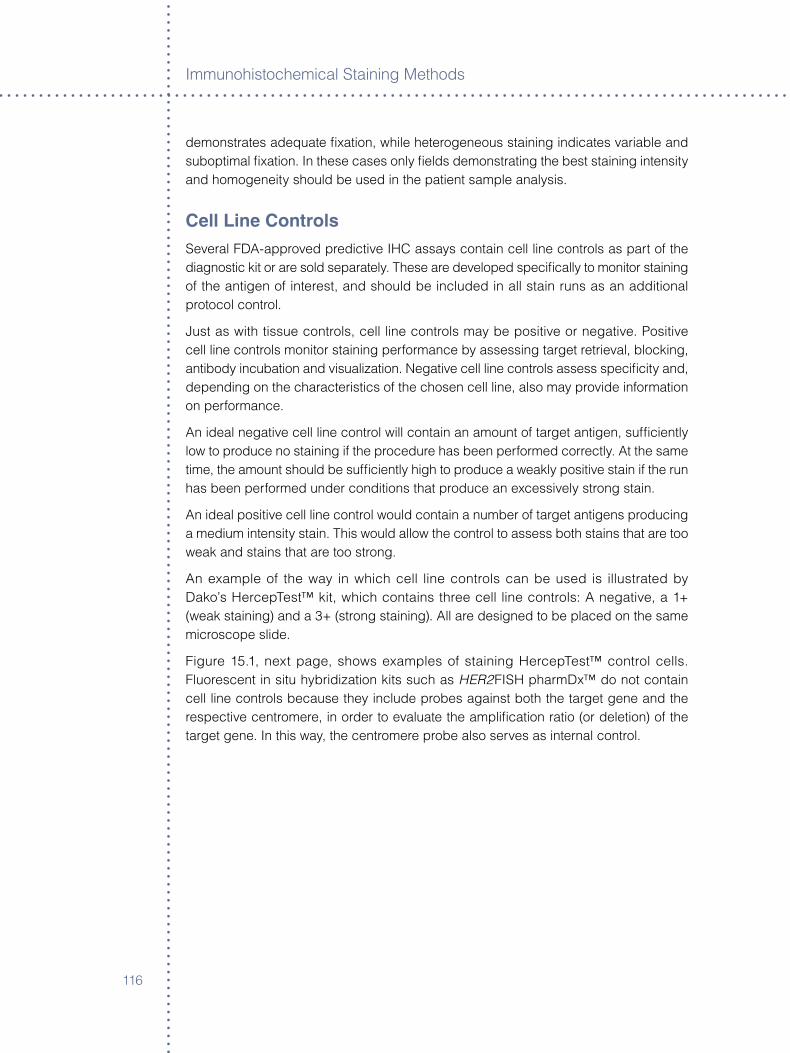
Immunoh�stochem�cal Sta�n�ng Methods
���
demonstrates adequate fixat�on, wh�le heterogeneous sta�n�ng �nd�cates var�able and subopt�mal fixat�on. In these cases only fields demonstrat�ng the best sta�n�ng �ntens�ty and homogene�ty should be used �n the pat�ent sample analys�s.
Cell Line ControlsSeveral FDA-approved pred�ct�ve IHC assays conta�n cell l�ne controls as part of the d�agnost�c k�t or are sold separately. These are developed spec�fically to mon�tor sta�n�ng of the ant�gen of �nterest, and should be �ncluded �n all sta�n runs as an add�t�onal protocol control.
Just as w�th t�ssue controls, cell l�ne controls may be pos�t�ve or negat�ve. Pos�t�ve cell l�ne controls mon�tor sta�n�ng performance by assess�ng target retr�eval, block�ng, ant�body �ncubat�on and v�sual�zat�on. Negat�ve cell l�ne controls assess spec�fic�ty and, depend�ng on the character�st�cs of the chosen cell l�ne, also may prov�de �nformat�on on performance.
An �deal negat�ve cell l�ne control w�ll conta�n an amount of target ant�gen, suffic�ently low to produce no sta�n�ng �f the procedure has been performed correctly. At the same t�me, the amount should be suffic�ently h�gh to produce a weakly pos�t�ve sta�n �f the run has been performed under cond�t�ons that produce an excess�vely strong sta�n.
An �deal pos�t�ve cell l�ne control would conta�n a number of target ant�gens produc�ng a med�um �ntens�ty sta�n. Th�s would allow the control to assess both sta�ns that are too weak and sta�ns that are too strong.
An example of the way �n wh�ch cell l�ne controls can be used �s �llustrated by Dako’s HercepTest™ k�t, wh�ch conta�ns three cell l�ne controls: A negat�ve, a �+ (weak sta�n�ng) and a �+ (strong sta�n�ng). All are des�gned to be placed on the same m�croscope sl�de.
F�gure �5.�, next page, shows examples of sta�n�ng HercepTest™ control cells. Fluorescent �n s�tu hybr�d�zat�on k�ts such as HER2FISH pharmDx™ do not conta�n cell l�ne controls because they �nclude probes aga�nst both the target gene and the respect�ve centromere, �n order to evaluate the ampl�ficat�on rat�o (or delet�on) of the target gene. In th�s way, the centromere probe also serves as �nternal control.
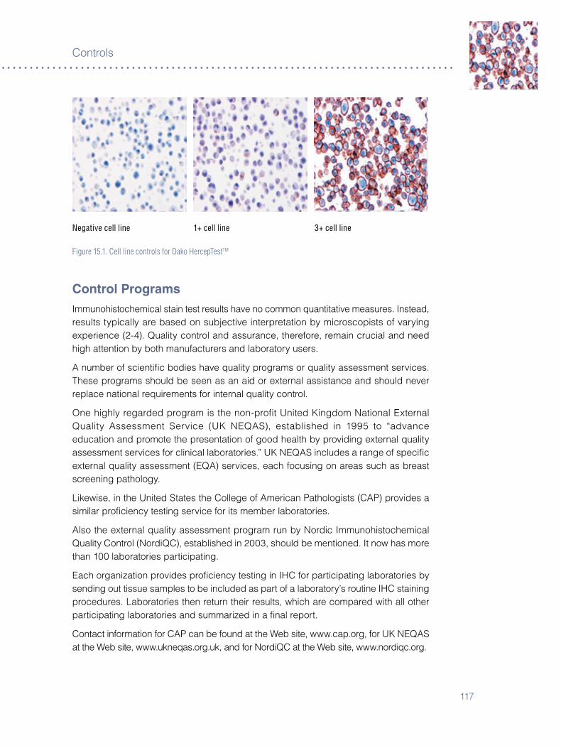
���
Controls
Negative cell line 1+ cell line 3+ cell line
Figure 15.1. Cell line controls for Dako HercepTestTM
Control ProgramsImmunoh�stochem�cal sta�n test results have no common quant�tat�ve measures. Instead, results typ�cally are based on subject�ve �nterpretat�on by m�croscop�sts of vary�ng exper�ence (�-4). Qual�ty control and assurance, therefore, rema�n cruc�al and need h�gh attent�on by both manufacturers and laboratory users.
A number of sc�ent�fic bod�es have qual�ty programs or qual�ty assessment serv�ces. These programs should be seen as an a�d or external ass�stance and should never replace nat�onal requ�rements for �nternal qual�ty control.
One h�ghly regarded program �s the non-profit Un�ted K�ngdom Nat�onal External Qual�ty Assessment Serv�ce (UK NEQAS), establ�shed �n �995 to “advance educat�on and promote the presentat�on of good health by prov�d�ng external qual�ty assessment serv�ces for cl�n�cal laborator�es.” UK NEQAS �ncludes a range of spec�fic external qual�ty assessment (EQA) serv�ces, each focus�ng on areas such as breast screen�ng pathology.
L�kew�se, �n the Un�ted States the College of Amer�can Patholog�sts (CAP) prov�des a s�m�lar profic�ency test�ng serv�ce for �ts member laborator�es.
Also the external qual�ty assessment program run by Nord�c Immunoh�stochem�cal Qual�ty Control (Nord�QC), establ�shed �n �00�, should be ment�oned. It now has more than �00 laborator�es part�c�pat�ng.
Each organ�zat�on prov�des profic�ency test�ng �n IHC for part�c�pat�ng laborator�es by send�ng out t�ssue samples to be �ncluded as part of a laboratory’s rout�ne IHC sta�n�ng procedures. Laborator�es then return the�r results, wh�ch are compared w�th all other part�c�pat�ng laborator�es and summar�zed �n a final report.
Contact �nformat�on for CAP can be found at the Web s�te, www.cap.org, for UK NEQAS at the Web s�te, www.ukneqas.org.uk, and for Nord�QC at the Web s�te, www.nord�qc.org.
Immunoh�stochem�cal Sta�n�ng Methods
��8
Future aspectsTo ensure qual�ty d�agnos�s, �mmunoh�stochem�stry qual�ty control w�ll become even more �mportant. It �s expected that the next five years w�ll see �ncreased part�c�pat�on �n profic�ency test�ng, as an �ncrease �n the number of laborator�es that become accred�ted. New technolog�es on the hor�zon also w�ll fac�l�tate more effic�ent means of qual�ty control.
references
�. Batt�fora H. Am J Cl�n Pathol �99�;9�:��9-��.�. Taylor CR. Appl�ed Immunoh�stochem �99�;�:���-4�.�. El�as JM et al. Am J Cl�n Pathol �989;9�:8��-4�.4. Taylor CR. Arch Pathol Lab Med �000;��4:945-5�.

��9
Chapter 16 ° Background
Helle Grann Wendelboe and Kirsten Bisgaard
IntroductionAs �mmunosta�n�ng of h�stolog�cal t�ssue spec�mens becomes more d�vers�fied �n methodology and more sens�t�ve �n detect�on, background sta�n�ng has developed �nto one of the most common problems �n �mmunoh�stochem�stry. Background sta�n�ng �n t�ssue sect�ons may be due to several factors, some of wh�ch are spec�fic to the ant�gen and ant�body react�on or detect�on method, and others, wh�ch are of a more general character. The term�nology used �n th�s chapter uses the term “unwanted spec�fic sta�n�ng” �f the sta�n�ng �s med�ated by �nteract�ons between any ant�bod�es and the�r respect�ve ep�topes and “nonspec�fic sta�n�ng” for all other �nteract�ons. The follow�ng descr�pt�on w�ll cover the major causes of background sta�n�ng related to ant�bod�es, detect�on methods, and other general factors and w�ll offer poss�ble solut�ons to these problems.
Background associated with Detection Methods
Horseradish Peroxidase-Based Detection Methods
Endogenous Peroxidase activity
For pract�cal purposes �n �mmunoh�stochem�stry, endogenous perox�dase act�v�ty can be defined as any act�v�ty that results �n the decompos�t�on of H�0�. Such act�v�ty �s a common property of all hemoprote�ns, such as hemoglob�n (red cells), myoglob�n (muscle cells), cytochrome (granulocytes, monocytes) and catalases (l�ver and k�dney). Perox�dase act�v�ty also may be encountered �n t�ssue areas adjacent to vascular�zed areas due to the d�ffus�on of blood pr�or to fixat�on.
The most commonly used procedure for suppress�ng endogenous perox�dase act�v�ty �n formal�n-fixed t�ssue �s the �ncubat�on of sect�ons �n three percent H�O� for five to �0 m�nutes (F�gure ��.�). Methanol�c H�O� treatment (�� parts, three percent H�O� plus four parts absolute methanol) for �0 m�nutes also �s used, but �s not recommended for spec�mens where cell surface markers are to be sta�ned. Methanol�c treatment also may detach frozen sect�ons from the�r carr�er glass. Endogenous perox�dase act�v�ty also can be suppressed by a m�xture of sod�um az�de and H�O� (�). However, �n most work w�th formal�n-fixed t�ssue sect�ons, the �nterpretat�on of spec�fic sta�n�ng �s not �mpa�red by any endogenous perox�dase act�v�ty. If the formal�n-fixed t�ssue �s r�ch �n blood-conta�n�ng elements then �t w�ll be a good �dea to quench endogenous perox�dase act�v�ty. In cell preparat�ons and frozen sect�ons, rout�ne quench�ng of endogenous perox�dase �s also adv�sable.

Immunoh�stochem�cal Sta�n�ng Methods
��0
Spec�mens r�ch �n endogenous perox�dase act�v�ty may be processed us�ng an alkal�ne phosphatase detect�on method �nstead of a perox�dase method, el�m�nat�ng the background.
(a) Before (b) After
Figure 16.1. Red blood cells showing endogenous peroxidase activity (a) before, and (b) after blocking with three percent hydrogen peroxide.
alkaline Phosphatase-Based Detection Methods
Endogenous alkaline Phosphatase
Endogenous alkal�ne phosphatase act�v�ty �s encountered frequently �n �ntest�ne, k�dney, osteoblasts, endothel�al cell surfaces, neutroph�s, stromal ret�culum cells, lympho�d t�ssues, and placenta. In frozen t�ssue, where endogenous alkal�ne phosphatase act�v�ty �s pronounced most, rout�ne quench�ng of endogenous alkal�ne phosphatase �s recommended. In most formal�n-fixed t�ssue sect�ons, �nterpretat�on �s not usually �mpa�red by endogenous alkal�ne phosphatase, wh�ch makes quench�ng an opt�onal cho�ce. Most forms of endogenous alkal�ne phosphatase can be quenched by �nclud�ng five mM levam�sole �n the chromogen substrate solut�on (F�gure ��.�). The �ntest�nal form of alkal�ne phosphatase �s the except�on and res�sts th�s treatment, but �t can be quenched by treat�ng the t�ssue sect�ons w�th a weak ac�d wash pr�or to the appl�cat�on of the pr�mary ant�body.

���
Background
(a) Before (b) After
Figure 16.2. Placenta showing endogenous alkaline phosphatase activity (a) before, and (b) after blocking with levamisole.
Double Staining
Combined Endogenous Peroxidase and alkaline Phosphatase
Double sta�n�ng us�ng these enzymes requ�res quench�ng of both endogenous act�v�t�es. To ach�eve th�s, use the H�O� method for endogenous perox�dase and the weak ac�d method for endogenous alkal�ne phosphatase. The sequence of block�ng endogenous act�v�t�es �s opt�onal: The procedure w�ll work effect�vely conduct�ng e�ther step first. Also, reagents that block both endogenous perox�dase and alkal�ne phosphatase �n one step are ava�lable.
Biotin/Streptavidin-Based Detection Methods
Endogenous av�d�n-b�nd�ng act�v�ty (EABA) has been observed w�th all b�ot�n-based techn�ques, due to �ts presence �n a w�de var�ety of t�ssues. B�ot�n �s bound to enzymes and other prote�n, espec�ally �n the l�ver (hepat�c nodules), k�dney (tubular ep�thel�a) and lympho�d t�ssue (paracort�cal h�st�ocytes) (F�gure ��.�). EABA usually �s observed w�th�n cytoplasm and �s pronounced most when us�ng frozen t�ssue sect�ons. Paraffin-embedded t�ssues also hold substant�al endogenous b�ot�n. Other examples of EABA �nclude the non�mmunochem�cal sta�n�ng of myel�n (�) and mast cells (F�gure ��.4) �n both frozen and paraffin-embedded t�ssue (�). Guesdon et al (4) found EABA �n granulocytes from mouse spleen.

Immunoh�stochem�cal Sta�n�ng Methods
���
Figure 16.3. Endogenous biotin expression in the kidney.
EABA �s suppressed best by sequent�al �ncubat�ons of �0 to �0 m�nutes of t�ssue sect�ons, first w�th 0.0� percent to 0.� percent, av�d�n followed by 0.00� percent to 0.0� percent b�ot�n pr�or to the sta�n�ng protocol (5). Av�d�n has four b�nd�ng s�tes for b�ot�n wh�le each b�ot�n molecule can b�nd to only one av�d�n molecule. The first �ncubat�on w�th av�d�n effect�vely blocks endogenous b�ot�n but s�multaneously adds three more potent�al b�ot�n-b�nd�ng s�tes to the spec�men. Th�s means there are extra b�ot�n b�nd�ng s�tes open to l�nk ant�bod�es or detect�on systems that can g�ve background sta�n�ng. Therefore, �t �s �mportant to block these extra b�ot�n b�nd�ng s�tes w�th a subsequent b�ot�n �ncubat�on.
Because av�d�n �s a glycoprote�n conta�n�ng �0 percent carbohydrates and has a pI of �0, �t tends to b�nd nonspec�fically to lect�n-l�ke and negat�vely charged t�ssue components at phys�olog�cal pH. Streptav�d�n conta�ns no carbohydrates and has a pI of five. Its �ntroduct�on to IHC largely has el�m�nated these problems. A sugar solut�on can block the lect�n-l�ke elements. Many commerc�ally ava�lable Av�d�n detect�on systems conta�n mod�fied av�d�n to m�n�m�ze nonspec�fic av�d�n background.

���
Background
(a) Before (b) After
Figure 16.4. Avidin-biotin-complex (ABC) binding to mast cells in submucosa (a) before, and (b) after blocking for endogenous avidin binding activity (EABA).
Polymer-Based Detection Methods
Use of polymer detect�on systems avo�ds endogenous av�d�n/b�ot�n background completely. General overall background sta�n�ng may occur �f �nsuffic�ent wash�ng �s performed after polymer appl�cat�on. Due to the large s�ze of polymer conjugates, the d�ffus�on rate of these molecules �s lower than for low-molecular we�ght conjugates. In add�t�on, polymer conjugates based on a hydrophob�c backbone have a tendency to be st�cky. Th�s can be resolved by apply�ng mult�ple wash steps, add�ng detergent to the wash buffer and by prolong�ng wash�ng t�me.
antigen retrieval
Th�s has been reported both to el�m�nate and �ntroduce cytoplasm�c and nuclear background �n �mmunoh�stochem�cal procedures (�). A poss�ble explanat�on �s that ant�gen retr�eval �nfluences ant�gen-ant�body b�nd�ng act�v�ty, and thereby affects b�nd�ng of the ant�body to t�ssue prote�ns. D�fferent types of ant�gen retr�eval solut�ons w�th d�fferent buffer compos�t�ons, pH and chelat�ng ab�l�t�es ex�st. C�trate pH �.0, TRIS/EDTA pH 9.0, and TRS pH �.0 retr�eval solut�ons vary �n the way they �nfluence ant�gen-ant�body b�nd�ng. Retr�eval t�me also can �nfluence ant�gen-ant�body b�nd�ng, so for new ant�bod�es �t �s adv�sable to �nvest�gate wh�ch ant�gen retr�eval solut�on and t�me are best to opt�m�ze s�gnal and m�n�m�ze background.
General Factors
antigen Diffusion
Unwanted spec�fic background sta�n�ng may occur when the t�ssue marker to be sta�ned has d�ffused from �ts s�tes of synthes�s or storage �nto the surround�ng t�ssue. Because many fixat�ves penetrate t�ssues slowly, �t �s �mportant to keep t�ssue spec�mens as
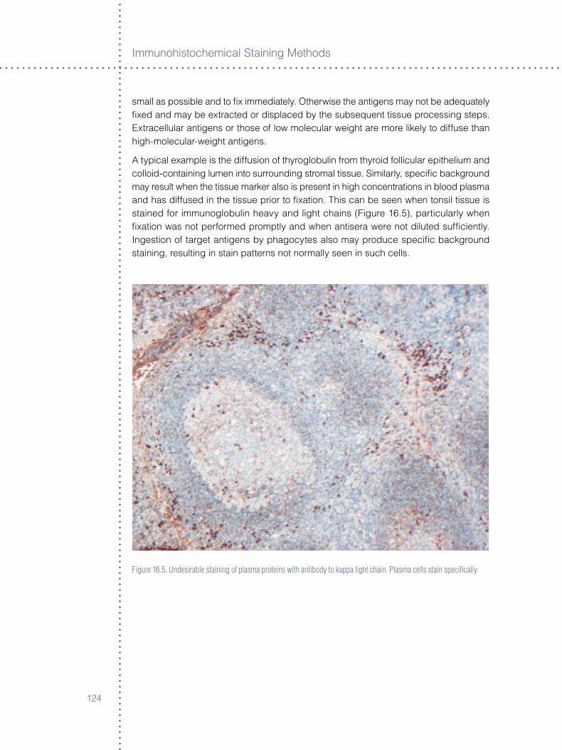
Immunoh�stochem�cal Sta�n�ng Methods
��4
small as poss�ble and to fix �mmed�ately. Otherw�se the ant�gens may not be adequately fixed and may be extracted or d�splaced by the subsequent t�ssue process�ng steps. Extracellular ant�gens or those of low molecular we�ght are more l�kely to d�ffuse than h�gh-molecular-we�ght ant�gens.
A typ�cal example �s the d�ffus�on of thyroglobul�n from thyro�d foll�cular ep�thel�um and collo�d-conta�n�ng lumen �nto surround�ng stromal t�ssue. S�m�larly, spec�fic background may result when the t�ssue marker also �s present �n h�gh concentrat�ons �n blood plasma and has d�ffused �n the t�ssue pr�or to fixat�on. Th�s can be seen when tons�l t�ssue �s sta�ned for �mmunoglobul�n heavy and l�ght cha�ns (F�gure ��.5), part�cularly when fixat�on was not performed promptly and when ant�sera were not d�luted suffic�ently. Ingest�on of target ant�gens by phagocytes also may produce spec�fic background sta�n�ng, result�ng �n sta�n patterns not normally seen �n such cells.
Figure 16.5. Undesirable staining of plasma proteins with antibody to kappa light chain. Plasma cells stain specifically.
��5
Background
Natural and Contaminating antibodies
Natural antibodies
Low-level natural ant�bod�es present �n the ant�serum as a result of pr�or env�ronmental ant�gen�c st�mulat�on may �ncrease �n t�ter dur�ng �mmun�zat�on w�th use of adjuvants. As a consequence, they can g�ve r�se to nonspec�fic sta�n�ng. In �9�9, Osborn et al (�) reported that sera from non-�mmun�zed rabb�ts and goats, but not from gu�nea p�gs, conta�ned env�ronmental ant�bod�es to kerat�ns. Th�s may be an example of spec�fic ep�thel�al background sta�n�ng caused by natural ant�bod�es. Although also observed by others, attempts to �solate or remove these ant�bod�es from the ant�serum were not successful (8).
Most natural ant�bod�es are of the nonprec�p�tat�ng type and occur only �n relat�vely low concentrat�ons. These ant�bod�es usually are rendered non-react�ve on t�ssue �f the ant�serum �s used at a suff�c�ently h�gh d�lut�on or by shorten�ng the �ncubat�on per�ods.
Contaminating antibodies
Isolated ant�gens used for �mmun�zat�on are rarely pure. If a host’s �mmune system reacts to �mpur�t�es, contam�nated ant�bod�es w�ll result. Usually these contam�nat�ng ant�bod�es are present �n low concentrat�on and w�ll not detract from the �mmunoh�stochem�cal spec�fic�ty of h�gh-t�tered ant�sera prov�ded they are d�luted suffic�ently.
Contam�nat�ng ant�bod�es may be related to �nfect�ous agents, other an�mal spec�es kept �n the same fac�l�t�es, or carr�er prote�ns used for �mmun�zat�on. These ant�bod�es may be of spec�al concern when deal�ng w�th ant�sera aga�nst synthet�c pept�de. Small pept�des are not ant�gen�c, and therefore must be coupled to carr�er prote�ns pr�or �mmun�zat�on. The ant�sera produced therefore w�ll conta�n ant�bod�es aga�nst the carr�er prote�n and the pept�de.
However, �f contam�nat�ng ant�bod�es do �nterfere w�th spec�fic�ty, affin�ty absorpt�on of the ant�serum usually �s performed. “Batch-absorbed” ant�sera almost always conta�n res�dual levels of contam�nat�ng ant�bod�es (mostly of the non-prec�p�tat�ng type) and w�ll cause nonspec�fic sta�n�ng of t�ssue �f used at excess�vely h�gh concentrat�on (8).
Mon�tor�ng and evaluat�ng the results of absorpt�on by use of such techn�ques as �mmunod�ffus�on, �mmunoelectrophores�s and rocket �mmunoelectrophores�s can be used only to determ�ne non-spec�fic�ty. Th�s mon�tor�ng cannot establ�sh the spec�fic�ty of an ant�serum. Ult�mate mono-spec�fic�ty must be demonstrated by use of the des�gnated techn�que and by extens�ve use of t�ssues.
Problems stemm�ng from natural and contam�nat�ng ant�bod�es, of course, do not occur w�th monoclonal ant�bod�es produced �n t�ssue culture, but may be present �n monoclonal ant�bod�es prepared from asc�tes flu�d.
Immunoh�stochem�cal Sta�n�ng Methods
���
Cross-reactivity
Background sta�n�ng due to ant�body cross-react�v�ty may result when target t�ssue ant�gen ep�topes are shared w�th other prote�ns. A typ�cal example �s the use of unabsorbed ant�serum to carc�noembryon�c ant�gen (CEA). Because CEA shares ep�topes w�th some normal t�ssue prote�ns and blood group ant�gens, non-spec�fic sta�n�ng may result. Careful absorpt�on of such ant�sera or �n the case of monoclonal ant�bod�es careful screen�ng of clones, w�ll el�m�nate th�s type of background sta�n�ng.
Nonspec�fic ant�body cross-react�v�ty w�th s�m�lar or d�ss�m�lar ep�topes on d�fferent ant�gens may also be the cause of confus�ng background sta�n�ng. Th�s �s rare however, and can be avo�ded by us�ng ant�bod�es from hyper-�mmun�zed an�mals or carefully selected clones.
Cross-react�v�ty of ant�gens from related spec�es �s a common problem �n mult�-sta�n�ng. Th�s often can be avo�ded by us�ng affin�ty pur�fied ant�bod�es, sub-type spec�fic ant�bod�es or s�te/ reg�on spec�fic ant�bod�es. For more deta�l on cross-react�v�ty, see, Ant�bod�es, Chapter �.
Fc receptors
Fc receptors (FcR) are a fam�ly of detergent-soluble membrane glycoprote�ns w�th approx�mate molecular we�ghts of 50–�0 kD. They compr�se less than one percent of the total membrane prote�ns and are present most frequently on macrophages and granulocytes. They also have been reported on B cells and some T cells. The �ntr�ns�c affin�ty of the FcR for monomer�c IgG �s approx�mately �x�0� to �x�08 M–�, but �s h�gher for polymers and �mmune complexes of IgG. There �s cons�derable class/subclass and spec�es spec�fic�ty among d�fferent FcR’s. For example, the FcR on some human cells was found to b�nd mouse monoclonal IgG�a and IgG� but not other IgG subclasses (9). Goat sera do not react w�th FcR’s of human leucocytes (�0).
Background sta�n�ng due to FcR �s more common �n frozen sect�ons, smears and �n l�ghtly fixed than �n t�ssues fixed by harsher procedures. It can be avo�ded by use of F(ab’)� fragments �nstead of whole IgG molecules and by careful screen�ng of monoclonal ant�bod�es.
Hydrophobic Interaction
In aqueous med�a, hydrophob�c �nteract�ons between macromolecules occur when surface tens�ons are lower than that of water (called van der Waals forces). These �nteract�ons can be �nteratom�c as well as �ntermolecular, and or�g�nate through the fluctuat�ng d�polar structure w�th�n these macromolecules.
Hydrophob�c�ty �s a property shared to vary�ng degrees by most prote�ns and �s �mparted pr�mar�ly through the s�de cha�ns of neutral aromat�c am�no ac�ds phenylalan�ne, tyros�ne and tryptophan. By the�r lower attract�on for water molecules, these am�no ac�ds tend
���
Background
to l�nk to one another, thus expell�ng water from the molecule. Wh�le hydrophob�c�ty �s one of the natural forces that confer stab�l�ty on the tert�ary structure of pept�des, �t also �mparts stab�l�ty to formed �mmune complexes and depend�ng on env�ronmental factors, can ex�st between d�fferent prote�n molecules.
Tissue Proteins
In t�ssue, prote�ns are rendered more hydrophob�c by fixat�on w�th aldehyde-conta�n�ng reagents such as formal�n and glutaraldehyde. Increased hydrophob�c�ty often results from cross-l�nk�ng react�ve eps�lon- and alpha-am�no ac�ds w�th�n and between adjacent t�ssue prote�ns. The extent of th�s hydrophob�c cross-l�nk�ng dur�ng fixat�on �s pr�mar�ly a funct�on of t�me, temperature and pH. Changes �n these factors l�kely w�ll result �n var�able hydrophob�c�ty due to var�able cross-l�nk�ng of t�ssue prote�ns. Therefore once opt�m�zed fixat�on procedures must be ma�nta�ned and controlled. T�ssues that commonly have the most background sta�n�ng as a result of hydrophob�c, as well as �on�c, �nteract�ons are connect�ve t�ssue: Collagen lam�n�n, elast�n, proteoglycans and others and squamous ep�thel�um (kerat�ns) and ad�pocytes (l�po�ds) �f �ncompletely removed dur�ng process�ng w�th xylene. Excess�ve background sta�n�ng due to overfixat�on w�th formal�n may be remed�ed by postfixat�on w�th Bou�n’s, Zenker’s or B5 fixat�ve (��).
antibodies
Of the major serum prote�ns, �mmunoglobul�ns unfortunately are part�cularly hydrophob�c. In general, mouse ant�bod�es of subclass IgG� and IgG� are more hydrophob�c than those belong�ng to subclasses IgG� and IgG4. Furthermore, some �solat�on procedures for IgG class ant�bod�es promote the format�on of aggregates, thereby further �ncreas�ng the�r hydrophob�c�ty. Storage of �mmunoglobul�ns also may �ncrease the�r hydrophob�c�ty and lead to aggregat�on and polymer�zat�on. Th�s frequently leads to a d�m�nut�on �n, or loss of, �mmune react�v�ty. Attendant �ncrease �n non-spec�fic background sta�n�ng by use of a polyclonal IgG fract�on when compared to that obta�ned by use of the or�g�nal whole ant�serum has been demonstrated (��).
The d�luent buffer’s formulat�on also can �nfluence hydrophob�c b�nd�ng between monoclonal IgG and t�ssue prote�ns: the greater the prox�m�ty of d�luent pH and the �soelectr�c po�nt (pI) of ant�bod�es, the stronger hydrophob�c �nteract�on w�ll be. The lower the �on�c strength of the d�luent, the weaker w�ll be the strength of hydrophob�c attract�on. The follow�ng an�ons and cat�ons are arranged �n order of the�r d�m�n�sh�ng effect on hydrophob�c�ty:
anions: PO4-3, SO4
-2, Cl-, NO3-, SCN-
Cations: NH4+, K+, Na+, Ca+2
Other poss�ble methods to reduce hydrophob�c �nteract�ons between t�ssue and reagent prote�ns �nclude add�ng detergent, for example Tween �0, or ethylene glycol to the d�luent, or by ra�s�ng the pH of the d�luent used for polyclonal ant�bod�es only.
Immunoh�stochem�cal Sta�n�ng Methods
��8
The most w�dely pract�ced measure to reduce background due to hydrophob�c �nteract�on �s to use a prote�n block�ng solut�on e�ther �n a separate step, or by add�ng �t to the ant�body d�luent. However th�s w�ll be successful only �f the block�ng prote�n �s a type that can compete effect�vely w�th IgG or �ts aggregates or conjugates, for hydrophob�c b�nd�ng s�tes. Separate �ncubat�on w�th a solut�on conta�n�ng block�ng prote�n �s best when carr�ed out �mmed�ately pr�or to appl�cat�on of the pr�mary ant�body. The solut�on should conta�n prote�ns �dent�cal to those present �n the secondary l�nk or labeled ant�body, but not to those �n the pr�mary ant�body, �n order to prevent nonspec�fic b�nd�ng of the secondary ant�body.
The add�t�on of one percent bov�ne serum album�n (BSA) to the pr�mary ant�body d�luent �s probably the most w�dely pract�ced step for reduc�ng non-spec�fic b�nd�ng due to hydrophob�c �nteract�on. Use of non-fat dry m�lk (��) or of case�n (�4) for reduc�ng background sta�n�ng also �s recommended. Case�n, when used as a block�ng agent, an ant�body d�luent and �n the wash buffer, was found to result �n s�gn�ficantly less background sta�n�ng compared to normal sw�ne and sheep sera (�4).
Because of the d�fferent uses of b�ot�nylated ant�bod�es today, �t should be of �nterest to note that b�ot�nylat�on can change the pI of the ant�body �n excess of three un�ts, for example from a pI of e�ght for the ant�body to less than five for the conjugate (�5). Th�s may have a marked effect on the solub�l�ty of these conjugates, poss�bly due to �ncreased hydrophob�c�ty.
Ionic and Electrostatic Interactions
Ion�c �nteract�ons are one of the pr�me forces that control �mmunochem�cal �nteract�on between ant�gens and the�r correspond�ng ant�bod�es. However, they also may contr�bute to non-spec�fic background.
The pI of the major�ty of polyclonal IgG ranges from approx�mately 5.8 to 8.5. At phys�olog�cal pH and at the pH commonly used for d�luents, ant�bod�es can have e�ther net negat�ve or pos�t�ve surface charges. Ion�c �nteract�on of some ant�bod�es w�th t�ssue prote�ns can be expected �f the latter possess oppos�te net surface charges. Negat�vely charged s�tes on endothel�a and collagen fibers have been reported to �nteract w�th cat�on�c conjugates composed of rabb�t Fab fragments and horserad�sh perox�dase type VI (pl �0.0) (��). In general, �nteract�ons of the �on�c type can be reduced by use of d�luent buffers w�th h�gher �on�c strength. Add�t�on of NaCl to the d�luent buffer can reduce background sta�n�ng stemm�ng from �on�c �nteract�ons but �ts rout�ne use �n d�luents for monoclonal ant�bod�es �s not recommended (��).
Unfortunately, most d�ffuse background sta�n�ng results from a comb�nat�on of �on�c and hydrophob�c �nteract�ons. Remed�es for one type of �nteract�on may aggravate the other.
��9
Background
Complement-mediated Binding
Complement-med�ated b�nd�ng occas�onally may be a cause of background �n frozen t�ssue when whole ant�sera are used. However, by the t�me large pools of ant�sera have been prepared for use, several of the complement factors usually are �nact�vated.
Miscellaneous Sources
Phys�cal �njury to t�ssue, dry�ng out pr�or to fixat�on or �ncomplete penetrat�on of fixat�ve may cause d�ffuse sta�n�ng of all or most t�ssue elements w�th�n an affected area. S�m�lar d�ffuse background sta�n�ng of both the sect�on and the glass sl�de, usually l�m�ted to the area of ant�body �ncubate, has been observed and may be due to res�dual embedd�ng med�um. Sect�ons mounted rout�nely �n water baths conta�n�ng prote�n add�t�ves such as Knox gelat�n or Elmer’s glue also may show th�s type of d�ffuse background, espec�ally �n procedures of h�gh sta�n�ng sens�t�v�ty. Water baths should be free of bacter�al or yeast contam�nat�on.
Nonspec�fic sta�n�ng due to und�ssolved chromogen granules also, on occas�on, may be encountered.
Non�mmunolog�c b�nd�ng of horserad�sh perox�dase (e�ther �n free form or as a conjugate) to HbsAg �n hepatocytes was reported by Omata et al (�8). The prec�se nature of th�s b�nd�ng was not known.
Necrot�c areas of t�ssue may sta�n w�th all reagents. Nadj� and Morales (�9) prov�de an excellent collect�on of color plates �llustrat�ng background sta�n�ng and accompany�ng explanat�ons.
Excess�ve countersta�n�ng may comprom�se the spec�fic sta�n�ng s�gnal.
General aspectsWh�le �t �s clear that background sta�n�ng can be caused by the factors outl�ned above, �t �s also �mportant to work w�th well-character�zed reagents and establ�shed protocols �n order to avo�d background or to troubleshoot background sta�n�ng. Many IHC reagent prov�ders offer “system solut�ons,” wh�ch are IHC product l�nes w�th carefully opt�m�zed buffers, target retr�eval reagents, pr�mary ant�bod�es, detect�on reagents and substrates to be run on an automated platform. These are des�gned to prov�de users w�th cons�stent, opt�mal sta�n�ng. Several countr�es have establ�shed nat�onal qual�ty programs, such as Un�ted K�ngdom Nat�onal External Qual�ty Assessment Serv�ce (UK NEQAS) (�0), and Nord�c Immunoh�stochem�cal Qual�ty Control (Nor�dQC) (��), formed to ra�se awareness of the need for qual�ty and best pract�ces �n IHC laborator�es, �nclud�ng �mprov�ng the reduct�on of background sta�n�ng. See Chapter �5, Controls, for further d�scuss�on. Informat�on on �nd�v�dual nat�onal programs can be found on each program’s Web s�te.

Immunoh�stochem�cal Sta�n�ng Methods
��0
references
�. L� C-Y et al. J H�stochem Cytochem �98�;�5:�45�-�0. �. Sternberger LA and Sternberger NH. J H�stochem Cytochem �98�; �4:599-�05. �. Hsu S-M and Ra�ne L. In DeLell�s RA(ed) Advances �n Immunoh�stochem�stry. New York:
Masson �984:��-4�. 4. Guesdon J-L et al. J H�stochem Cytochem �9�9;��:����-9. 5. Wood GS and Warnke R. J H�stochem Cytochem �98�;�9:��9�-�04. �. W�eczorek R et al. J H�stotech �99�;�0:��9-4�. �. Osborn M et al. Proc Natl Acad Sc� (USA) �9��;�4:�490-4. 8. Conway de Macar�o E et al. J Immunol Meth �98�;90:���-4�. 9. Gadd J and Ashman LK. Cl�n Exp Immunol �98�;54:8��-8. �0. Alexander EL and Sanders SK.J lmmunol �9��;��9:�084-8. ��. Caron BL and Banks PM. Lab Invest�g �9�9;40:�44-5. ��. Boen�sch T. Unpubl�shed observat�ons. ��. Duhamel RC and Johnson DA. J H�stochem Cytochem �985;�:���-4. �4. Tacha DE and McK�nney LA. J H�stotech �99�;�5:���-�. �5. Wadsley JJ and Watt RM. J Immunol Meth �98�;�0�:�-�. ��. P�no RM. J H�stochem Cytochem �985;��:55-8. ��. Boen�sch T. Appl Immunoh�stochem �999;�(4):�00-�. �8. Omata et al. Am J Cl�n Pathol �980;5:���-��. �9. Nadj� M and Morales AR Immunoperox�dase Techn�ques: A Pract�cal Approach to
Tumor D�agnos�s. Ch�cago: Amer�can Soc�ety of Cl�n�cal Patholog�sts, �98�. �0. Un�ted K�ngdom Nat�onal External Qual�ty Assessment Serv�ce (UK NEQAS):
www.ukneqas.org.uk. ��. Nord�c Immunoh�stochem�cal Qual�ty Control (Nord�QC): www.nord�qc.org.
���
Chapter 17 ° Troubleshooting
Karen N. Atwood and Dako Technical Support Group
IntroductionImmunoh�stochem�stry �s a mult�-step process that requ�res spec�al�zed tra�n�ng �n the process�ng of t�ssue, the select�on of appropr�ate reagents and �nterpretat�on of the sta�ned t�ssue sect�ons. In general, IHC sta�n�ng techn�ques allow for the v�sual�zat�on of ant�gens by sequent�al appl�cat�on of a spec�fic ant�body to the ant�gen, a secondary ant�body to the pr�mary ant�body, an enzyme complex and a chromogen�c substrate. The enzymat�c act�vat�on of the chromogen results �n a v�s�ble react�on product at the ant�gen s�te. Because of �ts h�ghly complex nature, the causes of unexpected negat�ve react�ons, undes�red spec�fic sta�n�ng or undes�red background could be d�fficult to �solate. The �nformat�on conta�ned �n th�s chapter should enable you to rap�dly p�npo�nt and resolve problems encountered dur�ng the sta�n�ng procedure.
Section One �s a comp�lat�on of common problems encountered when us�ng �mmunoh�stochem�cal-sta�n�ng reagents, the underly�ng causes of sta�n�ng fa�lure and recommended correct�ve act�ons. The chart �s d�v�ded �nto sect�ons descr�b�ng �nadequate or no sta�n�ng, general background sta�n�ng and l�m�ted background sta�n�ng.
Section Two presents a method of systemat�cally add�ng one IHC reagent at a t�me to determ�ne at wh�ch stage non-spec�fic or undes�red sta�n�ng may be occurr�ng �n a perox�dase, streptav�d�n-b�ot�n sta�n�ng system.
Section Three �s a s�mple chart used to define the type of t�ssue spec�men, the IHC sta�n�ng and anc�llary reagents already �n place �n the laboratory, and the sta�n�ng protocol used by the laboratory personnel. You are encouraged to copy th�s chart and use �t to help troubleshoot any problems you may encounter w�th your sta�n�ng systems.
Section Four �s a gu�de to read�ng manufacturers’ spec�ficat�on sheets for IVD ant�bod�es. Th�s �ncludes general �nformat�on for use �n �mmunoh�stochem�stry �nclud�ng fixat�on, recommended v�sual�zat�on systems, recommended t�ter and d�luent, pretreatment, and select�on of requ�red controls.
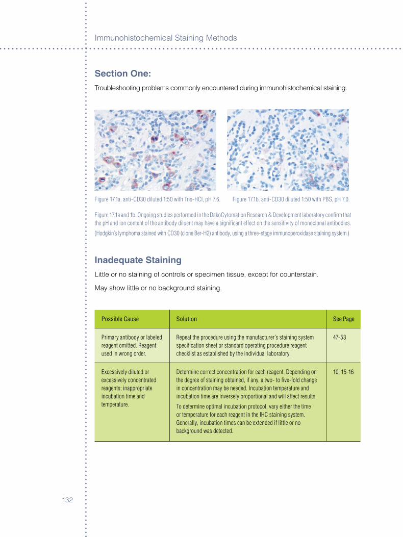
Immunoh�stochem�cal Sta�n�ng Methods
���
Section One:Troubleshoot�ng problems commonly encountered dur�ng �mmunoh�stochem�cal sta�n�ng.
Figure 17.1a. anti-CD30 diluted 1:50 with Tris-HCI, pH 7.6. Figure 17.1b. anti-CD30 diluted 1:50 with PBS, pH 7.0.
Figure 17.1a and 1b. Ongoing studies performed in the DakoCytomation Research & Development laboratory confirm that the pH and ion content of the antibody diluent may have a significant effect on the sensitivity of monoclonal antibodies.
(Hodgkin’s lymphoma stained with CD30 (clone Ber-H2) antibody, using a three-stage immunoperoxidase staining system.)
Inadequate StainingL�ttle or no sta�n�ng of controls or spec�men t�ssue, except for countersta�n.
May show l�ttle or no background sta�n�ng.
Possible Cause Solution See Page
Primary antibody or labeled reagent omitted. Reagent used in wrong order.
Repeat the procedure using the manufacturer’s staining system specification sheet or standard operating procedure reagent checklist as established by the individual laboratory.
47-53
Excessively diluted or excessively concentrated reagents; inappropriate incubation time and temperature.
Determine correct concentration for each reagent. Depending on the degree of staining obtained, if any, a two- to five-fold change in concentration may be needed. Incubation temperature and incubation time are inversely proportional and will affect results.
To determine optimal incubation protocol, vary either the time or temperature for each reagent in the IHC staining system. Generally, incubation times can be extended if little or no background was detected.
10, 15-16
���
Troubleshoot�ng
Possible Cause Solution See Page
Primary antibody diluted with inappropriate buffer.
° Use of PBS or TBS as an antibody diluent.
° Lack of stabilizing or carrier protein.
° Detergent in diluent.
Check formula and compatibility of antibody diluent. A change of ion content and/or pH of the antibody diluent can cause a diminution in the sensitivity of the antibody. Addition of NaCl should be avoided. This problem is seen primarily with monoclonal antibodies.
47-53
Primary antibody defective; one or several secondary or ancillary reagents defective. Do NOT use product after expiration date stamped on vial.
Replace defective or expired antibody; repeat staining protocol, replacing one reagent at a time with fresh, in-date reagents.° Store products according to each product specification
sheet or package insert.° If using a neat or concentrated antibody, and directed by
the manufacturer to store frozen, it may be aliquoted to avoid repeated freezing and thawing.
° Do not freeze ready-to-use or customer diluted products.° Follow manufacturer recommendations on product
specification sheets, package inserts and reagent labels.
10-11
Dissociation of primary antibody during washing or incubation with link antibodies.
A feature of low-affinity antibodies:° Polyclonal primary antiserum: Attempt staining at
low dilutions.° Monoclonal primary antibody: Replace with higher affinity
antibody of identical specificity.° Re-optimize incubation times for washing buffer and
link antibody.
7-8
Use of alcohol-based counterstain and/or alcohol-based mounting media with aqueous– based chromogens.
° Repeat staining, using water-based counterstain and mounting media.
° Use a permanent chromogen, such as DAB/DAB+, that is not affected by organic solvents.
21-22
Excessive counterstaining may compromise proper interpretation of results.
Use a counterstain that:° Will not excessively stain tissue sections.° Can be diluted so as not to obliterate the specific signal.° Reduce incubation time of the counterstain.
119-129
Incorrect preparation of substrate-chromogen mixture.
° Repeat substrate-chromogen treatment with correctly prepared reagent.
° Staining intensity is decreased when excess DAB/DAB+ is present in the working reagent.
Spec Sheet
Inadequate Staining (continued)

Immunoh�stochem�cal Sta�n�ng Methods
��4
Possible Cause Solution See Page
Incompatible buffer used for preparation of enzyme and substrate-chromogen reagents:
° Use of PBS wash buffer with an alkaline phosphatase staining system.
° Sodium azide in reagent diluent or buffer baths for immunoperoxidase methodologies.
Check compatibility of buffer ingredients with enzyme and substrate-chromogen reagents.Repeat staining.° Commercial phosphate buffers may contain additives that
will inhibit alkaline phosphates activity.° Avoid sodium azide in diluents and buffers.
A concentration of 15 mM/L sodium azide, which is added routinely to IHC reagents to inhibit bacterial growth, will not impair HRP conjugated labels.
47-53
Antigen levels are too low for detection by the employed visualization system.
May be due to loss of antigenic differentiation in some tumors or loss of antigenicity due to sub-optimal tissue fixation.
° Utilize a higher sensitivity staining system.° Prolong incubation time of primary antibody.° Re-optimize incubation times and concentrations of
ancillary reagents.° Perform antigen retrieval, if applicable, using a range
of pH buffers.
47-53
Steric hindrance due to high antigen level and possible prozone effect.
Re-optimize concentration of the primary antibody and ancillary reagents. Antibody concentration of the primary antibody may be too high.
47-53
Use of inappropriate fixative.
° Use of certain fixatives may damage or destroy antigens or epitopes in the tissue specimen.
° Use of non-cross linking fixatives may allow the elution of antigens soluble in IHC reagents.
° Different fixatives may affect standardization of cells.
Check manufacturer’s specifications regarding recommended fixative. 27-33
Inadequate Staining (continued)

��5
Troubleshoot�ng
Possible Cause Solution See Page
Immunoreactivity diminished or destroyed during embedding process.
Use a paraffin wax with a melting temperature ~ 55-58 ºC. Wax used for embedding should not exceed 60 OC.
27-33
Immunoreactivity diminished or destroyed during dewaxing at high oven temperature.
Oven temperature not to exceed 60 ºC.NOTE: The intensity of immunostaining may be diminished when tissue is exposed to prolonged heat. Refer to the primary antibody specification sheet for additional information.
27-33
Immunoreactivity diminished or destroyed on pre-cut tissue sections.
The intensity of immunostaining may be diminished when pre-cut tissue sections are exposed to air. Use freshly cut sections and reseal paraffin-embedded blocks.
27-33
Immunoreactivity diminished or destroyed by the enzyme blocking reagent altering a specific epitope.
More common on frozen sections: apply the primary antibody prior to the enzymatic block to insure its reaction. In such cases the blocking reagent can be applied at any point after the primary and before the enzyme labeled components.
13, 30, 31, 58, 107, 119, 126
Excessive wash buffer or blocking serum remaining on tissue section prior to application of IHC reagents.
Excess reagent will dilute the next consecutive reagent. Repeat staining, making sure to wipe away excess washing buffer and blocking serum.
15-16
Demasking protocol is inappropriate or has been omitted.
Some tissue antigens require proteolytic enzyme digestion or heat-induced antigen retrieval performed prior to staining.The need for pretreatment depends on the type and extent of fixation, specific characteristics of the antigen and the type of antibody used. Use the pretreatment method recommended by the manufacturer. No single pretreatment is suitable for all applications.
27-33
Repeated reuse of antigen retrieval buffer.
Do not reuse buffer. Spec Sheet
Sections incorrectly dewaxed.
Prepare new sections and deparaffinize according to standard laboratory protocol, using fresh xylene or xylene substitute.
119-129
Inadequate Staining (continued)

Immunoh�stochem�cal Sta�n�ng Methods
���
Possible Cause Solution See Page
Failure to achieve the optimal temperature required for heat induced antigen retrieval.
° When using a waterbath or steamer, allow sufficient time for the retrieval buffer to equilibrate to a temperature range of 95-99 ºC.
° At high altitude (greater than ~ 4,500 feet), the buffer will boil at less than 95 ºC.
° Utilize a closed heating system such as a pressure cooker, autoclave or Pascal, or utilize a low temperature protocol if standardization of the validated procedure is not affected.
41-44
Excessive or incomplete counterstaining.
Re-optimize concentration of counterstain and incubation time. 41-44
Instrument malfunction. Ensure automated stainer is programmed correctly and is running to manufacturer’s specifications.
95
Pos�t�ve control t�ssue shows adequate spec�fic sta�n�ng w�th l�ttle or no background sta�n�ng. Spec�men t�ssue shows l�ttle or no spec�fic sta�n�ng w�th var�able background sta�n�ng of several t�ssue elements.
Possible Cause Solution See Page
Specimen held for too long in a cross-linking fixative, usually in formalin, causing “masking” of antigenic determinants due to aldehydes cross-linking and increased hydrophobicity of tissue.
Standardize routine fixation. Proteolytic digestion or antigen retrieval will break down cross-linking and render some tissue antigens reactive. Refer to the primary antibody specification sheet for additional information.
41-44
Sectioned portion contains crush artifact caused by grossing tissue with dull scalpel or razor.
Serum proteins diffuse through tissue and are fixed in place.Re-cut tissue using sharp blade.
119-129
Sectioned portion of specimen contains necrotic or otherwise damaged elements.
Ignore physically damaged portions of stained tissue sections. 41-44
Inadequate Staining (continued)
���
Troubleshoot�ng
Possible Cause Solution See Page
Section portion of specimen not penetrated by fixative. Loss of antigenicity in unfixed tissue.
Fix tissue biopsy for longer period of time or fix smaller pieces to ensure complete penetration. Unfixed tissue tends to bind all reagents nonspecifically.
27-33, 119-129
General BackgroundBackground seen �n all control t�ssue and spec�men t�ssue. May see marked background sta�n�ng �n several t�ssue elements such as connect�ve t�ssue, ad�pose t�ssue and ep�thel�um.
Possible Cause Solution See Page
Excessive incubation with substrate- chromogen reagent.
Reduce incubation time. Spec Sheet
Substrate-chromogen reagent prepared incorrectly.
Repeat incubation with correctly prepared chromogen reagent. Spec Sheet
Secondary or link antibody cross-reacts with antigens from tissue specimen.
Absorb link antibody with tissue protein extract or species-specific normal serum from tissue donor.
47-53
Secondary or link antibody and /or tertiary reagents too concentrated.
Repeat staining. Determine correct concentration for each reagent. Incubation temperature and incubation time will affect results. To determine optimal incubation protocol, vary both the time and temperature for each reagent in the IHC staining protocol.
16-18
Slides inadequately rinsed. Gently rinse slide with wash buffer bottle and place in wash bath for five minutes. Gentle agitation of the wash bath may increase effectiveness when used with cytoplasmic or nuclear staining protocols.
7-8
Insufficient saline or detergent in wash buffer.
High-sensitivity staining systems may require higher concentrations of saline or detergent in the wash buffer. Refer to the staining system specification sheet for optimal formulation.
119-129
Inadequate Staining (continued)

Immunoh�stochem�cal Sta�n�ng Methods
��8
Possible Cause Solution See Page
Blocking serum or wrong blocking serum used.
Block with serum from the host of the secondary or link antibody. Avoid serum that contains auto-immune immunoglobulins. Alternatively, a serum-free protein block, lacking immunoglobulins, may be substituted for the serum block.
119-129
Sections incorrectly dewaxed. Prepare new sections and deparaffinize according to standard laboratory protocol using fresh xylene or xylene substitute.
119-129
Non-specific binding of the secondary antibody with an animal tissue specimen.
Use a secondary antibody that has been absorbed against a species specimen, or use a secondary antibody produced in a host that exhibits little or no cross-reactivity with the tissue source.
47-53
Instrument malfunction. Ensure automated stainer is programmed correctly and is running to manufacturer’s specification.
95
Spec�men t�ssue and negat�ve reagent control sl�des show background sta�n�ng. Pos�t�ve and negat�ve control t�ssue show appropr�ate spec�fic sta�n�ng. May �nvolve several t�ssue elements such as connect�ve t�ssue, ad�pose t�ssue and ep�thel�um.
Possible Cause Solution See Page
Specimen held for too long in a cross-linking fixative, usually in formalin, causing “masking” of antigenic determinants due to aldehydes cross-linking and increased hydrophobicity of tissue.
Standardize routine fixation. Proteolytic digestion or antigen retrieval will break down cross-linking and render some tissue antigens reactive. Refer to the primary antibody specification sheet for additional information.
27-33
Sectioned portion of specimen not penetrated by fixative. Loss of antigenicity in unfixed tissue. Unfixed tissue tends to bind all reagents nonspecifically.
Fix tissue biopsy for longer period of time or fix smaller pieces to ensure complete penetration.
27-33
General Background (continued)
��9
Troubleshoot�ng
Possible Cause Solution See Page
Sectioned portion contains crush artifact caused by grossing tissue with dull scalpel or razor. Serum proteins diffuse through tissue and are fixed in place.
Serum proteins diffuse through tissue and are fixed in place.Re-cut tissue using sharp blade.
119-129
Sectioned portion of specimen contains necrotic or otherwise damaged elements.
Ignore physically damaged portions of stained tissue sections. 119-129
Excessive or unevenly applied subbing agent on poly-L-lysine, charged, or silanized slides.
Some IHC reagents may bind to these products, resulting in a light stain over the entire slide surface. Some slides may be unevenly coated, and will exhibit the above problems on only a portion of the tissue or glass.
119-129
Antigen diffusion prior to fixation causing specific background outside the expected antigen site.
Avoid delays in fixation of the tissue. 119-129
Tissue sections too thick. Cut tissue sections thinner. Formalin-fixed paraffin-embedded tissue sections should be approximately 4-6 µm; cryostat section 10 <µm.
27-39
Incomplete permeabilization of tissue sections.
Seen in frozen sections, cell smears and non-paraffin embedded tissue: Incomplete permeabilization of cells allows unattached reagents to become trapped within the cells and resistant to removal by wash buffer.
99
General Background (continued)
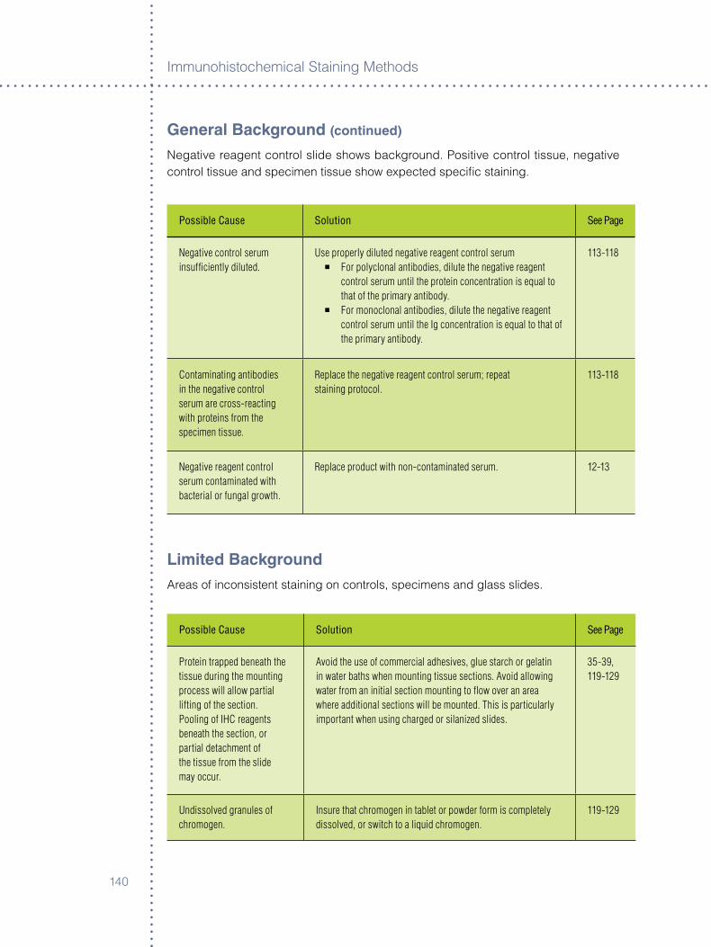
Immunoh�stochem�cal Sta�n�ng Methods
�40
General Background (continued)
Negat�ve reagent control sl�de shows background. Pos�t�ve control t�ssue, negat�ve control t�ssue and spec�men t�ssue show expected spec�fic sta�n�ng.
Possible Cause Solution See Page
Negative control serum insufficiently diluted.
Use properly diluted negative reagent control serum° For polyclonal antibodies, dilute the negative reagent
control serum until the protein concentration is equal to that of the primary antibody.
° For monoclonal antibodies, dilute the negative reagent control serum until the Ig concentration is equal to that of the primary antibody.
113-118
Contaminating antibodies in the negative control serum are cross-reacting with proteins from the specimen tissue.
Replace the negative reagent control serum; repeat staining protocol.
113-118
Negative reagent control serum contaminated with bacterial or fungal growth.
Replace product with non-contaminated serum. 12-13
Limited BackgroundAreas of �ncons�stent sta�n�ng on controls, spec�mens and glass sl�des.
Possible Cause Solution See Page
Protein trapped beneath the tissue during the mounting process will allow partial lifting of the section.Pooling of IHC reagents beneath the section, or partial detachment of the tissue from the slide may occur.
Avoid the use of commercial adhesives, glue starch or gelatin in water baths when mounting tissue sections. Avoid allowing water from an initial section mounting to flow over an area where additional sections will be mounted. This is particularly important when using charged or silanized slides.
35-39, 119-129
Undissolved granules of chromogen.
Insure that chromogen in tablet or powder form is completely dissolved, or switch to a liquid chromogen.
119-129
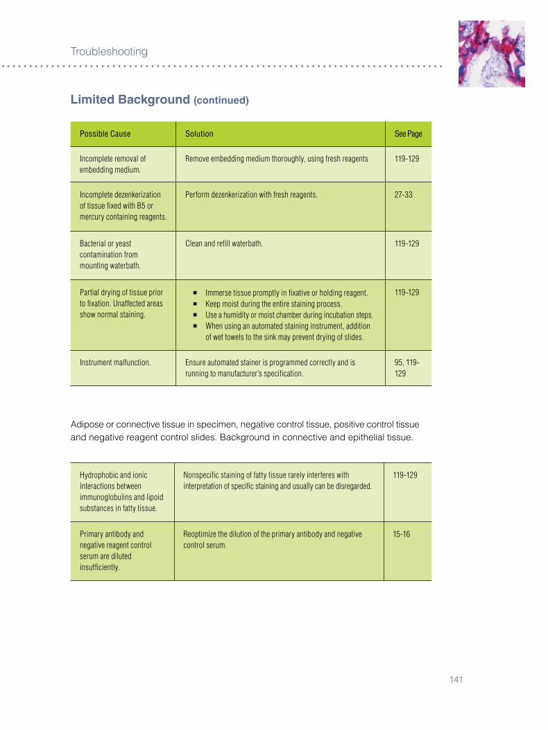
�4�
Troubleshoot�ng
Possible Cause Solution See Page
Incomplete removal of embedding medium.
Remove embedding medium thoroughly, using fresh reagents 119-129
Incomplete dezenkerization of tissue fixed with B5 or mercury containing reagents.
Perform dezenkerization with fresh reagents. 27-33
Bacterial or yeast contamination from mounting waterbath.
Clean and refill waterbath. 119-129
Partial drying of tissue prior to fixation. Unaffected areas show normal staining.
° Immerse tissue promptly in fixative or holding reagent.° Keep moist during the entire staining process.° Use a humidity or moist chamber during incubation steps.° When using an automated staining instrument, addition
of wet towels to the sink may prevent drying of slides.
119-129
Instrument malfunction. Ensure automated stainer is programmed correctly and is running to manufacturer’s specification.
95, 119-129
Ad�pose or connect�ve t�ssue �n spec�men, negat�ve control t�ssue, pos�t�ve control t�ssue and negat�ve reagent control sl�des. Background �n connect�ve and ep�thel�al t�ssue.
Hydrophobic and ionic interactions between immunoglobulins and lipoid substances in fatty tissue.
Nonspecific staining of fatty tissue rarely interferes with interpretation of specific staining and usually can be disregarded.
119-129
Primary antibody and negative reagent control serum are diluted insufficiently.
Reoptimize the dilution of the primary antibody and negative control serum.
15-16
Limited Background (continued)

Immunoh�stochem�cal Sta�n�ng Methods
�4�
Limited Background (continued)
Ep�thel�al t�ssue �n spec�men, negat�ve control t�ssue, pos�t�ve control t�ssue and negat�ve reagent control sl�des. Sta�n�ng �s moderate to marked, espec�ally �n ep�dermal ep�thel�um. Background �n ep�thel�a accompan�es background �n connect�ve t�ssue.
Possible Cause Solution See Page
Both the primary antibody and negative control serum contain contaminating antibodies to epithelial elements, possibly cytokeratins.
° Use a higher dilution of the primary antibody and negative control serum.
° Increase the incubation time.° Replace the antibody.
5-7, 119-129
Excessive formalin fixation of tissues may increase protein cross-linking, resulting in tissue hydrophobicity.
Proteolytic digestion or antigen retrieval will break down cross-linking and render some tissue antigens reactive. Refer to the primary antibody and/or the negative reagent control specification sheet for appropriate pretreatment.
27-33, 119-129
Focal cytoplasm�c sta�n�ng observed �n ep�thel�um �n the spec�men t�ssue.
Focal cytoplasmic staining is seen, particularly in intermediate and superficial layers of the epidermis. May be caused by passive absorption of plasma proteins into degenerating epidermal cells.
This observation is rare and should not interfere with interpretation of specific staining.
119-129
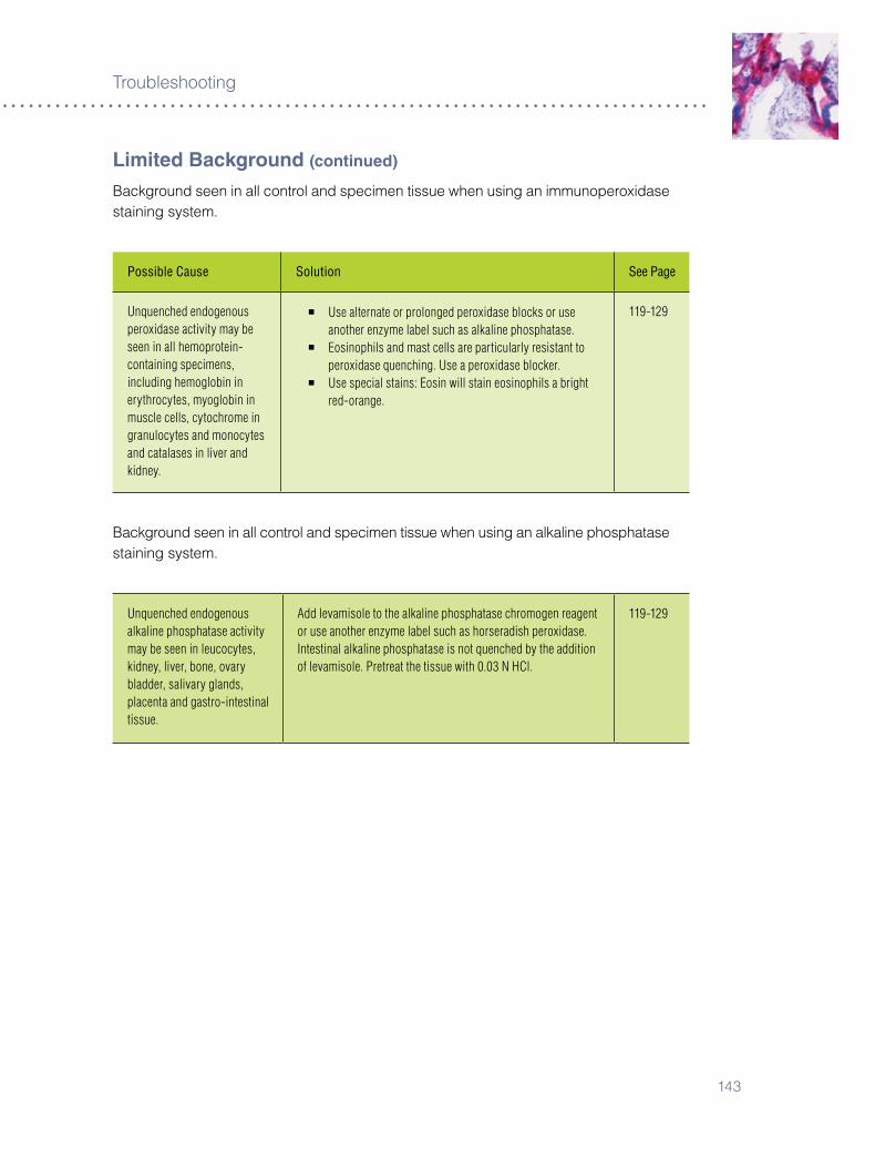
�4�
Troubleshoot�ng
Limited Background (continued)
Background seen �n all control and spec�men t�ssue when us�ng an �mmunoperox�dase sta�n�ng system.
Possible Cause Solution See Page
Unquenched endogenous peroxidase activity may be seen in all hemoprotein-containing specimens, including hemoglobin in erythrocytes, myoglobin in muscle cells, cytochrome in granulocytes and monocytes and catalases in liver and kidney.
° Use alternate or prolonged peroxidase blocks or use another enzyme label such as alkaline phosphatase.
° Eosinophils and mast cells are particularly resistant to peroxidase quenching. Use a peroxidase blocker.
° Use special stains: Eosin will stain eosinophils a bright red-orange.
119-129
Background seen �n all control and spec�men t�ssue when us�ng an alkal�ne phosphatase sta�n�ng system.
Unquenched endogenous alkaline phosphatase activity may be seen in leucocytes, kidney, liver, bone, ovary bladder, salivary glands, placenta and gastro-intestinal tissue.
Add levamisole to the alkaline phosphatase chromogen reagent or use another enzyme label such as horseradish peroxidase.Intestinal alkaline phosphatase is not quenched by the addition of levamisole. Pretreat the tissue with 0.03 N HCl.
119-129
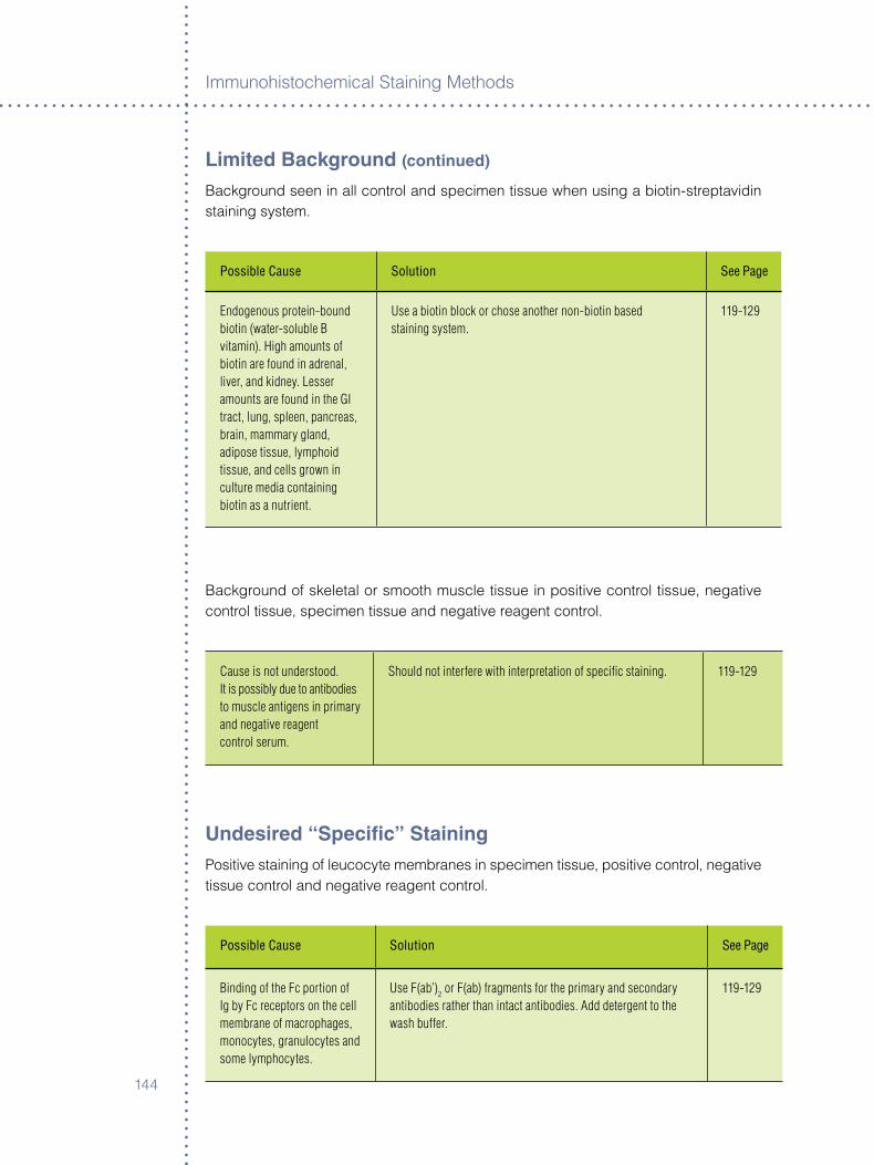
Immunoh�stochem�cal Sta�n�ng Methods
�44
Limited Background (continued)
Background seen �n all control and spec�men t�ssue when us�ng a b�ot�n-streptav�d�n sta�n�ng system.
Possible Cause Solution See Page
Endogenous protein-bound biotin (water-soluble B vitamin). High amounts of biotin are found in adrenal, liver, and kidney. Lesser amounts are found in the GI tract, lung, spleen, pancreas, brain, mammary gland, adipose tissue, lymphoid tissue, and cells grown in culture media containing biotin as a nutrient.
Use a biotin block or chose another non-biotin based staining system.
119-129
Background of skeletal or smooth muscle t�ssue �n pos�t�ve control t�ssue, negat�ve control t�ssue, spec�men t�ssue and negat�ve reagent control.
Cause is not understood. It is possibly due to antibodies to muscle antigens in primary and negative reagentcontrol serum.
Should not interfere with interpretation of specific staining. 119-129
Undesired “Specific” StainingPos�t�ve sta�n�ng of leucocyte membranes �n spec�men t�ssue, pos�t�ve control, negat�ve t�ssue control and negat�ve reagent control.
Possible Cause Solution See Page
Binding of the Fc portion of Ig by Fc receptors on the cell membrane of macrophages, monocytes, granulocytes and some lymphocytes.
Use F(ab’)2 or F(ab) fragments for the primary and secondary antibodies rather than intact antibodies. Add detergent to the wash buffer.
119-129
�45
Troubleshoot�ng
Pos�t�ve sta�n�ng of h�st�ocytes and granulocytes �n the spec�men t�ssue only, w�th a marker not normally react�ve w�th these cells.
Possible Cause Solution See Page
Phagocytosis of antigens may render phagocytes positive for the same.
Rare. Should not interfere with interpretation of specific staining. 119-129
Pos�t�ve membrane sta�n�ng of spec�men t�ssue and negat�ve reagent control t�ssue when us�ng a horserad�sh perox�dase sta�n�ng system.
Tissue from persons infected with Hepatitis B virus and expressing Hepatitis B surface antigen may exhibit undesired staining.
Utilize a non-peroxidase staining system. 119-129
MiscellaneousLoss of v�ab�l�ty of cell cultures.
Possible Cause Solution See Page
Some manufacturers produce antibodies and reagents for in vitro use only. These products may contain preservatives, usually sodium azide, which is a known poison.
Utilize an in vivo product for application on viable cells.For use on cell cultures only: Sodium azide may be dialyzed out of some reagents. Contact Dako Technical Support for additional information.
119-129
Immunoh�stochem�cal Sta�n�ng Methods
�4�
Section Two:
Troubleshooting flow chart: Use th�s flow chart to determ�ne source(s) of non-spec�fic sta�n�ng when us�ng an �mmunoh�stochem�cal protocol.
Background Staining Encountered with HrP-Peroxidase reagents
reagents result/action
Slid
e #1 Positive Control Tissue:
Counterstain with hematoxylin
NO STaINING SEEN. GO TO NEXT STEP.
q
Brown endogenous pigment (such as melanin) observed: ° To distinguish melanin pigment from DAB
chromogen, Azure B can be used as a counterstain. The melanin stains blue-green, while the DAB remains brown. ° An alternate method is to use AEC as the
chromogen. However, if high levels of pigment exist in the tissue, the red chromogen may be partially obscured. Since bleaching protocols to remove melanin may compromise tissue antigenicity, it should be avoided if at all possible.
Slid
e #2 Positive Control Tissue:
DAB/AEC + Counterstain
NO STaINING SEEN. GO TO NEXT STEP.
q
Brown/Red color observed: ° Indicates endogenous peroxidase activity in the
tissue sections. It is present in all hemoprotein containing tissue including erythrocytes, muscle, liver, kidney, granulocytes and monocytes. ° Block with three percent hydrogen peroxide or
other peroxidase blocking reagent. Using a new bottle of hydrogen peroxide, perform a three percent H202 peroxidase block, followed by DAB and an appropriate counterstain.
Slid
e #3 Positive Control Tissue:
Peroxidase Block + Secondary Antibody + Streptavidin-HRP + DAB/AEC + Counterstain
NO STaINING SEEN. GO TO NEXT STEP.
q
Brown/Red color observed: ° Indicates endogenous biotin activity in the tissue
sections. Protein-bound biotin may be found in adrenal, liver, kidney, GI tract, lung, spleen, brain, mammary gland, adipose tissue, lymphoid tissue and cell grown in culture media containing biotin (RPMI, NCTC, MEME). ° Block with a biotin block or switch to a staining
system that is not dependent on the streptavidin/biotin reaction.
�4�
Troubleshoot�ng
Slid
e #4
Positive Control Tissue:Peroxidase Block + Biotin Block (if required) + Secondary Antibody + Streptavidin-HRP + DAB/AEC + Counterstain
NO STaINING SEEN. GO TO NEXT STEP.
q
Brown/Red color observed: ° Indicates endogenous biotin activity in the tissue
sections. Protein-bound biotin may be found in adrenal, liver, kidney, GI tract, lung, spleen, brain, mammary gland, adipose tissue, lymphoid tissue and cell grown in culture media containing biotin (RPMI, NCTC, MEME). ° Block with a biotin block or switch to a staining
system that is not dependent on the streptavidin/biotin reaction.
Slid
e #5
Positive Control Tissue:Peroxidase Block + Biotin Block (if required)+ Negative Reagent Control + Secondary Antibody + Streptavidin-HRP + DAB/AEC
NO STaINING SEEN. GO TO NEXT STEP.
q
Brown/Red color observed: ° May indicate non-specific binding of the primary
antibody carrier-protein. Perform a protein block with normal serum from the host of the link antibody add 0.05-0.1% TWEEN 20 to wash buffer to decrease protein attachment.° Antigen retrieval lipofusion-artifact may appear as
granule staining in liver and cardiac tissue, or as specific staining in pancreatic sections.
Slid
e #6 Negative Control Tissue:
Perform complete staining protocol.
Brown/Red color observed on Negative Control Tissue:° Monoclonal antibody: Possible contamination.° Polyclonal antibody: Possible contamination or
undesired antibody in the host Ig fraction.° Antigen retrieval lipofusion-artifact may appear as
granule staining in liver and cardiac tissue, or as specific staining in pancreatic sections.
Background Staining Encountered with alkaline Phosphatase
reagents result/action
Slid
e #1 Positive Control Tissue:
Fast Red, Fuchsin or BCIP/NBT + Counterstain
NO STaINING SEEN. GO TO NEXT STEP.
q
Red/Blue color observed: ° Indicates endogenous alkaline phosphatase activity
in the tissue sections. It is present in liver, kidney, GI tract, bone, bladder, ovary, salivary gland, placenta, leukemic, necrotic or degenerated cells. ° Block with levamisole (Intestinal alkaline
phosphatase may be quenched by the addition of 0.03 N HCl prior to the addition of the alkaline phosphatase).

Immunoh�stochem�cal Sta�n�ng Methods
�48
Slid
e #2 Positive Control Tissue:
Streptavidin-AP + Fast Red, Fuchsin or BCIP/NBT + Counterstain
NO STaINING SEEN. GO TO NEXT STEP.
q
Red/Blue color observed: ° Indicates endogenous biotin activity in the tissue
sections. Protein-bound biotin may be found in adrenal, liver, kidney, GI tract, lung, spleen, brain, mammary gland, adipose tissue, lymphoid tissue and cells grown in culture media containing biotin (RPMI, NCTC, MEME). ° Block with a biotin block or switch to a staining
system that is not dependent on the streptavidin/biotin reaction.
Slid
e #3
Positive Control Tissue:Biotin Block (if required) + Secondary Antibody + Streptavidin-AP + Fast Red, Fuchsin or BCIP/NBT + Counterstain
NO STaINING SEEN. GO TO NEXT STEP.
q
Red/Blue color observed: ° Indicates non-specific or undesired binding of
the secondary antibody to the tissue sections. This primarily occurs when the secondary antiserum has not been prepared for use on a specific species tissue. ° To determine if this is the problem, absorb out non-
specific proteins by adding 2, 5 or 10 µL of normal serum (from the species of tissue to be stained) per 100 µL of the secondary antibody.
Slid
e #4
Positive Control Tissue:Biotin Block (if required) + Negative Reagent Control + Secondary Antibody + Streptavidin-AP + Fast Red, Fuchsin or BCIP/NBT + Counterstain.
NO STaINING SEEN. GO TO NEXT STEP.
q
Red/Blue color observed: ° May indicate non-specific binding of the primary
antibody carrier-protein. Perform a protein block with normal serum from the host of the link antibody or a protein block; add 0.05-0.1% TWEEN 20 to wash buffer to decrease protein attachment.° Antigen retrieval lipofusion-artifact may appear as
granule staining in liver and cardiac tissue or as specific staining in pancreatic sections.
Slid
e #5 Negative Control Tissue:
Perform complete staining protocol
Red/Blue color observed on Negative Control Tissue:° Monoclonal antibody: Possible contamination.° Polyclonal antibody: Possible contamination or
undesired antibody in the host Ig fraction.° Antigen retrieval lipofusion-artifact may appear as
granule staining in liver and cardiac tissue, or as specific staining in pancreatic sections.
�49
Troubleshoot�ng
Negative Control reagent
reagents result/action
Negative Control Reagent:Perform complete staining protocol.
° (Human tissue) Perform the peroxidase blocking protocol from Slide #2 under “Background Staining Encountered with HRP-Peroxidase Reagents.”° Perform a biotin block if required, protein block if
required, apply the appropriate negative reagent control (see below), apply biotinylated secondary antibody, apply streptavidin/HRP reagent and DAB.
Prepare a negative reagent control
° Polyclonal: Non-immunized sera from the same species, diluted to the same protein concentration as the primary antibody.° Monoclonal: Negative reagent control that matches the isotype as the primary antibody. Additionally, the diluent used
to manufacture a monoclonal primary antibody and isotypic negative control should contain the same ions. Diluents containing sodium or phosphate ions may change the sensitivity of some monoclonal antibodies.° Calculation:° Ig or total protein concentration of primary antibody divided by dilution factor of primary antibody = x.° Ig or total protein concentration of negative reagent control divided by x = dilution factor of negative reagent control.
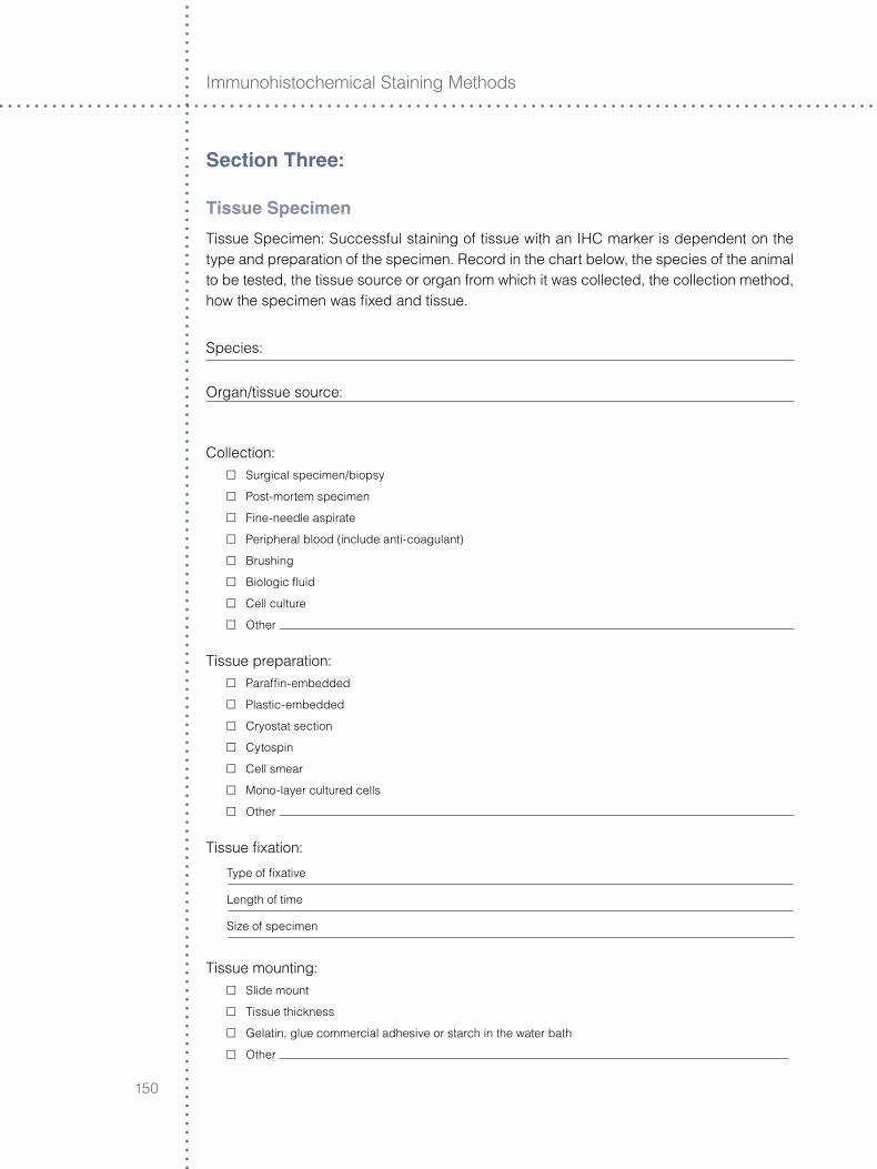
Immunoh�stochem�cal Sta�n�ng Methods
�50
Section Three:
Tissue Specimen
T�ssue Spec�men: Successful sta�n�ng of t�ssue w�th an IHC marker �s dependent on the type and preparat�on of the spec�men. Record �n the chart below, the spec�es of the an�mal to be tested, the t�ssue source or organ from wh�ch �t was collected, the collect�on method, how the spec�men was fixed and t�ssue.
Spec�es:
Organ/t�ssue source:
Collect�on:£ Surg�cal spec�men/b�opsy
£ Post-mortem spec�men
£ F�ne-needle asp�rate
£ Per�pheral blood (�nclude ant�-coagulant)
£ Brush�ng
£ B�olog�c flu�d
£ Cell culture
£ Other
T�ssue preparat�on: £ Paraffin-embedded
£ Plast�c-embedded
£ Cryostat sect�on
£ Cytosp�n
£ Cell smear
£ Mono-layer cultured cells
£ Other
T�ssue fixat�on:
Type of fixat�ve
Length of t�me
S�ze of spec�men
T�ssue mount�ng: £ Sl�de mount
£ T�ssue th�ckness
£ Gelat�n, glue commerc�al adhes�ve or starch �n the water bath
£ Other
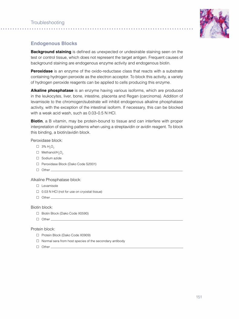
�5�
Troubleshoot�ng
Endogenous Blocks
Background staining �s defined as unexpected or undes�rable sta�n�ng seen on the test or control t�ssue, wh�ch does not represent the target ant�gen. Frequent causes of background sta�n�ng are endogenous enzyme act�v�ty and endogenous b�ot�n.
Peroxidase �s an enzyme of the ox�do-reductase class that reacts w�th a substrate conta�n�ng hydrogen perox�de as the electron acceptor. To block th�s act�v�ty, a var�ety of hydrogen perox�de reagents can be appl�ed to cells produc�ng th�s enzyme.
alkaline phosphatase �s an enzyme hav�ng var�ous �soforms, wh�ch are produced �n the leukocytes, l�ver, bone, �ntest�ne, placenta and Regan (carc�noma). Add�t�on of levam�sole to the chromogen/substrate w�ll �nh�b�t endogenous alkal�ne phosphatase act�v�ty, w�th the except�on of the �ntest�nal �soform. If necessary, th�s can be blocked w�th a weak ac�d wash, such as 0.0�-0.5 N HCl.
Biotin, a B v�tam�n, may be prote�n-bound to t�ssue and can �nterfere w�th proper �nterpretat�on of sta�n�ng patterns when us�ng a streptav�d�n or av�d�n reagent. To block th�s b�nd�ng, a b�ot�n/av�d�n block.
Perox�dase block:£ �% H�O�
£ Methanol/H�O�
£ Sod�um az�de
£ Perox�dase Block (Dako Code S�00�)
£ Other
Alkal�ne Phosphatase block: £ Levam�sole
£ 0.0� N HCl (not for use on cryostat t�ssue)
£ Other
B�ot�n block: £ B�ot�n Block (Dako Code X0590)
£ Other
Prote�n block: £ Prote�n Block (Dako Code X0909)
£ Normal sera from host spec�es of the secondary ant�body
£ Other
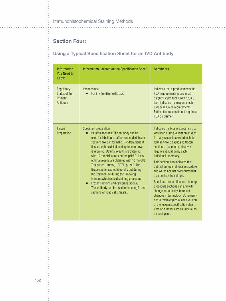
Immunoh�stochem�cal Sta�n�ng Methods
�5�
Section Four:
Using a Typical Specification Sheet for an IVD antibody
Information You Need to Know
Information Located on the Specification Sheet Comments
Regulatory Status of the Primary Antibody
Intended use° For in vitro diagnostic use.
Indicates that a product meets the FDA requirements as a clinical diagnostic product. Likewise, a CE icon indicates the reagent meets European Union requirements. Patient test results do not require an FDA disclaimer.
Tissue Preparation
Specimen preparation° Paraffin sections: The antibody can be
used for labeling paraffin- embedded tissue sections fixed in formalin. Pre-treatment of tissues with heat-induced epitope retrieval is required. Optimal results are obtained with 10 mmol/L citrate buffer, pH 6.0. Less optimal results are obtained with 10 mmol/L Tris buffer, 1 mmol/L EDTA, pH 9.0. The tissue sections should not dry out during the treatment or during the following immunocytochemical staining procedure.
° Frozen sections and cell preparations: The antibody can be used for labeling frozen sections or fixed cell smears .
Indicates the type of specimen that was used during validation studies. In many cases this would include formalin-fixed tissue and frozen sections. Use of other fixatives requires validation by each individual laboratory.
This section also indicates the optimal epitope retrieval procedure and warns against procedures that may destroy the epitope.
Specimen preparation and staining procedure sections can and will change periodically, to reflect changes in technology. So remem-ber to retain copies of each version of the reagent specification sheet. Version numbers are usually found on each page.
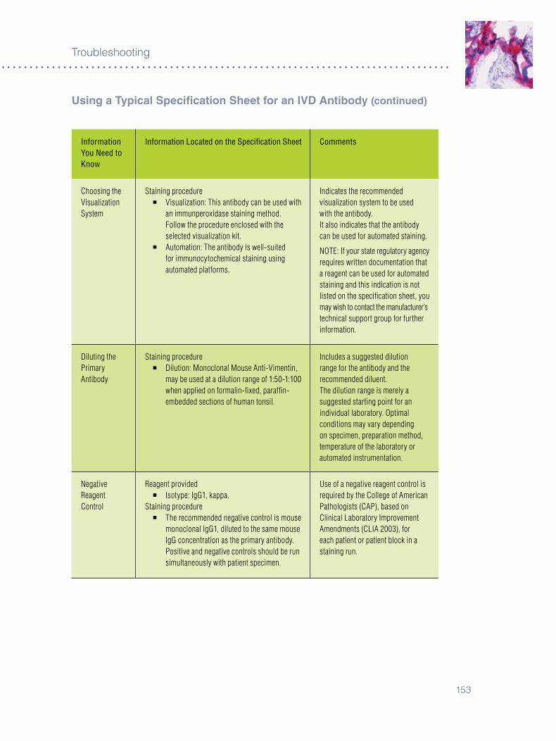
�5�
Troubleshoot�ng
Information You Need to Know
Information Located on the Specification Sheet Comments
Choosing the Visualization System
Staining procedure ° Visualization: This antibody can be used with
an immunperoxidase staining method. Follow the procedure enclosed with the selected visualization kit.
° Automation: The antibody is well-suited for immunocytochemical staining using automated platforms.
Indicates the recommended visualization system to be used with the antibody.It also indicates that the antibody can be used for automated staining.
NOTE: If your state regulatory agency requires written documentation that a reagent can be used for automated staining and this indication is not listed on the specification sheet, you may wish to contact the manufacturer’s technical support group for further information.
Diluting the Primary Antibody
Staining procedure° Dilution: Monoclonal Mouse Anti-Vimentin,
may be used at a dilution range of 1:50-1:100 when applied on formalin-fixed, paraffin- embedded sections of human tonsil.
Includes a suggested dilution range for the antibody and the recommended diluent.The dilution range is merely a suggested starting point for an individual laboratory. Optimal conditions may vary depending on specimen, preparation method, temperature of the laboratory or automated instrumentation.
Negative Reagent Control
Reagent provided° Isotype: IgG1, kappa.
Staining procedure ° The recommended negative control is mouse
monoclonal IgG1, diluted to the same mouse IgG concentration as the primary antibody. Positive and negative controls should be run simultaneously with patient specimen.
Use of a negative reagent control is required by the College of American Pathologists (CAP), based on Clinical Laboratory Improvement Amendments (CLIA 2003), for each patient or patient block in a staining run.
Using a Typical Specification Sheet for an IVD antibody (continued)
Immunoh�stochem�cal Sta�n�ng Methods
�54
Information You Need to Know
Information Located on the Specification Sheet Comments
Positive Control Tissue
Performance characteristics° Normal tissues: In general, most human
mesenchymal cells are labeled by the antibody, including fibrocytes, lipocytes, smooth muscle cells, vascular endothelial cells, astrocytes, peripheral nerve (Schwann) cells, macrophages (including Kupffer cells), as well as myoepithelial cells of sweat and salivary glands and of breast, which are all labeled strongly. Also positive, with variable intensity and distribution, are the follicular cells of the thyroid, adrenal cortex, renal distal tubules, and mesangial and endothelial cells of the renal glomerulus, as well as pancreatic acinar cells (1,2). In the human eye, the antibody labels the pigmented posterior and the anterior epithelia of the human iris, including the muscle portion (dilator pupillae) of the anterior epithelium, as well as the nonpigmented and pigmented ciliary epithelia (4). In the ciliary epithelia, vimentin was coexpressed with cytokeratin (4).
° Abnormal tissues: The antibody labeled 17/20 sarcomas, 16/18 melanomas, 4/4 meningeomas, and 3/3 schwannomas, and was the sole intermediate filament present in these tumours. In addition, variable percentages (10 to 57 percent) of carcinomas, neuroendocrine carcinomas, neuroblastomas, thymomas and mesotheliomas were positive with the antibody. With the exception of the neuroblastomas, cytokeratin was coexpressed with vimentin in these tumours. Among adenocarcinomas, more than 50 percent of papillary carcinomas of the thyroid as well as renal, endometrial, ovarian and lung carcinomas were labeled by the antibody and coexpressed keratins and vimentin.
CLIA 2003 Sec. 493.1273 (3) Mandates that fluorescent and immunohistochemical stains must be checked for appropriate positive and negative reactivity each time they are used.
Most IVD antibody specification sheet will list tissue that will exhibit positive and negative staining patterns in the Performance Characteristics section.
NOTE: abnormal tissue will not necessarily be labeled.
Both negative and positive tissue controls should be processed using the same fixation, embedding, mounting, drying, epitope retrieval and immunostaining protocols as the patient tissue.
Using a Typical Specification Sheet for an IVD antibody (continued)
�55
Troubleshoot�ng
Using a Typical Specification Sheet for an IVD antibody (continued)
Information You Need to Know
Information Located on the Specification Sheet Comments
Negative Control Tissue
Performance characteristics ° Normal tissues: Skeletal and cardiac muscle
cells, epidermal, squamous, urothelial, colonic and gastric mucosal, and glial cells, as well as neurons are consistently negative with the antibody .
references�. Wood G, et al. Suppress�on of Endogenous Av�d�n-B�nd�ng Act�v�ty �n T�ssues and
Its Relevance to B�ot�n-Av�d�n Detect�on Systems. Journal of H�stochem�stry and Cytochem�stry �98�;�9(�0):��9�-�04.
�. Sayak� H, et al. Azure B as a Countersta�n �n the Immunoh�stolog�cal Evaluat�on of Heav�ly P�gmented Nevomelanocyt�c Les�ons. Appl�ed Immunoh�stochem�stry �995;�(4):��8-��.
�. Federal Reg�ster: January �4, �00�;�8(��) 4�CFR Part 49�.4. College of Amer�can Pathology; Anatom�c Pathology Checkl�st, October �005.

Immunoh�stochem�cal Sta�n�ng Methods
�5�
�5�
Glossary
Th�s Glossary was not �ntended to be an all-encompass�ng l�st of term�nology as used �n �mmunochem�cal sta�n�ng. Rather, �t assumes a bas�c level of techn�cal knowledge beyond wh�ch the �ncluded defin�t�ons were selected to help �n clar�fy�ng the text of th�s Gu�de.
adjuvant In �mmunology, any substance that enhances the �mmunogen�c�ty of an ant�gen and results �n a super�or �mmune response. There are two types, those that possess the ab�l�ty to enhance both cellular and humoral response to a large number of ant�gens (general potent�at�on), and those that strengthen spec�fic response to only a few ant�gens (spec�fic potent�at�on). Adjuvants work by several mechan�sms �nclud�ng prolongat�on of ant�gen release, �mprov�ng �mmunogen�c�ty by ant�gen denaturat�on, recru�tment of other �mmunocompetent cells and �nduct�on of �nflammat�on.
affinity absorption A method of separat�on by affin�ty chromatography. It may be used, for example, to remove unwanted ant�bod�es from an ant�body preparat�on. The preparat�on �s passed through a column matr�x conta�n�ng ant�gens aga�nst wh�ch the unwanted ant�bod�es are d�rected. Thus, the unwanted ant�bod�es rema�n bound to the column. The ant�body solut�on leav�ng the column conta�ns only the des�red ant�bod�es, pur�fied by affin�ty absorpt�on.
affinity Isolation A method of separat�on by affin�ty chromatography. For example, affin�ty �solated ant�bod�es may be prepared by pass�ng the ant�body solut�on through a column matr�x to wh�ch ant�gens are coupled. Ant�bod�es d�rected aga�nst the coupled ant�gens rema�n bound on the column and may then be eluted us�ng a solut�on wh�ch d�srupts ant�gen-ant�body b�nd�ng.
agglutination The clump�ng of cells that are d�str�buted d�ffusely �n a flu�d. It �s caused by agglut�n�ns, ant�bod�es developed aga�nst that spec�fic cell type, and �s seen when a bacter�al culture �s treated w�th serum from an an�mal �mmun�zed aga�nst that part�cular organ�sm or when a suspens�on of cells, part�cularly red blood cells, �s exposed to ant�sera. Th�s phenomenon commonly �s employed �n blood bank�ng as an �nd�cator of ant�gen-ant�body react�on between red cells and spec�fic ant�serum or donor plasma.
antigen A molecule that �s capable of b�nd�ng to an ant�body.
antigenic Determinant See Ep�tope.
antigen retrieval (ar) Also known by the terms “ep�tope retr�eval” or “target retr�eval,” perta�ns to the restorat�on of ant�gen�c�ty (�mmunoreact�v�ty) to an �mmunogen.
antiserum A serum that conta�ns ant�bod�es.
ascites or ascitic Fluid An accumulat�on of flu�d �n the abdom�nal cav�ty.
Immunoh�stochem�cal Sta�n�ng Methods
�58
Background Unless defined otherw�se, background sta�n�ng �ncludes all nonspec�fic sta�n�ng as a result of procedural art�facts. Occas�onally, �t may also �nclude “undes�rable” sta�n�ng, eg, due to d�ffused ant�gen.
Chromogen One of a group of chem�cal spec�es that can form a part�cular colored mater�al or can be �dent�fied by such a react�on w�th an appropr�ate reagent.
Counterstain A second sta�n that prov�des a contrast�ng effect to another sta�n.
Cross-reactivity The ab�l�ty of an ant�body to react w�th ant�gens other than the �mmunogen. The term should not be used when referr�ng to react�ons occurr�ng between an ant�body and d�fferent cell or t�ssue components.
Epitope The structural part of an ant�gen that reacts w�th an ant�body. These are group�ngs of am�no ac�ds �n globular prote�ns and sugar s�de-cha�ns �n polysacchar�des. The most cr�t�cal part �s called the �mmunodom�nant po�nt.
Epitope retrieval See Ant�gen Retr�eval.
Expiration Date Th�s term s�gnals the m�n�mum expected shelf l�fe of b�olog�cal mater�als, �nclud�ng �mmunochem�cals. (See Shelf L�fe).
Hyperimmunization The pract�ce of establ�sh�ng a he�ghtened state of the act�vely acqu�red �mmun�ty by the adm�n�strat�on of repeated doses of ant�gen.
Idiotype Trad�t�onally, ant�gen�c determ�nants that relate to the spec�fic�ty of the ant�body. Id�otyp�c arrangement of several groups of am�no ac�ds �n the hypervar�able reg�ons of l�ght and heavy cha�ns were thought to bestow un�que ant�gen�c determ�nants to the ant�body molecule and, as a consequence, a h�gh degree of spec�fic�ty. However, ant�-sera d�rected aga�nst these ant�gen�c determ�nants have s�nce been found to cross-react w�th other ant�body molecules. The term �d�otype has yet to be redefined.
Immunochemistry The branch of �mmunology concerned w�th the chem�cal substances and react�ons of the �mmune system, the spec�fic study of ant�gens and ant�bod�es and the�r �nteract�ons w�th one another.
Immunocytochemistry Immunochem�stry appl�ed to the study of �ntracellular act�v�t�es. (Now frequently used �nterchangeably w�th �mmunoh�stochem�stry.)
Immunogen Any substance capable of generat�ng an �mmune react�on, �n contrast to any substance that b�nds to an ant�body (�e, an ant�gen).
Immunogenicity The ab�l�ty of an �mmunogen to el�c�t an �mmune response. Immunogen�c�ty depends upon fore�gnness to the host, the s�ze of the �mmunogen, the complex�ty of �ts molecular structure, the length of t�me �t rema�ns �n the host and �ts ab�l�ty to reach certa�n �mmunocompetent cells �n order to generate �mmun�ty.
Immunohistochemistry Immunochem�stry appl�ed to the study of cells and t�ssues. (Now frequently used �nterchangeably w�th �mmunocytochem�stry.)
In Situ Hybridization An assay for nucle�c ac�ds “on s�te” �n fixed t�ssue sect�ons by the use of heat to first denature and then to reanneal w�th spec�fic DNA, RNA or PNA probes.
�59
Glossary
Internal Tissue Control A spec�men from the pat�ent donor, wh�ch conta�ns the target marker, not only �n the tumor to be �dent�fied, but also �n adjacent normal t�ssue. Thus, no separate pos�t�ve control sect�ons are needed.
Ligand A molecule, �on or atom that �s bound to the central atom (usually a metal atom) of a coord�nat�on compound or chelate.
Link antibody See Secondary Ant�body.
Monoclonal antibodies Immunochem�cally �dent�cal ant�bod�es produced by one clone of plasma cells that react w�th a spec�fic ep�tope on a g�ven ant�gen. Produced commerc�ally us�ng hybr�domas.
Monospecific Hav�ng an effect only on a part�cular k�nd of cell or t�ssue, or react�ng w�th a s�ngle ant�gen, as a monospec�fic ant�serum.
Negative Tissue Control A t�ssue spec�men from the same organ lack�ng the target ant�gen and processed by use of the pr�mary ant�body.
Nonimmune Serum Serum obta�ned from an�mals wh�ch have not been �mmun�zed.
Polyclonal antibodies Immunochem�cally d�ss�m�lar ant�bod�es produced by d�fferent cells and react�ng w�th var�ous ep�topes on a g�ven ant�gen.
Positive Tissue Control A spec�men prev�ously shown to sta�n spec�fically for the target ant�gen after exposure to pr�mary ant�body. Nonspec�fic background sta�n�ng should be at a m�n�mum. Note that, for some target ant�gens (e.g., prostate spec�fic ant�gen), the sta�n�ng �ntens�ty �deally should be less than max�mal to allow mon�tor�ng not only for pos�t�v�ty, but also for var�at�on �n �ntens�ty.
Primary antibody The first ant�body used �n a sta�n�ng procedure.
Prozone Phenomenon The phenomenon exh�b�ted by some sera, wh�ch g�ve effect�ve agglut�nat�on react�ons when d�luted several hundred- or thousand-fold, but do not v�s�bly react w�th the ant�gen when und�luted or only sl�ghtly d�luted. The phenomenon �s not s�mply due to ant�body excess, but often �nvolves a spec�al class of ant�bod�es (block�ng or �ncomplete) wh�ch react w�th the correspond�ng ant�gen �n an anomalous manner. The bound ant�body not only fa�ls to el�c�t agglut�nat�on, but act�vely �nh�b�ts �t. The phenomenon may also occur w�th prec�p�tat�on or other �mmunolog�c react�ons.
Quenching Refers to the �nact�vat�on of a chem�cal act�v�ty by an excess of reactants or products. In enzymology, excess substrate or product may �nh�b�t the enzymat�c act�v�ty.
Secondary antibody The second ant�body used �n a sta�n�ng procedure; �t reacts w�th the pr�mary ant�body, now the ant�gen, and forms a br�dge between the pr�mary ant�body and a subsequent reagent, �f any. Also known as “l�nk” ant�body.
Immunoh�stochem�cal Sta�n�ng Methods
��0
Shelf Life Th�s term refers to the expected durat�on of the funct�onal stab�l�ty of b�olog�cal substances, �nclud�ng �mmunochem�cals, and most commonly �s assessed by exper�mental tests, stat�st�cal work and observat�on. W�th�n the user’s laboratory, per�od�cal compar�sons of the work�ng solut�on w�th al�quots kept frozen at –�0 °C �s recommended. The shelf l�fe �s term�nated by an Exp�rat�on Date.
Specific Staining Pos�t�ve sta�n�ng of t�ssue or cells by use of pr�mary ant�serum. Occas�onally th�s �ncludes d�ffused, absorbed or phagocytosed ant�gen, g�v�ng r�se to “undes�rable” sta�n�ng. The sta�n�ng seen due to contam�nat�ng ant�bod�es �n the pr�mary ant�serum should be cons�dered as nonspec�fic.
Standardization Class�cally, to standard�ze means to compare w�th or conform an assay of unknowns to establ�shed standards. In quant�tat�ve analyt�cal work numbers read�ly allow for conform�ng to such standards. In sem�-quant�tat�ve or qual�tat�ve assays such as �mmunocyto- or �mmunoh�stochem�stry, wh�ch frequently conclude w�th an op�n�on, only subject�ve compar�sons to carefully selected t�ssue and reagent controls can be used to mon�tor and ma�nta�n excellence.
Target retrieval See Ant�gen Retr�eval.
Titer In �mmunoh�stochem�stry, the h�ghest d�lut�on of an ant�serum, wh�ch results �n opt�mal spec�fic sta�n�ng w�th the least amount of background.
���
Index
aBC (avidin-biotin-enzyme-complex) 48 and catalyzed s�gnal 5�-5�, �4 ampl�ficat�on (see also CSa) ��, �8, 5�-54, ��-��, 89-9�, ���, ���, ���, ��� format�on of complex 48-50 general procedure 48
acetic acid-zinc chloride fixative �0
acetone �0, ��, 58
adipocytes ���
adjuvant 5, ��5
aEC (3-amino-9-ethylcarbazole) ��-��, �5, �4�-�4�
aerosil ��
affinity �, 9absorpt�on �, 58, ��5-���, �4�, �5� chromatography 5-�0 funct�onal �-8, ��, 55, �0�, ��0 and �nsoluble �mmune complex �-�0 �ntr�ns�c �-8, �5-�8, ��5, ���-��� maturat�on �-9 of ant�bod�es �-�0 and prec�p�tat�on �-8 and wash cycle �-8
air-drying 18, 29, 33, 58-59, 9�, 99, ��9
albumin, bovine ��
alcohol (see Methanol and Ethanol)
alkaline phosphatase ��-�5, �4-�5, �8, 99-�00, ��0-���, �4�, �4�, �5�
act�v�ty ��, ��9-��0chromogens ��endogenous 99, ��9-��0suppress�on of ��, ��, ��9, ����nh�b�tors �0, �4metal act�vators ��molecular we�ght ��substrates ��-��substrate-chromogen ��reagent �0-�5
alkaline phosphatase-anti-alkaline phosphatase complex (aPaaP)
�0, ��, ��, 5�advantages ��
calf �ntest�ne phosphatase (AP) �9-�� compos�t�on �0-�� method ��molecular we�ght ���-Am�no-9-ethylcarbazole (AEC) ��-��, �5, �4�-�4�ampl�ficat�on (see also CSa) 48, ��, �8, 5�-5�ampl�ficat�on reagent, 48, 5�-54and ABC 48
animal cross-reactivity �, 9, ��, ��-�5, ���, ��8
animal research Kit (arK™) Peroxidase �4
antibodies �, 5-��, �9, ��5-���, �5�-�59adsorpt�on ��affin�ty �-�0, �5-�8aggregates �0-��, �5, ���-��8ant�-rum�nant 5-�assoc�at�on constant �-8av�d�ty �-8, �5-�8conjugates ��-��conjugat�on ��, ��-��, 5�poss�ble effects of ���-��8 contam�nat�ng ��5cross-react�v�ty �, 9, ��, ��-�5, ���, ��8
an�mal 9-�0defin�t�on 9-�0and shared ep�topes 9-�0
d�lut�ons �5-�8and �ncubat�on temperature �5-�8and �ncubat�on t�me �5-�8and ��5and rap�d sta�n�ng ��, 4�, �05ser�al �5-�8�solat�on, effects of �0-��env�ronmental ���equ�l�br�um, w�th ant�gen �5-�8exp�rat�on dates �0-��Fab fragment �-�, �5, ��9F(ab’)� fragment �-�, �5, ��9Fc fragment �Fc receptor 58, ���format�on of �-�half l�fe �-4handl�ng �0-��h�nge reg�on �-4�ncubat�on �0, ��5
t�me �5-�8temperature �5-�8
�rrelevant spec�fic�ty �-9, 5�, 5�, ��, 8�, ���, ��5, ���, ��5, ���monoclonal (see Monoclonal antibodies) pI �8

Immunoh�stochem�cal Sta�n�ng Methods
���
polyclonal (see Polyclonal antibodies)polymer�zat�on ��-�� pred�luted ��-��product�on
latent per�od �n �-4react�on rates 9-�0
and molecular s�ze 9-�0s�gnal-to-no�se rat�o �5-�8stab�l�ty �0-��
and exp�rat�on �0-��tests for �0-��
storage �0-��conta�ners ��-��temperature ��-��
thaw�ng of ��-��t�ssue penetrat�on 9-�0t�ter �5-�8
defin�t�on of �5-�8enhancement of �5-�8
t�trat�on �5-�8use and care ���-��
antigens (see also Epitope) �-�0, �5-�8, �0, ��-��, �8, 4�-45, 4�, 50, 5�-5�, ��-�5, �8-�9, 99, ��0, ��5-���, ��9, ���, ���-�5�, �5�-��0
carbohydrate �, 5�, ��cytoplasm�c �0, ��, 9�, ���d�ffus�on ��, ���, ��8, ���-��5�nject�on 5-�l�p�d-conta�n�ng ��mask�ng ��, ��phagocyt�c �ngest�on ��4preservat�on �5-�8, 4�, ��4pur�fied ��, ���pur�ty ��. ���retr�eval (see Ant�gen retr�eval)surface membrane ��, 4�, 5�, ��, 9�, 99, ���unreact�ve detect�on of ��, ��9-��0
antigen-antibody interactions 8assoc�at�on constant of �-8d�ssoc�at�on �-8d�stance �-4equ�l�br�um 9-�0, �5-�8prec�p�tates 9-�0react�on rates �-�0
antigen-binding fragments (Fab’)2 , Fab) �
antigenic determinant (see Epitope)
antigen retrieval (ar) �-�0, �5-�8, �8, 4�-45, 50, ��, 99, ���, ��4-���, ��8, �4�
and cross-react�v�ty 9-�0and cytology 4�-44 of d�agnost�c markers 44and doublesta�n�ng 4�-44heat�ng methods 4�-44mechan�sm 4�-44pr�nc�ple 4�-4� and proteolyt�c d�gest�on �, �8-44, 58, ��, ��, 9�-9�retr�eval solut�ons 4�-4�and spec�fic�ty, loss of �-9, 5�, 5�, ��, 8�, ���, ��5, ���, ��5, ���techn�que 4�-4�
anti-ruminant antibodies 5-�
avidin-Biotin 48
B5 fixative �0, ���
Background 8, �0, �5-�8, 50, 5�, �9, ��5, ��9, ���-��9, ���-���, ���-��8, �5�, �58-�59
�n ad�pocytes ���ant�gen d�ffus�on ��8-��9and ant�gen retr�eval �-�0, �5-�8, �8, 4�-45, 50, ��, 99, ���, ��4-���, ��8, �4�and b�ot�nylated ant�bod�es 49, ��8, �49�n collagen ���complement �4, 5�, 59, �8, ��0, ��8connect�ve t�ssue ���contam�nat�ng ant�bod�es ��, �0, �0�, ��9, �4�, �4�-�48cross-react�v�ty �-�0, ��, ��-�5, ���, ��8endogenous av�d�n-b�nd�ng (EABA) ���-���elast�n ���endogenous enzyme act�v�ty
(see also aPaaP and PaP) ��, ��9-��0, �4�, �4�
Fc receptors 58, ���HbsAg ��8 hydrophob�c�ty ��, ���-��8, ���, ��8, �4��ncubat�on t�me �-�0, ��5 �on�c �nteract�ons �9, ��8m�scellaneous sources ��8natural ant�bod�es ��5 nuclear sta�n�ng ���phagocytos�s ��spec�fic �44squamous ep�thel�um ��4-���
Bacterial contamination ��-��
B cell(s) 5-�ant�gens and fixat�on 4�
���
Index
and Fc receptors 58, ���neoplasms �, ���nonreact�ve 5-�
Beta-lipoproteins ��-��
Biotin (see also avidin-biotin) 48endogenous 50, ���-���, �4�-�48, �5�
Biotinylated antibody 49, ��8, �49
Biotinylated probes 90-9�
Blood smears ��-��, ��
Bouin’s fixative ���
Bovine albumin ��
Bromelain 4�
5-Bromo-4-chloro-3-indoxyl phosphate (BCIP) ��-�4
Buffer ��, 8�-8�, �0�, ��8-��9, ��4Tr�s buffered sal�ne (TBS) 8�-84wash buffers ��, 8�
Carcinoembryonic antigen (CEa) 9-�0, ���
Calcium ��-��
Carbohydrates �, 5�, ��
CaP (College of american Pathologists) controls ��, ���, �5�
Catalases ��9, �4�
Catalyzed signal amplification (CSa) 49and ABC 49ampl�ficat�on reagent 49and ant�gen retr�eval �-�0, �5-�8, �8, 4�-45, 50, ��, 99, ���, ��4-���, ��8, �4�b�ot�nyl-tyram�de 48sens�t�v�ty 5�, �5
Cell membrane 4�, 5�, ��, 9�, 99, 45
Cell smears ��-�4, ��, 9�
Cell surface markers 4�
Chain polymer-conjugated technology ��, 4�, �05
dextran ��, ��, 50 EnV�s�on™ system 45, 50, �5and doublesta�n�ng �4-�5
EPOS™ system 50and rap�d sta�n�ng ��“sp�ne molecule” 5�
Checkerboard titrations ���-�8
4-Chloro-1-naphthol (CN) ��-��
Chloroform ��-��
Chromatin ��-��
Chromatolysis ��-��
Chromogen(s) (see also specific chromogens) �5–��
Chymotrypsin ��-��
Collagen ���
College of american Pathologists (CaP) controls ��, ���, �5�
Complement �4, 5�, 59, �8, ��0, ��8
Conjugation ��, ��-��, 5�enzyme-ant�body (see also Glutaraldehyde)and hydrophob�c�ty ��, ���-��8, ���, ��8, �4�of perox�dase-av�d�n ��-��of perox�dase-b�ot�n ��-��
Connective tissue ���
Contrast 5�d�fferent�al �nterference contrast 5� Hoffman modulat�on contrast (HMC) 5�
Controls 5�, �0, �00, ��0, ���-���, ���-���, �40, �5�-�54, ��0
affin�ty-absorbed ant�serum �, 8, �0, ��, �5, 58, ��5-���, ���, �48, �5�, �59-��0and ant�gen retr�eval �-�0, �5-�8, �8, 4�-45, 50, ��, 99, ���, ��4-���, ��8, �4�and automat�on �0�-�0�cell l�ne controls ���-��8d�luent, use of ��, ��, �9-8�, ���, ��8, ��4 �nternal t�ssue controls ��5non-�mmune serum ��4and �rrelevant spec�fic�ty �-9, 5�, 5�, ��, 8�, ���, ��5, ���, ��5, ���negat�ve 5�, �0, ��5, �5�om�ss�on of ��pur�fied ant�gen ��, ���qual�ty assurance ��
Immunoh�stochem�cal Sta�n�ng Methods
��4
qual�ty controls �, 8�, ���, ���-��8, ��8qual�ty control programs ��8reagent controls ���-��4, ��0
d�lut�on of ���-��8and soluble aggregates ���-��8monoclonal ant�bod�es, for ���-��8polyclonal ant�bod�es, for ���-��8and t�ssue culture med�a ���-��8�rrelevant ant�bod�es ���-��8“standard�zat�on” ���-��8
t�ssue controls ���-��8�nternal (“bu�lt-�n”) ���-��8negat�ve ���-��8pos�t�ve ���-��8
sens�t�v�ty ���-��8t�ssue reference ���-��8
Counterstain ��-�4
Cross-linking ��, �9, �8, 4�, 4�, 99, ���, ���, ��8, �4�
Cross-reactivity �, 9, ��, ��-�5, ���, ��8
Cryostat sections 58morphology �9, 4�, 4�, 4�, ��, 8�, 9�, 9�, 98-99
Cytochrome ��8
Cytocentrifuge preparations 9�
Cytology 4�, 90, 98, �00
Cytology smears 4�-44
Cytoplasmic antigens �0
Decalcification 4�-5�
Dextran ��, ��, 50
Dextran sulfate ��-��, 50
Diagnostic cytology 4�-5�
Diagnostic markersand ant�gen retr�eval �-�0, �5-�8, �8, 4�-45, 50, ��, 99, ���, ��4-���, ��8, �4�
3’-Diaminobenzidine tetrahydrochloride (DaB) ��-�4
Differential interference contrast (DIC) 5�
4,4’ Difluoro-3,3’ dinitrophenyl sulfone \(FNPS) ��-��
Diluent buffers �-8, ��9
and monoclonal ant�bod�es �-8, �5-��
Direct method ��-�4
Dissociationof �mmune complexes �, �-8reduct�ve �
Disulfide bridges �-4
DNa analysis (see also In situ hybridization) 89-9�
Double diffusion ���
Doublestaining �4-�5and ac�d elut�on �4-�5 and ant�gen retr�eval �-�0, �5-�8, �8, 4�-45, 50, ��, 99, ���, ��4-���, ��8, �4�and chromogens �4-�5and EnV�s�on™ procedure �4-�5
Elastin ���
Electronmicroscopy ��
Electrostatic interactions (see also Ionic interactions) �-8, ��8
Embedding (see also Paraffin) 4�, 5�-5�, �0�, �05, ��9
Endogenous (strept)avidin-binding activity (EaBa) ���-���
and b�ot�n 50, ���-���, �4�-�48, �5� block�ng of ��8
Endogenous enzyme activity �0, �8catalase ��9, �4� cytochrome ��8hemoglob�n ��8�ntest�nal �0, ��0non-�ntest�nal �0. ��0quench�ng of �0, 5�, ��9-���, �4�l�ver �0, 5�, ��5, ��9, ���myoglob�n ��8suppress�on of �0
Energy loss 5�non-rad�at�ve 5�rad�at�ve 5�
Environmental antibodies ��4
EnVision technique 45, 50, �5
Enzyme(s) (see also specific enzymes) �9-�5

��5
Index
act�v�ty �9-��endogenous �0, �8
chromogens ��-�4class�ficat�on �9-��conjugat�on ��-��, ��-��, 5�defin�t�on �9-��d�gest�on (see also spec�fic enzymes) �, �, �8-44, 58, ��, ��, 9�-9��nh�b�tors �9-��, �4and metal �ons �9-��prosthet�c groups �9-��react�ons �9-��select�on �9–�4substrates ��-�5
Enzyme-antienzyme Immune Complex (see aPaaP and PaP) �0,��, ��, 4�-48, 5�
Enzyme-substrate reactions (see also Enzyme) �9
Enzymology �9-�5
Epithelium ��4-���
Epitope �, �-9, ��, ��, �0, 4�, 45, �0, ��-��, �8, 80, 8�, ��5, ��5, �5�, �54, �5�, �59
and ant�body d�ssoc�at�on �and ant�gen retr�eval �-�0, �5-�8, �8, 4�-45, 50, ��, 99, ���, ��4-���, ��8, �4�carbohydrate-conta�n�ng �, 5�, ��conformat�onal changes 8�, 9�, 99and d�gest�on �, �8-44, 58, ��, ��, 9�-9�and fixat�on �-8, ��-45 formal�n-res�stant 40formal�n-sens�t�ve 40mask�ng 4�-4�and paraffin �-8react�v�ty �-�� un�queness �-�0, 5�, 90
Epitope retrieval (see antigen retrieval) �-�0, �5-�8, �8, 4�-45, 50, ��, 99, ���, ��4-���, ��8, �4�
Equilibriumof ant�gen-ant�body b�nd�ng 9-�0, ��-�8 of �njected �mmunogen �-4
EPOS™ technique 50
Ethanol �0-��, ��, 9�-98, ��9
Fading 57
Fast Garnet GBC ��-�4
Fast Blue BB ��-�4
Fast red LB ��-�4
Fast red Tr ��-�4
Fast red Substrate ��-�4
Fc receptor 58, ���
Ficin 4�
Fixation 9-�0, ��-��, �5, ��, �9, 4�-45, 50, 5�, 58, �0, ��, �5, 9�, 9�-98, ���, ��5-���, ��9, ��4, ���, ��9, ���
by a�r dry�ng 58, 9� and ant�gen retr�eval �-�0, �5-�8, �8, 4�-45, 50, ��, 99, ���, ��4-���, ��8, �4�and art�factual sta�n�ng ��of cell smears ��-�4, ��, 9�and chromatolys�s ��-��and conformat�on changes 8�, 9�, 99of cryostat sect�ons 58of cytocentr�fuge preparat�ons 9�of cytology smears 4�-44cytoplasm�c ant�gens �0ethanol �0-��, ��, 9�-98, ��9excess�ve ��-��formal�n ��-��
standard method ��-��by heat ��-��and hydrophob�c�ty ��, ���-��8, ���, ��8, �4�for �mmunoelectronm�croscopy ��of leukocyte surface markers ��-��of lympho�d t�ssue ��-��and monoclonal ant�bod�es ��-��of paraffin-embedded sect�ons ��-��post-fixat�on ��-��and proteolyt�c d�gest�on �, �8-44, 58, ��, ��, 9�-9�spray ��-��
Fixatives ��-��, �5-4�, 4�, ��, 95-98, ���, ��4, �5� (see also specific fixatives)
acet�c ac�d-z�nc chlor�de �0acetone �0-��, 58act�on of ��-��add�t�ve ��-��alcohol�c fixat�ves ��aldehyde-based fixat�ves ��Bou�n’s solut�on ��� B5 �0, ���cell smears ��-�4, ��, 9�carbowax ��-��coagulant 4�cryostat sect�ons 58effects of ��-��
Immunoh�stochem�cal Sta�n�ng Methods
���
on cell membrane 4�, 5�, ��, 9�, 99, 45on cytoplasm ��-��
ethanol �0-��, ��, 9�-98, ��9formaldehyde (formal�n)-based ��-��
and methylene br�dges ��-��neutral buffered formal�n ��-��
formal-acetone-based ��-��glutaraldehyde �0-��, ��-���nfra-red heat�ng ��-��mercur�c chlor�de-based ��-��methanol �0-��, ��, 9�-98, ��9molecular fixat�ves ��non-coagulant 4�and paraffin-embedd�ng 4�, 5�-5�, �0�, �05, ��9penetrat�on of �0, �9-�0, ��9paraformaldehyde-glutaraldehyde ��-�9per�odate-lys�ne-paraformaldehyde (PLP) ��-�9
and potass�um d�chromate (PLDP) ��-�9
spray �9Zenker’s solut�on �0, ���
Fluorescein Isothiocyanate 55-5�
Fluorescence In Situ Hybridization (FISH) 90
Fluorescence resonance Energy Transfer (FrET) 5�
Formalacetone ��-��subl�mate ��-��
Formaldehyde �8, ��, 8�, 98, ��4act�on of ��-��and conformat�onal changes 8�, 9�, 99and monoclonal ant�bod�es ��-��and proteolyt�c d�gest�on �, �8-44, 58, ��, ��, 9�-9�-based fixat�ves ��-��
Formalincompos�t�on of ��-��fixat�on ��-��
and �mmun�zat�on 5-��ncons�stency ��-��and proteolyt�c d�gest�on �, �8-44, 58, ��, ��, 9�-9�-res�stant ep�topes ��-��-sens�t�ve ep�topes ��-��standard method ��-��
and hydrophob�c�ty ��, ���-��8, ���, ��8, �4�
neutral buffered ��-��ten percent neutral buffered formal�n �8
Freezing ��of ant�bod�es �0-��of t�ssue �, ��, 50, ��0-���, ��9
Frozen sections ��, �0, ��, 58, �0�, ��9, ���
and Fc receptor(s) 58, ���
Giemsa staining ��-��
Glutaraldehyde �0-��, ��-��and electron m�croscopy ��and hydrophob�c�ty ��, ���-��8, ���, ��8, �4�and perox�dase conjugat�on ��, ��-��, 5�conjugat�on ��, ��-��, 5�
one-step ��-��two-step ��-��
Granulocytes �5, ���4, ��9-���, ���
Hanker-Yates reagent ��
Hardening agents ��-��
Hemoglobin ��8
Hemolysis ��
HercepTest™ ��4-���
Hoffman modulation contrast (HMC) 5�
Horseradish peroxidase (see Peroxidase) �9, 5�, �4, ��8-��9, �4�, �45
HPV 9�
Humidity chamber ��-�8
Hybridoma �
Hydrogen peroxide �0-�4. 5�-5�, 99-�00, ��0, �4�, �5�
Hydrophobicity ��, ���-��8, ���, ��8, �4�and ad�pocytes ���and aggregat�on 9-��, ���and aldehyde fixat�on �0-��, ��-�� and an�ons ���and ant�bod�es ���and ant�gen-ant�body b�nd�ng ���and background ���-��9and beta-l�poprote�ns ��-��and b�ot�nylated ant�bod�es 49, ��8, �49

���
Index
and block�ng prote�n ��8bov�ne album�n ��case�n ��9non-fat dry m�lk ��9
and cat�ons ���and conjugat�on ��, ��-��, 5�and connect�ve t�ssue ���and cross-l�nk�ng ��4-���and d�luent buffer ��4-���and ep�thel�um ��4-���and fixat�on ��-��and formal�n ��4-���and glutaraldehyde ��4-���and �mmunoglobul�ns ��4-���and �ons ���4-���and �on�c strength ��4-���and nonspec�fic b�nd�ng ��4-���and stab�l�ty �0-��, ��4-���and storage of ant�body �0-��postfixat�on ��-��reduct�on of ��4-���t�ssue 9-�0
Hyperimmunization �-4, �-8
Iga �
IgD �
IgE �
IgG (see Immunoglobulin G) �
IgG1 �-4
IgG2 �-4, ��4-���
IgG2a �-4, ��4-���
IgG2b �-4, �0-��
IgG3 �-4, ��4-���
IgG4 �-4, ��4-���
IgM (see Immunoglobulin M) �-4, ��4-���
IgM1 �-4
IgM2 �-4
Image analysis ��, ���-���
Immune complex �-8, ��and hydrophob�c�ty �-8, ��, ���-��8, ���, ��8, �4�d�ssoc�at�on �-8�nsoluble 9
�ntr�ns�c affin�ty of FcR for ��4-���soluble enzyme-ant�enzyme (see aPaaP and PaP) ��Immun�zat�on �-8
Immunoalkaline phosphatase method �, �00 (see aPaaP)
Immunoenzymatic staining method (see aPaaP and PaP) �9
Immunocytochemistry fixat�on ��-��
Immunodiffusion ��5
Immunoelectronmicroscopyfixat�on for ��-��
Immunoelectrophoresis ���, ��5
Immunogen �-4adjuvant 5-�dose 5-��nject�on 5-�pur�ty ��. ���
Immunoglobulins (see also IgG and IgM) 5–��
cha�ns �class �cytoplasm�c �0, ��, 9�, ���d�sulfide br�dges � fract�on �, �, �0-��, ��4, ���and hydrophob�c�ty �, ���-��8, ���, ��8, �4�l�ght cha�ns �-4membrane-bound ��pur�ficat�on �0-��, 5�, �4 stab�l�ty ���structure �–�subclass �-4, �0-��surface membrane ��, 4�, 5�, ��, 9�, 99, ���
Immunoglobulin G �-4, 5–��affin�ty �-8aggregates �0-��allotyp�c marker �-4am�no term�nal �-4and block�ng prote�n ��8and controls 5�, �0, �00, ��0, ���-���, ���-���, �40, �5�-�54, ��0and hydrophob�c�ty ��, ���-��8, ���, ��8, �4�and hyper�mmun�zat�on �-8ant�gen-comb�n�ng s�te �-4
Immunoh�stochem�cal Sta�n�ng Methods
��8
carbohydrate mo�ety �-4carboxyl term�nal �-4cha�ns �d�sulfide br�dges �doma�ns �-4formula �fragments �-4
molecular we�ght �glutaraldehyde-react�ve s�tes �0-��half-l�fe �-4h�nge reg�on �-4human �hypervar�able reg�on �-4�d�otyp�c determ�nants �-4�soelectr�c po�nts 50, �9, ���kappa/lambda rat�o �-4molecular we�ght �monoclonal (see Monoclonal antibodies)mouse �-4and papa�n �peps�n �polyclonal (see Polyclonal antibodies)proteolyt�c d�gest�on �, �, �8-44, 58, ��, ��, 9�-9�pur�ficat�on �0-��, 5�, �4reduct�ve d�ssoc�at�on 5storage �0-��structure �subclasses �-4surface charge ��8surv�val t�me �-4
Immunoglobulin M �-4 cha�ns �-4
d�sulfide br�dges 5enzym�c d�gest�on �, �8-44, 58, ��, ��, 9�-9�fragments �-4
molecular we�ght �-4format�on �-4formula �-4half-l�fe �-4J cha�n �-4pur�ficat�on, sens�t�v�ty to �0-��, 5�, �4reduct�ve cleavage �-4storage, sens�t�v�ty to structure �-4subclasses �-4surv�val t�me �-4t�ssue penetrat�on of �-4
Incubation temperature and time �-8, �5–�8, 4�, 58, �4, ��, �0� ���-���, ��9, ���, ��5, ��8, ���-�4�
and ant�body react�on rates �5-�8and ant�body t�ter �5-�8
Ind�rect method ��-�4three-step 4�-5�two-step 4�-5�
In situ hybridization (see also DNa and rNa analyses) ��-�8, 89-9�, �05, ���
target retr�eval for 44, ��, 9�, ��4, ���, ��9
Iodonitrotetrazolium Violet (INT) ��
Ionic interactions �-8, �9, ��8 and ant�body affin�ty �-8, �9, ��8and ant�gen retr�eval �-�0, �5-�8, �8, 4�-45, 50, ��, 99, ���, ��4-���, ��8, �4�and background �9, ��8and HRP conjugates �9, ��8and hydrophob�c�ty ��, ���-��8, ���, ��8, �4�reduct�on of �9, ��8
Isolationof ant�bod�es 5-�, �0-��, ���
Labeled avidin-biotin method (LaB) ��-�8, 48-49
Labeled streptavidin-biotin method (LSaB) ��-�8, ��, 49
Laminin ���
Leukocytesurface markers ��-��Fc receptors 58, ���
Levamisole ��, �4-�5, ��0-���, �4�, �4�, �5�
Link antibody ��affin�ty-absorbed �, 8, �0, ��, �5, 58, ��5-���, ���, �48, �5�, �59-��0b�ot�nylated 49, ��8, �49cross-react�v�ty �, 9, ��, ��-�5, ���, ��8and non-spec�fic b�nd�ng 8�, ��4, ��8requ�rements 48, 50, ��-�4, 80, �44
Lipemia ��-��
Lipids ��and aeros�l ��
Lipolysis ��
Lipoproteins ��-��
Lymphocyte(s) 5-�markers �, �0

��9
Index
Lymphoid tissue �, �0
Macrophages ���
Mercuric chloride-based fixatives ��metal act�vators ��
Methanol �0-��, ��, 9�-98, ��9
Methanolic H2O2 �0-��, ��, 9�-98, ��9
Monoclonal antibodies �, �-��, �5-��, ��, �0, 84, ��5-��8, ���-���, �40, �49
advantages �-8affin�ty �-8and asc�tes flu�d �5-�8and background �9, ��8and controls ���-��8and cell culture supernatant �5-�8concentrat�on �5-��cross-react�v�ty �, 9, ��, ��-�5, ���, ��8 d�luent buffer �-8, �5-�8d�lut�on �5-��
opt�mal �5-�� preparat�on of �5-�8
d�sadvantages �-8exp�rat�on dates �0-��Fc receptors 58, ���cross-react�v�ty �, 9, ��, ��-�5, ���, ��8and formal�n-fixat�on �9–��and frozen t�ssue �-8and prec�p�tat�on 9-�0and sta�n�ng procedures �-8, 49performance �0-��
and PBS �5-��, ��and pH �5-��and pI �5-��
product�on 5-�0propagat�on 5-�0propert�es �-8pur�ficat�on �0-��, 5�, �4screen�ng clones �-8sens�t�v�ty 8-�5
to solutes �-8, �5-��to pH �-8, �5-��
spec�fic�ty �0-��stab�l�ty �0-��storage �0-��t�ter �5-��
Morphology �9, 4�, 4�, 4�, ��, 8�, 9�, 9�, 98-99
Myeloma cells 5-�
Myoglobin ��8
Naphthol aS-BI phosphate ��-�4
Naphthol aS-MX phosphate ��-�4
Naphthol aS-Tr phosphate ��-�4
Natural antibodies ��5
Necrotic tissue �9
Negative control 5�, �0, ��5, �5�
Neutral buffered formalin ��-�0
New Fuchsin ��-�5
New Zealand White rabbit 5-�
Nitro blue tetrazolium (NBT) ��-�4
Noncoagulant fixatives 4�
Nonimmune serum (see also Preimmune serum) �59
Nonprecipitating antibodies 5-8
Nonspecific antibodies 5-�
Nonspecific binding (see Background)
Nonspecific cross-reactivity �, 9, ��, ��-�5, ���, ��8
Nonspecific staining (see Background)
Nordic Immunohistochemical Quality Control (NordiQC) ���, ��9
Northern blots 89, 9�
Nucleic acid Probes �5-��, 44, �58compos�t�on 89-9�detect�on methods 89-9�labels for 89-9�label�ng methods 89-9�probe length 89-9�and protease treatment 89-9�sample complex�ty 89-9�s�ze 89-9�sens�t�v�ty 89-9�and target retr�eval 44, ��, 9�, ��4, ���, ��9types 89-9�
Osmium tetroxide ��-��
Omnifix ��
Papain �

Immunoh�stochem�cal Sta�n�ng Methods
��0
Papanicolaou’s staining ���
Paraffin embedding 4�, 5�-5�, �0�, �05, ��9
Paraformaldehyde-glutaraldehyde solution ��-�9
PBS (see Phosphate-buffered saline)
Penetration �0, �9-�0, ��9of ant�bod�es 9-�0of fixat�ve ��-��
Pepsin �-�, 4�, 9�
Peptide nucleic acid probes (PNa) 88
Periodate-lysine-dichromate-paraformaldehyde (PLDP) ��-�9
Periodate-lysine-paraformaldehyde (PLP) ��-�9
Peroxidase �9-��, ��, 4�-5�, �4-�8, 99-�00, ��9-���, ��8-��9, ���, �4�, �45, �4�, �49
act�ve s�te �9-��act�v�ty ��-��
endogenous �9-��, ��9-��0ant�bod�es to �9-��-conjugated ant�bod�es ��-��conjugat�on ��, ��-��, 5�covalent b�nd�ng ��-��endogenous �9-��, ��9-��0enzyme class�ficat�on �9-���nh�b�t�on �9-���on�c �nteract�ons ��9-��0molecular we�ght �9-��noncovalent b�nd�ng �9-��prosthet�c group �9-��react�ve groups �9-��soluble �mmune complex (see also aPaaP and PaP) ��-��substrate-chromogen reagents ��-�4streptav�d�n conjugate ��-��
Peroxidase-antiperoxidase (PaP) complex 9, ��, �5, ��, �8
and endogenous perox�dase �9-��, ��, ��-��, 99, ��9, ��0-���, �4�, �4�compos�t�on 40, 89method 49molecular we�ght 9-�0, ��-��product�on 40, 49, 89sens�t�v�ty 40, 48, 89
pHand monoclonal ant�bod�es �5-��
and polyclonal ant�bod�es �5-��
Phagocytes ��4, �45
Phase contrast 5�
p-Phenylenediamine dihydrochloride ��-��
Phosphate-buffered saline (PBS) �5-��, 8�-8�
and monoclonal ant�bod�es 5-��, 8�-8�
Photobleaching 5�
pI �5-��
Picric acid ��-��
Polyclonal antibodies 5-�, ��4, ���, �40 (see also antiserum) 5, �
affin�ty �-8background ���-��9cross-react�v�ty �, 9, ��, ��-�5, ���, ��8hemolys�s ��-��hyper�mmun�zat�on �-4product�on 5-�proteolyt�c d�gest�on �, �8-44, 58, ��, ��, 9�-9�pur�ficat�on �0-��, 5�, �4rabb�t pr�mary 5-�t�ter �5-��
Pools of antisera 5-�
Postfixation ��-��
Preimmune serum �59
Primary antibody �0, ��, 48-5�, ��-�4, ��-�9, �9-80, �0�, ��4, ��9
affin�ty �-8and background ���-��9and block�ng reagent �9, 99-�00, ��5, �4�and checkerboard t�trat�on ��and controls ���-��8and �ncubat�on t�me ��5-�8and negat�ve control 5�, �0, ��5, �5�prec�p�tat�on of �mmune complexes �-8and prozone 9-�0concentrat�on �5-��spec�fic�ty �-9, 5�, 5�, ��, 8�, ���, ��5, ���, ��5, ���storage �0-��t�ssue penetrat�on 9-�0
Prosthetic group �9-��
���
Index
Pronase ��
Protein blocks �8
Protein structureconformat�onal changes 8�, 9�, 99
Proteinase K ��
Proteolytic digestion (see also Trypsin and Protease) �, �8-44, 58, ��, ��, 9�-9�
and ant�gen retr�eval �-�0, �5-�8, �8, 4�-45, 50, ��, 99, ���, ��4-���, ��8, �4�and coagulant fixat�ves 4�and noncoagulant fixat�ves 4�
Proteolytic enzymes 4�, 45, ��, ��
Prozone 9-�0
Pseudoperoxidase activity (see Endogenous peroxidase activity)
Quality control �, 8�, ���, ���-��8, ��8
Quantum dots ��
Quenching �0, 5�, ��9-���, �4�
rabbit antiserum 5, 50, ��5
rapid staining ��, 4�, �05and EPOS™ system 50“qu�ck” sta�n�ng method ��
reagent cost ��-�8
regulatory requirements �0-��
retrieval solution 4�-4�, ���
rNa analysis (see also In situ hybridization) 89-90
rocket immunoelectro-phoresis ���, ��5
romanowsky-type methods ��-��
Shelf life of reagents �0-��
Signal enhancement 4�-5�
Signal-to-noise ratio �5-�8
Silanized slides 58, 9�
Smears (see Blood smears and Cytology smears) ��-�4, ��, 9�
Sodium azide 8�, 99, �00, ��9
Sodium chloride �5, 8�-8�and monoclonal ant�bod�es �5-��, 8�-8�
Sodium thiosulfate ��-��
Soluble enzyme immune complex (see aPaaP and PaP)
Southern blots 89-9�
Specific background staining ���-��9
Specificity (see Controls) �-9, 5�, 5�, ��, 8�, ���, ��5, ���, ��5, ���
Specimen processing 9�bone marrow smears ��cell smears ��-�4, ��, 9�cytocentr�fuged cells 9�leukocyte smears (buffy coats) ��-��t�ssue sect�ons ��, �8, �5, �8, ��, ��, 4�-4�, 50, 58, �00, ��4, ��9, ��0-���, ���
frozen sect�ons �, �0, ��, 58, �0�, ��9, ���paraffin sect�ons ��
Stability �0-��, ��4-���and hydrophob�c�ty ��, ���-��8, ���, ��8, �4�conferred by fixat�on ��-��of ant�bod�es �0-��of �mmunoglobul�ns �0-��of pred�luted ant�bod�es �0-��of pur�fied IgG fract�ons �0-��
Stainingampl�ficat�on of ��, �8, 5�-54, ��-��, 89-9�, ���background (see Background)cons�stency �0-��controls (see Controls)cost ��-�8�ntens�ty ��-��, ��–���nterpretat�on �0, �8, ��-��, 44, 5�, 95, ���, ���, ���, ��9-��0, ���methods (see specific methods)qual�ty ��-��rap�d sta�n�ng ��-��, 4�, �05spec�fic val�dat�on (see Controls)
Streptavidin �5, �8, ��, ��
(Strept)avidin-biotin technologies �5-��, �8–�048-49, ���-���, �4�-�48
background �n ���-��9mod�ficat�ons ��, 4�-4�, �0�

Immunoh�stochem�cal Sta�n�ng Methods
���
Substrates �9-�5, 5�-5�, �5-��, �4-�5, ��4, ��0, ���, ���-��4, ���, �5�, �59
Substrate-chromogen solutions ��, ���-��4
Surface membrane antigens ��, 4�, 5�, ��, 9�, 99, ���
Target retrieval 44, ��, 9�, ��4, ���, ��9 (see also antigen retrieval)
T celland Fc receptors 58, ���
Temperatureant�gen-ant�body react�on �5-��enzyme act�v�ty �9-��fixat�on ��-��and �ncubat�on t�me �5-��and storage �0-��
Thyroglobulin ��4
Thyroid follicular epithelium ��4-���
Tissue controls ���-��8
Tissue marker 4�, 5�, ���-��4d�ffus�on of ��, ���, ��8, ���-��5
Tissue processing (see Specimen processing)
Titer of antibodies (see antibodies)
Troubleshooting ���-�55background ���-��9buffers ��4demask�ng ��5sta�n�ng ���
�nadequate ���undes�red �44
t�ssue spec�men �50
Trypsin 4�, ��
Tumor differentiation ��-�8
Unlabeled antibody method ��, �4 (see aPaaP and PaP)
United Kingdom National External Quality assessment Service (UK NEQaS) ���, ��9
van der Waals forces �-8, �9, ���
Virtual microscopy ��0-���
Washing 4�, ��, 8�, ��4, ��9, �40
Wet-fixation ��-��
Zenker’s fixative �0, ���

Notes
���

Immunoh�stochem�cal Sta�n�ng Methods
��4
Dako continues its commitment to training and education
with an extensive curriculum including instrument
training, slide interpretation workshops, and courses
related to immunohistochemistry, in situ hybridization,
special stains and flow cytometry.
Training & Education
0800
2 25
MAY
06
Australia +61 2 9316 4633
Austria +43 1 408 4334 50
Belgium +32 016 38 72 20
Canada +1 905 858 8510
Czech Republic +420 541 42 37 10
Denmark +45 44 85 97 56
France +33 1 30 50 00 50
Germany +49 40 69 69 470
Ireland +353 91 768150
Italy +39 02 58 078 1
Japan +81 75 211 3655
The Netherlands +31 20 42 11 100
Norway +47 23 14 05 40
Poland +48 58-661 1879
Spain +34 93 499 05 06
Sweden +46 08 556 20 600
Switzerland +41 41 760 11 66
United Kingdom +44 (0)1 353 66 99 11
United States of America +1 805 566 6655
Distributors in more than 50 countries
Corporate Headquarters Denmark +45 44 85 95 00
www.dako.com
SEND ME MORE...
OTHER EDUCATIONAL GUIDES
_____ Guide to Flow Cytometry _____ Guide to Special Stains
_____ Additional copies of Guide to Immunohistochemical Staining Methods
Fax: 805 566 6688 or drop this in the mail
Shipping Address:
Name
Bldg./Dept./Room No.
Institution
Address
City
State Zip
Phone
Tear off your bookmark before mailing.


