29
© Innova Biosciences Ltd. All rights reserved Quality – Consistency – Expertise
| Date post: | 16-Jul-2015 |
| Category: |
Science |
| Upload: | innova-biosciences |
| View: | 639 times |
| Download: | 2 times |
© Innova Biosciences Ltd. All rights reserved
Quality – Consistency – Expertise
© Innova Biosciences Ltd. All rights reserved
Welcome to our 14th webinar
An introduction to Immunohistochemistry:Basic principles and how to simplify your
staining procedures
Dr Andy Lane
© Innova Biosciences Ltd. All rights reserved
Dr Andy Lane
• What is immunohistochemistry?
• Tissue preparation and fixation
• Tissue pre-treatment
• Principles of immunostaining
• Staining protocols
• Choosing and using antibodies
• Troubleshooting
• Some special challenges
• Direct staining in IHC
© Innova Biosciences Ltd. All rights reserved
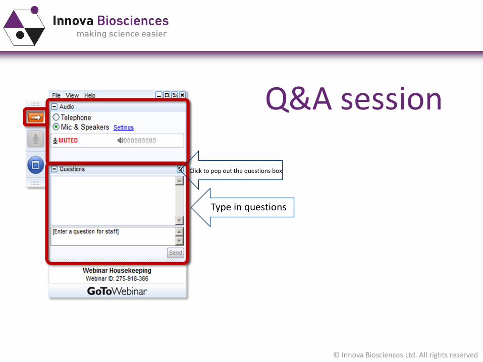
Q&A session
Type in questions
Click to pop out the questions box
© Innova Biosciences Ltd. All rights reserved
What is Immunohistochemistry?
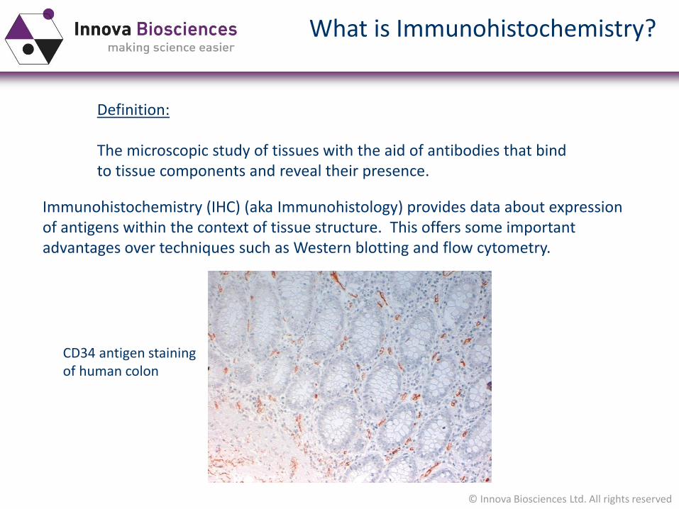
Definition:
The microscopic study of tissues with the aid of antibodies that bindto tissue components and reveal their presence.
Immunohistochemistry (IHC) (aka Immunohistology) provides data about expressionof antigens within the context of tissue structure. This offers some important advantages over techniques such as Western blotting and flow cytometry.
CD34 antigen staining of human colon

© Innova Biosciences Ltd. All rights reserved
Tissue preparation
The best option to maintain cellular structure, butdoes have a number of issues especially in relation to IHC.
Tissues needs to be fixed in formalin immediately after harvesting – generally for 4-24 hours. Over-fixation should be avoided.
Following fixation tissues are dehydrated and then embedded in hot paraffin (although plastic is sometimes used). Tissue blocks can be stored for many years.
5-10µm sections are prepared by microtome. Prior to labelling tissue sectionsare rehydrated.
Be aware that the fixation process can damage antigens, which can mean thatsome antibodies will not recognise their antigens.
Paraffin-embedded tissue
© Innova Biosciences Ltd. All rights reserved
Tissue preparation
Small pieces of tissue can be snap-frozen in liquid nitrogen or isopentane.
Sections can then be cut using a cryostat, which keeps tissue frozen during processing.
Fixation is then carried out immediately before staining, often using acetone.
Tissue morphology is generally not so good with frozen sections, but there is littleantigen damage and most antibodies will react as expected.
Frozen tissue
© Innova Biosciences Ltd. All rights reserved
Tissue pre-treatment
Formalin fixation can damage antigens due to cross-linking, which can mean that antibodies no longer recognise the antigen. Two general “antigen-retrieval” techniques are used to try to overcome this – proteolytic digestion and heat-induced epitope retrieval (HIER).
Proteolytic digestion uses incubation of tissue sections in either trypsin or pronase. Some antigens respond better to one enzyme than the other.
HIER may use either a pressure cooker, or commonly nowadays a microwave. There tends to be three cycles of heating, either in an acidic buffer (pH6) or alkaline buffer (pH9). Again, some antigens respond better to one pH than the other.
Paraffin-embedded tissue – antigen retrieval techniques
© Innova Biosciences Ltd. All rights reserved
Tissue pre-treatment
Peroxidase
A number of tissues contain endogenous peroxidase which can lead to backgroundstaining when using the HRP system. Blocking by pre-treatment with 0.3% hydrogen peroxide in PBS is recommended.
Alkaline phosphatase
Similarly, some tissues contain endogenous AP. This can be blocked using a pre-treatment with 5mM levamisole.
FFPE tissues tend to have much lowers levels of endogenous enzyme activity, so blocking is most commonly required for frozen tissues.
Frozen tissues - endogenous enzyme inhibition
© Innova Biosciences Ltd. All rights reserved
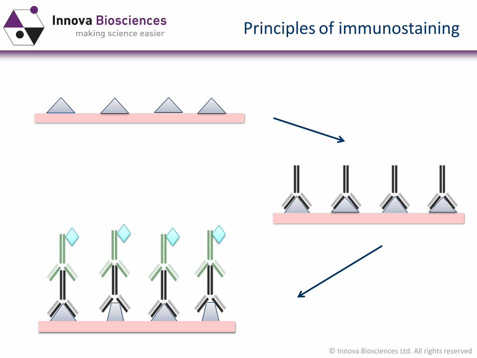
Principles of immunostaining
© Innova Biosciences Ltd. All rights reserved
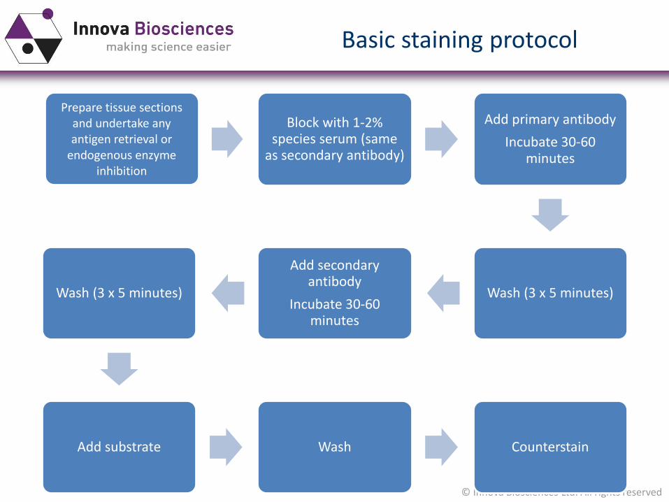
Basic staining protocol
Prepare tissue sections and undertake any antigen retrieval or
endogenous enzyme inhibition
Block with 1-2% species serum (same
as secondary antibody)
Add primary antibody
Incubate 30-60 minutes
Wash (3 x 5 minutes)
Add secondary antibody
Incubate 30-60 minutes
Wash (3 x 5 minutes)
Add substrate Wash Counterstain
© Innova Biosciences Ltd. All rights reserved
Choosing primary antibodies
Specificity and characterisation
Validation in IHC?
If using in FFPE is there information on requirement for antigen retrieval?
Check for supplier data, and if possible published references
On-line resources may be available with data http://www.proteinatlas.org/
© Innova Biosciences Ltd. All rights reserved
Choosing secondary antibodies
Specific for the primary antibody!
Not all secondary antibodies are the same…….consider epitope recognised, purification, absorption etc.
Enzyme conjugate (HRP or Alk Phos) or avidin/biotin system
Consider staining kits supplied by a number of suppliers
© Innova Biosciences Ltd. All rights reserved
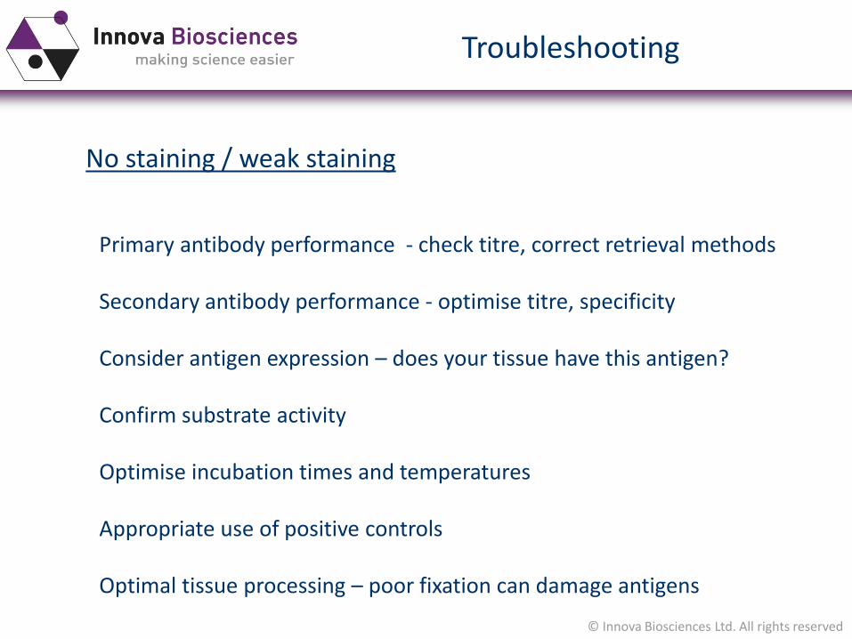
Troubleshooting
No staining / weak staining
Primary antibody performance - check titre, correct retrieval methods
Secondary antibody performance - optimise titre, specificity
Consider antigen expression – does your tissue have this antigen?
Confirm substrate activity
Optimise incubation times and temperatures
Appropriate use of positive controls
Optimal tissue processing – poor fixation can damage antigens
© Innova Biosciences Ltd. All rights reserved
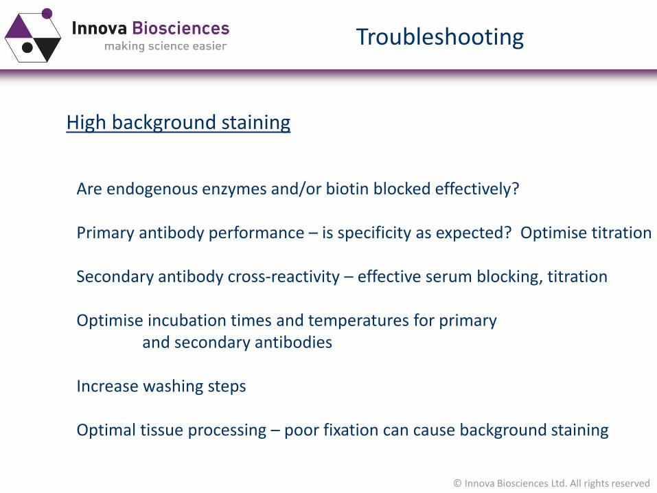
Troubleshooting
High background staining
Are endogenous enzymes and/or biotin blocked effectively?
Primary antibody performance – is specificity as expected? Optimise titration
Secondary antibody cross-reactivity – effective serum blocking, titration
Optimise incubation times and temperatures for primary and secondary antibodies
Increase washing steps
Optimal tissue processing – poor fixation can cause background staining
© Innova Biosciences Ltd. All rights reserved
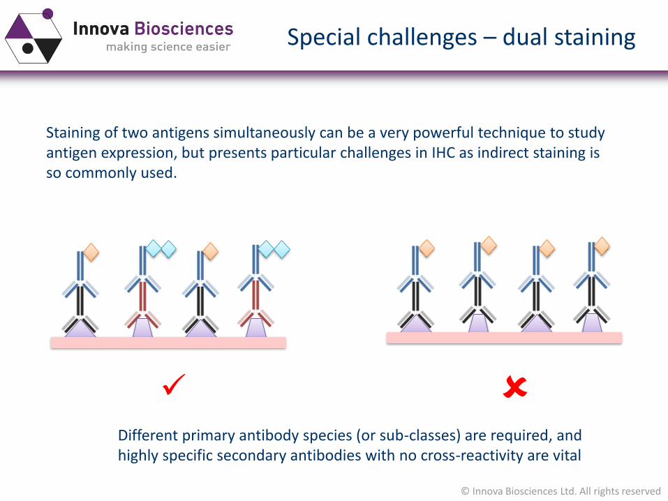
Special challenges – dual staining
Staining of two antigens simultaneously can be a very powerful technique to studyantigen expression, but presents particular challenges in IHC as indirect staining is so commonly used.
Different primary antibody species (or sub-classes) are required, and highly specific secondary antibodies with no cross-reactivity are vital
© Innova Biosciences Ltd. All rights reserved
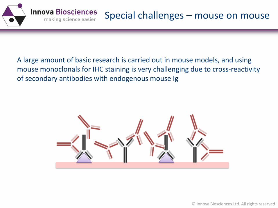
Special challenges – mouse on mouse
A large amount of basic research is carried out in mouse models, and using mouse monoclonals for IHC staining is very challenging due to cross-reactivityof secondary antibodies with endogenous mouse Ig
© Innova Biosciences Ltd. All rights reserved
Direct staining in IHC?
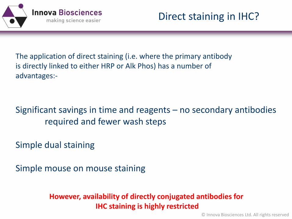
The application of direct staining (i.e. where the primary antibodyis directly linked to either HRP or Alk Phos) has a number ofadvantages:-
Significant savings in time and reagents – no secondary antibodiesrequired and fewer wash steps
Simple dual staining
Simple mouse on mouse staining
However, availability of directly conjugated antibodies for IHC staining is highly restricted
© Innova Biosciences Ltd. All rights reserved
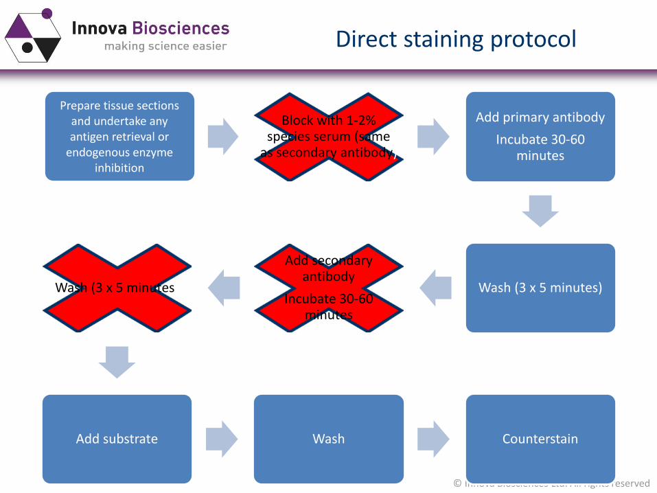
Direct staining protocol
Prepare tissue sections and undertake any antigen retrieval or
endogenous enzyme inhibition
Block with 1-2% species serum (same
as secondary antibody)
Add primary antibody
Incubate 30-60 minutes
Wash (3 x 5 minutes)
Add secondary antibody
Incubate 30-60 minutes
Wash (3 x 5 minutes)
Add substrate Wash Counterstain
© Innova Biosciences Ltd. All rights reserved
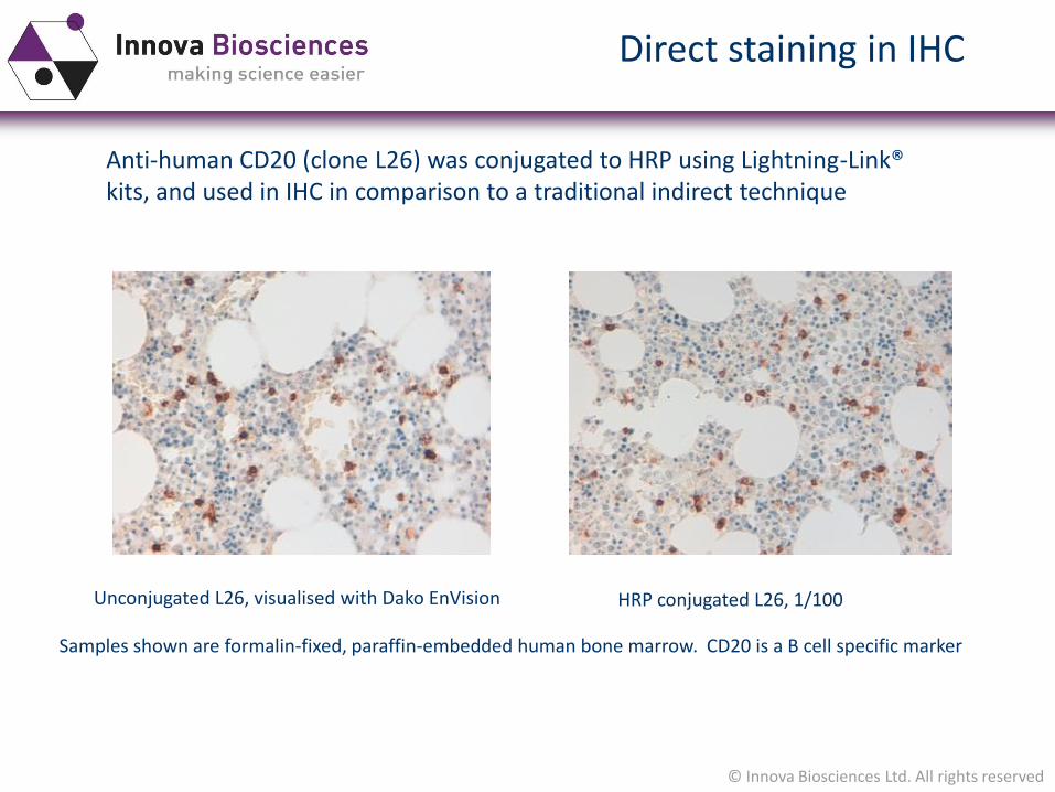
Anti-human CD20 (clone L26) was conjugated to HRP using Lightning-Link® kits, and used in IHC in comparison to a traditional indirect technique
Direct staining in IHC
Unconjugated L26, visualised with Dako EnVision HRP conjugated L26, 1/100
Samples shown are formalin-fixed, paraffin-embedded human bone marrow. CD20 is a B cell specific marker
© Innova Biosciences Ltd. All rights reserved
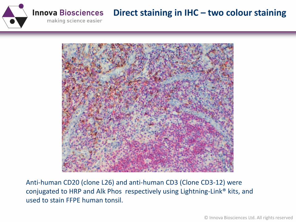
Anti-human CD20 (clone L26) and anti-human CD3 (Clone CD3-12) were conjugated to HRP and Alk Phos respectively using Lightning-Link® kits, and used to stain FFPE human tonsil.
Direct staining in IHC – two colour staining
© Innova Biosciences Ltd. All rights reserved
What if the antibody conjugate I need isn’t available?
The great majority of commercially available antibodies are not available directly conjugated to HRP or Alkaline Phosphatase for IHC use.
Having a custom made conjugate prepared, either of a commercialantibody, or of your own reagent, can be expensive and time consuming
Lightning-Link® conjugation kits fromInnova Biosciences offer a very quickand easy-to-use solution, that requiresas little as 10µg of antibody.
Preparing direct antibody conjugates
© Innova Biosciences Ltd. All rights reserved
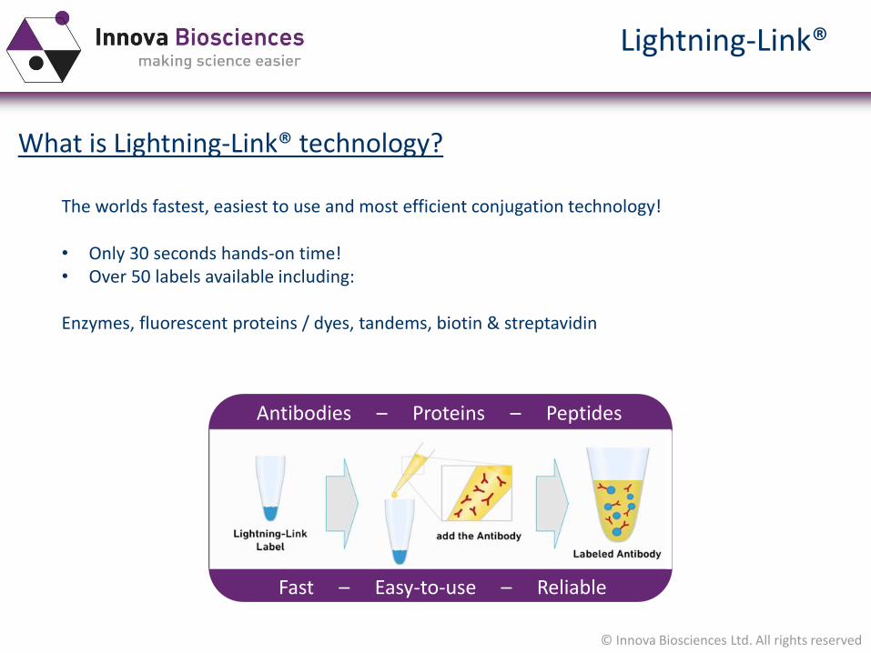
What is Lightning-Link® technology?
The worlds fastest, easiest to use and most efficient conjugation technology!
• Only 30 seconds hands-on time! • Over 50 labels available including:
Enzymes, fluorescent proteins / dyes, tandems, biotin & streptavidin
Lightning-Link®
Antibodies – Proteins – Peptides
Fast – Easy-to-use – Reliable
© Innova Biosciences Ltd. All rights reserved
What is Lightning-Link® technology?
• 100% antibody recovery
• Fully scalable from R&D (10µg<) to Production / Manufacture (>1g)
• Virtually eliminates batch to batch variability
• Coupling of the label to antibodies, proteins or other biomolecules
• Covalent conjugation ensures long term stability
Lightning-Link®
Lightning Link® kits are available for conjugating antibodies to HRP and Alkaline Phosphatase, as well as to Biotin and to a wide range of fluorescent dyes
© Innova Biosciences Ltd. All rights reserved
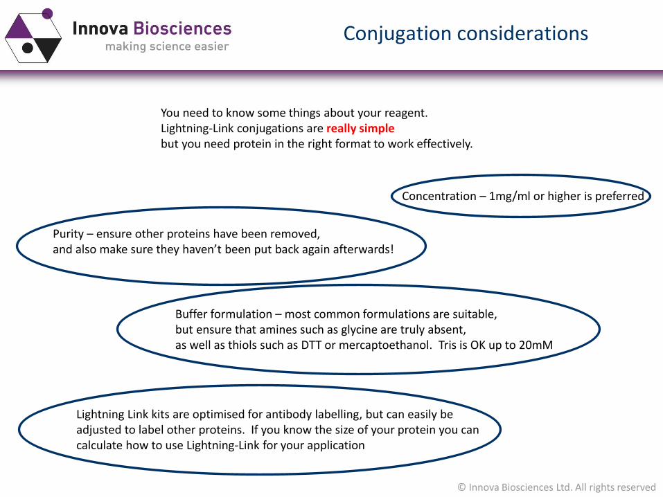
Conjugation considerations
You need to know some things about your reagent.Lightning-Link conjugations are really simplebut you need protein in the right format to work effectively.
Concentration – 1mg/ml or higher is preferred
Purity – ensure other proteins have been removed, and also make sure they haven’t been put back again afterwards!
Buffer formulation – most common formulations are suitable,but ensure that amines such as glycine are truly absent,as well as thiols such as DTT or mercaptoethanol. Tris is OK up to 20mM
Lightning Link kits are optimised for antibody labelling, but can easily be adjusted to label other proteins. If you know the size of your protein you cancalculate how to use Lightning-Link for your application

© Innova Biosciences Ltd. All rights reserved
Q&A session
Type in questions
Click to pop out the questions box
© Innova Biosciences Ltd. All rights reserved
San Diego, CAJune 23-26, 2014 Booth 4069
© Innova Biosciences Ltd. All rights reserved
Contact
If you would like any more information, please contact us at [email protected]
Please keep an eye out for our future webinars and other exciting news on our website and social media channels:
www.innovabiosciences.com/innova/webinars.html
YouTube: www.youtube.com/InnovaBiosciences
© Innova Biosciences Ltd. All rights reserved
Innova Biosciences Ltd.
Babraham Research Campus,
Cambridge, UK,
CB22 3AT
www.innovabiosciences.com
Lightning-Link® is a registered trademark of Innova BiosciencesDyLight® is a registered trademark of Thermo Fisher Scientific Inc. and its subsidiaries


