Fungal Diversity Ingoldian fungi In Hong Kong S.Y. Cban*, T.K. Gob and K.D. Hyde Centre for Research in Fungal Diversity, Department of Ecology and Biodiversity, The University of Hong Kong, Pokfulam Road, Hong Kong S.A.R., P.R. China; * e-mail: [email protected]Chan, S.Y., Goh, T.K. and Hyde, K.D. (2000). Ingoldian fungi in Hong Kong. In: Aquatic Mycology across the Millennium (eds K.D. Hyde, W.H. Ho and S.B. Pointing). Fungal Diversity 5: 89-107. A discussion on Ingoldian fungi is provided. The Ingoldian fungi known from Hong Kong are listed and a key for their identification is provided. Most of the fungi are illustrated and it is hoped that the paper will be a basis for the study of Ingoldian fungi in Hong Kong, at the student level. Key words: hyphomycetes, Ingoldian fungi. Introduction Freshwater hyphomycetes are often classified as those fungi which for part of their life cycle, or the whole of their life cycle, occur in freshwater environments. This definition, is however, quite vague as it includes all fungi that may be present in the freshwater ecosystem, regardless of their origins. Freshwater hyphomycetes can be classified into four ecological groups based on sporulation methods and mycelial growth. This gives a clearer definition of different freshwater fungal groups. The four ecological groups include the aero-aquatic hyphomycetes, terrestrial-aquatic hyphomycetes, submerged-aquatic (amphibious) hyphomycetes and Ingoldian fungi. Ingoldian fungi, which were the target group of this study, are classified as those fungal species actively growing and sporulating under water. They occur mostly on plant litter, and leaves in rivers or streams (Bm-Iocher, 1992). However, the grouping of these different kinds of freshwater hyphomycetes is quite arbitrary and some species overlap between the definitions. Ingoldian fungi were named in honor of C.T. Ingold, the "father" of this group of fungi, who was the mycologist to discover the typical habitat of these fungi (Iqbal, 1994). Habitat Ingoldian fungi are found in freshwater environments, mainly in rapidly flowing and turbulent water. The apparent preference for fast running, well- 89
Transcript
Fungal Diversity
Ingoldian fungi In Hong Kong
S.Y. Cban*, T.K. Gob and K.D. Hyde
Centre for Research in Fungal Diversity, Department of Ecology and Biodiversity, TheUniversity of Hong Kong, Pokfulam Road, Hong Kong S.A.R., P.R. China; * e-mail:[email protected]
Chan, S.Y., Goh, T.K. and Hyde, K.D. (2000). Ingoldian fungi in Hong Kong. In: AquaticMycology across the Millennium (eds K.D. Hyde, W.H. Ho and S.B. Pointing). FungalDiversity 5: 89-107.
A discussion on Ingoldian fungi is provided. The Ingoldian fungi known from Hong Kong arelisted and a key for their identification is provided. Most of the fungi are illustrated and it ishoped that the paper will be a basis for the study of Ingoldian fungi in Hong Kong, at thestudent level.
Key words: hyphomycetes, Ingoldian fungi.
IntroductionFreshwater hyphomycetes are often classified as those fungi which for part
of their life cycle, or the whole of their life cycle, occur in freshwaterenvironments. This definition, is however, quite vague as it includes all fungithat may be present in the freshwater ecosystem, regardless of their origins.
Freshwater hyphomycetes can be classified into four ecological groupsbased on sporulation methods and mycelial growth. This gives a clearerdefinition of different freshwater fungal groups. The four ecological groupsinclude the aero-aquatic hyphomycetes, terrestrial-aquatic hyphomycetes,submerged-aquatic (amphibious) hyphomycetes and Ingoldian fungi. Ingoldianfungi, which were the target group of this study, are classified as those fungalspecies actively growing and sporulating under water. They occur mostly onplant litter, and leaves in rivers or streams (Bm-Iocher, 1992). However, thegrouping of these different kinds of freshwater hyphomycetes is quite arbitraryand some species overlap between the definitions. Ingoldian fungi were namedin honor of C.T. Ingold, the "father" of this group of fungi, who was themycologist to discover the typical habitat of these fungi (Iqbal, 1994).
HabitatIngoldian fungi are found in freshwater environments, mainly in rapidly
flowing and turbulent water. The apparent preference for fast running, well-
89
aerated and non-polluted streams, indicates that they cannot tolerate lowoxygen levels (Barlocher, 1992). The majority of Ingoldian fungi are found instreams and rivers, but some have also been reported from lakes and terrestrialhabitats. They are saprotrophs and occur on almost any type of plant debris andare indeed most common on deciduous leaves, but they also colonize conifertwigs and needles. They also grow on submerged macrophytes and asendophytes in healthy roots of riparian trees (Barlocher, 1992).
Role in food webInterest in Ingoldian fungi was increased by the studies by Kaushik and
Hynes (1971), who found that autumn-shed leaves were an important foodsource for invertebrates in streams. The leaves undergo a process of microbial
degradation, in which Ingoldian fungi play an important role. The microbialdegradation makes the plant litter more palatable and nutritious to leafshredders (Suberkropp and Klug, 1976; Barlocher, 1992). These authorstherefore established that Ingoldian fungi are intimately involved in the energyflow in streams. Fungi are decomposers, which have been shown to produce arich array of enzymes active towards the major leaf polysaccharides(Suberkropp and Klug, 1980; Chamier, 1985; Suberkropp, 1991a), making theenergy from shredded leaves accessible to the community. Energy flows anddevelopment of communities in freshwater ecosystems are largely dependent onthe supply of allochthonous material, the majority of which is leaf litter fromadjacent terrestrial environments (Barlocher and Kendrick, 1976). The riparianvegetation therefore forms a close relationship with the stream ecosystem.Many previous studies of fungi in streams have focused on their role in theenergy flow and trophic dynamics of such detritus-based food chains(Suberkropp, 1992).
AdaptationIngoldian fungi have a large variety of conidial shapes that include
tetraradiate, branched or filiform. The most frequently observed spore conidialshape are tetraradiate. The function of this conidial shape in aquatichyphomycetes is to minimize downstream transport (Webster, 1959). When atetraradiate spore makes contact with a surface it does so at three points and thespore acts as a tripod, which represent a very stable form of attachment.Germination of Ingoldian fungi requires a contact stimulus and upon settling,the spore germinates to form a pad or appressorium, which further strengthensadhesion to surfaces (Webster, 1959). This mechanism may explain whyIngoldian fungi are successful colonizers on submerged plant material. Anotherexplanation for the abundance of the tetraradiate shape is that the shape might
90
Fungal Diversity
facilitate the dispersal in aqueous films, between layers of terrestrial leaf litter(Bandoni, 1975).
The second most common conidial shape typical of hyphomycetes issigmoid, a configuration which also aids attachment (Webster and Davey, 1984;Webster, 1987). Sigmoid spores in a slow moving current tend to role along thebottom, and conidial ends can make contact with surface (Webster and Davey,1984), which enhance the chance of colonizing the substratum. The twodominant conidial shapes of aquatic hyphomycetes increase their probability ofencountering a target (Cox, 1983) and hence facilitate attachment.
BiodiversityIn his first report, Ingold (1942) described 16 species ofIngoldian fungi, 10
of which were new, marking the starting point of a "minor mycologicalindustry" (Ainsworth, 1976). Later, over 150 species were described and manymore await description (Webster and Descals, 1981). In the recent report,approximately 300 species of Ingoldian fungi were thought to have beendescribed, most from temperate regions (Goh, 1997). This number is still..mcreasmg.
Geographical distributionIngoldian fungi exhibit morphological (Webster, 1959) and physiological
adaptations (Suberkropp and Klug, 1981) for plant litter degradation in flowingwater. Their conidia have been reported from a variety of habitats andgeographical locations (Webster and Descal, 1981, Wood-Eggenschwiler andBarlocher, 1983). They are cosmopolitan in their distribution, extending fromthe arctic Circle to the equator (Kobayasi et al., 1967, 1971; Muller-Haeckeland Marvanova, 1976, 1979; Webster and Descals, 1981; Engblom et al., 1986;Bhat and Chien, 1990). Geographic occurrences of fungi are broadly correlatedwith optimal temperature for in vitro growth and sporulation (Barlocher, 1992).Abundance and biodiversity of Ingoldian fungi vary in different temperaturezones. Most known Ingoldian fungi have been described from temperateregions, many tropical species are still unexplored.
Seasonal distributionThe concentration of conidia in stream water in temperate regions, has been
shown to be influenced by seasonal changes in leaf fall from riparian vegetation(Iqbal and Webster, 1973, 1977). This seasonal influence on the occurrence ofaquatic hyphomycete is more likely to be mediated through the availability offresh supply of autumn-shed leaves. The more leaves are available forcolonization, then the more conidia are found. A study found that most species
91
in England were more common from late summer to early winter than duringthe rest of the year (Ingold, 1942). In this period, there is an enormous amountof fallen leaves during autumn, and hence the concentration of conidia peaks.
Influence of riparian vegetation-typeThe occurrence and concentration of conidia and the species composition
of fungal communities not only vary with season, but also vary with the typesof riparian vegetation in different streams. Streams with similar physicalcharacteristics differ in their ecology according to the riparian vegetation. It iswell established that changes in the riparian flora often coincide with changesin the aquatic hyphomycete community (Gonczol, 1975, 1987, 1989; Barlocher,1982; Wood-Eggenschwiler and Barlocher, 1983; Thomas et al., 1989). Whenleaves of different species are collected from the same stream section,dominance patterns in the fungal communities of the leaves usually differ(Gonczol, 1975, 1989; Suberkropp and Klug, 1976; Chamier and Dixon, 1982;Bengtsson, 1983; Rossi et al., 1983; Shearer and Lane, 1983; Sridhar andKaveriappa, 1988, 1989). It can be concluded that riparian vegetation-typeplays an important role in determining the community composition of Ingoldianfungi in the stream.
Influence of water chemistryAu (1992) studied the influence of physical-chemical factors on the ability
of aquatic hyphomycetes to compete with other organisms for plant litterdecomposition. She found that among water temperature, dissolved oxygen,biological oxygen demand, pH, turbidity, oxygen availability would probablybe the major factors, since well-oxygenated water is required for growth andsporulation of aquatic hyphomycetes (Nilsson, 1964; Webster, 1975).
DispersalSince Ingoldian fungi do not have motile conidia, they are dispersed in
water currents. Apart from having independent conidia, Ingoldian fungi canalso attach to substrate during their dispersal. They can travel downstream bymeans of mycelium embedded in leaf tissue or wood submerged in the stream.Other dispersal mechanisms include animals, mycelium may attach to the feetof waterfowl (Barlocher, 1992) or aquatic invertebrates, which may transportthem to other areas. Asexual spore of aquatic fungi are generally too fragile forlong-range dispersal, while sexually produced spores are often airborne andallow long distance dispersal (Barlocher, 1992). This may help to explain theparadox of the worldwide distribution of freshwater fungi with passivelydispersed conidia.
92
Fungal Diversity
Table 1. List of Ingoldian fungi found in Hong Kong from different studies.
Dendrosporafusca Descals and WebsterDicranidion gracile Matsush.
Diplocladiella scalaroides Arnaud apud EllisFlabellospora acuminata Descals and WebsterFlabellospora crassa AlasoaduraFlabellospora spp.Flabellospora verticil/ata AlasoaduraFlagellospora curvula Ingold
Flagellospora penicilliodes IngoldHelicomyces colligatus MooreHelicomyces spp.Helicomyces torquatus Lane and ShearerIsthmolongispora spp.lsthmotricladia gombakiensis Nawawi
Lemonniera aquatica De WildemanLemonniera spp.
Lunulospora curvala IngoldLunulospora cymbiformis MiuraMycocentrospora filiform is Iqbal
References
Chan et al., 2000Au et al., 1992ChaD et al., 2000
ChaD et al., 2000; Tsui et al., 2000Ch an et al., 2000Chan et al., 2000
Au et al., 1992
Au et aI., 1992; ChaD et al., 2000
Chan et al., 2000
Ho, 1998; ChaD et al., 2000; Tsui etal., 2000Au et al., 1992
Chan et aI., 2000Chan et al., 2000ChaD et al., 2000Au et al., 1992Chan et aI., 2000Au et al., 1992ChaD et al., 2000Au et al., 1992
Chan et al., 2000ChaD et aI., 2000ChaD et al., 2000Au et al., 1992
Chan et al., 2000
Ho, 1998; Chan et al., 2000Chan et al., 2000Chan et al., 2000Chan et aI., 2000ChaD et al., 2000
Au et al., 1992; Chan et al., 2000
Au et al., 1992
Chan et aI., 2000Chan et al., 2000Chan et al., 2000Ch an et al., 2000Chan et al., 2000Chan et al., 2000
Au et al., 1992; Chan et al., 2000
Au et al., 1992
Au et al., 1992; Chan et al., 2000Au et al., 1992
93
Table 1. (continued).
Name
Nawawiafiliformis (Nawawi) Marvanova
Pseudoanguillospora stricta Iq balPyramidosporafluminea Miura and KudoScutisporus brunneus Ando and TubakiSigmoidea aurantiaca DescalsSubulispora procurvata Tubaki and YokohamaTetrachaetum elegans IngoldTetracladium marchalianum De WildemanTetracladium setigerum IngoldTricladim spp.Tricladium attenuatum IqbalTricladium indicum Sati rt N. Tiwari
Chan et aI., 2000Ch an et al., 2000Chan et al., 2000
Au et al., 1992; Tsui et al., 2000
Ho, 1998Chan et aI., 2000
Au et aI., 1992; Chan et aI., 2000
Chan et al., 2000
Au et al., 1992; Chan et al., 2000
Chan et al., 2000
Au et al., 1992Au et al., 1992
Ingoldian fungi in Hong KongStudies of Ingoldian fungi have been carried out in many countries, mostly
in temperate regions. In Hong Kong, 387 species of freshwater water fungihave been identified (Lu et al., 2000). Previous studies of Ingoldian fungi inHong Kong were carried out by comparing the biodiversity found on specific
leaf types in the polluted Lam Tsuen River and the unpolluted Tai Po KauForest Stream (Au et al., 1992). Twenty-five aquatic hyphomycetes specieswere found and most of them were cosmopolitian or frequently reported in
temperate regions. In other separate studies, Chan et al. (2000) reported 41species, Tsui et al. (2000) reported 3 species and Ho (1998) reported 4 speciesfrom Hong Kong. A total of 51 species of Ingoldian fungi known from Hong
Kong from several studies are lised in Table 1 and a key is provided below.
Key to the identified species found in Lam Tsuen River and Tai Po Kau ForestStream in Hong KongI. Conidia tetraradiate 2I. Conidia sigmoid 3I. Conidia with other shapes .4
2. Conidia hyaline 52. Conidia brown 6
94
Fungal Diversity
3. Conidia unicellular Flagellospora curvula (Fig. 27)3. Conidia with 2 or more cells 7
4. Conidia hyaline 84. Conidia brown 9
5. Conidia septate 105. Conidia non-septate 11
6. Conidia with 4-8 appendages slightly constricted at origin, much longer than 1.5 timesdiam. of central part; central part globose to pyramidal. Brachiosphaera tropicalis (Fig. 8)
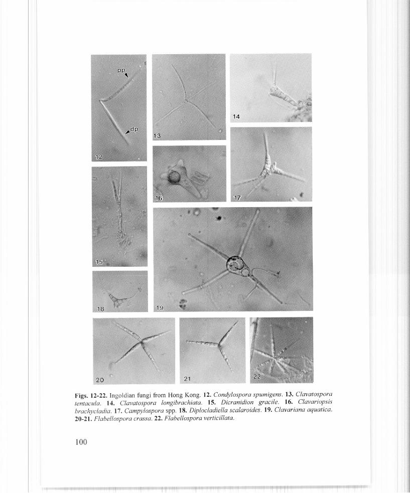
6. Conidia consisting of a clavate central body (triangular in outline), 5-8 !lm wide at base,24-33 ~lm wide above (crowned portion), and with 4, 0-3 septate, 53-160 !lm longappendages, 3-5 /lm at widest point, tapering to 2-2.5 /lm towards their ends; appendagesseptate but not constricted at their base Clavariana aquatica (Fig. 19)
7. Conidia filiform, not wider than 8 !lm 127. Conidia wider than 8 !lm in the middle, vermiform, with septum in middle .
.......... Anguillospora crassa (Fig. 3)
8. Conidia coiled 138. Conidia uncoiled 14
9. Conidia consisting of a biconic, symmetrical main axis, with a distinct, hyaline, transverseband Beltrania rhombica (Fig. 10)
9. Conidia not consisting ofa biconic, symmetrical main axis 15
10. Main axis clavate, truncate at apex, with non-septate appendages ................................................................................... Clavatospora longibrachiata (Fig. 14)
10. Main axis not clavate : 16
11. Conidia with spherical main axial cell 1711. Conidia without spherical main axial cell... 18
12. Conidia bicelled, not longer than 60 /lm Flagellospora penicilliodes (Fig. 49)12. Conidia with more than 2 cells 19
13. Secondary conidia usually formed, pate 11ate end of filament without flattened detachmentscar Helicomyces colligatus (Fig. 29)
13. Secondary conidia rarely formed, with flattened detachment seaL ............................ Helicomyces torquatus (Fig. 30)
14. Conidia branched 2014. Conidia unbranched 21
15. Conidia with filiform appendages 2215. Conidia without filiform appendages 23
16. Conidia consisting of a main axis widening at apex, where there is an oval to sphericalcentral knob, appendage with apical cell rounded, forming an eccentric knob .................................................................................... Tetracladium marchalianum (Fig. 35)
95
16. Conidia without central knob or eccentric knob 24
17. Conidia with 3-5 (mostly 4) appendages, which are obclavate, 3-9-septate (mostly 5), 3556/lm long, 3-3.5 /lm wide at apex, 5.5-7.5 /lm at widest point. Main axis 5-15 x 2-3 /lm,
with a 5-7 ~lm wide terminal swelling Flabellospora crassa (Figs. 20-21)17. Conidia with 4 divergent appendages, which are 25-120 x 2-5 /lm, 1-3 septate, uniform in
width. One of the appendages longer than the others Lemonniera aquatica (Fig. 31)
18. Conidial appendages rhomboid, obpyramidal, or obclavate 2518. Conidial appendages not rhomboid, obpyramidal or obclavate 26
19. Conidia with a basal filiform or falcate appendage, 5-35 x 1-2 /lm, uniform widththroughout its length Mycocentrosporafiliformis (Fig. 50)
19. Conidia with a basal filiform or falcate appendage, 75-125 x 2.5-30 /lm, tapering slightlytowards the apex Centrospora aquatica (Fig. 46)
19. Conidia without a basal filiform or falcate appendage 27
20. Condia multiradiate (with more than 4 appendages) 2820. Conidia not multiradiate 29
21. Conidia sickle-shape 3021. Conidia not sickle-shape 31
22. Conidia consisting a cylindrical main axis and 2 subapical appendages ................................................................................. .Camposporium antennatum (Fig. 9)
22. Conidia not consisting a cylindrical main axis 32
23. Conidia with 4-6 cylindrical or obclavate appendages with rounded apices, diverging atright angles to the main axis Dendrosporafusca (Fig. 48)
23. Conidia consisting of a main axis bent back on itself at about one third of its length, and 23 divergent appendages, shorter part of axis 2-celled, with one appendage; longer part ofaxis 3-5 celled, with 1-2 appendages Tripospermum porosporiferum (Fig. 32)
24. Conidia with axis rarely cylindrical, proximal part from cylindrical to narrowly clavate,distal part narrow-cymbiform Alatospora pulchella (Fig. 42)
24. Conidia with axis cylindrical 33
25. Conidia consisting of an obconical, 2 celled axis, and 3-4 romme appendages, either conicor obconical, much wider at base than at apex Clavariopsis brachycladia (Fig. 16)
26. Main axis clavate, with 3 equidistant divergent appendages arising from apex ........................................................................................... Clavatospora tentacula (Fig. 13)
26. Main axis not clavate 34
27. Conidia bent, with a detachment scar at where it bends at one -fifth of its length ............................................................................................. Calcarispora hiemalis (Fig. 44)
27. Con idia not bent 35
28. Conidia with 3 digitate (fmger-like) appendages Tetracladium setigerum (Figs. 36-37)28. Conidia without 3 digitate appendages 36
96
Fungal Diversity
29. Conidia unicellular, triangular-shaped and having long, hair-like divergent processes fromeach corner of the triangle Nawawiafiliformis (Fig. 58)
29. Conidia not unicellular 37
30. Conidia with two bends at the middle, proximal portion (Pp) straight to slightly curved;distal portion (dp) forming an angle of30-120° with the proximal portion .............. Condylospora spumigena (Fig. 12)
30. Conidia bent, with a rhombic detachment scar near the middle ........ Lunulospora cymbisformis (Fig. 25)
31. Conidia without filiform appendage Isthmolongispora quadricellularia (Fig. 26)31. Conidia with filiform appendage, conidia subulate-conoid, truncate at base .
32. Main body 4-celled, with a long appendage arising from each of the four corners of themain body SCutisporus brunneus (Fig. 34)
32. Main body more than 4-celled 38
33. Conidia consisting of 4 divergent appendages, one of them 1-3-septate, forming the mainaxis, the other 3 appendages inserted at the upper end of the main axis, one usually longerthan the others Articulospora tetracladia (Fig. 2)
33. Conidia consisting of 4 divergent appendages, one forming the main axis, the other 3appendages attached to the anterior part of the main axis, but not to the end of the mainaxis 39
34. Main axis 30-70 /lm x 2.4.5 /lm, with two 25-60 /lm x 2.5-3.5 /lm appendages fusing withmain axis Alatospora acuminata (Fig. 1)
34. Main axis 75-300 /lm x 2.5-5 /lm, bent at the insertion of appendages, with 2 divergingappendages of uniform width (septa indistinct) Tetrachaetum elegans (Fig. 54)
34. Main axis 150-200 /lm, widest (3-4 /lm) in the region between the two lateral appendages.The lower lateral appendages arising 50-70 /lm from the base and apparently causing aslight deflexion in the direction of growth of the principle axis, a further deflexion occuringwhen the second lateral arises 15-20 /lm above the first.. ......................................................................................... Tricladium chaetocladium (Fig. 56)
35. Conidia with a broad detachment scar or base truncate .40
35. Conidia without a broad detachment scar and tapering toward both ends .41
36. Conidia consisting of a 2-celled central axis, lateral outgrowths unbranched, hemisphericalor conic to cylindrical, with rounded ends Pyramidosporafluminea (Fig. 52)
36. Conidia not consisting ofa 2-celled central axis .42
38. Main axis consisting a smaller allantoid part and a larger triangular part 4438. Main axis triangular and 8-celled Diplocladiella scalaroides (Fig. 18)
39. Conidia constricted at septa .4539. Conidia not constricted at septa .46
97
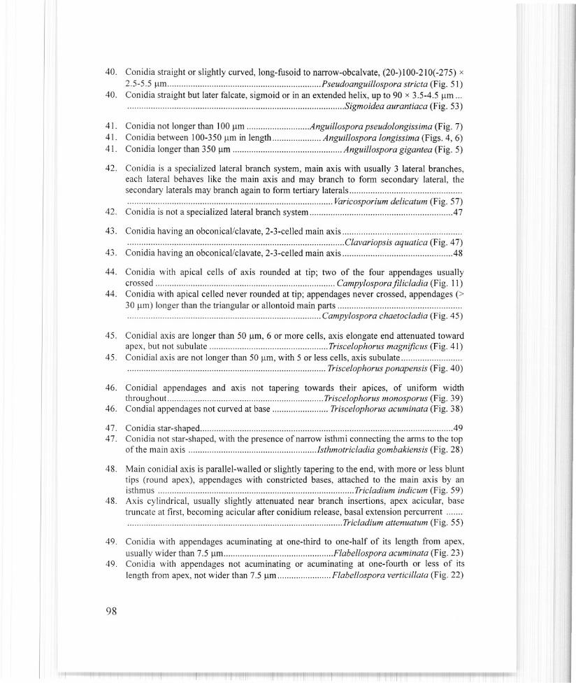
40. Conidia straight or slightly curved, long-fusoid to narrow-obcalvate, (20-) 100-210(-275) x
2.5-5.5 ~lm Pseudoanguillospora stricta (Fig. 51)40. Conidia straight but later falcate, sigmoid or in an extended helix, up to 90 x 3.5-4.5 Ilm ...
41. Conidia not longer than 100 Ilm Anguil/ospora pseudolongissima (Fig. 7)41. Conidia between 100-350 flm in length Anguillospora longissima (Figs. 4, 6)41. Conidia longer than 350 Ilm Anguil/ospora gigantea (Fig. 5)
42. Conidia is a specialized lateral branch system, main axis with usually 3 lateral branches,each lateral behaves like the main axis and may branch to form secondary lateral, thesecondary laterals may branch again to form tertiary laterals ......................................................................................... Varicosporium delicatum (Fig. 57)
42. Conidia is not a specialized lateral branch system 47
43. Conidia having an obconical/clavate, 2-3-celled main axis .................. Clavariopsis aquatica (Fig. 47)
43. Conidia having an obconical/clavate, 2-3-celled main axis .48
44. Conidia with apical cells of axis rounded at tip; two of the four appendages usuallycrossed Campylosporafilicladia (Fig. 11)
44. Conidia with apical celled never rounded at tip; appendages never crossed, appendages (>30 flm) longer than the triangular or allontoid main parts .................................................................................... Campylospora chaetocladia (Fig. 45)
45. Conidial axis are longer than 50 flm, 6 or more cells, axis elongate end attenuated towardapex, but not subulate Triscelophorus magnificus (Fig. 41)
45. Conidial axis are not longer than 50 flm, with 5 or less cells, axis subulate ...................................................................................... Triscelophorus ponapensis (Fig. 40)
46. Conidial appendages and axis not tapering towards their apices, of uniform widththroughout. Triscelophorus monosporus (Fig. 39)
46. Condial appendages not curved at base Triscelophorus acuminata (Fig. 38)
47. Con idia star-shaped 4947. Conidia not star-shaped, with the presence of narrow isthmi connecting the arms to the top
of the main axis Jsthmotricladia gombakiensis (Fig. 28)
48. Main conidial axis is parallel-walled or slightly tapering to the end, with more or less blunttips (round apex), appendages with constricted bases, attached to the main axis by anisthmus Tricladium indicum (Fig. 59)
48. Axis cylindrical, usually slightly attenuated near branch insertions, apex acicular, basetruncate at first, becoming acicular after conidium release, basal extension percurrent ......................................................... Tricladium attenuatum (Fig. 55)
49. Conidia with appendages acuminating at one-third to one-half of its length from apex,usually wider than 7.5 Ilm Flabellospora acuminata (Fig. 23)
49. Conidia with appendages not acuminating or acuminating at one-fourth or less of itslength from apex, not wider than 7.5 flm Flabellospora verticil/ata (Fig. 22)
AcknowledgementsHelen Leung is thanked for kind assistance.
References
Ainsworth, G.c. (1976). Introduction to the History of Mycology. U.K, Cambridge, Universityof Cambridge Press.
Au, D.WT., Hodgkiss, I.J. and Vrijmoed, L.L.P. (1992a). Fungi and cellulolyticactivityassociated with decomposition of Bauhinia purpurea leaf litter in a polluted andunpolluted Hong Kong waterway. Canadian Journal of Botany 70: 1071-1078.
Au, D.WT., Hodgkiss, I.J. and Vrijmoed, L.L.P. (1992b). Decomposition of Bauhinia purpurealeaf litter in a polluted and unpolluted Hong Kong waterway. Canadian Journal of Botany70: 1061-1069.
Bandoni, RJ. (1975). Significance of the tetraradiate form in dispersal of terrestrial Fungi.Report of the Tottori Mycological Institute 12: 105-113.
Barlocher, F. (1982). Conidium production from leaves and needles in four streams. CanadianJournal of Botany 60: 1487-1494.
Barlocher, F. (1992). The Ecology of Aquatic Hyphomycetes. Germany, Berlin, SpringerVerlag: Ecological Studies Vol. 94.
Barlocher, F. and Kendrick, B. (1976). Hyphomycetes as intermediaries of energy flow instreams. In: Recent Advances in Aquatic Mycology. (ed E.B.G. Jones). Elek Science,London, U.K.
Bengtsson, G. (1983). Habitat selection in two species of aquatic hyphomycetes. MicrobialEcology 9: 15-26.
Bhat, 0.1. and Chien, c.Y. (1990). Water-borne hyphomycetes found in Ethiopia. Transactionsof Mycological Society ofJapan 31: 147-158.
Chan, S.Y., Goh, T.K. and Hyde, KD. (2000). Ingoldian fungi in Lam Tsuen River and Tai PoKau Forest Stream, Hong Kong. Fungal Diversity 5: 109-118.
Chamier, A.C. and Dixon, P.A. (1982). Pectinases in leaf degradation by aquatic hyphomycetes.I: The field study. The colonization pattern of aquatic hyphomycetes on leaf packs in aSurrey stream. Oecologia 52: 109-115.
Chamier, A.C. (1985). Cell-wall degrading enzymes of aquatic hyphomycetes: a review.Botanical Journal of the Linnean Society 91: 67-81.
Cox, P.A. (1983). Search theory, random motion and the convergent evolution of pollen andspore morphology in aquatic plants. The American Naturalist 121: 9-31.
Engblom, E., Lingdell, P.E., Marvanova, L. and Muller-Haeckel, A. (1986). Foam spora inrunning water of southern Greenland. The Polar Regions 4: 47-51.
Goh, T.K. (1997). Tropical freshwater hyphomycetes. In: Biodiversity of Tropical Microfungi(ed KO. Hyde). Hong Kong University Press, Hong Kong: 189-227.
Gonczol, J. (1975). Ecological observations on the aquatic hyphomycetes of Hungary, I. ActaBotanica. Academiae Scientiarum Hungaicae 21: 243-264.
Gonczol, 1. (1987). Ecological observations on aquatic hyphomycetes of Hungary, Ill. ActaBotanica. Academiae Scientiarum Hungaicae 33: 41-49.
Gonczol, 1. (1989). Longitudinal distribution patterns of aquatic hyphomycetes in a mountainstream in Hungary. Experiment with leaf packs. Nova Hedwigia 48: 391-404.
Ho, WH. (1998). Biodiversity, ecological and ultrastructural observations of fungi on woodsubmerged in tropical streams. Ph.D. Thesis, University of Hong Kong, Hong Kong.
Iqbal, S.H. (1994). Species diversity of freshwater hyphomycetes in some streams of Pakistan.
105
1. Comparison of sampling techniques. Mycoscience 35: 331-343.Iqbal, S.H. and Webster, 1. (1973a). Aquatic hyphomycete spora of the River Exe and its
tributaries. Transactions of British Mycological Society 61: 331-346.Iqbal, S.H. and Webster, J. (1973b). The trapping of aquatic hyphomycete spora by air bubbles.
Transactions of British Mycological Society 60: 37-48.Iqbal, S.H. and Webster, J. (1977). Aquatic hyphomycetes spora of some Dartmoor streams.
Transactions of the British Mycological Society 69: 233-241.Ingold, C.T. (1942). Aquatic hyphomycetes of decaying alder leaves. Transactions of the British
Mycological Society 25: 339-417.Kaushik, N.K. and Hynes, H.B.N. (1971). The fate of the dead leaves that fall into streams.
Archiv fUr Hydrobiologia 68: 465-515.Kobayasi, Y., Hiratsuka, N., Korf, R.P., Tubaki, K, Aoshima, K, Soneda, M. and Sugiyama, J.
Cl 976). Mycological studies of the Alaskan Arctic. Annual Report of the Institute ofFermentation of Osaka 3: 1-138.
Kobayasi, Y., Hiratsuka, N., Otani, Y., Tubaki, K., Udagawa, S.L, Sugiyama, J. and Konno, KCl 971). Mycological studies of the Angmagssalik region of Greenland. Bulletin of theNational Science Museum of Tokyo. 14: 1-96.
Lu, 8.S., Hyde, K.D., Ho, W.H., Tsui, C.KM., Taylor, 1. E., Wong, KM., Yanna and Zhou,D.Q. (2000). Checklist of Hong Kong Fungi. Fungal Diversity Press, The University ofHong Kong, Hong Kong.
Muller-Haeckel, A. and Marvanova, L. (1976). Konidienproduktion und kolonisation vonSuddwasser-Hyphomyzeten im Kaltisjokk Lappland. Botaniska Notiser 129: 405-409.
Muller-Haeckel, A., Marvanova, L. (1979). Periodicity of aquatic hyphomycetes in thesubarctic. Transactions of British Mycological Society 73: 109-116.
Nilsson, S. Cl 964). Freshwater hyphomycetes. Taxonomy and morphology and ecology.Symbolae Botanica Upsalensis 18: 1-130.
Rossi, L., Fano, E.A., Basset, A., Fanelli, e. and Fabbri, A.A. (1983). An experimental study ofa microfungal community on plant detritus in a Mediterranean woodland stream.Mycologia 75: 887-896.
Shearer, e.A. and Lane, L.e. (1983). Comparison of three techniques for the study of aquatichyphomycete communities. Mycologia 75: 498-598.
Sridhar, K.R. and Kaveriappa, KM. (1988a). New host records of aquatic hyphomycetes.Indian Phytopathology 41: 160-161.
Sridhar, K.R. and Kaveriappa, K.M. (1988b). Colonization of leaf litter by aquatichyphomycetes in Western Ghat stream. Proceedings of Indian National Science AcademyB 54: 199-200.
Sridhar, K.R. and Kaveriappa, K.M. (1988c). Colonization of leaf litter by aquatichyphomycetes in a tropical stream. Archiv fUr Hydrobiologia 112: 627-630.
Sridhar, KR. and Kaveriappa, KM. (1988d). Occurrence and survival of aquatic hyphomycetesin brackish and seawater. Arch fur Hydrobiologia 113: 153-160.
Sridhar, K.R. and Kaveriappa, KM. (1988e). Survival of water-borne fungi - imperfecti undernon-aquatic conditions. Proceedings ofIndian National Science Academy B 54: 295-297.
Sridhar, K.R. and Kaveriappa, K.M. (1989a). Notes on aquatic hyphomycetes of mountainstreams in Western Ghat region, India. Feddes Report 100: 187-189.
Sridhar, K.R. and Kaveriappa, K.M. (1989b). Colonization of leaves by water-bornehyphomycetes in a tropical stream. Mycological Research 92: 392-396.
Sridhar, K.R. and Kaveriappa, K.M. (1989c). Water-borne hyphomycetes spora of twofreshwater streams. Environmental Ecology 7: 771-772.
Sridhar, K.R. and Kaveriappa, KM. (1989d). Observations on aquatic hyphomycetes of the
106
Il
Fungal Diversity
Western Ghat streams, India. Nova Hedwigia 42: 455-467.
Sridhar, K.R. and Kaveriappa, K.M. (198ge). New substrates of aquatic hyphomycetes. IndianPhytopathology 42: 203.
Suberkropp, K. (1991 a). Relationships between growth and sporulation of aquatichyphomycetes on decomposing leaflitter. Mycological Research 95: 843-850.
Suberkropp, K. (1992). Interactions with invertebrates. In: The Ecology of AquaticHyphomycetes. Germany, Berlin, Springer-Verlag: Ecological Studies Vol. 94: 118-134.
Suberkropp, K. and Klug, M.J. (1976). Changes in the chemical composition of leaves duringprocessing in a woodland stream. Ecology 57: 720-727.
Suberkropp, K. and Klug, MJ. (1980). The maceration of deciduous leaf litter by aquatichyphomycetes. Canadian Journal of Botany 58: 1025-1031.
Suberkropp, K. and Klug, MJ. (1981). Degradation of leaf litter by aquatic hyphomycetes. In:The Fungal Community (eds D.T. Wicklow and G.C. Carroll). Marcel Dekker, New York:761-776.
Thomas, K., Chilvers, G.A. and Norris, R.H. (1989). Seasonal occurrence of conidia of aquatichyphomycete in Lees Creek, Australian Capital Territory. Australian Journal of Marineand Freshwater Research 40: 11-23.
Tsui, C.K.M., Hyde, K.D. and Hodgkiss, I.J. (2000). Biodiversity of fungi on submerged woodin Hong Kong streams. Aquatic Microbial Ecology 21: 289-298.
Webster, J. (1959). Experiment with spores of aquatic hyphomycetes. I Sedimentation, andimpaction on smooth surfaces. Annals of Botany 23: 595-611.
Webster, 1. (1987). Convergent evolution and the functional significance of spore shape inaquatic and semi-aquatic fungi. In: Evolutionary Biology of the Fungi (eds A.D.M.Rayner, C.M. Brasier and D. Moore). Cambridge University Press, Cambridge, u.K.: 191201.
Webster, J. and Davey, R.A. (1984). Sigmoid conidial shape in aquatic fungi. Transactions ofBritish Mycological Society 83: 43-52.
Webster, 1. and Descals, E. (1981). Morphology, distribution and ecology of conidial fungi infreshwater habitat. In: Biology of Conidial Fungi. Vo!. I (eds G.T. Cole and B. Kendrick).Academic Press, New York, U.S.A.: 295-355.
Wood-Eggenschwiler, S. and Barlocher, F. (1983). Aquatic hyphomycetes in sixteen streams inFrance, Germany and Switzerland. Transactions of British Mycological Society 81: 371379.
(Received 18 December 1999, accepted 27 June 2000)