Janeway’s Immunobiology (8ed) Lecture-06 Kenneth Murphy, Paul Travers, Mark Walport Chapter 3 Antigen Recognition by B-cell and T-cell Receptors Prof. Wei Haiming [email protected]Institute of Immunology
Transcript
Janeway’s Immunobiology (8ed)
Lecture-06
Kenneth Murphy, Paul Travers, Mark Walport
Chapter 3 Antigen Recognition by B-cell and T-cell Receptors
Chapter 3 Antigen Recognition by B-cell and T-cell Receptors
Section-1 A structure of a typical antibody molecule Mini Summary
1、Ig单体有两条重链(H)和两条轻链(L)组成四肽链结构
3-1 IgG antibodies consist of four polypeptide chains.
2、Ig的H、L链分为可变区(V)和恒定区(C)
3-2 Immunoglobulin heavy and light chains are composed of constant and varible regions.
3、Ig可以被酶解为不同的功能片段
3-3 The antibody molecule can readily be cleaved into functionally distinct fragments.
4、铰链区赋予Ig柔性,以利于其Fab结合抗原
3-4 The Immunoglobulin molecule is flexible, especially at the hinge region.
5、Ig的结构域(domain)具有相似的结构
3-5 The domains of an immunoglobulin molecule have similar structures.
Antibodies are the secreted form of the B-cell receptor. An antibody is identical to the B-cell
receptor of the cell that secretes it except for a small portion of the C-terminus of the heavy-chain
constant region. In the case of the B-cell receptor the C-terminus is a hydrophobic membrane-
anchoring sequence, and in the case of antibody it is a hydrophilic sequence that allows secretion.
Since they are soluble, and secreted in large quantities, antibodies are easily obtainable and easily
studied. For this reason, most of what we know about the B-cell receptor comes from the study of
antibodies.
Antibody molecules are roughly Y-shaped molecules consisting of three equal-sized portions,
loosely connected by a flexible tether. Three schematic representations of antibody structure, which
has been determined by X-ray crystallography, are shown in Fig. 3.1. The aim of this part of the
chapter is to explain how this structure is formed and how it allows antibody molecules to carry out
their dual tasks—binding on the one hand to a wide variety of antigens, and on the other hand to a
limited number of effector molecules and cells. As we will see, each of these tasks is carried out by
separable parts of the molecule. The two arms of the Y end in regions that vary between different
antibody molecules, the V regions. These are involved in antigen binding, whereas the stem of the Y,
or the C region, is far less variable and is the part that interacts with effector cells and molecules.
All antibodies are constructed in the same way from paired heavy and light polypeptide chains, and
the generic term immunoglobulin is used for all such proteins. Within this general category,
however, five different classes of immunoglobulins— IgM, IgD, IgG, IgA, and IgE —can be
distinguished by their C regions, which will be described more fully in Chapter 4. More subtle
differences confined to the V region account for the specificity of antigen binding. We will use the
IgG antibody molecule as an example to describe the general structural features of
immunoglobulins.
IgG antibodies are large molecules, having a molecular weight of approximately 150 kDa, composed of two different
kinds of polypeptide chain. One, of approximately 50 kDa, is termed the heavy or H chain, and the other, of 25 kDa, is
termed the light or L chain (Fig. 3.2). Each IgG molecule consists of two heavy chains and two light chains. The two
heavy chains are linked to each other by disulfide bonds and each heavy chain is linked to a light chain by a disulfide
bond. In any given immunoglobulin molecule, the two heavy chains and the two light chains are identical, giving an
antibody molecule two identical antigen-binding sites (see Fig. 3.1), and thus the ability to bind simultaneously to two
identical structures.
Two types of light chain, termed lambda (λ) and kappa (κ), are found in antibodies. A given immunoglobulin either has
κ chains or λ chains, never one of each. No functional difference has been found between antibodies having λ or κ light
chains, and either type of light chain may be found in antibodies of any of the five major classes. The ratio of the two
types of light chain varies from species to species. In mice, the average κ to λ ratio is 20:1, whereas in humans it is 2:1
and in cattle it is 1:20. The reason for this variation is unknown. Distortions of this ratio can sometimes be used to detect
the abnormal proliferation of a clone of B cells. These would all express the identical light chain, and thus an excess of λ
light chains in a person might indicate the presence of a B-cell tumor producing λ chains.
The class, and thus the effector function, of an antibody, is defined by the structure of its heavy chain. There are five
main heavy-chain classes or isotypes, some of which have several subtypes, and these determine the functional activity
of an antibody molecule. The five major classes of immunoglobulin are immunoglobulin M (IgM), immunoglobulin D
(IgD), immunoglobulin G (IgG), immunoglobulin A (IgA), and immunoglobulin E (IgE). Their heavy chains are
denoted by the corresponding lower-case Greek letter (μ, δ, γ, α, and , respectively). IgG is by far the most abundant
immunoglobulin and has several subclasses (IgG1, 2, 3, and 4 in humans). Their distinctive functional properties are
conferred by the carboxy-terminal part of the heavy chain, where it is not associated with the light chain. We will
describe the structure and functions of the different heavy-chain isotypes in Chapter 4. The general structural features of all the isotypes are similar and we will consider IgG, the most abundant isotype in plasma, as a typical antibody
molecule.
The structure of the B-cell receptor structure is identical to that of its corresponding antibody except for a small portion
of the carboxy terminus is a heave-chain C region. In the B-cell receptor the carboxy terminus is a hydrophobic
sequence that anchors the molecule in the membrane, and in the antibody it is hydrophilic sequence that allows
secretion.
3-1 IgG antibodies consist of four polypeptide chains.
Figure 3.1. Structure of an antibody molecule. Panel a illustrates a ribbon diagram based on the X-ray
crystallographic structure of an IgG antibody, showing the course of the backbones of the polypeptide chains. Three
globular regions form a Y. The two antigen-binding sites are at the tips of the arms, which are tethered to the trunk
of the Y by a flexible hinge region. A schematic representation of the structure in a is given in panel b, illustrating
the four-chain composition and the separate domains comprising each chain. Panel c shows a simplified schematic
representation of an antibody molecule that will be used throughout this book. Photograph courtesy of A.
McPherson and L. Harris.
Figure 3-2
Figure 3.2. Immunoglobulin molecules are composed of two types of protein
chain:heavy chains and light chains. Each immunoglobulin molecule is made up of
two heavy chains (green) and two light chains (yellow) joined by disulfide bonds so that
each heavy chain is linked to a light chain and the two heavy chains are linked together.
The amino acid sequences of many immunoglobulin heavy and light chains have been
determined and reveal two important features of antibody molecules. First, each chain
consists of a series of similar, although not identical, sequences, each about 110 amino acids
long. Each of these repeats corresponds to a discrete, compactly folded region of protein
structure known as a protein domain. The light chain is made up of two such
immunoglobulin domains, whereas the heavy chain of the IgG antibody contains four (see
Fig. 3.1a). This suggests that the immunoglobulin chains have evolved by repeated
duplication of an ancestral gene corresponding to a single domain.
The second important feature revealed by comparisons of amino acid sequences is that the
amino-terminal sequences of both the heavy and light chains vary greatly between different
antibodies. The variability in sequence is limited to approximately the first 110 amino acids,
corresponding to the first domain, whereas the remaining domains are constant between
immunoglobulin chains of the same isotype. The amino-terminal variable or V domains of
the heavy and light chains (VH and VL, respectively) together make up the V region of the
antibody and confer on it the ability to bind specific antigen, while the constant domains (C
domains) of the heavy and light chains (CH and CL, respectively) make up the C region (see
Fig. 3.1b and c). The multiple heavy-chain C domains are numbered from the amino-
terminal end to the carboxy terminus, for example CH1, CH2, and so on.
3-2 Immunoglobulin heavy and light chains are composed of constant and varible regions.
The protein domains described above associate to form larger globular domains. Thus, when fully
folded and assembled, an antibody molecule comprises three equal-sized globular portions joined by
a flexible stretch of polypeptide chain known as the hinge region (see Fig. 3.1b). Each arm of this
Y-shaped structure is formed by the association of a light chain with the amino-terminal half of a
heavy chain, whereas the trunk of the Y is formed by the pairing of the carboxy-terminal halves of
the two heavy chains. The association of the heavy and light chains is such that the VH and VL
domains are paired, as are the CH1 and CL domains. The CH3 domains pair with each other but the
CH2 domains do not interact; carbohydrate side chains attached to the CH2 domains lie between the
two heavy chains. The two antigen-binding sites are formed by the paired VH and VL domains at the
ends of the two arms of the Y (see Fig. 3.1b).
Proteolytic enzymes (proteases) that cleave polypeptide sequences have been used to dissect the
structure of antibody molecules and to determine which parts of the molecule are responsible for its
various functions. Limited digestion with the protease papain cleaves antibody molecules into three
fragments (Fig. 3.3). Two fragments are identical and contain the antigen-binding activity. These are
termed the Fab fragments, for Fragment antigen binding. The Fab fragments correspond to the two
identical arms of the antibody molecule, which contain the complete light chains paired with the VH
and CH1 domains of the heavy chains. The other fragment contains no antigen-binding activity but
was originally observed to crystallize readily, and for this reason was named the Fc fragment, for
Fragment crystallizable. This fragment corresponds to the paired CH2 and CH3 domains and is the
part of the antibody molecule that interacts with effector molecules and cells. The functional
differences between heavy-chain isotypes lie mainly in the Fc fragment.
3-3 The antibody molecule can readily be cleaved into functionally distinct fragments.
The protein fragments obtained after proteolysis are determined by where the protease cuts the
antibody molecule in relation to the disulfide bonds that link the two heavy chains. These lie in the
hinge region between the CH1 and CH2 domains and, as illustrated in Fig. 3.3, papain cleaves the
antibody molecule on the amino-terminal side of the disulfide bonds. This releases the two arms of
the antibody as separate Fab fragments, whereas in the Fc fragment the carboxy-terminal halves of
the heavy chains remain linked.
Another protease, pepsin, cuts in the same general region of the antibody molecule as papain but on
the carboxy-terminal side of the disulfide bonds (see Fig. 3.3). This produces a fragment, the F(ab’)2
fragment, in which the two antigen-binding arms of the antibody molecule remain linked. In this
case the remaining part of the heavy chain is cut into several small fragments. The F(ab’)2 fragment
has exactly the same antigen-binding characteristics as the original antibody but is unable to interact
with any effector molecule. It is thus of potential value in therapeutic applications of antibodies as
well as in research into the functional role of the Fc portion.
Genetic engineering techniques also now permit the construction of many different antibody-related
molecules. One important type is a truncated Fab comprising only the V domain of a heavy chain
linked by a stretch of synthetic peptide to a V domain of a light chain. This is called single-chain Fv,
named from Fragment variable. Fv molecules may become valuable therapeutic agents because of
their small size, which allows them to penetrate tissues readily. For example, Fv molecules specific
for tumor antigens and coupled to protein toxins have potential applications in tumor therapy, as
discussed in Chapter 15.
Figure 3-3 part 2 of 2 Figure 3.3. The Y-shaped
immunoglobulin molecule can be
dissected by partial digestion with
proteases. Papain cleaves the
immunoglobulin molecule into three
pieces, two Fab fragments and one
Fc fragment (upper panels). The Fab
fragment contains the V regions and
binds antigen. The Fc fragment is
crystallizable and contains C
regions. Pepsin cleaves
immunoglobulin to yield one
F(ab′)2 fragment and many small
pieces of the Fc fragment, the
largest of which is called the pFc′
fragment (lower panels). F(ab′)2 is
written with a prime because it
contains a few more amino acids
than Fab, including the cysteines
that form the disulfide bonds.
The hinge region that links the Fc and Fab portions of the antibody molecule is in reality a flexible
tether, allowing independent movement of the two Fab arms, rather than a rigid hinge. This has
been demonstrated by electron microscopy of antibodies bound to haptens. These are small
molecules of various sorts, typically about the size of a tyrosine side chain. They can be recognized
by antibody but are only able to stimulate production of antihapten antibodies when linked to a
larger protein carrier. An antigen made of two identical hapten molecules joined by a short flexible
region can link two or more anti-hapten antibodies, forming dimers, trimers, tetramers, and so on,
which can be seen by electron microscopy (Fig. 3.4). The shapes formed by these complexes
demonstrate that antibody molecules are flexible at the hinge region. Some flexibility is also found
at the junction between the V and C domains, allowing bending and rotation of the V domain
relative to the C domain. For example, in the antibody molecule shown in Fig. 3.1a, not only are the
two hinge regions clearly bent differently, but the angle between the V and C domains in each of the
two Fab arms is also different. This range of motion has led to the junction between the V and C
domains being referred to as a ‘molecular balland-socket joint.' Flexibility at both the hinge and V-
C junction enables the binding of both arms of an antibody molecule to sites that are various
distances apart, for example, sites on bacterial cell-wall polysaccharides. Flexibility at the hinge
also enables the antibodies to interact with the antibody-binding proteins that mediate immune
effector mechanisms
3-4 The Immunoglobulin molecule is flexible, especially at the hinge region.
Figure 3-4
Figure 3.4. Antibody arms are
joined by a flexible hinge. An
antigen consisting of two hapten
molecules (red balls in diagrams) that
can cross-link two antigen-binding
sites is used to create antigen:antibody
complexes, which can be seen in the
electron micrograph. Linear,
triangular, and square forms are seen,
with short projections or spikes.
Limited pepsin digestion removes
these spikes (not shown in the figure),
which therefore correspond to the Fc
portion of the antibody; the F(ab′)2
pieces remain cross-linked by antigen.
The interpretation of the complexes is
shown in the diagrams. The angle
between the arms of the antibody
molecules varies, from 0° in the
antibody dimers, through 60° in the
triangular forms, to 90° in the square forms, showing that the connections
between the arms are flexible.
Photograph (× 300,000) courtesy of N.M. Green.
As we saw in Section 3-2, immunoglobulin heavy and light chains are composed of a series of discrete
protein domains. These protein domains all have a similar folded structure. Within this basic three-
dimensional structure, there are distinct differences between V and C domains. The structural
similarities and differences can be seen in the diagram of a light chain in Fig. 3.5. Each domain is
constructed from two β sheets, which are elements of protein structure made up of strands of the
polypeptide chain (β strands) packed together; the sheets are linked by a disulfide bridge and together
form a roughly barrel-shaped structure, known as a β barrel. The distinctive folded structure of the
immunoglobulin protein domain is known as the immunoglobulin fold.
Both the essential similarity of V and C domains and the critical difference between them are most
clearly seen in the bottom panels of Fig. 3.5, where the cylindrical domains are opened out to reveal
how the polypeptide chain folds to create each of the β sheets and how it forms flexible loops as it
changes direction. The main difference between the V and C domains is that the V domain is larger,
with an extra loop. We will see in Section 3-6 that the flexible loops of the V domains form the antigen-
binding site of the immunoglobulin molecule.
Many of the amino acids that are common to C and V domains of immuno-globulin chains lie in the
core of the immunoglobulin fold and are critical to its stability. For that reason, other proteins having
sequences similar to those of immunoglobulins are believed to form domains of similar structure, and
in many cases this has been demonstrated by crystallography. These immunoglobulin-like domains
are present in many other proteins of the immune system, and in proteins involved in cell-cell
recognition in the nervous system and other tissues. Together with the immunoglobulins and the T-cell
receptors, they make up the extensive immunoglobulin superfamily.
3-5 The domains of an immunoglobulin nilecule have similar structures
Figure 3-5 Figure 3.5. The structure of immuno-globulin
constant and variable domains. The upper
panels show schematically the folding pattern of
the constant (C) and variable (V) domains of an
immunoglobulin light chain. Each domain is a
barrel-shaped structure in which strands of
polypeptide chain (β strands) running in opposite
directions (antiparallel) pack together to form two
β sheets (shown in yellow and green in the
diagram of the C domain), which are held
together by a disulfide bond. The way the
polypeptide chain folds to give the final structure
• Block the active sites of toxins or pathogen-associated
molecules
• Block interactions between host and pathogen-associated
molecules
The (Fab)2 fragment can -
• Inflammatory and effector functions associated with cells
• Inflammatory and effector functions of complement
• The trafficking of antigens into the antigen processing
pathways
The Fc fragment can-
1、Ig可变区中的高变区是抗原结合部位
3-6 Localized regions of hypervariable sequence form the
antigen-binding site
2、抗体以表面互补的方式结合抗原
3-7 Antibody binding antigen via contacts with amino acids in
CDRs, but the details of binding depend upon the size and
shape of the antigen.
3-8 Antibody bind to conformational shapes on the surfaces
of antigens.
3、多种作用力参与抗原-抗体的相互作用
3-9 antigen-antibody interactions involve a varietyof forces.
Section-2 The interaction of the antibody molecule with specific antigen
Mini Summary
,
The V regions of any given antibody molecule differ from those of every other. Sequence variability is not,
however, distributed evenly throughout the V regions but is concentrated in certain segments of the V region. The
distribution of variable amino acids can be seen clearly in what is termed a variability plot (Fig. 3.6), in which the
amino acid sequences of many different antibody V regions are compared. Three segments of particular variability
can be identified in both the VH and VL domains. They are designated hypervariable regions and are denoted
HV1, HV2, and HV3. In the light chains these are roughly from residues 28 to 35, from 49 to 59, and from 92 to
103, respectively. The most variable part of the domain is in the HV3 region. The regions between the
hypervariable regions, which comprise the rest of the V domain, show less variability and are termed the
framework regions. There are four such regions in each V domain, designated FR1, FR2, FR3, and FR4.
The framework regions form the β sheets that provide the structural framework of the domain, whereas the
hypervariable sequences correspond to three loops at the outer edge of the β barrel, which are juxtaposed in the
folded domain (Fig. 3.7). Thus, not only is sequence diversity concentrated in particular parts of the V domain but
it is localized to a particular region on the surface of the molecule. When the VH and VL domains are paired in the
antibody molecule, the hypervariable loops from each domain are brought together, creating a single hypervariable
site at the tip of each arm of the molecule. This is the binding site for antigen, the antigen-binding site or antibody
combining site. The six hypervariable loops determine antigen specificity by forming a surface complementary to
the antigen, and are more commonly termed the complementarity-determining regions, or CDRs (there are three
CDRs from each of the heavy and light chains namely CDR1, CDR2, and CDR3). Because CDRs from both VH
and VL domains contribute to the antigen-binding site, it is the combination of the heavy and the light chain, and
not either alone, that determines the final antigen specificity. Thus, one way in which the immune system is able to
generate antibodies of different specificities is by generating different combinations of heavy- and light-chain V
regions. This means of producing variability is known as combinatorial diversity; we will encounter a second
form of combinatorial diversity when we consider in Chapter 4 how the genes encoding the heavy- and light-chain
V regions are created from smaller segments of DNA
3-6 Localized regions of hypervariable sequence form the antigen-binding site
Figure 3-6
Figure 3.6. There are discrete regions of hypervariability in V domains. A variability plot derived from comparison
of the amino acid sequences of several dozen heavy-chain and light-chain V domains. At each amino acid position the
degree of variability is the ratio of the number of different amino acids seen in all of the sequences together to the
frequency of the most common amino acid. Three hypervariable regions (HV1, HV2, and HV3) are indicated in red
and are also known as the complementarity-determining regions, CDR1, CDR2, and CDR3. They are flanked by less
variable framework regions (FR1, FR2, FR3, and FR4, shown in blue or yellow).
Figure 3-7
Figure 3.7. The
hypervariable regions
lie in discrete loops of
the folded structure.
When the hypervariable
regions (CDRs) are
positioned on the
structure of a V domain it
can be seen that they lie
in loops that are brought
together in the folded
structure. In the antibody
molecule, the pairing of a
heavy and a light chain
brings together the
hypervariable loops from
each chain to create a
single hypervariable
surface, which forms the
antigen-binding site at the
tip of each arm.
In early investigations of antigen binding to antibodies, the only available sources of large quantities of a
single type of antibody molecule were tumors of antibody-secreting cells. The antigen specificities of the
tumor-derived antibodies were unknown, so many compounds had to be screened to identify ligands that
could be used to study antigen binding. In general, the substances found to bind to these antibodies were
haptens (see Section 3-4) such as phosphorylcholine or vitamin K1. Structural analysis of complexes of
antibodies with their hapten ligands provided the first direct evidence that the hypervariable regions form the
antigen-binding site, and demonstrated the structural basis of specificity for the hapten. Subsequently, with
the discovery of methods of generating monoclonal antibodies, it became possible to make large amounts
of pure antibodies specific for many different antigens. This has provided a more general picture of how
antibodies interact with their antigens, confirming and extending the view of antibody-antigen interactions
derived from the study of haptens.
The surface of the antibody molecule formed by the juxtaposition of the CDRs of the heavy and light chains
creates the site to which an antigen binds. Clearly, as the amino acid sequences of the CDRs are different in
different antibodies, so are the shapes of the surfaces created by these CDRs. As a general principle,
antibodies bind ligands whose surfaces are complementary to that of the antibody. A small antigen, such as a
hapten or a short peptide, generally binds in a pocket or groove lying between the heavy- and light-chain V
domains (Fig. 3.8a and b). Some antigens, such as a protein, can be the same size as, or larger than, the
antibody itself. In these case, the interface between antigen and antibody is often an extended surface that
involves all of the CDRs and, in some cases, part of the framework region as well (Fig. 3.8c). This surface
need not be concave but can be flat, undulating, or even convex. In some case, antibody molecules with
elongated CDR3 loops can protrude a ‘finger’ into recesses in the surface of the antigen, as shown in
Fig.3.8d, where an antibody binding to the HIV gp120 antigen projects a long loop agaonst its target.
3-7 Antibody binding antigen via contacts with amino acids in CDRs, but the details of binding depend upon the size and shape of the antigen.
Figure 3-8
Figure 3.8. Antigens can bind in pockets or grooves, or on extended surfaces in the binding sites of antibodies.
The panels in the top row show schematic representations of the different types of binding site in a Fab fragment of an
antibody: left, pocket; center, groove; right, extended surface. Below are examples of each type. Panel a: space-filling
representation of the interaction of a small peptide antigen with the complementarity-determining regions (CDRs) of a
Fab fragment as viewed looking into the antigen-binding site. Seven amino acid residues of the antigen, shown in red,
are bound in the antigen-binding pocket. Five of the six CDRs (H1, H2, H3, L1, and L3) interact with the peptide,
whereas L2 does not. The CDR loops are colored as follows: L2, magenta; L3, green; H1, blue; H2, pale purple; H3,
yellow. Panel b: in a complex of an antibody with a peptide from the human immunodeficiency virus, the peptide
(orange) binds along a groove formed between the heavy- and light-chain V domains (green). Panel c: complex
between hen egg-white lysozyme and the Fab fragment of its corresponding antibody (HyHel5). Two extended
surfaces come into contact, as can be seen from this computer-generated image, where the surface contour of the
lysozyme molecule (yellow dots) is superimposed on the antigen-binding site. Residues in the antibody that make
contact with the lysozyme are shown in full (red); for the rest of the Fab fragment only the peptide backbone is shown
(blue). All six CDRs of the antibody are involved in the binding. Photographs a and b courtesy of I.A. Wilson and R.L.
Stanfield..
groove 3-8 Antibody bind to conformational shapes on the surfaces of antigens.
The biological function of antibodies is to bind to pathogens and their products, and to facilitate
their removal from the body. An antibody generally recognizes only a small region on the surface
of a large molecule such as a polysaccharide or protein. The structure recognized by an antibody
is called an antigenic determinant or epitope. Some of the most important pathogens have
polysaccharide coats, and antibodies that recognize epitopes formed by the sugar subunits of
these molecules are essential in providing immune protection from such pathogens. In many
cases, however, the antigens that provoke an immune response are proteins. For example,
protective antibodies against viruses recognize viral coat proteins. In such cases, the structures
recognized by the antibody are located on the surface of the protein. Such sites are likely to be
composed of amino acids from different parts of the polypeptide chain that have been brought
together by protein folding. Antigenic determinants of this kind are known as conformational or
discontinuous epitopes because the structure recognized is composed of segments of the protein
that are discontinuous in the amino acid sequence of the antigen but are brought together in the
three-dimensional structure. In contrast, an epitope composed of a single segment of polypeptide
chain is termed a continuous or linear epitope. Although most antibodies raised against intact,
fully folded proteins recognize discontinuous epitopes, some will bind peptide fragments of the
protein. Conversely, antibodies raised against peptides of a protein or against synthetic peptides
corresponding to part of its sequence are occasionally found to bind to the natural folded protein.
This makes it possible, in some cases, to use synthetic peptides in vaccines that aim at raising
antibodies against a pathogen protein
The interaction between an antibody and its antigen can be disrupted by high salt
concentrations, extremes of pH, detergents, and sometimes by competition with high
concentrations of the pure epitope itself. The binding is therefore a reversible noncovalent
interaction. The forces, or bonds, involved in these noncovalent interactions are outlined in Fig.
3.9.
Electrostatic interactions occur between charged amino acid side chains, as in salt bridges.
Interactions also occur between electric dipoles, as in hydrogen bonds, or can involve short-
range van der Waals forces. High salt concentrations and extremes of pH disrupt antigen-
antibody binding by weakening electrostatic interactions and/or hydrogen bonds. This principle
is employed in the purification of antigens using affinity columns of immobilized antibodies,
and vice versa for antibody purification. Hydrophobic interactions occur when two hydrophobic
surfaces come together to exclude water. The strength of a hydrophobic interaction is
proportional to the surface area that is hidden from water. For some antigens, hydrophobic
interactions probably account for most of the binding energy. In some cases, water molecules
are trapped in pockets in the interface between antigen and antibody. These trapped water
molecules especially those between polar amino acid residues, may also contribute to binding
and hence to the specificity of the antibody.
3-9 antigen-antibody interactions involve a variety of forces
The contribution of each of these forces to the overall interaction depends on the particular antibody
and antigen involved. A striking difference between antibody interactions with protein antigens and
most other natural protein-protein interactions is that antibodies possess many aromatic amino acids
in their antigen-binding sites. These amino acids participate mainly in van der Waals and hydrophobic
interactions, and sometimes in hydrogen bonds. In general, the hydrophobic and van der Waals forces
operate over very short ranges and serve to pull together two surfaces that are complementary in
shape: hills on one surface must fit into valleys on the other for good binding to occur. In contrast,
electrostatic interactions between charged side chains, and hydrogen bonds bridging oxygen and/or
nitrogen atoms, accommodate specific features or reactive groups while strengthening the interaction
overall. Amino acids that possess charged side chains, such as arginine, are also over-represented at
antigen-binding sites.
An example of a reaction involving a specific amino acid in the antigen can be see in the complex of
hen egg-white lysozyme with the antibody D1.3 (Fig. 3.10), where strong hydrogen bonds are formed
between the antibody and a particular glutamine in the lysozyme molecule that protrudes between the
VH and VL domains. Lysozymes from partridge and turkey have another amino acid in place of the
glutamine and do not bind to the antibody. In the high-affinity complex of hen egg-white lysozyme
with another antibody, HyHel5 (see Fig. 3.8c), two salt bridges between two basic arginines on the
surface of the lysozyme interact with two glutamic acids, one each from the VH CDR1 and CDR2
loops. Lysozymes that lack one of the two arginine residues show a 1000-fold decrease in affinity for
HyHel5. Overall surface complementarity must have an important role in antigen-antibody
interactions, but in most antibodies that have been studied at this level of detail only a few residues
make a major contribution to the binding energy and hence to the final specificity of the antibody.
Although many antibodies naturally bind their ligands with high affinity, genetic engineering by site-
directed mutagenesis can tailor an antibody's to bind even more strongly to its epitope.
Figure 3-9
Figure 3.9. The noncovalent forces that hold together the antigen:antibody complex. Partial charges found in
electric dipoles are shown as δ+ or δ-. Electrostatic forces diminish as the inverse square of the distance separating
the charges, whereas van der Waals forces, which are more numerous in most antigen-antibody contacts, fall off as
the sixth power of the separation and therefore operate only over very short ranges. Covalent bonds never occur
between antigens and naturally produced antibodies.
Figure 3-10 Figure 3.10. The complex of lysozyme
with the antibody D1.3. The
interaction of the Fab fragment of D1.3
with hen egg-white lysozyme is shown,
with the lysozyme in blue, the heavy
chain in purple and the light chain in
yellow. A glutamine residue of
lysozyme, shown in red, protrudes
between the two V domains of the
antigen-binding site and makes
hydrogen bonds important to the
antigen-antibody binding. Original
photograph courtesy of R.J. Poljak,
不同VH和VL氨基酸残基的变化频率表明,V region 氨基酸序列主要在三个区域存在很大差异,称为高变区(hypervariable region), 分别定为HV1、HV2和HV3。在高变区之间的区域氨基酸序列的变化则较小,称为骨架区(framework region,FR)。L chain and H chain各有4个FR,即FR1、FR2、FR3和FR4。
Major histocompatibility complex, MHC 能够引起急性移植排斥反应的同种异型抗原称为主要组织相容性抗原( major histocompatibility antigen),编码这组抗原的基因称为主要组织相容性复合体(major histocompatibility complex, MHC)。 人的MHC统称为HLA。小鼠为H-2。 在发现H-2复合体20多年后,直到发现了免疫应答基因(immuneresponse gene, Ir) 和 MHC限制现象(MHC restriction),才阐明了MHC分子的主要生物学功能是向T细胞呈递抗原,激发免疫应答。 T Cell Recognition of Antigen Recognize antigen peptide fragments bound to specialize cell surface molecules on antigen-presenting cells (APC). Molecules are encoded by major histocompatibility complex (MHC) Peptides are displayed to T cells as peptide:MHC complexes T cell antigen receptors recognize peptide:MHC complexes Each MHC molecule can bind numerous different peptides
二、TCR识别抗原的物质基础
3-12 There are two classes of MHC molecules with distinct subunit composition but similar three-dimensional structures.
There are two classes of MHC molecules--MHC class I and MHC class II –which differ in
both their structurr and expression pattern on the tissues of the bidy. Asd shown in Figs 3.15
and 3.16, MHC class I and MHC class II molecules are closely in overall structure but differ
in their subunit
Figure 3-20
Figure 3.15 The structure of an MHC class I molecule
determined by X-ray crystallography. Panel a shows a
computer graphic representation of a human MHC class I
molecule, HLA-A2, which has been cleaved from the cell surface
by the enzyme papain. The surface of the molecule is shown,
colored according to the domains shown in panels b-d and
described below. Panels b and c show a ribbon diagram of that
structure. Shown schematically in panel d, the MHC class I
molecule is a heterodimer of a membrane-spanning α chain
(molecular weight 43 kDa) bound noncovalently to β2-
microglobulin (12 kDa), which does not span the membrane. The
α chain folds into three domains: α1, α2, and α3. The α3 domain
and β2-microglobulin show similarities in amino acid sequence to
immunoglobulin C domains and have similar folded structures,
whereas the α1 and α2 domains fold together into a single
structure consisting of two segmented α helices lying on a sheet
of eight antiparallel β strands. The folding of the α1 and α2
domains creates a long cleft or groove, which is the site at which
peptide antigens bind to the MHC molecules. The transmembrane
region and the short stretch of peptide that connects the external
domains to the cell surface are not seen in panels a and b as they
have been removed by the papain digestion. As can be seen in
panel c, looking down on the molecule from above, the sides of
the cleft are formed from the inner faces of the two α helices; the
β-pleated sheet formed by the pairing of the α1 and α2 domains
creates the floor of the cleft. We shall use the schematic
representation in panel d throughout this text. The structure of an
MHC class I molecule determined by X-ray crystallography
Figure 3-21
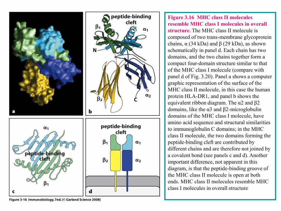
Figure 3.16 MHC class II molecules
resemble MHC class I molecules in overall
structure. The MHC class II molecule is
composed of two trans-membrane glycoprotein
chains, α (34 kDa) and β (29 kDa), as shown
schematically in panel d. Each chain has two
domains, and the two chains together form a
compact four-domain structure similar to that
of the MHC class I molecule (compare with
panel d of Fig. 3.20). Panel a shows a computer
graphic representation of the surface of the
MHC class II molecule, in this case the human
protein HLA-DR1, and panel b shows the
equivalent ribbon diagram. The α2 and β2
domains, like the α3 and β2-microglobulin
domains of the MHC class I molecule, have
amino acid sequence and structural similarities
to immunoglobulin C domains; in the MHC
class II molecule, the two domains forming the
peptide-binding cleft are contributed by
different chains and are therefore not joined by
a covalent bond (see panels c and d). Another
important difference, not apparent in this
diagram, is that the peptide-binding groove of
the MHC class II molecule is open at both
ends. MHC class II molecules resemble MHC
class I molecules in overall structure
Figure 3-22
Figure 3.17 MHC molecules bind peptides tightly within the cleft. When MHC molecules are crystallized with a
single synthetic peptide antigen, the details of peptide binding are revealed. In MHC class I molecules (panels a and
c) the peptide is bound in an elongated conformation with both ends tightly bound at either end of the cleft. In the
case of MHC class II molecules (panels b and d), the peptide is also bound in an elongated conformation but the
ends of the peptide are not tightly bound and the peptide extends beyond the cleft. The upper surface of the
peptide:MHC complex is recognized by T cells, and is composed of residues of the MHC molecule and the peptide.
In representations c and d, the electrostatic potential of the MHC molecule surface is shown, with blue areas
indicating a positive potential and red a negative potential. MHC molecules bind peptides tightly within the cleft
1、 MHC I类分子结构
经典MHC I类分子是由a 链和-2 微球蛋白 (2m)经非共价键连接成的异二聚体。属免疫球蛋白超家族, 细胞膜上HLA I类分子表达需要 a 链和 链同时存在。
Binding of a peptide to an MHC class I molecule is stabilized at both ends of the peptide-binding cleft by contacts
between atoms in the free amino and carboxy termini of the peptide and invariant sites that are found at each end of
the cleft of all MHC class I molecules (Fig. 3.18). These are thought to be the main stabilizing contacts for
peptide:MHC class I complexes because synthetic peptide analogues lacking terminal amino and carboxyl groups fail
to bind stably to MHC class I molecules. Other residues in the peptide serve as additional anchors. Peptides that bind
to MHC class I molecules are usually 8-10 amino acids long. Longer peptides are thought to be able to bind,
particularly if they can bind at their carboxy terminus , but are subsequently cleaved by exopeptidases present in the
endoplasmic reticulum, which is where MHC class I molecules bind peptides. The peptide lies in an elongated
conformation along the cleft; variations in peptide length appear to be accommodated, in most cases, by a kinking in
the peptide backbone. However, two examples of MHC class I molecules in which the peptide is able to extend out of
the cleft at the carboxy terminus suggest that some length variation may also be accommodated in this way.
These interactions give all MHC class I molecules their broad peptide-binding specificity. In addition, MHC
molecules are highly polymorphic. There are hundreds of different versions, or alleles, of the MHC class I genes in
the human population as a whole, and each individual carries only a small selection. The main differences between the
allelic MHC variants are found at certain sites in the peptide-binding cleft, resulting in different amino acids in key
peptide interaction sites in the different MHC variants. The consequence of this is that the different MHC variants
preferentially bind different peptides. The peptides that can bind to a given MHC variant have the same or very similar
amino acid residues at two or three particular positions along the peptide sequence. The amino acid side chains at
these positions insert into pockets in the MHC molecule that are lined by the polymorphic amino acids. Because the
binding of these side chains anchors the peptide to the MHC molecule, the peptide residues involved have been called
anchor residues. Both the position and identity of these anchor residues can vary, depending on the particular MHC
class I variant that is binding the peptide. However, most peptides that bind to MHC class I molecules have a
hydrophobic (or sometimes basic) anchor residue at the carboxy terminus (Fig. 3.19). Whereas changing an anchor
residue will in most case prevent the peptide from binding, not every synthetic peptides of suitable length that contain
these anchor residues will bind the appropriate MHC class I molecule, and so the overall binding must also depend on
the nature of the amino acids at other positions in the peptide.
3-14 MHC class I molecules bind short peptides of 8–10 amino acids by both ends.
Figure 3-23
Figure 3.18 Peptides are bound to MHC class I molecules by their ends. MHC class I molecules interact with the
back-bone of a bound peptide (shown in yellow) through a series of hydrogen bonds and ionic interactions (shown as
dotted blue lines) at each end of the peptide. The amino terminus of the peptide is to the left; the carboxy terminus to
the right. Black circles are carbon atoms; red are oxygen; blue are nitrogen. The amino acid residues in the MHC
molecule that form these bonds are common to all MHC class I molecules and their side chains are shown in full (in
gray) upon a ribbon diagram of the MHC class I groove. A cluster of tyrosine residues common to all MHC class I
molecules forms hydrogen bonds to the amino terminus of the bound peptide, while a second cluster of residues forms
hydrogen bonds and ionic interactions with the peptide backbone at the carboxy terminus and with the carboxy
terminus itself. Peptides are bound to MHC class I molecules by their ends
Figure 3-24
Fig 3.19 Peptides bind to MHC molecules through structurally related anchor residues. Peptides eluted from
two different MHC class I molecules are shown in the upper and lower panels, respectively. The anchor residues
(green) differ for peptides that bind different alleles of MHC class I molecules but are similar for all peptides that
bind to the same MHC molecule. The anchor residues that bind a particular MHC molecule need not be identical,
but are always related (for example, phenylalanine (F) and tyrosine (Y) are both aromatic amino acids, whereas
valine (V), leucine (L), and isoleucine (I) are all large hydrophobic amino acids). Peptides also bind to MHC class
I molecules through their amino (blue) and carboxy (red) termini.
Peptide binding to MHC class II molecules has also been analyzed by elution of bound peptides and by X-
ray crystallography, and differs in several ways from peptide binding to MHC class I molecules. Peptides
that bind to MHC class II molecules are at least 13 amino acids long and can be much longer. The clusters of
conserved residues that bind the two ends of a peptide in MHC class I molecules are not found in MHC class
II molecules, and the ends of the peptide are not bound. Instead, the peptide lies in an extended conformation
along the MHC class II peptide-binding groove. It is held in this groove both by peptide side chains that
protrude into shallow and deep pockets lined by polymorphic residues, and by interactions between the
peptide backbone and side chains of conserved amino acids that line the peptide-binding cleft in all MHC
class II molecules (Fig. 3.20). Although there are fewer crystal structures of MHC class II-bound peptides
than of MHC class I, the available data show that amino acid side chains at residues 1, 4, 6, and 9 of an
MHC class II-bound peptide can be held in these binding pockets.
The binding pockets of MHC class II molecules accommodate a greater variety of side chains than those of
the MHC class I molecule, making it more difficult to define anchor residues and to predict which peptides
will be able to bind particular MHC class II molecules (Fig. 3.21). Nevertheless, by comparing the
sequences of known binding peptides, it is usually possible to detect a pattern of permissive amino acids for
each of the different alleles of MHC class II molecules, and to model how the amino acids of this peptide
sequence motif will interact with the amino acids that make up the peptide-binding cleft in the MHC class II
molecule. Because the peptide is bound by its backbone and allowed to emerge from both ends of the
binding groove there is, in principle, no upper limit to the length of peptides that could bind to MHC class II
molecules. However, it seems that longer peptides bound to MHC class II molecules are trimmed by
peptidases to a length of 13-17 amino acids in most cases. Like MHC class I molecules, MHC class II
molecules that lack bound peptide are unstable, but the critical stabilizing interactions that the peptide makes
with the MHC class II molecule are not yet known.
3-15 The length of the peptides bound by MHC class II molecules is not constrained.
Figure 3-25
Fig. 3.20 Peptides bind to MHC class II molecules by interactions along the length of the binding groove. A
peptide (yellow; shown as the peptide backbone only, with the amino terminus to the left and the carboxy
terminus to the right), is bound by an MHC class II molecule through a series of hydrogen bonds (dotted blue
lines) that are distributed along the length of the peptide. The hydrogen bonds toward the amino terminus of the
peptide are made with the backbone of the MHC class II polypeptide chain, whereas throughout the peptide's
length bonds are made with residues that are highly conserved in MHC class II molecules. The side chains of
these residues are shown in gray upon the ribbon diagram of the MHC class II groove.
Figure 3-26
Fig 3.21 Peptides that bind MHC class II molecules are variable in length and their anchor residues lie at
various distances from the ends of the peptide. The sequences of a set of peptides that bind to the mouse MHC
class II Ak allele are shown in the upper panel. All contain the same core sequence (shaded) but differ in length. In
the lower panel, different peptides binding to the human MHC class II allele HLA-DR3 are shown. Anchor residues
are shown as green circles. The lengths of these peptides can vary, and so by convention the first anchor residue is
denoted as residue 1. Note that all of the peptides share a hydrophobic residue in position 1, a negatively charged
residue (aspartic acid (D) or glutamic acid (E)) in position 4, and a tendency to have a basic residue (lysine (K),
arginine (R), histidine (H), glutamine (Q), or asparagine (N)) in position 6 and a hydrophobic residue (for example,
tyrosine (Y), leucine (L), phenylalanine (F)) in position 9.
四、抗原肽与MHC分子相互作用及其分子基础
MHC I I类分子的抗原结合槽
MHCI类分子的a1和a2 Domain形成一个两端闭合的抗原结合槽,其中含有一条长度为8~11(一般为9肽)个氨基酸残基的肽。所容纳的肽不能伸出槽外。抗原肽一般含有一段与某个特定MHC分子结合的部位,称为锚定基,位于该部位上的氨基酸则称为锚定氨基酸残基(anchor residue)。与MHC I 类分子相结合的肽段的相应的锚定氨基酸残基插入MHC分子抗原结合凹槽中的“袋”(pocket)中,通过氢键与 I 类分子相结合。抗原肽中间部位一般均有一定程度的隆起,可作为T细胞表位被TCR识别。在正常情况下I类分子抗原结合槽内结合的往往是自身抗原肽。B2 domain含有与CD4分子和TC超抗原结合的保守部位。
Ab :an antibody is a protein that binds specifically to a particular substance—its antigen. Each antibody molecule has a unique structure that enables it to bind specifically to its corresponding antigen, but all antibodies have the same overall structure and are known collectively as immunoglobulins or Igs. Antibodies are produced by plasma cells in response to infection or immunization, and bind to and neutralize pathogens or prepare them for uptake and destruction by phagocytes.
Ig : all antibody molecules belong to a family of plasma proteins called immunoglobulins ( Ig ). Membrane-bound immunoglobulin serves as the specific antigen receptor on B lymphocytes (BCR).
BCR : the B-cell antigen receptor , or B-cell receptor ( BCR ), is the cell-surface receptor of B cells for specific antigen. It is composed of a transmembrane immunoglobulin molecule associated with the invariant Igα and Igβ chains in a noncovalent complex.
L chain : the immunoglobulin light chain ( L chain ) is the smaller of the two types of polypeptide chain that make up all immunoglobulins. It consists of one V and one C domain, and is disulfide-bonded to the heavy chain. There are two classes of light chain, known as κ and λ.
H chain : all immunoglobulin molecules have two types of chain, a heavy chain ( H chain ) of 50 kDa and a light chain of 25 kDa. The basic immunoglobulin unit consists of two identical heavy chains and two identical light chains. Heavy chains come in a variety of heavy-chain classes or isotypes, each of which confers a distinctive functional activity on the antibody molecule.
Glossary
V region : the variable region ( V region ) of an immunoglobulin or T-cell receptor is
formed of the amino-terminal domains of its component polypeptide chains. These are
called the variable domains (V domains) and are the most variable parts of the molecule.
They contain the antigen-binding sites.
C region : the constant region ( C region ) of an immunoglobulin or T-cell receptor is
that part of the molecule that is relatively constant in amino acid sequence between
different molecules. In an antibody molecule the constant regions of each chain are
composed of one or more C domains. The constant region of an antibody determines its
particular effector function.
Hinge region :the hinge region of antibody molecules is a flexible domain that joins the
Fab arms to the Fc piece. The flexibility of the hinge region in IgG and IgA molecules
allows the Fab arms to adopt a wide range of angles, permitting binding to epitopes spaced
variable distances apart.
Fab :IgG antibody molecules can be cleaved into three fragments by the enzyme papain.
Two of these are identical Fab fragments , so called because they are the F ragment with
specific a ntigen b inding. The Fab fragment consists of the light chain and the N-terminal
half of the heavy chain held together by an interchain disulfide bond. Another protease,
pepsin, cuts in the same general region of the antibody molecule as papain but on the
carboxy-terminal side of the disulfide bonds. This produces the F ( ab’)2 fragment , in
which the two arms of the antibody molecule remain linked..
Glossary
Fc : IgG antibody molecules can be cleaved into three fragments by the enzyme papain. One of these
is the Fc fragment , so-called for F ragment c rystallizable. The Fc fragment consists of the C-
terminal halves of the two heavy chains disulfide-bonded to each other by the residual hinge region.
HV : the hypervariable ( HV ) regions of immunoglobulin and T-cell receptor V domains are small
regions that make contact with the antigen and differ extensively from one receptor to the next.
FR : the V domains of immunoglobulins and T-cell receptors contain relatively invariant framework
regions (FR) that provide a protein scaffold for the hypervariable regions that make contact with
antigen.
CDRs : the complementarity determining regions ( CDRs ) of immuno-globulins and T-cell
receptors are the parts of these molecules that determine their specificity and make contact with
specific ligand. The CDRs are the most variable part of the molecule, and contribute to the diversity
of these molecules. There are three such regions (CDR1, CDR2, and CDR3) in each V domain.
Continuous epitopes , or linear epitopes, are antigenic determinants on proteins that are contiguous
in the amino acid sequence and therefore do not require the protein to be folded into its native
conformation for antibody to bind. The epitopes detected by T cells are continuous.
Haptens : hapten are molecules that can bind antibody but cannot by themselves elicit an adaptive
immune response. Haptens must be chemically linked to protein carriers to elicit antibody and T-cell
responses.
Glossary
β sheet : a β sheet is one of the fundamental structural building blocks of proteins,
consisting of adjacent, extended strands of amino acids ( β strands ) that are bonded
together by interactions between backbone amide and carbonyl groups. β Sheets can
be parallel, in which case the adjacent strands run in the same direction, or
antiparallel, where adjacent strands run in opposite directions. All immunoglobulin
domains are made up of antiparallel β-sheet structures. A β barrel or a β sandwich is
another way of describing the structure of the immunoglobulin domain
Ig domains : many proteins are partly or entirely composed of protein domains known
as immunoglobulin domains or Ig domains because they were first described in
antibody molecules. Immunoglobulin domains are characteristic of proteins of the
immunoglobulin superfamily, which includes antibodies, T-cell receptors, MHC
molecules, and many other proteins.
Ig fold: the immunoglobulin domain consists of a sandwich of two β sheets held
together by a disulfide bond and called the immunoglobulin fold (Ig fold).
Ig SF : many proteins involved in antigen recognition and cell–cell interaction in the
immune system and other biological systems are members of a protein family called
the immunoglobulin superfamily , or Ig superfamily (Ig SF), because their shared
structural features were first defined in immunoglobulin molecules. All members of
the immunoglobulin superfamily have at least one immunoglobulin or
immunoglobulin-like domain.
Glossary
MHC :The major histocompatibility complex ( MHC ) is a cluster of genes on human
chromosome 6 or mouse chromosome 17. It encodes a set of membrane glycoproteins called the
MHC molecules. The MHC class I molecules present peptides generated in the cytosol to CD8 T
cells, and the MHC class II molecules present peptides degraded in intracellular vesicles to CD4 T
cells. The MHC also encodes proteins involved in antigen processing and other aspects of host
defense. The MHC is the most polymorphic gene cluster in the human genome, having large numbers
of alleles at several different loci. Because this polymorphism is usually detected by using antibodies
or specific T cells, the MHC molecules are often called major histocompatibility antigens.
Co-receptor :A co-receptor is a cell-surface protein that increases the sensitivity of the antigen
receptor to antigen by binding to associated ligands and participating in signaling for activation. CD4
and CD8 are MHC-binding co-receptors on T cells, whereas CD19 is part of a complex that makes up
a co-receptor on B cells.
Antigen receptors: T and B lymphocytes collectively bear on their surface highly diverse antigen
receptors capable of recognizing a wide diversity of antigens. Each individual lymphocyte bears
receptors of a single antigen specificity.
Anchor residues : Peptide fragments of antigens are bound to specific MHC class I molecules by
anchor residues . These are residues of the peptide that have amino acid side chains that bind into
pockets lining the peptide-binding groove of the MHC class I molecule. Each MHC class I molecule
binds different patterns of anchor residues, called anchor motifs, giving some specificity to peptide
binding. Anchor residues exist but are less obvious for peptides that bind to MHC class II molecules.
Glossary
Review Question 1、名词概念:
Ag, hapten, Ab、Ig、Fab、Fc、FcR、Ig-fold, IgSF, HV, FR, linear epitope,