Biochem. J. (2012) 447, 321–334 (Printed in Great Britain) doi:10.1042/BJ20120813 321 REVIEW ARTICLE Lens and retina regeneration: new perspectives from model organisms Karla BARBOSA-SABANERO* 1 , Andrea HOFFMANN† 1 , Chelsey JUDGE*, Nicole LIGHTCAP*, Panagiotis A. TSONIS† 2 and Katia DEL RIO-TSONIS* 2 *Department of Zoology, Miami University, Oxford, OH 45056, U.S.A., and †Department of Biology and Center for Tissue Regeneration and Engineering, University of Dayton, Dayton, OH 45469-2320, U.S.A. Comparative studies of lens and retina regeneration have been conducted within a wide variety of animals over the last 100 years. Although amphibians, fish, birds and mammals have all been noted to possess lens- or retina-regenerative properties at specific developmental stages, lens or retina regeneration in adult animals is limited to lower vertebrates. The present review covers the newest perspectives on lens and retina regeneration from these different model organisms with a focus on future trends in regeneration research. Key words: lens, model organism, regeneration, retina. LENS REGENERATION Introduction to lens regeneration Lens regeneration has been studied for over a century, with the newt being the only adult animal that is capable of completely regenerating this complex organ [1,2]. The sheer unlimited regeneration potential of newt eyes following lens removal, even in older individuals, has established this system as indispensable for the study of organ regeneration and aging [3]. Newt lens regeneration is unique regarding the intrinsic capa- bility to regenerate the whole lens through transdifferentiation of dorsal IPE (iris pigment epithelial) cells without any remaining organ structure information. In contrast, other amphibians such as Xenopus can regenerate lenses from the corneal epithelium during developmental stages when certain developmental cues are provided, thus lacking the regeneration potential as adult individuals. In comparison with lower vertebrates, mammals in- cluding humans, rodents, cats and dogs are capable of regenerating the lens from LE (lens epithelial) cells when the capsular bag remains intact. The present review outlines and compares the different lens-regeneration processes in amphibians and mammals by delineating the molecular and cellular mechanisms. Physiology/anatomy of the lens The lens structure and physiology have been extensively reviewed by Kuszak et al. [4,5] and Al-Ghoul et al. [6]. Lenses are asymmetrical oblate spheroids that form from inverted lens vesicles and grow throughout life [4–6]. During lens development, LE cells differentiate into lens fibre cells that elongate into anterior and posterior directions of the lens equator [4–6]. When elongation is complete, the ends of lens fibres detach from the lens epithelium or capsule, and then lie alongside opposing fibres to form the lens sutures [4–6]. Throughout life, new lens fibre layers are added continuously within the lens periphery, resulting in formation of concentric growth shells [4–6]. The adult lens is composed of an exterior lens capsule with anterior LE cells positioned on the interior of the lens capsule. These epithelial cells expand into an LE/lens fibre cell transition zone towards the lens equator. The posterior part of the lens is free from LE cells. The inner part of the lens is filled with concentrically aligned lens fibre cells and a more dense fibre core within the middle of the lens (Figures 1A and 1B). Lens regeneration in Xenopus Species of the genus Xenopus are the only frogs known to regenerate lenses [7,8]. Of the lens-regenerating frogs, Xenopus laevis [9], Xenopus tropicalis [10] and Xenopus borealis [11] demonstrate lens regeneration at certain larval stages with declining regeneration potential during aging of the tadpole. In general, lens regeneration in Xenopus is based on the transdifferentiation of ectodermal central cornea epithelial cells into a lens vesicle that forms a new lens over time, also called CLT (corneal–lens transdifferentiation) [9]. Interestingly, Yoshii et al. [12] demonstrated that X. laevis is capable of regenerating the lens post-metamorphosis from the remaining LE cells left behind in the lens capsule [12]. Process of CLT-dependent lens regeneration Freeman [9] described five distinct phases of CLT in X. laevis on the basis of histological analysis. At stage 1 (1–2 days after lentectomy), cells of the inner corneal epithelium layer demonstrate a change from squamous to cuboidal epithelium. During stage 2, cells begin to thicken into a placodal structure with cells characteristic of LE cells. During stage 3 (3 days after lentectomy), a cell aggregate begins to separate from the corneal epithelium by invasion into the vitreous body for formation of a lens vesicle. During stage 4 (5 days after lentectomy), a definitive lens vesicle has formed by separation from the overlying cornea that contains elongated primary lens fibre Abbreviations used: BMP, bone morphogenetic protein; CB, ciliary body; CE, ciliary epithelium; CGZ, circumferential germinal zone; CLT, corneal–lens transdifferentiation; CMZ, ciliary marginal zone; E, embryonic day; ECM, extracellular matrix; FGF, fibroblast growth factor; HB-EGF, heparin-binding epidermal-like growth factor; INL, inner nuclear layer; IPE, iris pigment epithelial; iPSC, induced pluripotent stem cell; LE, lens epithelial; MAPK, mitogen- activated protein kinase; miRNA, microRNA; MSC, M¨ uller stem cell; NMDA, N-methyl-D-aspartate; ONL, outer nuclear layer; RPC, retinal progenitor cell; RPE, retinal pigmented epithelium; Shh, Sonic Hedgehog; TGFβ, transforming growth factor β; Wnt, wingless. 1 These authors made an equal contribution to this review. 2 Correspondence may be addressed to either of these authors (email [email protected] or [email protected]). c The Authors Journal compilation c 2012 Biochemical Society Biochemical Journal www.biochemj.org
Transcript
Biochem. J. (2012) 447, 321–334 (Printed in Great Britain) doi:10.1042/BJ20120813 321
REVIEW ARTICLELens and retina regeneration: new perspectives from model organismsKarla BARBOSA-SABANERO*1, Andrea HOFFMANN†1, Chelsey JUDGE*, Nicole LIGHTCAP*, Panagiotis A. TSONIS†2 andKatia DEL RIO-TSONIS*2
*Department of Zoology, Miami University, Oxford, OH 45056, U.S.A., and †Department of Biology and Center for Tissue Regeneration and Engineering, University of Dayton,Dayton, OH 45469-2320, U.S.A.
Comparative studies of lens and retina regeneration have beenconducted within a wide variety of animals over the last100 years. Although amphibians, fish, birds and mammals haveall been noted to possess lens- or retina-regenerative propertiesat specific developmental stages, lens or retina regeneration inadult animals is limited to lower vertebrates. The present review
covers the newest perspectives on lens and retina regenerationfrom these different model organisms with a focus on futuretrends in regeneration research.
Key words: lens, model organism, regeneration, retina.
LENS REGENERATION
Introduction to lens regeneration
Lens regeneration has been studied for over a century, with thenewt being the only adult animal that is capable of completelyregenerating this complex organ [1,2]. The sheer unlimitedregeneration potential of newt eyes following lens removal, evenin older individuals, has established this system as indispensablefor the study of organ regeneration and aging [3].
Newt lens regeneration is unique regarding the intrinsic capa-bility to regenerate the whole lens through transdifferentiation ofdorsal IPE (iris pigment epithelial) cells without any remainingorgan structure information. In contrast, other amphibians suchas Xenopus can regenerate lenses from the corneal epitheliumduring developmental stages when certain developmental cuesare provided, thus lacking the regeneration potential as adultindividuals. In comparison with lower vertebrates, mammals in-cluding humans, rodents, cats and dogs are capable of regeneratingthe lens from LE (lens epithelial) cells when the capsular bagremains intact. The present review outlines and compares thedifferent lens-regeneration processes in amphibians and mammalsby delineating the molecular and cellular mechanisms.
Physiology/anatomy of the lens
The lens structure and physiology have been extensively reviewedby Kuszak et al. [4,5] and Al-Ghoul et al. [6]. Lenses areasymmetrical oblate spheroids that form from inverted lensvesicles and grow throughout life [4–6]. During lens development,LE cells differentiate into lens fibre cells that elongate intoanterior and posterior directions of the lens equator [4–6]. Whenelongation is complete, the ends of lens fibres detach from thelens epithelium or capsule, and then lie alongside opposing fibresto form the lens sutures [4–6]. Throughout life, new lens fibrelayers are added continuously within the lens periphery, resultingin formation of concentric growth shells [4–6].
The adult lens is composed of an exterior lens capsule withanterior LE cells positioned on the interior of the lens capsule.These epithelial cells expand into an LE/lens fibre cell transitionzone towards the lens equator. The posterior part of the lensis free from LE cells. The inner part of the lens is filled withconcentrically aligned lens fibre cells and a more dense fibre corewithin the middle of the lens (Figures 1A and 1B).
Lens regeneration in Xenopus
Species of the genus Xenopus are the only frogs known toregenerate lenses [7,8]. Of the lens-regenerating frogs, Xenopuslaevis [9], Xenopus tropicalis [10] and Xenopus borealis [11]demonstrate lens regeneration at certain larval stages withdeclining regeneration potential during aging of the tadpole.In general, lens regeneration in Xenopus is based on thetransdifferentiation of ectodermal central cornea epithelial cellsinto a lens vesicle that forms a new lens over time, also called CLT(corneal–lens transdifferentiation) [9]. Interestingly, Yoshii et al.[12] demonstrated that X. laevis is capable of regenerating thelens post-metamorphosis from the remaining LE cells left behindin the lens capsule [12].
Process of CLT-dependent lens regeneration
Freeman [9] described five distinct phases of CLT in X. laevison the basis of histological analysis. At stage 1 (1–2 daysafter lentectomy), cells of the inner corneal epithelium layerdemonstrate a change from squamous to cuboidal epithelium.During stage 2, cells begin to thicken into a placodal structurewith cells characteristic of LE cells. During stage 3 (3 days afterlentectomy), a cell aggregate begins to separate from the cornealepithelium by invasion into the vitreous body for formation ofa lens vesicle. During stage 4 (5 days after lentectomy), adefinitive lens vesicle has formed by separation from theoverlying cornea that contains elongated primary lens fibre
1 These authors made an equal contribution to this review.2 Correspondence may be addressed to either of these authors (email [email protected] or [email protected]).
(A) Basic structures of the vertebrate eye. (B) Magnification of the anterior part of the eye, depicting the lens, iris, CE and CMZ. (C) Magnification of the posterior part of the eye, depicting mostly theretina and its specific cells. A colour-coded key has been included for each panel to help to identify the eye structures and cells.
cells on the side closest to the vitreous chamber. At stage5 (10 days after lentectomy), lens formation can be observedwith primary and secondary lens fibre formation and visibledisappearance of cell nuclei. The cornea has returned to its originalsquamous epithelial cell composition.
Like other vertebrates, Xenopus lenses express high levels ofcrystallin proteins that are important for lens structure and clarity.There are three major crystallin classes: α-, β- and γ -crystallins[13]. Crystallin accumulation follows a distinct temporal
and spatial expression pattern during lens development andregeneration. During lens development, α-, β- and γ -crystallinexpression appears simultaneously in the lens placode and lateron is limited to lens fibre cells [14]. During lens regeneration,α- and β-crystallins are detected in the posterior vesicle in themiddle of Freeman stage 3; however, by stage 4, the expressiondemonstrates clear restriction to the lens fibres [14–16]. On theother hand, γ -crystallin expression appears at a later time point(early stage 4) and is limited to lens fibres as well [14]. In contrast,
a recent microarray study by Day and Beck [17] demonstrated thatα-crystallin expression levels in regenerating or sham-operatedcorneas remains unchanged, whereas all β- and γ -crystallinsidentified are up-regulated during CLT. In addition, Day andBeck [17] also suggest similarities in the timing of α-, β- and γ -crystallin expression between regenerating and developing lenses.
Molecular mechanisms of Xenopus lens regeneration
The initiation of the CLT process is thought to include theexposure of the cornea to a vitreous humor unknown factorreleased from the neural retina [18–20]. Growth factors of theBMP (bone morphogenetic protein), FGF (fibroblast growthfactor) and Wnt (wingless) signalling pathways were identified assome of the potential candidates for induction of lens regeneration[17,21–23].
In detail, Day and Beck [17] demonstrated that overexpressionof the BMP inhibitor Noggin resulted in attenuated corneal celltransdifferentiation into lens vesicles. Day and Beck [17] alsosuggested that the canonical Wnt signalling pathway is activatedduring CLT for the induction of the bicoid-related homeoboxtranscription factors Pitx2, Pitx2a, Pitx1 and Pitx3 [17]. The roleof FGF signalling was first implicated by Bosco et al. [22] bydemonstrating FGF1-dependent induction of lentoid formationfrom outer cornea cultures. In a recent study, Fukui and Henry[21] found new evidence for the induction of lens regenerationthrough FGF family members by demonstrating up-regulation offgf1, fgf8 and fgf9 expression in corneal epithelium and retinaltissues before and during the process of lens regeneration. This issupported further by expression of the associated FGF receptorsfgfr1, fgfr2 and fgfr3 within the corneal epithelium throughoutthe regeneration process and a corresponding inhibition of thelens-regeneration process by the FGF inhibitor SU5402 [21].
Besides the BMP-, FGF- and Wnt-dependent signallingpathways, several transcription factors known to be fundamentalfor lens development are expressed during the process of CLTincluding Pax6, Prox1, Otx2 and Sox3 [17,24–28]. For instance,pax6 is expressed early during development in the anteriorembryonic ectoderm and represents a central transcriptionalregulator of eye development, lens cell differentiation andthe regulation of crystallin expression [25,27,29,30]. Similarly,the lens placode-expressed prox1 is important for lens fibre celldifferentiation and activation of crystallin expression [25,31]. Incontrast, otx2 is expressed in the early head ectoderm and issuggested to participate in lens formation during developmentthrough Notch-induced signalling [32].
Other molecules identified to play a role during CLT includetarget genes of the retinoic acid, Hedgehog, TGFβ (transforminggrowth factor β) and MAPK (mitogen-activated protein kinase)signalling pathways [17,24–28]. In addition, a huge variety ofother lens-regeneration expressed genes have been identified bygene expression profiling including proteins involved in RNA andprotein metabolism, and cell transport molecules [17,26].
Role of cellular growth matrix in Xenopus lens regeneration
Lentectomy leads to the disruption of the ECM (extracellularmatrix) barrier between the eye anterior aqueous chamber and theeye posterior vitreous chamber. The reconstitution of the existingECM, and, correspondingly, the expression of matrix-associatedgrowth factors, was suggested as an essential requirement forXenopus lens regeneration. For instance, gene expression profilingstudies of Day and Beck [17] and Malloch et al. [26] identifiedchanges in matrix metalloproteinase expression and associatedchanges in matrix modulating molecules such as TGFβ during
CLT. In addition, it has been suggested that the early appearance ofthe matrix-remodelling enzyme gelatinase B [Xmmp-9 (Xenopusmatrix metalloproteinase 9)] at the peripheral wound site plays arole in induction of wound healing-mediated responses that mightalso contribute to the induction of CLT [33].
Role of epigenetics and stem cell pluripotency factors in Xenopus lensregeneration
In contrast with lens regeneration in newts, Day and Beck[17] suggested that CLT regeneration in Xenopus does notinclude dedifferentiation of corneal cells into cells expressingpluripotency factors. According to Day and Beck [17], sox2was up-regulated in differentiated lenses when compared withcorneas undergoing CLT. Another pluripotency-associated gene,fut6, was significantly up-regulated in sham-operated corneas,but not in corneas undergoing CLT, whereas there was noexpression in differentiated lenses. In addition, Day and Beck [17]found evidence of genes associated with chromatin assembly anddisassembly during CLT, implying that epigenetic changes maybe taking place. Another study, by Perry et al. [34], demonstratedfurther evidence for changes in lens regeneration-associatedchromatin reassembly by identifying elevated histone H3S10Plevels following morpholino knockdown of the G-protein-coupledreceptor GPR84.
Lens regeneration in newts
To date, adult lens regeneration has been observed only innewts including the species Notophthalmus viridescens, Triturusviridescens and Cynops pyrrhogaster. Lens regeneration of theadult newt was first observed by Collucci in 1891 [2], andindependently by Wolff in 1895 [1], after whom the process isoften called Wolffian lens regeneration. Wolffian lens regenerationincludes the transdifferentiation of neuroectoderm-derived dorsalIPE cells into new lens [35–38].
Process of IPE-dependent lens regeneration
During the first 4 days after lentectomy, IPE cells of both dorsaland ventral regions dedifferentiate, visible by loss of pigmentationand initiation of proliferation [37,39,40]. However, at the mid-dorsal pupillary margin, IPE cells continue to proliferate, andlose pigmentation at approximately 8–10 days post-lentectomy.At this time, cell elongation and synthesis of lens-specific proteinscan be observed. Lens vesicle formation appears at day 10 ofthe regeneration process, which is followed by elongation of theposterior part of the vesicle where lens fibre differentiation isinitiated. The anterior cells become lens epithelium [41,42].
Cells in the ventral iris lack the lens transdifferentiationpotential. When ventral IPE cells are cultured for approximately2 weeks and implanted in the eye cavity or the limb blastema,they do not transdifferentiate into lens, whereas implantation ofdorsal IPE cell aggregates results in lens formation [43,44]. Onthe other hand, long-term in vitro cultures of dorsal or ventralIPE cells revealed that both cell types are capable of lentoid bodyformation [45]. This means that the ventral IPE cells have thepotential for transdifferentiation; however, this is not permittedin vivo or in short-term cultures.
Similar to Xenopus, expression of crystallins during newtlens regeneration was demonstrated to adhere to the expressionpattern during lens development. For instance, αA-, βB1- and γ -crystallins appear all simultaneously at the posterior lens vesicle atapproximately 10–12 days after lentectomy [40], with appearanceof γ -crystallins only in lens fibres [46].
Figure 2 Signalling overview during newt lens regeneration
Within the first days after lentectomy, FGF2 (in blue) and early lens genes such as pax6, sox2and mafB are suggested to promote induction of IPE cell-cycle re-entry and dedifferentiation inboth dorsal and ventral halves of the iris (as delineated by the horizontal line) [49–51]. Higherabundance of fgf2 expression in the dorsal iris is suggested to promote differential expressionof Wnt-pathway-associated signalling molecules (in red) such as Wnt2b and Frizzled4 that areactivated and confined to the dorsal iris [54]. Following Wnt pathway activation, induction ofdorsal iris transdifferentiation, including expression of lens-specific genes such as prox1 andsox1 can be observed together with initiation of crystallin expression [28,52,53].
Molecular mechanisms of lens regeneration in newts
Similar to lens regeneration in Xenopus, family members of theFGF signalling pathway have been suggested to control urodelelens regeneration [47–49]. In contrast with lens regeneration inXenopus, FGF2 and the FGF receptors FGFR1 and FGFR3 areessential for lens regeneration in the newt [47–49]. Followingfgf2 expression, induction of early lens genes such as pax6,sox2 and mafB can be observed within the first few days afterlentectomy [49–51] (Figure 2). Induction of late lens genes suchas prox1 and sox1 occurs shortly before expression of crystallingenes can be observed [28,52,53]. Besides the initial signallingactivity of FGF2, Hayashi et al. [54] suggested that a Wnt signalis activated during the second step of the lens-regeneratingprocess. According to Hayashi et al. [54], transcripts for the Wntligand and Frizzled receptor families including wnt2b, wnt5a,frizzled2 and frizzled4 can be detected in the iris undergoingregeneration. During early stages of lens removal, wnt5a andfrizzled2 are induced in both the dorsal and the ventral halves ofthe iris following lens removal or FGF2 injection, suggesting that
activation of these genes belong to the first-step process [54]. Incontrast, wnt2b and frizzled4 are induced only in the dorsal iris,suggesting their direct involvement in the dorsal-limited secondstep of the lens-regeneration process [54].
In addition, members of the Hedgehog signalling pathwaysuch as Shh (Sonic Hedgehog) and Ihh (Indian Hedgehog)are expressed during lens regeneration [55]. A broad-spectrumanalysis of mRNA expression patterns during dedifferentiationof the iris cells revealed further that several cancer and apoptosis-related genes, including signalling factors of the Ras family, p53tumour suppressor family, TNF (tumour necrosis factor), Rb(retinoblastoma) and Jun transcription factor family might alsobe involved in the lens-regeneration process [56].
Effect of cellular growth matrix on newt lens regeneration
Following lentectomy in newts, several changes in ECMremodelling that might contribute to IPE cell dedifferentiationcan be observed. For instance, higher accumulation ofglycosaminoglycans and hyaluronate in combination withincreased hyaluronidase activity can be found in the dorsal iristhan in the ventral iris [57,58]. In contrast, expression of tissue-remodelling enzymes such as collagenase and cathepsin are up-regulated during lens regeneration in both dorsal and ventraliris [59]. The glycoprotein fibronectin represents another ECMcomponent suggested to support the dedifferentiation of IPEcells [60]. Fibronectin was observed to increase at both thebasolateral and apical surface of the dedifferentiating IPE cellsand its accumulation continues until the cell surface is completelysurrounded. Within differentiating lens fibre cells, fibronectinexpression decreases [60]. Correspondingly, Elgert and Zalik [60]suggested that the decrease in fibronectin might be due to anincreasing activity in membrane-bound proteases.
A recent study by Godwin et al. [61] proposed that thetransmembrane protein TF (tissue factor) is expressed in a patch-like domain of the dorsal iris. Thrombin activation and recruitmentof blood cells was suggested to promote formation of a fibrinclot that allows localized expression of growth factors such asFGF2, as the first step for initiation of lens regeneration fromthe dorsal iris [61]. This idea is supported by prior studies byImokawa et al. [62,63] that found activation of thrombin at thedorsal margin after lentectomy, and could successfully blocklens regeneration by inhibiting thrombin activity. Godwin et al.[61] suggest that the localized FGF2 production is followedby induction of Wnt signalling and delayed activation of FGFexpression within the ventral iris. These differences in dorsaland ventral iris signalling are also suggested to contribute to theobserved differences in BMP and Wnt signalling between dorsaland ventral iris [44,54,61,64]. Correspondingly, differential BMPsignalling in dorsal compared with ventral iris might accountfor differences in the IPE cell differentiation potential of dorsalcompared with ventral iris. This is strongly supported by a studythat used inhibition of BMP to induce lens regeneration from theventral iris, which does not normally regenerate [44]. In addition,potential interspecies differences on the role of BMP regardingWolffian regeneration in newts and CLT in Xenopus might alsoaccount for the dedifferentiation potential of IPE cells in the newt.
Role of epigenetics and stem cell pluripotency factors in newt lens regeneration
A recent study of Wolffian lens regeneration using EST(expressed sequence tag) analysis identified several genetranscripts associated with pluripotency reprogramming, suchas histone deacetylases and the oncogene c-Myc [56]. That
changes in histone modification and expression are relevant forIPE cell-dependent lens regeneration is supported further byknockdown studies of histone B4 that resulted in alteration ofessential lens proteins [65]. In addition, increases in histone H3methylation and histone H4 acetylation, and a decrease in histoneH3 acetylation were observed during IPE cell dedifferentiation[66]. This study also identified differential epigenetic changes indorsal compared with ventral iris such as persistence of histoneH3 methylation in the dorsal iris, and increased histone H3methylation in the ventral iris [66].
When looking at proteins that play a role in IPE celldedifferentiation for establishment of multipotency, nucleosteminwas observed to accumulate in the nucleoli of dedifferentiatingpigmented epithelial cells 2 days before cell cycle re-entry [67].Furthermore, in contrast with the main pluripotency factors Oct4,Sox2, Klf4 and c-Myc that are required for induction of adultsomatic fibroblast dedifferentiation in higher vertebrates [68],expression of only three stem cell factors, e.g. sox2, klf4 and c-mycwas observed during lens regeneration in the newt [69]. Whereassox2 and klf4 are up-regulated during the very early stages oflens regeneration at day 2 for potential cell reprogramming andinitiation of cell cycle re-entry, c-myc expression peaked at day 8,together with the establishment of the lens vesicle [69]. However,the pluripotency factors Nanog and Oct4 were non-detectableduring lens regeneration [69]. Correspondingly, the lack of Nanogand Oct4 expression during newt lens regeneration might accountfor the difference between attaining the status of an ‘embryonic-like’ stem cell [i.e. iPSCs (induced pluripotent stem cells)] anda dedifferentiated cell capable of only re-creating a lens.
Another mechanism for regulation of dedifferentiation includespost-transcriptional regulation of gene expression by miRNA(microRNA). Makarev et al. [70] demonstrated that a numberof miRNAs including members of the miR-124 family, miR-125b,miR-181b, miR-133a, miR-21 and let-7b are expressed within theadult newt eye. In addition, several miRNAs have been identifiedthat play a role during lens regeneration such as members of thelet-7 family and miR-148 [71,72]. For instance, all of the let-7family members are down-regulated in the dorsal iris comparedwith the ventral iris during the initial dedifferentiation process oflens regeneration [71]. Specifically, let-7b that targets pax3, chrd(chordin) and tgfβr1 mRNA expression demonstrates a down-regulation in the regenerating dorsal iris compared with ventraliris and the experimental up-regulation was suggested to inhibitproliferation in both dorsal and ventral iris. In addition, miR-148,which targets tgfα, nog (noggin) and fgf13 mRNA expression,appears to be up-regulated in the ventral iris compared with thedorsal iris [72].
Mammalian lens regeneration
Compared with lens regeneration in Xenopus and newts,mammalian lenses do not regenerate completely and are notalways of normal shape. Lens regeneration in mammals, includingrabbits, dogs, cats, rats and mice, requires the existence of anintact lens capsule [73–79]. In contrast with the IPE cell-drivenlens regeneration in newts, lens regeneration in mammals wasdemonstrated to result from LE cells remaining in the lenscapsule [76,79,80]. The lens is sometimes well regenerated withbow regions where active differentiation to lens fibres occurs. Inmice, during the initial steps there is expression of inflammatoryfactors as well as factors involved in tissue remodelling andEMT (epithelial–mesenchymal transition). At later stages, normalexpression of crystallin genes is resumed [81]. Research in ratshas shown that preservation of the anterior lens epithelium and
surgical closure of the lens capsule results in clear lenses withparallel protein expression profiles resembling the ones duringlens development [79]. Lens currents also seem to play a criticalrole in the rat lens-regeneration process [82].
RETINA REGENERATION
Introduction to retina regeneration
Studies conducted over the last 100 years using a wide varietyof animals have contributed to the current knowledge on theprocess of retina regeneration [83]. Amphibians, fish, birds andmammals have all been noted to possess regenerative properties;however, even though most animals are capable of carrying outretina regeneration at specific developmental stages, only a fewhave been shown to regenerate their retina at maturity [84].
Retina-regeneration strategies used by vertebrates appearto be evolutionarily conserved. The main process of retinarepair/regeneration includes transdifferentiation of neural retina-supportive cells such as Muller glia cells or cells from the RPE(retinal pigmented epithelium). Regeneration through theactivation of cells located in the CB (ciliary body)/CMZ (ciliarymarginal zone) represents the other mode that plays a minor role inmost vertebrate animals with regenerative capacity. Regenerationvia RPE or Muller glia transdifferentiation generally involvesthe process of dedifferentiation, where the RPE/Muller glia cellslose their original features, proliferate and eventually differentiateinto retina cells. Regeneration through the CB/CMZ involves theactivation of stem/progenitor cells residing in this area, and thesein turn proliferate and eventually differentiate into retina cells[84,85] (Figure 3).
Physiology/anatomy of the retina
For a better understanding of the retina-regeneration process,we review the anatomy of this sophisticated tissue. The retinais a light-sensitive structure that absorbs light and converts itinto nerve impulses that are ultimately deciphered by the brain.The retina is composed of six different types of neural cells,including cone and rod photoreceptors, and horizontal, bipolar,amacrine and ganglion cells (Figure 1C). The photoreceptor cellsare distributed in the posterior retina where they capture light andtransform it into electrical signals. These cells are distributed inthe back of the retina next to the RPE. The cones are utilized fordaytime colour vision, whereas rods are used as low-light sensors.The retinal layers are divided into the GCL (ganglion cell layer),the INL (inner nuclear layer), where the cell bodies and nuclei ofamacrine, bipolar and horizontal cells are located, and the ONL(outer nuclear layer) where the nuclei of the photoreceptors reside.The OPL (outer plexiform layer) is the site of synaptic contactsbetween the cones or rods with horizontal and/or bipolar cells.The IPL (inner plexiform layer) is another synaptic region wherecommunication between bipolar and ganglion cells takes place[86].
The RPE that is located between the retina and the choroidconsists of a pigmented epithelial cell monolayer. The apicalmembrane of the RPE faces the photoreceptor layer, whereasits basolateral membrane faces the choroid (Figure 1C) [87].The RPE supports the retina by carrying out phagocytosis of thephotoreceptor outer segments, by maintaining the visual cycle,and by transporting nutrients, ions and fluids to the retina [88]. Inaddition, Muller glia cells are also essential for retina function andsurvival and they span the whole retina with their nuclei located inthe INL (Figure 1C). The CB is composed of the ciliary muscles
Figure 3 Different modes for retina regeneration in vertebrates
(A) The process of RPE transdifferentiation that takes place in amphibians. RPE cells lose their pigment and dedifferentiate to give rise to neuroepithelium that eventually differentiates into the differentretinal cells. The RPE also replenish itself, re-establishing the epithelium. (B) The RPE transdifferentiation that takes place in embryonic chickens. RPE dedifferentiates as in (A); however, the RPElayer is not replenished and the orientation of the regenerated retina is reversed. (C) Regeneration via activation of stem/progenitor cells from the CMZ. Progenitor cells are activated and proliferate togive rise to the different retinal cells. (D) Transdifferentiation of Muller glia cells that takes place in fish, birds and mammals. Muller glia cells dedifferentiate and proliferate to give rise to the differentretina cells. A colour-coded key has been included for each panel (the same key is used for both A and B) to indicate the different cells.
that are important for lens accommodation, and also containspigmented and non-pigmented epithelium, including the parsplana and the pars plicata (Figure 1B). The ciliary pigmented epi-thelium is related ontologically to the RPE, and the non-pigmentedepithelium is continuous from the CMZ/retina [89,90]. The CMZis a small transition zone located between the retina and the ciliarybody (Figure 1B) that, in lower vertebrates, represents the mainsource of proliferative progenitor cells for retina neurogenesis andcan be identified with progenitor markers [90–92].
Retina regeneration through the ciliary margin
Amphibians and fish
Retina growth persists until adulthood in amphibians and fishthrough the contribution of progenitor cells present in theCMZ [93–97] (Figure 1). In fish, this region is called theCGZ (circumferential germinal zone) and contains RPCs (retinalprogenitor cells) for the generation of all retinal cells, excludingrod photoreceptors [94,98] (Figure 3C). Rod photoreceptor cells
in fish are produced by specific rod progenitor cells located in thecentral retina [98–100]. Upon retinal injury in fish and amphibia,the CMZ/CGZ can contribute to retina regeneration by activatingprogenitor cells to proliferate and eventually differentiate toreplace various types of lost retinal cells [100–106] (Figure 3C).In fish, all retinal cells can be regenerated from the CGZexcept rod photoreceptors. It is important to note that retinaregeneration from the CMZ/CGZ only partially contributes torepair/regeneration since the major contributors of regenerationreside elsewhere in the retina. Whereas retina regeneration inamphibians is mainly based on regenerative cells within the RPE(Figure 3A), Muller glia cells represent the main regenerating cellsource in fish [12,98,104–110] (Figure 3D). Recently, Martinez-De Luna et al. [111] proposed a different model of regeneration inpre-metamorphic X. laevis that depends on the production of RPCsupon partial retinal resection. To contribute to retinal regeneration,these RPCs not only express retinal progenitor markers, but alsodepend on the expression of one of these progenitor genes, thehomeobox gene Rx.
Birds
Similar to fish and amphibians, the CMZ in birds can provideretinal cells for the growth of the retina, but only up to a fewweeks after hatching [112,113]. This activity is stimulated by thepresence of exogenous factors including FGF, endothelial growthfactor, insulin-like growth factor-1, insulin and Shh [112–114].Although post-hatched chickens cannot regenerate their retina viathe activation of the CMZ, Coulombre and Coulombre [115,116]showed that embryonic chicks [stage 23–25 or E (embryonic day)4–4.5] can regenerate their retinas upon removal from cells ofthe CMZ (Figure 3C) as long as a piece of retina is present.Park and Hollenberg [117] later identified that FGF1 couldinduce regeneration from the CMZ. Previous work has shownthat FGF2 regenerates a complete retina from E4 chick CMZvia the MAPK pathway [118]. Shh signalling is also sufficientto induce regeneration from this region; however, the Shh andFGF pathways are interdependent on each other, and are requiredfor the proliferation and survival of stem/progenitor cells of theCMZ during regeneration [118,119]. At this stage of development(stage 23–25), the chick eye is not fully differentiated andthe CB and CMZ cannot be distinguished, and therefore theregeneration from this region at this stage is referred to occurvia the CB/CMZ [118,119]. As a matter of fact, the CMZ is notconsidered morphologically/molecularly defined until E16, and,as mentioned above, the regenerative properties of the CMZ arelost postnatally [112,120]. Interestingly, a recent study suggeststhat the early optic cup lip that represents the boundary betweenpigmented and non-pigmented epithelium contains multi/pluri-potent stem/progenitors that participate during eye morphogenesisand that eventually feed into the CMZ [121].
The survival of stem/progenitor cells is regulated further viaanother FGF-dependent pathway, the BMP pathway that caninduce the CB/CMZ to replace lost retina [122]. BMP inducesproliferation of stem/progenitor cells via its canonical pathway(through SMADs) during the early stages of retina regeneration;however, later on, this pathway switches to one of its non-canonical pathways by activating p38 signalling and inducingapoptosis of the newly regenerated retina [122]. Recently, Wntsignalling has been determined to be essential for maintainingthe stem cell niche of the CB/CMZ after retina removal and themolecular mechanisms regulating this activity are currently beingdissected (K. Del Rio-Tsonis, unpublished work). Changes inthese signalling pathways might also explain the decrease
in regeneration potential of the chick CB/CMZ as the embryoages, having marginal regeneration at E5 (K. Del Rio-Tsonis,unpublished work).
Retina regeneration via transdifferentiation
Retina regeneration via RPE transdifferentiation is uniqueto amphibians; however, several animals have been reported toundergo RPE transdifferentiation during embryonic stages,including mammals and birds. In this process, the RPEcells dedifferentiate, losing their characteristics, proliferate anddifferentiate into retinal cells (Figures 3A and 3B).
Amphibians
In amphibians, retina regeneration is predominantly achievedvia RPE transdifferentiation. Notably, during the process oftransdifferentiation in amphibians, the RPE is replenished asit forms a complete retina. This is not the case when RPEtransdifferentitation takes place during embryonic stages inbirds [115,119] (Figure 3A compared with Figure 3B). Studieshave shown that retina regeneration can occur from earlydevelopmental stages to post-metamorphic stages [12,123,124].
Anurans. For many years, it was believed that adult post-metamorphic frogs are unable to regenerate their retina via RPEtransdifferentiation; however, Yoshii et al. [12] demonstrated thatpost-metamorphic X. laevis are capable of regeneration, even atmature stages. In their studies, the authors showed that if the retinais removed carefully from the eye cup of adult Xenopus, leavingthe retinal vascular membrane intact, the RPE cells can detachfrom the RPE monolayer, migrate towards this vascular membraneand, once attached, proliferate to make a neuroepithelium that willeventually differentiate into the different neural cell types [12].The importance of the vascular membrane for retina regenerationin anuran amphibians was first described by Reh and Nagy [104]in Rana catesbienna tadpoles. In that study, the authors describedthat the association of RPE cells with the vascular membrane isa crucial step in the process of transdifferentiation. In anotherstudy, the same group reported the importance of ECM proteins,particularly laminin, as a principal component of the vascularmembrane [125]. In addition, in vitro studies using RPE explantsplated on to different types of extracellular substrates indicatedthat only laminin is able to induce RPE transdifferentiation. Thesedata suggest that the process of retina regeneration in amphibiansis highly regulated by the inductive signals of the ECM [125].Future studies involving gene manipulations using transgenesiswill be very helpful for dissecting the molecular regulation ofretina regeneration. To this end, Ueda et al. [126] have recentlycreated transgenic X. laevis lines using the ef1-α promoter hookedto an eGFP (enhanced green fluorescence protein) gene to markthe tissues undergoing retina regeneration: the RPE and the CMZ.
Vergara and Del Rio-Tsonis [124] developed a new model toanalyse retinal regenerative properties in X. laevis tadpoles. Theretina was completely removed from stage 51–54 tadpoles,leaving only the RPE. Retina regeneration was induced with FGF2via RPE transdifferentiation. The regenerated retina maintainedthe proper lamination and the original orientation and alsoregenerated an optic nerve. The authors also determined thatthis FGF2-dependent RPE transdifferentiation process requiresactivation of the MAPK pathway for initiating regeneration. IfMAPK inhibitors are added, regeneration is halted.
Kuriyama et al. [127] utilized a three-dimensional MatrigelTM
culture to study RPE transdifferentiation in X. laevis tadpoles.
The authors cultured both RPE sheets with choroid, and RPEsheets isolated from choroid, in this three-dimensional MatrigelTM
system [127]. Their results showed that, in the absence of thechoroid which provides contact with the Bruch’s membrane (richin ECM proteins), the RPE is capable of transdifferentiating byfirst migrating towards the gel matrix, whereas the cultures of RPEsheets with choroid were unable to migrate and therefore unableto transdifferentiate [127]. The authors determined from theseresults that the loss of cell–cell/cell–ECM interactions triggersthe process of transdifferentiation, leading to the up-regulationof Pax6, which is independent of FGF during the first phase oftransdifferentiation. However, FGF is a crucial regulator of theoverall process of transdifferentiation, maintaining Pax6 andfurther driving RPE cells into neuronal progenitors.
These studies highlight the importance of environmental cues,cell–cell contact, cell–ECM interactions and their influence tosuccessfully achieve retina regeneration via transdifferentiation.
Urodeles. The adult newt can regenerate its entire retina withoutinduction from exogenous factors or without preserving thevascular membrane. Most importantly, the regenerated retinais fully functional [84,128–132]. The vast majority of newtretina regeneration occurs via RPE transdifferentiation; however,a small domain of the newly regenerated retina is derivedfrom the CMZ (Figures 3A and 3C). Chiba et al. [133]analysed the dynamics of the protein RPE65 present in theRPE, which is involved in the recycling of visual pigments. Intheir work, the authors documented the presence of RPE65 viaimmunohistochemistry during the process of retina regeneration,and showed that RPE65 was present even after the RPE cells beganto adopt a neural retinal fate [133]. However, their molecularanalysis suggested that the presence of this protein during RPEtransdifferentiation might represent lingering protein rather thannewly synthesized protein. This elegantly marked the domain ofRPE transdifferentiation compared with the regeneration domainderived from the CMZ, and a detailed description of the differentstages of the transdifferentiation process was suggested to includeearly stages 1–3 (E1–3; days 10–19), intermediates stages 1–3(1–3; days 19–23) and late stages (L1–2; days 45–65). Cell-cycleentry takes place at E1 (10 days after retinectomy). In addition,it was also clarified that there is a defined boundary at the CMZ,where no RPE65 protein was found [128,133].
The molecular mechanisms of these two regenerative processesare still being elucidated. A series of gene expression profileshas been performed during the different stages of newt retinaregeneration [134–136] and these results are nicely summarizedby Chiba and Mitashov [128]. Genes analysed include genesassociated with retina stem/progenitor cells such as pax6,chx10/vsx2, msi1 and notch (expressed early between E1 andE3) as well as with differentiating retina cells such as opsinand voltage-gated Na+ channel CpNaV1 (expressed from interme-diate stage 2 onwards). Further studies were performed with Msi1(Mushashi-1), a key regulatory molecule expressed in the retinaand necessary for photoreceptor survival [137]. msi1 is expressedin mature newt RPE, stem cells and photoreceptor cells. Uponretinectomy, RPE-transdifferentiating cells express msi1 in thenucleus and the cytoplasm. However, renewing RPE has less msi1expression, as does the differentiated RPE as it transitions intoneural retinal cells. Finally, the expression of msi1 re-establishesin the photoreceptor cells of the newly regenerated retina [138].
Nakamura and Chiba [135] suggested that the Notch signallingpathway plays a role in the process of transdifferentiation.This group analysed and compared the expression patterns ofNotch-1 via in situ hybridization in developing retina, as wellas during RPE transdifferentiation, concluding that expression
patterns in both processes are very similar, localizing to the earlyforming neuroepithelium, and eventually becoming restricted tothe peripheral retina. No expression was found in the adult newteye. Interestingly, expression of other components of the Notchsignalling pathway such as delta1 and hes1 were expressed in theadult RPE. Using a pharmacological inhibitor for Notch duringthe process of RPE transdifferentiation, Nakamura and Chiba[135] were able to detect premature neural differentiation,implicating the Notch pathway in RPC maintenance and inhibitionof retinal differentiation.
Na+ channels have been used to monitor ganglion celldifferentiation during RPE transdifferentiation [129,130,139].In contrast, a study by Vergara et al. [140] reported that the αsubunit of the Na+ /K+ -ATPase is up-regulated transiently in theRPE during early transdifferentiation when retinal progenitorsform, but is not present in the intact RPE or neural retina. It mustbe noted that more research is required to elucidate further therole of these channels/transporters during retina regeneration.
In vitro culture systems have been designed by several groupsto help to dissect the cellular and molecular mechanisms of RPEtransdifferentiation ([141–143], and reviewed in [107]). Mitsudaet al. [144] reported that RPE transdifferentiation in vitro islimited to RPE cells/explants that also have a choroid or a sourceof FGF. However, just separating the RPE from the choroid issufficient to push the RPE cells to enter the S-phase of the cellcycle [141]. Organ cultures of RPE still connected to the choroidnamed ‘retina-less eye cup’ have been used recently to moreclosely mimic what takes place in vivo [142]. Using this system,it was determined that cell-cycle entry is mediated via MAPKsignalling and that this activity is modulated by heparin that canaffect the overall influence of Wnt-, Shh- and thrombin-mediatedpathways. These pathways are able to regulate cell-cycle entry,although more studies must be conducted to determine the preciseregulation [142].
Birds
The embryonic chick is able to regenerate its retina via RPEtransdifferentiation during a small window of its developmentbetween stages 23 and 25 (E3.5–E4.5). A unique feature of thistype of transdifferentiation is that the RPE does not replenish itselfand the neuroepithelium formed, in consequence, will give rise toa retina with a flipped orientation (Figure 3B). After this stage, theRPE loses its plasticity and is unable to regenerate retina. RPEtransdifferentiation is dependent on the presence of exogenousFGF2 [119,145,146]. The molecular mechanism by which FGF2induces transdifferentiation includes the activation of the MAPKpathway and the up-regulation of Pax6 in the transdifferentiatingRPE cells [147]. Furthermore, it has been demonstrated that Shhinhibits this FGF-induced transdifferentation [119]. Recently, theWnt signalling pathway has been shown to protect the RPEphenotype and, when absent, RPE transdifferentiation can takeplace (K. Del Rio-Tsonis, unpublished work).
Although there is a restrictive period in which RPEtransdifferentiation (up to E4–E4.5) can occur, it has beensuggested that this period could be lengthened. Sakami et al.[148] cultured RPE explants from E5 chicks and inhibited activinsignalling, extending the time window of transdifferentationin vitro. Activin is an integral part of the TGFβ signallingpathway and a key molecule for maintaining the RPE fate.Other studies performed in vitro have shown that dissociatedRPE cells can be induced to transdifferentiate to retinal neurons,when certain key factors are overexpressed, including NeuroD,Six6/Optx2, Ash1, Sox2, neurog1, neurog2, neurog3 or Ath5
[149,150]. For example, RPE reprogramming via neurogenin 1 inembryonic chick cultures primarily generate photoreceptor-likeneurons that show the presence of photoreceptor markers as wellas key genes in photoreceptor development and components ofthe phototransduction pathway. These photoreceptor-like cellsrespond to light exposure in the same way that photoreceptorcells do, and develop inner segments rich in mitochondria, acharacteristic of mature photoreceptor cells [151].
The transdifferentiation process in the chick has been studiedanalysing the dynamic expression of several molecules, andby comparing the differences and similarities between RPEtransdifferentiation and regeneration via the activation cellsfrom the CMZ. However, the complexity of these processesmakes the analysis of the molecular mechanisms implicated intransdifferentiation difficult. To study the molecular mechanismsof transdifferentiation, it is important to keep in mind the gradualchange of cell fates, the timing of cell-cycle entry, the rate ofsynthesis and degradation of certain proteins, and the time pointat which analysis is performed.
Regeneration via Muller glia
Muller glia represent supportive cells localized in the neural retinathat function as a structural support and nourishment for neurons.They can also act as neurotransmitter transporters and as immune-response modulators [152–155]. However, Muller glia can alsoserve as a source of new retina neurons and play a critical role inretina regeneration in several vertebrate species.
Fish
In fish, Muller glia cells are the main source of retina regeneration.Muller glia can give rise to two different progenitor cellpopulations: rod precursor cells and RPCs in the INL. Rodprecursor cells are produced continuously in the adult fish tokeep up with the demands of the continuous growth of the retina[95,96]. However, upon injury, Muller glia cells dedifferentiateand proliferate generating multipotent progenitor cells that areultimately capable of differentiating to all retina cell types(Figure 3D). When rod precursor cells are produced, they migrateinto the photoreceptor layer and once they reach the ONL,they will only differentiate into rod cells, whereas the multipotentprogenitor cells can differentiate into all retinal cell types[98,108,110,156–159].
Recent work has concentrated on elucidating the molecularmechanism regulating this regenerative process. Some keymolecules include Ascl1a, Pax6b, c-Mycb, PCNA (proliferating-cell nuclear antigen), Rx, Chx10/Vsx2, Dkk, Notch, HB-EGF(heparin-binding epidermal-like growth factor) and Lin-28 vialet-7 miRNA [100,160–165]. Upon retinal injury in zebrafish,HB-EGF is up-regulated in Muller glia. This secreted factorsignals via its receptor through a MAPK signalling pathwayand in turn regulates the expression of Ascl1a, Lin-28, Notch andDkk, resulting in dedifferentiation and proliferation of Muller gliacells [165]. Thummel et al. [164] sought to study more preciselythe role of Pax6 during zebrafish retina regeneration. Knockingdown the two isoforms of Pax6, they found that, whereas thetwo isoforms have no effect on Muller glial cell division,the knockdown prevents Muller glia-derived INL neuronalprogenitor cell division; specifically, Pax6b regulates the first celldivision of neuronal progenitors, and Pax6a regulates later progen-itor cell divisions. Interestingly, Ramachandran et al. [162] claimthat Muller glia cells express markers of iPSCs, such as Oct4,Klf4 and c-Myc [162]. The findings described above demonstrate
the need for a cross-talk between several pathways/molecules thatactivate the molecular switch responsible for the reprogrammingof Muller glia to retinal neurons.
Birds
Muller glia cells were first reported to be a source of neuropro-genitor cells in the postnatal chicken [166]. Muller glia-derivedneuroprogenitors were observed after retina injury, or via injectionof cytotoxic compounds such as NMDA (N-methyl-D-aspartate),or even in the presence of exogenous growth factors, such asinsulin or FGF injected into the eye cup. Muller glia cells respondto these stimuli by re-entering the cell cycle, dedifferentiatingand expressing progenitor markers such as Pax6, Chx10/Vsx2 andCash1 [166]. Previous studies have tried to elucidate the molecularmechanisms of Muller glia cell activation in birds during retinaldamage [167–171]. It is clear that the FGF/MAPK pathway aswell as the Notch pathway play a role in the dedifferentiationprocess as well as in the differentiation of the Muller glia-derivedprogenitors ([168–171], and reviewed in [167]).
The neurogenic potential of Muller glia is very limited. Uponretinal damage, approximately 10% of the progenitor cellsderived from Muller glia give rise to amacrine and bipolar cells,but, in the presence of FGF or insulin, ganglion-like cells areproduced. Interestingly, Muller glia cells have a higher capacityto proliferate and differentiate in the peripheral region of theretina than in the central retina. On the other hand, of the Mullerglia cells that enter the cell cycle, only a few express progenitormarkers, and over 80% of the Muller glia-derived progenitors donot differentiate [166,167].
Mammals
The regenerative properties of mammals are restricted in compar-ison with other animals. For a long time, it was considered that theretina of adult mammals had no regenerative properties. However,strong evidence suggests that Muller glia cells are capable of re-sponding to injury by dedifferentiating and proliferating, leadingto retinal neurogenesis. In adult rats, Muller glia cells dedifferen-tiate and proliferate in response to the neurotransmitter NMDA,producing a limited number of bipolar cells and photoreceptors[172]. Moreover, when homeobox and bHLH (basic helix–loop–helix) genes are overexpressed in retinal explants from postnatalrats, Muller glia cells were able to give rise to several retinal celltypes. On the basis of the knowledge that the Wnt signalling path-way is able to regulate stem cell populations, Osakada et al. [173]analysed the role of the Wnt pathway during Muller glia-inducedretina regeneration in adult rats. In this study, the activation of theWNT pathway by Wnt3a or GSK3β (glycogen synthase kinase-3β) inhibitors promoted the proliferation of Muller glia [173].
Interestingly, enriched purified rat Muller glia can formneurospheres when induced by FGF2, and these neurosphereshave the potential to give rise to retinal neurons and glia. Inaddition, transplanted enriched Muller glia into postnatal day 1rat retina, gave rise to lamina-specific retinal neurons [174]. Adulthuman retinal explants treated with EGF showed that cells fromthe most anterior region of the retina are able to proliferate andexpress markers of Muller glia and stem cells, suggesting thatcells in this region of the human retina are normally dormant, buthave regenerative potential. This region corresponds to the CMZregion present in lower vertebrates [175]. Furthermore, humanMuller glia-derived cell lines treated with FGF2 or RA (retinoicacid) are able to express retinal progenitor markers, such as Pax6,Chx10/Vsx2 and Sox-2 [176]. On the other hand, Shh has been
identified as a participant in Muller glia transdifferentiation bothin vivo and in vitro models by up-regulating markers of retinalprogenitors. Post-retinal injury studies in rat also demonstrate thatShh promotes Muller glia activation and directs retinal progenitorsto differentiate towards the photoreceptor linage [177].
Intriguingly, Bhatia et al. [178] examined the differencesbetween the two cell populations that can carry out neurogenic andproliferative activity in the adult mammalian eye: Muller glia ofthe neural retina and the CE (ciliary epithelium). The morphologyof the two cell populations differed: the non-pigmented CEshowed epithelial morphology and MSCs (Muller stem cells)displayed neural morphology. Although both non-pigmented CEand MSCs possessed neural progenitor markers, only MSCsshowed the neural stem cell marker Nestin. This study alsodemonstrated that MSC have a superior proliferative ability whencompared with the cells of the CE and this suggests that MSCscould be a potential source for retinal neuron transplantation[178].
SUMMARY
The regenerative processes for lens and retina reviewedin the present paper share some common cellular andmolecular mechanisms, including cell signalling pathways, Pax6transcription factor as a central molecule for the eye and theregenerative process, the transdifferentiation process for tissueregeneration, the role of ECM proteins and finally the evolutionarymechanisms conserved in several animal models.
During the process of lens and retina regeneration, theactivation of cell signalling pathways such as FGF/MAPK, Wntand BMP seems to be conserved, especially the activation ofthe FGF/MAPK pathway which eventually can regulate Pax6[49,118,141,147]. Despite the similarities in signalling pathwaysinvolved in lens and retina regeneration, the regeneration potentialis highly dependent on the sequence and timing of these signallingevents and the diverse activation of downstream targets for eachpathway that are ultimately defined by the origin and, accordingly,the differentiation state of the cells that will undergo regeneration.For example, during IPE transdifferentiation in the adult newt, thedistinct timing and versatile expression of FGF family membersis critical, e.g. fgf2 in dorsal compared with ventral iris [49,179].In Xenopus CLT or chick RPE transdifferentiation, on the otherhand, the regenerative ability is limited to certain larval/embryonicstages, even though these processes share an FGF-dependentinduction with further participation of the BMP, Shh and Wntpathways [17,21,22,118,119,147,180].
The activation of some of these signalling pathways convergesand eventually targets common downstream effectors such as thePax6 transcription factor [49,118,141,147]. The activation of Pax6seems to be a crucial event for both lens and retina regeneration.The Pax6 gene is highly conserved through the evolution ofthe animal kingdom from invertebrates to vertebrates, stressingits critical role in eye development [181,182]. The importanceof Pax6 in eye development has been well documented; Pax6has been described as the master regulator of eye developmenton the basis of gain-of-function experiments or overexpressionstudies inducing ectopic eyes on appendages [183]. The complexregulation of Pax6 is still being investigated owing to its wideinteraction with several gene networks.
It is interesting to note that the contribution of ECM is onemore common mechanism for lens and retina regeneration. Itis important to highlight that the presence of ECM proteins iscritical and sometimes necessary to achieve these regenerativeprocesses, particularly during transdifferentiation [60,104,125].
However, to date, it remains to be determined whether woundhealing generated matrix provides the initial signal for lens orretinal regeneration, or if the regeneration of these organs istriggered by a mechanism that is intrinsic to the correspondingtissue of origin.
The transdifferentiation process seems to be one of the commoncellular mechanisms that regulates tissue regeneration in lensand retina [12,17,22,115,124,146,147,184]. Understanding thecorrelation of morphological stages that go along with keymolecular changes and their corresponding epigenetic code willhelp to unravel the molecular complexity of transdifferentiation.The role that the epigenetic code has on controlling the chromatinand DNA status still has to be explored further in the field oftissue regeneration. The current findings in the field of epigeneticregulation have influenced the study of the mechanisms implicatedin the maintenance of cell fate and cell reprogramming promotingeye tissue regeneration [66,185].
Finally, research has elucidated that certain animals during theirembryonic stages have the potential for lens or retina regeneration;however, this capacity is frequently lost postnatally except incertain lower vertebrates. It is interesting how the mechanismsfor lens and retina regeneration are also conserved throughevolution in several animals [84,186,187], particularly howlower vertebrates have been able to maintain their regenerativemechanisms, in contrast with higher vertebrates, such as mammalsthat, at some point during the course of evolution, havereduced their regenerative abilities. In this regard, we shouldconsider that the process of specialization through evolution hascontributed to the fact that mammals during this process lostsome regenerative capabilities. Moreover, there is the possibilitythat the regenerative capabilities are still present in mammals,but are repressed. Recent advances in genome and transcriptomeexpression profiling, the availability of tools to study epigeneticregulation, as well as the application of siRNA (small interferingRNA) technology and morpholino knockdown representpromising tools to study the complex mechanism of lens and retinaregeneration in vertebrates. Continued research in the field of eyetissue regeneration needs to be conducted to elucidate further thevarious mechanisms and molecules involved, in order to eventu-ally lead to therapies to cure human lens and retina degenerativediseases. The knowledge of the regenerative processes in lowervertebrates may lead us to understand or provide the knowledgeto activate the regenerative properties lost in other organisms.
ACKNOWLEDGEMENTS
We thank Dr Agustin Luz-Madrigal and Ellean Zhang for their assistance with the Figureediting.
FUNDING
This work was supported by the National Institutes of Health [grant numbers EY17319(to K.D.R.T.) and EY10540 (to P.A.T.)] and Consejo Nacional de Ciencia y Tecnologıa(CONACYT) [grant number 196021 (to K.B.S.)].
REFERENCES
1 Wolff, G. (1895) Entwicklungsphyiologische Studien. I. Die Regeneration derUrodelenlinse. Wilhelm Roux’ Arch. Entwicklungsmech. Org. 1, 380–390
2 Colluci, V. (1891) Sulla rigenerazione parziale deell’occhio nei tritoni: isogenesiesvilluppo-Studio seprimentale. Mem. Accad. Sci. Ist. Bologna, Cl. Sci. Fis. 5, 593–621
3 Eguchi, G., Eguchi, Y., Nakamura, K., Yadav, M. C., Millan, J. L. and Tsonis, P. A. (2011)Regenerative capacity in newts is not altered by repeated regeneration and ageing. Nat.Commun. 2, 384
4 Kuszak, J. R., Zoltoski, R. K. and Sivertson, C. (2004) Fibre cell organization incrystalline lenses. Exp. Eye Res. 78, 673–687
5 Kuszak, J. R., Mazurkiewicz, M., Jison, L., Madurski, A., Ngando, A. and Zoltoski, R. K.(2006) Quantitative analysis of animal model lens anatomy: accommodative range isrelated to fiber structure and organization. Vet. Ophthalmol. 9, 266–280
6 Al-Ghoul, K. J., Kuszak, J. R., Lu, J. Y. and Owens, M. J. (2003) Morphology andorganization of posterior fiber ends during migration. Mol. Vision 9, 119–128
7 Henry, J. J. (2003) The cellular and molecular bases of vertebrate lens regeneration. Int.Rev. Cytol. 228, 195–265
8 Filoni, S. (2009) Retina and lens regeneration in anuran amphibians. Semin. Cell Dev.Biol. 20, 528–534
9 Freeman, G. (1963) Lens regeneration from the cornea in Xenopus laevis. J. Exp. Zool.154, 39–65
10 Henry, J. J. and Elkins, M. B. (2001) Cornea–lens transdifferentiation in the anuran,Xenopus tropicalis. Dev. Genes Evol. 211, 377–387
11 Filoni, S., Bernardini, S. and Cannata, S. M. (2006) Experimental analysis oflens-forming capacity in Xenopus borealis larvae. J. Exp. Zool., Part A 305, 538–550
12 Yoshii, C., Ueda, Y., Okamoto, M. and Araki, M. (2007) Neural retinal regeneration in theanuran amphibian Xenopus laevis post-metamorphosis: transdifferentiation of retinalpigmented epithelium regenerates the neural retina. Dev. Biol. 303, 45–56
13 Piatigorsky, J. (1992) Lens crystallins: innovation associated with changes in generegulation. J. Biol. Chem. 267, 4277–4280
14 Mizuno, N., Mochii, M., Takahashi, T. C., Eguchi, G. and Okada, T. S. (1999) Lensregeneration in Xenopus is not a mere repeat of lens development, with respect tocrystallin gene expression. Differentiation 64, 143–149
15 Brahma, S. K. and McDevitt, D. S. (1974) Ontogeny and localization of the lenscrystallins in Xenopus laevis lens regeneration. J. Embryol. Exp. Morphol. 32, 783–794
16 Campbell, J. C. and Truman, D. E. (1977) Variations in differentiation in the regeneratinglens of Xenopus laevis. Exp. Eye Res. 25, 99–100
17 Day, R. C. and Beck, C. W. (2011) Transdifferentiation from cornea to lens in Xenopuslaevis depends on BMP signalling and involves upregulation of Wnt signalling. BMCDev. Biol. 11, 54
18 Filoni, S., Bosco, L. and Cioni, C. (1982) The role of neural retina in lens regenerationfrom cornea in larval Xenopus laevis. Acta Embryol. Morphol. Exp. 3, 15–28
19 Reeve, J. G. and Wild, A. E. (1981) Secondary lens formation from the cornea followingimplantation of larval tissues between the inner and outer corneas of Xenopus laevistadpoles. J. Embryol. Exp. Morphol. 64, 121–132
20 Bosco, L., Testa, O., Venturini, G. and Willems, D. (1997) Lens fibre transdifferentiationin cultured larval Xenopus laevis outer cornea under the influence of neuralretina-conditioned medium. Cell. Mol. Life Sci. 53, 921–928
21 Fukui, L. and Henry, J. J. (2011) FGF signaling is required for lens regeneration inXenopus laevis. Biol. Bull. 221, 137–145
22 Bosco, L., Venturini, G. and Willems, D. (1997) In vitro lens transdifferentiation ofXenopus laevis outer cornea induced by fibroblast growth factor (FGF). Development124, 421–428
23 Arresta, E., Bernardini, S., Gargioli, C., Filoni, S. and Cannata, S. M. (2005)Lens-forming competence in the epidermis of Xenopus laevis during development.J. Exp. Zool., Part A 303, 1–12
24 Henry, J. J., Carinato, M. E., Schaefer, J. J., Wolfe, A. D., Walter, B. E., Perry, K. J. andElbl, T. N. (2002) Characterizing gene expression during lens formation in Xenopuslaevis: evaluating the model for embryonic lens induction. Dev. Dyn. 224, 168–185
25 Schaefer, J. J., Oliver, G. and Henry, J. J. (1999) Conservation of gene expression duringembryonic lens formation and cornea–lens transdifferentiation in Xenopus laevis. Dev.Dyn. 215, 308–318
26 Malloch, E. L., Perry, K. J., Fukui, L., Johnson, V. R., Wever, J., Beck, C. W., King, M. W.and Henry, J. J. (2009) Gene expression profiles of lens regeneration and developmentin Xenopus laevis. Dev. Dyn. 238, 2340–2356
27 Gargioli, C., Giambra, V., Santoni, S., Bernardini, S., Frezza, D., Filoni, S. and Cannata,S. M. (2008) The lens-regenerating competence in the outer cornea and epidermis oflarval Xenopus laevis is related to pax6 expression. J. Anat. 212, 612–620
28 Mizuno, N., Mochii, M., Yamamoto, T. S., Takahashi, T. C., Eguchi, G. and Okada, T. S.(1999) Pax-6 and Prox 1 expression during lens regeneration from Cynops iris andXenopus cornea: evidence for a genetic program common to embryonic lensdevelopment. Differentiation 65, 141–149
29 Zygar, C. A., Cook, T. L. and Grainger, Jr, R. M. (1998) Gene activation during earlystages of lens induction in Xenopus. Development 125, 3509–3519
30 Cvekl, A. and Piatigorsky, J. (1996) Lens development and crystallin gene expression:many roles for Pax-6. BioEssays 18, 621–630
31 Mizuno, N., Ueda, Y. and Kondoh, H. (2005) Requirement for βB1-crystallin promoter ofXenopus laevis in embryonic lens development and lens regeneration. Dev. GrowthDiffer. 47, 131–140
32 Ogino, H., Fisher, M. and Grainger, R. M. (2008) Convergence of a head-field selectorOtx2 and Notch signaling: a mechanism for lens specification. Development 135,249–258
33 Carinato, M. E., Walter, B. E. and Henry, J. J. (2000) Xenopus laevis gelatinase B(Xmmp-9): development, regeneration, and wound healing. Dev. Dyn. 217, 377–387
34 Perry, K. J., Johnson, V. R., Malloch, E. L., Fukui, L., Wever, J., Thomas, A. G., Hamilton,P. W. and Henry, J. J. (2010) The G-protein-coupled receptor, GPR84, is important foreye development in Xenopus laevis. Dev. Dyn. 239, 3024–3037
35 Stone, L. S. (1957) Further experiments on lens regeneration from retina pigment cellsin adult newt eyes. J. Exp. Zool. 136, 75–87
36 Stone, L. S. (1953) An experimental analysis of lens regeneration. Am. J. Ophthalmol.36, 31–39
37 Eguchi, G. and Shingai, R. (1971) Cellular analysis on localization of lens formingpotency in the newt iris epithelium. Dev. Growth Differ. 13, 337–349
38 Reyer, R. W., Woolfitt, R. A. and Withersty, L. T. (1973) Stimulation of lens regenerationfrom the newt dorsal iris when implanted into the blastema of the regenerating limb. Dev.Biol. 32, 258–281
39 Reyer, R. W. (1977) Repolarization of reversed, regenerating lenses in adult newts,Notophthalmus viridescens. Exp. Eye Res. 24, 501–509
40 Yamada, T. (1977) Control mechanisms in cell-type conversion in newt lensregeneration. Monogr. Dev. Biol. 13, 1–126
41 Eguchi, G. (1963) Electron microscopic studies on lens regeneration. 1. Mechanism ofdepigmentation of iris. Embryologia 8, 45–62
42 Eguchi, G. (1964) Electiron microscopic studies on lens regeneration II. Formation andgrowth of lens vesicle and differentiation of lens fibers. Embryologia 8, 247–287
43 Ito, M., Hayashi, T., Kuroiwa, A. and Okamoto, M. (1999) Lens formation by pigmentedepithelial cell reaggregate from dorsal iris implanted into limb blastema in the adultnewt. Dev. Growth Differ. 41, 429–440
44 Grogg, M. W., Call, M. K., Okamoto, M., Vergara, M. N., Del Rio-Tsonis, K. and Tsonis,P. A. (2005) BMP inhibition-driven regulation of six-3 underlies induction of newt lensregeneration. Nature 438, 858–862
45 Eguchi, G., Abe, S. I. and Watanabe, K. (1974) Differentiation of lens-like structures fromnewt iris epithelial cells in vitro. Proc. Natl. Acad. Sci. U.S.A. 71, 5052–5056
46 Mizuno, N., Agata, K., Sawada, K., Mochii, M. and Eguchi, G. (2002) Expression ofcrystallin genes in embryonic and regenerating newt lenses. Dev. Growth Differ. 44,251–256
47 McDevitt, D. S., Brahma, S. K., Courtois, Y. and Jeanny, J. C. (1997) Fibroblast growthfactor receptors and regeneration of the eye lens. Dev. Dyn. 208, 220–226
48 Del Rio-Tsonis, K., Trombley, M. T., McMahon, G. and Tsonis, P. A. (1998) Regulation oflens regeneration by fibroblast growth factor receptor 1. Dev. Dyn. 213, 140–146
49 Hayashi, T., Mizuno, N., Ueda, Y., Okamoto, M. and Kondoh, H. (2004) FGF2 triggersiris-derived lens regeneration in newt eye. Mech. Dev. 121, 519–526
50 Madhavan, M., Haynes, T. L., Frisch, N. C., Call, M. K., Minich, C. M., Tsonis, P. A. andDel Rio-Tsonis, K. (2006) The role of Pax-6 in lens regeneration. Proc. Natl. Acad. Sci.U.S.A. 103, 14848–14853
51 Del Rio-Tsonis, K., Washabaugh, C. H. and Tsonis, P. A. (1995) Expression of pax-6during urodele eye development and lens regeneration. Proc. Natl. Acad. Sci. U.S.A. 92,5092–5096
52 Markitantova, Y. V., Makariev, E. O., Pavlova, G. V., Zinovieva, R. D. and Mitashov, V. I.(2003) Location of the Prox1 gene expression during newt lens and retina regeneration.Dokl. Biol. Sci. 391, 361–364
53 Del Rio-Tsonis, K., Tomarev, S. I. and Tsonis, P. A. (1999) Regulation of Prox1 duringlens regeneration. Invest. Ophthalmol. Visual Sci. 40, 2039–2045
54 Hayashi, T., Mizuno, N., Takada, R., Takada, S. and Kondoh, H. (2006) Determinative roleof Wnt signals in dorsal iris-derived lens regeneration in newt eye. Mech. Dev. 123,793–800
55 Tsonis, P. A., Vergara, M. N., Spence, J. R., Madhavan, M., Kramer, E. L., Call, M. K.,Santiago, W. G., Vallance, J. E., Robbins, D. J. and Del Rio-Tsonis, K. (2004) A novelrole of the hedgehog pathway in lens regeneration. Dev. Biol. 267, 450–461
56 Maki, N., Martinson, J., Nishimura, O., Tarui, H., Meller, J., Tsonis, P. A. and Agata, K.(2010) Expression profiles during dedifferentiation in newt lens regeneration revealed byexpressed sequence tags. Mol. Vision 16, 72–78
57 Kulyk, W. M. and Zalik, S. E. (1982) Synthesis of sulfated glycosaminoglycan in newtiris during lens regeneration. Differentiation 23, 29–35
58 Kulyk, W. M., Zalik, S. E. and Dimitrov, E. (1987) Hyaluronic acid production andhyaluronidase activity in the newt iris during lens regeneration. Exp. Cell Res. 172,180–191
59 Makarev, E., Call, M. K., Grogg, M. W., Atkinson, D. L., Milash, B., Odelberg, S. J. andTsonis, P. A. (2007) Gene expression signatures in the newt irises during lensregeneration. FEBS Lett. 581, 1865–1870
60 Elgert, K. L. and Zalik, S. E. (1989) Fibronectin distribution during cell type conversionin newt lens regeneration. Anat. Embryol. 180, 131–142
61 Godwin, J. W., Liem, Jr, K. F. and Brockes, J. P. (2010) Tissue factor expression in newtiris coincides with thrombin activation and lens regeneration. Mech. Dev. 127, 321–328
62 Imokawa, Y., Simon, A. and Brockes, J. P. (2004) A critical role for thrombin in vertebratelens regeneration. Philos. Trans. R. Soc. London Ser. B 359, 765–776
63 Imokawa, Y. and Brockes, J. P. (2003) Selective activation of thrombin is a criticaldeterminant for vertebrate lens regeneration. Curr. Biol. 13, 877–881
64 Hayashi, T., Mizuno, N. and Kondoh, H. (2008) Determinative roles of FGF and Wntsignals in iris-derived lens regeneration in newt eye. Dev. Growth Differ. 50, 279–287
65 Maki, N., Suetsugu-Maki, R., Sano, S., Nakamura, K., Nishimura, O., Tarui, H., DelRio-Tsonis, K., Ohsumi, K., Agata, K. and Tsonis, P. A. (2010) Oocyte-type linker histoneB4 is required for transdifferentiation of somatic cells in vivo. FASEB J. 24, 3462–3467
66 Maki, N., Tsonis, P. A. and Agata, K. (2010) Changes in global histone modificationsduring dedifferentiation in newt lens regeneration. Mol. Vision 16, 1893–1897
67 Maki, N., Takechi, K., Sano, S., Tarui, H., Sasai, Y. and Agata, K. (2007) Rapidaccumulation of nucleostemin in nucleolus during newt regeneration. Dev. Dyn. 236,941–950
68 Takahashi, K. and Yamanaka, S. (2006) Induction of pluripotent stem cells from mouseembryonic and adult fibroblast cultures by defined factors. Cell 126, 663–676
69 Maki, N., Suetsugu-Maki, R., Tarui, H., Agata, K., Del Rio-Tsonis, K. and Tsonis, P. A.(2009) Expression of stem cell pluripotency factors during regeneration in newts. Dev.Dyn. 238, 1613–1616
70 Makarev, E., Spence, J. R., Del Rio-Tsonis, K. and Tsonis, P. A. (2006) Identification ofmicroRNAs and other small RNAs from the adult newt eye. Mol. Vision 12, 1386–1391
71 Tsonis, P. A., Call, M. K., Grogg, M. W., Sartor, M. A., Taylor, R. R., Forge, A., Fyffe, R.,Goldenberg, R., Cowper-Sal-lari, R. and Tomlinson, C. R. (2007) MicroRNAs andregeneration: Let-7 members as potential regulators of dedifferentiation in lens andinner ear hair cell regeneration of the adult newt. Biochem. Biophys. Res. Commun.362, 940–945
72 Nakamura, K., Maki, N., Trinh, A., Trask, H. W., Gui, J., Tomlinson, C. R. and Tsonis,P. A. (2010) miRNAs in newt lens regeneration: specific control of proliferation andevidence for miRNA networking. PLoS ONE 5, e12058
73 Cocteau, M. M. and D’Etoille, L. (1827) Reproduction du crystallin. J. Physiol. Exp.Pathol. 1, 730–744
74 Kessler, J. (1975) Lens refilling and regrowth of lens substance in the rabbit eye. Ann.Ophthalmol. 7, 1059–1062
75 Gwon, A., Enomoto, H., Horowitz, J. and Garner, M. H. (1989) Induction of de novosynthesis of crystalline lenses in aphakic rabbits. Exp. Eye Res. 49, 913–926
76 Gwon, A. E., Gruber, L. J. and Mundwiler, K. E. (1990) A histologic study of lensregeneration in aphakic rabbits. Invest. Ophthalmol. Visual Sci. 31, 540–547
77 Gwon, A. E., Jones, R. L., Gruber, L. J. and Mantras, C. (1992) Lens regeneration injuvenile and adult rabbits measured by image analysis. Invest. Ophthalmol. Visual Sci.33, 2279–2283
78 Call, M. K., Grogg, M. W., Del Rio-Tsonis, K. and Tsonis, P. A. (2004) Lens regenerationin mice: implications in cataracts. Exp. Eye Res. 78, 297–299
79 Huang, Y. and Xie, L. (2010) Expression of transcription factors and crystallin proteinsduring rat lens regeneration. Mol. Vision 16, 341–352
80 Randolph, R. L. (1900) The regeneration of the crystallin lens: an experimental study.Johns Hopkins Hosp. Rep. 9, 237–263
81 Medvedovic, M., Tomlinson, C. R., Call, M. K., Grogg, M. and Tsonis, P. A. (2006) Geneexpression and discovery during lens regeneration in mouse: regulation of epithelial tomesenchymal transition and lens differentiation. Mol. Vision 12, 422–440
82 Lois, N., Reid, B., Song, B., Zhao, M., Forrester, J. and McCaig, C. (2010) Electriccurrents and lens regeneration in the rat. Exp. Eye Res. 90, 316–323
83 Griffini, L. and Marcchio, G. (1889) Sulla rigenerazione totale della retina nei tritoni.Riforma Med. 5, 86–93
84 Del Rio-Tsonis, K. and Tsonis, P. A. (2003) Eye regeneration at the molecular age. Dev.Dyn. 226, 211–224
85 Haynes, T. and Del Rio-Tsonis, K. (2004) Retina repair, stem cells and beyond. Curr.Neurovasc. Res. 1, 231–239
86 Zhu, J., Zhang, E. and Del Rio-Tsonis, K. (2012) Eye Anatomy. eLS, John Wiley & SonsLtd, Chichester; http://www.els.net, doi:10.1002/9780470015902.a0000108.pub2
87 Steinberg, R. H. (1985) Interactions between the retinal-pigment epithelium and theneural retina. Doc. Ophthalmol. 60, 327–346
88 Strauss, O. (2005) The retinal pigment epithelium in visual function. Physiol. Rev. 85,845–881
89 Dias da Silva, M. R., Tiffin, N., Mima, T., Mikawa, T. and Hyer, J. (2007) FGF-mediatedinduction of ciliary body tissue in the chick eye. Dev. Biol. 304, 272–285
90 Locker, M., El Yakoubi, W., Mazurier, N., Dullin, J. P. and Perron, M. (2010) A decade ofmammalian retinal stem cell research. Arch. Ital. Biol. 148, 59–72
91 Perron, M., Kanekar, S., Vetter, M. L. and Harris, W. A. (1998) The genetic sequence ofretinal development in the ciliary margin of the Xenopus eye. Dev. Biol. 199, 185–200
92 Wetts, R., Serbedzija, G. N. and Fraser, S. E. (1989) Cell lineage analysis revealsmultipotent precursors in the ciliary margin of the frog retina. Dev. Biol. 136, 254–263
93 Hollyfield, J. G. (1968) Differential addition of cells to the retina in Rana pipienstadpoles. Dev. Biol. 18, 163–179
94 Johns, P. R. (1977) Growth of the adult goldfish eye. III. Source of the new retinal cells.J. Comp. Neurol. 176, 343–357
95 Johns, P. R. and Easter, Jr, S. S. (1977) Growth of the adult goldfish eye. II. Increase inretinal cell number. J. Comp. Neurol. 176, 331–341
96 Lyall, A. H. (1957) The growth of the trout retina. Q. J. Microsc. Sci. 98, 101–11097 Straznicky, K. and Gaze, R. M. (1971) The growth of the retina in Xenopus laevis: an
autoradiographic study. J. Embryol. Exp. Morphol. 26, 67–7998 Bernardos, R. L., Barthel, L. K., Meyers, J. R. and Raymond, P. A. (2007) Late-stage
neuronal progenitors in the retina are radial Muller glia that function as retinal stemcells. J. Neurosci. 27, 7028–7040
99 Johns, P. R. and Fernald, R. D. (1981) Genesis of rods in teleost fish retina. Nature 293,141–142
100 Raymond, P. A., Barthel, L. K., Bernardos, R. L. and Perkowski, J. J. (2006) Molecularcharacterization of retinal stem cells and their niches in adult zebrafish. BMC Dev. Biol.6, 36
101 Hitchcock, P., Ochocinska, M., Sieh, A. and Otteson, D. (2004) Persistent andinjury-induced neurogenesis in the vertebrate retina. Prog. Retinal Eye Res. 23, 183–194
102 Mitashov, V. I. (1968) [Autoradiographic study of the retinal regeneration in the newt(Triturus cristatus)]. Dokl. Akad. Nauk SSSR 181, 1510–1513
103 Reh, T. A. (1987) Cell-specific regulation of neuronal production in the larval frog retina.J. Neurosci. 7, 3317–3324
104 Reh, T. A. and Nagy, T. (1987) A possible role for the vascular membrane in retinalregeneration in Rana catesbienna tadpoles. Dev. Biol. 122, 471–482
105 Reyer, R. W. (1971) The origins of the regenerating neural retina in two species ofurodele. Anat. Rec. 169, 410–411
106 Stenkamp, D. L., Powers, M. K., Carney, L. H. and Cameron, D. A. (2001) Evidence fortwo distinct mechanisms of neurogenesis and cellular pattern formation in regeneratedgoldfish retinas. J. Comp. Neurol. 431, 363–381
107 Araki, M. (2007) Regeneration of the amphibian retina: role of tissue interaction andrelated signaling molecules on RPE transdifferentiation. Dev. Growth Differ. 49, 109–120
108 Fausett, B. V. and Goldman, D. (2006) A role for α1 tubulin-expressing Muller glia inregeneration of the injured zebrafish retina. J. Neurosci. 26, 6303–6313
109 Stenkamp, D. L. (2007) Neurogenesis in the fish retina. Int. Rev. Cytol. 259, 173–224110 Thummel, R., Kassen, S. C., Enright, J. M., Nelson, C. M., Montgomery, J. E. and Hyde,
D. R. (2008) Characterization of Muller glia and neuronal progenitors during adultzebrafish retinal regeneration. Exp. Eye Res. 87, 433–444
111 Martinez-De Luna, R. I., Kelly, L. E. and El-Hodiri, H. M. (2011) The retinal homeobox(Rx) gene is necessary for retinal regeneration. Dev. Biol. 353, 10–18
112 Fischer, A. J. and Reh, T. A. (2000) Identification of a proliferating marginal zone ofretinal progenitors in postnatal chickens. Dev. Biol. 220, 197–210
113 Moshiri, A., McGuire, C. R. and Reh, T. A. (2005) Sonic hedgehog regulates proliferationof the retinal ciliary marginal zone in posthatch chicks. Dev. Dyn. 233, 66–75
114 Fischer, A. J. and Reh, T. A. (2003) Growth factors induce neurogenesis in the ciliarybody. Dev. Biol. 259, 225–240
115 Coulombre, J. L. and Coulombre, A. J. (1965) Regeneration of neural retina from thepigmented epithelium in the chick embryo. Dev. Biol. 12, 79–92
116 Coulombre, J. L. and Coulombre, A. J. (1970) Influence of mouse neural retina onregeneration of chick neural retina from chick embryonic pigmented epithelium. Nature228, 559–560
117 Park, C. M. and Hollenberg, M. J. (1991) Induction of retinal regeneration in vivo bygrowth factors. Dev. Biol. 148, 322–333
118 Spence, J. R., Aycinena, J. C. and Del Rio-Tsonis, K. (2007) Fibroblast growthfactor-hedgehog interdependence during retina regeneration. Dev. Dyn. 236, 1161–1174
119 Spence, J. R., Madhavan, M., Ewing, J. D., Jones, D. K., Lehman, B. M. and DelRio-Tsonis, K. (2004) The hedgehog pathway is a modulator of retina regeneration.Development 131, 4607–4621
120 Ghai, K., Stanke, J. J. and Fischer, A. J. (2008) Patterning of the circumferential marginalzone of progenitors in the chicken retina. Brain Res. 1192, 76–89
121 Venters, S. J., Cuenca, P. D. and Hyer, J. (2011) Retinal and anterior eye compartmentsderive from a common progenitor pool in the avian optic cup. Mol. Vision 17,3347–3363
122 Haynes, T., Gutierrez, C., Aycinena, J. C., Tsonis, P. A. and Del Rio-Tsonis, K. (2007)BMP signaling mediates stem/progenitor cell-induced retina regeneration. Proc. Natl.Acad. Sci. U.S.A. 104, 20380–20385
123 Lopashov, G. V. and Sologub, A. A. (1972) Artificial metaplasia of pigmented epitheliuminto retina in tadpoles and adult frogs. J. Embryol. Exp. Morphol. 28, 521–546
124 Vergara, M. N. and Del Rio-Tsonis, K. (2009) Retinal regeneration in the Xenopus laevistadpole: a new model system. Mol. Vision 15, 1000–1013
125 Reh, T. A., Nagy, T. and Gretton, H. (1987) Retinal pigmented epithelial cells induced totransdifferentiate to neurons by laminin. Nature 330, 68–71
126 Ueda, Y., Mizuno, N. and Araki, M. (2012) Transgenic Xenopus laevis with the ef1-αpromoter as an experimental tool for amphibian retinal regeneration study. Genesis 50,642–650
127 Kuriyama, F., Ueda, Y. and Araki, M. (2009) Complete reconstruction of the retinallaminar structure from a cultured retinal pigment epithelium is triggered by altered tissueinteraction and promoted by overlaid extracellular matrices. Dev. Neurobiol. 69,950–958
128 Chiba, C. and Mitashov, V. I. (2007) Cellular and molecular events in the adult newtretinal regeneration. In Strategies for Retinal Tissue Repair and Regeneration inVertebrates: From Fish to Human (Chiba, C., ed.), pp. 15– 33, Research Signpost,Trivandrum
129 Saito, T., Kaneko, Y., Maruo, F., Niino, M. and Sakaki, Y. (1994) Study of the regeneratingnewt retina by electrophysiology and immunohistochemistry (bipolar- and cone-specificantigen localization). J. Exp. Zool. 270, 491–500
130 Chiba, C., Oi, H. and Saito, T. (2005) Changes in somatic sodium currents of ganglioncells during retinal regeneration in the adult newt. Dev. Brain Res. 154, 25–34
131 Kaneko, Y. and Saito, T. (1992) Appearance and maturation of voltage-dependentconductances in solitary spiking cells during retinal regeneration in the adult newt.J. Comp. Physiol., A 170, 411–425
132 Chiba, C., Matsushima, O., Muneoka, Y. and Saito, T. (1997) Time course of appearanceof GABA and GABA receptors during retinal regeneration in the adult newt. Dev. BrainRes. 98, 204–210
133 Chiba, C., Hoshino, A., Nakamura, K., Susaki, K., Yamano, Y., Kaneko, Y., Kuwata, O.,Maruo, F. and Saito, T. (2006) Visual cycle protein RPE65 persists in new retinal cellsduring retinal regeneration of adult newt. J. Comp. Neurol. 495, 391–407
134 Kaneko, Y., Matsumoto, G. and Hanyu, Y. (1999) Pax-6 expression during retinalregeneration in the adult newt. Dev. Growth Differ. 41, 723–729
135 Nakamura, K. and Chiba, C. (2007) Evidence for Notch signaling involvement in retinalregeneration of adult newt. Brain Res. 1136, 28–42
136 Sakami, S., Hisatomi, O., Sakakibara, S., Liu, J., Reh, T. A. and Tokunaga, F. (2005)Downregulation of Otx2 in the dedifferentiated RPE cells of regenerating newt retina.Dev. Brain Res. 155, 49–59
137 Susaki, K., Kaneko, J., Yamano, Y., Nakamura, K., Inami, W., Yoshikawa, T., Ozawa, Y.,Shibata, S., Matsuzaki, O., Okano, H. and Chiba, C. (2009) Musashi-1, an RNA-bindingprotein, is indispensable for survival of photoreceptors. Exp. Eye Res. 88, 347–355
138 Kaneko, J. and Chiba, C. (2009) Immunohistochemical analysis of Musashi-1expression during retinal regeneration of adult newt. Neurosci. Lett. 450, 252–257
139 Cheon, E. W., Kaneko, Y. and Saito, T. (1998) Regeneration of the newt retina: order ofappearance of photoreceptors and ganglion cells. J. Comp. Neurol. 396, 267–274
140 Vergara, M. N., Smiley, L. K., Del Rio-Tsonis, K. and Tsonis, P. A. (2009) The α1 isoformof the Na+ /K+ ATPase is up-regulated in dedifferentiated progenitor cells that mediatelens and retina regeneration in adult newts. Exp. Eye Res. 88, 314–322
141 Susaki, K. and Chiba, C. (2007) MEK mediates in vitro neural transdifferentiation of theadult newt retinal pigment epithelium cells: is FGF2 an induction factor? Pigm. Cell Res.20, 364–379
142 Yoshikawa, T., Mizuno, A., Yasumuro, H., Inami, W., Vergara, M. N., Rio-Tsonis, K. D.and Chiba, C. (2012) MEK-ERK and heparin-susceptible signaling pathways areinvolved in cell-cycle entry of the wound edge retinal pigment epithelium cells in theadult newt. Pigm. Cell Melanoma Res. 25, 66–82
143 Ikegami, Y., Mitsuda, S. and Araki, M. (2002) Neural cell differentiation from retinalpigment epithelial cells of the newt: an organ culture model for the urodele retinalregeneration. J. Neurobiol. 50, 209–220
144 Mitsuda, S., Yoshii, C., Ikegami, Y. and Araki, M. (2005) Tissue interaction between theretinal pigment epithelium and the choroid triggers retinal regeneration of the newtCynops pyrrhogaster. Dev. Biol. 280, 122–132
145 Park, C. M. and Hollenberg, M. J. (1989) Basic fibroblast growth factor induces retinalregeneration in vivo. Dev. Biol. 134, 201–205
146 Park, C. M. and Hollenberg, M. J. (1993) Growth factor-induced retinal regenerationin vivo. Int. Rev. Cytol. 146, 49–74
147 Spence, J. R., Madhavan, M., Aycinena, J. C. and Del Rio-Tsonis, K. (2007) Retinaregeneration in the chick embryo is not induced by spontaneous Mitf downregulation butrequires FGF/FGFR/MEK/Erk dependent upregulation of Pax6. Mol. Vision 13, 57–65
148 Sakami, S., Etter, P. and Reh, T. A. (2008) Activin signaling limits the competence forretinal regeneration from the pigmented epithelium. Mech. Dev. 125, 106–116
149 Toy, J., Yang, J. M., Leppert, G. S. and Sundin, O. H. (1998) The optx2 homeobox geneis expressed in early precursors of the eye and activates retina-specific genes. Proc.Natl. Acad. Sci. U.S.A. 95, 10643–10648
150 Wang, S. Z., Ma, W., Yan, R. T. and Mao, W. (2010) Generating retinal neurons byreprogramming retinal pigment epithelial cells. Expert Opin. Biol. Ther. 10, 1227–1239
151 Yan, R. T., Liang, L., Ma, W., Li, X., Xie, W. and Wang, S. Z. (2010) Neurogenin1effectively reprograms cultured chick retinal pigment epithelial cells to differentiatetoward photoreceptors. J. Comp. Neurol. 518, 526–546
152 Kawasaki, A., Otori, Y. and Barnstable, C. J. (2000) Muller cell protection of rat retinalganglion cells from glutamate and nitric oxide neurotoxicity. Invest. Ophthalmol. VisualSci. 41, 3444–3450
153 Hollander, H., Makarov, F., Dreher, Z., van Driel, D., Chan-Ling, T. L. and Stone, J.(1991) Structure of the macroglia of the retina: sharing and division of labour betweenastrocytes and Muller cells. J. Comp. Neurol. 313, 587–603
154 Forrester, J. V., Liversidge, J. and Dua, H. S. (1990) Regulation of the local immuneresponse by retinal cells. Curr. Eye Res. 9 (Suppl.), 183–191
155 Roberge, F. G., Caspi, R. R. and Nussenblatt, R. B. (1988) Glial retinal Muller cellsproduce IL-1 activity and have a dual effect on autoimmune T helper lymphocytes:antigen presentation manifested after removal of suppressive activity. J. Immunol. 140,2193–2196
156 Faillace, M. P., Julian, D. and Korenbrot, J. I. (2002) Mitotic activation of proliferativecells in the inner nuclear layer of the mature fish retina: regulatory signals and molecularmarkers. J. Comp. Neurol. 451, 127–141
157 Kassen, S. C., Thummel, R., Burket, C. T., Campochiaro, L. A., Harding, M. J. and Hyde,D. R. (2008) The Tg(ccnb1:EGFP) transgenic zebrafish line labels proliferating cellsduring retinal development and regeneration. Mol. Vision 14, 951–963
158 Yurco, P. and Cameron, D. A. (2005) Responses of Muller glia to retinal injury in adultzebrafish. Vision Res. 45, 991–1002
159 Wu, D. M., Schneiderman, T., Burgett, J., Gokhale, P., Barthel, L. and Raymond, P. A.(2001) Cones regenerate from retinal stem cells sequestered in the inner nuclear layer ofadult goldfish retina. Invest. Ophthalmol. Visual Sci. 42, 2115–2124
160 Brockerhoff, S. E. and Fadool, J. M. (2011) Genetics of photoreceptor degeneration andregeneration in zebrafish. Cell. Mol. Life Sci. 68, 651–659
161 Fausett, B. V., Gumerson, J. D. and Goldman, D. (2008) The proneural basichelix–loop–helix gene ascl1a is required for retina regeneration. J. Neurosci. 28,1109–1117
162 Ramachandran, R., Fausett, B. V. and Goldman, D. (2010) Ascl1a regulates Muller gliadedifferentiation and retinal regeneration through a Lin-28-dependent, let-7 microRNAsignalling pathway. Nat. Cell Biol. 12, 1101–1107
163 Ramachandran, R., Zhao, X. F. and Goldman, D. (2011) Ascl1a/Dkk/β-catenin signalingpathway is necessary and glycogen synthase kinase-3β inhibition is sufficient forzebrafish retina regeneration. Proc. Natl. Acad. Sci. U.S.A. 108, 15858–15863
164 Thummel, R., Enright, J. M., Kassen, S. C., Montgomery, J. E., Bailey, T. J. and Hyde,D. R. (2010) Pax6a and Pax6b are required at different points in neuronal progenitor cellproliferation during zebrafish photoreceptor regeneration. Exp. Eye Res. 90, 572–582
165 Wan, J., Ramachandran, R. and Goldman, D. (2012) HB-EGF is necessary and sufficientfor Muller glia dedifferentiation and retina regeneration. Dev. Cell 22, 334–347
166 Fischer, A. J. and Reh, T. A. (2001) Muller glia are a potential source of neuralregeneration in the postnatal chicken retina. Nat. Neurosci. 4, 247–252
167 Fischer, A. J. and Bongini, R. (2010) Turning Muller glia into neural progenitors in theretina. Mol. Neurobiol. 42, 199–209
168 Hayes, S., Nelson, B. R., Buckingham, B. and Reh, T. A. (2007) Notch signalingregulates regeneration in the avian retina. Dev. Biol. 312, 300–311
169 Fischer, A. J., Scott, M. A., Ritchey, E. R. and Sherwood, P. (2009) Mitogen-activatedprotein kinase-signaling regulates the ability of Muller glia to proliferate and protectretinal neurons against excitotoxicity. Glia 57, 1538–1552
170 Fischer, A. J., Scott, M. A. and Tuten, W. (2009) Mitogen-activated proteinkinase-signaling stimulates Muller glia to proliferate in acutely damaged chicken retina.Glia 57, 166–181
171 Fischer, A. J., McGuire, C. R., Dierks, B. D. and Reh, T. A. (2002) Insulin and fibroblastgrowth factor 2 activate a neurogenic program in Muller glia of the chicken retina. J.Neurosci. 22, 9387–9398
172 Ooto, S., Akagi, T., Kageyama, R., Akita, J., Mandai, M., Honda, Y. and Takahashi, M.(2004) Potential for neural regeneration after neurotoxic injury in the adult mammalianretina. Proc. Natl. Acad. Sci. U.S.A. 101, 13654–13659
173 Osakada, F., Ooto, S., Akagi, T., Mandai, M., Akaike, A. and Takahashi, M. (2007) Wntsignaling promotes regeneration in the retina of adult mammals. J. Neurosci. 27,4210–4219
174 Das, A. V., Mallya, K. B., Zhao, X., Ahmad, F., Bhattacharya, S., Thoreson, W. B., Hegde,G. V. and Ahmad, I. (2006) Neural stem cell properties of Muller glia in the mammalianretina: regulation by Notch and Wnt signaling. Dev. Biol. 299, 283–302
175 Bhatia, B., Singhal, S., Lawrence, J. M., Khaw, P. T. and Limb, G. A. (2009) Distributionof Muller stem cells within the neural retina: evidence for the existence of a ciliarymargin-like zone in the adult human eye. Exp. Eye Res. 89, 373–382
176 Lawrence, J. M., Singhal, S., Bhatia, B., Keegan, D. J., Reh, T. A., Luthert, P. J., Khaw, P.T. and Limb, G. A. (2007) MIO-M1 cells and similar muller glial cell lines derived fromadult human retina exhibit neural stem cell characteristics. Stem Cells 25, 2033–2043
177 Wan, J., Zheng, H., Xiao, H. L., She, Z. J. and Zhou, G. M. (2007) Sonic hedgehogpromotes stem-cell potential of Muller glia in the mammalian retina. Biochem. Biophys.Res. Commun. 363, 347–354
178 Bhatia, B., Jayaram, H., Singhal, S., Jones, M. F. and Limb, G. A. (2011) Differencesbetween the neurogenic and proliferative abilities of Muller glia with stem cellcharacteristics and the ciliary epithelium from the adult human eye. Exp. Eye Res. 93,852–861
179 Tsonis, P. A. and Makarev, E. (2008) On dorsal/ventral-specific genes in the iris duringlens regeneration. Cell. Mol. Life Sci. 65, 41–44
180 Spence, J., Gutierrez, C and Del Rio-Tsonis, K. (2008) Retina regeneration inthe embryonic chick. In Strategies for Retinal Tissue Repair and Regenerationin Vertebrates: From Fish to Human (Chiba, C., ed.), pp. 97–112, Research Signpost,Trivandrum
181 Gehring, W. J. (2011) Chance and necessity in eye evolution. Genome Biol. Evol. 3,1053–1066
182 Graw, J. (1996) Genetic aspects of embryonic eye development in vertebrates. Dev.Genet. 18, 181–197
183 Halder, G., Callaerts, P. and Gehring, W. J. (1995) Induction of ectopic eyes by targetedexpression of the eyeless gene in Drosophila. Science 267, 1788–1792
184 Fischer, A. J. and Reh, T. A. (2001) Transdifferentiation of pigmented epithelial cells: asource of retinal stem cells? Dev. Neurosci. 23, 268–276
185 Cvekl, A. and Mitton, K. P. (2010) Epigenetic regulatory mechanisms in vertebrate eyedevelopment and disease. Heredity 105, 135–151
186 Sanchez Alvarado, A. and Tsonis, P. A. (2006) Bridging the regeneration gap: geneticinsights from diverse animal models. Nat. Rev. Genet. 7, 873–884
187 Tsonis, P. A. and Del Rio-Tsonis, K. (2004) Lens and retina regeneration:transdifferentiation, stem cells and clinical applications. Exp. Eye Res. 78, 161–172
Received 16 May 2012/6 August 2012; accepted 9 August 2012Published on the Internet 5 October 2012, doi:10.1042/BJ20120813


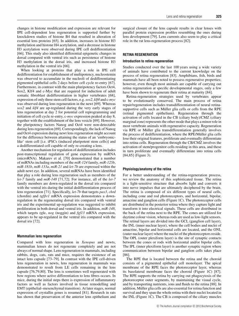
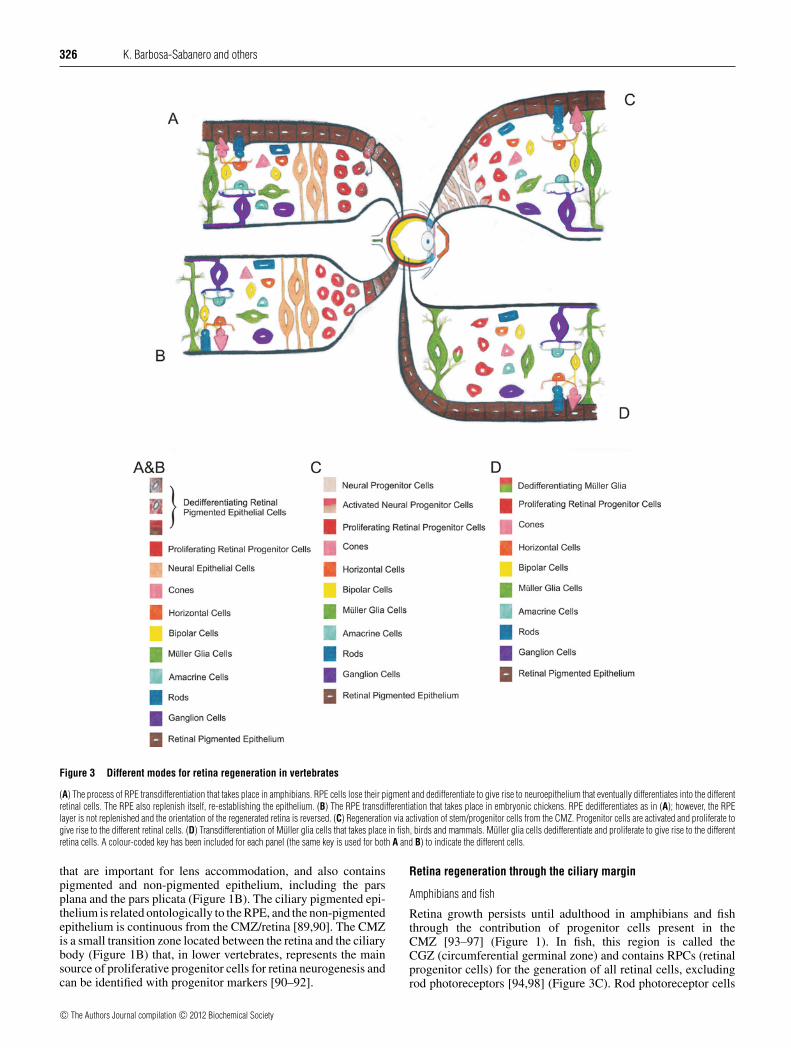


![Artificial retina [shweta]](https://static.documents.pub/doc/80x56/5874c3ae1a28ab8f508b5dd3/artificial-retina-shweta.jpg)

