2 Manganese: A New Emerging Contaminant in the Environment Annalisa Pinsino 1,2 , Valeria Matranga 2 and Maria Carmela Roccheri 1 1 Dipartimento di Scienze e Tecnologie Molecolari e Biomolecolari (Sez. Biologia Cellulare), Università di Palermo 2 Istituto di Biomedicina e Immunologia Molecolare “Alberto Monroy” CNR, Palermo Italy 1. Introduction The environment is composed of the atmosphere, earth and water. According to the World Health Organization, more than 100,000 chemicals are released into the global ambient every year as a consequence of their production, use and disposal. The fate of a chemical substance depends on its chemical application and physical-chemical properties, in combination with the characteristics of the environment where it is released. Chemical substances or contaminants discharged into the environment may be “natural” or “man- made”. One of the most misunderstood concepts regarding contamination is the miss- interpretation of term “natural”. A “natural” contaminant is one substance that can occur without human introduction. For example, trace metals, such as iron, zinc, manganese, copper, cobalt and nickel, can be considered naturally-occurring contaminants. Generally, these metals are found in the environment only in moderate amounts that do not cause health threats. However, “natural” contaminants can also have anthropogenic origins: in fact human activities often cause the release of a large amount of naturally-occurring minerals into the environment. Moreover, it is not the mere presence of a contaminant that makes it toxic, but its concentration. Paracelsus’ famous aphorism “only dose makes the difference” has laid the groundwork for the development of the modern toxicology by recognizing the importance of the dose-response relationship. In the last century, the massive production of manganese-containing compounds (metallurgic and chemical products, municipal wastewater discharges, sewage sludge, alloys, steel, iron, ceramics, fungicide products) has attracted the attention of scientists who investigated manganese as a potential emerging contaminant in the environment, and especially in the marine environment (CICAD 63, 2004). In humans, manganese excess is renowned for its role in neurotoxicity, associated with a characteristic syndrome called ‘manganese madness’ or ‘Parkinson-like’ diseases (Perl & Olanow, 2007). This neurodegenerative disorder is due to the accumulation of manganese inside intracellular compartments, such as the Golgi apparatus and mitochondria. In mammals, prenatal and postnatal exposure to manganese is associated with embryo-toxicity, fetal-toxicity, and decreased postnatal growth (Sanchez et al., 1993; Colomina et al., 1996). In marine organisms some studied showed that an excessive amount of manganese causes toxicity, although the cause-effect evidence is not extensive. www.intechopen.com
Transcript
2
Manganese: A New Emerging Contaminant in the Environment
Annalisa Pinsino1,2, Valeria Matranga2 and Maria Carmela Roccheri1 1Dipartimento di Scienze e Tecnologie Molecolari e Biomolecolari
(Sez. Biologia Cellulare), Università di Palermo 2Istituto di Biomedicina e Immunologia Molecolare “Alberto Monroy” CNR, Palermo
Italy
1. Introduction
The environment is composed of the atmosphere, earth and water. According to the World Health Organization, more than 100,000 chemicals are released into the global ambient every year as a consequence of their production, use and disposal. The fate of a chemical substance depends on its chemical application and physical-chemical properties, in combination with the characteristics of the environment where it is released. Chemical substances or contaminants discharged into the environment may be “natural” or “man-made”. One of the most misunderstood concepts regarding contamination is the miss-interpretation of term “natural”. A “natural” contaminant is one substance that can occur without human introduction. For example, trace metals, such as iron, zinc, manganese, copper, cobalt and nickel, can be considered naturally-occurring contaminants. Generally, these metals are found in the environment only in moderate amounts that do not cause health threats. However, “natural” contaminants can also have anthropogenic origins: in fact human activities often cause the release of a large amount of naturally-occurring minerals into the environment. Moreover, it is not the mere presence of a contaminant that makes it toxic, but its concentration. Paracelsus’ famous aphorism “only dose makes the difference” has laid the groundwork for the development of the modern toxicology by recognizing the importance of the dose-response relationship.
In the last century, the massive production of manganese-containing compounds (metallurgic and chemical products, municipal wastewater discharges, sewage sludge, alloys, steel, iron, ceramics, fungicide products) has attracted the attention of scientists who investigated manganese as a potential emerging contaminant in the environment, and especially in the marine environment (CICAD 63, 2004). In humans, manganese excess is renowned for its role in neurotoxicity, associated with a characteristic syndrome called ‘manganese madness’ or ‘Parkinson-like’ diseases (Perl & Olanow, 2007). This neurodegenerative disorder is due to the accumulation of manganese inside intracellular compartments, such as the Golgi apparatus and mitochondria. In mammals, prenatal and postnatal exposure to manganese is associated with embryo-toxicity, fetal-toxicity, and decreased postnatal growth (Sanchez et al., 1993; Colomina et al., 1996).
In marine organisms some studied showed that an excessive amount of manganese causes toxicity, although the cause-effect evidence is not extensive.
www.intechopen.com
Environmental Contamination
18
In this chapter, we will provide: firstly the information available regarding the natural behaviour of manganese in the environment and its role in the living organisms, with particular emphasis on the marine environment. Secondly, we will discuss how and why the manganese contamination has become a global problem recently. Thirdly, we will cover some aspects regarding the adverse effects resulting from the exposure of whole organisms to high levels of manganese. Advantage of the notion that marine invertebrates express qualitatively similar types of induced damage to those found in higher organisms, we will focus our attention on the toxicity of manganese at different levels of organization: whole-organism, cellular and embryonic levels. In this review chapter we intend to promote the embryos and the immune cells of echinoderms as useful models to study manganese toxicity.
2. Manganese: Environmental aspects
Manganese is one of the most abundant and widely distributed metals in nature. In fact it is typically found in rocks, soils and waters. The Earth’s crust consists of 0.1% of manganese.
As constituent of the soil, its concentrations range from 40 to 900 mg kg-1. Pure manganese is a silver-stained metal; however, it does not occur in the environment in a pure form. Rather, it occurs in manganese-compounds, combined with other elements such as oxygen, sulphur, carbon, silicon and chlorine. These forms of manganese are solid and some of them can dissolve in water or be suspended in the air as small particles. The small dust particles in the air usually settle at the bottom within a few days, depending on their size, weight, density and weather conditions. Manganese can exist in 11 oxidation states, ranging from –3 to +7, but the most common ones are: +2 (e.g., MnCl2) and +4 (e.g., MnO2).
2.1 Manganese behaviour in the aquatic environment
Natural waters, such as lakes, streams, rivers and oceans, contain variable quantities of dissolved manganese, ranging from 10 to 10,000 μg l-1. In water, most manganese compounds tend to attach to circulating particles or settle as sediment. Ocean spray, forest fires, vegetation, crustal rock and volcanic activity are the major natural atmospheric sources of manganese (CICAD 63, 2004).
Manganese exists in the aquatic environment in two main forms: Mn2+ and Mn4+. Oscillation between these two forms occurs via oxidation and reduction reactions that may be abiotic or biotic (Schamphelaire et al., 2008). The interconversions between these forms is of particular importance to the aquatic chemistry of manganese, as Mn2+ forms are soluble whereas Mn4+ is present in insoluble oxides.
Since the late 1970s, the bacterially-catalyzed oxidation of manganese has been receiving increasing attention, because of its important role in geochemical cycles. Three well-studied and phylogenetically distinct manganese-oxidizing bacteria have been described: i) the ┚-Proteobacterium Leptothrix discophora, isolated from a swamp; ii) the ┛-Proteobacterium Pseudomonas putida, which is a ubiquitous freshwater and soil bacterium; iii) the Bacillus sp spores, isolated from a near-shore manganese sediment (De Schamphelaire et al., 2007). A number of related metal-reducing micro-organisms have been identified and classified as the Geobacteraceae (Caccavo et al., 1994; Coates et al., 1995, 2001; Holmes et al., 2004; Vandieken et al., 2006). The biochemical pathways involved in Mn2+ oxidation have not yet
www.intechopen.com
Manganese: A New Emerging Contaminant in the Environment
19
been completely elucidated. A general scheme of the manganese cycle occurring in a sediment-water system is showed in Figure 1. The main oxidant in natural water is dissolved oxygen.
Fig. 1. A flux model for manganese interconversion. Manganese oxidation is performed in the oxic layer (water), while manganese reduction occurs in the anoxic layer (sediment). The oxic-anoxic boundary is located at the sediment-water interface. Note: all figures presented in this work are original.
The marine environmental chemistry of manganese is largely governed by pH, oxygen concentration of the solution and redox conditions. In fact, manganese oxidation increases with the decrease in acidity of the medium. The redox cycle of manganese in the oceans occurs at the oxic-anoxic boundary, which is often located at the sediment-water interface. Manganese oxides are present on the ocean floor as concretions, crusts and fine disseminations in sediments. It is well known, for example, that the soft bottom sediments of the oceans are particularly rich in manganese aggregates in the form of nodules (Bonatti & Nayudu, 1965; Wang et al., 2011).
Free manganese ions are released in the water by means of the photochemical and chemical reduction of manganese oxides coming from the organic matter (Sunda & Huntsman, 1998; De Schamphelaire et al., 2007). The process is initialised after the increase in temperature, the decrease in oxygen concentrations and the upward movement of the redox-cline (Balzer, 1982; Hunt, 1983). The transport of the dissolved manganese ions is governed by molecular diffusion in the water pores and it follows a manganese concentration gradient (the gradient decreases towards the oxic zone). In the marine environment, in absence of micro-organisms or mineral particles, manganese oxidation is a slow process (Wehrli et al., 1995). A reduced dissolved oxygen condition (called hypoxia) causes the rise of the ionic flux of manganese, which goes from the sediment to the overlying waters, where it reaches concentrations 1,000-folds higher than those normally occurring in seawater (up to 22 mg l-1) (Trefry et al., 1984; Aller, 1994). Hypoxia in the marine environment can be natural or human-induced.
www.intechopen.com
Environmental Contamination
20
At present, costal hypoxia is increasing because of man-made alterations of coastal ecosystems and changes in oceanographic conditions due to global warming. In the deep ocean water, hypoxia is influenced mostly by the variations in the up-welling that is driven by the wind. Hypoxic areas are marine dead zones in the world's oceans which can happen for example in the fjords, coastlines or closed sea (such as Black, Baltic and Mediterranean Seas), where the water turnover, that should increase the oxygen content, is very slow or not present (Middelburg & Levin, 2009).
3. Biological functions of manganese in living organisms: General aspects
While manganese is abundant and widely distributed in nature, it is required only in trace amounts in the organisms during their life span, where it guides normal development and body function. In fact, it plays essential roles in many metabolic and non-metabolic regulatory functions, such as: i) bone mineralization; ii) connective tissue formation; iii) energetic metabolism; iv) enzyme activation; v) immunological and nervous system activities; vi) reproductive hormone regulation; vii) cellular defence; viii) amino acid, lipid, protein, and carbohydrate metabolisms; ix) glycosaminoglycans formation; x) blood clotting (ATSDR, 2008; Santamaria, 2008). Manganese works as a constituent of metallo-enzymes or as an enzyme activator. Examples of manganese-containing enzymes include: arginase, the cytosolic enzyme responsible for the urea formation; pyruvate carboxylase, the enzyme that catalyses the first step of the carbohydrate synthesis from pyruvate; and manganese-superoxide dismutase, the enzyme that catalyzes the dismutation of superoxide into oxygen and hydrogen peroxide (Wedler, 1994; Crowley et al., 2000). In contrast to the relatively few manganese metallo-enzymes, there are a large number of manganese-activated enzymes, including the hydrolases, the kinases, the decarboxylases, the DNA and RNA polymerases and the transferases (Missiaen et al., 2004). Activation of these enzymes can occur either as a direct consequence of the binding of manganese to the proteins, which causes subsequent conformational changes, or by its binding to the substrate, as in the case of ATP. Mechanisms regulating manganese homeostasis in cells are largely unknown. Some studies indicate the importance of regulated intracellular trafficking of manganese transporters to balance its absorption and secretion. Multiple transporters mediate intracellular manganese uptake including: i) natural resistance-associated macrophage proteins (Nramp); ii) cation/H+ antiporter; iii) zinc-regulated transporter/iron-regulated transporter (ZRT/IRT1)-related proteins (ZIP); iv) transferrin receptors; v) various calcium-transport ATPases; vi) glutamate ionotropic receptors (Au et al., 2008). Some of these transporters are localized within specific intracellular compartments, but none of them are manganese-specific transporters. In yeast, under normal conditions, the intracellular manganese concentration is regulated by adjustments of surface levels of the Nramp transporter, which regulates its degradation by endocytosis and ubiquitin-mediated targeting to vacuoles (Culotta et al., 2005). In mammals, the Golgi-associated secretory pathway Ca2+-ATPase (SPCA) is known to pump cytosolic manganese into the lumen of the Golgi complex, in order to be used by glycosylation pathway enzymes. However, SPCA role in manganese detoxification has not been well elucidated (Missiaen et al., 2004).
4. Manganese toxicity: Causes and concerns
Manganese is considered an emerging contaminant because it is a perceived or real threat to the human health and the environment. Manganese exposure occurs at different levels and
www.intechopen.com
Manganese: A New Emerging Contaminant in the Environment
21
through a wide variety of industrial sources such as mining, alloy production, goods processing, iron-manganese operations, welding, agrochemical production and other anthropogenic activities. Manganese products can be discharged into the sea and become an unforeseen toxic metal in the marine environment. As manganese bioavailability increases, its uptake into living organisms occurs predominately through the water. The manganese rates of accumulation, as well as its elimination, are relatively fast-regulated processes. The exposure to high levels of manganese causes toxicity and decreases the fitness of the organisms (Roth, 2006). In humans, the neurological damage induced by excessive manganese exposure has been well documented for over a century (Cooper, 1837; Mena et al., 1967; Normandin & Hazel, 2002; Takeda, 2003). On the contrary, data on the effects of high manganese exposure in marine organisms are not well documented. In fact, although marine environment contains high natural concentrations of manganese, especially in the hypoxic zones, the potential danger to benthic and planktonic organisms has attracted the attention of scientists only recently.
4.1 Manganese toxicity in marine invertebrates
In all marine organisms manganese is accumulated into tissues; its amount reflects the concentrations of the bio-available manganese dissolved in sea water (Weinstein et al., 1992; Hansen & Bjerregaard, 1995; Baden & Eriksson, 2006). At the cellular level, manganese balance is proficiently managed by processes controlling cellular uptake, retention, and excretion (Roth, 2006), but these elaborate homeostatic mechanisms are altered under high levels of the available metal. Thus, it is important to consider that manganese dissolved in sea water is bio-concentrated significantly more at lower than at higher trophic levels (CICAD 63, 2004). The Bio Concentration Factor (BCF) correlates the concentration of a substance in animal tissues to the concentration of the same substance in the surrounding water. The reported BCF values range between: 100-600 for fish, 10,000-20,000 for marine and freshwater plants, 10,000-40,000 for invertebrates (ATSDR, 2008). In aquatic invertebrates manganese uptake significantly increases with temperature increase and salinity and with pH decrease. Dissolved oxygen has no significant effect (Baden et al., 1995).
Crustaceans and molluscs are the most manganese-sensitive invertebrates, followed by arthropods and echinoderms. The first studies on the effects of manganese in crustaceans (species Homarus gammarus and H. vulgari) were carried out by Bryan & Ward (1965).
High levels of manganese have been found in the haemolymph and body tissues of the lobster Nephrops norvegicus living in the SE Kattegat, Swedish west coast, as well as in lobsters living in the hypoxic areas near sludge dumping sites in the Firth of Clyde, Scotland (Baden & Neil, 1998). Exposure to high manganese impairs the lobster’s antennular ficking activity, causing disorientation and inability to locate food (Krång & Rosenqvist, 2006). Likewise, unhealthy blue crabs, Callinectes sapidus, have been found in a manganese-contaminated area of North Carolina, USA (Gemperline et al., 1992; Weinstein et al., 1992). In general, internal tissues such as the intestine, nervous system, haemolymph and reproductive organs accumulate much more manganese than other tissues such as exoskeleton, but in the latter case, manganese elimination is a very slow process. In Nephrops norvegicus, manganese accumulation reached a plateau after 1.25 days of exposure in all tissues except for the mid-gut gland, which continued to accumulate manganese over time
www.intechopen.com
Environmental Contamination
22
(Baden et al., 1999). A similar accumulation pattern of manganese in soft tissues has also been described in mussels (Regoli et al., 1991). Specifically, in the species Donacilla cornea, manganese was rapidly accumulated; reaching a maximum after 3 days of exposure, and it was rapidly excreted (60% loss) after 3 days in clean sea water. Seasonal and sex differences in the manganese accumulation levels have been reported for both mussels (e.g. Mytilus edulis and Mytilus californianus) and oysters (e.g. Crassostrea gigas and Crassostrea virginica) (Nørum et al., 2005). For example, in the species Mytilus edulis the gonads of females accumulated manganese more than males. Manganese accumulation in the sea star Asterias rubens has shown linearity with time up to 23 days at low concentrations (0.1 mg l-1), but its saturation kinetics were very fast at higher concentrations (Hansen & Bjerregaard, 1995). In fact, it was found that steady-state levels were reached in the coelomic fluids after only 5 days of exposure to 5.5 mg l-1 (Oweson et al., 2008).
Manganese excess may cause a Ca2+ pump dysfunction, affecting neuro-muscular transmission in benthic marine invertebrates (Hagiwara & Takahashi, 1967; Baden & Neil, 1998; Holmes et al., 1999). For example, in crustaceans, manganese acts as a competitive inhibitor of the calcium-regulated ion channels present in nerve and muscle membranes, thus inhibiting synaptic and neuromuscular transmission and muscle excitation (Hagiwara & Takahashi, 1967; Holmes et al., 1999).
Manganese affects the immune system of marine invertebrates in a species-specific manner.
In the immune system of Nephrops norvegicus (haemolymph), manganese is mainly found in the protein fraction that includes haemocyanin and immune cells (called haemocytes). Recent studies showed that high levels of manganese affect Nephrops norvegicus haemocytes causing: i) apoptosis-induced reduction of the number of circulating haemocytes ; ii) inhibition of their maturation to granular haemocytes; iii) inhibition of the recruitment of haematopoietic stem cells (Hernroth et al., 2004; Oweson et al., 2006). These immune suppressive effects were also found in Mytilus edulis (Oweson et al., 2009). In addition, manganese alters the immune system of sponges (Geodia cydonium, Crella elegans and Chondrosia reniformis) by inhibiting the activity of the 2’, 5’-oligoadenylate synthetase (2-5A synthetase), an enzyme known to be involved in the functioning of the immune system of vertebrates (Saby et al., 2009).
Surprisingly, in contrast to what was recorded in crustaceans and molluscs, in echinoderms (Asterias rubens) manganese exposure stimulated haematopoiesis, thus causing an increase in the number of circulating immune cells (Oweson et al., 2008). Manganese effects on Asterias rubens immune system will be discussed in detail in the next sections.
4.2 How does manganese affect echinoderm immune cells?
Echinoderms play a key role in the maintenance of the integrity of the ecosystem where they live (Hereu et al., 2005) and are constantly exposed to pollutants deriving from different kinds of human activities (Bellas et al., 2008; Rosen et al., 2008). They are phylogenetically related to vertebrates and have a sophisticated and sensitive immune system. In echinoderms, immune cells (called coelomocytes) are a heterogeneous population of free moving cells found in all coelomic spaces, including the perivisceral coelomic cavities and the water-vascular system (reviewed in Matranga, 1996; Glinski & Jarosz, 2000; Smith et al., 2010). They are also present sparsely in the connective tissue (mesodermal stromal tissue)
www.intechopen.com
Manganese: A New Emerging Contaminant in the Environment
23
and amongst tissues of various organs (Muñoz-Chápuli et al., 2005; Pinsino et al., 2007). Coelomocytes participate as immune cells in function similar to their vertebrate’s immune system homologues. In fact, they are involved in: clot formation, phagocytosis, encapsulation and clearance of pathogens, as well as oxygen transport. The coelomic fluid in which the immunocytes or coelomocytes reside and move is a key factor governing the immunological capabilities of echinoderms, as it contains essential trophic and activating factors (for a review see Matranga et al., 2005; Smith et al., 2010). Four different morphotypes have been described in the asteroid Asterias rubens, with the phagocytes as the most abundant type, accounting for approximately 95% of the total population (Pinsino et al., 2007).
As previously reported, the accumulation of manganese into the coelomic fluid of exposed sea stars (Asterias rubens) induces the proliferation of haematopoietic cells (Oweson et al., 2008). Specifically, by using the substitute nucleotide 5-bromo-2’-deoxyuridine (BrdU) for tracing cell division, and by recording the mitotic index after nuclei staining, authors found that manganese induced the proliferation of cells from a putative haematopoietic tissue, the coelomic epithelium. In addition, the haematopoietic tissue and coelomocytes showed stress response in terms of changes in HSP70 levels and protein carbonyls. Incubation with heat-killed FITC-labelled yeast cells (Saccharomyces cerevisiae) exhibited an inhibited phagocyte capacity of coelomocytes. Moreover, measurement of dehydrogenase activity, using MTS/PMS, revealed that manganese showed cytotoxic properties. Although manganese was revealed as stressful to the coelomocytes and affected their ability to phagocyte, the increased number of coelomocytes compensated these impairments. In summary, the authors concluded that the exposure of Asterias rubens to manganese impaired their immune response, but induced renewal of coelomocytes, assuring survival. Co-occurrence of manganese with hypoxic conditions does not inhibit the elevated production of coelomocytes, but probably affects the composition of the subpopulations of these immune cells since hypoxia, but not manganese, increased the mRNA expression of Runt, a transcription factor, assumed necessary for cell differentiation (Oweson et al., 2010).
5. Sea urchin embryonic development
To address this issue, at the beginning of this section we will describe the basic steps of the
sea urchin development. Briefly, upon appropriate stimulation, millions of eggs and sperm
are released into the sea water; after fertilization, the single-celled zygote is converted into a
multi-cellular embryo through rapid and repeated mitotic cell divisions (cleavage). Founder
cells of the three germ layers ecto- meso- and endoderm, are the basic units where
regulatory information is localized during cleavage. In particular, ┚-catenin is required for
the development of all endo-mesoderm territories, including the archenteron, the primary
mesenchyme cells (PMCs) and the secondary mesenchyme cells (SMCs) (Logan et al., 1999).
Cell fates are fully specified by the blastula-early gastrula stage of development, when cells
have begun to express particular sets of territory-specific genes (Davidson et al., 1998).
Although maternal determinants are required for founder cell specification during
development, interactions between the PMCs and external cues derived from the ectoderm
specify many phases of the skeleton formation (Armstrong et al., 1993; Ettensohn &
Malinda, 1993; Guss & Ettensohn, 1997; Zito et al., 1998). The blastula stage is characterized
by the presence of a large fluid-filled blastocoels, surrounded by a single layer of cells.
www.intechopen.com
Environmental Contamination
24
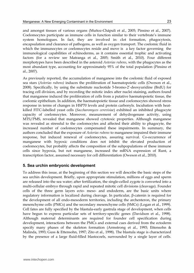
During gastrulation extensive cellular rearrangements occur which convert the hollow-
spherical-blastula into a multi-layered gastrula. Changes in shape and differentiation of
embryo structures lead to the formation of a pluteus, the first larval stage. Genus-specific
spicule growth and patterning is completed at this stage, directed by the spatial-temporal
regulated expression of bio-mineralization related genes (Zito et al., 2005; Matranga et al.,
2011). Sea urchin development from the blastula to the pluteus stage is showed in figure 2.
Fig. 2. Sea urchin development from the blastula to the pluteus stage. A) hatching blastula; B) mesenchyme blastula; C) middle gastrula; D) pluteus. Note: all figures presented in this work are original.
5.1 Sea urchin embryos as an in vivo model for the assessment of toxicity
The sea urchin is estimated to have 23,300 genes with representatives of nearly all vertebrate gene families (Sea Urchin Genome Sequencing Consortium, 2006). Since it has been demonstrated that the sea urchin genome shares at least 70% of the genes with the mankind, we shall consider how this provides an important tool kit to aid our understanding of eco- embryo- and geno-toxicological studies as well as for studies on embryonic development. Sea urchins are marine invertebrates with two life stages: i) an early and brief developmental stage (planktonic) and ii) a remarkably long-lived adult stage with life spans extending to over a century (epi-benthonic). Sea urchins are pivotal components of sub-tidal marine ecology (Hereu et al., 2005) and they are continuously exposed to environmental pressure, including changes in temperature, hypoxia, pathogens, UV radiation, free radicals, metals and toxicants. These marine invertebrates produce large numbers of susceptible, but not vulnerable, transparent embryos. The keys for their developmental success are the potent cellular mechanisms that provide them with protection, robustness and resistance, as well as the regulatory pathways that alter their developmental course in response to the conditions encountered (Hamdoun & Epel, 2007). The integrated network of genes, proteins and pathways that allow an organism to defend itself against chemical agents is known as the “chemical defensome” (Goldstone et al., 2006). In sea urchin embryos, many “defensome” genes are also expressed during their normal development as integral part of the developmental program, suggesting a dual function regulating both defence and development. In addition, genes involved in signal transduction often respond to environmental stress, activating alternative signalling pathways as a defence strategy for survival (Hamdoun & Epel, 2007). Thus, the sea urchin becomes an excellent candidate for the understanding of the two-fold function of genes/proteins and signalling pathways involved in both defence and regulation/preservation of development during environmental changes.
www.intechopen.com
Manganese: A New Emerging Contaminant in the Environment
25
To date, several researchers have shown that exposure of sea urchin embryos to chemical and physical agents involve a selective set of defence “macromolecules” (Geraci et al., 2004; Roccheri et al., 2004; Bonaventura et al., 2005; Matranga et al., 2010; Russo et al., 2010; Pinsino et al., 2011a). Of interest, for example are reports about the biochemical and molecular changes occurring in response to cadmium exposure in Paracentrotus lividus embryos. Briefly, the toxic effects have been studied by examining the: accumulation, embryonic malformation, stress gene expression, stress protein induction, apoptosis and related pathways (Roccheri & Matranga, 2010). Specifically, it was found that the exposure of embryos to sub-acute/sub-lethal cadmium concentrations was able to trigger the expression of one of the metallothionein genes which binds metal ions in the cytoplasm for storage and/or detoxification (Russo et al., 2003). Simultaneously, or alternatively, cadmium was able to induce the new synthesis of several stress proteins—HSPs that usually facilitate the repair of miss-folded proteins or the elimination of aggregated proteins (Roccheri et al., 2004). The authors found that 9 hours of cadmium exposure were required to induce the synthesis of HSPs 70 and 72, while at least 15 hours were needed to observe the induction of hsp56 and 25kDa synthesis. In addition, it has been demonstrated that a long-lasting exposure (over 24 hours) triggers DNA fragmentation and causes the activation of caspase-3, one of the key molecules promoting apoptosis, which increased in a time-dependent way (Agnello et al. 2006, 2007). In sea urchin embryos, apoptosis is an important part of the defence strategy, both in physiological or stress conditions (Agnello & Roccheri, 2010).
Recently, it has also been demonstrated that in sea urchin embryos autophagy is a further defence strategy activated in response to cadmium exposure (Chiarelli et al., 2011). These authors found that autophagy reaches its maximum peak after 18 hours, when apoptosis is just beginning, suggesting that this degradation process starts before apoptosis and after the failure of HSP and metallothionein function. In conclusion, data demonstrate the wide range of alternative strategies that can occur at different levels of stress.
5.2 Manganese embryo-toxicity in sea urchin embryos: Biochemical and molecular studies
As previously mentioned, prenatal and postnatal exposure to manganese in mammals is associated with embryo-toxicity, fetal-toxicity, and decreased postnatal growth (Sanchez et al., 1993; Colomina et al., 1996; Doyle & Kapron, 2002; Giordano et al., 2009). Nevertheless, functional data on the effect of high manganese exposure on gene expression and on cellular mechanisms involved in embryonic development remain scant. Recently, we took advantage of the amenable embryonic model, the Mediterranean sea urchin Paracentrotus lividus, to investigate the potential toxicity of manganese on embryonic development, using different biological and biochemical approaches.
In our studies, embryos were continuously exposed to manganese from fertilization, at concentrations ranging from 1.0 to 61.6 mg l-1 (or from 0.018 to 1,120 mM), and harvested at different developmental stages (Pinsino et al., 2010; Pinsino et al., 2011b). The biological study was carried out according to classical toxicological criteria, namely: concentration- and time-dependent responses, analysis of the impact on development, manganese accumulation. We found that embryos showed an elevated tolerance/resistance to manganese, as they accumulated high amounts into cells in a time- and concentration-dependent manner. Here we show, just as an example, the time course of manganese
www.intechopen.com
Environmental Contamination
26
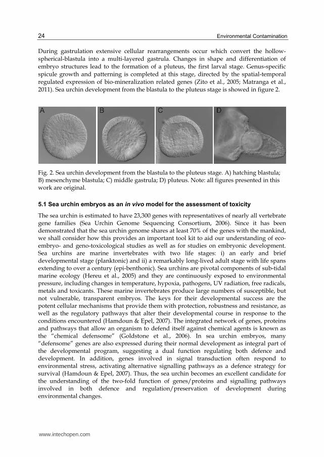
accumulation analyzed from 24 to 72 hours of exposure/development in embryos exposed to different manganese concentrations (Figure 3A).
Fig. 3. Time course of manganese accumulation and calcium content determined by AAS in embryos exposed to different manganese concentrations (0, 1.0, 7.7, 15.4, 30.8, 61.6 and 122 mg l -1). Note: all figures presented in this work are original.
Results were compared to the physiological calcium content measured in the same samples (Figure 3B). We found that calcium content diminished in an inversely proportional way to manganese accumulation. The amount of manganese accumulated and the calcium content in cells were determined, in exposed and control embryos, by atomic absorption spectrophotometer (AAS). Moreover, AAS data for calcium content was consistent with its poor detection in PMCs observed by in vivo labelling with the cell-permeable fluorescent dye, calcein (Pinsino et al., 2011b).
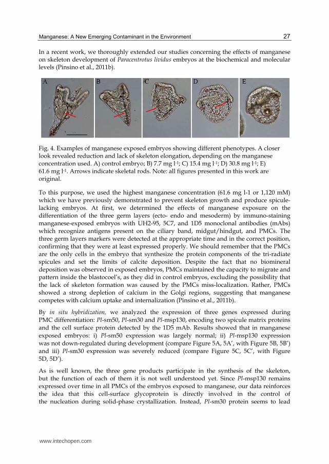
Rising manganese exposure concentrations from 1.0 to 61.6 or 122 mg l-1 did not produce lethal effects. Rather, we observed a concentration-dependent increase in the number of morphological abnormalities found 48 hours post-fertilization (pluteus stage) (Figure 4). The impact on embryonic development was analysed considering that normal embryos should satisfy some morphological criteria as the correct schedule in reaching the developmental endpoint (pluteus) and the correct skeleton development and patterning (Pinsino et al., 2010). Major developmental defects consisted in the reduced elongation of skeletal rods (spicules) (see arrows Figure 4), suggesting a key role for manganese in embryonic skeleton development.
In addition, a correlation was observed when comparing malformations, accumulation of manganese and the regulation of key stress proteins that provide protection against stressors. In fact, embryos exposed to high manganese concentrations (15.4 mg l-1 or above) showed an increase of the hsc70 and hsc60 protein levels at the 48 hours. The proteins are useful to protect them from apoptosis, in accordance to the finding that no DNA fragmentation was induced by manganese exposure (see Pinsino et al., 2010). By a fluorescent detection assay on live embryos, we found no induction of the reactive oxygen species (ROS), indicating no correlation between manganese toxicity and oxidative stress (see Pinsino et al., 2010).
www.intechopen.com
Manganese: A New Emerging Contaminant in the Environment
27
In a recent work, we thoroughly extended our studies concerning the effects of manganese on skeleton development of Paracentrotus lividus embryos at the biochemical and molecular levels (Pinsino et al., 2011b).
Fig. 4. Examples of manganese exposed embryos showing different phenotypes. A closer look revealed reduction and lack of skeleton elongation, depending on the manganese concentration used. A) control embryo; B) 7.7 mg l-1; C) 15.4 mg l-1; D) 30.8 mg l-1; E) 61.6 mg l-1. Arrows indicate skeletal rods. Note: all figures presented in this work are original.
To this purpose, we used the highest manganese concentration (61.6 mg l-1 or 1,120 mM) which we have previously demonstrated to prevent skeleton growth and produce spicule-lacking embryos. At first, we determined the effects of manganese exposure on the differentiation of the three germ layers (ecto- endo and mesoderm) by immuno-staining manganese-exposed embryos with UH2-95, 5C7, and 1D5 monoclonal antibodies (mAbs) which recognize antigens present on the ciliary band, midgut/hindgut, and PMCs. The three germ layers markers were detected at the appropriate time and in the correct position, confirming that they were at least expressed properly. We should remember that the PMCs are the only cells in the embryo that synthesize the protein components of the tri-radiate spicules and set the limits of calcite deposition. Despite the fact that no biomineral deposition was observed in exposed embryos, PMCs maintained the capacity to migrate and pattern inside the blastocoel’s, as they did in control embryos, excluding the possibility that the lack of skeleton formation was caused by the PMCs miss-localization. Rather, PMCs showed a strong depletion of calcium in the Golgi regions, suggesting that manganese competes with calcium uptake and internalization (Pinsino et al., 2011b).
By in situ hybridization, we analyzed the expression of three genes expressed during
PMC differentiation: Pl-sm50, Pl-sm30 and Pl-msp130, encoding two spicule matrix proteins
and the cell surface protein detected by the 1D5 mAb. Results showed that in manganese
was not down-regulated during development (compare Figure 5A, 5A’, with Figure 5B, 5B’)
and iii) Pl-sm30 expression was severely reduced (compare Figure 5C, 5C’, with Figure
5D, 5D’).
As is well known, the three gene products participate in the synthesis of the skeleton, but the function of each of them it is not well understood yet. Since Pl-msp130 remains expressed over time in all PMCs of the embryos exposed to manganese, our data reinforces the idea that this cell-surface glycoprotein is directly involved in the control of the nucleation during solid-phase crystallization. Instead, Pl-sm30 protein seems to lead
www.intechopen.com
Environmental Contamination
28
the elongation phase as supported by the down-regulation of its transcript over time (Figure 4), in agreement with reports on the American species Lythechinus pictus (Guss & Ettenhson, 1997).
It has been widely demonstrated that Extracellular signal-Regulated Kinase (ERK) MapK mediated signalling controls the expression of several regulatory genes which participate in the specification and differentiation of the sea urchin skeleton (Fernandez-Serra et al., 2004; Röttinger et al., 2004).
Fig. 5. Whole-mount in situ hybridizations in control (A, A’, C, C’) and manganese-exposed embryos (B, B’, D, D’), performed with the following probes: Pl-msp130 (A-B, A’-B’) encoding for PMCs surface protein; Pl-sm30 (C-D, C’-D’) encoding for an integral spicule matrix protein.Note: all figures presented in this work are original.
Thus, we analyzed, by Western blotting, the activation of ERK in manganese exposed embryos during development. We found a persistent phosphorylated state at all stages examined, as proteins levels were only partially modulated during development of exposed embryos, contrary to the physiological oscillations observed in normal embryos.
In conclusion, our results showed for the first time the ability of manganese to interfere with
calcium uptake and internalisation into PMCs, and the involvement of endogenous calcium
content in regulating the activation/inactivation of ERK during the sea urchin embryo
morphogenesis (Pinsino et al., 2011b).
6. Conclusion
Metals are one of the most abundant classes of contaminants generated by human activities
and represent an actual hazard for marine ecosystems and organisms’ health. In fact,
although metals are terrestrially produced, they flow into the sea through effluent and
Pl-sm30 Pl-msp130
A B C D
A’ B’ C’ D’
www.intechopen.com
Manganese: A New Emerging Contaminant in the Environment
29
sewage or are directly discharged from industries placed on the sea water front. Marine
organisms can take up from the sea and from their diet these metals, which may consist of
particles in suspension or might be deposited in the sediment. A great number of factors
may influence dose-effect and dose-response relationships between metals and organisms;
their tolerance to and use of trace metals reflect sea water concentrations. Marine
invertebrates accumulate and bio-concentrate metals more than higher organisms; thus,
their peculiar position in the marine trophic chain, where pelagic larvae are part of the diet
of several planktonic and benthic organisms (bio-magnification), increases the interest of
many researchers. Trace essential metals are of environmental interest both as limiting
nutrients (Fe, Zn, Mn, Cu, Co, Mo and Ni), playing important roles in metal-requiring and
metal-activated enzyme systems, and as toxicants when present at high concentrations. On
the contrary, non-essential heavy metals as Cd, Hg, Ag, Pb, Sn and Cr, are toxic for living
organisms even at low concentrations.
The effects of several metals on echinoderm embryos have been studied for many years. The response of sea urchin embryos to metal exposure involves selective sets of defence “macromolecules”. The type of defence response elicited greatly depends on the different sensitivity of the organisms to the different metal used. Metal toxicity can trigger several biochemical and cellular events that include: the induction of a set of highly conserved proteins, such as the heat-shock proteins and the metallothioneins; the efflux transporters; the activation of anti-oxidative enzymes; autophagy and apoptosis. In this chapter it was reported that manganese-exposed embryos do not activate a fast defence response, but activate or repress signalling pathways and transcriptional activities involved in the regulation/preservation of development. This surprising behaviour is probably due to the fact that a moderate manganese increase is not recognized by the cell as stressful event. In fact, low manganese concentrations stimulate embryos growth (not shown), probably as a consequence of their role in the metabolic-enzymatic reactions. On the contrary, non-essential heavy metals, such as cadmium, activate several cellular defence strategies aimed at permitting embryo survival. In fact, Paracentrotus lividus embryos showed an elevated tolerance to manganese, due to the increased HSC levels, did not activate the synthesis of the HSPs inducible forms and did not enter into the apoptotic program. However, manganese interferes with calcium uptake and it’s internalization into embryos, suggesting that skeletal growth is highly dependent on calcium signalling. The use of manganese-exposed embryos as a new model to study signalling pathways involved in skeletogenesis provides new insights into the mechanisms involved in manganese embryo-toxicity and emphasizes the role of calcium trafficking, recruitment and storage in the bio-mineralization process.
Interestingly, recent studies suggested that the homeostatic mechanisms that control the amount of trace metals necessary to metabolic activities are regulated by authophagy. Autophagy is a ubiquitous, non-selective degradation process involving the lysosomal/ vesicular pathway that protects the cells by clearing the damaged organelles and toxic protein aggregates. A fascinating suggestion which links manganese-affected homeostasis and embryo survival would involve authophagy. Future studies in this direction are needed to clarify this hypothesis.
Lastly, we described that, in echinoderm immune cells, manganese acts as a trigger for the proliferation of coelothelial cells and thus increases the number of circulating coelomocytes.
www.intechopen.com
Environmental Contamination
30
Studies on the effects of manganese on the activation of putative progenitor cells, including their proliferation and differentiation, should make an important contribution to our understanding of the operating mechanisms, and to the identification of those genes expressed before their release into the coelom.
7. Acknowledgements
This work has been supported by 60% MIUR and FSE (PON 2000/2006) grants to MCR and the BIOMINTEC Project (PITN-GA-2008-215507) to VM. The first author has been the recipient of a Doctoral fellowship from the University of Palermo. The project was the backbone of AP doctoral studies.
8. References
Aller, R.C. (1994). The sedimentary Mn cycle in long island sound: its role as intermediate oxidant and the influence of bioturbation, O2, and Corg flux on diagenetic reaction balances. Journal of Marine Research, Vol. 52, No. 2, (March 1994), pp. 259–295, ISSN: 1543-9542
Agnello, M., Filosto, S., Scudiero, R., Rinaldi, A.M., & Roccheri, M.C. (2006). Cadmium accumulation induces apoptosis in P. lividus embryos. Caryologia, Vol. 59, No. 4, pp. 403–408. Available from
http://www1.unifi.it/caryologia/past_volumes/59_4/59-4_gei11.pdf Agnello, M., Filosto, S., Scudiero, R., Rinaldi, A.M., & Roccheri, M.C. (2007). Cadmium
induces apoptotic response in sea urchin embryos. Cell Stress & Chaperones, Vol. 12, No. 1, (Sprinter 2007), pp. 44–50, 1355-8145
Agnello, M., & Roccheri, M.C. (2010). Apoptosis: focus on sea urchin development. Apoptosis, Vol. 15, No. 3, (March 2010), pp. 322-330, ISSN 1360-8185
Armstrong, N., Hardin, J., & McClay, D.R. (1993). Cell-cell interactions regulate skeleton formation in the sea urchin embryo. Development, Vol. 119, No. 3, (November 1993), pp. 833-840, ISSN 0950-1991
ATSDR (2008). Draft toxicological profile for manganese. Agency for toxic substances and disease registry. Division of toxicology and environmental medicine/applied toxicology branch, Atlanta, Georgia, Available from
http://www.atsdr.cdc.gov/toxprofiles/tp151-p.pdf Au, C., Benedetto, A., & Aschner, M. (2008). Manganese transport in eukaryotes: the role of
DMT1. Neurotoxicology, Vol. 29, No. 4, (July 2008), pp. 569–576, ISSN 0161-813X Baden, S.P., Eriksson, S.P., & Weeks, J.M. (1995). Uptake, accumulation and regulation of
manganese during experimental hypoxia and normoxia by the decapod Nephrops nor6egicus (L.). Marine Pollution Bulletin, Vol. 31, No. 1-3, 93-102, ISSN 0025-326X
Baden, S.P., & Neil, D.M. (1998). Accumulation of Manganese in the Haemolymph, Nerve and Muscle Tissue of Nephrops norvegicus (L.) and Its Effect on Neuromuscular Performance. Comparative Biochemistry and Physiology, Vol. 119A, No.1, (January 1998), pp. 351-359, ISSN 1095-6433
Baden, S.P., Eriksson, S.P., & Gerhardt, L. (1999). Accumulation and elimination kinetics of manganese from different tissues of the Norway lobster Nephrops norvegicus (L.). Aquatic Toxicology, Vol. 46, No. 2, (July 1999), pp. 127–137, ISSN 0166-445X
www.intechopen.com
Manganese: A New Emerging Contaminant in the Environment
31
Baden, S.P., & Eriksson, S.P. (2006). Oceanography and marine biology: an annual review. In: Role, routes and effects of manganese in crustaceans, R.N. Gibson, R.J.A. Atkinson, J.D.M. Gordon (eds), 61-83, Taylor and Francis, ISBN 978-0-8493-7044-1, London
Balzer, W. (1982). On the distribution of iron and manganese at the sediment/water interface: thermodynamic versus kinetic control. Geochimica et Cosmochimica Acta, Vol. 46, No. 7, (July 1982), pp. 1153–1161, ISSN 0016-7037
Bellas, J., Fernández, N., Lorenzo, I., & Beiras, R. (2008). Integrative assessment of coastal pollution in a Ría coastal system (Galicia, NW Spain): correspondence between sediment chemistry and toxicity. Chemosphere, Vol. 72, No. 5, (April 2008), pp. 826–835, ISSN 0045-6535
Bonatti, E., & Nayudu, Y.R. (1965). The origin of manganese nodules on the ocean floor. American Journal of Science, Vol. 263, (January 1965), pp. 17-39. ISSN 1945-452X
Bonaventura, R., Poma, V., Costa, C., & Matranga, V. (2005). UVB radiation prevents skeleton growth and stimulates the expression of stress markers in sea urchin embryos. Biochemical and Biophysical Research Communications, Vol. 328, No. 1, (March 2005), pp. 150-157, ISSN 0006-291X
Bryan, G. W. & Ward, E. (1965). The absorption and loss of radioactive and non-radioactive manganese by the lobster, Homarus vulgaris. Journal of the Marine Biological Association of the United Kingdom, Vol. 45, No. 1, pp. 65-95, ISSN 0025-3154
Caccavo, F., Lonergan, D.J., Lovley, D.R., Davis, M., Stolz, J.F., & McInerney, M.J. (1994). Geobacter sulfurreducens sp-nov, a hydrogen-oxidizing and acetate-oxidizing dissimilatory metal-reducing microorganism. Applied and Environmental Microbiology, Vol. 60, No. 10, (October 1994), pp. 3752–3759, ISSN 0099-2240
Chiarelli, R., Agnello M., & Roccheri, M.C. (2011). Sea urchin embryos as a model system for studying autophagy induced by cadmium stress. Autophagy, Vol. 7, No 9, (September 2011), pp. 1028-1034, ISSN 1554-8627
CICAD (2004) Manganese and its compounds: environmental aspects. Concise international chemical assessment document 63. WHO, Geneva, Switzerland, Available from http://www.who.int/ipcs/publications/cicad/cicad63_rev_1.pdf
Coates, J.D., Lonergan, D.J., Philips, E.J.P., Jenter, H., & Lovley, D.R. (1995). Desulfuromonas palmitatis sp nov, a marine dissimilatory Fe(III) reducer that can oxidize long-chain fatty acids. Archives of Microbiology, Vol.164, No. 6, (December 1995), pp. 406–413, ISSN 0302-8933
Coates, J.D., Bhupathiraju, V.K., Achenbach, L.A., McInerney, M.J., & Lovley, D.R. (2001). Geobacter hydrogenophilus, Geobacter chapellei and Geobacter grbiciae, three new, strictly anaerobic, dissimilatory Fe(III)-reducers. International Journal of Systematic and Evolutionary Microbiology, Vol. 51, No. 2, (March 2001), pp. 581–588, ISSN 1466-5026
Colomina, M.T., Domingo, J.L., Llobet, J.M., & Corbella, J. (1996). Effect of day of exposure on the developmental toxicity of manganese in mice. Veterinary & Human Toxicology, Vol. 38, No. 1, pp. 7–9, ISSN 0145-6296
Couper, J. (1837). On the effects of black oxide of manganese when inhaled into the lungs. Brain Annual Medical Pharmacology, Vol. 1, pp. 41-42
Crowley, J.A., Traynor, D.A., & Weatherburn, D.C. (2000). Enzymes and proteins containing manganese: an overview. Metal ions in biological systems, Vol. 37, pp. 209-278, ISSN 0161-5149
www.intechopen.com
Environmental Contamination
32
Culotta, V. C., Yang, M., & Hall, M. D. (2005). Manganese transport and trafficking: lessons learned from Saccharomyces cerevisiae. Eukaryotic Cell, Vol. 4, No. 7, (July 2005), pp. 1159–1165, ISSN 1535-9778
Davidson, E.H., Cameron, R.A., & Ransick, A. (1998). Specification of cell fate in the sea urchin embryo: summary and some proposed mechanisms. Development, Vol.125, No. 17, (September 1998), pp. 3269-3290, ISSN 0950-1991
De Schamphelaire, L., Rabaey, K., Boon, N., Verstraete, W.,& Boeckx, P. (2007). Minireview: The potential of enhanced manganese redox cycling for sediment oxidation. Geomicrobiology Journal, Vol. 24, No. 7-8, 547–558, ISSN 0149-0451
Doyle, D., & Kapron, C.M. (2002). Inhibition of cell differentiation by manganese chloride in micromass cultures of mouse embryonic limb bud cells. Toxicology in Vitro, Vol. 16, No. 2, (April 2002), pp. 101-106, ISSN 0887-2333
Ettensohn, C.A., & Malinda, K.M. (1993). Size regulation and morphogenesis: A cellular analysis of skeletogenesis in the sea urchin embryo. Development, Vol. 119, No. 1, (September 1993), pp. 155–167, ISSN 0950-1991
Fernandez-Serra, M., Consales, C., Livigni, A., & Arnone, M.I. (2004). Role of the ERK mediated signaling pathway in mesenchyme formation and differentiation in the sea urchin embryo. Developmental Biology, Vol. 268, No. 2, (April 2004), pp. 384–402, ISSN 0012-1606
Gemperline, P.J., Miller, K.H., West, T.L., Weinstein, J.E., Hamilton, C.J., & Bray, J.T. (1992). Principal component analysis, trace elements, and Blue Crab Shell disease. Analytical Chemistry, Vol. 64, No. 9, (May 1992), pp. 523–532, ISSN 0003-2700
Geraci, F., Pinsino, A., Turturici, G., Savona, R., Giudice, G., & Sconzo, G. (2004). Nickel, lead, and cadmium induce differential cellular responses in sea urchin embryos by activating the synthesis of different HSP70s. Biochemical and Biophysical Research Communications, Vol. 322, No. 3, (September 2004), pp. 873–877, ISSN 0006-291X
Giordano, G., Pizzurro, D., VanDeMark, K., Guazzetti, M., & Costa, L.G. (2009). Manganese inhibits the ability of astrocytes to promote neuronal differentiation. Toxicology and Applied Pharmacology, Vol. 240, No. 2, (October 2009), pp. 226–235, ISSN 0041-008X
Glinski, Z., & Jarosz, J. (2000). Immune phenomena in echinoderms. Archivum Immunologiae et Therapiae Experimentalis, Vol. 48, No. 3, pp. 189–193, ISSN 0004-069X
Goldstone, J.V., Hamdoun, A., Cole, B.J., Howard-Ashby, M., Nebert, D.W., Scally, M., Dean, M., Epel, D., Hahn, M.E., & Stegeman, J.J. (2006). The chemical defensome: Environmental sensing and response genes in the Strongylocentrotus purpuratus genome. Developmental Biology, Vol. 300, No. 1, (December 2006), pp. 366-384, ISSN 0012-1606
Guss, K.A., & Ettensohn, C.A. (1997). Skeletal morphogenesis in the sea urchin embryo: regulation of primary mesenchyme gene expression and skeletal rod growth by ectoderm-derived cues. Development, Vol. 124, No. 10, (May 1997), pp. 1899-1908, ISSN 0950-1991
Hagiwara, S., & Takahashi, K. (1967). Surface density of calcium ion and calcium spikes in the barnacle muscle fibre membrane. The Journal of General Physiology, Vol. 50, No. 3, (January 1967), pp. 583–601, ISSN 0022-1295
www.intechopen.com
Manganese: A New Emerging Contaminant in the Environment
33
Hamdoun, A., & Epel, D. (2007). Embryo stability and vulnerability in an always changing world. Proceedings of the National Academy of Sciences, Vol. 104, No. 6, (February 2007), pp. 1745–1750, ISSN 0027-8424
Hansen, S.N., & Bjerregaard, P. (1995). Manganese kinetics in the sea star Asterias rubens (L.) exposed via food or water. Marine Pollution Bulletin, Vol. 31, No. 1-3, pp. 127-132, ISSN 0025-326X
Hereu, B., Zabala, M., Linares, C., & Sala, E. (2005). The effects predator abundance and habitat structural complexity on survival juvenile sea urchins. Marine Biology, Vol. 146, No. 2, (January 2005), pp. 293-299, ISSN 0025-3162
Hernroth, B., Baden, S.P., Holm, K., André, T., & Söderhäll, I. (2004). Manganese induced immune suppression of the lobster, Nephrops norvegicus. Aquatic Toxicology, Vol. 70, No. 3, (December 2004), pp. 223-231, ISSN 0166-445X
Holmes, J.M., Gräns, A.S., Neil, D.M., & Baden S.P. (1999). Effects of the metal ions Mn2+ and Co2+ on muscle contraction in the Norway lobster, Nephrops norvegicus. Journal of Comparative Physiology B, Vol. 169, No. 6, (September 1999), pp. 402-410, ISSN 0174-1578
Holmes, D.E., Nevin, K.P., & Lovley, D.R. (2004). Comparison of 16S rRNA, nifD, recA, gyrB, rpoB and fusA genes within the family Geobacteraceae fam. nov. International Journal of Systematic and Evolutionary Microbiology, Vol. 54, No. 5, (September 2004), pp. 1591–1599, ISSN 1466-5026
Hunt, C.D. (1983). Variability in the benthic Mn flux in coastal marine ecosystems resulting from temperature and primary production. Limnology and Oceanography, Vol. 28, No. 5, pp. 913–923, ISSN 0024-3590
Krång, A.S., & Rosenqvist, G. (2006). Effects of manganese on chemically induced food search behaviour of the Norway lobster, Nephrops norvegicus (L.). Aquatic Toxicology, Vol. 78, No. 3, (Jun 2006), pp. 284-291, ISSN 0166-445X
Logan, C.Y., Miller, J.R., Ferkowicz, M.J., & McClay, D.R. (1999). Nuclear beta-catenin is required to specify vegetal cell fates in the sea urchin embryo. Development, Vol. 126, No. 2, (January 1999), pp. 345-357, ISSN 0950-1991
Matranga, V (1996). Molecular aspects of immune reactions in Echinodermata. Progress in Molecular & Subcellular Biology, Vol. 15, pp. 235–247, ISSN 0079-6484
Matranga, V., Pinsino, A., Celi, M., Natoli, A., Bonaventura, R., Schröder, H.C., & Müller, W.E.G. (2005). Monitoring chemical and physical stress using sea urchin immune cells. Progress in Molecular & Subcellular Biology, Vol. 39, pp. 85–110, ISSN 0079-6484
Matranga, V., Zito, F., Costa, C., Bonaventura, R., Giarrusso, S., & Celi, F. (2010). Embryonic development and skeletogenic gene expression affected by X-rays in the Mediterranean sea urchin Paracentrotus lividus. Ecotoxicology, Vol. 19, No. 3, (March 2010), pp. 530–537, ISSN 0963-9292
Matranga, V., Bonaventura, R., Costa, C., Karakostis, K., Pinsino, A., Russo, R., & Zito, F. (2011). Echinoderms as blueprints for biocalcification: regulation of skeletogenic genes and matrices. Progress in Molecular and Subcellular Biology, Vol. 52, pp. 225-248, ISSN 0079-6484
Mena, I., Mario, O., Fuenzalida, S., & Cotzias, G.C. (1967) Chronic manganese poisoning-clinical picture and manganese turnover. Neurology, Vol. 17, No. 2, (February 1967), pp. 128-136, ISSN 0028-3878
www.intechopen.com
Environmental Contamination
34
Middelburg, J.J., & Levin, L.A. (2009). Coastal hypoxia and sediment biogeochemistry. Biogeosciences, 6, 3655–3706. Available from
G., Smedt, H.D., Segaert, S., & Wuytack, F. (2004). SPCA1 pumps and Hailey-Hailey disease. Biochemical and Biophysical Research Communications, Vol. 322, No. 4, (October 2004), pp. 1204–1213, ISSN 0006-291X
Muñoz-Chápuli, R., Carmona, R., Guadix, J.A., Macías, D., & Pérez-Pomares, J.M. (2005). The origin of the endothelial cells: an evo-devo approach for the invertebrate/vertebrate transition of the circulatory system. Evolution & Development, Vol. 7, No. 4, (July-August 2005), pp. 351–358, ISSN 1520-541X
Normandin, L., & Hazell, A.S. (2002). Manganese neurotoxicity: An update of pathophysiologic mechanisms. Metabolic Brain Disease, Vol. 17, No. 4, (December 2002), pp. 375-387, ISSN 0885-7490
Nørum, U., Lai, V.W., & Cullen, W.R. (2005). Trace element distribution during the reproductive cycle of female and male spiny and Pacific scallops, with implications for biomonitoring. Marine Pollution Bulletin, Vol. 50, No. 2, (February 2005), pp. 175-184, ISSN 0025-326X
Oweson, C.A., Baden, S.P., & Hernroth, B.E. (2006). Manganese induced apoptosis in haematopoietic cells of Nephrops norvegicus (L.). Aquatic Toxicology, Vol. 77, No. 3, (May 2006), pp. 322-328, ISSN 0166-445X
Oweson, C., Sköld, H., Pinsino, A., Matranga, V., & Hernroth, B. (2008). Manganese effects on haematopoietic cells and circulating coelomocytes of Asterias rubens (Linnaeus). Aquatic Toxicology, Vol. 89, No. 2, (August 2008), pp. 75-81, ISSN 0166-445X
Oweson, C., & Hernroth B. (2009). A comparative study on the influence of manganese on the bactericidal response of marine invertebrates. Fish & Shellfish Immunology, Vol. 27, No. 3, (September 2009), pp. 500-507, ISSN 1050-4648
Oweson, C., Li, C., Söderhäll, I., & Hernroth, B. (2010). Effects of manganese and hypoxia on coelomocyte renewal in the echinoderm, Asterias rubens (L.). Aquatic Toxicology, Vol. 100, No. 1, (October 2010), pp. 84-90, ISSN 0166-445X
Perl, D.P., & Olanow, C.W. (2007). The neuropathology of manganese-induced parkinsonism. Journal of Neuropathology & Experimental Neurology, Vol. 66, No. 8, (August 2007), pp. 675–682, ISSN 0022-3069
Pinsino, A., Thorndyke, M.C., & Matranga, V. (2007). Coelomocytes and post-traumatic response in the common sea star Asterias rubens. Cell Stress & Chaperones, Vol. 12, No.4, (Winter 2007), pp. 331–341, ISSN 1355-8145
Pinsino, A., Matranga, V., Trinchella, F., & Roccheri, M. C. (2010). Sea urchin embryos as an in vivo model for the assessment of manganese toxicity: developmental and stress response effects. Ecotoxicology, Vol. 19, Vo. 3, (March 2010), pp. 555-562, ISSN 0963-9292
Pinsino, A., Turturici, G., Sconzo, G., & Geraci, F. (2011). Rapid changes in heat-shock cognate 70 levels, heat-shock cognate phosphorylation state, heat-shock transcription factor, and metal transcription factor activity levels in response to heavy metal exposure during sea urchin embryonic development. Ecotoxicology, Vol. 20, No.1, (January 2011), pp. 246-254, ISSN 0963-9292
www.intechopen.com
Manganese: A New Emerging Contaminant in the Environment
35
Pinsino, A., Roccheri, M. C., Costa, C., & Matranga, V., (2011). Manganese interferes with calcium, perturbs ERK signalling and produces embryos with no skeleton. Toxicological Sciences, Vol. 123, No. 1, (September 2011), pp. 217-30, ISSN 1096-6080
Regoli, F., Orlando, E., Mauri, M., Nigro, M., & Cognetti, G.A. (1991). Heavy metal accumulation and calcium content in the bivalve Donacilla cornea. Marine Ecology Progress Series, Vol. 74, No 2-3, (August 1991), pp. 219–224, ISSN 0171-8630
Roccheri, M.C., Agnello, M., Bonaventura, R., & Matranga, V. (2004). Cadmium induces the expression of specific stress proteins in sea urchin embryos. Biochemical and Biophysical Research Communications, Vol. 321, No. 1, (August 2004), pp. 80-87, 0006-291X
Roccheri, M.C., & Matranga, V. (2010). Cellular, Biochemical and molecular effects of cadmium on marine invertebrates: focus on Paracentrotus lividus sea urchin development. In: Cadmium in the Environment, R.G. Parvau (ed), 337-366, Nova Science Publishers, Inc., ISBN: 978-1-60741-934-1, United States of America
Rosen, G., Rivera-Duarte, I., Chadwick, D.B., Ryan, A., Santore, R.C., & Paquin, P.R. (2008). Critical tissue copper residues for marine bivalve (Mytilus galloprovincialis) and echinoderm (Strongylocentrotus purpuratus) embryonic development: conceptual, regulatory and environmental implications. Marine Environmental Research, Vol. 66, No. 3, (September 2008), pp. 327–336, ISSN 0141-1136
Roth, J.A. (2006). Homeostatic and toxic mechanisms regulating manganese uptake, retention, and elimination. Biological Research, Vol. 39, No. 1, (April 2005), pp. 45–57, ISSN 0716-9760
Röttinger, E., Besnardeau, L., & Lepage, T. (2004). A Raf/MEK/ERK signalling pathway is required for development of the sea urchin embryo micromere lineage through phosphorylation of the transcription factor Ets. Development, Vol. 131, No. 5, (March 2004), pp. 1075-1087, ISSN 0950-1991
Russo, R., Bonaventura, R., Zito, F., Schroder, H.C., Muller, I., Muller, W. E.G., & Matranga, V. (2003). Stress to cadmium monitored by metallothionein gene induction in Paracentrotus lividus embryos. Cell Stress & Chaperones, Vol. 8, No. 3, pp. 232–241, ISSN 1355-8145
Russo, R., Zito, F., Costa, C., Bonaventura, R., & Matranga, V. (2010). Transcriptional increase and misexpression of 14-3-3 epsilon in sea urchin embryos exposed to UV-B. Cell Stress & Chaperones, Vol. 15, No. 6, (November 2010), pp. 993-1001, ISSN 1355-8145
Saby, E., Justesen, J., Kelve, M., & Uriz, M.J. (2009). In vitro effects of metal pollution on Mediterranean sponges: species-specific inhibition of 2',5'-oligoadenylate synthetase. Aquatic Toxicology, Vol. 94, No. 3, (September 2009), pp. 204-210, ISSN 0166-445X
Sanchez, D.J., Domingo, J.L., Llobet, J.M., & Keen, C.L. (1993). Maternal and developmental toxicity of manganese in the mouse. Toxicology Letters, Vol. 69, No. 1, (July 1993), pp. 45-52, ISSN 0378-4274
Santamaria, A.B. (2008). Manganese exposure, essentiality and toxicity. Indian Journal of Medical Research, Vol. 128, No. 4, ( October 2008), pp.484–500, ISSN 0971-5916
Schamphelaire, L., Rabaey, K., Boeckx, P.,Boon, N., & Verstrate, W. (2008). Outlook for benefits of sediment microbial fuel cells with two bio-electrodes. Microbial Biotechnology, Vol. 1, No. 6, (November 2008), pp. 446-462, ISSN 1751-7907
www.intechopen.com
Environmental Contamination
36
Sea Urchin Genome Sequencing Consortium (2006). The genome of the sea urchin Strongylocentrotus purpuratus. Science, Vol. 314, No. 5801, (November 2006), pp. 941–952, ISSN 1095-9203
Smith, L., Ghosh, J., Buckley, K.M., Clow, L.A., Dheilly, N.M., Haug, T., Henson, J.H., Li, C., Lun, C.M., Majeske, A.J., Matranga, V., Nair, S.V., Rast, J.P., Raftos, D.A., Roth, M., Sacchi, S., Schrankel, C.C., & Stensvåg, K. (2010). Echinoderm Immunity. In: Invertebrate Immunology, Soderhall K (ed), pp. 261-301, Landes Bioscience, Available from
Sunda, W.G., & Huntsman, S.A. (1998). Processes regulating cellular metal accumulation and physiological effects: Phytoplankton as model systems. Science of the Total Environment, Vol. 219, No. 2-3, (August 1998), pp. 165–181, ISSN 0048-9697
Takeda, A. (2003). Manganese action in brain function. Brain Research Reviews, Vol. 41, No. 1, (Jenuary 2003), pp. 79-87, ISSN 0165-0173
Trefry, J.H., Presley, B.J., Keeney-Kennicutt, W.L., & Trocine, R.P. (1984). Distribution and chemistry of manganese, iron, and suspended particulates in orca basin. Geo-Marine Letters, Vol. 4, No. 2, pp. 125–130, ISSN 0276-0460
Vandieken, V., Mussmann, M., Niemann, H., & Jorgensen, B.B. (2006). Desulfuromonas svalbardensis sp nov and Desulfuromusa ferrireducens sp nov., psychrophilic, Fe(III)-reducing bacteria isolated from Arctic sediments, Svalbard. International Journal of Systematic and Evolutionary Microbiology , Vol. 56, No. 5, (May 2006), pp. 1133–1139, ISSN 1466-5026
Wang, X., Gan, L., Wiens, M., Schloßmacher, U., Schröder, H.C., & Müller, W.E. (2011). Distribution of Microfossils Within Polymetallic Nodules: Biogenic Clusters Within Manganese Layers. Marine Biotechnology, (May 31), [Epub ahead of print], ISSN 1436-2236
Wedler, F. (1994). Biochemical and nutritional role of manganese: an overview. In: Manganese in health and disease, D. Klimis-Tavantzis (ed), CRC Press, 1–38, ISBN: 9780849378416, Boca Raton, Florida
Wehrli, B., Friedl, G., & Manceau, A. (1995) Reaction Rates and Products of Manganese Oxidation at the Sediment-Water Interface, In: Aquatic Chemistry: Principles and Applications of Interfacial and Inter-Species Interactions in Aquatic Systems, C.P. Huang, C. O’ Melia, J.J. Morgan (eds), Washington, 111-134, ISBN: 9780841224261, American Chemical Society
Weinstein, J.E., West, T.L., & Bray, J.T. (1992). Shell disease and metal content of blue crabs, Callinectes sapidus, from the Albemarle-Pamlico estuarine system, North Carolina. Archives of Environmental Contamination and Toxicology, Vol. 23, No. 3, (October 1992), pp. 355-362, ISSN 0090-4341
Zito, F., Tesoro, V., McClay, D.R., Nakano, E., & Matranga, V. (1998). Ectoderm cell--ECM interaction is essential for sea urchin embryo skeletogenesis. Developmental Biology, Vol. 96, No. 2, ( April 1998), pp. 184-192, ISSN 0012-1606
Zito, F., Costa, C., Sciarrino, S., Cavalcante, C., Poma, V., & Matranga, V. (2005). Cell adhesion and communication: a lesson from echinoderm embryos for the exploitation of new therapeutic tools. Progress in Molecular & Subcellular Biology, Vol. 39, pp. 7–44, ISSN 0079-6484
www.intechopen.com
Environmental ContaminationEdited by Dr. Jatin Srivastava
ISBN 978-953-51-0120-8Hard cover, 220 pagesPublisher InTechPublished online 29, February, 2012Published in print edition February, 2012
InTech ChinaUnit 405, Office Block, Hotel Equatorial Shanghai No.65, Yan An Road (West), Shanghai, 200040, China
Phone: +86-21-62489820 Fax: +86-21-62489821
Nature minimizes the hazards, while man maximizes them. This is not an assumption, but a basic idea of thefindings of scientists from all over the world. The last two centuries have witnessed the indiscriminatedevelopment and overexploitation of natural resources by man causing alterations and impairment of our ownenvironment. Environmental contamination is the result of the irrational use of resources at the wrong placeand at the wrong time. Environmental contamination has changed the lifestyle of people virtually all over theworld, and has reduced the extent of life on earth. Today, we are bound to compromises with suchenvironmental conditions, which was not anticipated for the sustenance of humanity and other life forms. Letus find out the problem and its management within this book.
How to referenceIn order to correctly reference this scholarly work, feel free to copy and paste the following:
Annalisa Pinsino, Valeria Matranga and Maria Carmela Roccheri (2012). Manganese: A New EmergingContaminant in the Environment, Environmental Contamination, Dr. Jatin Srivastava (Ed.), ISBN: 978-953-51-0120-8, InTech, Available from: http://www.intechopen.com/books/environmental-contamination/manganese-a-new-emerging-contaminant-in-the-environment