+ All Categories
Home > Documents > Memorial Medical Center - Digisonics

Memorial Medical Center - Digisonics

Date post: 22-Oct-2021
Category:
Upload: others
View: 0 times
Download: 0 times
Share this document with a friend
4
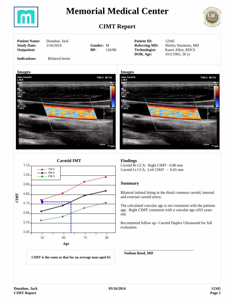
Memorial Medical Center CIMT Report Patient Name: Donahue, Jack Study Date: 3/16/2014 Outpatient Gender: M BP: 126/88 Patient ID: 12345 Referring MD: Shirley Simmons, MD Technologist: Karen Allen, RDCS DOB, Age: 10/2/1963, 50 yr Indications: Bilateral bruits Images Images Carotid IMT CIMT is the same as that for an average man aged 63 Findings Carotid Rt CCA: Right CIMT - 0.80 mm Carotid Lt CCA: Left CIMT - 0.65 mm Summary Bilateral intimal lining in the distal common carotid, internal and external carotid artery. The calculated vascular age is not consistent with the patients age. Right CIMT consistent with a vascular age of 63 years old. Recommend follow up - Carotid Duplex Ultrasound for full evaluation _____________________________________ Nathan Reed, MD Donahue, Jack 03/16/2014 12345 CIMT Report Page 1
Transcript

Memorial Medical Center

CIMT Report

Patient Name: Donahue, JackStudy Date: 3/16/2014Outpatient

Gender: MBP: 126/88

Patient ID: 12345Referring MD: Shirley Simmons, MDTechnologist: Karen Allen, RDCSDOB, Age: 10/2/1963, 50 yr

Indications: Bilateral bruits

Images Images

Carotid IMT

CIMT is the same as that for an average man aged 63

FindingsCarotid Rt CCA: Right CIMT - 0.80 mm Carotid Lt CCA: Left CIMT - 0.65 mm

Summary

Bilateral intimal lining in the distal common carotid, internal and external carotid artery.

The calculated vascular age is not consistent with the patients age. Right CIMT consistent with a vascular age of 63 years old.

Recommend follow up - Carotid Duplex Ultrasound for full evaluation

_____________________________________Nathan Reed, MD

Donahue, Jack 03/16/2014 12345CIMT Report Page 1

Memorial Medical Center

Lower Extremity Venous Ultrasound Report

Patient Name: Donahue, JackStudy Date: 3/19/2014Weight: 209 lbOutpatientICD9: 729.5, 782.3, V12.51

Gender: MPatient ID: 12345Priority: ROUTINEReferring MD: Amy Jones, MDTechnologist: Barnaby Franklin, RVTDOB, Age: 10/2/1963, 50 yrCPT4: 93882, 93981

Indications: Left leg edema, varicose veins, painHistory/Clinical: History of Left leg DVTProcedure: LE Venous - Bilateral, Right Lower Extremity RefluxPrevious Study: Date: 12/08/2013

GSV Right LeftLeg Leg

Diam Time Diam Time(msec) (msec)(cm) (cm)

SF Confluence 2 2000 5 6000Thigh Mid 2.1 2000 6 7500Thigh Dist 2.3 1000 4.5 2500Knee 2.6 1000 4.6 3000Calf Mid 2.3 1000 4.5 2600Ankle 2.1 1000 3.5 1500

SSVProx 1.5 2000 1.5 500Mid 1.8 1000 1.8 300Dist 1.6 1000 1.6 300PerforatorsCalf Dist 4.3 1000

Incompetent Perforator

Findings

Reflux

Rt Reflux: Evaluation of the deep and superficial system of the right lower extremity is performed and appears to have no evidence of venous thrombus or insufficiency. Normal phasic flow and compressibility is demonstrated. Reflux analysis indicates normal valvular function.

Lt Reflux: Venous insufficiency evalution of the superficial system demonstrates reflux disease in the proximal to distal GSV. moderate varicosity is identified within the lateral thigh, knee, and calf associated with the GSV.

Summary

Technically adequate 2D, Doppler and color-flow exam performed. significant venous insufficieny is noted in the left greater saphenous vein. Recommend endovenous laser ablation procedure.

_____________________________________Dolores Osborne, MD

Donahue, Jack 03/19/2014 12345LE Venous Report Page 1

Memorial Outpatient Clinic

Upper Extremity Venous Ultrasound Report

Patient Name: Raymond, Richard LStudy Date: 3/4/2014InpatientICD9: 453.4

Gender: MPatient ID: 10428Priority: ROUTINEReferring MD: Amy Jones, MDTechnologist: Barry Jones, RVTDOB, Age: 11/6/1927, 86 yrCPT4: 93971

Indications: DVT

CO

MP

RE

SS

IBL

E

NO

RM

AL

FL

OW

FL

OW

AU

GM

EN

TS

TH

RO

MB

US

Internal Jugular R Y Y Y NInnominate R Y Y Y NSubclavian R Y Y Y NAxillary R Y Y Y NBrachial Upper R Y Y Y NBrachial Mid R Y Y Y NBrachial Antecube R Y Y Y NRadial Antecube R Y Y Y NRadial Forearm R Y Y Y NInternal Jugular L Y Y Y NInnominate L Y Y Y NSubclavian L Y Y Y NAxillary L N N N YBrachial Upper L N N N YBrachial Mid L N N N YBrachial Antecube L Y Y Y NRadial Antecube L Y Y Y NRadial Forearm L Y Y Y N

Y = Yes i = Reduced N = No P = Partial = Erase

Thrombus

Summary

Rt Upper Ext: No evidence of acute or chronic thrombosis noted in the deep or superficial veins. The contralateral Subclavian vein was assessed and found to be patent.Lt Upper Ext: Left Axillary through mid Brachial vein not compressible and flow cannot be augmented. Consistent with Venous Thrombus in Left Upper Extremity.

Preliminary interpretation by Jason Andre, M.D..

<Electronic Signature> 11/17/2015 01:59 PM_____________________________________Tony Oh, MD Revised

Raymond, Richard L 03/04/2014 10428UE Venous Report Page 1

Memorial Outpatient Clinic

Lower Extremity Arterial Ultrasound Report

Patient Name: Donahue, JackStudy Date: 3/12/2014OutpatientICD9: 440.3

Gender: MPatient ID: 12345Priority: ROUTINEReferring MD: Shirley Simmons, MDTechnologist: Karen Allen, RDCSDOB, Age: 10/2/1963, 50 yrCPT4: 93925

Indications: Claudication -Right LegHistory/Clinical: Hypertension, Peripheral vascular diseaseProcedure: Left LE Arterial, ABIPrevious Study: Date: 01/31/2014

Doppler Right Left (cm/s) PSV EDV PSV EDVCFA Prox 101 120Profunda 125 123SFA Prox 350 109SFA Mid 25 111SFA Dist 36 90Pop Prox 40 85Pop Dist 45 76PTA Dist 32 67Peroneal Dist 25 80ATA Dist 30 67DPA 25 56

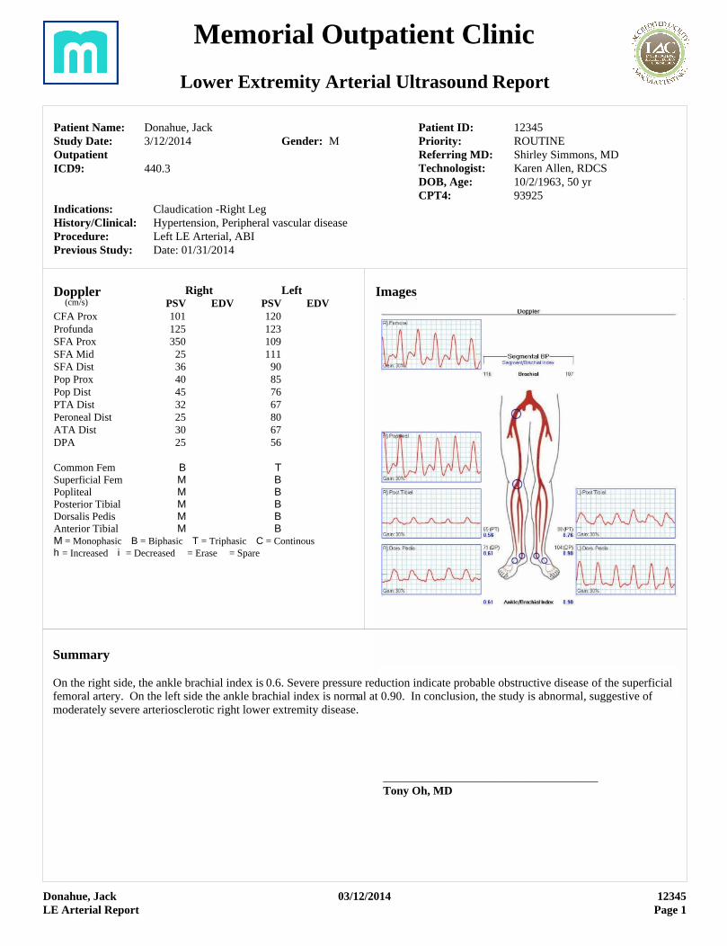
Common Fem B TSuperficial Fem M BPopliteal M BPosterior Tibial M BDorsalis Pedis M BAnterior Tibial M BM = Monophasic B = Biphasic T = Triphasic C = Continoush = Increased i = Decreased = Erase = Spare

Images

Donahue, Jack 03/12/2014 12345LE Arterial Report Page 1

Summary

On the right side, the ankle brachial index is 0.6. Severe pressure reduction indicate probable obstructive disease of the superficial femoral artery. On the left side the ankle brachial index is normal at 0.90. In conclusion, the study is abnormal, suggestive of moderately severe arteriosclerotic right lower extremity disease.

_____________________________________Tony Oh, MD


Recommended