MOL #96529 1 Novel Thiosemicarbazones Regulate the Signal Transducer and Activator of Transcription 3 (STAT3) Pathway: Inhibition of Constitutive and Interleukin 6 (IL6)- Induced Activation by Iron Depletion. Goldie Y.L. Lui, Zaklina Kovacevic, Sharleen Menezes, Danuta S. Kalinowski, Angelica M. Merlot, Sumit Sahni and Des R. Richardson Department of Pathology and Bosch Institute, School of Medical Sciences, Sydney Medical School, The University of Sydney, Sydney, NSW 2006, Australia (G.Y.L.L., Z.K., S.M., D.S.K., A.M.M., S.S., and D.R.R.). This article has not been copyedited and formatted. The final version may differ from this version. Molecular Pharmacology Fast Forward. Published on January 5, 2015 as DOI: 10.1124/mol.114.096529 at ASPET Journals on September 9, 2018 molpharm.aspetjournals.org Downloaded from
Transcript
MOL #96529
1
Novel Thiosemicarbazones Regulate the Signal Transducer and Activator of
Transcription 3 (STAT3) Pathway: Inhibition of Constitutive and Interleukin 6 (IL6)-
Induced Activation by Iron Depletion.
Goldie Y.L. Lui, Zaklina Kovacevic, Sharleen Menezes, Danuta S. Kalinowski, Angelica
M. Merlot, Sumit Sahni and Des R. Richardson
Department of Pathology and Bosch Institute, School of Medical Sciences, Sydney Medical
School, The University of Sydney, Sydney, NSW 2006, Australia (G.Y.L.L., Z.K., S.M., D.S.K.,
A.M.M., S.S., and D.R.R.).
This article has not been copyedited and formatted. The final version may differ from this version.Molecular Pharmacology Fast Forward. Published on January 5, 2015 as DOI: 10.1124/mol.114.096529
protein kinase; NAC, N-acetylcysteine; ROS, reactive oxygen species; STAT3, signal
transducer and activator of transcription factor 3; TGF-β, transforming growth factor β.
This article has not been copyedited and formatted. The final version may differ from this version.Molecular Pharmacology Fast Forward. Published on January 5, 2015 as DOI: 10.1124/mol.114.096529
Pharmacological manipulation of metal pools in tumor cells is a promising strategy for cancer
treatment. Here, we reveal the iron-binding ligands, desferrioxamine (DFO), di-2-
pyridylketone-4,4-dimethyl-3-thiosemicarbazone (Dp44mT) and di-2-pyridylketone 4-
cyclohexyl-4-methyl-3-thiosemicarbazone (DpC), inhibit constitutive and interleukin 6 (IL6)-
induced activation of signal transducer and activator of transcription 3 (STAT3) signaling,
which promotes proliferation, survival and metastasis of cancer cells. We demonstrate that
DFO, Dp44mT, and DpC significantly decrease constitutive phosphorylation of the STAT3
transcription factor at Tyr705 in the pancreatic cancer cell lines, PANC-1 and MIAPaCa-2, as
well as the prostate cancer cell line, DU145. These compounds also significantly decrease
dimerized STAT3 levels, binding of nuclear STAT3 to its target DNA, and expression of
downstream targets of STAT3, including cyclin D1, c-myc and Bcl-2. Examination of
upstream mediators of STAT3 in response to these ligands revealed Dp44mT and DpC could
significantly decrease activation of the non-receptor tyrosine kinase, Src, and activation of
cAbl in DU145 and MIAPaCa-2 cells. In contrast to the effects of Dp44mT, DpC or DFO on
inhibiting STAT3 activation, the negative control compound, di-2-pyridylketone 2-methyl-3-
thiosemicarbazone (Dp2mT), or the DFO:Fe complex, which cannot bind cellular iron, had
no effect. This demonstrates the role of iron-binding in the activity observed.
Immunohistochemical staining of PANC-1 tumor xenografts showed a marked decrease in
STAT3 in the tumors of mice treated with Dp44mT or DpC, compared to the vehicle.
Collectively, these studies demonstrate suppression of STAT3 activity by iron depletion in
vitro and in vivo, and reveal insights into regulation of the critical oncogenic STAT3
pathway.
This article has not been copyedited and formatted. The final version may differ from this version.Molecular Pharmacology Fast Forward. Published on January 5, 2015 as DOI: 10.1124/mol.114.096529
Unlike the classical iron chelator used to treat iron overload disease, desferrioxamine (DFO;
Fig. 1A), DpT thiosemicarbazones form redox active metal complexes that generate reactive
oxygen species (ROS), which play key roles in their cytotoxic activity (Lovejoy et al., 2011;
Richardson et al., 2006; Yuan et al., 2004). The mechanism of action of these ligands also
involves cellular iron depletion that is known to affect cellular targets critical for cell
proliferation, survival and metastasis, including ribonucleotide reductase (Yu et al., 2011),
cyclin D1 (Gao and Richardson, 2001; Kulp et al., 1996; Nurtjahja-Tjendraputra et al., 2007),
p21 (Darnell and Richardson, 1999; Fu and Richardson, 2007), and the metastasis suppressor,
This article has not been copyedited and formatted. The final version may differ from this version.Molecular Pharmacology Fast Forward. Published on January 5, 2015 as DOI: 10.1124/mol.114.096529
that phosphorylate STAT3 at Tyr705 (p-STAT3) (Turkson and Jove, 2000). This leads to
dimer formation, nuclear translocation, binding to STAT3-specific DNA-binding elements
and transcription of target genes (e.g., Bcl2, cyclin D1 and c-myc) that promote cell
proliferation, survival, migration, apoptosis inhibition and immune evasion (Bromberg et al.,
This article has not been copyedited and formatted. The final version may differ from this version.Molecular Pharmacology Fast Forward. Published on January 5, 2015 as DOI: 10.1124/mol.114.096529
1999; Darnell et al., 1994; Ihle, 1995; Sasse et al., 1997; Wang et al., 2004; Xie et al., 2004;
Zushi et al., 1998). Constitutive activation of STAT3 is a hallmark of many human
malignancies, including those of the prostate, pancreas and breast, as well as leukemias and
melanomas (Bowman et al., 2000; Scholz et al., 2003). Indeed, several studies have
demonstrated that STAT3 inhibition can induce apoptosis in a range of cancer-types,
including prostate and pancreatic cancer (Haura et al., 2005; Lewis et al., 2008).
Understanding the role of STAT3 signaling in tumor progression and how this affects
responsiveness to therapies is an active area of clinical investigation and is critical for the
successful development of personalized cancer treatments.
Herein, for the first time, we demonstrate that inducing cellular iron depletion using the iron
chelators DFO, Dp44mT, and DpC can inhibit STAT3 signaling in vitro in prostate and
pancreatic cancer cells, as well as in vivo using pancreatic tumor xenografts grown in nude
mice. These findings reveal key insights into the role of iron in tumor progression through
STAT3 pathway inhibition and highlight its potential application in cancer therapy through
the use of potent and novel thiosemicarbazones.
This article has not been copyedited and formatted. The final version may differ from this version.Molecular Pharmacology Fast Forward. Published on January 5, 2015 as DOI: 10.1124/mol.114.096529
acids (100 mM) and sodium pyruvate (100 mM; all supplements from Life Technologies).
Primary human pancreatic epithelial cells were purchased from the Applied Cell Biology
Research Institute (ACBRI; Kirkland, WA), grown and maintained in Cell Systems
Corporation complete medium containing 10% serum, as well as 2% CultureBoost-R
(ACBRI) to support cell growth. All cells were incubated at 37°C in a humidified atmosphere
containing 5% CO2.
Cell treatments
The thiosemicarbazones, Dp44mT, DpC, and Dp2mT, were synthesized and characterized
using standard methods (Lovejoy et al., 2012; Richardson et al., 2006), while DFO was
purchased from Novartis (Basel, Switzerland). Dp44mT, DpC and di-2-pyridylketone 2-
methyl-3-thiosemicarbazone (Dp2mT) were dissolved in DMSO at 10 mM and then diluted
in media containing 10% (v/v) fetal bovine serum (FBS; Sigma-Aldrich, St. Louis, MO,
USA) so that the final [DMSO] was ≤ 0.1% (v/v). Control studies have previously shown that
these low DMSO levels do not affect cellular proliferation or iron metabolism (Richardson et
This article has not been copyedited and formatted. The final version may differ from this version.Molecular Pharmacology Fast Forward. Published on January 5, 2015 as DOI: 10.1124/mol.114.096529
Bcl-2 (Cat.#: 2876). Antibodies against c-myc (Cat.#: sc-40) and cyclin D1 (Cat.#: sc-8396)
were from Santa Cruz Biotechnology (Santa Cruz, CA) and antibody against β-actin (Cat.#:
A1978) was purchased from Sigma-Aldrich. All primary antibodies were used at a 1:1,000
dilution, except β-actin (1:10,000). All secondary antibodies were from Sigma and used at a
1:10,000 dilution.
Membranes were probed for β-actin as a loading control and all sample data values were
normalized to the corresponding β-actin values. Densitometric analysis was performed using
Image Lab 4.0.1 software (Bio-Rad, Hercules, CA).
Native PAGE
Native PAGE was performed according to the method of Shin et al. (Shin et al., 2009).
Briefly, native cell extracts were prepared using ice-cold isotonic buffer containing 20 mM
Tris (pH 7.0), 150 mM NaCl, 6 mM MgCl2, 0.8 mM PMSF, and 20% glycerol, and
This article has not been copyedited and formatted. The final version may differ from this version.Molecular Pharmacology Fast Forward. Published on January 5, 2015 as DOI: 10.1124/mol.114.096529
This article has not been copyedited and formatted. The final version may differ from this version.Molecular Pharmacology Fast Forward. Published on January 5, 2015 as DOI: 10.1124/mol.114.096529
Cells were visualized by immunofluorescence as described previously (Chen et al., 2012).
Briefly, cells were fixed with 4% (w/v) paraformaldehyde (Sigma-Aldrich) for 10 min/20oC
then permeabilized using 0.1% (v/v) Triton X-100 for 5 min/20oC. Blocking was performed
using 5% BSA in PBS for 1 h/20oC at room temperature and then cells were incubated with
primary antibodies overnight at 4°C. Cells were then incubated with fluorescent secondary
antibody for 1 h/20oC, washed with PBS, and set onto slides using mounting solution
containing 4',6-diamidino-2-phenylindole (DAPI; Invitrogen). Imaging was performed using
an Olympus Zeiss AxioObserver Z1 fluorescence microscope (Olympus) with a 63x oil
objective. Mean fluorescence intensities were measured using Image J (Schneider et al.,
2012), with average values taken from three random images per sample.
Preparation of tumor xenografts in nude mice
The following in vivo study was approved by the Sydney University Animal Ethics
Committee. Tumor xenografts were established in 8-week-old female nude mice (BALBc
nu/nu), which were routinely fed basal rodent chow, watered ad libitum and housed under a
12 h light/dark cycle. Briefly, 2x10⁶ PANC-1 cells grown in culture were harvested and
suspended in Matrigel (BD Biosciences) and injected subcutaneously into the right flank of
each mouse. Tumor size and volume were monitored daily and treatments began when
tumors reached an average size of 90 mm3. Mice were separated into three groups (n=6), with
each group receiving either Dp44mT (0.4 mg/kg), DpC (5 mg/kg), or the vehicle Control
intravenously via tail vein injection 5 days/week. This treatment regimen was established
based on previous studies performed in our laboratory using these agents demonstrating that
this administration schedule was well tolerated and showed high anti-tumor efficacy
(Kovacevic et al., 2011; Lovejoy et al., 2012; Whitnall et al., 2006). The Dp44mT and DpC
This article has not been copyedited and formatted. The final version may differ from this version.Molecular Pharmacology Fast Forward. Published on January 5, 2015 as DOI: 10.1124/mol.114.096529
treatments were prepared by dissolving each of the compounds in 30% propylene
glycol/0.9% saline, while the vehicle Control treatment consisted of 30% propylene
glycol/0.9% saline only (Kovacevic et al., 2011; Lovejoy et al., 2012; Whitnall et al., 2006).
After 22 days of treatment, mice were euthanized and tumors were harvested, fixed in 10%
neutral buffered formalin for 24 h then processed to paraffin wax.
Immunohistochemistry
Tumor tissue sections were sectioned at 5 μm onto Superfrost Plus slides (Menzel-Gläser
Braunschweig, Germany), de-paraffinized through xylene rinses, and re-hydrated through
graded ethanols. Antigen retrieval was performed by boiling slides in a water bath for 30 min
using Dako Target Retrieval Solution (Dako, Carpinteria, CA) at pH 9.0 for p-STAT3
staining, and pH 6.0 for STAT3 staining. Endogenous peroxidase activity was blocked by 5
min incubation with 3% hydrogen peroxide, followed by a wash with 1x TBS/Tween
(0.05%). The samples were incubated with primary antibody for 30 min/20oC, followed by a
TBS/Tween wash. Bound antibody was detected using Dako Envision+ anti-rabbit HRP-
labeled polymer secondary antibody (Dako) for 30 min/20oC. Slides were rinsed in a series of
TBS/Tween washes, then incubated with DAB+ chromogen (Cat.#: K3468, Dako) for 10
min. Finally, slides were counterstained with Harris hematoxylin, dehydrated in graded
ethanols, cleared through xylene rinses, and mounted. Immunohistochemical staining was
performed using the following primary antibodies from Cell Signaling: p-STAT3 (Cat.#:
9145) and STAT3 (Cat.#: 9132).
Evaluation of immunohistochemical staining
As immunohistochemical p-STAT3 staining demonstrated nuclear localization in this study,
we quantified nuclear p-STAT3 staining in ImageJ (Schneider et al., 2012) using the
This article has not been copyedited and formatted. The final version may differ from this version.Molecular Pharmacology Fast Forward. Published on January 5, 2015 as DOI: 10.1124/mol.114.096529
ImmunoRatio plug-in (Tuominen et al., 2010), as previously described (Jahangiri et al.,
2013). The average values were taken from three random images per sample and background
correction was performed for each slide using a blank field image.
As STAT3 staining was not exclusively localized in the nucleus, the above method of
quantification was not appropriate. Instead, STAT3 staining was scored for each sample by
considering both the percentage and intensity of cells displaying positive staining. Scores for
the percentage of positive cells were assigned based on standard procedures (Gong et al.,
2005; Krajewska et al., 1996), as follows: 0-10%, 0; 11-25%, 1; 26-50%, 2; 51-75%, 3; 75-
100%, 4. Scores for staining intensity were assigned as follows: light brown, weak staining,
1; brown, moderate staining, 2; dark brown, intense staining, 3. The individual scores for
percentage and intensity were multiplied to obtain an IHC score, as previously described
(Gong et al., 2005; Krajewska et al., 1996). Two researchers scored three random fields for
each sample in a blinded manner independently, with the average IHC score being presented.
Statistical analysis
Data are expressed as mean ± SD. Statistical analyses were performed using Graphpad Prism
(GraphPad Software Inc., La Jolla, CA). Data were compared against the respective Control
in each experiment by one-way analysis of variance followed by Dunnett’s post-test. Results
were considered statistically significant when p<0.05.
This article has not been copyedited and formatted. The final version may differ from this version.Molecular Pharmacology Fast Forward. Published on January 5, 2015 as DOI: 10.1124/mol.114.096529
Previous studies have revealed that the MAPK, AKT and TGF-β pathways are molecular
targets that play a role in the anti-proliferative activity of DFO and Dp44mT (Dixon et al.,
2013; Kovacevic et al., 2013; Yu and Richardson, 2011). To further elucidate the molecular
mechanisms of thiosemicarbazones, we considered evidence suggesting crosstalk between
these and other major signaling pathways, including the STAT3 pathway (Assinder et al.,
2009; Jain et al., 1998; Zhao et al., 2008), and examined the potential role of the latter
pathway in mediating the anti-proliferative activities of the potent DpT thiosemicarbazones,
DpC and Dp44mT. In the current investigation, we utilized the pancreatic cancer cell lines,
PANC-1 and MIAPaCa2, as well as the prostate cancer cell line, DU145, as the potent anti-
tumor efficacy of Dp44mT and DpC have previously been demonstrated in these cell-types
(Dixon et al., 2013; Kovacevic et al., 2011; Sun et al., 2013; Whitnall et al., 2006).
Furthermore, these cell lines display constitutive phosphorylation of STAT3 that leads to its
activation (Lian et al., 2004; Mora et al., 2002).
Cells were incubated with DFO (250 μM), Dp44mT (5 μM), or DpC (5 μM) for 24 h/37°C
(Fig. 1A). These conditions have been previously demonstrated to efficiently induce iron
depletion and increase the expression of iron-regulated molecules (Kovacevic et al., 2011;
Richardson et al., 1994; Yuan et al., 2004). Furthermore, we have shown that Dp44mT
uptake by cells saturates at 5-10 μM (Merlot et al., 2013). At this concentration, the level of
agent is pharmacologically relevant in humans, as the structurally-related thiosemicarbazone,
Triapine, has been observed in the serum at similar levels (Chao et al., 2012; Wadler et al.,
2004). The much higher DFO concentration was utilized due to its limited membrane
permeability compared to Dp44mT and DpC, which display high permeability and marked
iron chelation efficacy (Kovacevic et al., 2011; Lovejoy et al., 2012; Sun et al., 2013; Yuan et
This article has not been copyedited and formatted. The final version may differ from this version.Molecular Pharmacology Fast Forward. Published on January 5, 2015 as DOI: 10.1124/mol.114.096529
al., 2004). As a negative control, under the same conditions, cells were also incubated with
Dp2mT (5 μM) (Fig. 1A), a structural analogue of Dp44mT and DpC that cannot bind metal
ions and displays negligible anti-tumor activity (Chen et al., 2012; Richardson et al., 2006;
Sun et al., 2013; Yuan et al., 2004).
Dp44mT and DpC decrease constitutive STAT3 phosphorylation at Tyr705.
Initial studies revealed that incubation of PANC-1, MIAPaCa2, and DU145 cells with DFO,
Dp44mT, or DpC significantly (p<0.001-0.01) decreased levels of phosphorylated (Tyr705)
STAT3 (i.e., p-STAT3; Fig. 1B-D). Under the same conditions, total STAT3 expression was
also significantly (p<0.05) decreased after incubation with Dp44mT or DpC, but not with
DFO. In contrast, the negative control compound for Dp44mT, namely Dp2mT, which cannot
bind metal ions (Chen et al., 2012; Yuan et al., 2004), did not significantly (p>0.05) alter p-
STAT3 or STAT3 levels in all cell-types examined (Fig. 1B-D). Herein, levels of STAT3
activation were determined by densitometric analysis of the western blots and calculation of
the p-STAT3/STAT3 ratio.
To understand the role of iron depletion in the decrease of the p-STAT3/STAT3 ratio induced
by these compounds, the cells were also incubated with the iron complexes of these ligands
that were prepared by pre-complexing the chelators with FeCl3 using standard methods (Yu
and Richardson, 2011). Since DFO is a hexadentate ligand (Merlot et al., 2013), the iron
complex of DFO (DFO:Fe) was prepared in a 1:1 ligand/iron molar ratio to saturate the
coordination sphere at a concentration of 250 μM. In contrast, the iron complexes of the
tridentate chelators, Dp44mT and DpC (Lovejoy et al., 2012; Merlot et al., 2013; Richardson
et al., 2006) (i.e., Dp44mT:Fe and DpC:Fe, respectively), were prepared in 2:1 ligand/iron
molar ratios to saturate the coordination sphere at a final concentration of 5 μM. As another
This article has not been copyedited and formatted. The final version may differ from this version.Molecular Pharmacology Fast Forward. Published on January 5, 2015 as DOI: 10.1124/mol.114.096529
relevant control, cells were also incubated with FeCl3 alone (250 μM), which resulted in no
significant (p>0.05) effects on p-STAT3 or STAT3 levels versus the Control (Fig. 1B-D).
For all three cell-types, incubation with the non-redox-active DFO:Fe complex (Kalinowski
and Richardson, 2005) resulted in p-STAT3/STAT3 levels that were significantly (p<0.001-
0.01) greater than those found for DFO alone, and comparable to its respective Control (Fig.
1B-D). This finding indicates that the ability of DFO to inhibit constitutively activated
STAT3 depends on its ability to bind intracellular iron. In contrast, incubation of PANC-1,
MIAPaCa-2 or DU145 cells with the Dp44mT:Fe or DpC:Fe complex significantly (p<0.001-
0.05) decreased the p-STAT3/STAT3 ratio compared to the Control. The extent of the
decrease in the p-STAT3/STAT3 ratio by the complexes was similar (PANC1) or slightly
less (MIAPaCa-2, DU-145) than that observed for Dp44mT or DpC alone. These results
suggest that in contrast to the non-redox active DFO:Fe complex, the well-characterized
ability of Dp44mT:Fe and DpC:Fe complex to generate ROS (Lovejoy et al., 2011;
Richardson et al., 2006; Yuan et al., 2004) may also play a role in the decreased p-
STAT3/STAT3 ratio.
To compare the effects of these compounds on mortal, non-transformed cells, studies also
examined the effects of DFO, Dp44mT, or DpC on primary cultures of normal pancreatic
epithelial cells (PaECs; Fig. 1E). Incubation of PaECs with DFO, Dp44mT and DpC for 24
h/37°C did not result in any significant (p>0.05) alteration in p-STAT3 or STAT3 levels
relative to the Control. In fact, p-STAT3 expression was barely detectable in these normal
cells (Fig. 1E) compared to their malignant counterparts (Fig. 1B-D). This observation is
consistent with: (1) previous reports that STAT3 is over-activated in pancreatic carcinoma
specimens, but not in normal pancreatic tissues (Lian et al., 2004); and (2) that these
This article has not been copyedited and formatted. The final version may differ from this version.Molecular Pharmacology Fast Forward. Published on January 5, 2015 as DOI: 10.1124/mol.114.096529
compounds can selectively target cancer cells over normal cells in vitro and in vivo (Dixon et
al., 2013; Kovacevic et al., 2011; Lovejoy et al., 2012; Yuan et al., 2004).
Collectively, these studies demonstrate that iron chelation decreases the pSTAT3/STAT3
ratio and that the redox activity of the Dp44mT:Fe and DpC:Fe complexes may be important
in inducing this effect.
The anti-oxidant and GSH precursor, NAC, prevents the ability of the Dp44mT- or DpC-
Fe complexes to decrease the pSTAT3/STAT3 ratio in PANC-1 and MIAPaCa-2 Cells
Considering that incubation of PANC-1, MIAPaCa-2 and DU145 cells with Dp44mT:Fe or
DpC:Fe also significantly decreased the pSTAT3/STAT3 ratio (Fig. 1B), we further
examined whether the redox activity of these complexes (Lovejoy et al., 2011; Richardson et
al., 2006; Yuan et al., 2004) was at least partly responsible for this effect (Fig. 2). This
hypothesis was investigated by utilizing the anti-oxidant, NAC, which has been well
characterized to increase intracellular GSH and quench ROS (Schafer and Buettner, 2001;
Watts and Richardson, 2001). Previous studies have demonstrated that NAC markedly
reduces the anti-proliferative activity of Dp44mT and prevents its ability to induce lysosomal
membrane permeabilisation due to its ability to act as a GSH donor (Lovejoy et al., 2011). In
this investigation, cells were initially pre-incubated for 1 h/37°C with Control medium alone
or NAC (10 mM) dissolved in medium. Then, cells were incubated with Dp44mT:Fe (1 µM
or 5 µM) or DpC:Fe (1 µM or 5 µM) in Control medium alone, or in medium containing
NAC (10 mM) for a further 24 h/37°C.
Consistent with the results in Fig. 1B, incubation of PANC-1 cells with Dp44mT:Fe or
DpC:Fe decreased the pSTAT3/STAT3 ratio, particularly at 5 µM relative to 1 µM (Fig. 2A).
This article has not been copyedited and formatted. The final version may differ from this version.Molecular Pharmacology Fast Forward. Published on January 5, 2015 as DOI: 10.1124/mol.114.096529
However, incubation of cells with Dp44mT:Fe (1 µM) or DpC:Fe (1 µM) in the presence of
NAC prevented the decrease in the pSTAT3/STAT3 ratio in PANC-1 cells (Fig. 2A). This
finding suggests the ability of Dp44mT:Fe and DpC:Fe to inhibit STAT3 activation could
depend on their ability to generate ROS (Yuan et al., 2004). When the concentration of the
Dp44mT:Fe and DpC:Fe complexes was increased to 5 µM, NAC no longer significantly
prevented their ability to decrease the pSTAT3/STAT3 ratio (Fig. 2A). This observation
suggested ROS generation by the complexes at this higher concentration was markedly
greater than that induced at 1 µM and could not be sufficiently rescued by NAC.
A similar result was observed with the MIAPaCa-2 cells (Fig. 2B), with both Dp44mT:Fe
and DpC:Fe significantly (p<0.01-0.05) decreasing the pSTAT3/STAT3 ratio relative to the
control, as shown in Fig. 1C. The presence of NAC inhibited the decrease in the
pSTAT3/STAT3 ratio for both Dp44mT:Fe (1 and 5 µM) and DpC:Fe (1 and 5 µM) (Fig.
2B). Examining DU145 cells, both Dp44mT:Fe and DpC:Fe complexes also markedly
(p<0.01) reduced the pSTAT3/STAT3 ratio at the 5 µM concentration (Fig. 2C) which was
consistent with the results in Fig. 1D. However, in contrast to the PANC-1 and MIAPaCa-2
cells, incubation of the DU145 cells with the Dp44mT:Fe or DpC:Fe complexes in the
presence of NAC did not significantly (p>0.05) rescue the effect of these agents on reducing
the pSTAT3/STAT3 ratio (Fig. 2C). Considering these results, it could be suggested that
DU145 cells have an altered redox state, which may modulate their response to NAC and the
complexes. Indeed, a previous study showed that DU145 cells have higher basal GSH levels
and a reducing redox environment when compared to other prostate cancer cells, enhancing
their tolerability to ROS (Jayakumar et al., 2014). Alternatively, the Dp44mT:Fe and DpC:Fe
complexes may have additional molecular effects in this cell line that are not accounted for
by ROS production alone.
This article has not been copyedited and formatted. The final version may differ from this version.Molecular Pharmacology Fast Forward. Published on January 5, 2015 as DOI: 10.1124/mol.114.096529
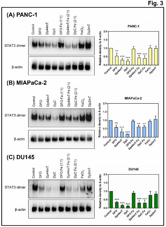
Activation of STAT3 by phosphorylation at Tyr705 induces dimerization, which allows
subsequent nuclear translocation (Bromberg et al., 1999; Ihle, 1995). Thus, native PAGE was
utilized to assess whether the decrease in p-STAT3/STAT3 levels induced by the ligands
(Fig. 1B-D) resulted in a reduction in STAT3 dimers, as this is vital for STAT3 activity
(Haura et al., 2005). Incubation of PANC-1, MIAPaCa-2 and DU145 cells with DFO,
Dp44mT, or DpC for 24 h significantly (p<0.001-0.01) decreased dimerized STAT3 levels
compared to the untreated Controls (Fig. 3). Notably, Dp44mT, and particularly DpC, were
significantly (p<0.05) more effective than DFO at decreasing STAT3 dimer formation.
Further, incubation of these cells with Dp44mT:Fe or DpC:Fe complexes also resulted in
significantly (p<0.001-0.01) decreased levels of dimerized STAT3, whereas incubation with
either DFO:Fe, FeCl3, or Dp2mT, did not significantly (p>0.05) alter dimerized STAT3
levels compared to the Control (Fig. 3). These findings agree with the decrease in the p-
STAT3/STAT3 ratio observed in Figure 1, indicating that the ability of DFO to inhibit
STAT3 dimer formation depends solely on its iron-binding activity. In contrast, the ability of
Dp44mT and DpC to inhibit STAT3 dimer formation could also be dependent on ROS
generation in addition to their known ability to chelate intracellular iron (Kovacevic et al.,
2011; Lovejoy et al., 2012; Whitnall et al., 2006; Yuan et al., 2004). It is notable that the
effect observed after treating cells with the ligands could not be reproduced by adding the
agents directly to Control cell lysates, indicating a biological response that required cellular
and metabolic integrity.
DFO, Dp44mT and DpC inhibit STAT3 DNA-binding activity
To examine whether the chelator-induced decrease in p-STAT3/STAT3 and STAT3 dimer
levels resulted in decreased STAT3 DNA-binding activity, we performed EMSA using a
This article has not been copyedited and formatted. The final version may differ from this version.Molecular Pharmacology Fast Forward. Published on January 5, 2015 as DOI: 10.1124/mol.114.096529
biotin-labeled, double-stranded DNA probe containing the STAT3 consensus-binding motif
(Fig. 4). In these studies, incubation of nuclear extracts with this probe resulted in a
pronounced band (see arrow; Fig. 4A, lane 2), that was not present with the probe alone (Fig.
4A, lane 1).
To assess if the STAT3 DNA-binding activity observed in Figure 4A (lane 2) was a specific
interaction, a series of controls was utilized. First, addition of a 50-fold molar excess of
unlabeled specific competitor oligonucleotide (i.e., Spec. comp.; Fig. 4A) markedly and
significantly (p<0.001) reduced the intensity of this band (Fig. 4A, lane 3), while addition of
a 50-fold molar excess of a non-specific competitor oligonucleotide (i.e., Non-spec. comp.;
Fig. 4A) did not significantly (p>0.05) alter the intensity of the STAT3 band compared to the
Control in lane 2 (Fig. 4A, lane 4). Second, incubation with a biotin-labeled probe containing
a mutated STAT3-binding motif did not result in the appearance of any clear bands (Fig. 4A,
lane 5). Third, addition of an antibody against STAT3 (i.e., Spec. Ab; Fig. 4A) to the binding
reaction resulted in a significant (p<0.001) reduction of the STAT3 band, but also the
apparent presence of a smear that ran higher in the gel (see asterisk), suggesting an antibody-
induced supershift of STAT3 (Fig. 4A, lane 6). Fourth, as a further control, incubation of an
isotype control antibody (i.e., Control Ab; Fig. 4A) to the binding reaction under the same
conditions (Fig. 4A, lane 7), did not significantly (p>0.05) alter the appearance of the band
compared to the Control in lane 2 (Fig. 4A). Collectively, these controls demonstrate the
identified STAT3 band was representative of STAT3 DNA-binding activity.
To assess the effect of the ligands on STAT3 DNA-binding activity, PANC-1 cells were
incubated with DFO (250 μM), Dp44mT (5 μM), or DpC (5 μM) for 24 h/37oC, nuclear
lysates prepared and EMSA performed (Fig. 4B). Incubation with these ligands significantly
This article has not been copyedited and formatted. The final version may differ from this version.Molecular Pharmacology Fast Forward. Published on January 5, 2015 as DOI: 10.1124/mol.114.096529
(p<0.001) reduced the intensity of the STAT3 band compared to Control cells (Fig. 4B; cf.
lanes 3-5 to lane 2). Moreover, this effect was also observed in MIAPaCa-2 (Fig. 4C) and
DU145 (Fig. 4D) cells, indicating that DFO, Dp44mT, and DpC could significantly
(p<0.001-0.01) reduce binding of STAT3 to its target DNA in each cell line examined. Thus,
these data agree with the effects of the ligands on STAT3 activation (pSTAT3/STAT3 ratio;
Fig. 1), and STAT3 dimerization (Fig. 3) and demonstrate that they inhibit STAT3 DNA-
binding activity, which may compromise its down-stream oncogenic effects.
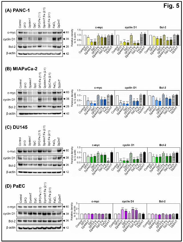
DFO, Dp44mT and DpC down-regulate the expression of the downstream STAT3 targets,
c-myc, cyclin D1 and Bcl-2.
Considering the results indicating these ligands can decrease STAT3 activation, dimerization
and DNA-binding (Figs. 1-4), we examined whether this subsequently affected the
expression of multiple downstream STAT3 targets that regulate cell cycle progression and
apoptosis, namely c-myc, cyclin D1 and Bcl-2 (Bromberg et al., 1999; Darnell, 1997; Sasse
et al., 1997; Zhong et al., 1999; Zushi et al., 1998). In these studies, incubation of cells with
DFO (250 μM), Dp44mT (5 μM), or DpC (5 μM) for 24 h/37oC significantly (p<0.001-0.05)
reduced protein levels of c-myc, and cyclin D1 in PANC-1 (Fig. 5A), MIAPaCa-2 (Fig. 5B)
and DU145 cells (Fig. 5C). Notably, Bcl-2 was also significantly (p<0.01) reduced by
Dp44mT and DpC in each of the three cell-types, while DFO only significantly (p<0.01)
reduced Bcl-2 levels in DU145 cells (Fig. 5C). These findings are in agreement with previous
reports that cellular iron depletion using chelators can regulate cyclin D1 and Bcl-2 levels,
which could mediate the potent anti-proliferative activity of these compounds (Dixon et al.,
2013; Gao and Richardson, 2001; Kovacevic et al., 2011; Yuan et al., 2004). The iron
complex, DFO:Fe, did not significantly alter the expression of these downstream targets
compared to the Control, indicating that the ability of DFO to sequester cellular iron is
This article has not been copyedited and formatted. The final version may differ from this version.Molecular Pharmacology Fast Forward. Published on January 5, 2015 as DOI: 10.1124/mol.114.096529
important for its ability to inhibit STAT3 signaling and its downstream effectors. In contrast,
incubation of cells with the Dp44mT:Fe and DpC:Fe complexes significantly (p<0.001-0.05)
reduced expression of the downstream targets, c-myc, cyclin D1 and Bcl-2 in all three cell-
types (Fig. 5A-C). Again, this suggests that the redox activity of Dp44mT:Fe and DpC:Fe
complexes could play a role in Dp44mT- and DpC-mediated suppression of downstream
STAT3 signaling.
Utilization of FeCl3 as a control for the Dp44mT:Fe and DpC:Fe complexes indicated that it
did not significantly (p>0.05) affect c-myc expression in PANC-1 or MIAPaCa-2 cells (Fig.
5A-B), nor did it significantly (p<0.05) affect cyclin D1 or Bcl-2 expression in all cell-types
examined. Interestingly, for DU145 cells only, FeCl3 was found to significantly (p<0.001)
decrease c-myc expression (Fig. 5C). This observation may reflect cell-type heterogeneity
that results in enhanced sensitivity of c-myc to high intracellular iron levels in DU145 cells.
For all cell-types, incubation with the negative control compound, Dp2mT, which cannot
bind metals (Chen et al., 2012; Yuan et al., 2004) resulted in no significant (p>0.05)
alteration in the levels of c-myc, cyclin D1 or Bcl-2, demonstrating the key role of iron
chelation by Dp44mT and DpC in decreasing the expression of these proteins (Fig. 5).
To compare the activity of these agents in mortal, non-transformed cells, we also examined
the effects of DFO, Dp44mT and DpC on these downstream target molecules in primary
PaEC cultures (Fig. 5D). In agreement with the results indicating that iron chelation did not
significantly alter STAT3 activation in these cells (Fig. 1E), c-myc and Bcl-2 expression
levels were not significantly (p>0.05) altered compared to the Control in response to any of
the ligands examined (Fig. 5D). Interestingly, cyclin D1 expression was significantly
(p<0.001) increased in response to Dp44mT, DpC, Dp44mT:Fe, and DpC:Fe in PaECs (Fig.
This article has not been copyedited and formatted. The final version may differ from this version.Molecular Pharmacology Fast Forward. Published on January 5, 2015 as DOI: 10.1124/mol.114.096529
5D), which may possibly reflect a pro-survival response of these normal cells to the stress
conditions induced by these compounds.
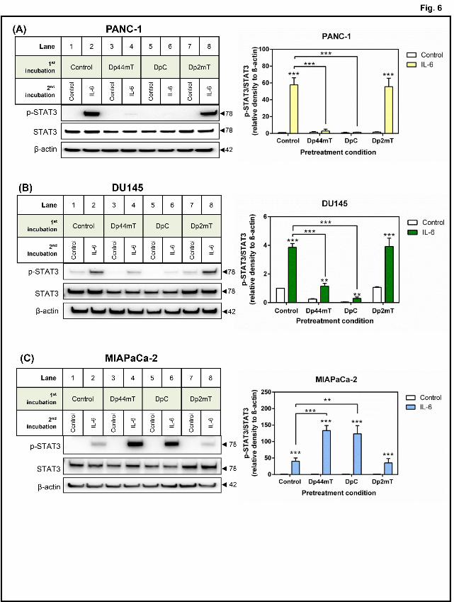
Pre-treatment of PANC-1 and DU145 cells with Dp44mT or DpC inhibits IL-6-induced
activation of STAT3.
IL-6 is implicated in the hyper-activation of STAT3 signaling in tumor progression by
stimulating this pathway through a receptor-mediated mechanism (Turkson and Jove, 2000).
To investigate whether cellular iron depletion could also inhibit IL-6-induced activation of
STAT3, we pre-incubated PANC-1 and DU145 cells with Dp44mT, DpC, or Dp2mT in
serum-free media or serum-free media alone for 24 h/37oC (to remove cytokines/growth
factors (Hahm and Singh, 2010)), then stimulated the cells with IL-6 (10 ng/mL) in serum-
free media or serum-free media alone for 30 min/37oC (Fig. 6).
Pre-incubation with Control media alone followed by incubation with IL-6 for 30 min
markedly and significantly (p<0.001) increased the p-STAT3/STAT3 ratio in PANC-1 (Fig.
6A; lane 2) and DU145 (Fig. 6B; lane 2) cells compared to the same cells incubated with
media alone (Fig. 6A, B; lane 1). Notably, the low pSTAT3 levels in Fig. 6 relative to Fig. 1,
are consistent with serum-starvation (Hahm and Singh, 2010). Pre-incubation of cells with
Dp44mT or DpC significantly (p<0.001) inhibited IL-6-induced activation of STAT3 in
PANC-1 and DU145 cells (Fig. 6A, B; lanes 4 and 6). In contrast to Dp44mT and DpC,
Dp2mT which does not bind metal ions (Chen et al., 2012; Richardson et al., 2006; Sun et al.,
2013; Yuan et al., 2004), had no significant (p>0.05) effect on STAT3 activation induced by
IL-6 relative to the Control (Fig. 6A, B; lane 8).
In contrast to the PANC1 and DU145 cell-types, MIAPaCa-2 cells showed a different
response to the ligands and incubation with IL-6 (Fig. 6C). In fact, IL-6 stimulation of cells
This article has not been copyedited and formatted. The final version may differ from this version.Molecular Pharmacology Fast Forward. Published on January 5, 2015 as DOI: 10.1124/mol.114.096529
pre-incubated with Dp44mT or DpC resulted in a significant (p<0.001-0.01) increase in p-
STAT3/STAT3 levels compared to cells pre-incubated with Control media alone (Fig. 6C; cf.
lane 2 to lanes 4 and 6). This observation was unexpected given the previous results
demonstrating that Dp44mT and DpC could inhibit constitutive STAT3 activation and its
downstream targets in this particular cell line (Fig. 1C, 2B, 3B, 4C, 5B) and may reflect
tumor cell heterogeneity. Alternatively, this result may reflect a compensatory mechanism in
the MIAPaCa-2 cells in response to Dp44mT or DpC that leads to a more marked IL-6
response, which is then subsequently inhibited by these agents downstream, as demonstrated
in Figs. 1C, 2B, 3B, 4C, and 5B.
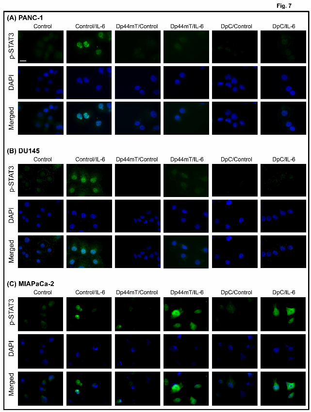
The positive findings above in PANC-1 and DU145 cells using IL-6 (Fig. 6A, B) were
further confirmed by immunofluorescence staining which demonstrated a marked induction
of p-STAT3 expression (green fluorescence) upon incubation of Control cells with IL-6 that
was predominantly nuclear, as demonstrated by the merged stain showing overlap with DAPI
staining (blue fluorescence; Fig. 7A, B). Compared to their respective Control cells, the mean
p-STAT3 staining intensities in cells treated with IL-6 were significantly (p<0.01) increased
by 3.9-fold in PANC-1 cells and 3.5-fold in DU145 cells. However, stimulation with IL-6 of
PANC-1 or DU145 cells pre-incubated with Dp44mT or DpC did not result in marked p-
STAT3 staining, with mean p-STAT3 staining intensities that were not significantly different
to Control cells (Fig. 7A, B).
Using MIAPaCa-2 cells, the mean p-STAT3 staining was also significantly (p<0.05)
increased 2.1-fold upon treatment with IL-6 when compared to their respective Control cells
(Fig. 7C). However, as demonstrated in Fig. 6C, pre-incubation of these cells with either
Dp44mT or DpC resulted in a significantly (p<0.01-0.05) greater 2.4-fold and 2.5-fold
This article has not been copyedited and formatted. The final version may differ from this version.Molecular Pharmacology Fast Forward. Published on January 5, 2015 as DOI: 10.1124/mol.114.096529
increase in p-STAT3 levels in response to IL-6 treatment, respectively, when compared to the
IL-6 treated Control cells (Fig. 7C). Together, the results above from immunoblotting and
fluorescence microscopy demonstrate that pre-treatment of PANC-1 and DU145 cells with
Dp44mT or DpC inhibits IL-6-induced STAT3 activation, while this process is stimulated by
these ligands in MIAPaCa-2 cells.
DFO, Dp44mT and DpC decrease activation of kinases upstream of STAT3, namely Src
and cAbl
It is possible that the observed decrease in STAT3 activation induced by DFO, Dp44mT and
DpC may be mediated by their effect on upstream kinases known to promote STAT3
signaling (Yu and Richardson, 2011). To investigate this hypothesis, the effect of these
compounds was examined on key protein tyrosine kinases that phosphorylate and activate
STAT3, namely the non-receptor tyrosine kinases, Src and cAbl, and the receptor-associated
Janus tyrosine kinase (JAK2) (Haura et al., 2005; Hedvat et al., 2009; Srinivasan et al., 2008;
Turkson et al., 1998). Specifically, activation of Src and cAbl were examined through
assessing levels of an activating phosphorylation of Src (p-SrcY416), an inactivating
phosphorylation of Src (p-SrcY527) (Hunter, 1987), and an activating phosphorylation of cAbl
(p-cAblY245) (Brasher and Van Etten, 2000). Activation of JAK2 was also assessed by
investigating the levels of phospho-JAK2Y1007/8 (p-JAK2), an activating phosphorylation that
occurs in response to receptor-mediated cytokine or growth factor stimulation (Feng et al.,
1997; Hedvat et al., 2009). It is important to note that JAK2, rather than JAK1, was
investigated as numerous studies have identified that the JAK2/STAT3 cascade is specifically
implicated in pancreatic (Corcoran et al., 2011; Thoennissen et al., 2009) and other cancers
(Colomiere et al., 2009; Marotta et al., 2011; Xiong et al., 2008; Zhang et al., 2009) to
promote tumour progression.
This article has not been copyedited and formatted. The final version may differ from this version.Molecular Pharmacology Fast Forward. Published on January 5, 2015 as DOI: 10.1124/mol.114.096529
Incubation with Dp44mT, DpC, Dp44mT:Fe, or DpC:Fe significantly (p<0.001-0.01)
decreased p-SrcY416/Src ratio relative to the Control, with no significant (p>0.05) alteration
being observed with total Src levels in PANC-1 (Fig. 8A), MIAPaCa-2 (Fig. 8B), and DU145
(Fig. 8C) cells. In contrast, incubation of these cells with DFO, DFO:Fe, FeCl3, or Dp2mT
did not significantly (p>0.05) alter levels of p-SrcY416 or total Src compared to the untreated
Control. Further, none of the treatments were found to significantly (p>0.05) alter p-SrcY527
levels, indicating these compounds did not affect Src activity by regulating the
phosphorylation of this site. Examination of cAbl activity revealed that incubation with
Dp44mT, DpC, Dp44mT:Fe, and DpC:Fe could significantly (p<0.001-0.05) reduce the p-
cAbl/cAbl ratio with no significant (p>0.05) alterations to total cAbl levels in MIAPaCa-2
(Fig. 8B) and DU145 (Fig. 8C) cells, but not in PANC-1 cells (Fig. 6A). Interestingly,
incubation of DU145 cells with FeCl3 alone resulted in significantly (p<0.05) decreased p-
cAbl and cAbl levels (Fig. 8C). This result appeared analogous to that seen with c-myc (Fig.
5C), and again, may reflect an enhanced sensitivity of these molecules to high cellular iron
levels in DU145 cells (Li et al., 2011).
In all three cell-types, incubation with any of the chelators, their corresponding iron
complexes, FeCl3, or the negative control compound, Dp2mT, did not significantly (p>0.05)
alter p-JAK2 or JAK2 levels (Fig. 8). This indicates that the ability of iron chelators to inhibit
STAT3 activation in these cells likely does not occur through inhibition of constitutive JAK2
activation.
Collectively, these findings indicate the ability of Dp44mT, DpC, Dp44mT:Fe, and DpC:Fe
to inhibit STAT3 activation could potentially be mediated by the upstream inhibition of Src
This article has not been copyedited and formatted. The final version may differ from this version.Molecular Pharmacology Fast Forward. Published on January 5, 2015 as DOI: 10.1124/mol.114.096529
This article has not been copyedited and formatted. The final version may differ from this version.Molecular Pharmacology Fast Forward. Published on January 5, 2015 as DOI: 10.1124/mol.114.096529
Immunohistochemical staining of total STAT3 in Control-treated tumor samples was
noticeably marked and intense in both the cytoplasm and nuclei, with an average IHC score
of 10.89 ± 1.77 (n = 18). However, STAT3 staining was found to be significantly (p<0.001-
0.01) lower in tumor samples from mice treated with Dp44mT or DpC, with IHC scores from
these groups of 6.38 ± 2.70 (n = 18) and 5.50 ± 2.06 (n = 18), respectively (Fig. 9B).
Collectively, these results demonstrate that Dp44mT and DpC can also inhibit STAT3
signaling in vivo, which corroborates well with our findings in vitro.
This article has not been copyedited and formatted. The final version may differ from this version.Molecular Pharmacology Fast Forward. Published on January 5, 2015 as DOI: 10.1124/mol.114.096529
In this study, we have further elucidated the anti-tumor mechanism of the novel di-2-pyridyl
thiosemicarbazones, Dp44mT and DpC, which are emerging as a promising class of anti-
cancer agents that demonstrate marked and selective anti-tumor and anti-metastatic efficacy
in vitro and in vivo in a range of cancer cell-types (Chen et al., 2012; Kovacevic et al., 2011;
Liu et al., 2012; Lovejoy et al., 2012; Sun et al., 2013; Whitnall et al., 2006). Here, for the
first time, we have identified that these agents can inhibit the STAT3 pathway, which could
be a crucial mechanism by which they exhibit their potent anti-tumor activity (Fig. 10A).
We have demonstrated that DFO, Dp44mT and DpC, inhibit the STAT3 pathway by
markedly decreasing constitutive phosphorylation and STAT3 activation in three cancer cell
lines, i.e., PANC-1, MIAPaCa-2 and DU145 (Fig. 1B-C), but not in primary cultures of
mortal PaECs (Fig. 1D). We also showed these compounds inhibit STAT3 dimerization (Fig.
3), binding of nuclear STAT3 to its target DNA (Fig. 4B-D), and subsequently attenuate
expression of the downstream STAT3 targets, c-myc, cyclin D1 and Bcl-2 (Fig. 5A-C). These
effects were dependent on the ability of the ligands to bind iron, because incubation of cells
with the inert DFO:Fe complex which is not redox active (Kalinowski and Richardson,
2005), or the control compound, Dp2mT, which does not bind metal ions (Chen et al., 2012;
Yuan et al., 2004), did not lead to inhibition.
Further studies herein demonstrate the redox-active Dp44mT:Fe and DpC:Fe complexes
inhibit STAT3 phosphorylation, dimerization and expression of downstream STAT3 targets.
These observations suggest the ability of these complexes to form redox-active species within
cells also plays a role in their STAT3-inhibitory properties. In fact, the decreased STAT3
activation induced by Dp44mT:Fe and DpC:Fe could be prevented by the anti-oxidant NAC
This article has not been copyedited and formatted. The final version may differ from this version.Molecular Pharmacology Fast Forward. Published on January 5, 2015 as DOI: 10.1124/mol.114.096529
interference, chemopreventive agents and G-quartet oligodeoxynucleotides (Jing and
Tweardy, 2005; Sansone and Bromberg, 2012; Siveen et al., 2014; Wang et al., 2012).
Recently, resistance to standard anti-cancer therapies has been correlated to constitutive or
unabated activation of STAT3, indicating that combination therapy with STAT3 inhibitors
may be important for patient treatment (Tan et al., 2014). Hence, this finding underlines the
importance of developing new STAT3 inhibitors, such as Dp44mT and DpC, that demonstrate
potent and selective anti-tumor activity.
To investigate whether the decreased STAT3 activation observed with DFO, Dp44mT and
DpC was due to the reduced activity of well-known upstream activators of this protein,
studies examined the effect of these agents on JAK2, Src and cAbl activation. These agents
did not significantly alter constitutive phosphorylation of JAK2 in all three cell-types (Fig. 8).
This article has not been copyedited and formatted. The final version may differ from this version.Molecular Pharmacology Fast Forward. Published on January 5, 2015 as DOI: 10.1124/mol.114.096529
However, Dp44mT and DpC were found to decrease activation of the non-receptor kinase,
Src, in all cell lines, as well as activation of cAbl in MIAPaCa-2 and DU145 cells (Fig. 8).
This observation suggests the ability of Dp44mT, DpC, Dp44mT:Fe and DpC:Fe to inhibit
STAT3 activation in these cells could potentially be mediated by the upstream inhibition of
Src activation. However, considering that cAbl activation was not significantly altered in
response to Dp44mT or DpC in PANC-1 cells, as well as that DFO did not significantly alter
Src and cAbl activation, suggests these latter proteins may not be the only contributors to
inhibiting the STAT3 pathway.
In addition to inhibiting constitutive activation of STAT3 in tumor cells, Dp44mT and DpC
could inhibit IL-6-induced activation of STAT3 in PANC-1 and DU145 cells (Figs. 6 and 7).
This finding is critical given that several studies have indicated that IL-6-mediated activation
of STAT3 is a principal pathway implicated in promoting tumorigenesis (Sansone and
Bromberg, 2012). Interestingly, this effect was not observed with MIAPaCa-2 cells, where
Dp44mT and DpC enhanced subsequent IL-6-induced STAT3 phosphorylation compared to
the Control (Figs. 6C and 7C). This latter observation was unexpected given that these
compounds inhibit constitutive STAT3 activation in MIAPaCa-2 cells, and may reflect cell-
type genetic heterogeneity resulting in a differential response to the combined stimulus of
Dp44mT or DpC with IL-6. Importantly, this effect is not pro-oncogenic, because these cells
are sensitive to the anti-tumor activity of these compounds in vitro and in vivo (Kovacevic et
al., 2011). In fact, Dp44mT and DpC markedly inhibit STAT3 activation (Fig. 1C, 2B, 3B,
4C), as well as expression of downstream tumorigenic effectors, c-myc, cyclin D1 and Bcl-2
(Fig. 5B) in MIAPaCa-2 cells. Moreover, Dp44mT and DpC increase expression of the
cyclin-dependent kinase inhibitor, p21, the apoptotic markers, cleaved PARP and Bax, and
This article has not been copyedited and formatted. The final version may differ from this version.Molecular Pharmacology Fast Forward. Published on January 5, 2015 as DOI: 10.1124/mol.114.096529
the growth and metastasis suppressor, NDRG1, which mediate the pronounced inhibition of
proliferation by these compounds in MIAPaCa-2 cells (Kovacevic et al., 2011).
In light of a recent study demonstrating the potential of Dp44mT and DpC to be effective for
pancreatic cancer treatment in vivo (Kovacevic et al., 2011), we utilized tumors from mice
treated with Dp44mT, DpC, or the vehicle alone and performed immunohistochemical
staining to examine p-STAT3 and STAT3 levels. Both p-STAT3 and STAT3 staining were
significantly lower in tumors from mice treated with Dp44mT and DpC compared to the
vehicle (Fig. 9). These studies suggest Dp44mT and DpC prevent tumor growth, in part, by
inhibition of STAT3.
Apart from the effect of these agents on the STAT3 pathway, it has been demonstrated that
these compounds have multiple molecular targets in tumor cells, which leads to their marked
anti-proliferative efficacy (Fig. 10B). These include: (1) enhancement of iron mobilization
from cells (Lovejoy et al., 2011; Richardson et al., 2006; Yuan et al., 2004); (2) inhibition of
iron uptake from transferrin (both (1) and (2)), leading to cellular iron depletion which is
essential for growth; (Lovejoy et al., 2011; Richardson et al., 2006; Yuan et al., 2004); (3) up-
regulation of the potent growth and metastasis suppressor, NDRG1 (Kovacevic et al., 2011;
Kovacevic et al., 2013; Le and Richardson, 2004), leading to inhibition of the epithelial
mesenchymal transition (Chen et al., 2012); (4) lysosomal membrane permeabilization
mediated by the generation of redox active copper complexes (Lovejoy et al., 2011;
Richardson et al., 2006; Yuan et al., 2004), resulting in impaired autophagy (Gutierrez et al.,
2014) and the induction of apoptosis (Gutierrez et al., 2014; Sahni et al., 2014); (5) inhibition
of multiple oncogenic cellular signaling pathways e.g., phosphoinositide 3-kinase/protein
kinase B (PI3K/AKT) and transforming growth factor-β (TGF-β) pathways and up-regulation
This article has not been copyedited and formatted. The final version may differ from this version.Molecular Pharmacology Fast Forward. Published on January 5, 2015 as DOI: 10.1124/mol.114.096529
of tumor-suppressive phosphatase and tensin homologue deleted on chromosome 10 (PTEN)
and SMAD4 (Dixon et al., 2013; Kovacevic et al., 2013); (6) inhibition of cyclin D1
expression (Gao and Richardson, 2001; Kulp et al., 1996; Nurtjahja-Tjendraputra et al.,
2007); and (7) inhibition of the rate-limiting step of DNA synthesis catalyzed by the iron-
containing enzyme, ribonucleotide reductase (Yu et al., 2011). The relative contribution of
each of these targets to the inhibition of tumor cell growth is yet to be deciphered, but may
depend on the type and stage of the neoplasm. In fact, the multiple molecular targets of these
agents probably explains their marked effect on a broad range of aggressive tumor types
(Kovacevic et al., 2011; Lovejoy et al., 2012; Whitnall et al., 2006) and their ability to
overcome drug resistance (Kovacevic et al., 2011; Lovejoy et al., 2012; Whitnall et al.,
2006).
Considering other molecular targets of thiosemicarbazones, it is notable that the loss of
STAT1 expression, which is closely related to STAT3, corresponds to advanced stage
pancreatic cancer with lymph node metastasis and poor prognosis (Sun et al., 2014).
Conversely, STAT1 signaling has also been described as a tumor promoter (Kovacic et al.,
2006), and thus, the importance of STAT1 in pancreatic and prostate tumor progression
remains to be more extensively characterized. However, it may be of interest to examine the
potential role of STAT1 in mediating the activities of thiosemicarbazones in future studies.
This investigation is the first to examine in detail the regulation of the STAT3 pathway by
cellular iron in pancreatic and prostate cancer (Fig. 10A). Importantly, our findings provide
further rationale for the use of novel thiosemicarbazones in chemotherapy. Moreover, since
STAT3 inhibition increases response to conventional therapies and delays pancreatic cancer
This article has not been copyedited and formatted. The final version may differ from this version.Molecular Pharmacology Fast Forward. Published on January 5, 2015 as DOI: 10.1124/mol.114.096529
progression (Venkatasubbarao et al., 2013), this suggests Dp44mT and DpC could be an
effective chemotherapeutic strategy in combination with existing anti-cancer agents.
This article has not been copyedited and formatted. The final version may differ from this version.Molecular Pharmacology Fast Forward. Published on January 5, 2015 as DOI: 10.1124/mol.114.096529
The authors wish to kindly thank Sanaz Maleki for her valuable assistance with
immunohistochemical staining.
AUTHORSHIP CONTRIBUTIONS
Participated in research design: Lui, Kovacevic and Richardson.
Conducted experiments: Lui, Kovacevic and Menezes.
Contributed new reagents or analytic tools: Richardson.
Performed data analysis: Lui, Kovacevic and Richardson.
Wrote or contributed to the writing of the manuscript: Lui, Kovacevic, Menezes, Kalinowski,
Merlot, Sahni and Richardson.
This article has not been copyedited and formatted. The final version may differ from this version.Molecular Pharmacology Fast Forward. Published on January 5, 2015 as DOI: 10.1124/mol.114.096529
Colomiere M, Ward AC, Riley C, Trenerry MK, Cameron-Smith D, Findlay J, Ackland L and Ahmed
N (2009) Cross talk of signals between EGFR and IL-6R through JAK2/STAT3 mediate
epithelial-mesenchymal transition in ovarian carcinomas. Br J Cancer 100: 134-144.
This article has not been copyedited and formatted. The final version may differ from this version.Molecular Pharmacology Fast Forward. Published on January 5, 2015 as DOI: 10.1124/mol.114.096529
Goldstein LJ, Galski H, Fojo A, Willingham M, Lai SL, Gazdar A, Pirker R, Green A, Crist W,
Brodeur GM and et al. (1989) Expression of a multidrug resistance gene in human cancers. J
Natl Cancer Inst 81: 116-124.
Gong W, Wang L, Yao JC, Ajani JA, Wei D, Aldape KD, Xie K, Sawaya R and Huang S (2005)
Expression of activated signal transducer and activator of transcription 3 predicts expression
This article has not been copyedited and formatted. The final version may differ from this version.Molecular Pharmacology Fast Forward. Published on January 5, 2015 as DOI: 10.1124/mol.114.096529
Jahangiri A, De Lay M, Miller LM, Carbonell WS, Hu YL, Lu K, Tom MW, Paquette J, Tokuyasu
TA, Tsao S, Marshall R, Perry A, Bjorgan KM, Chaumeil MM, Ronen SM, Bergers G and
Aghi MK (2013) Gene expression profile identifies tyrosine kinase c-Met as a targetable
mediator of antiangiogenic therapy resistance. Clin Cancer Res 19: 1773-1783.
Jain N, Zhang T, Fong SL, Lim CP and Cao X (1998) Repression of Stat3 activity by activation of
mitogen-activated protein kinase (MAPK). Oncogene 17: 3157-3167.
Jayakumar S, Kunwar A, Sandur SK, Pandey BN and Chaubey RC (2014) Differential response of
DU145 and PC3 prostate cancer cells to ionizing radiation: role of reactive oxygen species,
GSH and Nrf2 in radiosensitivity. Biochim Biophys Acta 1840: 485-494.
Jing N and Tweardy DJ (2005) Targeting Stat3 in cancer therapy. Anticancer Drugs 16: 601-607.
This article has not been copyedited and formatted. The final version may differ from this version.Molecular Pharmacology Fast Forward. Published on January 5, 2015 as DOI: 10.1124/mol.114.096529
Kalinowski DS and Richardson DR (2005) The evolution of iron chelators for the treatment of iron
overload disease and cancer. Pharmacol Rev 57: 547-583.
Kamran MZ, Patil P and Gude RP (2013) Role of STAT3 in cancer metastasis and translational
advances. Biomed Res Int 2013: 421821.
Kovacevic Z, Chikhani S, Lovejoy DB and Richardson DR (2011) Novel thiosemicarbazone iron
chelators induce up-regulation and phosphorylation of the metastasis suppressor N-myc
down-stream regulated gene 1: a new strategy for the treatment of pancreatic cancer. Mol
Pharmacol 80: 598-609.
Kovacevic Z, Chikhani S, Lui GY, Sivagurunathan S and Richardson DR (2013) The iron-regulated
metastasis suppressor NDRG1 targets NEDD4L, PTEN, and SMAD4 and inhibits the PI3K
and Ras signaling pathways. Antioxid Redox Signal 18: 874-887.
Kovacic B, Stoiber D, Moriggl R, Weisz E, Ott RG, Kreibich R, Levy DE, Beug H, Freissmuth M and
Sexl V (2006) STAT1 acts as a tumor promoter for leukemia development. Cancer Cell 10:
77-87.
Krajewska M, Krajewski S, Epstein JI, Shabaik A, Sauvageot J, Song K, Kitada S and Reed JC (1996)
Immunohistochemical analysis of bcl-2, bax, bcl-X, and mcl-1 expression in prostate cancers.
Am J Pathol 148: 1567-1576.
Kulp KS, Green SL and Vulliet PR (1996) Iron deprivation inhibits cyclin-dependent kinase activity
and decreases cyclin D/CDK4 protein levels in asynchronous MDA-MB-453 human breast
cancer cells. Exp Cell Res 229: 60-68.
Le NT and Richardson DR (2004) Iron chelators with high antiproliferative activity up-regulate the
expression of a growth inhibitory and metastasis suppressor gene: a link between iron
metabolism and proliferation. Blood 104: 2967-2975.
Lee JT, Lehmann BD, Terrian DM, Chappell WH, Stivala F, Libra M, Martelli AM, Steelman LS and
McCubrey JA (2008) Targeting prostate cancer based on signal transduction and cell cycle
pathways. Cell Cycle 7: 1745-1762.
This article has not been copyedited and formatted. The final version may differ from this version.Molecular Pharmacology Fast Forward. Published on January 5, 2015 as DOI: 10.1124/mol.114.096529
Jansson PJ, Kalinowski DS, Bernhardt PV and Richardson DR (2012) Novel second-
generation di-2-pyridylketone thiosemicarbazones show synergism with standard
chemotherapeutics and demonstrate potent activity against lung cancer xenografts after oral
and intravenous administration in vivo. J Med Chem 55: 7230-7244.
Mamoon NM, Smith JK, Chatti K, Lee S, Kundrapu K and Duhe RJ (2007) Multiple cysteine residues
are implicated in Janus kinase 2-mediated catalysis. Biochemistry 46: 14810-14818.
Marotta LL, Almendro V, Marusyk A, Shipitsin M, Schemme J, Walker SR, Bloushtain-Qimron N,
Kim JJ, Choudhury SA, Maruyama R, Wu Z, Gonen M, Mulvey LA, Bessarabova MO, Huh
SJ, Silver SJ, Kim SY, Park SY, Lee HE, Anderson KS, Richardson AL, Nikolskaya T,
Nikolsky Y, Liu XS, Root DE, Hahn WC, Frank DA and Polyak K (2011) The JAK2/STAT3
This article has not been copyedited and formatted. The final version may differ from this version.Molecular Pharmacology Fast Forward. Published on January 5, 2015 as DOI: 10.1124/mol.114.096529
autophagy in cancer cells. J Biol Chem 289: 9692-9709.
Sansone P and Bromberg J (2012) Targeting the interleukin-6/Jak/stat pathway in human
malignancies. J Clin Oncol 30: 1005-1014.
This article has not been copyedited and formatted. The final version may differ from this version.Molecular Pharmacology Fast Forward. Published on January 5, 2015 as DOI: 10.1124/mol.114.096529
Sasse J, Hemmann U, Schwartz C, Schniertshauer U, Heesel B, Landgraf C, Schneider-Mergener J,
Heinrich PC and Horn F (1997) Mutational analysis of acute-phase response factor/Stat3
activation and dimerization. Mol Cell Biol 17: 4677-4686.
Schafer FQ and Buettner GR (2001) Redox environment of the cell as viewed through the redox state
of the glutathione disulfide/glutathione couple. Free Radic Biol Med 30: 1191-1212.
Schneider CA, Rasband WS and Eliceiri KW (2012) NIH Image to ImageJ: 25 years of image
analysis. Nat Methods 9: 671-675.
Scholz A, Heinze S, Detjen KM, Peters M, Welzel M, Hauff P, Schirner M, Wiedenmann B and
Rosewicz S (2003) Activated signal transducer and activator of transcription 3 (STAT3)
supports the malignant phenotype of human pancreatic cancer. Gastroenterology 125: 891-
905.
Shin DS, Kim HN, Shin KD, Yoon YJ, Kim SJ, Han DC and Kwon BM (2009) Cryptotanshinone
inhibits constitutive signal transducer and activator of transcription 3 function through
blocking the dimerization in DU145 prostate cancer cells. Cancer Res 69: 193-202.
Siveen KS, Sikka S, Surana R, Dai X, Zhang J, Kumar AP, Tan BK, Sethi G and Bishayee A (2014)
Targeting the STAT3 signaling pathway in cancer: role of synthetic and natural inhibitors.
Biochim Biophys Acta 1845: 136-154.
Srinivasan D, Sims JT and Plattner R (2008) Aggressive breast cancer cells are dependent on
activated Abl kinases for proliferation, anchorage-independent growth and survival.
Oncogene 27: 1095-1105.
Sun J, Zhang D, Zheng Y, Zhao Q, Zheng M, Kovacevic Z and Richardson DR (2013) Targeting the
metastasis suppressor, NDRG1, using novel iron chelators: regulation of stress fiber-mediated
tumor cell migration via modulation of the ROCK1/pMLC2 signaling pathway. Mol
Pharmacol 83: 454-469.
Sun Y, Yang S, Sun N and Chen J (2014) Differential expression of STAT1 and p21 proteins predicts
pancreatic cancer progression and prognosis. Pancreas 43: 619-623.
Tan FH, Putoczki TL, Stylli SS and Luwor RB (2014) The Role Of Stat3 Signaling In Mediating
Tumor Resistance To Cancer Therapy. Curr Drug Targets.
This article has not been copyedited and formatted. The final version may differ from this version.Molecular Pharmacology Fast Forward. Published on January 5, 2015 as DOI: 10.1124/mol.114.096529
Thoennissen NH, Iwanski GB, Doan NB, Okamoto R, Lin P, Abbassi S, Song JH, Yin D, Toh M, Xie
WD, Said JW and Koeffler HP (2009) Cucurbitacin B induces apoptosis by inhibition of the
JAK/STAT pathway and potentiates antiproliferative effects of gemcitabine on pancreatic
cancer cells. Cancer Res 69: 5876-5884.
Tuominen VJ, Ruotoistenmaki S, Viitanen A, Jumppanen M and Isola J (2010) ImmunoRatio: a
publicly available web application for quantitative image analysis of estrogen receptor (ER),
progesterone receptor (PR), and Ki-67. Breast Cancer Res 12: R56.
Turkson J, Bowman T, Garcia R, Caldenhoven E, De Groot RP and Jove R (1998) Stat3 activation by
Src induces specific gene regulation and is required for cell transformation. Mol Cell Biol 18:
2545-2552.
Turkson J and Jove R (2000) STAT proteins: novel molecular targets for cancer drug discovery.
Oncogene 19: 6613-6626.
Venkatasubbarao K, Peterson L, Zhao S, Hill P, Cao L, Zhou Q, Nawrocki ST and Freeman JW
(2013) Inhibiting signal transducer and activator of transcription-3 increases response to
gemcitabine and delays progression of pancreatic cancer. Mol Cancer 12: 104.
Vincent A, Herman J, Schulick R, Hruban RH and Goggins M (2011) Pancreatic cancer. Lancet 378:
607-620.
Wadler S, Makower D, Clairmont C, Lambert P, Fehn K and Sznol M (2004) Phase I and
pharmacokinetic study of the ribonucleotide reductase inhibitor, 3-aminopyridine-2-
carboxaldehyde thiosemicarbazone, administered by 96-hour intravenous continuous infusion.
J Clin Oncol 22: 1553-1563.
Wang T, Niu G, Kortylewski M, Burdelya L, Shain K, Zhang S, Bhattacharya R, Gabrilovich D,
Heller R, Coppola D, Dalton W, Jove R, Pardoll D and Yu H (2004) Regulation of the innate
and adaptive immune responses by Stat-3 signaling in tumor cells. Nat Med 10: 48-54.
Wang X, Crowe PJ, Goldstein D and Yang JL (2012) STAT3 inhibition, a novel approach to
enhancing targeted therapy in human cancers. Int J Oncol 41: 1181-1191.
This article has not been copyedited and formatted. The final version may differ from this version.Molecular Pharmacology Fast Forward. Published on January 5, 2015 as DOI: 10.1124/mol.114.096529
Yuan J, Lovejoy DB and Richardson DR (2004) Novel di-2-pyridyl-derived iron chelators with
marked and selective antitumor activity: in vitro and in vivo assessment. Blood 104: 1450-
1458.
Zhang X, Yin P, Di D, Luo G, Zheng L, Wei J, Zhang J, Shi Y, Zhang J and Xu N (2009) IL-6
regulates MMP-10 expression via JAK2/STAT3 signaling pathway in a human lung
adenocarcinoma cell line. Anticancer Res 29: 4497-4501.
This article has not been copyedited and formatted. The final version may differ from this version.Molecular Pharmacology Fast Forward. Published on January 5, 2015 as DOI: 10.1124/mol.114.096529
Zhao S, Venkatasubbarao K, Lazor JW, Sperry J, Jin C, Cao L and Freeman JW (2008) Inhibition of
STAT3 Tyr705 phosphorylation by Smad4 suppresses transforming growth factor beta-
mediated invasion and metastasis in pancreatic cancer cells. Cancer Res 68: 4221-4228.
Zhong H, De Marzo AM, Laughner E, Lim M, Hilton DA, Zagzag D, Buechler P, Isaacs WB,
Semenza GL and Simons JW (1999) Overexpression of hypoxia-inducible factor 1 alpha in
common human cancers and their metastases. Cancer Res 59: 5830-5835.
Zushi S, Shinomura Y, Kiyohara T, Miyazaki Y, Kondo S, Sugimachi M, Higashimoto Y, Kanayama
S and Matsuzawa Y (1998) STAT3 mediates the survival signal in oncogenic ras-transfected
intestinal epithelial cells. Int J Cancer 78: 326-330.
This article has not been copyedited and formatted. The final version may differ from this version.Molecular Pharmacology Fast Forward. Published on January 5, 2015 as DOI: 10.1124/mol.114.096529
This work was supported by a National Health and Medical Research Council of Australia
(NHMRC) Project Grant [APP1060482], NHMRC Senior Principal Research Fellowship
[APP1062607] and a NHMRC Peter Doherty Biomedical Post-Doctoral Fellowship
[APP1037323] and a Cancer Institute New South Wales Early Career Development
Fellowship [12/ECF/2-17].
Reprint requests to be sent to Prof. Des R. Richardson (Room 555, Blackburn Building D06,
University of Sydney, NSW 2006, Australia). Email: [email protected]
This article has not been copyedited and formatted. The final version may differ from this version.Molecular Pharmacology Fast Forward. Published on January 5, 2015 as DOI: 10.1124/mol.114.096529
This article has not been copyedited and formatted. The final version may differ from this version.Molecular Pharmacology Fast Forward. Published on January 5, 2015 as DOI: 10.1124/mol.114.096529
Dp44mT:Fe (2:1; 5 µM), DpC:Fe (2:1; 5 µM), FeCl3 (250 µM), or Dp2mT (5 µM). Western
blots are typical of three independent experiments, with densitometric analysis representing
mean ± SD. Relative to Control: *p<0.05, **p<0.01, ***p<0.001.
Figure 6. Pre-treatment of PANC-1 and DU145 cells with Dp44mT or DpC inhibits IL-
6-induced STAT3 phosphorylation. The expression of p-STAT3 and STAT3 protein levels
as determined by western blotting of: (A) PANC-1, (B) DU145 and (C) MIAPaCa-2 cells
after a 24 h/37°C incubation with: serum-free medium alone, Dp44mT (5 µM), or DpC (5
This article has not been copyedited and formatted. The final version may differ from this version.Molecular Pharmacology Fast Forward. Published on January 5, 2015 as DOI: 10.1124/mol.114.096529
µM). Western blots are typical of three independent experiments, with densitometric analysis
representing mean ± SD. Relative to Control: *p<0.05, **p<0.01, ***p<0.001.
Figure 9. Dp44mT and DpC inhibit STAT3 signaling in vivo in a PANC-1 tumor
xenograft model. PANC-1 tumor xenografts were grown subcutaneously in nude mice that
were separated into three groups, with each group receiving Dp44mT (0.4 mg/kg), DpC (5
mg/kg), or the vehicle Control intravenously as described in detail in the Materials and
This article has not been copyedited and formatted. The final version may differ from this version.Molecular Pharmacology Fast Forward. Published on January 5, 2015 as DOI: 10.1124/mol.114.096529
B (PI3K/AKT) and transforming growth factor-β (TGF-β) pathways and up-regulation of
tumour-suppressive phosphatase and tensin homologue deleted on chromosome 10 (PTEN)
This article has not been copyedited and formatted. The final version may differ from this version.Molecular Pharmacology Fast Forward. Published on January 5, 2015 as DOI: 10.1124/mol.114.096529
and SMAD4; (6) inhibition of cyclin D1 expression; and (7) inhibition of the rate-limiting
step of DNA synthesis catalyzed by the iron-containing enzyme, ribonucleotide reductase.
This article has not been copyedited and formatted. The final version may differ from this version.Molecular Pharmacology Fast Forward. Published on January 5, 2015 as DOI: 10.1124/mol.114.096529
This article has not been copyedited and formatted. The final version may differ from this version.Molecular Pharmacology Fast Forward. Published on January 5, 2015 as DOI: 10.1124/mol.114.096529
This article has not been copyedited and formatted. The final version may differ from this version.Molecular Pharmacology Fast Forward. Published on January 5, 2015 as DOI: 10.1124/mol.114.096529
This article has not been copyedited and formatted. The final version may differ from this version.Molecular Pharmacology Fast Forward. Published on January 5, 2015 as DOI: 10.1124/mol.114.096529
This article has not been copyedited and formatted. The final version may differ from this version.Molecular Pharmacology Fast Forward. Published on January 5, 2015 as DOI: 10.1124/mol.114.096529
This article has not been copyedited and formatted. The final version may differ from this version.Molecular Pharmacology Fast Forward. Published on January 5, 2015 as DOI: 10.1124/mol.114.096529
This article has not been copyedited and formatted. The final version may differ from this version.Molecular Pharmacology Fast Forward. Published on January 5, 2015 as DOI: 10.1124/mol.114.096529
This article has not been copyedited and formatted. The final version may differ from this version.Molecular Pharmacology Fast Forward. Published on January 5, 2015 as DOI: 10.1124/mol.114.096529
This article has not been copyedited and formatted. The final version may differ from this version.Molecular Pharmacology Fast Forward. Published on January 5, 2015 as DOI: 10.1124/mol.114.096529
This article has not been copyedited and formatted. The final version may differ from this version.Molecular Pharmacology Fast Forward. Published on January 5, 2015 as DOI: 10.1124/mol.114.096529
This article has not been copyedited and formatted. The final version may differ from this version.Molecular Pharmacology Fast Forward. Published on January 5, 2015 as DOI: 10.1124/mol.114.096529