PEROXIDASE-ANTI-PEROXIDASE TECHNIQUE FOR THE IDENTIFICATION OF ANTIGENS IN PARAFFIN WAX SECTIONS (USE SLIDES COATED WITH POLY-L-LYSINE TO PREVENT SECTION LOSS). 1. Dewax se cti ons an d tra nsf er to 100% a lcohol . 2. Block e ndogenou s peroxidase with 0.5% hydr ogen per oxide i n methan ol - 30 minutes. 3. Pl ace i n tr is- sal ine b uff er a t 20 o C. 4. Treat with 0.05% t rypsin in tris-saline buff er cont aining 0.1% calcium chloride at 37 o C (must be accurate). Add the trypsin to the rest after preheating and just before use - 40 minutes. 5. Was h in se ver al changes of tris-sal ine a t 20 o C. 6. Treat with P.B. S. containing 1% normal serum (P.B.S./N.S. ) from t he speci es the secondary was raised in - 2 x 5 minutes. 7. Treat with spe cifi c primar y antib ody diluted to the order of 1:100 to 1:250 forimmunoglobulins or 1:250 to 1:500 for hormones with P.B.S./N.S. - 30 minutes in a damp chamber.It may be necessary to extend this period of incubation to 12-24 hours. 8. Was h in P.B.S. /N. S. - 2 x 5 minutes. 9. Treat wi th seconda ry anti body (ant i-(pr imar y host)- IgG) dil uted 1:40 wi th P.B.S . - 20 minutes. 10. Wash in P.B.S./ N.S. - 2 x 5 minutes . 11. Treat with P.A.P. ( from same species as the primary) dilut ed 1:40 with P.B.S. - 20 minutes. 12. Wash in P.B.S./ N.S. - 2 x 5 minutes . 13. Rinse in 0.05M pH5 acetate buffer for ethylcarbazole; T.B.S. for D.A.B. 14. Treat with peroxidase substrate solution...ethy lcarbazole or D.A.B. - 5 minutes. 15. Rinse in di stil led water . 16. Stain in Mayer's haematoxy lin - 30 seconds. 17. Wash an d blue in runn ing wat er. 18. Mount in glycerine jell y or a suitable aqueous mountant. (For D.A.B. dehydrate in alcohol, clear in xylene or Histo-Clear® and mount in D.P.X.). CONTROLS1. Omit t he prima ry ant ibody by l eaving gri ds in the wash/bl ock solut ion at st ep 6. and continuing to step 8. (Checks the secondary and substrate). 2. Repla ce the primary anti body wit h another b ut inappr opria te antib ody. (Ch ecks the primary). 3. Repla ce the primary anti body wit h normal ( non-i mmune) s erum obt ained fr om the same animal as the primary. (Checks the primary). SOLUTIONS
PEROXIDASE-ANTI-PEROXIDASE TECHNIQUEFOR THE IDENTIFICATION OF ANTIGENS INPARAFFIN WAX SECTIONS
(USE SLIDES COATED WITH POLY-L-LYSINE TO PREVENT SECTION LOSS).
1. Dewax sections and transfer to 100% alcohol.2. Block endogenous peroxidase with 0.5% hydrogen peroxide in methanol - 30
minutes.3. Place in tris-saline buffer at 20 oC.4. Treat with 0.05% trypsin in tris-saline buffer containing 0.1% calcium chloride at
37oC (must be accurate). Add the trypsin to the rest after preheating and just before use - 40 minutes.
5. Wash in several changes of tris-saline at 20 oC.6. Treat with P.B.S. containing 1% normal serum (P.B.S./N.S.) from the species the
secondary was raised in - 2 x 5 minutes.7. Treat with specific primary antibody diluted to the order of 1:100 to 1:250 for
immunoglobulins or 1:250 to 1:500 for hormones with P.B.S./N.S. - 30 minutes ina damp chamber.It may be necessary to extend this period of incubation to 12-24hours.
8. Wash in P.B.S./N.S. - 2 x 5 minutes.9. Treat with secondary antibody (anti-(primary host)-IgG) diluted 1:40 with P.B.S.
- 20 minutes.10. Wash in P.B.S./N.S. - 2 x 5 minutes.11. Treat with P.A.P. (from same species as the primary) diluted 1:40 with P.B.S. - 20
minutes.12. Wash in P.B.S./N.S. - 2 x 5 minutes.13. Rinse in 0.05M pH5 acetate buffer for ethylcarbazole; T.B.S. for D.A.B.14. Treat with peroxidase substrate solution...ethylcarbazole or D.A.B. - 5 minutes.15. Rinse in distilled water.16. Stain in Mayer's haematoxylin - 30 seconds.17. Wash and blue in running water.18. Mount in glycerine jelly or a suitable aqueous mountant. (For D.A.B. dehydrate in
alcohol, clear in xylene or Histo-Clear ® and mount in D.P.X.).
CONTROLS
1. Omit the primary antibody by leaving grids in the wash/block solution at step 6.and continuing to step 8. (Checks the secondary and substrate).
2. Replace the primary antibody with another but inappropriate antibody. (Checksthe primary).
3. Replace the primary antibody with normal (non-immune) serum obtained fromthe same animal as the primary. (Checks the primary).
0.1M sodium acetate (4.1g anhydrous or 6.8g trihydrate in 500mls distilled water) -150mls.
Distilled water - 200mls.
Ethylcarbazole reagent
0.002g 3-amino-ethylcarbazole dissolved in 0.5mls N,N,dimethylformamide in a DRYglass container. Add 9.5mls pH5 0.05M acetate buffer and use immediately.
Return to the Veterinary Pathology Home page USEOF THE LIGHT MICROSCOPE
• Each time the microscope is to be used it should be set up correctly to give a goodimage. Most often users forget to adjust the iris diaphragm and focus thecondenser to give its optimum performance, this could result in objects in the
preparation being undetected.• There are many microscope set up procedures for specific functionality but the
most versatile for general use is ‘Critical Illumination’ as detailed below.
1. Examine the microscope and identify the parts as siting of some controls mayvary and some have ‘Fixed’ or specialised condensers.
2. Ensure the light intensity control is set to low, turn on the light and increase the brightness to give a white light output.
3. Raise the substage condenser fully and open the iris diaphragm fully.4. Rotate the nosepiece to the lowest power objective (x4).5. Rack the stage down using the coarse adjustment control and place the specimen
slide on the stage using the retaining mechanism to hold in place.6. While looking carefully rack the stage up close to the objective lens (do not rack
the lens into the slide; this cannot normally occur when a low power objective isselected).
7. Set the eye pieces to the correct width for your eyes so you can look down bothtogether comfortably.
8. Focus the Microscope.9. Look down the eyepieces with both eyes open and slowly lower the stage to bring
the specimen into focus, (use the coarse adjustment control initially and then thefine). Slight movement of the specimen while focusing by using the mechanicalstage controls may aid you to find the object. If the image is not sharp focusadjustment of the eyepieces may be necessary. If one eyepiece is fixed and oneadjustable, close the eye over the adjustable one. Now focus using the fine controluntil a sharp image is obtained, (if you cannot achieve this ask for assistance asthe problem may lie elsewhere). Now open the other eye (keep both open) andusing the adjustment on the eyepiece itself rotate to obtain a sharp image.
10. Focus the Condenser.11. The condenser should be fully raised to start with. Rotate the nose piece to select
the x10 objective and check the focus using fine adjustment. Position the point of a pencil into the middle of the light source over the lamp housing. Using thecondenser control, slowly rack it down until the outline image of the pencil
becomes sharp and crisp, (this is the ‘Critical’ point for the condenser for optimum performance from where the technique derives it’s name). Note thismust be done only after a specimen has been focused on the stage.
13. When the iris diaphragm is fully open the image is flooded with light anddefinition is lost due to ‘white-out’. As the diaphragm is closed controlling theamount of light passing through the condenser the image is darkened and outlinesappear thickened and defined as more contrast is achieved. Remove one eyepieceand place safely to one side. Close the diaphragm down ,then open slowly until
you achieve a dark ring blanking off about 1/5th of the original field of view togive a ‘bicycle tyre’ effect. The correct amount of light should thus be obtained.14. Examine the specimen, moving it by means of the mechanical stage controls. No
further adjustment to the condenser or iris diaphragm is usually necessary to viewother slides of the same type/density at low power. The Vernier scale readingsmay be used to identify and re-locate a point of interest on a slide provided it is a‘mounted’ preparation and the slide is placed on the stage in the same orientation,i.e. well labeled.
You are a mixed animal practitioner working in Southeastern Pennsylvania.You are called by one of your clients—a local poultry producer. The producer has two houses (45,000 / house) of white leghorn commercial layers. Recently,the producer has noticed some production "problems" as well as increasedmortality and would like for you to come and investigate these recent problems.
During your drive to the farm, you try not to be distracted by the beautiful color changes of the leaves associated with the fall season in this area. As you pull upto the farm, you think back to your poultry training that you underwent inveterinary school.
The farmer greets you as you emerge from your vehicle. After donning theappropriate biosecure clothing and boots, you enter the house.
What do you want to do next
The farmer states that the problem started in house #1 about 7 days ago. The birds in house #1 are 58 week-old commercial white leghorns. He noticed thatthe birds didn’t eat as much as normal (the feed records for the house reflectthis observation) and that they seemed to be a little bit lethargic and depressed.
In the last couple of days, he has noticed that he’s had some problems with"soft egg shells" and "shell-less eggs" from that house. Overall egg productionfrom the house has dropped approximately 12% in the last week. He’s not sure,
but he might have heard some respiratory "snicking" from some of the birds, but only a very few. Mortality is about double what it "normally" is for thistype / age of bird. All birds from both houses have have been vaccinated with a"typical" vaccination protocol for this type of bird.
You have access to an excellent regional poultry diagnostic laboratory in your area.
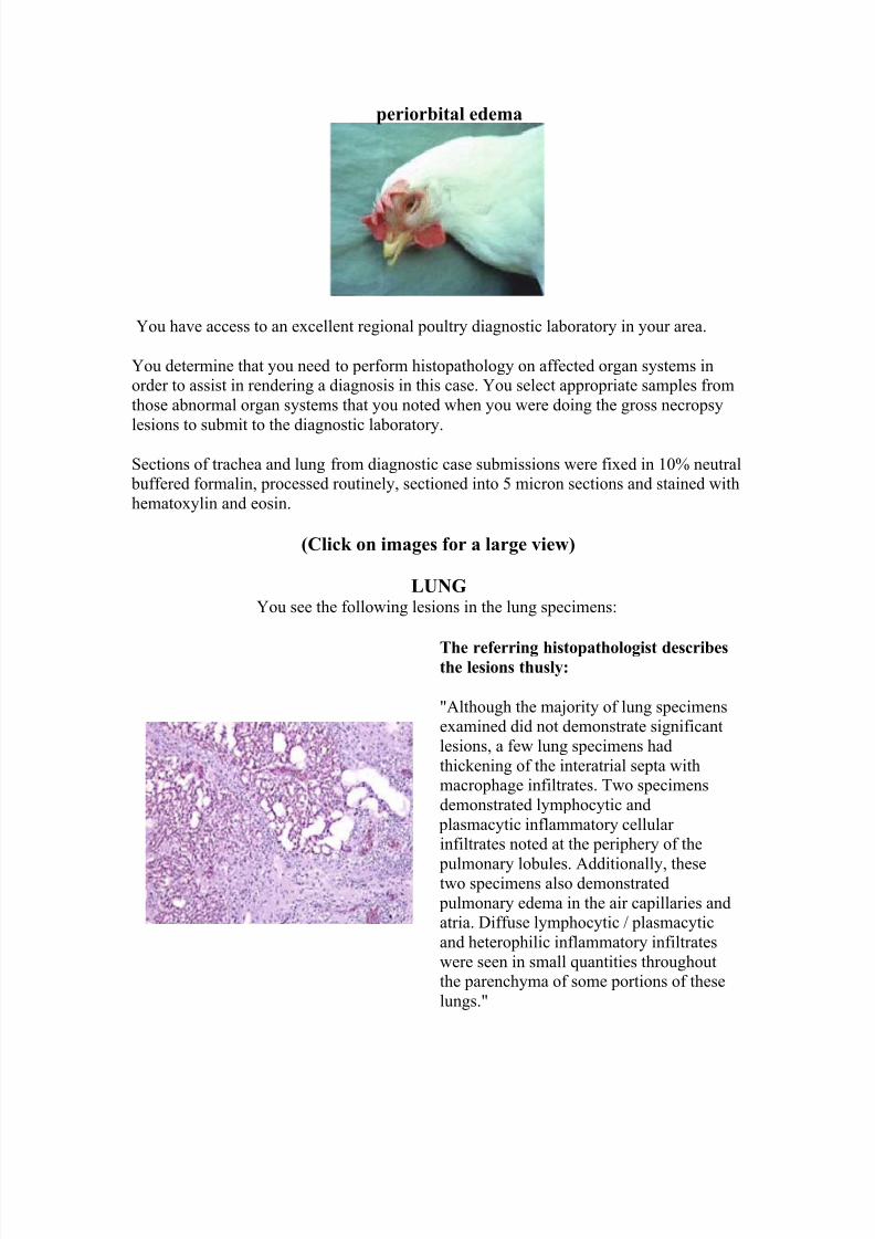
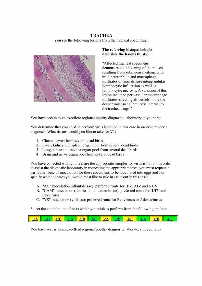
You determine that you need to perform histopathology on affected organ systems inorder to assist in rendering a diagnosis in this case. You select appropriate samples fromthose abnormal organ systems that you noted when you were doing the gross necropsylesions to submit to the diagnostic laboratory.
Sections of trachea and lung from diagnostic case submissions were fixed in 10% neutral buffered formalin, processed routinely, sectioned into 5 micron sections and stained withhematoxylin and eosin.
(Click on images for a large view)
LUNGYou see the following lesions in the lung specimens:
The referring histopathologist describesthe lesions thusly:
"Although the majority of lung specimensexamined did not demonstrate significantlesions, a few lung specimens hadthickening of the interatrial septa withmacrophage infiltrates. Two specimensdemonstrated lymphocytic and
plasmacytic inflammatory cellular infiltrates noted at the periphery of the
pulmonary lobules. Additionally, thesetwo specimens also demonstrated pulmonary edema in the air capillaries andatria. Diffuse lymphocytic / plasmacyticand heterophilic inflammatory infiltrateswere seen in small quantities throughoutthe parenchyma of some portions of theselungs."
TRACHEAYou see the following lesions from the tracheal specimens:
The referring histopathologistdescribes the lesions thusly:
"Affected tracheal specimensdemonstrated thickening of the mucosaresulting from submucosal edema withmild heterophilic and macrophageinfiltrates or from diffuse interglandular lymphocytic infiltration as well aslymphocytic necrosis. A variation of thislesion included perivascular macrophage
infiltrates affecting all vessels in the thedeeper mucosa / submucosa internal tothe tracheal rings."
You have access to an excellent regional poultry diagnostic laboratory in your area.
You determine that you need to perform virus isolation in this case in order to render adiagnosis. What tissues would you like to take for VI?
1. Choanal swab from several dead birds2. Liver, kidney and spleen organ pool from several dead birds
3. Lung, airsac and trachea organ pool from several dead birds4. Brain and nerve organ pool from several dead birds
You have collected what you feel are the appropriate samples for virus isolation. In order to assist the diagnostic laboratory in requesting the appropriate tests, you must request a
particular route of inoculation for these specimens to be inoculated into eggs and / or specify which viruses you would most like to rule in / rule out in this case:
A. "AC" inoculation (allantoic sac): preferred route for IBV, AIV and NDVB. "CAM" inoculation (chorioallantoic membrane): preferred route for ILTV and
Poxviruses
C. "YS" inoculation (yolksac): preferred route for Reoviruses or Adenoviruses
Select the combination of tests which you wish to perform from the following options:
1/A 1/B 1/C 2/A 2/B 2/C 3/A 3/B 3/C 4/A 4/B 4/C
You have access to an excellent regional poultry diagnostic laboratory in your area.
You determine that you need to perform microbiological tests in order to render adiagnosis in this case. Select only those tests that you think would be appropriate inthis case to assist with your diagnosis.
1. Aerobic bacterial culture of airsacs of affected birds
2. Anaerobic bacterial culture of liver of affected birds3. Fungal culture of lung / airsacs of affected birds4. Aerobic culture of intestinal contents of affected birds5. Salmonella culture of organ pool of affected birds
You have access to an excellent regional poultry diagnostic laboratory in your area.
You determine that you need to perform serological testing from this flock in order torender a diagnosis. You draw blood from approximately twenty birds in the house in your
collection efforts. Due to cost-related issues, you are limited to choosing no more than 6tests to perform on these collected specimens.
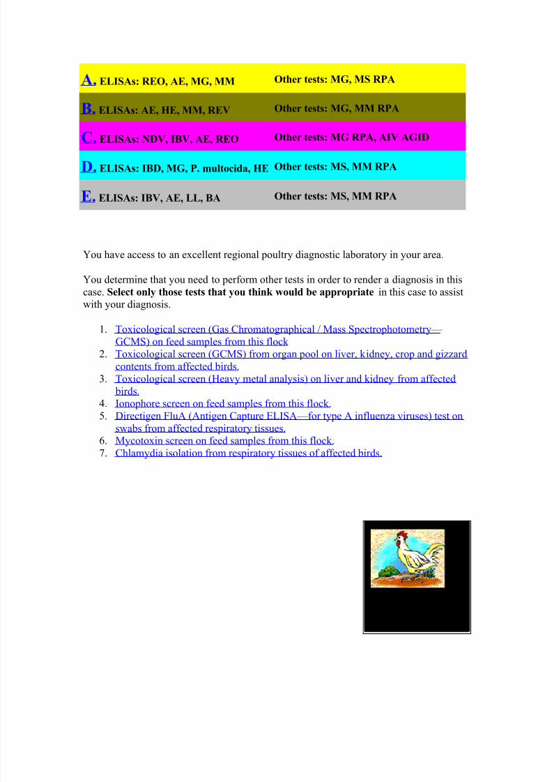
The diagnostic laboratory can run the following tests:
A. ELISAs: REO, AE, MG, MM Other tests: MG, MS RPA
B. ELISAs: AE, HE, MM, REV Other tests: MG, MM RPA
C. ELISAs: NDV, IBV, AE, REO Other tests: MG RPA, AIV AGID
D. ELISAs: IBD, MG, P. multocida, HE Other tests: MS, MM RPA
E. ELISAs: IBV, AE, LL, BA Other tests: MS, MM RPA
You have access to an excellent regional poultry diagnostic laboratory in your area.
You determine that you need to perform other tests in order to render a diagnosis in thiscase. Select only those tests that you think would be appropriate in this case to assistwith your diagnosis.
1. Toxicological screen (Gas Chromatographical / Mass Spectrophotometry— GCMS) on feed samples from this flock
2. Toxicological screen (GCMS) from organ pool on liver, kidney, crop and gizzardcontents from affected birds.
3. Toxicological screen (Heavy metal analysis) on liver and kidney from affected birds.
4. Ionophore screen on feed samples from this flock. 5. Directigen FluA (Antigen Capture ELISA—for type A influenza viruses) test on
swabs from affected respiratory tissues. 6. Mycotoxin screen on feed samples from this flock. 7. Chlamydia isolation from respiratory tissues of affected birds.