30
Physical Exam & History Taking dr. Fitria Nurul Hidayah, M.Sc., Sp.PD Interna Departement FKIK UMY

Physical Exam
& History
Taking
dr. Fitria Nurul Hidayah, M.Sc., Sp.PD
Interna Departement
FKIK UMY

Anamnesis


7 dimensi anamnesis
– Location
– Quality
– Quantity or severity
– Timing (onset, duration, and frequency)
– The setting in which it occurs
– Factor that aggravated or relieved the symptomps

Review Of System (ROS)
– General
– Usual weight, recent weight change, clothing that fits more tightly or loosely than before; weakness, fatigue, fever.
– Skin
– Rashes, lumps, sores, itching, dryness, color change; changes in hair or nails; changes in size or color of moles.
– Head, Eyes, Ears, Nose, Throat (HEENT).
– Head: Headache, head injury, dizziness, lightheadedness.
– Eyes: Vision, glasses or contact lenses, last examination, pain, redness, excessive tearing, double or blurred vision, spots, specks, flashing lights, glaucoma, cataracts.
– Ears: Hearing, tinnitus, vertigo, earache, infection, discharge. If hearing is decreased, use or nonuse of hearing aid.
– Nose and sinuses: Frequent colds, nasal stuffiness, discharge or itching, hay fever, nosebleeds, sinus trouble.
– Throat (or mouth and pharynx): Condition of teeth and gums; bleeding gums; dentures, if any, and how they fit; last dental examination; sore tongue; dry mouth; frequent sore throats; hoarseness

– Neck. Lumps, “swollen glands,” goiter, pain, stiffness.
– Breasts. Lumps, pain or discomfort, nipple discharge, self-examination practices.
– Respiratory. Cough, sputum (color, quantity), hemoptysis, dyspnea, wheezing, pleurisy, last chest x-ray. You may wish to include asthma, bronchitis, emphysema, pneumonia, and tuberculosis.
– Cardiovascular. “Heart trouble,” hypertension, rheumatic fever, heart murmurs, chest pain or discomfort, palpitations, dyspnea, orthopnea, paroxysmal nocturnal dyspnea, edema, past electrocardiographic or other cardiovascular tests.
– Gastrointestinal. Trouble swallowing, heartburn, appetite, nausea. Bowel movements, color and size of stools, change in bowel habits, rectal bleeding or black or tarry stools, hemorrhoids, constipation, diarrhea. Abdominal pain, food intolerance, excessive belching or passing of gas. Jaundice, liver or gallbladder trouble, hepatitis.

– Peripheral Vascular. Intermittent claudication; leg cramps; varicose veins; past clots in veins; swelling in calves, legs, or feet; color change in fingertips or toes during cold weather; swelling with redness or tenderness.
– Urinary. Frequency of urination, polyuria, nocturia, urgency, burning or pain on urination, hematuria, urinary infections, kidney stones, incontinence; in males, reduced caliber or force of urinary stream, hesitancy, dribbling.
– Genital.
– Male
– Female
– Musculoskeletal. Muscle or joint pain, stiffness, arthritis, gout, backache. Neck or low back pain. Joint pain with systemic features such as fever, chills, rash, anorexia, weight loss, or weakness.
– Psychiatric. Nervousness; tension; mood, including depression, memory change, suicide attempts, if relevant.
– Neurologic. Changes in mood, attention, or speech; changes in orientation, memory, insight, or judgment; headache, dizziness, vertigo; fainting, blackouts, seizures, weakness, paralysis, numbness or loss of sensation, tingling or “pins and needles,” tremors or other involuntary movements, seizures.
– Hematologic. Anemia, easy bruising or bleeding, past transfusions, transfusion reactions.
– Endocrine. “Thyroid trouble,” heat or cold intolerance, excessive sweating, excessive thirst or hunger, polyuria, change in glove or shoe size.

Physical
Examination

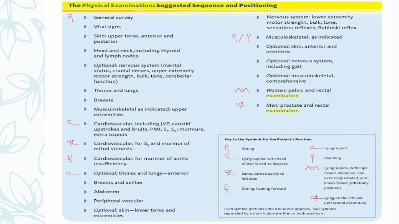
Preparing for the Physical Examination
Choose Choose the sequence of examination
Make Make the patient comfortable.
Check Check your equipment.
Adjust Adjust the lighting and the environment.
Reflect on Reflect on your approach to the patient.


Vital Sign
dr. Fitria Nurul Hidayah, M.Sc., Sp.PD
Interna Departement
FKIK UMY

Vital Sign

Tekanan Darah

– Center the inflatable bladder over the brachial artery. The lower border of the cuff should be about 2.5 cm above the antecubital crease. Position the patient’s arm so that it is slightly flexed at the elbow
– Systolic palpatoar
– place the bell of a stethoscope lightly over the brachial artery
– deflate it slowly, at a rate of about 2 to 3 mm Hg per second
– Wait 2 or more minutes and repeat. Average your readings. If the first two readings differ by more than 5 mm Hg, take additional readings.

Tekanan darah


Nadi
– Rate
– Rhytme
– Tekanan nadi

Respirasi

Temperature
– Oral temperature
– is not recommended when patients are unconscious or restless, unable to close the mouth
– a glass or an electronic thermometer
– When using a glass thermometer, shake the thermometer down to 35°C (96°F) or below, insert it under the tongue, instruct the patient to close both lips, and wait 3 to 5 minutes. Then read the thermometer
– reinsert it for a minute, and read it again.
– If the temperature is still rising, repeat this procedure until the reading remains stable.
– Note that hot or cold liquids, and even smoking, can alter the temperature reading.
– In these situations, it is best to delay measuring the temperature for 10 to 15 minutes.

Temperatur
– Axilla temperature
– are lower than oral temperatures by approximately 1°, but take 5 to 10 minutes to register and are generally considered less accurate than other measurements
– Rectal temperature
– ask the patient to lie on one side with the hip flexed.
– Select a rectal thermometer with a stubby tip, lubricate it, and insert it about 3 cm
to 4 cm (1½ inches) into the anal canal, in a direction pointing to the umbilicus. Remove it after 3 minutes, then read.
– Alternatively, use an electronic thermometer after lubricating the probe cover. Wait about 10 seconds for the digital temperature recording to appear

– Tympanic Membrane Temperatures.
– quick, safe, and reliable if performed properly.
– Make sure the external auditory canal is free of cerumen, which lowers
temperature readings.
– Position the probe in the canal so that the infrared beam is aimed at the
tympanic membrane (otherwise the measurement will be invalid). Wait 2
to 3 seconds until the digital temperature reading appears.
– This method measures core body temperature, which is higher than the
normal oral temperature by approximately 0.8°C (1.4°F)

Head and Neck
– Bentuk kepala
– Konjunctiva mata, sclera
– Leher : benjolan

Thorax

Abdomen


