NATURAL PRODUCTS: FROM CHEMISTRY TO PHARMACOLOGY (C HO, SECTION EDITOR) Phytocompounds of Rheum emodi, Thymus serpyllum, and Artemisia annua Inhibit Spike Protein of SARS-CoV-2 Binding to ACE2 Receptor: In Silico Approach Rajan Rolta 1 & Deeksha Salaria 1 & PremPrakash Sharma 2 & Bhanu Sharma 1 & Vikas Kumar 1 & Brijesh Rathi 2 & Mansi Verma 3 & Anuradha Sourirajan 1 & David J. Baumler 4 & Kamal Dev 1 Accepted: 24 May 2021 # The Author(s), under exclusive licence to Springer Nature Switzerland AG 2021 Abstract COVID-19, the disease caused by SARS-CoV-2, has been declared as a global pandemic. Traditional medicinal plants have long history to treat viral infections. Our in silico approach suggested that unique phytocompounds such as emodin, thymol and carvacrol, and artemisinin could physically bind SARS-CoV-2 spike glycoproteins (6VXX and 6VYB), SARS-CoV-2 B.1.351 South Africa variant of Spike glycoprotein (7NXA), and even with ACE2 and prevent the SARS-CoV-2 binding to the host ACE2, TMPRSS2 and neutrapilin-1 receptors. Since Chloroquine has been looked as potential therapy against COVID-19, we also compared the binding of chloroquine and artemisinin for its interaction with spike proteins (6VXX, 6VYB) and its variant 7NXA, respectively. Molecular docking study of phytocompounds and SARS-CoV-2 spike protein was performed by using AutoDock/Vina software. Molecular dynamics (MD) simulation was performed for 50ns. Among all the phytocompounds, molecular docking studies revealed lowest binding energy of artemisinin with 6VXX and 6VYB, with E total −10.5 KJ mol −1 and −10.3 KJ mol −1 respectively. Emodin showed the best binding affinity with 6VYB with E total −8.8 KJ mol −1 and SARS-CoV- 2 B.1.351 variant (7NXA) with binding energy of −6.4KJ mol −1 . Emodin showed best interactions with TMPRSS 2 and ACE2 with E total of −7.1 and −7.3 KJ mol −1 respectively, whereas artemisinin interacts with TMPRSS 2 and ACE2 with E total of −6.9 and −7.4 KJ mol −1 respectively. All the phytocompounds were non-toxic and non-carcinogenic. MD simulation showed that artemisinin has more stable interaction with 6VYB as compared to 6VXX, and hence proposed as potential phytochemical to prevent SARS-CoV-2 interaction with ACE-2 receptor. Keywords COVID-19 . Antimalarial drugs . Chloroquine . Artemisinin . Phytocompounds . Emodin . Thymol . In silico . MD simulation Introduction COVID-19 caused by a member of family Coronaries (CoV) has threatened the survival of human beings on the Earth and it has been declared as global health emergency by the World Health Organization (WHO) [1••]. Coronaviruses (CoV) are known for their ability to cause illness, with severe diseases such as Middle East respiratory syndrome (MERS-CoV) and severe acute respiratory syndrome (SARS-CoV). Though there are other coronaviruses known to infect humans (hCoVsOC43 and 229E), but they are mild pathogens respon- sible for common cold. In Latin, Corona means “halo” or “crown”; thus, the name represents the structure of the virus which consists of crown like projections on its surface [2]. In 1937, coronavirus was isolated from an infectious bronchitis virus in birds which was responsible to ruin the poultry stocks Rajan Rolta, PremPrakash Sharma, Bhanu Sharma , and Deeksha Salaria contributed equally to this work. This article is part of the Topical Collection on Natural Products: From Chemistry to Pharmacology * Kamal Dev [email protected]1 Faculty of Applied Sciences and Biotechnology, Shoolini University, Pin, Solan, Himachal Pradesh 173212, India 2 Laboratory for Translational Chemistry and Drug Discovery, Hansraj College University of Delhi, Delhi 110007, India 3 Sri Venkateswara College, University of Delhi, New Delhi 110021, India 4 Department of Food Science and Nutrition, University of Minnesota, St. Paul, MN, USA https://doi.org/10.1007/s40495-021-00259-4 / Published online: 15 July 2021 Current Pharmacology Reports (2021) 7:135–149
Transcript
NATURAL PRODUCTS: FROM CHEMISTRY TO PHARMACOLOGY (C HO, SECTION EDITOR)
Phytocompounds of Rheum emodi, Thymus serpyllum,and Artemisia annua Inhibit Spike Protein of SARS-CoV-2 Bindingto ACE2 Receptor: In Silico Approach
Mansi Verma3 & Anuradha Sourirajan1& David J. Baumler4 & Kamal Dev1
Accepted: 24 May 2021# The Author(s), under exclusive licence to Springer Nature Switzerland AG 2021
AbstractCOVID-19, the disease caused by SARS-CoV-2, has been declared as a global pandemic. Traditional medicinal plants have longhistory to treat viral infections. Our in silico approach suggested that unique phytocompounds such as emodin, thymol andcarvacrol, and artemisinin could physically bind SARS-CoV-2 spike glycoproteins (6VXX and 6VYB), SARS-CoV-2 B.1.351South Africa variant of Spike glycoprotein (7NXA), and even with ACE2 and prevent the SARS-CoV-2 binding to the hostACE2, TMPRSS2 and neutrapilin-1 receptors. Since Chloroquine has been looked as potential therapy against COVID-19, wealso compared the binding of chloroquine and artemisinin for its interaction with spike proteins (6VXX, 6VYB) and its variant7NXA, respectively. Molecular docking study of phytocompounds and SARS-CoV-2 spike protein was performed by usingAutoDock/Vina software. Molecular dynamics (MD) simulation was performed for 50ns. Among all the phytocompounds,molecular docking studies revealed lowest binding energy of artemisinin with 6VXX and 6VYB, with Etotal −10.5 KJ mol−1
and −10.3 KJmol−1 respectively. Emodin showed the best binding affinity with 6VYBwith Etotal −8.8 KJmol−1and SARS-CoV-2 B.1.351 variant (7NXA) with binding energy of −6.4KJ mol−1. Emodin showed best interactions with TMPRSS 2 and ACE2with Etotal of −7.1 and −7.3 KJ mol−1 respectively, whereas artemisinin interacts with TMPRSS 2 and ACE2 with Etotal of −6.9and −7.4 KJ mol−1 respectively. All the phytocompounds were non-toxic and non-carcinogenic. MD simulation showed thatartemisinin has more stable interaction with 6VYB as compared to 6VXX, and hence proposed as potential phytochemical toprevent SARS-CoV-2 interaction with ACE-2 receptor.
COVID-19 caused by a member of family Coronaries (CoV)has threatened the survival of human beings on the Earth andit has been declared as global health emergency by the WorldHealth Organization (WHO) [1••]. Coronaviruses (CoV) areknown for their ability to cause illness, with severe diseasessuch as Middle East respiratory syndrome (MERS-CoV) andsevere acute respiratory syndrome (SARS-CoV). Thoughthere are other coronaviruses known to infect humans(hCoVsOC43 and 229E), but they are mild pathogens respon-sible for common cold. In Latin, Corona means “halo” or“crown”; thus, the name represents the structure of the viruswhich consists of crown like projections on its surface [2]. In1937, coronavirus was isolated from an infectious bronchitisvirus in birds which was responsible to ruin the poultry stocks
Rajan Rolta, PremPrakash Sharma, Bhanu Sharma , and Deeksha Salariacontributed equally to this work.
This article is part of the Topical Collection on Natural Products: FromChemistry to Pharmacology
[3]. Coronaviruses are the type of viruses that directly affectthe respiratory tract. These are associated with the commoncold, pneumonia, gut, and severe acute respiratory syndrome.Coronaviruses are zoonotic, which means they are transmittedbetween animals and humans [4]. A new strain, novel coro-navirus (nCoV) has come into knowledge since 2019 and hasemerged as a threat to mankind.
The first case of Middle East respiratory syndrome(MERS-CoV) was seen in the year 2012, a businessman inSaudi Arabia who died from viral pneumonia [5]. In 2016, areport on 1998 was published by the World HealthOrganization (WHO) regarding the confirmed cases ofMERS-CoV infection and the death rate was approximately36% (Middle East respiratory coronavirus (MERS-CoV) [5].The biggest outbreak with first ever confirmed case of thisdisease came into existence in the year 2015 in South Korea.Including the China, the confirmed cases extend to 186 withtotal 36 deaths [6, 7]. Cases regarding the novel coronaviruscame in to existence among the population of Wuhan, China,on December 8, 2019. Pneumonia was the first symptom ofinfection and most of the cases were linked to a local fish andanimal market. During the research, it was seen that 2019novel coronavirus was recognized as pathogenic agent re-sponsible for evolution of pneumonia [8]. On January 20,2020, laboratory in Korea confirmed the first case of corona-virus. On 23 January, 2020, the government of China an-nounced total shutdown of country and advised the peoplefor undergoing personal isolation. In the USA, there are fivevariants of SARS-Cov-2. B.1.1.7: This variant was discoveredfor the first time in December 2020 in the USA. It was firstdiscovered in the UK. B.1.351: This variant was discoveredfor the first time in the USA at the end of January 2021. It wasfirst discovered in December 2020 in South Africa. P.1: InJanuary 2021, this variant was discovered for the first timein the USA. B.1.427 and B.1.429: These two variants werediscovered in February 2021 in California (https://www.cdc.gov/coronavirus/2019-ncov/transmission/variant.html).
SARS-CoV-2 consists of four structural proteins: spike (S),membrane (M), envelop (E), and nucleocapsid (N) proteins[9]. Among all, S protein plays an important role in viralattachment, fusion, entry, and also act as a target for develop-ment of antibodies, entry inhibitors, and vaccines [10, 11].The S1 domains of coronaviruses contain receptor-bindingdomains (RBDs) that directly bind to the cellular receptors[12, 13]. In general, SARS-CoV surface exhibits two compo-nents: S1, which contains the receptor binding domain (RBD);and S2, which contains the fusion peptide. SARS-CoV gainsentry into cells through interaction of the SARS-SRBD withthe cell surface receptor angiotensin-converting enzyme 2(ACE2) [14, 15]. These interactions are followed by endocy-tosis, and at the low pH in endosomes, SARS-S is cleaved by acellular protease called cathepsin L, thereby exposing the S2domain of the spike protein for membrane fusion [16, 17]. The
minimal RBD of SARS-CoV S protein is located in the S1subunit (AA 318–510) and is responsible for viral binding tohost cell receptors [18, 19]. Besides the main receptor for theangiotensin-converting enzyme 2, there are several alternativereceptors, such as dendritic cell-specific intercellular adhesionmolecule-3-grabbing non-integrin and liver/lymph node-spe-cific intercellular adhesion molecule-3-grabbing integrin [20].SARS-CoVs recognizes angiotensin-converting enzyme 2(ACE2) as its receptor, whereas MERS-CoV recognizesdipeptidyl peptidase 4 (DPP4) as its receptor [21, 22]. Tworesidues (AA 479 and AA 487) in RBD determine SARSprogression and tropism, and their mutations may enhanceanimal-to-human or human-to-human transmission [13].Some residues (AA 109, 118, 119, 158, 227, 589, and 699)in S protein are critical strategies against this deadly viralagent, especially in high-risk groups, including people of ev-ery age group [23]. According to the previous data, the ACE2receptor expressing cell fused with SARS-S-expressing cellsadds to the cell surface by pH-independent mechanism [19]. Itenhances the cell stress responses and apoptosis [24]. Bindingis very critical for pathogenesis and if the binding of SARS-Swith ACE2 receptor is blocked, infection can be stopped.Traditional medicinal plants produce large number of com-pounds which are used as therapeutics to kill the pathogens[25]. In the recent years, many reports published on antimi-crobial activity of the medicinal plants [25–27]. It is expectedthat plant extracts and phytocompounds showing the targetsite other than antibiotics, a very little information is availableon this type of activity of medicinal plants [26, 27]. Extracts ofmedicinal plants have been used from ancient times and theseplants are known for their antiviral properties and less sideeffects. Traditionally, thyme was acclimated to treat asthmaand loosen congestion in the throat and stomach [28]. Thepharmacological manuscript of Chailander medical codex (fif-teenth and sixteenth centuries) mentions the utilizations ofwild thyme for the treatment of headaches caused by coldand laryngitis [29]. During the Renaissance period (sixteenthand seventeenth centuries), wild thyme was utilized internallyto treat malaria and epilepsy [30]. Traditionally in many coun-tries, areal part of T. serpyllum is utilized as anthelmintic, avigorous antiseptic, an antispasmodic, a carminative, deodor-ant, diaphoretic, disinfectant, expectorant, sedative, and tonic.Thymus serpyllum additionally used to treat respiratory quan-daries [29]. In western Balkans, thymus species used to amendb l ood c i r cu l a t i on and a s an t i cho l e s t e r o l em ic ,immunostimulant [31]. Carvacrol and thymol are isomers, be-longing to the group of monoterpenic phenols with potentantiseptic properties. Chauhan et al. [32] reported thymol(25–200 mg kg−1) as immunomodulatory in cyclosporine A-treated Swiss albino mice by enhancing the expression ofcluster of differentiation 4 (CD4), cluster of differentiation 8(CD8), and Th1 cytokines via upregulation of IFN-4 expres-sion and enhanced secretion of interleukin-12 (IL-12).
Antiviral property of Thymus serpyllum [33] and thymol isalready reported [34]. Pilau et al. [35] reported the antiviralactivity of carvacrol from Lippia graveolens against humanand animal virus (herpes simplex virus, acyclovir-resistantherpes simplex virus 1, bovine herpesvirus 2, respiratory syn-cytial virus; human rotavirus, bovine viral diarrhea virus).Antiviral nature of Emodin was also reported in several stud-ies [36, 37]. Study from Efferth et al. [38] showed in vitroantiviral properties of artemisinin against hepatitis B virus,hepatitis C virus, and bovine viral diarrhea. Keeping in viewthe antiviral potential of Himalayan herbs, the current studywas focused on the identification of potent phytocompoundsfrom Himalayan herbs (Rheum emodi, Thymus serpyllum, andArtemisia annua) to cure a dangerous COVID-19.
Material and Methods
Bioinformatics Tools Open Babel GUI [39], UCSF Chimera1.8.1 [40], Pubchem (www.pubchem.com), RCSB PDB(http://www.rscb.org/pdb), PDBsum (www.ebi.ac.uk/pdbsum), and Autodock/vina software [41, 42] were used inthe present investigation.
Ligand Preparation
Four major phytocompounds of three medicinal plants—em-odin of Rheum emodi, thymol and carvacrol of Thymusserpyllum, and artemisinin of Artemisia annua—were usedfor the docking studies. The 3-dimensional structures of allthe phytocompounds and chloroquine were obtained fromPubChem (www.pubchem.com) in .sdf format. The .sdf fileof the phytocompounds was converted into PDB and pdbqtformat by using the Open Babel tool [43]. Table 1 showsmolecular structure, molecular weight, pharmacologicalproperties, plant source, and percentage of phytocompoundsin the respective plants and antimalarial drug chloroquine.Targets of phytocompounds and standard drug chloroquinewere predicted by using SwissADME online server.
Protein Preparation
Two spike proteins of SARS-CoV-2 spike glycoprotein (PDBID: 6VXX, closed conformation), SARS-CoV-2 spikeectodomain structure (PDB ID: 6VYB, open conformation)[52] and one mutated variant of SARS-CoV-2 B.1.351(South African variant) variant of Spike glycoprotein (PDBID: 7NXA) [53] and two receptor of SARS-CoV-2 (HumanTMPRSS2 (PDB ID: 7MEQ) [54], Angiotensin-convertingenzyme-2 (ACE2 PDB ID: 6M1D)) [55••], and neuropilin-1(PDB ID: 4DEQ) were used to analyze the interactions ofmajor phytocompounds of R. emodi, T. serpyllum, and A.
annua. It has been shown that SARS CoV-2 SB openprotein conformation is necessary for binding with ACE2 athost surface; and coronavirus with open surface S-glycoprotein trimers found to be highly pathogenic tohuman [56••].
The 3-dimensional structures of selected target proteinswere retrieved from the Protein Data Bank (PDB) (http://www.rscb.org/pdb). Non-essential water molecules,including heteroatoms, were removed from the targetreceptor molecule and hydrogen atoms were added to thetarget receptor molecule. Binding site of both the targetproteins of COVID-19 (SARS-CoV-2 spike glycoprotein(PDB ID: 6VXX), SARS-CoV-2 spike ectodomain structure(PDB ID: 6VYB)), SARS-CoV-2 B.1.351 variant Spikeglycoprotein (PDB ID: 7NXA), Human TMPRSS2 (7MEQ),Angiotensin-converting enzyme-2, ACE2 (PDB ID: 6M1D),and neuropilin-1 (PDB ID: 4DEQ) was determined by gridbox generation. Grid box was generated by adjusting the gridparameter x, y, z coordinate values; grid value for 6VYB and6VXXwas center x: −189.229, y: −255.9, z: 229.87 Å; 7NXAwas x: −14.806, y: −19.528, z: −51.972 Å; 7MEQ was x: −1.028, y: −0.352, z: 10.912; and 6MID was x: 126.806, y: 133.196, z:121.533. Size of the grid was same for all the targetproteins (i.e., x—40, y—40, z—40 Å) using AutoDocksoftware. The grid values were recorded in the config.txt fileformat [57••].
Prediction of Drug Likeness of SelectedPhytocompounds
The aim of the drug scan was to see whether selected phyto-chemicals met the drug-likeness criteria. Lipinski’s filtersusing Molinspiration (http://www.molinspiration.com) wereapplied for examining drug-likeness attributes, includingquantity of hydrogen acceptors (should not be more than 10), quantity of hydrogen donors (should not be more than 5),molecular weight (mass should be more than 500 Daltons),and partition coefficient log P (should not be less than 5). Thesmiles format of each of the phytochemical was uploaded forthe analysis [58].
ADME and Toxicity Prediction of SelectedPhytocompounds
Absorption, distribution, metabolism, excretion, and toxicity(ADMET) screening was done to determine the absorption,toxicity, and drug-likeness properties of the selected ligands.The 3-dimensional structures of ligands such as emodin, thy-mol, carvacrol, artemisinin, and chloroquine were saved in.smiles format and chloroquine was uploaded onSWISSADME (Molecular Modeling Group of the SIB(Swiss Institute of Bioinformatics), Lausanne, Switzerland),admetSAR (Laboratory of Molecular Modeling and Design,
Shanghai, China), and PROTOX web servers (ChariteUniversity of Medicine, Institute for Physiology, StructuralBioinformatics Group, Berlin, Germany) for ADMET screen-ing. SWISSADME is a web tool used for the prediction ofADME and pharmacokinetic properties of a molecule. Thepredicted results consist of lipophilicity, water solubility,
physicochemical properties, pharmacokinetics, drug likeness,medicinal chemistry, and Brain or Intestinal Estimated perme-ation method (blood-brain barrier and PGP ± prediction).AdmetSAR provides ADMET profiles for query moleculesand can predict about fifty ADMET properties. Toxicity clas-ses are as follows: (i) category I contains compounds with
Table 1 Molecular structure, molecular weight, pharmacological properties, plant source, and percentage of selected phytocompounds andchloroquine
Phytocompounds
Plant source Molecular structures
Percentage (%) ofphytocompounds in
plants
Molecular weight
(g mol-1)
Pharmacological properties
Emodin Rheum emodi 23.24 (Rolta et al.,
2020a) [44]
270.24 Antiviral [47], antimicrobial
[44,48]
Artemisinin Artemesia annua
0.77–1.06 (Castilho et
al., 2018) [45]
282.33 Antimalarial [49]; Antiviral [38]
Thymol Thymusserpyllum
8.3 (Gul et al., 2018)
[46]
150.22 Antiseptic, antibacterial,
antifungal and antioxidant
properties[30] Antivirotic [35]
Carvacrol Thymusserpyllum
3.03 [46] 150.22 Antimicrobial, antithrombotic,
anti-inflammatory,acetylcholinesterase inhibitory
properties [30] Antiparasitic
[50], Antiviral [36]
Chloroquine Standard
antimalarial
drug
Standard Antimalarial
drug
319.9 FDA has allowing the use
hydroxychloroquine to treat
coronavirus disease 2019
(COVID-19) [51••].
138 Curr Pharmacol Rep (2021) 7:135–149
LD50 values ≤ 50 mg kg−1, (ii) category II contains com-pounds with LD50 values > 50 mg kg−1 but 500 mg kg−1 but5000 mg kg−1 (Cheng, 2020). PROTOX is a rodent oral tox-icity server predicting LD50 value and toxicity class of querymolecule. The toxicity classes are as follows: class 1: fatal ifswallowed (LD50 ≤5), class 2: fatal if swallowed (55000)[59].
Docking of COVID-19 Receptors and Phytocompounds
The docking of selected ligands to the catalytic triad of proteinwas performed by using AutoDock/Vina [41]. Docking wasperformed to obtain populations of conformations and orien-tation for ligands at binding sites. Docking was performed tostudy the interactions between SARS-CoV-2 receptors suchas 6VXX (closed state), 6VYB (open state), 7NXA (SARS-CoV-2 B.1.351 variant), 7MEQ, 6MID, and neuropilin-1(4DEQ) with major phytocompounds of R. emodi, T.serpyllum, and A. annua and .pdb file of proteins-ligand com-plexes was generated. All the bonds of ligands were set to berotatable. All calculations for protein-ligand docking wereperformed using the Lamarckian genetic algorithm (LGA)method. The best conformation was chosen with the lowestdocked energy, after the completion of docking search. The.pdb complex of protein and ligand was analyzed by usingDiscovery Studio (https://discover.3ds.com/d) to study thelist of interactions between protein and ligand complex.Detailed visualization and comparison of the docked sites oftarget proteins and ligands were done by using Chimera [60].
Molecular Dynamics (MD) Simulation
In order to further verify the accuracy of docking observa-tions, the two best complexes of S-protein were selected forextensive MD simulation for 50ns. Both the complexes wereintroduced into Desmond software to study the binding stabil-ity of the ligand within the binding site of S-protein [60]. Bothcomplexes were prepared prior to simulation to remove anystructural error as described earlier [61, 62]. Both the com-plexes were solvated in TIP3P water model and 0.15 M NaClto mimic a physiological ionic concentration. This MD simu-lation was performed with OPLS3e force field.
Results
Drug Likeness of Selected Phytocompounds
Early preclinical production is supported by drug-likeness fil-ters, which help to prevent expensive late-stage preclinical andclinical failure. The Lipinski rule of 5 was used to evaluate thedrug-likeness properties of the emodin, thymol, carvacrol,artemisinin, and chloroquine. The emodin, thymol, carvacrol,
and artemisinin followed the Lipinski’s rule of five, whereaschloroquine showed one violation (Table 2).
ADMET Prediction and Toxicity Analysis of SelectedPhytocompounds
The comparat ive ADME propert ies predicted bySwissADME of emodin, thymol, carvacrol, artemisinin, andchloroquine are summarized in Table 3. Consensus Log Po/wvalue of ˂5 indicates good aqueous solubility, which meansthat an adequate amount of drug can reach and be maintainedinside the body through oral administration. The emodin, thy-mol, carvacrol, artemisinin, and chloroquine showed consen-sus Log Po/w value of < 5 (Table 3). TPSA indicates perme-ability of compounds into the cells. A TPSA value of < 140Å2 is required for good permeation of compound into the cellmembrane and value < 90 Å2 is required to permeate throughblood-brain barrier. All the selected phytocompounds showedTPSA value < 90 Å2, except 94.83 Å2 for emodin, indicatinggood permeability of selected phytocompounds into the cell aswell as through blood-brain barrier. Lipinski’s rule of fivehelps to determine drug likeness of the compound and orallyactive drug should not violate the Lipinski’s rule. The emodin,thymol, carvacrol, artemisinin, and chloroquine followedLipinski’s rule of five (Table 3). The predicted cellular targetsof emodin, thymol, carvacrol, artemisinin, and chloroquineare shown in Table 4.
Toxicity of emodin, thymol, carvacrol, artemisinin, andchloroquine was predicted by PROTOX-II and admetSARand results are summarized in Table 5. It was observed thatemodin, thymol, carvacrol, artemisinin, and chloroquine arenon-carcinogenic and non-cytotoxic in nature and are safe toadminister. However, LD50 value of emodin (5000 mg kg−1)and artemisinin (4228 mg kg−1) calculated from Protox II washigher than that of all other phytocompounds and chloroquine.This suggests that natural phytocompounds are safer even athigher dosage than that of chemically synthesized chloroquine.
Molecular Docking (MD) Analysis of Phytocompoundsand Chloroquine With Spike Protein and Its Variant ofSARS-CoV-2
Docking results of phytocompounds of medicinal plantsshowed good binding affinity and modes of interaction withboth the spike protein of SARS-CoV-2 (6VXX closed stateand 6VYB open state), SARS-CoV-2 B.1.351 variant, HumanTMPRSS2 (7MEQ), Angiotensin-converting enzyme-2,ACE2, and neuropilin-1 as compared to chloroquine.Among all the selected phytocompounds, artemisininshowed the best binding affinity (−10.5 kcal mol−1),followed by thymol (−6.9 kcal mol−1), carvacrol (−6.8 kcalmol−1), emodin (−6.4 kcal mol−1), and chloroquine (−5.6 kcalmol−1) with SARS-CoV-2 spike glycoprotein (6VXX).
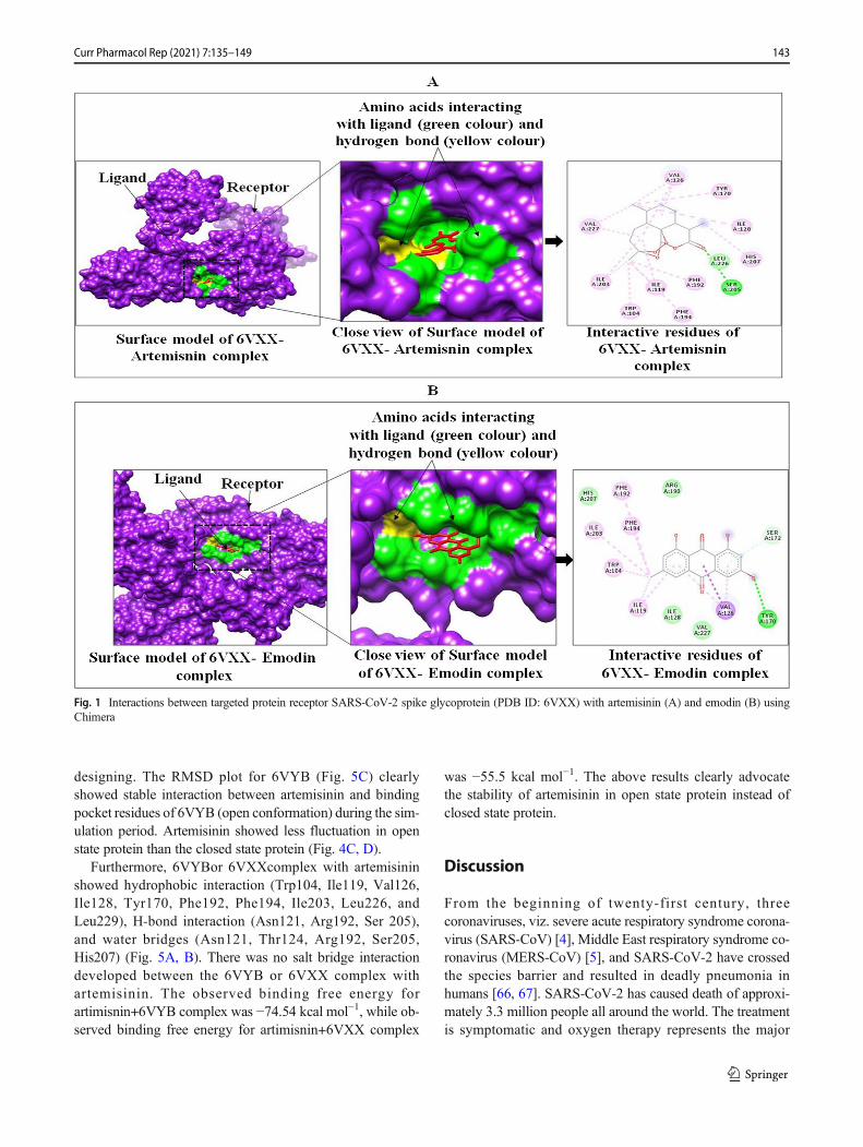
Artemisinin makes week hydrogen bonds with SER 205, HIS207, and hydrophobic interactions with ILE 119, VAL 126,ILE 128, PHE 192, PHE 194, ILE 203, LEU 226, and VAL227 (Table 6, Fig. 1, and Figure S1).
The binding interaction almost follows the similar patternof interaction with SARS-CoV-2 spike ectodomain structure(6VYB, open state) with a binding affinity of −10.3 kcalmol−1 (artemisinin), −8.8 kcal mol−1 (emodin), −6.7 kcalmol−1 (thymol), −6.8 kcal mol−1 (carvacrol), and −5.9 kcalmol−1 (chloroquine). Artemisinin makes week hydrogenbonds with SER 730 and THR 778 and showed hydrophobicinteractions with TRP 104, ILE 119, ILE 126, VAL 128, PHE194, and VAL 227 (Table 6 and Fig. 2 and S2). Artemisinin incomplex with 6VYB makes week hydrogen bonds with SER730 and THR 778 and showed hydrophobic interactions withTRP 104, ILE 119, ILE 126, VAL 128, PHE 194, and VAL227 (Table 5 and Fig. 2 and Figure S2).
In case of SARS-CoV-2 B.1.351 variant, the binding affin-ity was −6.4, −4.4, −4.7, −5.9, and −4.9 kcal mol−1 for emodin,thymol, carvacrol, artemisinin, and chloroquine respectively.Emodin in complex with SARS-CoV-2 B.1.351 variant(7NXA) makes strong hydrogen bonds with GLU 6, GLN111, and LYS 207 and showed hydrophobic interactions withSER 7, GLY 9, VAL 92, GLY 112, THR 113, PRO 155, andPRO 208 (Table 6 and Fig. S3). Emodin also showed the bestinteractions (−7.1 kcal mol−1) with Human TMPRSS2(7MEQ), followed by artemisinin (−6.9 kcal mol−1), chloro-quine (−5.7 kcal mol−1), thymol (−5.5 kcal mol−1), and
carvacrol (−5.3 kcal mol−1). Emodin makes hydrogen bonds(moderate strength) with ASN 277 and showed hydrophobicinteractions with HIS 274, GLN 276, VAL 278, TRP 306,THR 309, PHE 311, TYR 322, GLN 323, ALA 324, GLY325, andGLN 327 (Table 6 and Fig. S4). Neuropilin-1 interactswith emodin (−6.6 kcal mol−1), carvacrol and artemisinin (−5.8kcal mol−1), thymol (−5.6 kcal mol−1), and chloroquine (4.9kcal mol−1). Emodinmakes hydrogen bond (moderate strength)with ILE 147 and hydrophobic interactions with TYR 24, TRP28, THR 43, ASP 47, THR 76, LYS 78, TYR 80, andGLY 141(Table 6 and Fig. S5). Similarly, Angiotensin-convertingenzyme-2, ACE2 showed best interaction with artemisinin(−7.4 kcal mol−1), followed by emodin (−7.3 kcal mol−1),thymol (−6.9 kcal mol−1), chloroquine (−6.5 kcal mol−1), andcarvacrol (−6.1 kcal mol−1). Artemisinin do not make anyhydrogen bonds with ACE2, but showed hydrophobicinteractions with LEU 545, ALA 273, LEU 495, SER 487,LEU 269, SER 491, ILE 492, ASP 270, TYR 488, VAL 552,PHE 549, and GLU 553 (Table 6 and Figure S6).
The nature of hydrogen bonds was determined on the basisof donor-acceptor distances in protein secondary structure el-ements. Jeffrey and Jeffrey [63••] categorize hydrogen bondswith donor-acceptor distances of 2.2–2.5 Å as “strong,” 2.5–3.2 Å as “moderate” (mostly electrostatic), and 3.2–4.0 Å as“weak, electrostatic.” Hydrogen bond interaction and hydro-phobic interactions of these phytocompounds with target pro-teins were analyzed through discovery studio and results aresummarized in Table 6.
Table 2 Prediction of drug-likeness activity of selected phytocompounds
Phytocompounds miLogP TPSA natoms MW nON nOHNH nviolations
Emodin 3.01 94.83 20 270.24 5 3 0
Thymol 3.34 20.23 11 150.22 1 1 0
Carvacrol 3.81 20.23 11 150.22 1 1 0
Artemisinin 3.32 54.01 20 282.34 5 0 0
Chloroquine 5 28.16 22 319.88 3 1 1
Table 3 ADME properties of selected phytocompounds and chloroquine predicted by SwissADME
Phytocompounds SwissADME
Consensus LogPO/W
Water solubility GIabsorption
TPSA(Å2)
Lipinski’srule
Ghoserule
Veberrule
Eganrule
Mueggerule
Emodin 1.87 Soluble High 94.83 Yes Yes Yes Yes Yes
Thymol 2.8 Soluble High 20.23 Yes No Yes Yes No
Carvacrol 2.82 Soluble High 20.83 Yes No Yes Yes No
Artemisinin 2.50 Soluble High 53.99 Yes Yes Yes Yes Yes
Molecular Dynamics Analysis of Complexes of 6VXXand 6VYB With Artemisinin
Among all the selected phytocompounds, artemisinin showedthe best binding affinity with both the spike proteins of SARS-CoV-2 (6VXX closed conformation, 6VYB open confirma-tion) and these complexes were selected for MD simulation[60, 64, 65]. The binding and conformational stability of theartemisinin complex with the spike receptor protein is a majorfactor to advocate the inhibitory action of the artemisininagainst SARS-CoV-2 infection. MD simulations were carriedout for 50ns at 300 Kelvin temperature and 1.01325 bar pres-sure. Both the complexes of artemisinin with spike receptors(PDB ID: 6VXX, and 6VYB) have 6Na+ ions to neutralizecomplexes. The artemisinin complexes with 6VXX and6VYB have water molecules 58601 and 72196, respectively.The Na+ and Cl− ions were added in the environment to thesecomplexes, such that artemisinin +6VXX (Cl− 50.573 nM; Na+
52.435 nM) and artemisinin+6VYB (Cl− 50.620 nM; Na+
50.620 nM) could mimic physiological ionic concentration.
The Stereochemical Geometry of S-Protein inComplex With Ligand After Molecular DynamicsSimulation
The Ramachandran mapping of S-protein residues after ana-lyzing the stereochemical geometry of artemisinin+6VXX,
and artemisinin+6VYB after MD simulation revealed a veryacceptable number of residues in favored region (Fig. 3; Table7). These complexes possess outlier residues within the ac-ceptable range (less than 1.0%). Overall, spike protein in com-plex with artemisinin showed a sterically acceptable confor-mation of molecule after MD simulation, indicating the stabil-ity of the complexes.
Conformational Deviation in Cα of S-Protein (6VXXand 6VYB) in Complex With Artemisinin DuringMolecular Dynamics Simulation for 50ns
The root mean square deviation (RMSD) of the Cα of the S-protein molecule was analyzed for 50-ns simulation for 6VXXand 6VYB complexed with artemisinin. The Cα RMSD plotfor 6VXX (Fig. 4A) gets stabilized at 35ns and remains sta-bled for the entire simulation period. Ligand RMSDwas 4Å at30ns and then changed to 10Å at 40ns, which is furtherchanged to 14Å at 50-ns time line (Fig. S7). The ligandRMSF plot for artemisinin fit over 6VXX protein showedligand fluctuation with respect to protein. The high RMSF ofatom 7 and 16–18 is mainly due to the exposure to solvent(Fig. 4B). The trajectory analysis reveals conformational shiftin artemisinin within binding pocket, which indicatesartemisinin is less stable in the binding pocket of 6VXX(closed conformation) complex. It has been proposed that6VYB (open conformation) is more significant for drug
Table 4 Predicted targets of phytocompounds and standard drug chloroquine
Chloroquine Non-carcinogen 2.684 (II) 311 (class 4) Inactive
141Curr Pharmacol Rep (2021) 7:135–149
Table 6 E-total of ligands (emodin, thymol, carvacrol, artemisinin, and chloroquine) with targets of SARS-CoV-2using Autodock/vina software
Receptor Ligands E total
(kcalmol−1)
Interacting amino acids
H-bonding Hydrophobic interaction
SARS-CoV-2 spikeglycoprotein (6VXX,closed state)
Emodin −6.4 TYR 170 (weak) TRP 104, ILE 119, VAL 126, PHE 192, PHE194, ILE 128, SER 172, VAL227
Thymol −6.9 - TRP 104, ILE 119, ASN 121, PHE 192, PHE 194, ILE 203, LEU 226Carvacrol −6.8 HIS 203 (moderate) TRP 104, ILE 119, ASN 121, ARG 190, PHE 192, PHE 194, ILE 203Artemisinin −10.5 SER 205, HIS 207
(week)ILE 119, VAL 126, ILE 128, PHE 192, PHE 194, ILE 203, LEU 226, VAL
227Chloroquine −5.6 - TRP 104, ILE 119, ASN121, VAL 126, SER 172, ARG190, PHE 192, PHE
194, ILE 203, HIS 207, LEU 226, VAL 227SARS-CoV-2 spike
ectodomain structure(6VYB, open state)
Emodin −8.8 TYR 170, SER 172(weak), ARG 190(moderate)
TRP 104, ILE 119, VAL 126, ILE 128, TYR 170, SER 172, ARG 190, PHE192, ILE 203, VAL 227
Thymol −6.7 SER 730, THR 778(moderate)
LEU 865, PRO 863, PHE 782, ILE 870, ALA 1056, GLY 1059
Carvacrol −6.8 ARG 190 (moderate) TRP 104, ILE 119, ILE 128, ARG 190, PHE 192, ILE 203, SER 205, HIS207 LEU 226
Artemisinin −10.3 SER 730, THR 778(weak)
TRP 104, ILE 119, ILE 126, VAL 128, PHE 194, VAL 227
Chloroquine −5.9 - TRP 104, ILE 119, ASN 121, VAL 126, ILE 128, TYR 170, ARG 190, PHE192, PHE 194, ILE 203, SER 205, HIS 207, LEU 226, VAL 227
(moderate)SER 487, SER 491, ILE 492, GLY 591, VAL 587, VAL 586, ALA 590,
VAL 552, PHE 549Thymol −6.9 PHE 279 (moderate) TYR 129, PHE 277, PHE 283, PHE 48, SER 431, CYS 49, SER 280, LEU
281, LEU 494, VAL 125, GLY 490, PHE 279Carvacrol −6.1 GLU 89 (moderate) LEU 88, GLU 501, VAL 505, ILE 92, MET 502, LEU 85, ILE 287Artemisinin −7.4 - ILE 545, ALA 273, LEU 495, SER 487, LEU 269, SER 491, ILE 492, ASP
270, TYR 488, VAL 552, PHE 549, GLU 553Chloroquine −6.5 - PHE 546, ILE 545, LEU 495, ILE 492, GLY 490, ALA 273, LEU 269, PHE
277, ARG 57, TYR 488, SER 491, SER 487, ASP 270, ASP 486, GLN274
designing. The RMSD plot for 6VYB (Fig. 5C) clearlyshowed stable interaction between artemisinin and bindingpocket residues of 6VYB (open conformation) during the sim-ulation period. Artemisinin showed less fluctuation in openstate protein than the closed state protein (Fig. 4C, D).
Furthermore, 6VYBor 6VXXcomplex with artemisininshowed hydrophobic interaction (Trp104, Ile119, Val126,Ile128, Tyr170, Phe192, Phe194, Ile203, Leu226, andLeu229), H-bond interaction (Asn121, Arg192, Ser 205),and water bridges (Asn121, Thr124, Arg192, Ser205,His207) (Fig. 5A, B). There was no salt bridge interactiondeveloped between the 6VYB or 6VXX complex withartemisinin. The observed binding free energy forartimisnin+6VYB complex was −74.54 kcal mol−1, while ob-served binding free energy for artimisnin+6VXX complex
was −55.5 kcal mol−1. The above results clearly advocatethe stability of artemisinin in open state protein instead ofclosed state protein.
Discussion
From the beginning of twenty-first century, threecoronaviruses, viz. severe acute respiratory syndrome corona-virus (SARS-CoV) [4], Middle East respiratory syndrome co-ronavirus (MERS-CoV) [5], and SARS-CoV-2 have crossedthe species barrier and resulted in deadly pneumonia inhumans [66, 67]. SARS-CoV-2 has caused death of approxi-mately 3.3 million people all around the world. The treatmentis symptomatic and oxygen therapy represents the major
Fig. 1 Interactions between targeted protein receptor SARS-CoV-2 spike glycoprotein (PDB ID: 6VXX) with artemisinin (A) and emodin (B) usingChimera
143Curr Pharmacol Rep (2021) 7:135–149
treatment intervention for patients with severe infection.Mechanical ventilation may be necessary in cases of failureof respiratory, whereas hemodynamic support is essential for
managing septic shock. Researchers fromWorldwide are con-tinuing to work on developing vaccine againstSARS-CoV-2.Professor Didier Raoult from infectious diseases institute,
Fig. 2 Interactions between targeted protein receptor SARS-CoV-2 spike ectodomain structure (PDB ID: 6VYB) with artemisinin (A) and emodin (B)using Chimera
Table 7 The Ramachandran mapping of S-protein residues for analyzing stereochemical geometry of artemisinin complexes with 6VXX and 6VYBafter MD simulation
Entry Protein Favored region Additional allowed region Generously allowed region Outlier region
IHU Méditerranée Infection in Marseille (France), has report-ed successful results from a new treatment for COVID-19,with early tests suggesting it can stop the virus from beingcontagious in just 6 days. Chloroquine phosphate and
hydroxychloroquine have previously been used to treat coro-navirus patients in China, in ongoing COVID-19 clinical tri-als. Kaletra, a US-based antiviral drug used to treat HIV, isanother medicine that is being tested in the fight against the
Fig. 3 Ramachandran plots of S-proteins: (A) 6VXX (closed conformation) complex with artemisinin, and (B) 6VYB (open conformation) complexwith artemisinin
Fig. 4 The RMSD and RMSF plots of S-protein (6VXX and 6VYB) incomplex with artemisinin. (A) RMSD of artemisinin + 6VXX complex,(B) RMSF of artemisinin + 6VXX complex, (C) RMSD of artemisinin +
6VYB complex, and (D) RMSF of artemisinin + 6VYB complex asindicated for the backbone and ligand
145Curr Pharmacol Rep (2021) 7:135–149
SARS-CoV-2. Emodin has been shown to act as inhibitor of3a ion channel of SARS-CoV and HCoV-OC43coronaviruses as well as virus release from HCoV-OC43. Ithas been reported that emodin is a potent inhibitor of the 3achannel with a K1/2 value of about 20 M. The reduction ofextracellular viral RNA copies by emodin reflects inhibitionof virus release. At high concentrations of emodin, intracellu-lar levels of viral RNA were reduced suggesting that the highconcentrations may also inhibit other stages of the virus lifecycle [67]. Ho et al. [68] identified emodin as an effective toblock the interaction of the SARS-CoV S protein with theACE2 and the infection by S protein-pseudo-typed retrovirus.Ahmed et al. [69] reported SARS-CoV-2 spike protein(6VYB) is highly stable protein and it is difficult to un-stabi-lize the integrity of these proteins by individual drugs. Theyalso reported that inserting of NH2 halogen and vinyl groupcan increase the binding affinity of coulerpin with 6VYB,while inserting an alkyl group decreases the binding affinityof coulerpin with 6VYB. This work is unique in a way that insilico approach has been utilized to compare the open (6YVB)and closed (6VXX) conformations of spike proteins. It is in-teresting that open state of the spike protein (6YVB) which ismore pathogenic showed more stable interaction withartemisinin as compared to closed state (6VXX) (Data is
Fig. 5). Also, artemisinin contacts with amino acid residuesof S-protein were different for open and closed conformation(Fig. 5B). Similar to our study, Kumar [60] reported the bind-ing affinity of Nelfinavir (−8.4), Rhein (−8.1), Withanolide D(−7.8), Withaferin A (−7.7), Enoxacin (−7.4), and Aloe-emo-din (−7.4) with COVID-19 main protease (6LU7). Rolta et al.[70••] reported the binding affinity of emodin, aloe-emodin,anthrarufin, alizarine, and dantron phytocompound Rheumemodi with three active sites of RNA binding domain of nu-cleocapsid phospho protein of COVID-19. They reported thebinding energies of emodin, aloe-emodin, anthrarufin,alizarine, and dantron were −8.299, −8.508, −8.456,−8.441, and −8.322 Kcal mol−1 respectively with binding siteA and −7.714, −6.433, −6.354, −6.598, and −6.99 Kcalmol−1 respectively with binding site B, and −8.299, 8.508,8.538, 8.841, and 8.322 Kcal mol−1 respectively with bindingsite C. Similarly, Adem et al. [71] reported khainaoside C, 6-O-Caffeoylarbutin, khainaoside B, khainaoside C, andvitexfolin are potent modulator of open and closed state ofSARS-CoV-2 spike S2 proteins. Suravajhala et al. [72] re-ported the antiviral binding affinity of curcumin with differentSARS-CoV-2 Proteins (Spike Glycoprotein-6VYB, nucleocap-sid phosphoprotein- 6VYO, membrane glycoprotein-6M17,nsp10-6W4H, and RNA-dependent RNA polymerase-
Fig. 5 Histogram of ligand contacts with amino acid residues of S-protein. (A) Artemisinin + 6VXX (closed conformation) complex, (B)artemisinin + 6VYB (open conformation) complex. Color codes for
hydrogen bonds, hydrophobic, and water bridges interactions are asindicated. X-axis showed amino acid residues and y-axis indicatedinteraction fraction
146 Curr Pharmacol Rep (2021) 7:135–149
6M71). Selailia and Chemat [73••] reported hydroxyl chloro-quine and artemisinin interact in the same binding pocket ofSARS-CoV-2 protein (6LZG); artesunate, artemisinin, andartenimol showed two mode of interaction with LYS 353and LYS 31; and they also reported the extraction protocolof artemisinin from Artemisia annua. Walls et al. [52] sug-gested that S-protein is highly pathogenic in humancoronaviruses and appears to exist in partially opened states,while S-protein remains largely closed in humancoronaviruses that are responsible for common colds. It wasalso proposed that the S-protein of pathogenic coronavirusesexists in open and close conformation. The current in silicostudy provides evidence that ligand binding affinity is differ-ent for open and closed conformation of S protein andartemisinin interacts more stably with the open conformationof spike protein, than the closed conformation, thus can beused as a potent drug to cure COVID-19. Basu et al. [74••]studied the molecular docking of five phytocompounds (hes-peridin, anthraquinone, thein, chrysin, and emodin) with spikeprotein of SARS-CoV2 and ACE2 receptor. It was shown thathesperidin can bind with ACE2 protein and bound structure ofACE2 protein and spike protein of SARS-CoV2noncompetitively. The study proposed that the presence ofhesperidin, the bound structure of ACE2, and spike proteinfragment become unstable. Srivasta et al. [75••] reported theinteractions of antimalarial compounds (Mepacrine,Chloroquine, Quinin, Hydroxychloroquine, Artemisinin,Phomarin, and Proguanil) with main protease (PDB ID6LU7) of SARS-CoV2. They found that mepacrine showedbest interactions with 6LU7 (−8.89 kcal mol−1) followed bychloroquine (−8.15 kcal mol−1), quinin (−7.77 kcal mol−1),hydroxychloroquine (−7.62 kcal mol−1), artemisinin (−7.34kcal mol−1), phomarin (−7.13 kcal mol−1), and proguanil(−6.69 kcal mol−1). Previous studies reported the binding af-finity of emodin, artemisinin, and chloroquine against RNAbinding domain of nucleocapsid phosphoprotein and mainprotease of SARS-CoV-2. The current study provides evi-dence that ligand binding affinity is different for open andclosed conformation of S protein. This study also providesthe evidence that phytocompounds can inhibit spike proteinvariant of SARS-CoV-2. Artemisinin interacts more stablywith the open conformation of spike protein, than the closedconformation and emodin binds strongly with variant of spikeprotein SARS-CoV-2; thus, artemisinin and emodin need fur-ther attention through in vitro and in vivo studies to be testedto inactivate the SARS-CoV-2.
Conclusions
In this study, we are proposing the artemisinin as a leadphytocompound to inactivate the SARS-CoV-2 virus throughinhibiting S-protein, especially in open state conformation.
The MD simulation for 50ns showed both the S-protein com-plexes were stable as there are less than 1.0% outlier aminoacid residues in Ramachandran plot developed after MD. TheRMSD plot for both complexes and ligand RMSF has shownthe stability of artemisinin within the binding pocket of S-protein. The present study proposed a safe and less toxicartemisinin for the treatment for SARS-CoV-2 infection,which can be further validated through in vitro and in vivostudies.
Supplementary Information The online version contains supplementarymaterial available at https://doi.org/10.1007/s40495-021-00259-4.Acknowledgements The authors acknowledge Shoolini University,Solan, for providing infrastructure support to conduct the research work.Authors also acknowledge the support provided by Yeast BiologyLaboratory, School of Biotechnology, Shoolini University, Solan, India.
Availability of Data and Material Main data and supporting data areprovided in manuscript and supplementary data.
Author Contribution All the experimental work was done jointly byRajan Rolta, Deeksha Salaria, and Prem Prakash Sharma. Dr. BrijeshRathi helped in MD simulations. Er. Bhanu Sharma, Dr. Mansi Verma,Dr. Vikas Kumar, and Dr. David J. Baumler provided the technical inputsin designing and data analysis. Prof. Anuradha Sourirajan and Prof.Kamal Dev conceived the idea and provided guidance to execute theresearch project. All the authors have read the manuscript.
Declarations
Ethics Approval and Consent to Participate Not applicable.
Consent for Publication Not applicable.
Conflict of Interest The authors declare that they have no competinginterests.
Human and Animal Rights and Informed Consent This article does notcontain any studies with human or animal subjects performed by any ofthe authors.
References
Papers of particular interest, published recently, have beenhighlighted as:•• Of major importance
1.•• Sohrabi C, Alsafi Z, O’Neill N, Khan M, Kerwan A, Al-Jabir A,et al. Agha R World Health Organization declares global emer-gency: a review of the 2019 novel coronavirus (COVID-19). Int JSurg. 2020;76:71–6. This reference is relevant to SRAS-COV-2 and published recently.
2. Islam MT. Studies on coronaviruses causing enteric infections indomestic animals in Japan. Graduate School of VeterinaryMedicine and Life Science Nippon Veterinary and Life ScienceUniversity. 2017; 1–91.
3. Bracewell CD. Serological studies of avian infectious bronchitisvirus. D.V.M., University of Tolima, Colombia. 1977; 1–166.
4. Ksiazek TG, Erdman D, Goldsmith CS, Zaki SR, Peret T, Emery S,et al. A novel coronavirus associated with severe acute respiratorysyndrome. N Engl J Med. 2003;348:1953–66.
5. Zaki AM, Van BS, Bestebroer TM, Osterhaus AD, Fouchier RA.Isolation of a novel coronavirus from a man with pneumonia inSaudi Arabia. N Engl J Med. 2012;367:1814–20.
6. Bialek SR, Allen D, Alvarado-Ramy F, Arthur R, Balajee A, BellD, et al. First confirmed cases of Middle East respiratory syndromecoronavirus (MERS-CoV) infection in the United States, updatedinformation on the epidemiology of MERS-CoV infection, andguidance for the public, clinicians, and public health authorities—May 2014. MMWR Morb Mortal Wkly Rep. 2014;63:431–6.
7. Cowling BJ, Park M, Fang VJ, Wu P, Leung GM, Wu JT.Preliminary epidemiologic assessment of MERS-CoV outbreak inSouth Korea, May–June 2015. Euro surveillance: bulletinEuropeensur les maladies transmissibles. Euro Surveill. 2015;20:21175.
8. Zhu YY, Yu G, Wang YY, Xu JH, Xu FZ, Fu H, et al. Antiviralactivity and molecular docking of active constituents from the rootof Aconitum carmichaelii. Chem Nat Compd. 2019;55:189–93.
9. Zhou Y, Jiang S, Du L. Prospects for a MERS-CoV spike vaccine.Expert Rev Vaccines. 2018;17:677–86.
10. Du L, He Y, Zhou Y, Liu S, ZhengBJ, Jiang S. The spike protein ofSARS-CoV—a target for vaccine and therapeutic development. NatRev Microbiol. 2019;7:226–36.
11. Wang Q, Wong G, Lu G, Yan J, Gao GF. MERS-CoV spike pro-tein: targets for vaccines and therapeutics. Antivir Res. 2016;133:165–77.
12. Babcock GJ, Esshaki DJ, Thomas WD, Ambrosino DM. Aminoacids 270 to 510 of the severe acute respiratory syndrome corona-virus spike protein are required for interaction with receptor. JVirol. 2004;78:4552–60.
13. Wong SK, Li W, Moore MJ, Choe H, Farzan MA. 193-amino acidfragment of the SARS coronavirus S protein efficiently binds an-giotensin-converting enzyme 2. Int J Biol Chem Sci. 2004;279:3197–201.
15. Perlman S, Netland J. Coronaviruses post-SARS: update on repli-cation and pathogenesis. Nat Rev Microbiol. 2009;7:439–50.
16. Bosch BJ, Vander ZR, de HAAn CA, Rottier PJ. The coronavirusspike protein is a class I virus fusion protein: structural and func-tional characterization of the fusion core complex. J Virol. 2003;77:8801–11.
17. Yang ZY, HuangY, Ganesh L, LeungK, KongWP, Schwartz O, etal. pH-dependent entry of severe acute respiratory syndrome coro-navirus is mediated by the spike glycoprotein and enhanced bydendritic cell transfer through DC-SIGN. J Virol. 2004;78:5642–50.
18. Dimitrov DS. The secret life of ACE2 as a receptor for the SARSvirus. Cell. 2003;115:652–3.
19. Xiao X, Chakraborti S, Dimitrov AS, Gramatikoff K, Dimitrov DS.The SARS-CoV S glycoprotein: expression and functional charac-terization. Biochem Bioph Res Co. 2003;312:1159–64.
20. De Diego ML, Nieto-Torres JL, Jiménez-Guardeño JM, Regla-Nava JA, Alvarez E, Oliveros JC, et al. Severe acute respiratorysyndrome coronavirus envelope protein regulates cell stress re-sponse and apoptosis. PLoS Pathog. 2011;7:e1002315.
21. Li W, Moore MJ, Vasilieva N, Sui J, Wong SK, Berne MA, et al.Angiotensin-converting enzyme 2 is a functional receptor for theSARS coronavirus. Nature. 2003;426:450–4.
22. Raj VS,MouH, Smits SL, Dekkers DH,MüllerMA, Dijkman R, etal. Dipeptidyl peptidase 4 is a functional receptor for the emerginghuman coronavirus-EMC. Nature. 2013;495:251–4.
23. Cinatl J, Morgenstern B, Bauer G, Chandra P, Rabenau H, DoerrHW. Glycyrrhizin, an active component of liquorice roots, andreplication of SARS-associated coronavirus. Lancet. 2003;361:2045–6.
24. Jeffers SA, Tusell SM, Gillim-Ross L, Hemmila EM, AchenbachJE, Babcock GJ, et al. CD209L (L-SIGN) is a receptor for severeacute respiratory syndrome coronavirus. Proc Natl Acad Sci.2004;101:15748–53.
25. Iyengar MA. Study of crude drugs. 2nd ed. Manipal: College ofPharmaceutical Sciences; 1985. p. 13–78.
26. Hasegawa H, Matsumya S, Yamasak K. Reversal of efflux medi-ated tetracycline resistance in Staphylococcus aureus clinical iso-lates by Ginseng prosaponenins. Phytother Res. 1995;9:260–3.
27. Lee CK, Kin H, Moon KH, Shun KH. Screening and isolation ofantibiotic resistance inhibitors from herb materials - resistance inhi-bition of volatile components of Korean aromatic herbs. ArchPharm Res. 1998;21:62–6.
28. Heilmeyer M. Ancient herbs: Getty Publications; 2007.29. Jaric S, Mitrović M, Pavlović P. Review of ethnobotanical, phyto-
chemical, and pharmacological study of Thymus serpyllum L. Evid.Based Complementary Altern. Med. 2015; 1–10. https://doi.org/10.1155/2015/101978
30. Adams M, Schneider SV, Kluge M, Kessler M, Hamburger M.Epilepsy in the renaissance: a survey of remedies from 16th and17th century German herbals. J Ethnopharmacol. 2012;143:1–13.
31. Mustafa B, Hajdari A, Pieroni A, Pulaj B, Koro X, Quave CL. Across-cultural comparison of folk plant uses among Albanians,Bosniaks, Gorani and Turks living in south Kosovo. J EthnobiolEthnomed. 2015;11:39.
32. Chauhan PS, Satti NK, Suri KA, Amina M, Bani S. Stimulatoryeffects of Cuminum cyminum and flavonoid glycoside onCyclosporine-A and restraint stress induced immune-suppressionin Swiss albino mice. Chem Biol Interact. 2010;185:66–72.
33. Herrmann EC Jr, Kucera LS. Antiviral substances in plants of themint family (Labiatae). III. Peppermint (Menthapiperita) and othermint plants. Exp Biol Med. 1967;124:874–8.
34. Rustaiyan A, Masoudi S, Monfared A, Kamalinejad M, LajevardiT, Sedaghat S, et al. Volatile constituents of three Thymus speciesgrown wild in Iran. Planta Med. 2000;66:197–8.
35. Pilau MR, Alves SH, Weiblen R, Arenhart S, Cueto AP, LovatoLT. Antiviral activity of the Lippia graveolens (Mexican oregano)essential oil and its main compound carvacrol against human andanimal viruses. Braz J Microbiol. 2011;42:1616–24.
36. Hsiang CY, Ho TY. Emodin is a novel alkaline nuclease inhibitorthat suppresses herpes simplex virus type 1 yields in cell cultures.Br J Pharmacol. 2008;155:227–35.
37. Dai JP,Wang QW, Su Y, Gu LM, Zhao Y, Chen XX, et al. Emodininhibition of influenza A virus replication and influenza viral pneu-monia via the Nrf2, TLR4, p38/JNK and NF-kappaB pathways.Molecules. 2017;22:1754.
38. Efferth T, Romero MR, Wolf DG, Stamminger T, Marin JJ,Marschall M. The antiviral activities of artemisinin and artesunate.Arch Clin Infect Dis. 2008;47:804–11.
39. O’Boyle NM, Banck M, James CA, Morley C, Vandermeersch T,Hutchison GR. Open Babel: An open chemical toolbox. JCheminform. 2011;3:33.
40. Pettersen EF, Goddard TD, Huang CC, Couch GS, Greenblatt DM,Meng EC, et al. UCSFChimera—a visualization system for explor-atory research and analysis. J Comput Chem. 2004;25:1605–12.
41. Trott O, Olson AJ. AutoDockVina: improving the speed and accu-racy of docking with a new scoring function, efficient optimization,and multithreading. J Comput Chem. 2010;31:455–61.
42. Salaria D, Rolta R, Sharma N, Dev K, Sourirajan A, Kumar V. Insilico and In vitro evaluation of the anti-inflammatory and antioxi-dant potential of Cymbopogon citratus from North-westernHimalayas. bioRxiv. 2020; https://doi.org/10.1101/2020.05.31.124982.
43. Wang K, Tang D, Wang M, Lu J, Yu H, Liu J, et al. MER3 isrequired for normal meiotic crossover formation, but not for pre-synaptic alignment in rice. J Cell Sci. 2009;122:2055–63.
44. Rolta R, Kumar V, Sourirajan A, Upadhyay NK, Dev K. Bioassayguided fractionation of rhizome extract of Rheum emodi wall asbio-availability enhancer of antibiotics against bacterial and fungalpathogens. J Ethnopharmacol. 2020a;257:112867.
45. Castilho PC, Gouveia SC, Rodrigues AI. Quantification ofartemisinin in Artemisia annua extracts by 1H-NMR. PhytochemAnal. 2018;19:329–34. https://doi.org/10.1002/pca.1053.
46. Gul R, Jan SU, Taimor M, Rabbani T, Jahan N. Validation of ahigh-performance liquid chromatography technique for the investi-gation of thymol and carvacrol in Thymus serpyllum l. indigenousto balochistan. Indo Am J Pharm. 2018;5:7429–35.
47. Schwarz S, Wang K, Yu W, Sun B, Schwarz W. Emodin inhibitscurrent through SARS-associated coronavirus 3a protein. AntivirRes. 2011;90:64–9.
48. Cao F, PengW, Li X, LiuM, Li B, Qin R, et al. Emodin is identifiedas the active component of ether extracts from Rhizoma PolygoniCuspidati, for anti-MRSA activity. Can J Physiol Pharmacol.2015;93:485–93. https://doi.org/10.1139/cjpp-2014-0465.
49. Tu Y. The discovery of artemisinin (qinghaosu) and gifts fromChinese medicine. Nat Med. 2011;17:1217–20.
50. Can Baser KH. Biological and pharmacological activities of carva-crol and carvacrol bearing essential oils. Curr Pharm Des. 2008;14:3106–19. https://doi.org/10.2174/138161208786404227.
51.•• Jaffe S. Regulators split on antimalarials for COVID-19. Lancet.2020;395:1179. This reference is relevant to SRAS-COV-2and published recently.
52. Walls AC, Park YJ, Tortorici MA, Wall A, McGuire AT, VeeslerD. Structure, function, and antigenicity of the SARS-CoV-2 spikeglycoprotein. Cell. 2020;180:281–92.
53. Dejnirattisai W, Zhou D, Supasa P, Liu C, Mentzer AJ, Ginn HM,et al. Antibody evasion by the P. 1 strain of SARS-CoV-2. Cell.2021.
54. Fraser B, Beldar S, HutchinsonA, Li Y, SeitovaA, Edwards AM, etal. Crystal structure of human TMPRSS2 in complex withNafamostat: Proten data bank; 2021.
55.•• Yan R, ZhangY, Li Y, Xia L, Guo Y, Zhou Q. Structural basis forthe recognition of SARS-CoV-2 by full-length human ACE2.Science. 2020;367:1444–8. This reference is relevant toSRAS-COV-2 and published recently.
56.•• Wan Y, Shang J, Graham R, Baric RS, Li F. Receptor recognitionby the novel coronavirus from Wuhan: an analysis based on de-cade-long structural studies of SARS coronavirus. J Virol.2020;94. This reference is relevant to SRAS-COV-2 and pub-lished recently.
57.•• Choudhury A, Mukherjee S. In silico studies on the comparativecharacterization of the interactions of SARS-CoV-2 spike glyco-protein with ACE-2 receptor homologs and human TLRs. J MedVirol. 2020;92:2105–13. This reference is relevant to SRAS-COV-2 and published recently.
58. Pollastri MP. Overview on the Rule of Five. Curr ProtocPharmacol. 2010;49:9–12.
59. Banerjee P, Eckert AO, Schrey AK. PreissnerR.ProTox-II: awebserver for the prediction of toxicity of chemicals. NucleicAcids Res Spec Publ. 2018;46:W257–63.
60. Kumar S, Sharma PP, Shankar U, Kumar D, Joshi SK, Pena L, etal., Discovery of new hydroxyethylamine analogs against 3CLproprotein target of SARS-CoV-2: Molecular docking, molecular
dynamics simulation and structure-activity relationship studies. JChem Inform and Mod. 2020; 60:5754–5770.
61. Schrödinger Release 2020-2: Desmond Molecular DynamicsSystem, D. E. Shaw Research, New York, NY, 2020. Maestro-Desmond Interoperability Tools, Schrödinger, New York, NY;2020.
62. Jeffrey GA, Jeffrey GA. An introduction to hydrogen bonding, vol.12. New York: Oxford university press; 1997. p. 228.
63.•• Kumar D, Chandel V, Raj S, Rathi B. In silico identification ofpotent FDA approved drugs against Coronavirus COVID-19main protease: a drug repurposing approach. Chem Biol Lett.2020;7:166–75. This reference is relevant to SRAS-COV-2and published recently.
64. Schrödinger LLC, Schrödinger, LLC; New York, NY. SchrödingerSuite, 2; 2017-1 2017.
66. Huang C, Wang Y, Li X, Ren L, Zhao J, Hu Y, et al. Clinicalfeatures of patients infected with 2019 novel coronavirus inWuhan, China. Lancet. 2020;395:497–506.
67. Zhu N, Zhang D, Wang W, Li X, Yang B, Song J, et al. A novelcoronavirus from patients with pneumonia in China, 2019. N Engl JMed. 2020;382:727–33.
68. Ho TY, Wu SL, Chen JC, Li CC, Hsiang CY. Emodin blocks theSARS coronavirus spike protein and angiotensin-converting en-zyme 2 interaction. Antivir Res. 2007;74:92–101.
69. Ahmed SA, Abdelrheem DA, Abd El-Mageed HR, Mohamed HS,Rahman AA, Elsayed KN, et al. Destabilizing the structural integ-rity of COVID-19 by caulerpin and its derivatives along with someantiviral drugs: an in-silico approaches for a combination therapy.Struct Chem. 2020:1–22. https://doi.org/10.1007/s11224-020-01586-w
70.•• Rolta R, Yadav R, Salaria D, Trivedi S, ImranM, Sourirajan A, etal. In silico screening of hundred phytocompounds of ten medic-inal plants as potential inhibitors of nucleocapsid phosphoproteinof COVID-19: an approach to prevent virus assembly. J BiomolStruct Dyn. 2020b:1–18. This reference is relevant to SRAS-COV-2 and published recently.
71. Adem S, Eyupoglu V, Sarfraz I, Rasul A, Zahoor AF, Ali M, et al.Caffeic acid derivatives (CAFDs) as inhibitors of SARS-CoV-2:CAFDs-based functional foods as a potential alternative approachto combat COVID-19. Phytomed. 2020:153310. https://doi.org/10.1016/j.phymed.2020.153310.
72. Suravajhala R, Parashar A, Malik B, Nagaraj VA, Padmanaban G,KaviKishor P, Polavarapu R, Suravajhala P. Comparative dockingstudies on curcumin with COVID-19 proteins. Preprints 2020,2020050439 https://doi.org/10.20944/preprints202005.0439.v3.
73.•• Sehailia M, Chemat S. Antimalarial-agent artemisinin and deriv-atives portray more potent binding to Lys353 and Lys31-bindinghotspots of SARS-CoV-2 spike protein than hydroxychloroquine:potential repurposing of artenimol for COVID-19. J BiomolStruct Dyn. 2020:1–11. This reference is relevant to SRAS-COV-2 and published recently.
74.•• Basu A, Sarkar A, Maulik U. Molecular docking study of poten-tial phytochemicals and their effects on the complex of SARS-CoV2 spike protein and human ACE2. Sci Rep. 2020;10(1):1–15.
75.•• Srivastava, A.K., Kumar, A., Tiwari, G., Kumar, R. and Misra,N., 2020. In silico investigations on the potential inhibitors forCOVID-19 protease. arXiv preprint arXiv:2003.10642. This ref-erence is relevant to SRAS-COV-2 and published recently.
Publisher’s Note Springer Nature remains neutral with regard to jurisdic-tional claims in published maps and institutional affiliations.