29
Placenta and Amniotic fluid- Structure, Function, and Abnormalities
Placenta and Amniotic fluid- Structure, Function, and
Abnormalities
Placenta
• Human placenta develops from two sources
Fetal component- Chorionic frondosum Maternal component- decidua basalis
• Trophoblast cells (syncytiotrophoblast , cytotrophoblast )
Human placenta is • Discoid in shape
• Haemochorial
• Deciduate
Placenta at Term- Gross Anatomy
• Fleshy• Weight-500gm• Diameter- 15-20 cm• Thickness-2.5 cm• Spongy to feel• Occupies 30% of the uterine wall• Two surfaces- Maternal and fetal• 4/5th of the placenta is of fetal origin and 1/5 is of
maternal origin
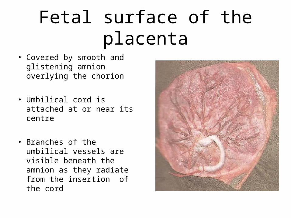
Fetal surface of the placenta• Covered by smooth and
glistening amnion overlying the chorion
• Umbilical cord is attached at or near its centre
• Branches of the umbilical vessels are visible beneath the amnion as they radiate from the insertion of the cord
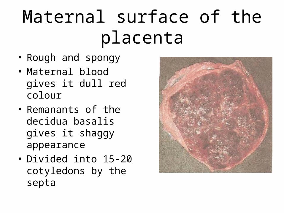
Maternal surface of the placenta
• Rough and spongy• Maternal blood gives
it dull red colour• Remanants of the
decidua basalis gives it shaggy appearance
• Divided into 15-20 cotyledons by the septa
• Margins of the placenta are formed by fused chorionic and the basal plate
• Placenta is attached to the upper part of the uterine body either at the posterior or anterior wall
• After delivery ,placenta separates with the line of separation being through decidua spongiosum (intermediate spongy layer of the decidua basalis
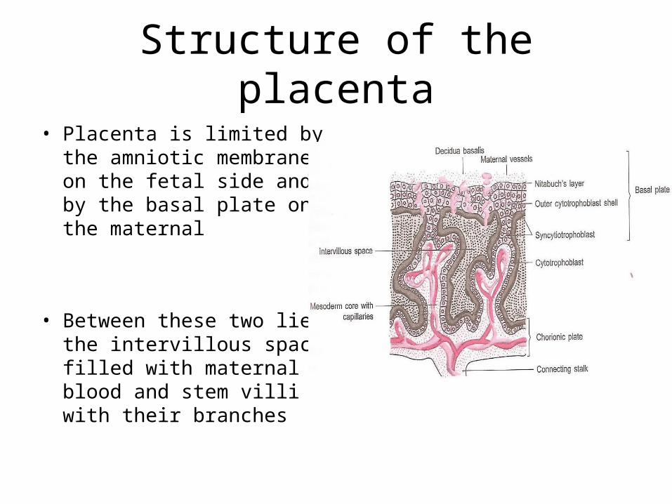
Structure of the placenta• Placenta is limited by the
amniotic membrane on the fetal side and by the basal plate on the maternal
• Between these two lies the intervillous space filled with maternal blood and stem villi with their branches
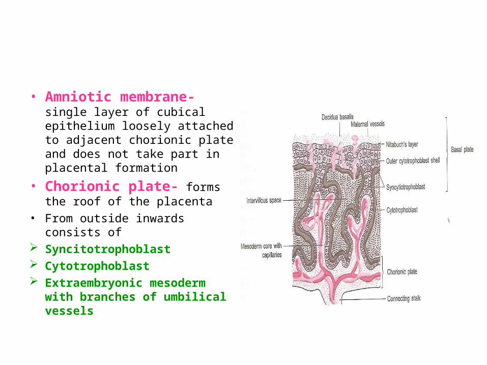
• Amniotic membrane- single layer of cubical epithelium loosely attached to adjacent chorionic plate and does not take part in placental formation
• Chorionic plate- forms the roof of the placenta
• From outside inwards consists of Syncitotrophoblast Cytotrophoblast Extraembryonic mesoderm
with branches of umbilical vessels
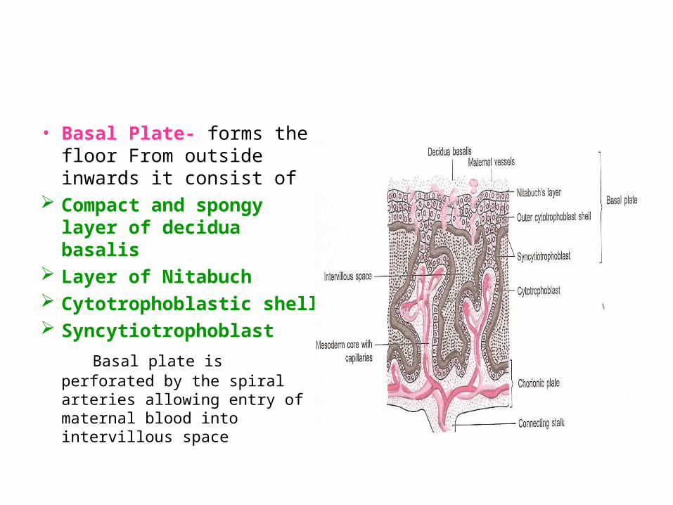
• Basal Plate- forms the floor From outside inwards it consist of
Compact and spongy layer of decidua basalis
Layer of Nitabuch Cytotrophoblastic shell Syncytiotrophoblast Basal plate is perforated by the
spiral arteries allowing entry of maternal blood into intervillous space
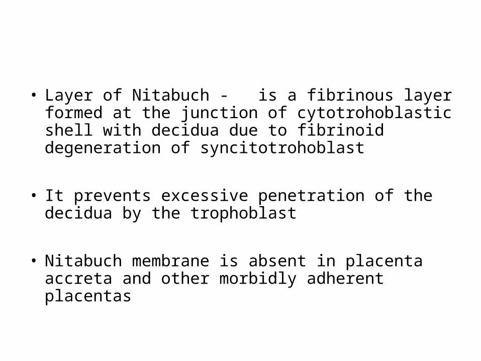
• Layer of Nitabuch - is a fibrinous layer formed at the junction of cytotrohoblastic shell with decidua due to fibrinoid degeneration of syncitotrohoblast
• It prevents excessive penetration of the decidua by the trophoblast
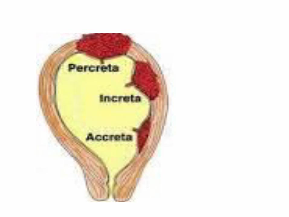
• Nitabuch membrane is absent in placenta accreta and other morbidly adherent placentas
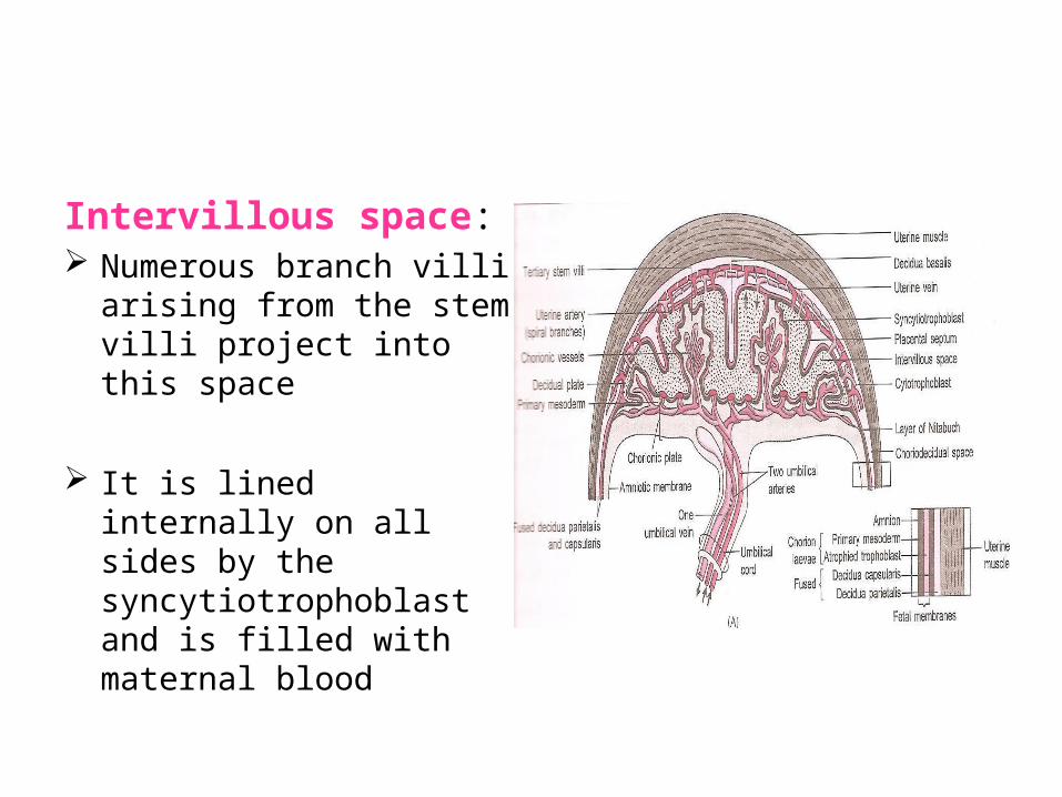
Intervillous space: Numerous branch villi
arising from the stem villi project into this space
It is lined internally on all sides by the syncytiotrophoblast and is filled with maternal blood
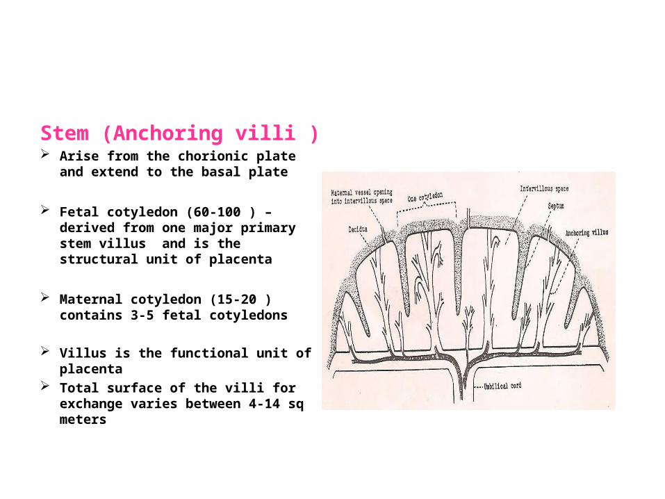
Stem (Anchoring villi ) Arise from the chorionic plate and
extend to the basal plate
Fetal cotyledon (60-100 ) – derived from one major primary stem villus and is the structural unit of placenta
Maternal cotyledon (15-20 ) contains 3-5 fetal cotyledons
Villus is the functional unit of placenta
Total surface of the villi for exchange varies between 4-14 sq meters
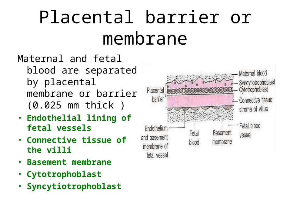
Placental barrier or membraneMaternal and fetal blood
are separated by placental membrane or barrier (0.025 mm thick )
• Endothelial lining of fetal vessels
• Connective tissue of the villi
• Basement membrane• Cytotrophoblast• Syncytiotrophoblast
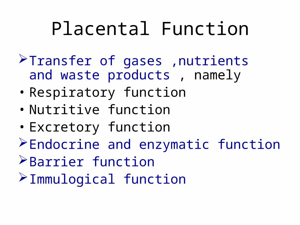
Placental FunctionTransfer of gases ,nutrients and waste
products , namely• Respiratory function• Nutritive function• Excretory functionEndocrine and enzymatic functionBarrier functionImmulogical function
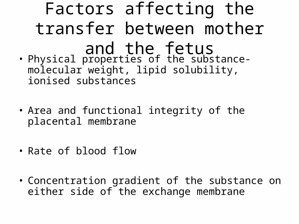
Factors affecting the transfer between mother and the fetus
• Physical properties of the substance- molecular weight, lipid solubility, ionised substances
• Area and functional integrity of the placental membrane
• Rate of blood flow
• Concentration gradient of the substance on either side of the exchange membrane
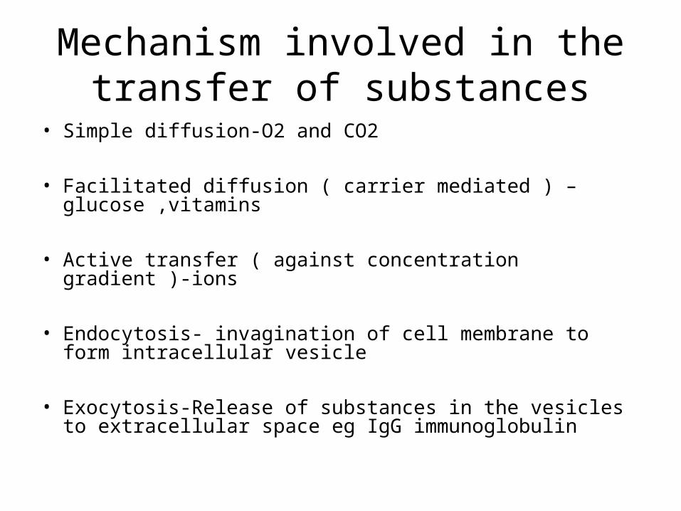
Mechanism involved in the transfer of substances
• Simple diffusion-O2 and CO2
• Facilitated diffusion ( carrier mediated ) –glucose ,vitamins
• Active transfer ( against concentration gradient )-ions
• Endocytosis- invagination of cell membrane to form intracellular vesicle
• Exocytosis-Release of substances in the vesicles to extracellular space eg IgG immunoglobulin
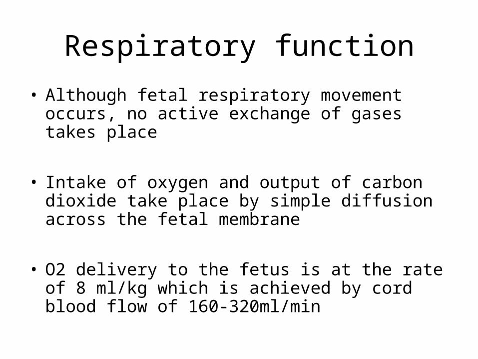
Respiratory function• Although fetal respiratory movement occurs, no
active exchange of gases takes place
• Intake of oxygen and output of carbon dioxide take place by simple diffusion across the fetal membrane
• O2 delivery to the fetus is at the rate of 8 ml/kg which is achieved by cord blood flow of 160-320ml/min
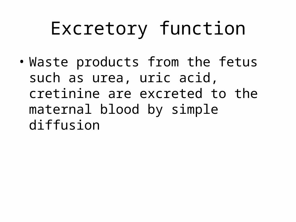
Excretory function
• Waste products from the fetus such as urea, uric acid, cretinine are excreted to the maternal blood by simple diffusion
Nutritive functionFetus obtains its nutrients from the maternal blood
• Glucose- transferred to the fetus by facilitated diffusion
• Lipids for fetal growth and development has dual origin. They are transferred across the fetal membrane or synthesised in the fetus
• Amino acids are transferred by active transport
• Water and electrolytes- Na, K ,Cl cross by simple diffusion, Ca , P, and Fe cross by active transport
• Water soluble vitamins are transferred by active transport but the fat soluble vitamins are transferred slowly
Barrier Function• Placental membrane is thought to be a protective barrier for the
fetus against harmful agents in the maternal blood
• Substances with large molecular weight or size like insulin or heparin are transferred minimally
• Only IgG ( not IgA or Ig M )antibodies and antigens can cross the placental barrier
• Most drugs can cross the placental barrier and some can be teratogenic
• Various viruses, bacteria, protozoa can cross the placenta and affect the fetus in utero
Immunological function
• Inspite of foreign paternally inherited antigens in the fetus and placenta, there is no graft rejection due to immunological protection provided by the placenta
Endocrine and Enzymatic function
• Placenta secretes various hormones – Protein hormones like HCG, human placental lactogen,pregnancy specific beta 1 glycoprotein,,pregnancy associated plasma protein, steroidal hormones like estrogen and progestrone
• Enzymes secreted are diamine oxidase-which activates the circulatory pressor amines,oxytocinase which neutralizes oxytocin, phospholipase A2 which synthesizes arachidonic acid
Placental abnormalities
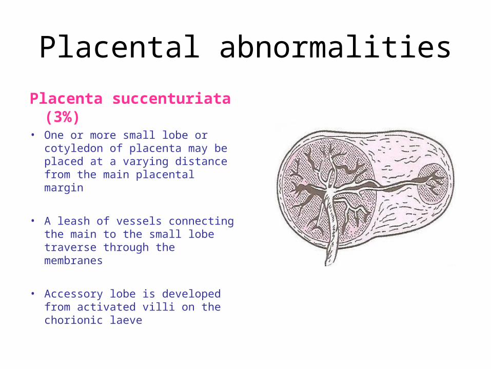
Placenta succenturiata (3%)
• One or more small lobe or cotyledon of placenta may be placed at a varying distance from the main placental margin
• A leash of vessels connecting the main to the small lobe traverse through the membranes
• Accessory lobe is developed from activated villi on the chorionic laeve
Clinical significance- If succenturiate lobe is retained
following birth of placenta it may lead to
PPH Subinvolution Uterine sepsis Polyp formationTreatment- exploration of the uterus
and removal of the lobe
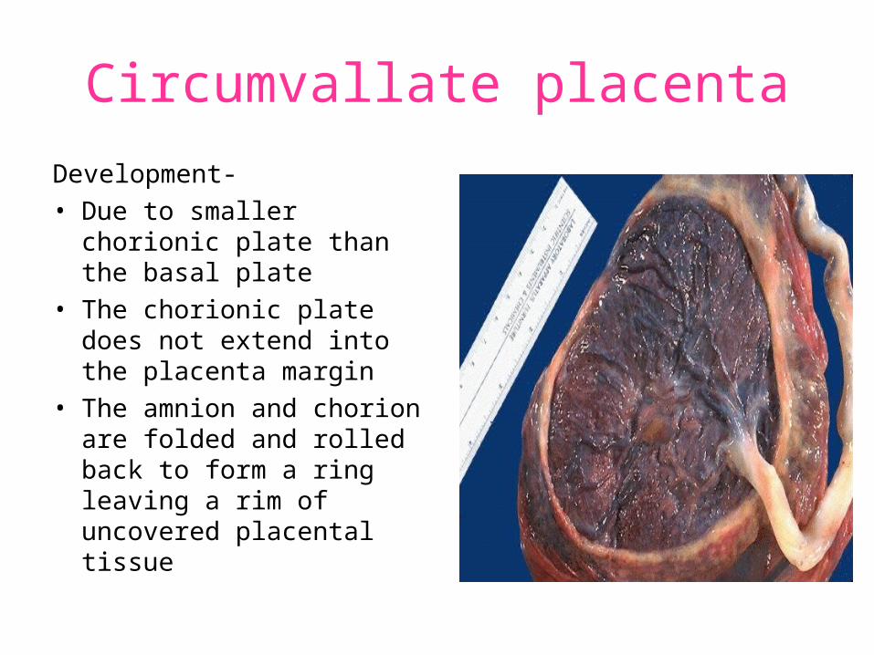
Circumvallate placentaDevelopment- • Due to smaller chorionic
plate than the basal plate• The chorionic plate does
not extend into the placenta margin
• The amnion and chorion are folded and rolled back to form a ring leaving a rim of uncovered placental tissue
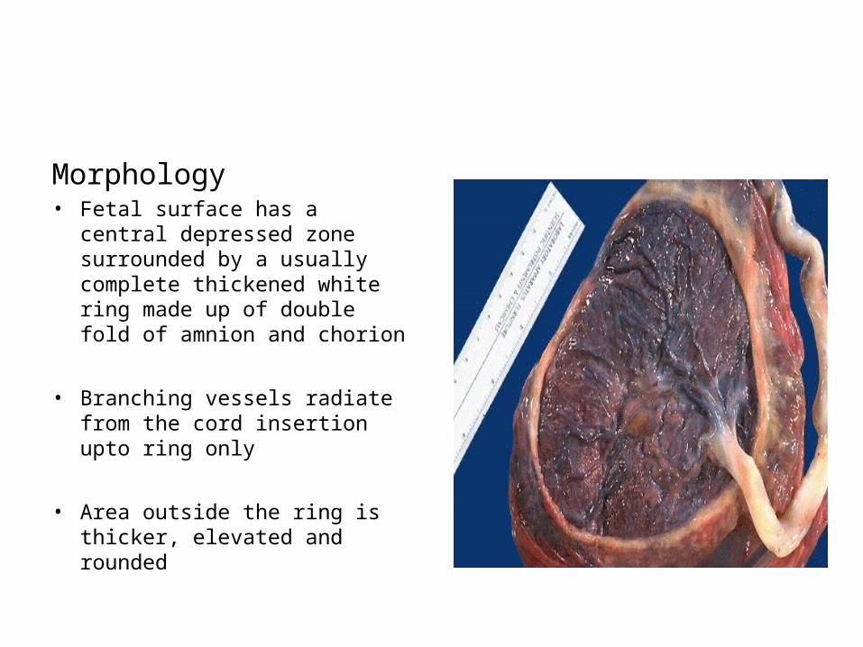
Morphology• Fetal surface has a central
depressed zone surrounded by a usually complete thickened white ring made up of double fold of amnion and chorion
• Branching vessels radiate from the cord insertion upto ring only
• Area outside the ring is thicker, elevated and rounded
Abnormal Placenta
• Placenta Previa.• Placenta acreta.• Placenta increta.• Placenta percreta.