Neuroscience and Biobehavioral Reviews 71 (2016) 865–877
Contents lists available at ScienceDirect
Neuroscience and Biobehavioral Reviews
jou rn al h om epage: www.elsev ier .com/ locate /neubiorev
lant polyphenols as natural drugs for the management of Downyndrome and related disorders
osa Anna Vaccaa,∗, Daniela Valenti a, Salvatore Caccameseb, Maria Dagliac, Nady Braidyd
Seyed Mohammad Nabavie,f,∗∗
Institute of Biomembranes and Bioenergetics, Italian National Council of Research, Bari, ItalyDepartment of Chemical Sciences, University of Catania, ItalyDepartment of Drug Sciences, Medicinal Chemistry and Pharmaceutical Technology Section, University of Pavia, ItalyCentre for Healthy Brain Ageing, School of Psychiatry, University of New South Wales, AustraliaApplied Biotechnology Research Center, Baqiyatallah University of Medical Sciences, P.O. Box 19395 5487, Tehran, IranBioactive Natural Products, Pharmaceutical Sciences Research Center, Tehran University of Medical Sciences, Tehran, Iran
r t i c l e i n f o
rticle history:eceived 19 September 2016eceived in revised form 17 October 2016ccepted 21 October 2016vailable online 5 November 2016
eywords:olyphenolsGCGesveratrol
a b s t r a c t
Polyphenols are secondary metabolites of plants largely found in fruits, vegetables, cereals and beverages,and therefore represent important constituents of the human diet. Increasing studies have demonstratedthe potential beneficial effects of polyphenols on human health. Extensive reviews have discussed theprotective effects of polyphenols against a series of diseases such as cancer, cardiovascular diseases,diabetes, and neurodegenerative disorders. Limited studies have investigated the potential therapeu-tic effects of these natural compounds on neurodevelopmental disorders associated with intellectualdisability, such as Down syndrome (DS), for which mitochondrial dysfunctions and oxidative stressare hallmarks and contribute to the deleterious symptoms and cognitive decline. This review, start-ing from the structure, source, bioavailability and pharmacokinetics of relevant polyphenols, highlights
recent studies on the effect and potential molecular mechanism(s) of action of the phenolic compoundsepigallocatechin-3-gallate, resveratrol and hydroxytyrosol in restoring mitochondrial energy deficit andin reversing phenotypical alteration in DS. The clinical implications of plant polyphenol dietary sup-
Down Syndrome (DS) is the most common chromosomal abnor-ality. About 95% of affected individuals have the free trisomy
f human chromosome 21 i.e. an extra copy of chromosome 21,nd their chromosome count is 47. The leading cause of trisomys attributed to meiotic nondisjunction which occurs mainly in thevum although the reason for this phenomenon is not completelylear (Hultén et al., 2010). Indeed, the maternal origin for trisomyf chromosome 21 is prevailing being less than 10% the cases ofaternal origin and the maternal age plays a major risk factor onhe onset of DS (Coppedè, 2016).
Clinically, DS is a neurodevelopmental disease and the mostrequent genetic cause of intellectual disability characterised byymptoms of premature aging as well as cognitive decline (Griecot al., 2015; Hamlett et al., 2016). The characteristic features ofS are atypical craniofacial profile composed of a combinationf epicanthic folds, flat facial projections and protruding tongue.ffected persons have from mild to severe mental retardation and
Q varies between 25 and 50. Congenital malformations are delete-ious and debilitating. Approximately 40% of patients with DS sufferrom congenital heart defects, immune disorders and increasedusceptibility to infection. Moreover DS patients have higher inci-ence of obesity, diabetes mellitus and lymphoblastic and myeloid
eukaemia than healthy people (Van Cleve and Cohen, 2006; Vanleve et al., 2006).
In recent years, a large number of cytogenetic studies has beenonducted, but the mechanism(s) by which this aneuploidy pro-uces the clinical phenotype and induces cognitive impairment hasot been fully elucidated. In DS, on the long arm of chromosome1 the overexpression of specific genes (i.e. the dual-specificityyrosine (Y)-phosphorylation regulated kinase 1A (DYRK1A) andown Syndrome Critical Region 1 (DSCR1 or RCAN1) genes), haveeen linked to the complex metabolic derangement observed inS (Rachidi and Lopes, 2007). Several studies have also identifiedutritional deficiencies in DS population resulting from imbalance
n biochemical pathways, due to overexpression of other chro-osome 21 genes and their targets. A metabolic derangement of
he homocysteine/folate/transulfuration pathways and abnormalNA methylation have been observed in children with DS involv-
ng cystathionine beta-synthase (CBS) and the folate transporterFC1 genes (Gueant et al., 2005; Iacobazzi et al., 2014).
As well, impaired mitochondria and altered homeostasis ofeactive oxygen species (ROS), and oxidative stress have beenssociated with DS pathogenesis and in the aetiology of otherntellectual disability (Valenti et al., 2014). Oxidative stress iselieved to be due to an imbalance between the chromosome1-encoded superoxide dismutase 1 (SOD1) and glutathione per-xidase activities (Rodríguez-Sureda et al., 2015) as well asitochondrial respiratory chain dysfunctions (Valenti et al., 2011).
lteration in signalling pathways, including cAMP-dependent pro-
ein kinase A (PKA) phosphorylation, NAD-dependent sirtuin-1SIRT1) acetylation and AMP-activated protein kinase (AMPK)-
dependent phosphorylation, have also been associated withimpaired redox metabolism and mitochondrial dysfunction in cellsisolated from both DS patients and murine models of DS (Valentiet al., 2016; Zuo et al., 2014).
Although the overall prognosis of DS has increased over thelast decade due to advances in treatment against infections, DSremains uncured. The majority of patients that reach middleages develop histopathological and neurochemical features whichmimic Alzheimer’s disease (AD) and related dementias (Dick et al.,2016). On this basis, the molecular basis of DS has been investigatedin the hope of increasing our understanding of the neurobiology ofAD and identifying efficacious drug targets.
Over the last two decades, there has been a growing interestfor the use of naturally occurring plant-based polyphenolic com-pounds for the treatment of several degenerative diseases dueto their potent therapeutic effects such as antimicrobial, anti-cancer, antioxidant and anti-inflammatory activities both in vitroand in vivo. The favourable safety profile of polyphenolic com-pounds represents another important advantage of these bioactivemolecules. Several studies have shown that polyphenolic com-pounds possess potent neuroprotective effects under both in vitroand in vivo conditions (Daglia et al., 2014; Nabavi et al., 2014, 2015).Of these compounds, flavonoids are thought to be highly bioac-tive and are found in several plants (Hwang et al., 2012; Nabaviet al., 2012; Vauzour et al., 2013). However, the commercialisa-tion of these compounds is limited because only one in thousandlead molecules can be developed as a successful drug (Sharmaand Gupta, 2015; Molinari, 2009). Newer systematic and scien-tific approaches are necessary for the development of active drugsderived from plants. Moreover, extracting large amounts of com-pounds from natural sources remains a challenge, and requires thedevelopment of newer biotechnology approaches and total organicsynthesis (Atanasov et al., 2015). Despite these shortcomings, cur-rent research suggests that natural products are likely to representa major source of new drugs in the future.
Numerous studies have examined the clinical effects of polyphe-nolic compounds in neurodegenerative pathologies such as AD,supporting the notion that the neuroprotective properties of thesenatural molecules can suppress neuroinflammation and potentiallyenhance memory, learning and cognitive functions (Libro et al.,2016; Pérez-Hernández et al., 2016).
Herein, we focus on recent discoveries concerning the biologicaleffects of plant polyphenols on DS. Emphasis will be given to resver-atrol, epigallocatechin-3-gallate (EGCG), and hydroxytyrosol,whose molecular mechanisms underlying their neuroprotectiveactions extend beyond their usual well-established antioxidant andanti-inflammatory activities. We also review studies which analyzethe effects of other polyphenols exerting protection in neurologicaldiseases associated with cognitive impairment. To give a completepicture of the selected polyphenols, their chemistry, bioavailability
and studies aimed to improve their pharmacokinetic parameterswill also be examined.
R.A. Vacca et al. / Neuroscience and Biobeh
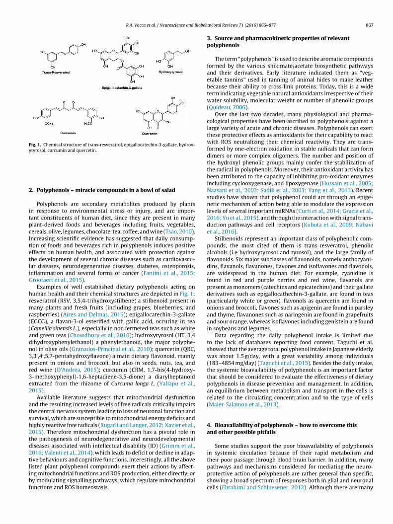
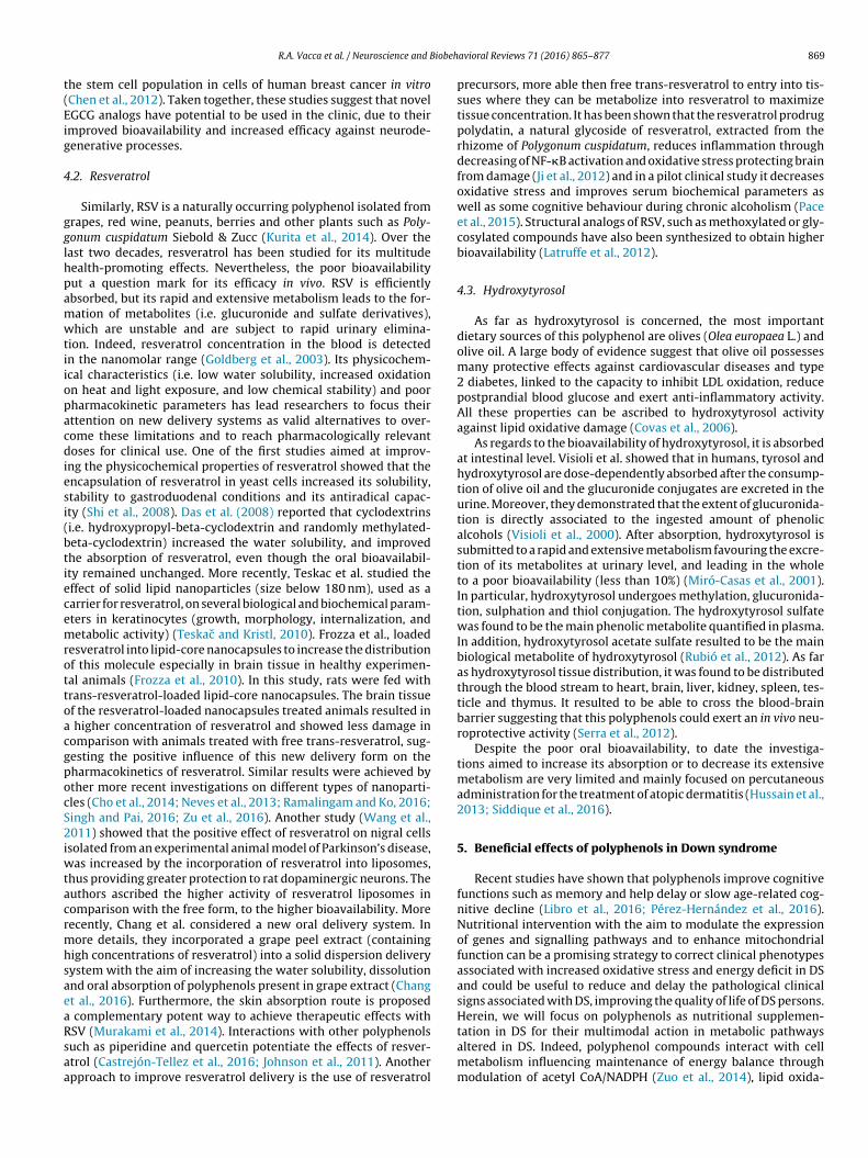
Fig. 1. Chemical structure of trans-resveratrol, epigallocatechin-3-gallate, hydrox-ytyrosol, curcumin and quercetin.
2
itpcItetliG
hrmr((adn3pr3e2
atsh2td2tlibf
pathways and mechanisms considered for mediating the neuro-protective action of polyphenols are rather general than specific,showing a broad spectrum of responses both in glial and neuronal
. Polyphenols – miracle compounds in a bowl of salad
Polyphenols are secondary metabolites produced by plantsn response to environmental stress or injury, and are impor-ant constituents of human diet, since they are present in manylant-derived foods and beverages including fruits, vegetables,ereals, olive, legumes, chocolate, tea, coffee, and wine (Tsao, 2010).ncreasing scientific evidence has suggested that daily consump-ion of foods and beverages rich in polyphenols induces positiveffects on human health, and associated with protection againsthe development of several chronic diseases such as cardiovascu-ar diseases, neurodegenerative diseases, diabetes, osteoporosis,nflammation and several forms of cancer (Fantini et al., 2015;rootaert et al., 2015).
Examples of well established dietary polyphenols acting onuman health and their chemical structures are depicted in Fig. 1:esveratrol (RSV, 3,5,4-trihydroxystilbene) a stilbenoid present inany plants and fresh fruits (including grapes, blueberries, and
aspberries) (Aires and Delmas, 2015); epigallocatechin-3-gallateEGCG), a flavan-3-ol esterified with gallic acid, occuring in teaCamellia sinensis L.), especially in non fermeted teas such as whitend green teas (Chowdhury et al., 2016); hydroxytyrosol (HT, 3,4ihydroxyphenylethanol) a phenylethanoid, the major polyphe-ol in olive oils (Granados-Principal et al., 2010); quercetin (QRC,,3′,4′,5,7-pentahydroxyflavone) a main dietary flavonoid, mainlyresent in onions and broccoli, but also in seeds, nuts, tea, anded wine (D’Andrea, 2015); curcumin (CRM, 1,7-bis(4-hydroxy--methoxyphenyl)-1,6-heptadiene-3,5-dione) a diarylheptanoidxtracted from the rhizome of Curcuma longa L. (Yallapu et al.,015).
Available literature suggests that mitochondrial dysfunctionnd the resulting increased levels of free radicals critically impairshe central nervous system leading to loss of neuronal function andurvival, which are susceptible to mitochondrial energy deficits andighly reactive free radicals (Rugarli and Langer, 2012; Xavier et al.,015). Therefore mitochondrial dysfunction has a pivotal role inhe pathogenesis of neurodegenerative and neurodevelopmentaliseases associated with intellectual disability (ID) (Grimm et al.,016; Valenti et al., 2014), which leads to deficit or decline in adap-ive behaviours and cognitive functions. Interestingly, all the aboveisted plant polyphenol compounds exert their actions by affect-ng mitochondrial functions and ROS production, either directly, ory modulating signalling pathways, which regulate mitochondrialunctions and ROS homeostasis.
avioral Reviews 71 (2016) 865–877 867
3. Source and pharmacokinetic properties of relevantpolyphenols
The term “polyphenols” is used to describe aromatic compoundsformed by the various shikimate/acetate biosynthetic pathwaysand their derivatives. Early literature indicated them as “veg-etable tannins” used in tanning of animal hides to make leatherbecause their ability to cross-link proteins. Today, this is a wideterm indicating vegetable natural antioxidants irrespective of theirwater solubility, molecular weight or number of phenolic groups(Quideau, 2006).
Over the last two decades, many physiological and pharma-cological properties have been ascribed to polyphenols against alarge variety of acute and chronic diseases. Polyphenols can exertthese protective effects as antioxidants for their capability to reactwith ROS neutralizing their chemical reactivity. They are trans-formed by one-electron oxidation in stable radicals that can formdimers or more complex oligomers. The number and position ofthe hydroxyl phenolic groups mainly confer the stabilization ofthe radical in polyphenols. Moreover, their antioxidant activity hasbeen attributed to the capacity of inhibiting pro-oxidant enzymesincluding cyclooxygenase, and lipoxygenase (Hussain et al., 2005;Naasani et al., 2003; Sadik et al., 2003; Yang et al., 2013). Recentstudies have shown that polyphenol could act through an epige-netic mechanism of action being able to modulate the expressionlevels of several important miRNAs (Curti et al., 2014; Gracia et al.,2016; Yu et al., 2015), and through the interaction with signal trans-duction pathways and cell receptors (Kubota et al., 2009; Nabaviet al., 2016).
Stilbenoids represent an important class of polyphenolic com-pounds, the most cited of them is trans-resveratrol, phenolicalcohols (i.e hydroxytyrosol and tyrosol), and the large family offlavonoids. Six major subclasses of flavonoids, namely anthocyani-dins, flavanols, flavanones, flavones and isoflavones and flavonols,are widespread in the human diet. For example, cyanidine isfound in red and purple berries and red wine, flavanols arepresent as monomers (catechins and epicatechins) and their gallatederivatives such as epigallocathechin-3-gallate, are found in teas(particularly white or green), flavonols as quercetin are found inonions and broccoli, flavones such as apigenin are found in parsleyand thyme, flavanones such as naringenin are found in grapefruitsand sour orange, whereas isoflavones including genistein are foundin soybeans and legumes.
Data regarding the daily polyphenol intake is limited dueto the lack of databases reporting food content. Taguchi et al.showed that the average total polyphenol intake in Japanese elderlywas about 1.5 g/day, with a great variability among individuals(183–4854 mg/day) (Taguchi et al., 2015). Besides the daily intake,the systemic bioavailability of polyphenols is an important factorthat should be considered to evaluate the effectiveness of dietarypolyphenols in disease prevention and management. In addition,an equilibrium between metabolism and transport in the cells isrelated to the circulating concentration and to the type of cells(Maier-Salamon et al., 2013).
4. Bioavailability of polyphenols – how to overcome thisand other possible pitfalls
Some studies support the poor bioavailability of polyphenolsin systemic circulation because of their rapid metabolism andtheir poor passage through blood brain barrier. In addition, many
cells (Ebrahimi and Schluesener, 2012). Although there are many
868 R.A. Vacca et al. / Neuroscience and Biobehavioral Reviews 71 (2016) 865–877
Table 1Approaches to improve polyphenols bioavailability.
EGCG, RSV, QCT Zhang et al., 2013; Das et al., 2008; Frozza et al., 2010
Prodrugs PolydatinePro-EGCG
RSV, EGCG Ji et al., 2012; Pace et al., 2015; Ahmed et al., 2016
Analogs Structural analogs, RSV, EGCG Latruffe et al., 2012; Chen et al., 2012
E
ipDdmplcItFtt2moelo
np
4
teca(lp1Aftotiba
bniaTi
methoxylated or glycosylated compoundsAlternative to oral delivery Percutaneous
Transdermal
n vivo and in vitro investigations on neuroprotective effects ofolyphenols, their pharmacokinetic properties (ADME, Absorption,istribution, Metabolism, Elimination) have not been fully eluci-ated. The discussed health effects of natural polyphenols dependainly on their bioavailability. After oral intake and absorption,
olyphenols undergo extensive enzymatic modifications, whichead to the synthesis of glucuronidated, methylated and sulphatedompounds at intestinal and liver levels (D’Archivio et al., 2010).ndeed, cells recognize plant polyphenols as xenobiotic compoundshat are quickly enzymatically modified in order to eliminate them.or example, the clinical administration of curcumin is limited dueo poor solubility and limited adsorption in the gastrointestinalract, its rapid metabolism and rapid renal clearance (Gupta et al.,013). Quercetin is present in foods as various glycosides, which areore bioavailable than the free aglycone (quercetin itself) because
f efficient deglycosylation process at the intestinal level (Russot al., 2012). The poor bioavailability of the aglycon form due toowered absorption in the gastrointestinal tract raises a questionf the clinical translation of quercetin.
The bioavailability and the pharmacokinetics of plant polyphe-ols can be employed to potentially enhance the delivery ofolyphenols and optimize their efficacy in humans (see Table 1).
.1. Epigallocatechin-3-gallate
EGCG is the most common bioactive catechin present in greenea. The portion that is not degraded during digestion (Marcheset al., 2014) is adsorbed at the intestinal level through epithelialells. Afterwards, it is metabolized in the liver to give glucuronidend sulfide metabolite and, especially, methylated metabolitesYong Feng, 2006). In the blood, EGCG has been found to be methy-ated, as reported by Meng and co-workers. The human plasmaeak concentrations of EGCG and its methylated form were about45 and 20 nM, respectively, after 2 h following green tea ingestion.fter 24 h, the urinary excretion of EGCG methylated derivative was
ound to be approximately 140 �g, corresponding to about 0.1% ofhe ingested EGCG (Meng et al., 2002). Although the bioavailabilityf EGCG is poor due to the degradation occurring during diges-ion, poor absorption, and rapid metabolism and excretion, EGCGs a promising substance. In addition, it has been found to crosslood-brain-barrier, which is a prerequisite for the efficacy of EGCGgainst neurodegenerative disorders (Li et al., 2012; Lin et al., 2007).
Considering the beneficial properties of EGCG and its lowioavailability, many recent studies have focused on the search ofew delivery forms and combinations of substances to improve
ts pharmacokinetic properties. One of the earliest studies thatddressed the problem was published in 2004 by Lambert et al.he study showed that the combination of black pepper piper-ne and EGCG (EGCG/piperine 163.8/70.2 �mol/kg) increased the
GCG, RSV, HT Lambert et al., 2006; Hussain et al., 2013
plasma concentration of EGCG by 1.3-fold in comparison withmice treated with EGCG alone. This increase seemed to be causedby the inhibition of EGCG glucuronidation at gastrointestinal lev-els, thus slowing its metabolism (Lambert et al., 2004). The sameresearch group showed that genistein exerted the same effect ofpiperine in an in vitro model system (HT-29 human colon cancercells) (Lambert et al., 2008). Similar results were achieved by Wanget al. and Kale et al. that showed that co-treatment with quercetininduced an increase in EGCG bioavailability (Kale et al., 2010; Wanget al., 2012). The possibility of increasing EGCG bioavailability withnutrient combinations was also confirmed in humans by Gawandeet al. (2008). The study showed that supplementation of 5 humanvolunteers with a single dose of green tea catechins, ascorbic acid,selenium, N-acetyl cysteine, and black grapes induced an increasein EGCG bioavailability by 27%. More recently, Giunta et al. reportedthat in Tg2576 mice the treatment with EGCG (62.5 mg/kg/day or12.5 mg/kg/day) and fish oil (8mg/kg/day), which induced a sta-tistically significant increase in EGCG bioavailability (Giunta et al.,2010), showed a synergist effect on the inhibition of cerebral beta-amyloid peptide deposits, which are implicated in the pathogenesisof AD. As far as new delivery forms of EGCG are concerned, Lambertet al. reported that transdermal gel, containing EGCG applied onSKH-1 mice epidermis (50 mg/kg), could be an alternative promis-ing form, to deliver EGCG to plasma and tissues, in comparisonwith the oral form (Lambert et al., 2006). More recently, a num-ber of studies have focused on using nanotechnology to improveEGCG bioavailability; the effect of EGCG encapsulated chitosan-coated nanoliposomes on the intracellular concentration of EGCGin THP-1-derived macrophages and MCF7 breast cancer cells hasbeen reported (de Pace et al., 2013; Zhang et al., 2013). The resultsshowed that these nanoliposomes significantly increase the stabil-ity and the concentration of EGCG in the cells, improving also EGCGbioactivities.
Several EGCG analogues have been also developed. Pro-EGCG,a prodrug of EGCG has been shown to demonstrate greater sta-bility, and higher bioavailability and biological activity in vivo, incomparison to the naturally derived EGCG (Ahmed et al., 2016).Similarly, compounds 2a and 4a showed lower susceptibility tomethylation/inhibition by the catalytic activity of catechol-O-methyltransferase, a key enzyme involved in the metabolism ofEGCG (Ahmed et al., 2016). Additionally, these drugs demonstratedpotent antiproliferative, antiangiogenic, and antifibrotic activitiesin human uterine leiomyoma cells in vitro. Another study showedthat synthetic EGCG analogs 4 and 6 resulted to be more potentAMPK activators than EGCG and metformin, which is the drug com-
monly used to treat type 2 diabetes. Activation of AMPK by theseEGCG synthetic analogs inhibited cell proliferation, induced theover expression of the cyclin-dependent kinase inhibitor p21 andthe down-regulation of mTOR signaling pathway, and suppressed
he stem cell population in cells of human breast cancer in vitroChen et al., 2012). Taken together, these studies suggest that novelGCG analogs have potential to be used in the clinic, due to theirmproved bioavailability and increased efficacy against neurode-enerative processes.
.2. Resveratrol
Similarly, RSV is a naturally occurring polyphenol isolated fromrapes, red wine, peanuts, berries and other plants such as Poly-onum cuspidatum Siebold & Zucc (Kurita et al., 2014). Over theast two decades, resveratrol has been studied for its multitudeealth-promoting effects. Nevertheless, the poor bioavailabilityut a question mark for its efficacy in vivo. RSV is efficientlybsorbed, but its rapid and extensive metabolism leads to the for-ation of metabolites (i.e. glucuronide and sulfate derivatives),hich are unstable and are subject to rapid urinary elimina-
ion. Indeed, resveratrol concentration in the blood is detectedn the nanomolar range (Goldberg et al., 2003). Its physicochem-cal characteristics (i.e. low water solubility, increased oxidationn heat and light exposure, and low chemical stability) and poorharmacokinetic parameters has lead researchers to focus theirttention on new delivery systems as valid alternatives to over-ome these limitations and to reach pharmacologically relevantoses for clinical use. One of the first studies aimed at improv-
ng the physicochemical properties of resveratrol showed that thencapsulation of resveratrol in yeast cells increased its solubility,tability to gastroduodenal conditions and its antiradical capac-ty (Shi et al., 2008). Das et al. (2008) reported that cyclodextrinsi.e. hydroxypropyl-beta-cyclodextrin and randomly methylated-eta-cyclodextrin) increased the water solubility, and improvedhe absorption of resveratrol, even though the oral bioavailabil-ty remained unchanged. More recently, Teskac et al. studied theffect of solid lipid nanoparticles (size below 180 nm), used as aarrier for resveratrol, on several biological and biochemical param-ters in keratinocytes (growth, morphology, internalization, andetabolic activity) (Teskac and Kristl, 2010). Frozza et al., loaded
esveratrol into lipid-core nanocapsules to increase the distributionf this molecule especially in brain tissue in healthy experimen-al animals (Frozza et al., 2010). In this study, rats were fed withrans-resveratrol-loaded lipid-core nanocapsules. The brain tissuef the resveratrol-loaded nanocapsules treated animals resulted in
higher concentration of resveratrol and showed less damage inomparison with animals treated with free trans-resveratrol, sug-esting the positive influence of this new delivery form on theharmacokinetics of resveratrol. Similar results were achieved byther more recent investigations on different types of nanoparti-les (Cho et al., 2014; Neves et al., 2013; Ramalingam and Ko, 2016;ingh and Pai, 2016; Zu et al., 2016). Another study (Wang et al.,011) showed that the positive effect of resveratrol on nigral cells
solated from an experimental animal model of Parkinson’s disease,as increased by the incorporation of resveratrol into liposomes,
hus providing greater protection to rat dopaminergic neurons. Theuthors ascribed the higher activity of resveratrol liposomes inomparison with the free form, to the higher bioavailability. Moreecently, Chang et al. considered a new oral delivery system. Inore details, they incorporated a grape peel extract (containing
igh concentrations of resveratrol) into a solid dispersion deliveryystem with the aim of increasing the water solubility, dissolutionnd oral absorption of polyphenols present in grape extract (Changt al., 2016). Furthermore, the skin absorption route is proposed
complementary potent way to achieve therapeutic effects with
SV (Murakami et al., 2014). Interactions with other polyphenolsuch as piperidine and quercetin potentiate the effects of resver-trol (Castrejón-Tellez et al., 2016; Johnson et al., 2011). Anotherpproach to improve resveratrol delivery is the use of resveratrol
avioral Reviews 71 (2016) 865–877 869
precursors, more able then free trans-resveratrol to entry into tis-sues where they can be metabolize into resveratrol to maximizetissue concentration. It has been shown that the resveratrol prodrugpolydatin, a natural glycoside of resveratrol, extracted from therhizome of Polygonum cuspidatum, reduces inflammation throughdecreasing of NF-�B activation and oxidative stress protecting brainfrom damage (Ji et al., 2012) and in a pilot clinical study it decreasesoxidative stress and improves serum biochemical parameters aswell as some cognitive behaviour during chronic alcoholism (Paceet al., 2015). Structural analogs of RSV, such as methoxylated or gly-cosylated compounds have also been synthesized to obtain higherbioavailability (Latruffe et al., 2012).
4.3. Hydroxytyrosol
As far as hydroxytyrosol is concerned, the most importantdietary sources of this polyphenol are olives (Olea europaea L.) andolive oil. A large body of evidence suggest that olive oil possessesmany protective effects against cardiovascular diseases and type2 diabetes, linked to the capacity to inhibit LDL oxidation, reducepostprandial blood glucose and exert anti-inflammatory activity.All these properties can be ascribed to hydroxytyrosol activityagainst lipid oxidative damage (Covas et al., 2006).
As regards to the bioavailability of hydroxytyrosol, it is absorbedat intestinal level. Visioli et al. showed that in humans, tyrosol andhydroxytyrosol are dose-dependently absorbed after the consump-tion of olive oil and the glucuronide conjugates are excreted in theurine. Moreover, they demonstrated that the extent of glucuronida-tion is directly associated to the ingested amount of phenolicalcohols (Visioli et al., 2000). After absorption, hydroxytyrosol issubmitted to a rapid and extensive metabolism favouring the excre-tion of its metabolites at urinary level, and leading in the wholeto a poor bioavailability (less than 10%) (Miró-Casas et al., 2001).In particular, hydroxytyrosol undergoes methylation, glucuronida-tion, sulphation and thiol conjugation. The hydroxytyrosol sulfatewas found to be the main phenolic metabolite quantified in plasma.In addition, hydroxytyrosol acetate sulfate resulted to be the mainbiological metabolite of hydroxytyrosol (Rubió et al., 2012). As faras hydroxytyrosol tissue distribution, it was found to be distributedthrough the blood stream to heart, brain, liver, kidney, spleen, tes-ticle and thymus. It resulted to be able to cross the blood-brainbarrier suggesting that this polyphenols could exert an in vivo neu-roprotective activity (Serra et al., 2012).
Despite the poor oral bioavailability, to date the investiga-tions aimed to increase its absorption or to decrease its extensivemetabolism are very limited and mainly focused on percutaneousadministration for the treatment of atopic dermatitis (Hussain et al.,2013; Siddique et al., 2016).
5. Beneficial effects of polyphenols in Down syndrome
Recent studies have shown that polyphenols improve cognitivefunctions such as memory and help delay or slow age-related cog-nitive decline (Libro et al., 2016; Pérez-Hernández et al., 2016).Nutritional intervention with the aim to modulate the expressionof genes and signalling pathways and to enhance mitochondrialfunction can be a promising strategy to correct clinical phenotypesassociated with increased oxidative stress and energy deficit in DSand could be useful to reduce and delay the pathological clinicalsigns associated with DS, improving the quality of life of DS persons.Herein, we will focus on polyphenols as nutritional supplemen-
tation in DS for their multimodal action in metabolic pathwaysaltered in DS. Indeed, polyphenol compounds interact with cellmetabolism influencing maintenance of energy balance throughmodulation of acetyl CoA/NADPH (Zuo et al., 2014), lipid oxida-
8 iobeh
te(tsdeiNt
5m
irtrrerlfoapros
mtiieeDt3mtpietdhe
cmc(dsliwaadtbi
70 R.A. Vacca et al. / Neuroscience and B
ion (Wang et al., 2015), homocysteine metabolism (Kołodziejczykt al., 2011) and through inhibition of oxidative DNA damageWang et al., 2014). In additions, polyphenols can modulate signalransduction pathways, which regulate mitochondrial functionsuch as respiration, oxidative phosphorylation and mitochondrial-ependent apoptosis. Polyphenols also regulate ROS homeostasisfficiently scavenging mitochondrial ROS and upregulate antiox-dant transcriptional programmes in cells (Gibellini et al., 2015)otwithstanding this, until now only EGCG, RSV and HT have been
ested in DS.
.1. Epigallocatechin-3-gallate in DS: studies in vitro, in animalodels and in humans
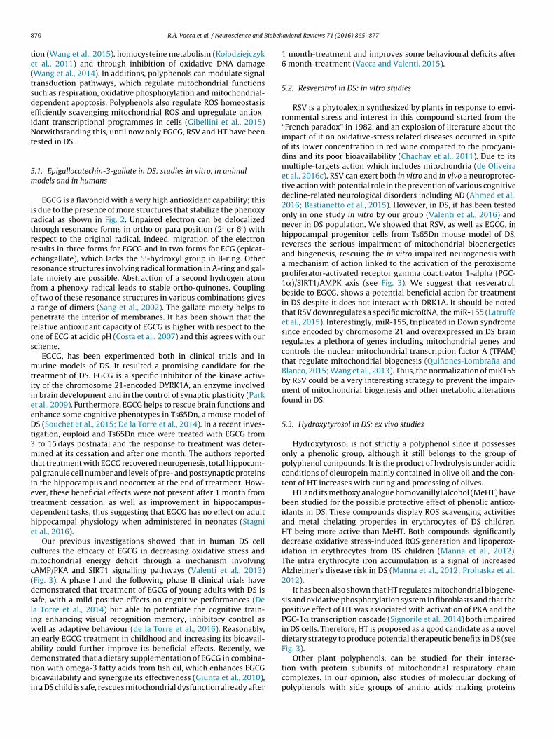
EGCG is a flavonoid with a very high antioxidant capability; thiss due to the presence of more structures that stabilize the phenoxyadical as shown in Fig. 2. Unpaired electron can be delocalizedhrough resonance forms in ortho or para position (2′ or 6′) withespect to the original radical. Indeed, migration of the electronesults in three forms for EGCG and in two forms for ECG (epicat-chingallate), which lacks the 5′-hydroxyl group in B-ring. Otheresonance structures involving radical formation in A-ring and gal-ate moiety are possible. Abstraction of a second hydrogen atomrom a phenoxy radical leads to stable ortho-quinones. Couplingf two of these resonance structures in various combinations gives
range of dimers (Sang et al., 2002). The gallate moiety helps toenetrate the interior of membranes. It has been shown that theelative antioxidant capacity of EGCG is higher with respect to thene of ECG at acidic pH (Costa et al., 2007) and this agrees with ourcheme.
EGCG, has been experimented both in clinical trials and inurine models of DS. It resulted a promising candidate for the
reatment of DS. EGCG is a specific inhibitor of the kinase activ-ty of the chromosome 21-encoded DYRK1A, an enzyme involvedn brain development and in the control of synaptic plasticity (Parkt al., 2009). Furthermore, EGCG helps to rescue brain functions andnhance some cognitive phenotypes in Ts65Dn, a mouse model ofS (Souchet et al., 2015; De la Torre et al., 2014). In a recent inves-
igation, euploid and Ts65Dn mice were treated with EGCG from to 15 days postnatal and the response to treatment was deter-ined at its cessation and after one month. The authors reported
hat treatment with EGCG recovered neurogenesis, total hippocam-al granule cell number and levels of pre- and postsynaptic proteins
n the hippocampus and neocortex at the end of treatment. How-ver, these beneficial effects were not present after 1 month fromreatment cessation, as well as improvement in hippocampus-ependent tasks, thus suggesting that EGCG has no effect on adultippocampal physiology when administered in neonates (Stagnit al., 2016).
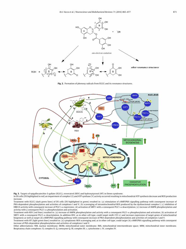
Our previous investigations showed that in human DS cellultures the efficacy of EGCG in decreasing oxidative stress anditochondrial energy deficit through a mechanism involving
AMP/PKA and SIRT1 signalling pathways (Valenti et al., 2013)Fig. 3). A phase I and the following phase II clinical trials haveemonstrated that treatment of EGCG of young adults with DS isafe, with a mild positive effects on cognitive performances (Dea Torre et al., 2014) but able to potentiate the cognitive train-ng enhancing visual recognition memory, inhibitory control as
ell as adaptive behaviour (de la Torre et al., 2016). Reasonably,n early EGCG treatment in childhood and increasing its bioavail-bility could further improve its beneficial effects. Recently, we
emonstrated that a dietary supplementation of EGCG in combina-ion with omega-3 fatty acids from fish oil, which enhances EGCGioavailability and synergize its effectiveness (Giunta et al., 2010),
n a DS child is safe, rescues mitochondrial dysfunction already after
avioral Reviews 71 (2016) 865–877
1 month-treatment and improves some behavioural deficits after6 month-treatment (Vacca and Valenti, 2015).
5.2. Resveratrol in DS: in vitro studies
RSV is a phytoalexin synthesized by plants in response to envi-ronmental stress and interest in this compound started from the“French paradox” in 1982, and an explosion of literature about theimpact of it on oxidative-stress related diseases occurred in spiteof its lower concentration in red wine compared to the procyani-dins and its poor bioavailability (Chachay et al., 2011). Due to itsmultiple-targets action which includes mitochondria (de Oliveiraet al., 2016c), RSV can exert both in vitro and in vivo a neuroprotec-tive action with potential role in the prevention of various cognitivedecline-related neurological disorders including AD (Ahmed et al.,2016; Bastianetto et al., 2015). However, in DS, it has been testedonly in one study in vitro by our group (Valenti et al., 2016) andnever in DS population. We showed that RSV, as well as EGCG, inhippocampal progenitor cells from Ts65Dn mouse model of DS,reverses the serious impairment of mitochondrial bioenergeticsand biogenesis, rescuing the in vitro impaired neurogenesis witha mechanism of action linked to the activation of the peroxisomeproliferator-activated receptor gamma coactivator 1-alpha (PGC-1�)/SIRT1/AMPK axis (see Fig. 3). We suggest that resveratrol,beside to EGCG, shows a potential beneficial action for treatmentin DS despite it does not interact with DRK1A. It should be notedthat RSV downregulates a specific microRNA, the miR-155 (Latruffeet al., 2015). Interestingly, miR-155, triplicated in Down syndromesince encoded by chromosome 21 and overexpressed in DS brainregulates a plethora of genes including mitochondrial genes andcontrols the nuclear mitochondrial transcription factor A (TFAM)that regulate mitochondrial biogenesis (Quinones-Lombrana andBlanco, 2015; Wang et al., 2013). Thus, the normalization of miR155by RSV could be a very interesting strategy to prevent the impair-ment of mitochondrial biogenesis and other metabolic alterationsfound in DS.
5.3. Hydroxytyrosol in DS: ex vivo studies
Hydroxytyrosol is not strictly a polyphenol since it possessesonly a phenolic group, although it still belongs to the group ofpolyphenol compounds. It is the product of hydrolysis under acidicconditions of oleuropein mainly contained in olive oil and the con-tent of HT increases with curing and processing of olives.
HT and its methoxy analogue homovanillyl alcohol (MeHT) havebeen studied for the possible protective effect of phenolic antiox-idants in DS. These compounds display ROS scavenging activitiesand metal chelating properties in erythrocytes of DS children,HT being more active than MeHT. Both compounds significantlydecrease oxidative stress-induced ROS generation and lipoperox-idation in erythrocytes from DS children (Manna et al., 2012).The intra erythrocyte iron accumulation is a signal of increasedAlzheimer’s disease risk in DS (Manna et al., 2012; Prohaska et al.,2012).
It has been also shown that HT regulates mitochondrial biogene-sis and oxidative phosphorylation system in fibroblasts and that thepositive effect of HT was associated with activation of PKA and thePGC-1� transcription cascade (Signorile et al., 2014) both impairedin DS cells. Therefore, HT is proposed as a good candidate as a noveldietary strategy to produce potential therapeutic benefits in DS (seeFig. 3).
Other plant polyphenols, can be studied for their interac-tion with protein subunits of mitochondrial respiratory chaincomplexes. In our opinion, also studies of molecular docking ofpolyphenols with side groups of amino acids making proteins
R.A. Vacca et al. / Neuroscience and Biobehavioral Reviews 71 (2016) 865–877 871
Fig. 2. Formation of phenoxy radicals from EGCG and its resonance structures.
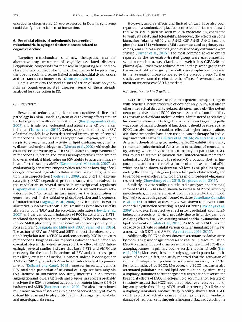
Fig. 3. Targets of epigallocatechin-3-gallate (EGCG), resveratrol (RSV) and hydroxytyrosol (HT) in Down syndrome.In DS cells (DS highlighted in red) an impairment of complex I (I) and ATP synthase (V) activity occurred resulting in mitochondrial ATP synthesis decrease and ROS productionincrease.Treatment with EGCG (dark green lines) of DS cells (DS highlighted in green) resulted in: (a) stimulation of cAMP/PKA signalling pathway with consequent increase ofPKA-dependent phosphorylation and activities of complexes I and V; (b) scavenging of intramitochondrial ROS produced by the dysfunctional complex I; (c) inhibition ofDRK1A activity with consequent increase of PGC1-� expression; (d) activation of SIRT1 with a consequent PGC1-� deacetylation; (e) increase of AMPK phosphorylation andactivity with a consequent PGC1-� phosphorylation and activation.Treatment with RSV (red lines) resulted in: (a) increase of AMPK phosphorylation and activity with a consequent PGC1-� phosphorylation and activation; (b) activation ofSIRT1 with a consequent PGC1-� deacetylation. In addition RSV, as in other cell type, could target mirR-155 (c) and increase expression of target genes of mitochondrialbiogenesis as well as target (d) cAMP/PKA signalling pathway with consequent increase of PKA-dependent phosphorylation and activities of complexes I and V.Treatment with HT (light green lines) resulted in: (a) cytoplasmic ROS scavenging and, as in other cell type, could target (b) cAMP/PKA signalling pathway with consequentincrease of PKA-dependent phosphorylation and activities of complexes I and V.Other abbreviations: NM, nuclear membrane; MOM, mitochondrial outer membrane; IMS, mitochondrial intermembrane space; MIM, mitochondrial inner membrane.Respiratory chain complexes: II, complex II; Q, coenzyme Q; III, complex III; c, cytochrome c; IV, complex IV.
8 iobeh
ec
6mc
aPsta
na
6
pt2iomrwemkleetct(vgoaa2mirTtmeentAiR(aiimea
72 R.A. Vacca et al. / Neuroscience and B
ncoded in chromosome 21 overexpressed in Down’s syndromeould clarify the mechanism of interaction.
. Beneficial effects of polyphenols by targetingitochondria in aging and other diseases related to
ognitive decline
Targeting mitochondria is a new therapeutic area forlternative-drug treatment of cognitive-associated diseases.olyphenolic compounds for their role in regulating ROS homeo-tasis and modulating mitochondrial function could be promisingherapeutic tools in diseases linked to mitochondrial dysfunctionsnd aberrant redox homeostasis (Arun et al., 2016).
Herein we review the mechanisms of action of some polyphe-ols in cognitive-associated diseases, some of them alreadynalysed for their action in DS.
.1. Resveratrol
Resveratrol reduces aging-dependent cognitive decline andathology in animal models system of AD exerting effects similaro that registered with caloric restriction (Karuppagounder et al.,009) and is safe, well-tolerated, and alters some AD biomarker
n human (Turner et al., 2015). Dietary supplementation with RSVf animal models have been determined improvement of severalitochondrial functions such as oxygen consumption, activity of
espiratory enzymes, and activity of lipid-oxidizing enzymes asell as mitochondrial biogenesis (Murase et al., 2009). Although the
xact molecular events by which RSV mediates the improvement ofitochondrial function and exerts neuroprotective action are not
nown in detail, it likely relies on RSV ability to activate intracel-ular effectors such as AMPK (Dasgupta and Milbrandt, 2007), anvolutionarily conserved enzyme which senses the lowering of cellnergy status and regulates cellular survival with emerging func-ion in neuroprotection (Poels et al., 2009), and SIRT1 an enzymeatalyzing NAD+-dependent protein deacetylation, involved inhe modulation of several metabolic transcriptional regulatorsLagouge et al., 2006). Both SIRT1 and AMPK are well known acti-ators of PGC-1�, which in turn improves the transcription ofenes involved in the oxidative phosphorylation and biogenesisf mitochondria (Lagouge et al., 2006). RSV has been shown tollosterically interact with SIRT1, thus resulting in the increase of itsffinity for both NAD+ and the acetylated substrates (Howitz et al.,003) and the consequent induction of PGC1� activity by SIRT1-ediated deacetylation. On the other hand, RSV has been shown to
nduce AMPK phosphorylation in neuronal cell lines, primary neu-ons and brain (Dasgupta and Milbrandt, 2007; Valenti et al., 2016).he action of RSV on AMPK and SIRT1 impact the phosphoryla-ion/acetylation status of PGC1� and consequently PGC1� activates
itochondrial biogenesis and improves mitochondrial function, anssential step in the whole neuroprotective effect of RSV. Inter-stingly, several studies indicate that both SIRT1 and AMPK areecessary for the metabolic actions of RSV and that these pro-eins likely exert their function in concert. Indeed, blocking eitherMPK or SIRT1 prevents RSV-induced mitochondrial biogenesis
n vivo (Kulkarni and Cantó, 2015). Another important point isSV-mediated protection of neuronal cells against beta-amyloidA�)-induced neurotoxicity. RSV likely interferes in A� proteinggregation and lowers A� levels and plaques, in a process probablynvolving the RSV-dependent activation of protein kinase C (PKC)
soforms and AMPK (Bastianetto et al., 2000). The above-mentioned
ultimodal action of RSV can explain the observed ability of RSV toxtend life span and to play protective function against metabolicnd neurological diseases.
avioral Reviews 71 (2016) 865–877
However, adverse effects and limited efficacy have also beenreported in a randomized, placebo-controlled multicenter phase 2trial with RSV in patients with mild to moderate AD, conductedto verify its safety and tolerability. Moreover, the effects on somebiomarker (plasma A�40 and A�42, CSF A�40, A�42, tau, andphospho-tau 181), volumetric MRI outcomes (used as primary out-comes) and clinical outcomes (used as secondary outcomes) werestudied (Turner et al., 2015). The most common adverse eventsreported in the resveratrol-treated group were gastrointestinalsymptoms such as nausea, diarrhea, and weight loss. CSF A�40 andplasma A�40 levels were reduced more in the placebo group thanthe resveratrol-treated group; as well brain atrophy was increasedin the resveratrol group compared to the placebo group. Furtherstudies are warranted to elucidate the effects of resveratrol treat-ment on trajectories of AD biomarkers.
6.2. Epigallocatechin-3-gallate
EGCG has been shown to be a multipotent therapeutic agentwith beneficial neuroprotective effects not only in DS, but also inother intellectual disability-related diseases, such AD. The potentneuroprotective role of EGCG derives essentially from its abilityto act as an anti-oxidant molecule when administered at relativelylow concentrations, and to target mitochondria and signalling path-ways controlling mitochondrial functions. It should be stressed thatEGCG can also exert pro-oxidant effects at higher concentrations,and these properties have been used in cancer therapy for induc-ing cancer cell death (de Oliveira et al., 2016b; Valenti et al., 2013).As a mitochondrial-targeted molecule, EGCG exhibits the abilityto maintain mitochondrial function in conditions of neurotoxic-ity, among which amyloid-induced toxicity. In particular, EGCGwas found to restore respiration rate, mitochondrial membranepotential and ATP levels and to reduce ROS production both in hip-pocampus, striatum and cerebral cortex of a mouse model of AD inwhich has been shown to decrease A� levels and plaques by pro-moting the antiamyloidogenic �-secretase proteolytic activity, andto remodel �-synuclein amyloid fibrils into disordered oligomers,respectively (Chowdhury et al., 2016; Dragicevic et al., 2011).
Similarly, in vitro studies (in cultured astrocytes and neurons)showed that EGCG has been shown to increase ATP production bymitochondria, with different kinetic parameters and without toxic-ity, likely by activating cytochrome C oxidase (Castellano-Gonzálezet al., 2016). In other studies, EGCG was shown to prevent mito-chondrial dysfunction occurring in aged rat brain (Srividhya et al.,2009) and to exert a protective effect in toxic pollutant (cadmium)-induced mitotoxicity, in vitro, probably due to its antioxidant andchelating effects, finally countering mitochondrial dysfunction andlipid peroxidation (Abib et al., 2011). As RSV, also EGCG has thecapacity to activate or inhibit various cellular signalling pathways,among which SIRT1 and AMPK (Valenti et al., 2016, 2013).
Additionally, EGCG has been shown to attenuate oxidative stressby modulating autophagic processes to reduce lipid accumulation.EGCG treatment induced an increase in the generation of LC3-II andautophagosomes in primary bovine aortic endothelial cells (Kimet al., 2013). Moreover, the same study suggested a potential mech-anism of action. In fact, the study reported that the activation ofcalmodulin-dependent protein kinase � was necessary for LC3-IIformation induced by EGCG. Moreover, the EGCG treatment alsoattenuated palmitate-induced lipid accumulation, by stimulatingautophagy. Inhibition of autophagosomal degradation reversed thebeneficial effects of EGCG in ectopic lipid accumulation. Results ofthis study suggest that EGCG mediates protective effects by enhanc-
ing autophagic flux. Using ATG5 small interfering (si) RNA andautophagy inhibitors, another study recently showed that EGCGexerts protective activity against human prion protein-induceddamage of neuronal cells through inhibition of Bax and cytochrome
iobeh
ctwiio
6
tbtr2stmiMtatpd(Qcaar
6
cpmacfeio2clorA
raraf
iti(nddo2
R.A. Vacca et al. / Neuroscience and B
translocation (Lee et al., 2015). The study further showed thathe beneficial effects of EGCG on autophagic pathways in neuronsere dependent on SIRT1 activation. Taken together, these results
nduce to speculate that EGCG may be a potential therapeutic agentn neuroinflammatory conditions where there is marked evidencef disrupted autophagy.
.3. Quercetin
Another polyphenol with potential health benefits and pro-ective effects against neurological diseases is quercetin. Thisioflavonoid can improve mitochondrial function and structurehrough modulation of mitochondrial biogenesis and respiration,edox signaling as well as energy production (de Oliveira et al.,016a). QRC protects mitochondria against the damages caused byeveral cellular stressors. In addition, it was found to be effective inhe modulation of serotonergic action by reducing mitochondrial
onoamino oxidase (MAO) activity in the brain, thus attenuat-ng hydrogen peroxide generation accompanying the reaction of
AO (Yoshino et al., 2011). QRC supplementation was also effec-ive in improving mitochondrial dysfunctions in 3-nitropropioniccid induced rat model of Huntington’s disease through inhibi-ion of respiratory chain complexes, restoring of ATP levels andreventing mitochondrial swelling, thus ameliorating mitochon-rial dysfunctions, oxidative stress and neurobehavioral deficitsSandhir and Mehrotra, 2013). Finally, a recent study showed thatRC reduces aluminum-induced mitochondrial swelling, loss ofristae and chromatin condensation and decreases ROS productionnd increases mitochondrial superoxide dismutase activity actings an effective antioxidant in vivo against aluminum-induced neu-odegeneration in rat hippocampus (Sharma et al., 2016).
.4. Curcumin
Curcumin is a phenolic compound occurring in turmeric (Cur-uma longa) as a yellow pigment, is a promising compound for itsotentially helpful nutraceutical properties in the prevention ofitochondrial dysfunction. Curcumin is an antioxidant compound
ble to react directly with ROS and to induce the expression ofytoprotective and antioxidant proteins through the transcriptionactor nuclear factor-erythroid-2-related factor 2 (Nrf2) (Trujillot al., 2014). CMN also attenuates the effects of chronic admin-stration in mice of D-galactose-induced cognitive impairment,xidative stress as well as mitochondrial dysfunction (Kumar et al.,011). Moreover, curcumin mitigates oxidative stress and alleviatesognitive dysfunction in patients with AD, as well as reduces amy-oid accumulation among the neuronal tissues in animal modelsf AD (Baum et al., 2008). Dietary QRC administration remarkablyestored the aging-related cerebrovascular via the up-regulation ofMPK pathway (Pu et al., 2013).
CMN supplementation was shown to be an effective therapy toeduce the negative effects on homeostatic control of energy bal-nce and cognitive function of traumatic brain injury (TBI), ofteneducing cognitive ability (Sharma et al., 2009). Dietary CMN is
safe treatment useful in the management of TBI patients withunctional restoration.
As far as neurodevelopmental diseases, in propanoic acid-nduced autism rats, a model of autism, CMN was found to restorehe associated symptoms of autistic phenotype by suppress-ng oxidative-nitrosative stress and mitochondrial dysfunctionBhandari and Kuhad, 2015), therefore, it is proposed as a potentialeuropsychopharmaco-therapeutic adjunct for autism spectrum
isorders. Also for Rett syndrome (RTT) – a severe neurologicalisorder, one of the leading cause of ID in female, associated withxidative stress and mitochondrial dysfunction (De Filippis et al.,015) – it has been shown that dietary supplementation with
avioral Reviews 71 (2016) 865–877 873
curcumin in a mouse model of RTT reduces intravascular ROS over-production and reverses alterations critical for RTT (Panighini et al.,2013).
6.5. Hydroxytyrosol
HT is a phenolic alcohol occurring in olive oil able to reduceoxidative stress and improve mitochondrial function. In murine-dissociated brain cells, an extract rich in HT resulted to attenuateFe2+- and nitric oxide-induced cytotoxicity associated with a severeloss of cellular ATP and a markedly depolarized mitochondrialmembrane potential (Schaffer et al., 2007). In the same study micefeeding experiments were performed to assess the brain bioactiv-ity of HT ex vivo. Sub-chronic administration of HT (100 mg/kgbody weight) for 12 days ameliorates the resistance of dissoci-ated brain cells to oxidative stress, as shown by reduced basaland stress-induced lipid peroxidation. Also, basal mitochondrialmembrane potential was moderately hyperpolarized, an effect sug-gestive of cytoprotection. In synthesis, the ex vivo data providethe first evidence of neuroprotective effects of oral HT intake orsupplementation (Schaffer et al., 2007).
The neuroprotective effects of HT have been shown on acommonly used model system of prenatal restraint stress whichinduces long-lasting neurobiological and behavioural alterations,including altered neuroplasticity and cognitive dysfunction (Zhenget al., 2015). Oxidative stress and mitochondrial impairment inprenatally stressed rats were reported with abnormalities in pro-tein oxidation, SOD activity, decreased expression of mitochondrialcomplexes and copy number of mitochondrial DNA. Maternal sup-plementation with high-dose HT significantly increased levels ofoverall mitochondrial respiratory complex components comparedto the stress group and mitochondrial DNA copy number. Similarly,the learning and memory impairment induced by prenatal stress inboth male and female offspring were found significantly modifiedin the HT supplement groups thus, indicating HT as an effectivenutritional supplement ameliorating neurogenesis and cognitivefunction in prenatally stressed offspring (Zheng et al., 2015).
7. Concluding remarks
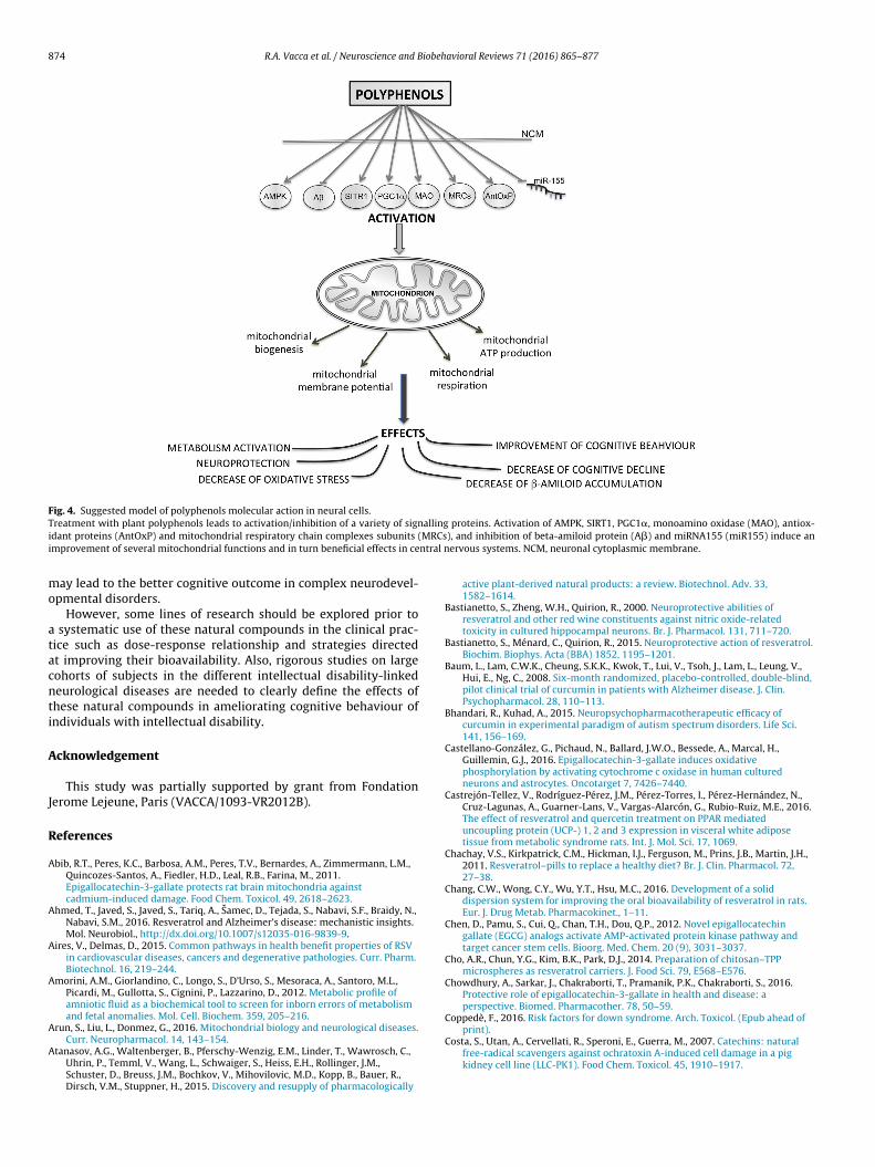
In this review we have shown as plant polyphenols, affecting amultitude of functions that have been correlated with the biology ofthe mitochondria, exert positive effects for development and func-tion of neurons (see Fig. 4). Therefore, dietary supplements withplant polyphenol compounds may have important neuroprotectiveeffects in preventing or managing some diseases associated withintellectual disability for which brain mitochondrial dysfunctionsand oxidative stress are critical. Food supplements used earlierinstead of later during development could be more effective inenhanching cognitive outcomes (Stagni et al., 2015). For instance,brain alterations and mitochondrial dysfunctions are alreadypresent prenatally in patients with DS (Olmos-Serrano et al., 2016;Valenti et al., 2011) as well as errors in the metabolism of someaminoacids, particularly Glutamate/Glutamine ratio (Amorini et al.,2012). Therefore, polyphenol extracts, for their safety, efficacy inreversing critical alterations in DS, should be considered for ther-apeutic interventions in early childhood or the prenatal period torescue brain development and prevent cognitive behaviour impair-ment; EGCG can cross the blood-brain and feto-maternal placentalbarriers (Martel et al., 2010). For example EGCG administered atneonatal and embryonic life stages in the Ts65Dn mouse model of
DS, fully restores hippocampal neurogenesis but not in adulthood(Stagni et al., 2016) thus suggesting a long-term assumption of thispolyphenol. We also suggest that the use of these phytochemicalsearly during development associated with other therapeutic agents
874 R.A. Vacca et al. / Neuroscience and Biobehavioral Reviews 71 (2016) 865–877
Fig. 4. Suggested model of polyphenols molecular action in neural cells.T allingi (MRCsi ntral n
mo
atacnti
A
J
R
A
A
A
A
A
A
reatment with plant polyphenols leads to activation/inhibition of a variety of signdant proteins (AntOxP) and mitochondrial respiratory chain complexes subunits
mprovement of several mitochondrial functions and in turn beneficial effects in ce
ay lead to the better cognitive outcome in complex neurodevel-pmental disorders.
However, some lines of research should be explored prior to systematic use of these natural compounds in the clinical prac-ice such as dose-response relationship and strategies directedt improving their bioavailability. Also, rigorous studies on largeohorts of subjects in the different intellectual disability-linkedeurological diseases are needed to clearly define the effects ofhese natural compounds in ameliorating cognitive behaviour ofndividuals with intellectual disability.
cknowledgement
This study was partially supported by grant from Fondationerome Lejeune, Paris (VACCA/1093-VR2012B).
eferences
bib, R.T., Peres, K.C., Barbosa, A.M., Peres, T.V., Bernardes, A., Zimmermann, L.M.,Quincozes-Santos, A., Fiedler, H.D., Leal, R.B., Farina, M., 2011.Epigallocatechin-3-gallate protects rat brain mitochondria againstcadmium-induced damage. Food Chem. Toxicol. 49, 2618–2623.
hmed, T., Javed, S., Javed, S., Tariq, A., Samec, D., Tejada, S., Nabavi, S.F., Braidy, N.,Nabavi, S.M., 2016. Resveratrol and Alzheimer’s disease: mechanistic insights.Mol. Neurobiol., http://dx.doi.org/10.1007/s12035-016-9839-9.
ires, V., Delmas, D., 2015. Common pathways in health benefit properties of RSVin cardiovascular diseases, cancers and degenerative pathologies. Curr. Pharm.Biotechnol. 16, 219–244.
morini, A.M., Giorlandino, C., Longo, S., D’Urso, S., Mesoraca, A., Santoro, M.L.,Picardi, M., Gullotta, S., Cignini, P., Lazzarino, D., 2012. Metabolic profile ofamniotic fluid as a biochemical tool to screen for inborn errors of metabolismand fetal anomalies. Mol. Cell. Biochem. 359, 205–216.
proteins. Activation of AMPK, SIRT1, PGC1�, monoamino oxidase (MAO), antiox-), and inhibition of beta-amiloid protein (A�) and miRNA155 (miR155) induce anervous systems. NCM, neuronal cytoplasmic membrane.
active plant-derived natural products: a review. Biotechnol. Adv. 33,1582–1614.
Bastianetto, S., Zheng, W.H., Quirion, R., 2000. Neuroprotective abilities ofresveratrol and other red wine constituents against nitric oxide-relatedtoxicity in cultured hippocampal neurons. Br. J. Pharmacol. 131, 711–720.
Bastianetto, S., Ménard, C., Quirion, R., 2015. Neuroprotective action of resveratrol.Biochim. Biophys. Acta (BBA) 1852, 1195–1201.
Baum, L., Lam, C.W.K., Cheung, S.K.K., Kwok, T., Lui, V., Tsoh, J., Lam, L., Leung, V.,Hui, E., Ng, C., 2008. Six-month randomized, placebo-controlled, double-blind,pilot clinical trial of curcumin in patients with Alzheimer disease. J. Clin.Psychopharmacol. 28, 110–113.
Bhandari, R., Kuhad, A., 2015. Neuropsychopharmacotherapeutic efficacy ofcurcumin in experimental paradigm of autism spectrum disorders. Life Sci.141, 156–169.
Castellano-González, G., Pichaud, N., Ballard, J.W.O., Bessede, A., Marcal, H.,Guillemin, G.J., 2016. Epigallocatechin-3-gallate induces oxidativephosphorylation by activating cytochrome c oxidase in human culturedneurons and astrocytes. Oncotarget 7, 7426–7440.
Castrejón-Tellez, V., Rodríguez-Pérez, J.M., Pérez-Torres, I., Pérez-Hernández, N.,Cruz-Lagunas, A., Guarner-Lans, V., Vargas-Alarcón, G., Rubio-Ruiz, M.E., 2016.The effect of resveratrol and quercetin treatment on PPAR mediateduncoupling protein (UCP-) 1, 2 and 3 expression in visceral white adiposetissue from metabolic syndrome rats. Int. J. Mol. Sci. 17, 1069.
Chachay, V.S., Kirkpatrick, C.M., Hickman, I.J., Ferguson, M., Prins, J.B., Martin, J.H.,2011. Resveratrol–pills to replace a healthy diet? Br. J. Clin. Pharmacol. 72,27–38.
Chang, C.W., Wong, C.Y., Wu, Y.T., Hsu, M.C., 2016. Development of a soliddispersion system for improving the oral bioavailability of resveratrol in rats.Eur. J. Drug Metab. Pharmacokinet., 1–11.
Chen, D., Pamu, S., Cui, Q., Chan, T.H., Dou, Q.P., 2012. Novel epigallocatechingallate (EGCG) analogs activate AMP-activated protein kinase pathway andtarget cancer stem cells. Bioorg. Med. Chem. 20 (9), 3031–3037.
Cho, A.R., Chun, Y.G., Kim, B.K., Park, D.J., 2014. Preparation of chitosan–TPPmicrospheres as resveratrol carriers. J. Food Sci. 79, E568–E576.
Chowdhury, A., Sarkar, J., Chakraborti, T., Pramanik, P.K., Chakraborti, S., 2016.Protective role of epigallocatechin-3-gallate in health and disease: aperspective. Biomed. Pharmacother. 78, 50–59.
Coppedè, F., 2016. Risk factors for down syndrome. Arch. Toxicol. (Epub ahead ofprint).
Costa, S., Utan, A., Cervellati, R., Speroni, E., Guerra, M., 2007. Catechins: naturalfree-radical scavengers against ochratoxin A-induced cell damage in a pigkidney cell line (LLC-PK1). Food Chem. Toxicol. 45, 1910–1917.
ovas, M.I., De La Torre, K., Farré-Albaladejo, M., Kaikkonen, J., Fitó, M.,López-Sabater, C., Pujadas-Bastardes, M.A., Joglar, J., Weinbrenner, T.,Lamuela-Raventós, R.M., 2006. Postprandial LDL phenolic content and LDLoxidation are modulated by olive oil phenolic compounds in humans. FreeRadic. Biol. Med. 40, 608–616.
urti, V., Capelli, E., Boschi, F., Nabavi, S.F., Bongiorno, A.I., Habtemariam, S., Nabavi,S.M., Daglia, M., 2014. Modulation of human miR-17-3p expression by methyl3-O-methyl gallate as explanation of its in vivo protective activities. Mol. Nutr.Food Res. 58, 1776–1784.
’Archivio, M., Filesi, C., Varì, R., Scazzocchio, B., Masella, R., 2010. Bioavailability ofthe polyphenols: status and controversies. Int. J. Mol. Sci. 11, 1321–1342.
’Andrea, G., 2015. Quercetin: a flavonol with multifaceted therapeuticapplications? Fitoterapia 106, 256–271.
aglia, M., Di Lorenzo, A., Nabavi, S.F., Talas, Z.S., Nabavi, S.M., 2014. Polyphenols.well beyond the antioxidant capacity: gallic acid and related compounds asneuroprotective agents: you are what you eat! Curr. Pharm. Biotechnol. 15,362–372.
as, S., Lin, H.S., Ho, P.C., Ng, K.Y., 2008. The impact of aqueous solubility and doseon the pharmacokinetic profiles of resveratrol. Pharm. Res. 25, 2593–2600.
e Filippis, B., Valenti, D., de Bari, L., De Rasmo, D., Musto, M., Fabbri, A., Ricceri, L.,Fiorentini, C., Laviola, G., Vacca, R.A., 2015. Mitochondrial free radicaloverproduction due to respiratory chain impairment in the brain of a mousemodel of Rett syndrome: protective effect of CNF1. Free Radic. Biol. Med. 83,167–177.
e la Torre, R., De Sola, S., Pons, M., Duchon, A., de Lagran, M.M., Farré, M., Fitó, M.,Benejam, B., Langohr, K., Rodriguez, J., Pujadas, M., Bizot, J.C., Cuenca, A., Janel,N., Catuara, S., Covas, M.I., Blehaut, H., Herault, Y., Delabar, J.M., Dierssen, M.,2014. Epigallocatechin-3-gallate, a DYRK1A inhibitor, rescues cognitive deficitsin down syndrome mouse models and in humans. Mol. Nutr. Food Res. 58,278–288.
e Oliveira, M.R., Nabavi, S.M., Braidy, N., Setzer, W.N., Ahmed, T., Nabavi, S.F.,2016a. Quercetin and the mitochondria: a mechanistic view. Biotechnol. Adv.34 (5), 532–549.
e Oliveira, M.R., Nabavi, S.F., Daglia, M., Rastrelli, L., Nabavi, S.M., 2016b.Epigallocatechin gallate and mitochondria—a story of life and death.Pharmacol. Res. 104, 70–85.
e Oliveira, M.R., Nabavi, S.F., Manayi, A., Daglia, M., Hajheydari, Z., Nabavi, S.M.,2016c. Resveratrol and the mitochondria: from triggering the intrinsicapoptotic pathway to inducing mitochondrial biogenesis, a mechanistic view.Biochim. Biophys. Acta (BBA) 1860, 727–745.
e Pace, R.C.C., Liu, X., Sun, M., Nie, S., Zhang, J., Cai, Q., Gao, W., Pan, X., Fan, Z.,Wang, S., 2013. Anticancer activities of (−)-epigallocatechin-3-gallateencapsulated nanoliposomes in MCF7 breast cancer cells. J. Liposome Res. 23,187–196.
e la Torre, R., de Sola, S., Hernandez, G., Farré, M., Pujol, J., Rodriguez, J., Espadaler,J.M., Langohr, K., Cuenca-Royo, A., Principe, A., 2016. Safety and efficacy ofcognitive training plus epigallocatechin-3-gallate in young adults with down’ssyndrome (TESDAD): a double-blind randomised, placebo-controlled, phase 2trial. Lancet Neurol. 15, 801–810.
ick, M.B., Doran, E., Phelan, M., Lott, I.T., 2016. Cognitive profiles on the severeimpairment battery are similar in Alzheimer disease and down syndrome withdementia. Alzheimer Dis. Assoc. Disord. 30 (3), 251–257.
brahimi, A., Schluesener, H., 2012. Natural polyphenols against neurodegenerativedisorders: potentials and pitfalls. Ageing Res. Rev. 11, 329–345.
antini, M., Benvenuto, M., Masuelli, L., Frajese, G.V., Tresoldi, I., Modesti, A., Bei, R.,2015. In vitro and in vivo antitumoral effects of combinations of polyphenols,or polyphenols and anticancer drugs: perspectives on cancer treatment. Int. J.Mol. Sci. 16, 9236–9282.
rozza, R.L., Bernardi, A., Paese, K., Hoppe, J.B., Silva T. d. Battastini, A.M., Pohlmann,A.R., Guterres, S.S., Salbego, C., 2010. Characterization oftrans-resveratrol-loaded lipid-core nanocapsules and tissue distributionstudies in rats. J. Biomed. Nanotechnol. 6, 694–703.
awande, S., Kale, A., Kotwal, S., 2008. Effect of nutrient mixture and black grapeson the pharmacokinetics of orally administered (−) epigallocatechin-3-gallatefrom green tea extract: a human study. Phytother. Res. 22, 802–808.
ibellini, L., Bianchini, E., De Biasi, S., Nasi, M., Cossarizza, A., Pinti, M., 2015.Natural compounds modulating mitochondrial functions. Evid. BasedComplement. Altern. Med. 2015, 527209.
iunta, B., Hou, H., Zhu, Y., Salemi, J., Ruscin, A., Shytle, R.D., Tan, J., 2010. Fish oilenhances anti-amyloidogenic properties of green tea EGCG in Tg2576 mice.Neurosci. Lett. 471, 134–138.
oldberg, D.M., Yan, J., Soleas, G.J., 2003. Absorption of three wine-relatedpolyphenols in three different matrices by healthy subjects. Clin. Biochem. 36,79–87.
racia, A., Miranda, J., Fernández-Quintela, A., Eseberri, I., Garcia-Lacarte, M.,
Milagro, F.I., Martínez, J.A., Aguirre, L., Portillo, M.P., 2016. Involvement ofmiR-539-5p in the inhibition of de novo lipogenesis induced by resveratrol inwhite adipose tissue. Food Funct. 7, 1680–1688.
Grieco, J., Pulsifer, M., Seligsohn, K., Skotko, B., Schwartz, A., 2015. Downsyndrome: cognitive and behavioral functioning across the lifespan. Am. J.Med. Genet. Part C: Semin. Med. Genet. Wiley Online Library, 135–149.
Grimm, A., Friedland, K., Eckert, A., 2016. Mitochondrial dysfunction: the missinglink between aging and sporadic Alzheimer’s disease. Biogerontology 17,281–296.
Grootaert, C., Kamiloglu, S., Capanoglu, E., Van Camp, J., 2015. Cell systems toinvestigate the impact of polyphenols on cardiovascular health. Nutrients 7,9229–9255.
Gueant, J., Anello, G., Bosco, P., Gueant-Rodriguez, R., Romano, A., Barone, C.,Gérard, P., Romano, C., 2005. Homocysteine and related geneticpolymorphisms in down’s syndrome IQ. J. Neurol. Neurosurg. Psychiatry 76,706–709.
Gupta, S.C., Patchva, S., Aggarwal, B.B., 2013. Therapeutic roles of curcumin:lessons learned from clinical trials. AAPS J. 15, 195–218.
Hamlett, E.D., Boger, H.A., Ledreux, A., Kelley, C.M., Mufson, E.J., Falangola, M.F.,Guilfoyle, D.N., Nixon, R.A., Patterson, D., Duval, N., 2016. Cognitiveimpairment, neuroimaging, and Alzheimer neuropathology in mouse modelsof down syndrome. Curr. Alzheimer Res. 13, 35–52.
Howitz, K.T., Bitterman, K.J., Cohen, H.Y., Lamming, D.W., Lavu, S., Wood, J.G.,Zipkin, R.E., Chung, P., Kisielewski, A., Zhang, L.-L., 2003. Small moleculeactivators of sirtuins extend Saccharomyces cerevisiae lifespan. Nature 425,191–196.
Hultén, M.A., Patel, S., Jonasson, J., Iwarsson, E., 2010. On the origin of the maternalage effect in trisomy 21 down syndrome: the oocyte mosaicism selectionmodel. Reproduction 139, 1–9.
Hussain, T., Gupta, S., Adhami, V.M., Mukhtar, H., 2005. Green tea constituentepigallocatechin-3-gallate selectively inhibits COX-2 without affecting COX-1expression in human prostate carcinoma cells. Int. J. Cancer 113, 660–669.
Hussain, Z., Katas, H., Amin, M.C.I.M., Kumolosasi, E., Buang, F., Sahudin, S., 2013.Self-assembled polymeric nanoparticles for percutaneous co-delivery ofhydrocortisone/hydroxytyrosol: an ex vivo and in vivo study using an NC/Ngamouse model. Int. J. Pharm. 444, 109–119.
Iacobazzi, V., Infantino, V., Castegna, A., Andria, G., 2014. Hyperhomocysteinemia:related genetic diseases and congenital defects: abnormal DNA methylationand newborn screening issues. Mol. Genet. Metab. 113, 27–33.
Ji, H., Zhang, X., Du, Y., Liu, H., Li, S., Li, L., 2012. Polydatin modulates inflammationby decreasing NF-�B activation and oxidative stress by increasing Gli1, Ptch1,SOD1 expression and ameliorates blood-brain barrier permeability for itsneuroprotective effect in pMCAO rat brain. Brain Res. Bull. 87, 50–59.
Johnson, J.J., Nihal, M., Siddiqui, I.A., Scarlett, C.O., Bailey, H.H., Mukhtar, H., Ahmad,N., 2011. Enhancing the bioavailability of resveratrol by combining it withpiperine. Mol. Nutr. Food Res. 55, 1169–1176.
Kale, A., Gawande, S., Kotwal, S., Netke, S., Roomi, W., Ivanov, V., Niedzwiecki, A.,Rath, M., 2010. Studies on the effects of oral administration of nutrientmixture, quercetin and red onions on the bioavailability of epigallocatechingallate from green tea extract. Phytother. Res. 24, S48–S55.
Karuppagounder, S.S., Pinto, J.T., Xu, H., Chen, H.-L., Beal, M.F., Gibson, G.E., 2009.Dietary supplementation with resveratrol reduces plaque pathology in atransgenic model of Alzheimer’s disease. Neurochem. Int. 54, 111–118.
Kim, H.S., Montana, V., Jang, H.J., Parpura, V., Kim, J.A., 2013. Epigallocatechingallate (EGCG) stimulates autophagy in vascular endothelial cells: a potentialrole forreducing lipid accumulation. J. Biol. Chem. 288, 22693–22705.
Kołodziejczyk, J., Malinowska, J., Olas, B., Stochmal, A., Oleszek, W., Erler, J., 2011.The polyphenol-rich extract from grape seeds suppresses toxicity ofhomocysteine and its thiolactone on the fibrinolytic system. Thromb. Res. 127,489–491.
Kubota, S., Kurihara, T., Mochimaru, H., Satofuka, S., Noda, K., Ozawa, Y., Oike, Y.,Ishida, S., Tsubota, K., 2009. Prevention of ocular inflammation inendotoxin-induced uveitis with resveratrol by inhibiting oxidative damageand Nuclear factor–�B activation. Invest. Ophthalmol. Vis. Sci. 50, 3512–3519.
Kulkarni, S.S., Cantó, C., 2015. The molecular targets of resveratrol. Biochim.Biophys. Acta (BBA) 1852, 1114–1123.
Kumar, A., Prakash, A., Dogra, S., 2011. Protective effect of curcumin (Curcumalonga) against D-galactose-induced senescence in mice. J. Asian Nat. Prod. Res.13, 42–55.
Kurita, S., Kashiwagi, T., Ebisu, T., Shimamura, T., Ukeda, H., 2014. Content ofresveratrol and glycoside and its contribution to the antioxidative capacity ofPolygonum cuspidatum (Itadori) harvested in Kochi. Biosci. Biotechnol.Biochem. 78, 499–502.
Lagouge, M., Argmann, C., Gerhart-Hines, Z., Meziane, H., Lerin, C., Daussin, F.,Messadeq, N., Milne, J., Lambert, P., Elliott, P., 2006. Resveratrol improvesmitochondrial function and protects against metabolic disease by activatingSIRT1 and PGC-1�. Cell 127, 1109–1122.
Lambert, J.D., Hong, J., Kim, D.H., Mishin, V.M., Yang, C.S., 2004. Piperine enhancesthe bioavailability of the tea polyphenol (−)-epigallocatechin-3-gallate in
mice. J. Nutr. 134, 1948–1952.
Lambert, J.D., Kim, D.H., Zheng, R., Yang, C.S., 2006. Transdermal delivery of(−)-epigallocatechin-3-gallate, a green tea polyphenol, in mice. J. Pharm.Pharmacol. 58, 599–604.
ambert, J.D., Kwon, S.J., Ju, J., Bose, M., Lee, M.J., Hong, J., Hao, X., Yang, C.S., 2008.Effect of genistein on the bioavailability and intestinal cancer chemopreventiveactivity of (−)-epigallocatechin-3-gallate. Carcinogenesis 29, 2019–2024.
atruffe, N., Delmas, D., Lizard, G., Tringali, C., Spatafora, C., Vervandier-Fasseur, D.,Meunier, P., 2012. Resveratrol against major pathologies: from diet preventionto possible alternative chemotherapies with new structural analogues. In:Tringali, C. (Ed.), Bioactive Compounds from Natural Sources, Natural Productsas Lead Compounds in Drug Discovery. , 2nd ed. CRC Press-Taylor&Francis,Boca Raton, pp. 339–378.
atruffe, N., Lancon, A., Frazzi, R., Aires, V., Delmas, D., Michaille, J.J., Djouadi, F.,Bastin, J., Cherkaoui-Malki, M., 2015. Exploring new ways of regulation byresveratrol involving miRNAs, with emphasis on inflammation. Ann. N. Y.Acad. Sci. 1348, 97–106.
ee, J.H., Moon, J.H., Kim, S.W., Jeong, J.K., Nazim, U.M., Lee, Y.J., Seol, J.W., Park, S.Y.,2015. EGCG-mediated autophagy flux has a neuroprotection effect via a classIII histone deacetylase in primaryneuron cells. Oncotarget 6, 9701–9717.
i, J., Ye, L., Wang, X., Liu, J., Wang, Y., Zhou, Y., Ho, W., 2012. (−)-Epigallocatechingallate inhibits endotoxin-induced expression of inflammatory cytokines inhuman cerebral microvascular endothelial cells. J. Neuroinflamm. 9, 161.
ibro, R., Giacoppo, S., Rajan, T.S., Bramanti, P., Mazzon, E., 2016. Naturalphytochemicals in the treatment and prevention of dementia: an overview.Molecules 21, 518.
in, L.C., Wang, M.N., Tseng, T.Y., Sung, J.S., Tsai, T.H., 2007. Pharmacokinetics of(−)-epigallocatechin-3-gallate in conscious and freely moving rats and itsbrain regional distribution. J. Agric. Food Chem. 55, 1517–1524.
aier-Salamon, A., Böhmdorfer, M., Riha, J., Thalhammer, T., Szekeres, T., Jager, W.,2013. Interplay between metabolism and transport of resveratrol. Ann. N. Y.Acad. Sci. 1290, 90–106.
anna, C., Tagliafierro, L., Scala, I., Granese, B., Andria, G., Zappia, V., 2012. The roleof iron toxicity in oxidative stress-induced cellular degeneration in downsyndrome: protective effects of phenolic antioxidants. Curr. Nutr. Food Sci. 8,206–212.
archese, A., Coppo, E., Sobolev, A.P., Rossi, D., Mannina, L., Daglia, M., 2014.Influence of in vitro simulated gastroduodenal digestion on the antibacterialactivity, metabolic profiling and polyphenols content of green tea (Camelliasinensis). Food Res. Int. 63, 182–191.
artel, F., Monteiro, R., Calhau, C., 2010. Effect of polyphenols on the intestinal andplacental transport of some bioactive compounds. Nutr. Res. Rev. 23, 47–64.
eng, X., Sang, S., Zhu, N., Lu, H., Sheng, S., Lee, M.J., Ho, C.T., Yang, C.S., 2002.Identification and characterization of methylated and ring-fission metabolitesof tea catechins formed in humans mice, and rats. Chem. Res. Toxicol. 15,1042–1050.
iró-Casas, E., Albaladejo, M.F., Covas, M.I., Rodriguez, J.O., Colomer, E.M.,Raventós, R.M.L., De La Torre, R., 2001. Capillary gas chromatography–massspectrometry quantitative determination of hydroxytyrosol and tyrosol inhuman urine after olive oil intake. Anal. Biochem. 294, 63–72.
olinari, G., 2009. Natural products in drug discovery: present status andperspectives. Adv. Exp. Med. Biol. 655, 13–27.
urakami, I., Chaleckis, R., Pluskal, T., Ito, K., Hori, K., Ebe, M., Yanagida, M.,Kondoh, H., 2014. Metabolism of skin-absorbed resveratrol into itsglucuronized form in mouse skin. PLoS One 9, e115359.
urase, T., Haramizu, S., Ota, N., Hase, T., 2009. Suppression of the aging-associateddecline in physical performance by a combination of resveratrol intake andhabitual exercise in senescence-accelerated mice. Biogerontology 10, 423–434.
aasani, I., Oh-hashi, F., Oh-hara, T., Feng, W.Y., Johnston, J., Chan, K., Tsuruo, T.,2003. Blocking telomerase by dietary polyphenols is a major mechanism forlimiting the growth of human cancer cells in vitro and in vivo. Cancer Res. 63,824–830.
abavi, S.F., Nabavi, S.M., Latifi, A.M., Mirzaei, M., Habtemariam, S., Moghaddam,A.H., 2012. Mitigating role of quercetin against sodium fluoride-inducedoxidative stress in the rat brain. Pharmaceut. Biol. 50, 1380–1383.
abavi, S.M., Daglia, M., Moghaddam, A.H., Nabavi, S.F., Curti, V., 2014. Teaconsumption and risk of ischemic stroke: a brief review of the literature. Curr.Pharm. Biotechnol. 15, 298–303.
abavi, S.F., Sureda, A., Habtemariam, S., Nabavi, S.M., 2015. Ginsenoside Rd andischemic stroke; a short review of literatures. J. Ginseng Res. 39, 299–303.
abavi, S.F., Barber, A.J., Spagnuolo, C., Russo, G.L., Daglia, M., Nabavi, S.M.,Sobarzo-Sánchez, E., 2016. Nrf2 as molecular target for polyphenols: a noveltherapeutic strategy in diabetic retinopathy. Crit. Rev. Clin. Lab. Sci. 53,293–312.
eves, A.R., Lucio, M., Martins, S., Lima, J., Reis, S., 2013. Novel resveratrolnanodelivery systems based on lipid nanoparticles to enhance its oralbioavailability. Int. J. Nanomed. 8, 177–187.
érez-Hernández, J., Zaldívar-Machorro, V.J., Villanueva-Porras, D., Vega-Ávila, E.,Chavarría, A., 2016. A potential alternative against neurodegenerativediseases: phytodrugs. Oxid. Med. Cell. Longev. 2016.
ace, M.C., Passavanti, M.B., Aurilio, C., Sansone, P., Aurilio, R., De Maria, S., Lama, S.,
Federico, A., Ravagnan, G., Caraglia, M., 2015. Polydatin administrationimproves serum biochemical parameters and oxidative stress markers duringchronic alcoholism: a pilot study. In Vivo 29, 405–408.
anighini, A., Duranti, E., Santini, F., Maffei, M., Pizzorusso, T., Funel, N., Taddei, S.,Bernardini, N., Ippolito, C., Virdis, A., 2013. Vascular dysfunction in a mouse
avioral Reviews 71 (2016) 865–877
model of Rett syndrome and effects of curcumin treatment. PLoS One 8,e64863.
Park, J., Song, W.-J., Chung, K.C., 2009. Function and regulation of Dyrk1A: towardsunderstanding down syndrome. Cell. Mol. Life Sci. 66, 3235–3240.
Poels, J., Spasic, M.R., Callaerts, P., Norga, K.K., 2009. Expanding roles forAmP-activated protein kinase in neuronal survival and autophagy. Bioessays31, 944–952.
Prohaska, R., Sibon, O.C., Rudnicki, D.D., Danek, A., Hayflick, S.J., Verhaag, E.M.,Vonk, J.J., Margolis, R.L., Walker, R.H., 2012. Brain, blood, and iron: perspectiveson the roles of erythrocytes and iron in neurodegeneration. Neurobiol. Dis. 46,607–624.
Quinones-Lombrana, A., Blanco, J.G., 2015. Chromosome 21-derivedhsa-miR-155-5p regulates mitochondrial biogenesis by targetingmitochondrial transcription factor a (TFAM). Biochim. Biophys. Acta (BBA)1852, 1420–1427.
Quideau, S., 2006. Why bother with polyphenols. Polyphén. Actual. 24, 10–14.Rachidi, M., Lopes, C., 2007. Mental retardation in down syndrome: from gene
dosage imbalance to molecular and cellular mechanisms. Neurosci. Res. 59,349–369.
Rodríguez-Sureda, V., Vilches Á, Sánchez, O., Audí, L., Domínguez, C., 2015.Intracellular oxidant activity, antioxidant enzyme defense system, and cellsenescence in fibroblasts with Trisomy 21. Oxid. Med. Cell. Longev., 509241.
Rubió, L., Valls, R.M., Macià, A., Pedret, A., Giralt, M., Romero, M.P., de la Torre, R.,Covas, M.I., Solà, R., Motilva, M.J., 2012. Impact of olive oil phenolicconcentration on human plasmatic phenolic metabolites. Food Chem. 135,2922–2929.
Rugarli, E.I., Langer, T., 2012. Mitochondrial quality control: a matter of life anddeath for neurons. EMBO J. 31, 1336–1349.
Russo, M., Spagnuolo, C., Tedesco, I., Bilotto, S., Russo, G.L., 2012. The flavonoidquercetin in disease prevention and therapy: facts and fancies. Biochem.Pharmacol. 83, 6–15.
Sadik, C.D., Sies, H., Schewe, T., 2003. Inhibition of 15-lipoxygenases by flavonoids:structure–activity relations and mode of action. Biochem. Pharmacol. 65,773–781.
Sandhir, R., Mehrotra, A., 2013. Quercetin supplementation is effective inimproving mitochondrial dysfunctions induced by 3-nitropropionic acid:implications in Huntington’s disease. Biochim. Biophys. Acta 1832, 421–430.
Sang, S., Cheng, X., Stark, R.E., Rosen, R.T., Yang, C.S., Ho, C.-T., 2002. Chemicalstudies on antioxidant mechanism of tea catechins: analysis of radical reactionproducts of catechin and epicatechin with 2, 2-diphenyl-1-picrylhydrazyl.Bioorg. Med. Chem. 10, 2233–2237.
Schaffer, S., Podstawa, M., Visioli, F., Bogani, P., Müller, W.E., Eckert, G.P., 2007.Hydroxytyrosol-rich olive mill wastewater extract protects brain cells in vitroand ex vivo. J. Agric. Food Chem. 55, 5043–5049.
Serra, A., Rubió, L., Borràs, X., Macià, A., Romero, M.P., Motilva, M.J., 2012.Distribution of olive oil phenolic compounds in rat tissues after administrationof a phenolic extract from olive cake. Mol. Nutr. Food Res. 56, 486–496.
Sharma, S.B., Gupta, R., 2015. Drug development from natural resource: asystematic approach. Mini Rev. Med. Chem. 15, 52–57.
Sharma, S., Zhuang, Y., Ying, Z., Wu, A., Gomez-Pinilla, F., 2009. Dietary curcuminsupplementation counteracts reduction in levels of molecules involved inenergy homeostasis after brain trauma. Neuroscience 161, 1037–1044.
Sharma, D., Wani, W., Sunkaria, A., Kandimalla, R., Sharma, R., Verma, D., Bal, A.,Gill, K., 2016. Quercetin attenuates neuronal death against aluminum-inducedneurodegeneration in the rat hippocampus. Neuroscience 324, 163–176.
Shi, G., Rao, L., Yu, H., Xiang, H., Yang, H., Ji, R., 2008. Stabilization and encapsulationof photosensitive resveratrol within yeast cell. Int. J. Pharm. 349, 83–93.
Siddique, M.I., Katas, H., Amin, M.C.I.M., Ng, S.-F., Zulfakar, M.H., Jamil, A., 2016.In-vivo dermal pharmacokinetics, efficacy, and safety of skin targetingnanoparticles for corticosteroid treatment of atopic dermatitis. Int. J. Pharm.507, 72–82.
Signorile, A., Micelli, L., De Rasmo, D., Santeramo, A., Papa, F., Ficarella, R., Gattoni,G., Scacco, S., Papa, S., 2014. Regulation of the biogenesis of OXPHOScomplexes in cell transition from replicating to quiescent state: involvement ofPKA and effect of hydroxytyrosol. Biochim. Biophys. Acta (BBA) 1843, 675–684.
Singh, G., Pai, R.S., 2016. In vitro and in vivo performance of supersaturableself-nanoemulsifying system of trans-resveratrol. Artif. cells Nanomed.Biotechnol. 44, 510–516.
Srividhya, R., Zarkovic, K., Stroser, M., Waeg, G., Zarkovic, N., Kalaiselvi, P., 2009.Mitochondrial alterations in aging rat brain: effective role of (−)-epigallo
catechin gallate. Int. J. Dev. Neurosci. 27, 223–231.
Stagni, F., Giacomini, A., Guidi, S., Ciani, E., Bartesaghi, R., 2015. Timing of therapiesfor Down syndrome: the sooner, the better. Front. Behav. Neurosci. 9, 265.
Stagni, F., Giacomini, A., Emili, M., Trazzi, S., Guidi, S., Sassi, M., Ciani, E., Rimondini,R., Bartesaghi, R., 2016. Short- and long-term effects of neonatal
pharmacotherapy with epigallocatechin-3-gallate onhippocampaldevelopment in the Ts65Dn mouse model of Down syndrome.Neuroscience 333, 277–301.
aguchi, C., Fukushima, Y., Kishimoto, Y., Suzuki-Sugihara, N., Saita, E., Takahashi,Y., Kondo, K., 2015. Estimated dietary polyphenol intake and major food andbeverage sources among elderly Japanese. Nutrients 7, 10269–10281.
eskac, K., Kristl, J., 2010. The evidence for solid lipid nanoparticles mediated celluptake of resveratrol. Int. J. Pharm. 390, 61–69.
rujillo, J., Granados-Castro, L.F., Zazueta, C., Andérica-Romero, A.C., Chirino, Y.I.,Pedraza-Chaverrí, J., 2014. Mitochondria as a target in the therapeuticproperties of curcumin. Arch. Pharm. 347, 873–884.
sao, R., 2010. Chemistry and biochemistry of dietary polyphenols. Nutrients 2,1231–1246.
urner, R.S., Thomas, R.G., Craft, S., Van Dyck, C.H., Mintzer, J., Reynolds, B.A.,Brewer, J.B., Rissman, R.A., Raman, R., Aisen, P.S., 2015. A randomized,double-blind, placebo-controlled trial of resveratrol for Alzheimer disease.Neurology 85, 1383–1391.
acca, R.A., Valenti, D., 2015. Green tea EGCG plus fish oil omega-3 dietarysupplements rescue mitochondrial dysfunctions and are safe in a Down’ssyndrome child. Clin. Nutr. 34, 783–784.
alenti, D., Manente, G.A., Moro, L., Marra, E., Vacca, R.A., 2011. Deficit of complex Iactivity in human skin fibroblasts with chromosome 21 trisomy andoverproduction of reactive oxygen species by mitochondria: involvement ofthe cAMP/PKA signalling pathway. Biochem. J. 435, 679–688.
alenti, D., De Rasmo, D., Signorile, A., Rossi, L., de Bari, L., Scala, I., Granese, B.,Papa, S., Vacca, R.A., 2013. Epigallocatechin-3-gallate prevents oxidativephosphorylation deficit and promotes mitochondrial biogenesis in human cellsfrom subjects with Down’s syndrome. Biochim. Biophys. Acta (BBA) 1832,542–552.
alenti, D., de Bari, L., De Filippis, B., Henrion-Caude, A., Vacca, R.A., 2014.Mitochondrial dysfunction as a central actor in intellectual disability-relateddiseases: an overview of down syndrome, autism, Fragile X and Rett syndrome.Neurosci. Biobehav. Rev. 46, 202–217.
alenti, D., de Bari, L., de Rasmo, D., Signorile, A., Henrion-Caude, A., Contestabile,A., Vacca, R.A., 2016. The polyphenols resveratrol andepigallocatechin-3-gallate restore the severe impairment of mitochondria inhippocampal progenitor cells from a Down syndrome mouse model. Biochim.Biophys. Acta (BBA) 1862, 1093–1104.
an Cleve, S.N., Cohen, W.I., 2006. Part I: clinical practice guidelines for childrenwith Down syndrome from birth to 12 years. J. Pediatr. Health Care 20, 47–54.
an Cleve, S.N., Cannon, S., Cohen, W.I., 2006. Part II: clinical practice guidelines foradolescents and young adults with Down syndrome: 12 to 21 years. J. Pediatr.Health Care 20, 198–205.
auzour, D., Rattray, M., Williams, R.J., Spencer, J.P.E., 2013. Potential
neuroprotective actions of dietary flavonoids. In: Ramawat, K.G., Merillion,J.-M. (Eds.), Handbook of Natural Products. Springer, pp. 2617–2640.
isioli, F., Galli, C., Bornet, F., Mattei, A., Patelli, R., Galli, G., Caruso, D., 2000. Oliveoil phenolics are dose-dependently absorbed in humans. FEBS Lett. 468,159–160.
avioral Reviews 71 (2016) 865–877 877
Wang, Y., Xu, H., Fu, Q., Ma, R., Xiang, J., 2011. Protective effect of resveratrolderived from Polygonum cuspidatum and its liposomal form on nigral cells inParkinsonian rats. J. Neurol. Sci. 304, 29–34.
Wang, P., Heber, D., Henning, S.M., 2012. Quercetin increased bioavailability anddecreased methylation of green tea polyphenols in vitro and in vivo. FoodFunct. 3, 635–642.
Wang, X., Zhao, Y., Zhang, X., Badie, H., Zhou, Y., Mu, Y., Loo, L.S., Cai, L., Thompson,R.C., Yang, B., 2013. Loss of sorting nexin 27 contributes to excitatory synapticdysfunction by modulating glutamate receptor recycling in Down’s syndrome.Nat. Med. 19, 473–480.
Wang, L., Gao, S., Jiang, W., Luo, C., Xu, M., Bohlin, L., Rosendahl, M., Huang, W.,2014. Antioxidative dietary compounds modulate gene expression associatedwith apoptosis, DNA repair, inhibition of cell proliferation and migration. Int. J.Mol. Sci. 15, 16226–16245.
Wang, Y., Li, F., Zhuang, H., Li, L., Chen, X., Zhang, J., 2015. Effects of plantpolyphenols and �-tocopherol on lipid oxidation, microbiologicalcharacteristics, and biogenic amines formation in dry-cured bacons. J. Food Sci.80, C547–C555.
Yallapu, M.M., Nagesh, P.K.B., Jaggi, M., Chauhan, S.C., 2015. Therapeuticapplications of curcumin nanoformulations. AAPS J. 17, 1341–1356.
Yang, W.H., Deng, Y.T., Kuo, M.Y.P., Liu, C.M., Chang, H.H., Chang, J.Z.C., 2013.Epigallocatechin-3-gallate blocks triethylene glycol dimethacrylate–inducedcyclooxygenase-2 expression by suppressing extracellular signal-regulatedkinase in human dental pulp and embryonic palatal mesenchymal cells. J.Endod. 39, 1407–1412.
Yong Feng, W., 2006. Metabolism of green tea catechins: an overview. Curr. DrugMetab. 7, 755–809.
Yoshino, S., Hara, A., Sakakibara, H., Kawabata, K., Tokumura, A., Ishisaka, A., Kawai,Y., Terao, J., 2011. Effect of quercetin and glucuronide metabolites on themonoamine oxidase-A reaction in mouse brain mitochondria. Nutrition 27,847–852.
Yu, D., An, F., He, X., Cao, X., 2015. Curcumin inhibits the proliferation and invasionof human osteosarcoma cell line MG-63 by regulating miR-138. Int. J. Clin. Exp.Pathol. 8, 14946.
Zhang, J., Nie, S., Wang, S., 2013. Nanoencapsulation enhancesepigallocatechin-3-gallate stability and its antiatherogenic bioactivities inmacrophages. J. Agric. Food Chem. 61, 9200–9209.
Zheng, A., Li, H., Cao, K., Xu, J., Zou, X., Li, Y., Chen, C., Liu, J., Feng, Z., 2015. Maternalhydroxytyrosol administration improves neurogenesis and cognitive functionin prenatally stressed offspring. J. Nutr. Biochem. 26, 190–199.
Zu, Y., Zhang, Y., Wang, W., Zhao, X., Han, X., Wang, K., Ge, Y., 2016. Preparationand in vitro/in vivo evaluation of resveratrol-loaded carboxymethyl chitosan
nanoparticles. Drug Deliv. 23, 971–981.
Zuo, X., Tian, C., Zhao, N., Ren, W., Meng, Y., Jin, X., Zhang, Y., Ding, S., Ying, C., Ye, X.,2014. Tea polyphenols alleviate high fat and high glucose-induced endothelialhyperpermeability by attenuating ROS production via NADPH oxidasepathway. BMC Res. Notes 7, 1.