Abstract It is reported a novel method to prepare magnetic core (iron oxidespinels)–shell (silica) composites containing well-dispersed magnetic nanoparticlesin aqueous solution. The synthetic process consists of two steps. In a first step, ironoxide nanoparticles obtained through co-precipitation are dispersed in an aqueoussolution containing tetramethylammonium hydroxide; in a second step, particles ofthis sample are coated with silica, through hydrolyzation of tetraethyl orthosilicate.The intrinsic atomic structure and essential properties of the core–shell system wereassessed with powder X-ray diffraction, Fourier transform infrared spectrometry,Mössbauer spectroscopy and transmission electron microscopy. The heat releasedby this ferrofluid under an AC-generated magnetic field was evaluated by following
A. L. AndradeDepartment of Chemistry, Federal University of Ouro Preto (UFOP),35400-000 Ouro Preto, Minas Gerais, Brazil
J. D. Fabris (B)Federal University of Jequitinhonha and Mucuri Valleys (UFVJM),39100-000 Diamantina, Minas Gerais, Brazile-mail: [email protected]
M. C. PereiraInstitute of Science, Engineering and Technology,Federal University of Jequitinhonha and Mucuri Valleys (UFVJM),39803-371 Teófilo Otoni, Minas Gerais, Brazil
R. Z. DominguesDepartment of Chemistry, Federal University of Minas Gerais (UFMG),31270-901 Belo Horizonte, Minas Gerais, Brazil
J. D. ArdissonLaboratory of Applied Physics,Development Center of Nuclear Technology (CNEN/CDTN),31270-901 Belo Horizonte, Minas Gerais, Brazil
72 A.L. Andrade et al.
the temperature evolution under increasing magnetic field strengths. Results stronglyindicate that this ferrofluid based on silica-coated iron oxide spinels is technologicallya very promising material to be used in medical practices, in oncology.
Keywords Biomedicine · Nanotechnology · Magnetic hyperthermia · Ferrofluid
1 Introduction
Different chemical methods have been used or reportedly proposed to synthesize andprepare magnetic nanoparticles (MNPs) of iron oxide spinels, namely maghemite(γ-Fe2O3) and magnetite (Fe3O4), with variable mean sizes and homogeneousmorphology, for technological applications in clinical diagnostics and medical ther-apies [1–6]. This increasing interest in designing and developing MNPs is due totheir unique physical and chemical properties for such purposes. In particular, totheir magnetic anisotropy, which can be pronouncedly higher than those of thecorresponding bulk specimen.
With proper surface coating, those MNPs can be dispersed into selected solventsto form homogeneous suspensions, known as ferrofluids [7, 8]. The surfaces ofparticles may be suitably modified with an organic polymer, a metallic (e.g. gold) oran oxide (e.g. silica or alumina) layer to make them suitably functional, as by furthercoating with various bioactive molecules [9]. Such suspensions can be driven, underthe action of an externally applied magnetic field, to be concentrated in selectedinternal parts of the living body, enabling them to be used in medical therapiesthat demand local tissue heating or delivering of bioactive molecules [10]. For suchapplications, the particle must combine high magnetic saturation, biocompatibilityand interactive functions at its surface.
Biomedical applications of ferrofluids include magnetic separation, drug delivery,magnetic resonance imaging (MRI), and hyperthermia [11–14]. Hyperthermia meansthat the magnetic nanosystems can heat up under the action of a magnetic field,releasing thermal energy that may be dimensioned to directly kill tumor cells (amoderate degree of heating may result in an effective cell destruction), or to beused as a programmable local delivery of chemicals, in chemotherapy, or even ofradiotherapic agents [15].
The use of hyperthermia (heating) to treat tissues with malignant tumors is as oldas the medicine itself. Hippocrates, the Father of Medicine, proposed that the surfaceof tumors should be cauterized by heating them with a hot iron piece. In our moderntimes, more sophisticated methods (hot water bath, pyrogens such as mixed bacterialtoxins, perfusion heating, high-frequency radiation, magnetic fluid hyperthermia) areused to heat, and hopefully destroy tumors [16].
Magnetically induced hyperthermia, one of the sought procedures for local ther-apy to treat cancers, is based on the exposition of cancerous tissues, which was madecovered with a ferrofluid, to an alternating magnetic field. Ferrofluids for this pur-pose imply a system containing MNPs in a carrier fluid that would be directly injectedto the tumor area in the body or to the blood stream, alternatively also mixed with aspecific tumor antibody targeting. Once the ferrofluid is placed in the selected vitalarea of the living body, an alternating magnetic field is externally applied. The fieldstrength must be dimensioned basing on the resulting hyperthermic temperature,
Silica-coated nanoparticles of iron oxide for applications based on hyperthermia 73
being limited to a value that is assured to not affect the healthy cells. Up to this limit,the magnetic field can be safely applied so to reach any deeper parts of the livingbody. The magnetic particles respond to the variable magnetic field by releasingheat due to magnetic hysteresis losses. Magnetic particles embedded around a tumorsite and subject to an oscillating magnetic field will heat up to a temperature thatdepends on the magnetic properties of the material, the strength of the magneticfield, on the oscillation frequency and on the cooling capacity of the blood flowsurrounding the tumor site. Cancer cells are destroyed at temperatures slightly higherthan 43 ◦C; the normal cells can survive even at temperatures moderately above thatlimit. Hyperthermia in this sense is still an under development technique for localtherapy in oncology. The procedure has also the advantage to prevent many sideeffects, so commonly observed in patients under treatment with chemotherapy orradiotherapy.
Herein, we present the preparation of a ferrofluid based on well dispersed core-shell system formed by iron oxide spinels (particle cores) coated with silica (shell)to produce magnetically induced hyperthermia. The core-shell system was preparedvia a two-step process: firstly, iron oxide nanoparticles (sample Mt) were obtainedthrough co-precipitation, and this material was dispersed in an aqueous solutioncontaining tetramethylammonium hydroxide (TMAOH) (sample Mt1). In a secondstep, particles of the sample Mt1 were coated with silica (sample Mt1-Si2), throughhydrolyzation of tetraethyl orthosilicate (TEOS). The obtained ferrofluid is intendedto be a basic material for potential technological applications in medical therapies inoncology.
2 Experimental
2.1 Materials and reagents
All chemicals used in this work, namely iron (III) chloride hexahydrate, FeCl3·6H2O(Riedel-de Haen); hydrochloride acid, HCl (Fluka); sodium sulfide, Na2SO3 (Sigma,Aldrich); ammonium hydroxide, NH4OH (Fluka); 25 % aqueous tetramethylam-monium hydroxide solution, C4H13NO·5H2O (TMAOH) (Vetec); absolute ethanol,CH3CH2OH (Panreac, 99.5 %) and tetraethyl orthosilicate, (C2H5O)4Si (TEOS)(Aldrich, 98 %) were of analytical grade standards and used as received.
2.2 Iron oxide nanoparticles preparation
The synthesis procedure followed to prepare these iron oxide spinels samples wasdescribed in more details in ref. [2]. It was based on the method earlier proposedby Qu et al. [17]: 30 mL of a FeCl3.6H2O stock solution containing 2 mol L−1
(dissolved in 0.5 mol L−1 HCl), 20 mL of a Na2SO3 stock solution with 1 mol L−1 and50.8 mL of a NH4OH solution diluted to a total volume of 800 mL were used. Justafter mixing Fe3+ and SO2−
3 , the obtained solution was kept under vacuum pressurefor 1 min. Initially, the color of the solution changed from yellow to red; after fewminutes, the yellow reappeared. Diluted ammonia solution was then quickly pouredinto the mixture under vigorous stirring, and vacuum, for 1 min. The stirring wascontinued for an additional 30 min, after the black precipitate was formed. The
74 A.L. Andrade et al.
suspension containing the precipitated was centrifuged at 2,000 rpm for 3 min; thesupernatant was discarded. This procedure was repeated five times by re-dispersingthe resulting cakes in distilled water. The as obtained precipitate was labeled Mt. Thenanoparticles were then coated with tetramethylammonium hydroxide (TMAOH).Typically, 1 mL of commercial 25 % TMAOH solution was added to each centrifugetube containing an amount of wet cake corresponding to about 1 g dry powder and re-dispersed with a thin glass rod until obtaining homogeneous suspensions. This samplewas labeled Mt1. 1 mL of this suspension was diluted ten times in distilled waterand the obtained ferrofluid was used for further coating with silica, to produce thesample Mt1-Si2, in an alkaline alcohol/water mixture at room temperature by usingthe ferrofluid as seed, according to the method originally proposed by Stöber [18],with some modification [19]. This Mt1-Si2 sample was prepared by adding 2 mL ofdiluted Mt1 suspension in 160 mL of absolute ethanol at room temperature, followedby ultrasonication for 5 min. Following that, 2 mL of TEOS and 40 mL of distilledwater were added and the mixture was stirred at room temperature for 1 day long.The as coated magnetic powder was centrifuged at 16,000 rpm for 20 min, washedthrice with distilled water and acetone.
The resulting products were separated with a small hand magnet, washed out withdistilled water and acetone, and dried at room temperature.
2.3 Preparation of glass used as control sample for the zeta potential measurements
A silicon-based glass was prepared the following way: TEOS was added to anacidified (HCl—pH 1.7) aqueous solution, and next added with ethanol in a molarproportion of H2O:TEOS:CH3CH2OH of 4:1:4. The solution was magneticallystirred until gelling. The material was placed under a humidified atmosphereovernight, and treated next at 400 ◦C for 2 h. The resulting glass was used as a controlsample for zeta potential measurements.
2.4 Characterization
Electron micrographs of the iron oxide spinels dispersions were carried out witha conventional transmission electron microscopy (TEM). The iron oxide spinelsdispersions powder were embedded in an Epofix TM resin to form a block whichwas cleaved; thin slices (50 nm) were then cut with an Ultramicrotome (LeicaUC6), provided with a diamond knife, and deposited onto a holey carbon coatedTEM grid. This TEM grid was then dried in a vacuum desiccator. The crystallinestructure of the magnetic nanoparticles, after the liquid carrier of samples wasevaporated, were assessed with powder X-ray diffraction (XRD) measurements,using a Shimadzu XRD 6000 diffractometer equipped with an iron tube (40 kVand 30 mA) and a graphite monochromator. The scans were done between 15 and70◦ (2θ) with a scanning speed of 1◦/min. The identification of crystalline phaseswas performed with the Jade + software (Materials Data, Inc.). Silicon was usedas an internal standard. FTIR data were obtained with a Perkin Elmer SpectrumGX spectrophotometer. The solids were homogeneously dispersed in KBr (1 wt%approximately) and pressed into discs. Spectra were recorded with a resolution of4 cm−1 with 16 scans per spectrum. Mössbauer spectra were collected with a constantacceleration transmission mode setup, with a ∼50 mCi 57Co/Rh gamma-ray source.
Silica-coated nanoparticles of iron oxide for applications based on hyperthermia 75
Spectra at 298 K and 80 K were obtained with a spectrometer equipped with atransducer (CMTE model MA250) controlled by a linear function driving (CMTEmodel MR351). Values of Mössbauer isomer shifts are quoted relatively to α-Fe.The experimental reflections were fitted to Lorentzian functions by least-squarefitting with software NORMOS™-90 (developed by R. A. Brand, at Laboratoriumfür Angewandte Physik, Universität Duisburg, D-47048, Duisburg-Germany).
The determination of zeta potential (ζ ) was performed on a Coulter Delsa 440SX,by using a stock suspension of the ground material in deionized water, preparedand homogenized by ultrasonication for 15 min. Drops of this stock suspensionwere then added to an aqueous solution of KCl 10−3 mol L−1 for the zeta potentialmeasurements. The pH of the measuring solution was varied and adjusted to severalvalues, in the pH range of 2–8, by using 10−3 mol L−1 aqueous solutions of KOH andHNO3.
Heat dissipation experiments were carried out by transferring the suspensionswith dispersed iron oxide spinels in both water and hydrogel into a test tube. Athree-loop coil (Nova Star 5kW RF Power Supply, Ameritherm, Inc) with resonantfrequency of 220 kHz and 198 kHz was used in the experiments, in order to study thecorrelation between the applied magnetic field and the AC magnetically inducedheating temperature. The sample concentration was approximately 4 mg mL−1
in water and 4 mg g−1 in polyvinyl alcohol hydrogel. In the hydrogel-dispersedsamples, rotation of particles was restricted and the magnetic moment relaxed onlythrough Néel relaxation. The temperature of the magnetic suspension was measuredwith an optical fiber thermometer. Results were taken as the mean of triplicatemeasurements. The amount of heat generated by pure water and pure hydrogel inthe two AC magnetic fields was measured, in an attempt to obtain any contributionof water and hydrogel to rise the temperature by dispersing nanoparticles. The wholecontribution of both pure water and gel was discounted from the final temperaturevalue for each ferrofluid.
The polyvinyl alcohol hydrogel was based on the method earlier proposed byHyon et al. [20], with minor modifications [21]. A homogeneous PVA solution witha PVA concentration of 15 wt% was obtained by heating the mixture of PVA and amixed water/DMSO solvent at 140 ◦C for 2 h. The mixing ratio of water to DMSOwas kept to 20:80, by weight. Then the casted PVA was placed in a freezer at −20 ◦Cfor 24 h.
3 Results and discussion
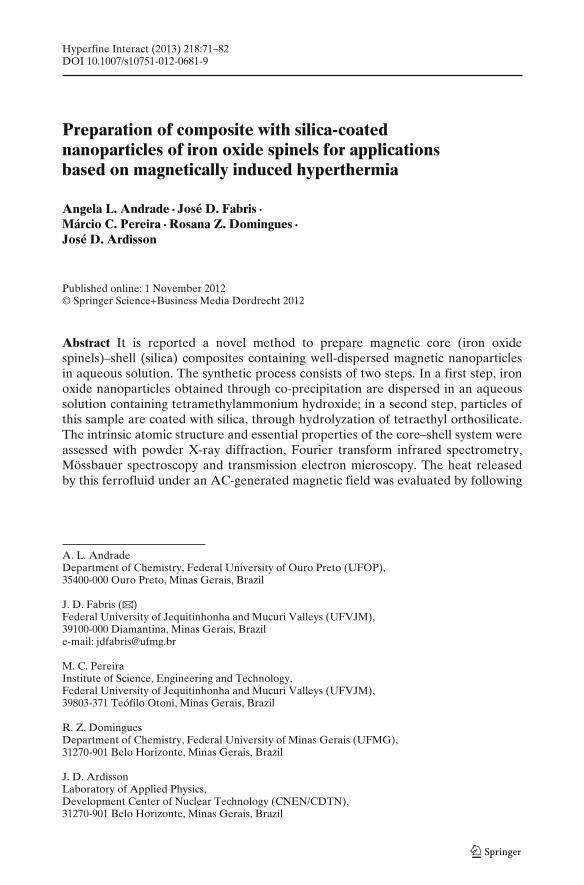
The size and morphology of particles by comparing TEM pictures of the samplesMt and Mt1 was described in details in ref. [1]. It can be observed that the particlesin Mt1 were better dispersed than in Mt. This may be explained by assuming thatthe tetramethylammonium hydroxide (TMAOH) acts as a surface-active agent [22]favoring the dispersion of iron oxide nanoparticles. Figure 1 shows typical TEMimages of the Mt1-Si2 sample. It was observed that the core particles are sphericalin shape and is covered with a uniform and continuous dense layer on a surfacecontaining multi-cores of iron oxide nanoparticles. Because the atomic number foriron is higher than that for silicon, the TEM micrographs show cores in darker thanshells.
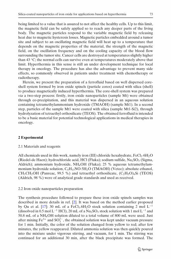
Figure 2 depicts the FTIR spectra of the tetramethylammonium hydroxide(TMAOH), iron oxide spinels (Mt), TMAOH coated iron oxide spinels (Mt1), andsilica coated Mt1 (Mt1-Si2) samples. The spectrum for sample TMAOH shows astrong band at 950 cm−1, assignable to the asymmetric methyl deformation mode C–N, which is generally observed in the domain 900–1000 cm−1 [23]. Consistently, thissame band at 950 cm−1 is observed for sample Mt1-Si2, originated from the TMAOHitself or to Si-OH, resulting from the incomplete condensation of TEOS [24]. Theabsorption peak of iron oxide spinels of Fe-O bond at around 570 cm−1 [25–27] isclearly visible in the spectrum for samples Mt, Mt1 and Mt1-Si2. Comparably, onenew absorption peak for samples Mt1 and Mt1-Si2 that appears at 950 cm−1 is dueto the transmittance bands typical of TMAOH. Furthermore, two absorption peakscentered at around 805 and 1093 cm−1 can also be observed for Mt1-Si2 sample,corresponding to the stretching of Si–O–Si bond from the silica particles. From theabove results, it can be inferred that the TMAOH and SiO2 have been successfullycoated on the surface of iron oxide nanoparticles and that the sample Mt does notchange chemically or physically after the treatment with TMAOH and TEOS.
Mössbauer spectra at 298 K for all the samples (Fig. 3a; hyperfine parameters,Table 1) exhibited rather complex spectral patterns, with broad and asymmetricresonance lines due to distribution of particle size and different degrees of oxidation
Silica-coated nanoparticles of iron oxide for applications based on hyperthermia 77
Fig. 3 Mössbauer spectra and corresponding probability profiles of hyperfine field values forsamples Mt, Mt1, and MtSi2 at: a 80 K and b 298 K
of Fe2+ in the iron oxide spinels. A typical spectrum for magnetite is characterizedby two subspectra: one due to Fe3+ in tetrahedral coordination site and other tothe mixed valence Fe3+/Fe2+ in octahedral sites. From the spectrum for the sample
78 A.L. Andrade et al.
Table 1 Hyperfine parameters of the fitted Mössbauer spectra recorded at 298 K
δ = isomer shift relative to αFe; ε = quadrupole shift; � = quadrupole splitting; Bhf = hyperfinefield and RA = relative subspectral areaaHyperfine field at maximum probability of the hyperfine field distribution profile#Value kept fixed during the minimization procedure on least squares fitting experimental data
Table 2 Averaged values for cubic lattice parameters and mean crystallite size (actually, meancoherent length) as determined from X-ray diffraction data for this solid solution of iron oxide spinels
Sample Lattice parameter/Å Mean coherent length/nm
Number in parentheses is the uncertainty over the last significant digit as estimated from the standarddeviation of the corresponding mean value
Mt at 298 K, it can be observed a subtle difference of intensities between lines 1(broader and less intense) and 6, which could eventually indicate the occurrenceof some Fe2+/Fe3+ character, of a residual magnetite in this sample. From corre-sponding spectra for samples Mt1 and Mt1Si2, this difference much less evident or isvirtually absent, suggesting that only structural Fe3+ responds for their correspondinghyperfine structures. From these observations, any eventually existing magnetite inthese samples containing mixtures of iron oxide spinels cannot be unequivocallydetected, for its relatively very low amount. This assumption is in agreement withvalues of isomer shift from distribution profiles (Fig. 3b) and with the latticeparameters values obtained from Rietveld refinement of powder X-ray diffractiondata (Table 2). Values of hyperfine field for all samples are markedly lower thanthose expected for a bulk maghemite or even iron oxide spinels. This is certainly dueto the small sizes of particles. Spectrum for the sample Mt1Si2 is formed by a doubletof Fe3+ in octahedral coordination due to the iron oxide spinel dispersed on the silicamatrix.
Mössbauer patterns at 80 K are shown in Fig. 3b (hyperfine parameters, Table 3).Values of isomer shift from the hyperfine field distribution are systematically higherfor the Mt than for Mt1 or Mt1Si2 samples. This characteristic is a direct consequenceof the higher proportion of Fe2+ in Mt relatively to the other two samples.
Spectra at 80 K, thus below the Verwey temperature (TV ≈ 120 K), reveal thatfor part (relative subspectral area, RA = 31 %) of sample Mt, isomer shift valuesvary from 0.65 mm s−1 to 0.82 mm s−1, which may also be an evidence of somesmall ferrous character in the spinel phases mixture. Complementarily, subspectradue to octahedral and tetrahedral sites of maghemite would be slightly separatedat this temperature, with different values of isomer shift and hyperfine field. The
Silica-coated nanoparticles of iron oxide for applications based on hyperthermia 79
Table 3 Hyperfine parameters of the fitted Mössbauer spectra recorded at 80 K
Sample δ/mm s−1 ε/mm s−1 Bhf/T RA/%
Mt 0.31 0.02 51.5 190.55 0 52.2a 81
Mt1 0.31 0.04 51.4 200.50 −0.01 52.2a 80
Mt1Si2 0.31 −0.03 51.3 220.50 0.01 51.8a 78
δ = isomer shift relative to αFe; ε = quadrupole shift; � = quadrupole splitting; Bhf = hyperfinefield and RA = relative subspectral areaaHyperfine field at maximum probability of the hyperfine field distribution profile
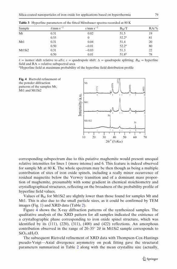
Fig. 4 Rietveld refinement ofthe powder diffractionpatterns of the samples Mt,Mt1 and Mt1Si2
corresponding subspectrum due to this putative maghemite would present unequalrelative intensities for lines 1 (more intense) and 6. This feature is indeed observedfor sample Mt at 80 K. The whole spectrum may be then though as being a multiplecontribution of sites of iron oxide spinels, including a really minor occurrence ofresidual magnetite below the Verwey transition and of a dominant mass propor-tion of maghemite, presumably with some gradient in chemical stoichiometry andcrystallographical structures, reflecting on the broadness of the probability profile ofhyperfine field values.
Values of Bhf for Mt1Si2 are slightly lower than those found for samples Mt andMt1. This is also due to the small particle sizes, as it could be confirmed by TEMimages (Fig. 1) and XRD data (Table 2).
Figure 4 shows the X-ray diffraction patterns of the synthesized samples. Thequalitative analysis of the XRD pattern for all samples indicated the existence ofa crystallographic phase corresponding to iron oxide spinel structure, which wasidentified by its (111), (220), (311), (400) and (422) reflections. An amorphouscontribution observed in the range of 20–35◦ 2θ in Mt1Si2 sample corresponds toSiO2.nH2O.
The subsequent Rietveld refinement of XRD data with Thompson-Cox-Hastingspseudo-Voigt—Axial divergence asymmetry on peak fitting gave the structuralparameters summarized in Table 2 along with the mean crystallite size (actually,
the mean coherent length) as determined from the Scherrer equation. The averageapparent crystallite size practically does not change with the treatment performed inthese samples.
Fitting XRD data (patterns, Fig. 4) with Rietveld method for all samples yieldeda profile residual factor, Rp, of approximately 4.7 % for each sample, indicativeof good quality refined models. The pattern for sample Mt corresponds to a cubiclattice with a = 8.3625(5) Å. After treatment with TMAOH the unit cell of ironoxide spinel decreases from 8.3625(5) Å in Mt sample to 8.3455(8) Å in Mt1 sample,indicating some oxidation of the Fe2+ in the octahedral sites of the iron oxide spinelstructure, confirming what could be inferred from Mössbauer data. The unit cell ofMt1Si2 is 8.3353 Å (Table 2), suggesting a compression of the unit cell of iron oxidespinel structure. The ionic radius of the high spin Fe3+ on octahedral coordinationis 65 pm; the corresponding value for Fe2+ is 78 pm. The contraction of the unit cellas it is observed for Mt1 and Mt1Si2 samples relatively to the Mt sample is due tothe oxidation of Fe2+ to produce Fe3+, with cationic vacancies in the iron oxidesstructures. The Fe2+ oxidation in the iron oxide structure should affect directly themagnetization values of these samples and consequently the amount of heat releasedby these nanoparticles.
To study the surface properties of the non-coated and silica-coated nanoparticles,the electrokinetics of the particles were measured and the results are presented aszeta potentials as a function of pH in Fig. 5. For comparison, the zeta potentials ofsilica powder are also included in this figure. The isoelectric point (IEP) of Mt wasfound around at pH 5.0 which is in the range of the reported value of IEP for ironoxide spinels [28]. The zeta potential is positive below the IEP and negative above theIEP. It is noted that there is different zeta potentials dependence after silica coating,which further shows that the surface properties of coated Mt sample have changed.It must be noticed that the IEP for the SiO2-coated particles is nearly same as thatfound for the pure SiO2 particles. This confirms that SiO2 is effectively coating theiron oxide particles, as expected.
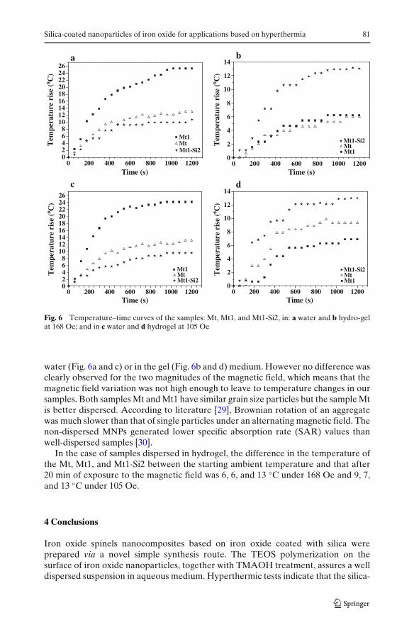
Figure 6 shows the increase in the temperature of the samples in applied ACmagnetic fields of 105 Oe and 168 Oe at 198 kHz in water and hydrogel. In the caseof samples dispersed in water, the difference in the temperature of the Mt, Mt1, andMt1-Si2 between the starting ambient temperature and that after 20 min of exposureto the magnetic field was 13, 25, and 11 ◦C under 168 Oe and 13, 24, and 10 ◦C under105 Oe. The used magnetic field led to temperature changes for samples either in
Silica-coated nanoparticles of iron oxide for applications based on hyperthermia 81
0 200 400 600 800 1000 120002468
101214161820222426
MtMt1
Tem
pera
ture
ris
e (0 C
)T
empe
ratu
re r
ise
(0 C)
Tem
pera
ture
ris
e (0 C
)
Time (s)0 200 400 600 800 1000 1200
Time (s)
0 200 400 600 800 1000 1200Time (s)
0 200 400 600 800 1000 1200Time (s)
Mt1-Si20
2
4
6
8
10
12
14
Tem
pera
ture
ris
e (0 C
)
MtMt1
Mt1-Si2
02468
101214161820222426
MtMt1
Mt1-Si20
2
4
6
8
10
12
14
MtMt1
Mt1-Si2
ba
dc
Fig. 6 Temperature–time curves of the samples: Mt, Mt1, and Mt1-Si2, in: a water and b hydro-gelat 168 Oe; and in c water and d hydrogel at 105 Oe
water (Fig. 6a and c) or in the gel (Fig. 6b and d) medium. However no difference wasclearly observed for the two magnitudes of the magnetic field, which means that themagnetic field variation was not high enough to leave to temperature changes in oursamples. Both samples Mt and Mt1 have similar grain size particles but the sample Mtis better dispersed. According to literature [29], Brownian rotation of an aggregatewas much slower than that of single particles under an alternating magnetic field. Thenon-dispersed MNPs generated lower specific absorption rate (SAR) values thanwell-dispersed samples [30].
In the case of samples dispersed in hydrogel, the difference in the temperature ofthe Mt, Mt1, and Mt1-Si2 between the starting ambient temperature and that after20 min of exposure to the magnetic field was 6, 6, and 13 ◦C under 168 Oe and 9, 7,and 13 ◦C under 105 Oe.
4 Conclusions
Iron oxide spinels nanocomposites based on iron oxide coated with silica wereprepared via a novel simple synthesis route. The TEOS polymerization on thesurface of iron oxide nanoparticles, together with TMAOH treatment, assures a welldispersed suspension in aqueous medium. Hyperthermic tests indicate that the silica-
82 A.L. Andrade et al.
coated composites are potentially very good materials to be used in local therapies,in oncology.
Acknowledgements Work financially supported by CNPq and FAPEMIG (Brazil; particularlygrant # PPM 00419-10, APQ-04333-10, and APQ-00651-11). CAPES (Brazil) grants the VisitingProfessor PVNS fellowship to JDF at UFVJM.