Proteomic Characterization of EvolutionarilyConserved and Variable Proteins of ArabidopsisCytosolic Ribosomes1[w]
Ing-Feng Chang2, Kathleen Szick-Miranda2, Songqin Pan, and Julia Bailey-Serres*
Center for Plant Cell Biology, Department of Botany and Plant Sciences, University of California,Riverside, California 92521–0124 (I.-F.C., S.P., J.B.-S.); and Department of Biology,California State University, Bakersfield, California 93311 (K.S.-M.)
Analysis of 80S ribosomes of Arabidopsis (Arabidopsis thaliana) by use of high-speed centrifugation, sucrose gradientfractionation, one- and two-dimensional gel electrophoresis, liquid chromatography purification, and mass spectrometry(matrix-assisted laser desorption/ionization time-of-flight and electrospray ionization) identified 74 ribosomal proteins(r-proteins), of which 73 are orthologs of rat r-proteins and one is the plant-specific r-protein P3. Thirty small (40S) subunit and44 large (60S) subunit r-proteins were confirmed. In addition, an ortholog of the mammalian receptor for activated proteinkinase C, a tryptophan-aspartic acid-domain repeat protein, was found to be associated with the 40S subunit and polysomes.Based on the prediction that each r-protein is present in a single copy, the mass of the Arabidopsis 80S ribosome was estimatedas 3.2 MD (1,159 kD 40S; 2,010 kD 60S), with the 4 single-copy rRNAs (18S, 26S, 5.8S, and 5S) contributing 53% of the mass.Despite strong evolutionary conservation in r-protein composition among eukaryotes, Arabidopsis 80S ribosomes are variablein composition due to distinctions in mass or charge of approximately 25% of the r-proteins. This is a consequence of aminoacid sequence divergence within r-protein gene families and posttranslational modification of individual r-proteins (e.g.amino-terminal acetylation, phosphorylation). For example, distinct types of r-proteins S15a and P2 accumulate in ribosomesdue to evolutionarily divergence of r-protein genes. Ribosome variation is also due to amino acid sequence divergence anddifferential phosphorylation of the carboxy terminus of r-protein S6. The role of ribosome heterogeneity in differential mRNAtranslation is discussed.
The ribosome is a two-subunit ribonucleoproteincomplex that catalyzes the peptidyl transferase re-action of polypeptide synthesis, an absolute require-ment for cellular growth and differentiation. Thestructure and function of both prokaryotic and eu-karyotic ribosomes have been investigated, with theeukaryotic emphasis on ribosomes of Baker’s yeast(Saccharomyces cerevisiae) and rat (Rattus rattus andRattus norvegicus). The cytosolic ribosomes of eukar-yotes are composed of a large number of ribosomalproteins (r-proteins) and four distinct rRNAs, the 18SrRNA of the 40S subunit, and the 5S, 5.8S, and 23S-like(25–28S) rRNAs of the 60S subunit (Bielka, 1982). Earlyevaluation of the buoyant density and sedimentationcoefficients of eukaryotic ribosomes predicted that therat 80S ribosome has a higher mass (4.2–4.6 MD) thanthat of pea (Pisum sativum; 3.9 MD) due to distinctionsin the 60S subunit (Cammarano et al., 1972). Further
evidence that the plant ribosome is smaller than that ofmammals was provided by the three-dimensionalreconstruction of wheat (Triticum aestivum) and rabbit(Oryctolagus cuniculus) ribosomes by use of cryo-electron microscopy (Verschoor et al., 1996). Despitean overall similarity in architecture, the 60S subunit ofwheat appeared approximately 20% smaller thanthat of rabbit. This variation in mass was attributedto differences in the length of highly variable loopregions of the 23S-like rRNA, which ranges in sizefrom approximately 3,300 bp (25–26S) in plants toapproximately 4,700 bp (28S) in mammals (Schnareet al., 1996). However, to date there has been nodetailed biochemical comparison of the protein com-ponent of plant and animal ribosomes.
The systematic analysis of two-dimensional (2D) gelfractionated r-proteins of 80S ribosomes and genesequences of animals and fungi led to the recogni-tion of 79 eukaryotic r-proteins of common evolu-tionary origin (32 small subunit and 47 large subunitr-proteins; Warner, 1989; Wool et al., 1995; Goffeauet al., 1997; Planta and Mager, 1998; Veuthey and Bittar,1998; Yoshihama et al., 2002; Nakao et al., 2004). Themouse, human, and Drosophila melanogaster genomesencode all 79 proteins, whereas some r-proteins areabsent in the genomes of Baker’s yeast (L28), andCaenorhabditis elegans and Schizosaccharomyces pombe(S27a; Nakao et al., 2004). Based on biochemicalanalyses of ribosomes of mammals and yeast, each
1 This research was supported by the National Science Founda-tion (grant no. DBI 021187 to J.B.-S.), by the U.S. Department ofAgriculture (grant no. 00–35301–9108 to J.B.-S.), and by the Ministryof Education, Republic of China, Taiwan (grant to I.-F.C.).
2 These authors contributed equally to the paper.* Corresponding author; e-mail [email protected]; fax 951–827–
4437.[w] The online version of this article contains Web-only data.Article, publication date, and citation information can be found at
848 Plant Physiology, March 2005, Vol. 137, pp. 848–862, www.plantphysiol.org � 2005 American Society of Plant Biologists www.plantphysiol.orgon May 26, 2018 - Published by Downloaded from
r-protein is present in unimolar amounts with theexception of P1 and P2 of the 60S subunit, which canbe either entirely absent or present as 2 or 4 monomersin yeast and 2 dimers in animals (Tsurugi and Ogata,1985; Guarinos et al., 2003).
Recently, mass spectrometry (MS) has become anefficient technology for proteomic characterization ofmacromolecular complexes (for review, see Aebersoldand Mann, 2003). MS analyses confirmed the presenceof the 32 predicted r-proteins in the 40S subunit of ratfibroblast ribosomes (Louie et al., 1996) and 75 of the78 putative r-proteins of yeast 80S ribosomes (Linket al., 1999; Lee et al., 2002; Inada et al., 2002). Fouradditional putative r-proteins were noted in the ratanalysis and two ribosome-associated proteins wereidentified in the yeast analyses. MS analysis alsoallowed for the identification of plastidic r-proteinsof spinach (Spinacia oleracea) and Chlamydomonas rein-hardtii (Yamaguchi et al., 2000, 2002; Yamaguchi andSubramanian, 2000, 2003), as well as mitochondrialr-proteins of yeast and mammals (Koc et al., 2000,2001; Saveanu et al., 2001; Suzuki et al., 2001a, 2001b;Gan et al., 2002). In addition, MS technology has beenapplied to identify covalent posttranslational modifi-cation of r-proteins. For example, the sequential phos-phorylation of carboxy-terminal sites of Drosophilaand maize (Zea mays) r-protein S6 was validated by useof matrix-assisted laser desorption/ionization time-of-flight (MALDI-TOF) following digestion with trypsinand b-elimination of phosphate groups by treatmentwith barium hydroxide (Radimerski et al., 2000;Williams et al., 2003).
There have been limited biochemical analyses of theprotein components of plant ribosomes. 2D gel elec-trophoresis was employed to resolve r-proteins ofseveral higher plant species, including wheat, soybean(Glycine max), tomato (Lycopersicon peruvianum), maize,tobacco (Nicotiana tabacum), and barley (Hordeum vul-gare; Capel and Bourque, 1982; Gantt and Key, 1983;Sikorski et al., 1983; Scharf and Nover, 1987; Bailey-Serres and Freeling, 1990; Koyama et al., 1996). Theestimates of small and large subunit proteins rangefrom 26 to 40 and 41 to 59 proteins, respectively. Thestrong conservation in eukaryotic r-protein primarysequence was utilized to identify orthologs of the well-characterized rat r-protein collection in Arabidopsis(Arabidopsis thaliana; Barakat et al., 2001). By use ofexpressed sequence tag and the complete genomicsequence accessions, 249 genes (including 22 apparentpseudogenes) were estimated to encode 80 putativetypes of r-proteins (32 small subunit and 48 largesubunit), including a previously identified plant-specific r-protein of the large subunit, P3 (Bailey-Serreset al., 1997; Szick et al., 1998).
To identify the evolutionarily conserved r-proteinsof plant 80S ribosomes and to determine if there areadditional plant-specific r-proteins, we combined 2Dgel electrophoresis and MS to characterize the ribo-somal proteome of Arabidopsis. With this approach,we identified 74 r-proteins, including 2 products of
evolutionarily distinct r-protein genes. We character-ized a number of covalent posttranslational modifica-tions of r-proteins, including the phosphorylation ofS6. In addition, we identified a Trp-Asp (WD)-repeatdomain protein associated with the 40S subunit that isa known scaffold for regulatory proteins.
RESULTS
Identification of Arabidopsis 80S R-Proteins by 2D
Gel Fractionation and MS
In a previous study, we identified 249 genes thatencode 80 putative types of cytosolic r-proteins inArabidopsis (Barakat et al., 2001; an update of thisanalysis indicates that there are 251 genes, including17 pseudogenes, that encode the 80 types of r-proteins;http://www.arabidopsis.org/info/genefamily/athr.html). To confirm the presence of these proteins in Ara-bidopsis ribosomes, a ribosome pellet fraction wasisolated from dark-grown suspension culture cells bydetergent extraction and differential centrifugation.The majority of r-proteins are basic and a few arehighly acidic (Barakat et al., 2001). The basic proteinswere optimally separated in a first dimension basic-urea gel that achieved separation of proteins rangingin pI from 9 to 13 (Williams et al., 2003). Proteins werefurther fractionated by apparent molecular mass in thesecond dimension by SDS-PAGE and stained withCoomassie Blue (Fig. 1A). The acidic and neutralproteins of the ribosome pellet fraction were optimallyseparated by nonequilibrium pH gel electrophoresis(NEpHGE; O’Farrell, 1975) in the first dimension andSDS-PAGE in the second dimension (Fig. 2A). Theprotein spots were excised and digested in-gel withtrypsin and analyzed by either peptide mass finger-printing (PMF) using MALDI-TOF, or tandem MS(MS/MS) using triple quadropole TOF (Q-TOF) withorthogonal MALDI (oMALDI) or electro-spray ioniza-tion (ESI) as the ion source.
The proteomic analyses of the ribosome pellet frac-tion resulted in the identification of 70 of the 80 types ofputative cytosolic r-proteins (Table I; SupplementalTable I). These included 58 r-proteins identified withthe basic-urea gel system, 15 with the NEpHGE system,and 3 with both systems (Table I; Supplemental Table I;Figs. 1, B and C, and 2B). None of the spots containedknown proteins of plastid or mitochondrial ribosomes,indicating that organellar ribosomes were not abun-dant in the pellet fraction. The percentage of the totalresidues of each protein detected (sequence coverage)in peptide fragments by MALDI-TOF ranged from 14%to 70%, with more than one-half of the proteins iden-tified with over 41% sequence coverage. MS/MS wasused for peptide sequencing to confirm the identity ofproteins with low sequence coverage. Peptides corre-sponding to the products of 2 or more distinct geneswere detected for 33 of the 70 (approximately 47%)r-proteins (Supplemental Table I), consistent with the
Proteomics of Arabidopsis Cytosolic Ribosomes
Plant Physiol. Vol. 137, 2005 849 www.plantphysiol.orgon May 26, 2018 - Published by Downloaded from
finding that most r-proteins are encoded by 3 to 4expressed genes (Barakat et al., 2001).
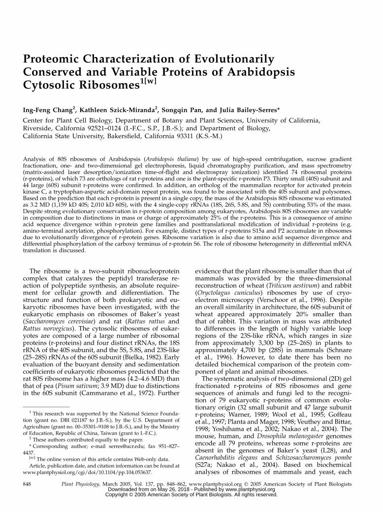
Biochemical variation in r-proteins was apparentfrom the detection of 18 r-proteins (approximately26%) identified in more than one gel spot (Table I). Ofthese, peptides specific to a gene family member wereidentified in spots corresponding to S7, S11, S15a, S19,P2, and L10a, indicating that the difference in mobilitycan reflect biochemical distinctions of individual geneproducts. For other r-proteins found in two or morespots (S6, S25, P0, L5, L10a, L18, L19, L26, and L31), thesame gene product was identified in multiple spots,suggesting that the variation in apparent charge ormass could be due to covalent posttranslational mod-ifications. For example, the product of RPL19A wasresolved in 2 forms with the same migration in thebasic-urea gel but with different apparent molecularmasses (34 and 36 kD; Fig. 1C), whereas 2 forms of L5and P0 had slightly different pIs (Fig. 2B). Distinctionsin protein migration did not appear to be due toN-terminal acetylation (142 D), which was confirmedfor nine r-proteins (Supplemental Table I). Unequalstoichiometry of some r-proteins was suggested fromthe Coomassie Blue staining; however, variation in the
staining intensity of both basic and acidic proteinscould be due to differences in avidity of proteins to thestain.
Identification of R-Proteins and Non-R-Proteins by
Liquid Chromatography and MS/MS
The ribosome pellet isolated by differential centri-fugation reproducibly included several acidic andneutral proteins that are not r-proteins (Fig. 2B; Sup-plemental Table II). These proteins included coreproteasome subunits, several abundant membraneproteins, a mitochondrial chaperonin, an abundantcytosolic enzyme, and a WD-repeat domain protein.To evaluate if these proteins were present in theribosome pellet fraction due to an association withtranslational complexes or cosedimentation of macro-molecular complexes, the pellet was subjected to Sucdensity gradient fractionation, the proteins in the re-sulting 15 fractions were separated by one-dimensional(1D) SDS-PAGE, and visualized by Coomassie Bluestaining. Immunoblot analysis with anti-maize S6 andanti-yeast L15 (ortholog of rat L12) antisera, specificfor small and large ribosomal subunits, respectively,
Figure 1. 2D basic-urea/SDS-PAGE of Arabidopsis r-proteins. A, Total ribosomes isolated from 6-d-old dark grown suspensioncultured cells of Arabidopsis were treated with glacial acetic acid to extract rRNA and basic r-proteins were separated using 2Dbasic-urea/SDS-PAGE. The first-dimension fractionated proteins through a specialized basic-urea gel and the second dimensionfractionated proteins by apparent molecular mass (15% [w/v] SDS-PAGE). The direction and polarity of electrophoresis is shownwith arrows and charge symbols. R-proteins were visualized by staining with Coomassie Blue. The migration of molecular massmarkers is indicated on the right. B and C, Identification of small (40S) subunit (B) and large (60S) subunit (C) r-proteins. Proteinspots resolved in Awere excised, digested in-gel with trypsin, and analyzed byMS. The r-protein designation is directly above theprotein spot or by an arrow; a number or Roman numeral after a protein indicates evidence of a gene-specific or modifiedproduct. The proteins in the boxed region of the gel were further resolved in D. D, Basic-urea gel electrophoresis/SDS-PAGE ofRPS6 forms. R-protein fractionation as in A, except that 12% (w/v) SDS-PAGE was used to increase protein separation. The 8forms of r-protein S6 detected by MALDI-TOF MS are designated as S6, S6a, S6b, S6c, S6d, S6e, S6f, and S6g. Protein spots thatwere not reproducibly observed on gels are not identified.
Chang et al.
850 Plant Physiol. Vol. 137, 2005 www.plantphysiol.orgon May 26, 2018 - Published by Downloaded from
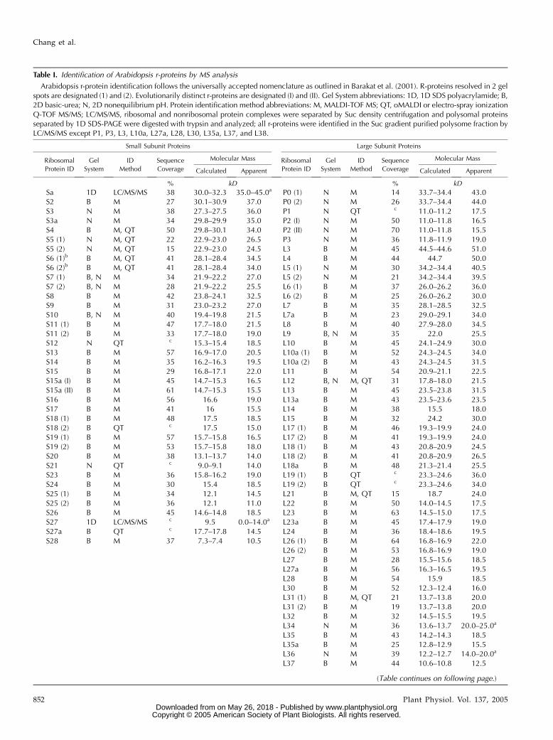
was performed to identify fractions enriched in 40Sand 60S subunits, 80S ribosomes, and polysomes (Fig.3). The stained gel was divided into seven sections forthe polysomal fraction, which were subjected to in-geltrypsin digestion followed by liquid chromatography(LC) coupled to Q-TOF ESI/MS/MS. In this manner,the polysome fraction was found to include 60 of the70 r-proteins detected after 2D gel separation, as well
as 4 additional putative r-proteins (Sa, S27, L37a, andL40; Table I; Supplemental Table I), bringing the totalnumber of identified r-proteins to 74. Of the non-r-proteins detected on 2D gels, the abundant tonoplastmembrane-associated reversibly glycosylated poly-peptide was present in the nonpolysomal fractionand absent in the polysomal fraction, whereas protea-some a-subunits A, B, and E were identified in bothfractions (Fig. 3; Supplemental Table II).
A 36-kD protein resolved as spot number 10 byNEpHGE/SDS-PAGE was identified as a WD-repeatdomain protein related to mammalian receptor foractivated protein kinase C, RACK1 (Fig. 2B; Supple-mental Table II). Three Arabidopsis genes (At1g18080,At1g48630, and At3g18130) encode a protein with 65%amino acid sequence identity to human RACK1. Pep-tides corresponding to the product of 2 of these geneswere detected in spot number 10 and in a 36-kD bandobserved in the 40S subunit, 80S ribosome, and poly-some fractions (Fig. 3, arrow; Supplemental Table II).Consistent with the conclusion that the ArabidopsisRACK1 ortholog associates with ribosomes via aninteraction with the 40S subunit, this protein wasdetected in the 40S, 80S, and polysome fractions atlevels similar to that observed for r-protein S6 (Fig. 3).
Arabidopsis Ribosomes Contain Differentially
Phosphorylated Forms of S6
The detection of r-protein S6 in multiple proteinspots led to the evaluation of the phosphorylation of2 S6 gene products (RPS6A, At4g31700; RPS6B,At5g10360). S6A and S6B are 95.2% identical in aminoacid sequence but have divergent carboxy terminiwith 5 and 3 potential phosphorylation sites, respec-tively. Eight forms of S6 (S6, S6a–g) were identifiedamong the constellation of proteins with an apparentpI of 10 to 11 and molecular mass of 28 to 30 kD (Fig.1D; Supplemental Table II). The forms S6 and S6a tod displayed more rapid mobility in the SDS-PAGEdimension than the forms S6e to g.
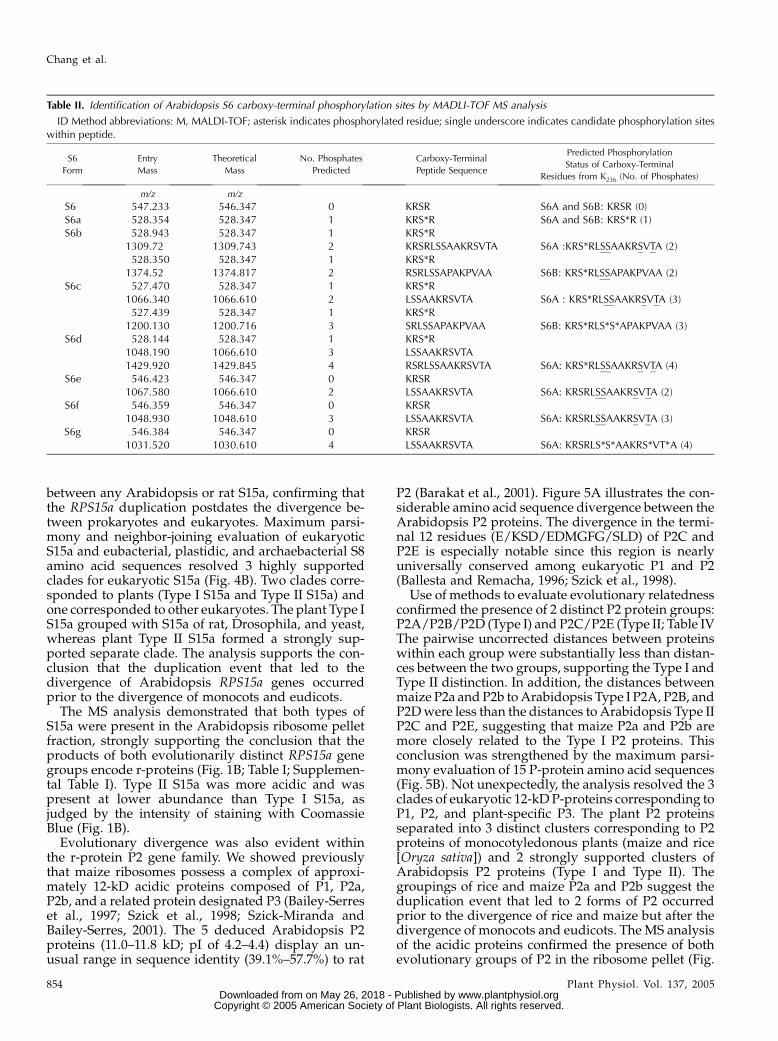
MS analysis of each of the 8 S6 forms by in-geltrypsin-digestion and barium hydroxide-treatmentprovided evidence of carboxy-terminal phosphoryla-tion in S6A and S6B (Table II). Phosphorylation (180 D)of Ser or Thr decreases the charge of tryptic peptidesand reduces the frequency of peptide ionization duringMALDI-TOF MS analysis. To circumvent this difficulty,trypsin digestion was coupled with treatment withbarium hydroxide, which causes b-elimination ofH3PO4 (298 D). Thus, the ions of phosphorylatedpeptides show an 18 mass-to-charge ratio (m/z) re-duction for each phosphorylated residue. When thisprocedure was carried out with the extremely basic S6form, only an unmodified carboxy-terminal peptidewas detected. On the other hand, the m/z values of ionsfrom the S6a to d digests corresponded to the predictedmasses of mono-, di-, tri-, and tetra-phosphorylatedS6A, respectively. Peptide ions from S6a to c were de-tected that also corresponded to the predicted masses
Figure 2. 2D NEpHGE of Arabidopsis r-proteins. A, Total ribosomesisolated as described in Figure 1 were separated by 2D NEpHGE/SDS-PAGE to analyze acidic and neutral proteins. The first-dimensionfractionated proteins by ionic charge through a pH gradient (pH3–10). The second-dimension fractionated proteins by apparent mo-lecular mass (12% [w/v] SDS-PAGE). The direction and polarity ofelectrophoresis is shown with arrows and charge symbols. R-proteinswere visualized by staining with Coomassie Blue. The migration ofmolecular mass markers is indicated on the right. B, Identification ofproteins. Proteins resolved in A were excised, digested in gel withtrypsin, and analyzed by MS. The protein designation is directly abovethe protein spot or by an arrow; a number or Roman numeral aftera protein indicates evidence of a gene-specific or modified product.Nonribosomal proteins are identified with a number: 1, proteasomea-subunit A (At2g05840); 2, proteasome a-subunit B (At1g16470); 3,proteasome a-subunit E (At1g53850; At3g14290); 4, vacuolar H1
ATPase subunit E (At4g11150); 5, vacuolar H1 ATPase subunit G; 6,chaperonin heat shock protein 60 (At3g23990); 7, mitochondrial F0ATPase chain D (At3g52300); 8, vacuolar reversibly glycosylatedprotein (At3g02230; At5g15650); 9, Gln synthase (At3g17820); 10,WD-repeat domain repeat protein human RACK1 ortholog(At1g18080; At1g48630). Protein spots that were not reproduciblyobserved on gels are not identified.
Proteomics of Arabidopsis Cytosolic Ribosomes
Plant Physiol. Vol. 137, 2005 851 www.plantphysiol.orgon May 26, 2018 - Published by Downloaded from
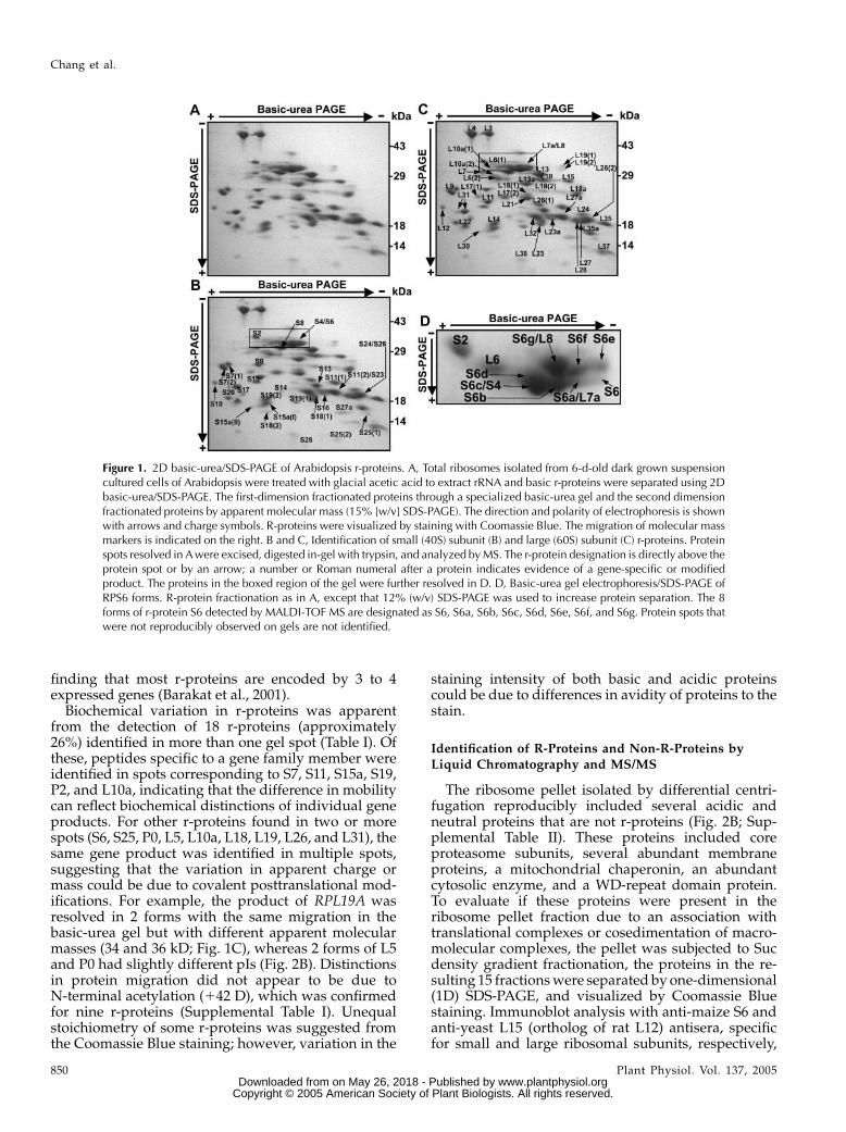
Table I. Identification of Arabidopsis r-proteins by MS analysis
Arabidopsis r-protein identification follows the universally accepted nomenclature as outlined in Barakat et al. (2001). R-proteins resolved in 2 gelspots are designated (1) and (2). Evolutionarily distinct r-proteins are designated (I) and (II). Gel System abbreviations: 1D, 1D SDS polyacrylamide; B,2D basic-urea; N, 2D nonequilibrium pH. Protein identification method abbreviations: M, MALDI-TOF MS; QT, oMALDI or electro-spray ionizationQ-TOF MS/MS; LC/MS/MS, ribosomal and nonribosomal protein complexes were separated by Suc density centrifugation and polysomal proteinsseparated by 1D SDS-PAGE were digested with trypsin and analyzed; all r-proteins were identified in the Suc gradient purified polysome fraction byLC/MS/MS except P1, P3, L3, L10a, L27a, L28, L30, L35a, L37, and L38.
Small Subunit Proteins Large Subunit Proteins
Ribosomal
Protein ID
Gel
System
ID
Method
Sequence
Coverage
Molecular Mass Ribosomal
Protein ID
Gel
System
ID
Method
Sequence
Coverage
Molecular Mass
Calculated Apparent Calculated Apparent
% kD % kD
Sa 1D LC/MS/MS 38 30.0–32.3 35.0–45.0a P0 (1) N M 14 33.7–34.4 43.0S2 B M 27 30.1–30.9 37.0 P0 (2) N M 26 33.7–34.4 44.0S3 N M 38 27.3–27.5 36.0 P1 N QT c 11.0–11.2 17.5S3a N M 34 29.8–29.9 35.0 P2 (I) N M 50 11.0–11.8 16.5S4 B M, QT 50 29.8–30.1 34.0 P2 (II) N M 70 11.0–11.8 15.5S5 (1) N M, QT 22 22.9–23.0 26.5 P3 N M 36 11.8–11.9 19.0S5 (2) N M, QT 15 22.9–23.0 24.5 L3 B M 45 44.5–44.6 51.0S6 (1)b B M, QT 41 28.1–28.4 34.5 L4 B M 44 44.7 50.0S6 (2)b B M, QT 41 28.1–28.4 34.0 L5 (1) N M 30 34.2–34.4 40.5S7 (1) B, N M 34 21.9–22.2 27.0 L5 (2) N M 21 34.2–34.4 39.5S7 (2) B, N M 28 21.9–22.2 25.5 L6 (1) B M 37 26.0–26.2 36.0S8 B M 42 23.8–24.1 32.5 L6 (2) B M 25 26.0–26.2 30.0S9 B M 31 23.0–23.2 27.0 L7 B M 35 28.1–28.5 32.5S10 B, N M 40 19.4–19.8 21.5 L7a B M 23 29.0–29.1 34.0S11 (1) B M 47 17.7–18.0 21.5 L8 B M 40 27.9–28.0 34.5S11 (2) B M 33 17.7–18.0 19.0 L9 B, N M 35 22.0 25.5S12 N QT c 15.3–15.4 18.5 L10 B M 45 24.1–24.9 30.0S13 B M 57 16.9–17.0 20.5 L10a (1) B M 52 24.3–24.5 34.0S14 B M 35 16.2–16.3 19.5 L10a (2) B M 43 24.3–24.5 31.5S15 B M 29 16.8–17.1 22.0 L11 B M 54 20.9–21.1 22.5S15a (I) B M 45 14.7–15.3 16.5 L12 B, N M, QT 31 17.8–18.0 21.5S15a (II) B M 61 14.7–15.3 15.5 L13 B M 45 23.5–23.8 31.5S16 B M 56 16.6 19.0 L13a B M 43 23.5–23.6 23.5S17 B M 41 16 15.5 L14 B M 38 15.5 18.0S18 (1) B M 48 17.5 18.5 L15 B M 32 24.2 30.0S18 (2) B QT c 17.5 15.0 L17 (1) B M 46 19.3–19.9 24.0S19 (1) B M 57 15.7–15.8 16.5 L17 (2) B M 41 19.3–19.9 24.0S19 (2) B M 53 15.7–15.8 18.0 L18 (1) B M 43 20.8–20.9 24.5S20 B M 38 13.1–13.7 14.0 L18 (2) B M 41 20.8–20.9 26.5S21 N QT c 9.0–9.1 14.0 L18a B M 48 21.3–21.4 25.5S23 B M 36 15.8–16.2 19.0 L19 (1) B QT c 23.3–24.6 36.0S24 B M 30 15.4 18.5 L19 (2) B QT c 23.3–24.6 34.0S25 (1) B M 34 12.1 14.5 L21 B M, QT 15 18.7 24.0S25 (2) B M 36 12.1 11.0 L22 B M 50 14.0–14.5 17.5S26 B M 45 14.6–14.8 18.5 L23 B M 63 14.5–15.0 17.5S27 1D LC/MS/MS c 9.5 0.0–14.0a L23a B M 45 17.4–17.9 19.0S27a B QT c 17.7–17.8 14.5 L24 B M 36 18.4–18.6 19.5S28 B M 37 7.3–7.4 10.5 L26 (1) B M 64 16.8–16.9 22.0
L26 (2) B M 53 16.8–16.9 19.0L27 B M 28 15.5–15.6 18.5L27a B M 56 16.3–16.5 19.5L28 B M 54 15.9 18.5L30 B M 52 12.3–12.4 16.0L31 (1) B M, QT 21 13.7–13.8 20.0L31 (2) B M 19 13.7–13.8 20.0L32 B M 32 14.5–15.5 19.5L34 N M 36 13.6–13.7 20.0–25.0a
L35 B M 43 14.2–14.3 18.5L35a B M 25 12.8–12.9 15.5L36 N M 39 12.2–12.7 14.0–20.0a
L37 B M 44 10.6–10.8 12.5
(Table continues on following page.)
Chang et al.
852 Plant Physiol. Vol. 137, 2005 www.plantphysiol.orgon May 26, 2018 - Published by Downloaded from
of mono-, di-, and tri-phosphorylated S6B, respectively.By comparison of theoretical and observed mass val-ues, we determined that S6a was phosphorylated atSer-238; S6b to d were phosphorylated at Ser-238 andadditional sites in the region R239LSSAAKRSVTA. Therapid migrating S6e to g forms were sequentially di-,tri-, and tetra-phosphorylated forms of S6A. Inter-estingly, the detection of the ion corresponding tononphosphorylated form of the peptide K236RSR in-dicated that Ser-238 was skipped by the S6 kinase ordephosphorylated by an unknown phosphatase in the
formation of S6e to g. Although the absence of cleavagesites in the carboxy terminus precluded identificationof the exact sites of phosphorylation in some S6 forms,these results confirm extensive heterogeneity in thecarboxyl-terminal phosphorylation sites of S6A and S6B.
The Arabidopsis Genome Encodes Evolutionarily
Distinct S15a and P2 Types That Accumulatein Ribosomes
The percent amino acid sequence identity betweenthe deduced Arabidopsis and rat r-protein orthologswas extremely consistent among gene family members,except for the RPS15a, RPP2, and RPL7 multigenefamilies (Barakat et al., 2001). Evolutionary divergenceamong these gene families was confirmed by use of se-quence alignment, pairwise distance calculations aswell as distance- and parsimony-based phylogeneticanalyses. Of the 6 Arabidopsis RPS15a genes, 4(RPS15aA, RPS15aC, RPS15aD, and RPS15aF) encodea protein with 73.1% to 77.7% identity and 2 (RPS15aBand RPS15aE) encode a protein with 47.6% and 48.8%identity to rat S15a, respectively (Barakat et al., 2001).Eukaryotic S15a is orthologous to S8 of eubacteria,plastids, and mitochondria of lower eukaryotes (Woolet al., 1995; Nevskaya et al., 1998; Adams et al., 2002),which binds 16S rRNA and is crucial for ribosomeassembly (Held et al., 1974; Svensson et al., 1988).Conservation between plant and animal S15a includesthe carboxy-terminal amino acid region that corre-sponds to an rRNA-binding site in eubacteria (Mougelet al., 1986; Wower et al., 1992; Davies et al., 1996;Nevskaya et al., 1998; Fig. 4A, underlined). In addition,a number of residues within this region are highlyconserved in eubacterial, plastidic, and archaebacterialS8 (Fig. 4A, shaded residues).
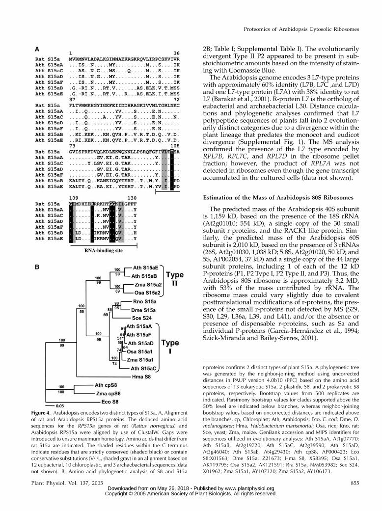
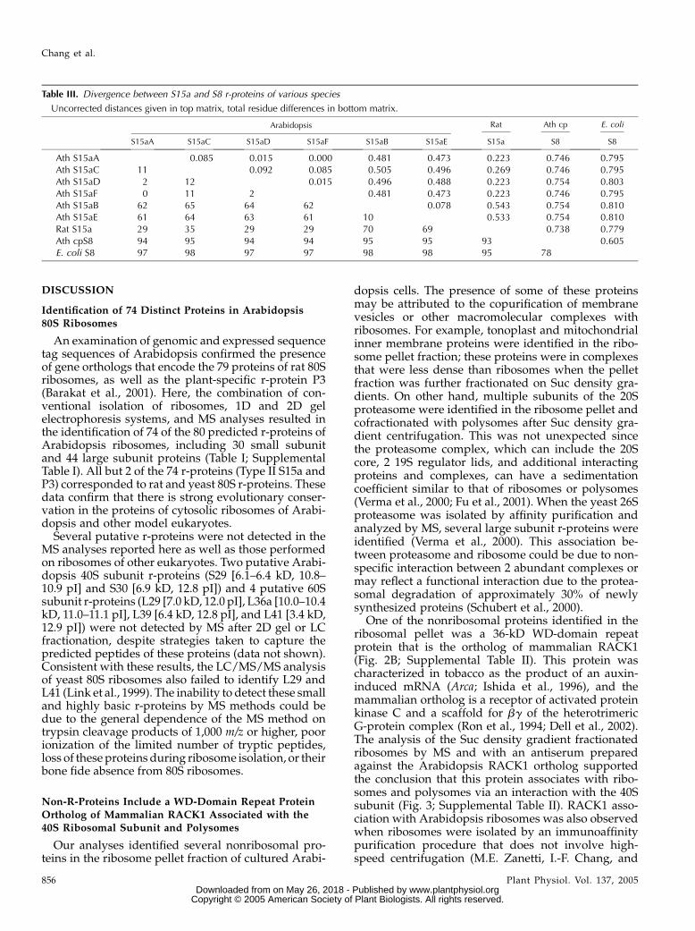
The calculation of pairwise, uncorrected distancesbetween Arabidopsis S8 and S15a proteins confirmedthat S15a falls into 2 distinct groups: S15aA/S15aC/S15aD/S15aF (Type I) and S15aB/S15aE (Type II; TableIII). This analysis revealed that evolutionary distancebetween Arabidopsis S15a proteins within each of thetwo groups was substantially less than betweengroups. The distance between Type I or Type II S15ato plastid S8 was quite similar, whereas the distancebetween Escherichia coli and plastid S8 was less than
Table I. (Continued from previous page.)
Small Subunit Proteins Large Subunit Proteins
Ribosomal
Protein ID
Gel
System
ID
Method
Sequence
Coverage
Molecular Mass Ribosomal
Protein ID
Gel
System
ID
Method
Sequence
Coverage
Molecular Mass
Calculated Apparent Calculated Apparent
% kD
L37a 1D LC/MS/MS 10.0–10.4 0.0–14.0a
L38 B M 60 8.1 11.5L40 1D LC/MS/MS 14.7 0.0–14.0a
aProteins identified from the 1D gel were from gel slices that had a range in apparent molecular mass. bMultiple isoforms of S6 were detectedin both the rapid mobility group and the slow mobility group. cIdentification from sequencing one or more peptide.
Figure 3. Suc density gradient fractionation of total ribosomes. Totalribosomes isolated from 6-d-old dark grown suspension cultured cellsof Arabidopsis by high speed centrifugation were further fractionatedthrough a Suc density gradient. The fractions were resolved by 15%(w/v) SDS-PAGE and visualized by staining with Coomassie Blue.Fractions corresponding to the nonpolysomal fraction (np), 40S subunit,60S subunit, 80S ribosome, and polysomes (poly) are shown. Themigration of molecular mass markers is indicated on the left. The SDS-PAGE gel lanes were cut into seven even sections as indicated on the right(I, 35.0–45.0 kD; II, 32.5–35.0 kD; III, 30.0–32.5 kD; IV, 25.0–30.0 kD;V, 20.0–25.0 kD; VI, 14.0–20.0 kD; and VII, 0.0–14.0 kD). Individualsections of the nonpolysomal and polysomal fractions were subjected toin-gel trypsin digestion and LC/MS/MS. Arrows indicated the position ofthe Coomassie Blue-stained Arabidopsis ortholog of human RACK1, asdetermined by LC/MS/MS. Immunological detection of protein in thegradient fractions was performed with anti-maize S6, anti-yeast L15(ortholog of rat and plant L12), and anti-Arabidopsis RACK1.
Proteomics of Arabidopsis Cytosolic Ribosomes
Plant Physiol. Vol. 137, 2005 853 www.plantphysiol.orgon May 26, 2018 - Published by Downloaded from
between any Arabidopsis or rat S15a, confirming thatthe RPS15a duplication postdates the divergence be-tween prokaryotes and eukaryotes. Maximum parsi-mony and neighbor-joining evaluation of eukaryoticS15a and eubacterial, plastidic, and archaebacterial S8amino acid sequences resolved 3 highly supportedclades for eukaryotic S15a (Fig. 4B). Two clades corre-sponded to plants (Type I S15a and Type II S15a) andone corresponded to other eukaryotes. The plant Type IS15a grouped with S15a of rat, Drosophila, and yeast,whereas plant Type II S15a formed a strongly sup-ported separate clade. The analysis supports the con-clusion that the duplication event that led to thedivergence of Arabidopsis RPS15a genes occurredprior to the divergence of monocots and eudicots.
The MS analysis demonstrated that both types ofS15a were present in the Arabidopsis ribosome pelletfraction, strongly supporting the conclusion that theproducts of both evolutionarily distinct RPS15a genegroups encode r-proteins (Fig. 1B; Table I; Supplemen-tal Table I). Type II S15a was more acidic and waspresent at lower abundance than Type I S15a, asjudged by the intensity of staining with CoomassieBlue (Fig. 1B).
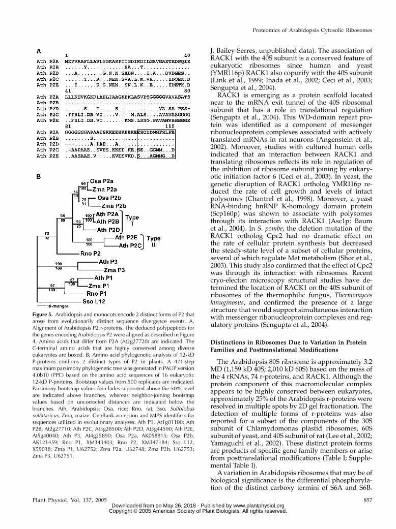
Evolutionary divergence was also evident withinthe r-protein P2 gene family. We showed previouslythat maize ribosomes possess a complex of approxi-mately 12-kD acidic proteins composed of P1, P2a,P2b, and a related protein designated P3 (Bailey-Serreset al., 1997; Szick et al., 1998; Szick-Miranda andBailey-Serres, 2001). The 5 deduced Arabidopsis P2proteins (11.0–11.8 kD; pI of 4.2–4.4) display an un-usual range in sequence identity (39.1%–57.7%) to rat
P2 (Barakat et al., 2001). Figure 5A illustrates the con-siderable amino acid sequence divergence between theArabidopsis P2 proteins. The divergence in the termi-nal 12 residues (E/KSD/EDMGFG/SLD) of P2C andP2E is especially notable since this region is nearlyuniversally conserved among eukaryotic P1 and P2(Ballesta and Remacha, 1996; Szick et al., 1998).
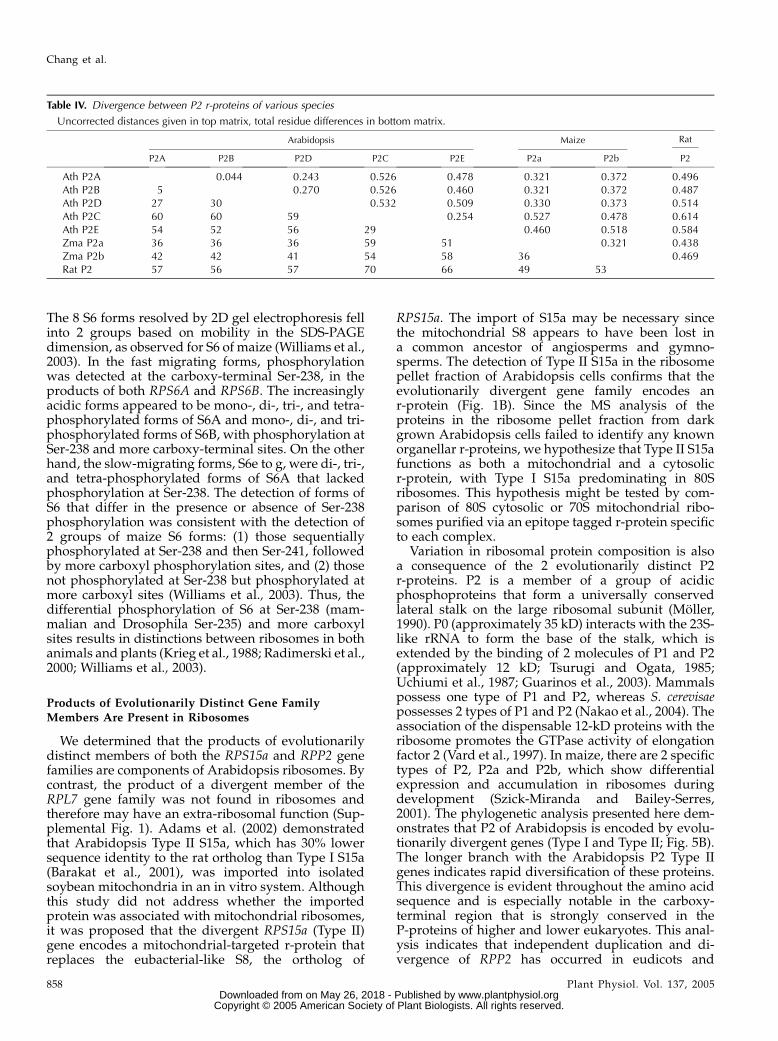
Use of methods to evaluate evolutionary relatednessconfirmed the presence of 2 distinct P2 protein groups:P2A/P2B/P2D (Type I) and P2C/P2E (Type II; Table IVThe pairwise uncorrected distances between proteinswithin each group were substantially less than distan-ces between the two groups, supporting the Type I andType II distinction. In addition, the distances betweenmaize P2a and P2b to Arabidopsis Type I P2A, P2B, andP2D were less than the distances to Arabidopsis Type IIP2C and P2E, suggesting that maize P2a and P2b aremore closely related to the Type I P2 proteins. Thisconclusion was strengthened by the maximum parsi-mony evaluation of 15 P-protein amino acid sequences(Fig. 5B). Not unexpectedly, the analysis resolved the 3clades of eukaryotic 12-kD P-proteins corresponding toP1, P2, and plant-specific P3. The plant P2 proteinsseparated into 3 distinct clusters corresponding to P2proteins of monocotyledonous plants (maize and rice[Oryza sativa]) and 2 strongly supported clusters ofArabidopsis P2 proteins (Type I and Type II). Thegroupings of rice and maize P2a and P2b suggest theduplication event that led to 2 forms of P2 occurredprior to the divergence of rice and maize but after thedivergence of monocots and eudicots. The MS analysisof the acidic proteins confirmed the presence of bothevolutionary groups of P2 in the ribosome pellet (Fig.
Table II. Identification of Arabidopsis S6 carboxy-terminal phosphorylation sites by MADLI-TOF MS analysis
ID Method abbreviations: M, MALDI-TOF; asterisk indicates phosphorylated residue; single underscore indicates candidate phosphorylation siteswithin peptide.
2B; Table I; Supplemental Table I). The evolutionarilydivergent Type II P2 appeared to be present in sub-stoichiometric amounts based on the intensity of stain-ing with Coomassie Blue.
The Arabidopsis genome encodes 3 L7-type proteinswith approximately 60% identity (L7B, L7C ,and L7D)and one L7-type protein (L7A) with 38% identity to ratL7 (Barakat et al., 2001). R-protein L7 is the ortholog ofeubacterial and archaebacterial L30. Distance calcula-tions and phylogenetic analyses confirmed that L7polypeptide sequences of plants fall into 2 evolution-arily distinct categories due to a divergence within theplant lineage that predates the monocot and eudicotdivergence (Supplemental Fig. 1). The MS analysisconfirmed the presence of the L7 type encoded byRPL7B, RPL7C, and RPL7D in the ribosome pelletfraction; however, the product of RPL7A was notdetected in ribosomes even though the gene transcriptaccumulated in the cultured cells (data not shown).
Estimation of the Mass of Arabidopsis 80S Ribosomes
The predicted mass of the Arabidopsis 40S subunitis 1,159 kD, based on the presence of the 18S rRNA(At2g01010; 554 kD), a single copy of the 30 smallsubunit r-proteins, and the RACK1-like protein. Sim-ilarly, the predicted mass of the Arabidopsis 60Ssubunit is 2,010 kD, based on the presence of 3 rRNAs(26S, At2g01030, 1,038 kD; 5.8S, At2g01020, 50 kD; and5S, AP002054, 37 kD) and a single copy of the 44 largesubunit proteins, including 1 of each of the 12 kDP-proteins (P1, P2 Type I, P2 Type II, and P3). Thus, theArabidopsis 80S ribosome is approximately 3.2 MD,with 53% of the mass contributed by rRNA. Theribosome mass could vary slightly due to covalentposttranslational modifications of r-proteins, the pres-ence of the small r-proteins not detected by MS (S29,S30, L29, L36a, L39, and L41), and/or the absence orpresence of dispensable r-proteins, such as Sa andindividual P-proteins (Garcıa-Hernandez et al., 1994;Szick-Miranda and Bailey-Serres, 2001).
Figure 4. Arabidopsis encodes two distinct types of S15a. A, Alignmentof rat and Arabidopsis RPS15a proteins. The deduced amino acidsequences for the RPS15a genes of rat (Rattus norvegicus) andArabidopsis RPS15a were aligned by use of ClustalW. Gaps wereintroduced to ensure maximum homology. Amino acids that differ fromrat S15a are indicated. The shaded residues within the C terminusindicate residues that are strictly conserved (shaded black) or containconservative substitutions (V/I/L, shaded gray) in an alignment based on12 eubacterial, 10 chloroplastic, and 3 archaebacterial sequences (datanot shown). B, Amino acid phylogenetic analysis of S8 and S15a
r-proteins confirms 2 distinct types of plant S15a. A phylogenetic treewas generated by the neighbor-joining method using uncorrecteddistances in PAUP version 4.0b10 (PPC) based on the amino acidsequences of 13 eukaryotic S15a, 2 plastidic S8, and 2 prokaryotic S8r-proteins, respectively. Bootstrap values from 500 replicates areindicated. Parsimony bootstrap values for clades supported above the50% level are indicated below branches, whereas neighbor-joiningbootstrap values based on uncorrected distances are indicated abovethe branches. cp, Chloroplast; Ath, Arabidopsis; Eco, E. coli; Dme, D.melanogaster; Hma, Halobacterium marismortui; Osa, rice; Rno, rat;Sce, yeast; Zma, maize. GenBank accession and MIPS identifiers forsequences utilized in evolutionary analyses: Ath S15aA, At1g07770;Ath S15aB, At2g19720; Ath S15aC, At2g39590; Ath S15aD,At3g46040; Ath S15aE, At4g29430; Ath cpS8, AP000423; EcoS8:X01563; Dme S15a, Z21673; Hma S8, X58395; Osa S15a1,AK119795; Osa S15a2, AK121591; Rra S15a, NM053982; Sce S24,X01962; Zma S15a1, AY107320; Zma S15a2, AY106173.
Proteomics of Arabidopsis Cytosolic Ribosomes
Plant Physiol. Vol. 137, 2005 855 www.plantphysiol.orgon May 26, 2018 - Published by Downloaded from
Identification of 74 Distinct Proteins in Arabidopsis
80S Ribosomes
An examination of genomic and expressed sequencetag sequences of Arabidopsis confirmed the presenceof gene orthologs that encode the 79 proteins of rat 80Sribosomes, as well as the plant-specific r-protein P3(Barakat et al., 2001). Here, the combination of con-ventional isolation of ribosomes, 1D and 2D gelelectrophoresis systems, and MS analyses resulted inthe identification of 74 of the 80 predicted r-proteins ofArabidopsis ribosomes, including 30 small subunitand 44 large subunit proteins (Table I; SupplementalTable I). All but 2 of the 74 r-proteins (Type II S15a andP3) corresponded to rat and yeast 80S r-proteins. Thesedata confirm that there is strong evolutionary conser-vation in the proteins of cytosolic ribosomes of Arabi-dopsis and other model eukaryotes.
Several putative r-proteins were not detected in theMS analyses reported here as well as those performedon ribosomes of other eukaryotes. Two putative Arabi-dopsis 40S subunit r-proteins (S29 [6.1–6.4 kD, 10.8–10.9 pI] and S30 [6.9 kD, 12.8 pI]) and 4 putative 60Ssubunit r-proteins (L29 [7.0 kD, 12.0 pI], L36a [10.0–10.4kD, 11.0–11.1 pI], L39 [6.4 kD, 12.8 pI], and L41 [3.4 kD,12.9 pI]) were not detected by MS after 2D gel or LCfractionation, despite strategies taken to capture thepredicted peptides of these proteins (data not shown).Consistent with these results, the LC/MS/MS analysisof yeast 80S ribosomes also failed to identify L29 andL41 (Link et al., 1999). The inability to detect these smalland highly basic r-proteins by MS methods could bedue to the general dependence of the MS method ontrypsin cleavage products of 1,000 m/z or higher, poorionization of the limited number of tryptic peptides,loss of these proteins during ribosome isolation, or theirbone fide absence from 80S ribosomes.
Non-R-Proteins Include a WD-Domain Repeat Protein
Ortholog of Mammalian RACK1 Associated with the40S Ribosomal Subunit and Polysomes
Our analyses identified several nonribosomal pro-teins in the ribosome pellet fraction of cultured Arabi-
dopsis cells. The presence of some of these proteinsmay be attributed to the copurification of membranevesicles or other macromolecular complexes withribosomes. For example, tonoplast and mitochondrialinner membrane proteins were identified in the ribo-some pellet fraction; these proteins were in complexesthat were less dense than ribosomes when the pelletfraction was further fractionated on Suc density gra-dients. On other hand, multiple subunits of the 20Sproteasome were identified in the ribosome pellet andcofractionated with polysomes after Suc density gra-dient centrifugation. This was not unexpected sincethe proteasome complex, which can include the 20Score, 2 19S regulator lids, and additional interactingproteins and complexes, can have a sedimentationcoefficient similar to that of ribosomes or polysomes(Verma et al., 2000; Fu et al., 2001). When the yeast 26Sproteasome was isolated by affinity purification andanalyzed by MS, several large subunit r-proteins wereidentified (Verma et al., 2000). This association be-tween proteasome and ribosome could be due to non-specific interaction between 2 abundant complexes ormay reflect a functional interaction due to the protea-somal degradation of approximately 30% of newlysynthesized proteins (Schubert et al., 2000).
One of the nonribosomal proteins identified in theribosomal pellet was a 36-kD WD-domain repeatprotein that is the ortholog of mammalian RACK1(Fig. 2B; Supplemental Table II). This protein wascharacterized in tobacco as the product of an auxin-induced mRNA (Arca; Ishida et al., 1996), and themammalian ortholog is a receptor of activated proteinkinase C and a scaffold for bg of the heterotrimericG-protein complex (Ron et al., 1994; Dell et al., 2002).The analysis of the Suc density gradient fractionatedribosomes by MS and with an antiserum preparedagainst the Arabidopsis RACK1 ortholog supportedthe conclusion that this protein associates with ribo-somes and polysomes via an interaction with the 40Ssubunit (Fig. 3; Supplemental Table II). RACK1 asso-ciation with Arabidopsis ribosomes was also observedwhen ribosomes were isolated by an immunoaffinitypurification procedure that does not involve high-speed centrifugation (M.E. Zanetti, I.-F. Chang, and
Table III. Divergence between S15a and S8 r-proteins of various species
Uncorrected distances given in top matrix, total residue differences in bottom matrix.
J. Bailey-Serres, unpublished data). The association ofRACK1 with the 40S subunit is a conserved feature ofeukaryotic ribosomes since human and yeast(YMR116p) RACK1 also copurify with the 40S subunit(Link et al., 1999; Inada et al., 2002; Ceci et al., 2003;Sengupta et al., 2004).
RACK1 is emerging as a protein scaffold locatednear to the mRNA exit tunnel of the 40S ribosomalsubunit that has a role in translational regulation(Sengupta et al., 2004). This WD-domain repeat pro-tein was identified as a component of messengerribonucleoprotein complexes associated with activelytranslated mRNAs in rat neurons (Angenstein et al.,2002). Moreover, studies with cultured human cellsindicated that an interaction between RACK1 andtranslating ribosomes reflects its role in regulation ofthe inhibition of ribosome subunit joining by eukary-otic initiation factor 6 (Ceci et al., 2003). In yeast, thegenetic disruption of RACK1 ortholog YMR116p re-duced the rate of cell growth and levels of intactpolysomes (Chantrel et al., 1998). Moreover, a yeastRNA-binding hnRNP K-homology domain protein(Scp160p) was shown to associate with polysomesthrough its interaction with RACK1 (Asc1p; Baumet al., 2004). In S. pombe, the deletion mutation of theRACK1 ortholog Cpc2 had no dramatic effect onthe rate of cellular protein synthesis but decreasedthe steady-state level of a subset of cellular proteins,several of which regulate Met metabolism (Shor et al.,2003). This study also confirmed that the effect of Cpc2was through its interaction with ribosomes. Recentcryo-electon microscopy structural studies have de-termined the location of RACK1 on the 40S subunit ofribosomes of the thermophilic fungus, Thermomyceslanuginosus, and confirmed the presence of a largestructure that would support simultaneous interactionwith messenger ribonucleoprotein complexes and reg-ulatory proteins (Sengupta et al., 2004).
Distinctions in Ribosomes Due to Variation in ProteinFamilies and Posttranslational Modifications
The Arabidopsis 80S ribosome is approximately 3.2MD (1,159 kD 40S; 2,010 kD 60S) based on the mass ofthe 4 rRNAs, 74 r-proteins, and RACK1. Although theprotein component of this macromolecular complexappears to be highly conserved between eukaryotes,approximately 25% of the Arabidopsis r-proteins wereresolved in multiple spots by 2D gel fractionation. Thedetection of multiple forms of r-proteins was alsoreported for a subset of the components of the 30Ssubunit of Chlamydomonas plastid ribosomes, 60Ssubunit of yeast, and 40S subunit of rat (Lee et al., 2002;Yamaguchi et al., 2002). These distinct protein formsare products of specific gene family members or arisefrom posttranslational modifications (Table I; Supple-mental Table I).
A variation in Arabidopsis ribosomes that may be ofbiological significance is the differential phosphoryla-tion of the distinct carboxy termini of S6A and S6B.
Figure 5. Arabidopsis and monocots encode 2 distinct forms of P2 thatarose from evolutionarily distinct sequence divergence events. A,Alignment of Arabidopsis P2 r-proteins. The deduced polypeptides forthe genes encoding Arabidopsis P2 were aligned as described in Figure4. Amino acids that differ from P2A (At2g27720) are indicated. TheC-terminal amino acids that are highly conserved among diverseeukaryotes are boxed. B, Amino acid phylogenetic analysis of 12-kDP-proteins confirms 2 distinct types of P2 in plants. A 471-stepmaximum parsimony phylogenetic tree was generated in PAUP version4.0b10 (PPC) based on the amino acid sequences of 16 eukaryotic12-kD P-proteins. Bootstrap values from 500 replicates are indicated.Parsimony bootstrap values for clades supported above the 50% levelare indicated above branches, whereas neighbor-joining bootstrapvalues based on uncorrected distances are indicated below thebranches. Ath, Arabidopsis; Osa, rice; Rno, rat; Sso, Sulfolobussolfataricus; Zma, maize. GenBank accession and MIPS identifiers forsequences utilized in evolutionary analyses: Ath P1, At1g01100; AthP2B, At2g27710; Ath P2C, At3g28500; Ath P2D, At3g44590; Ath P2E,At5g40040; Ath P3, At4g25890; Osa P2a, AK058815; Osa P2b,AK121459; Rno P1, XM343403; Rno P2, XM347184; Sso L12,X59038; Zma P1, U62752; Zma P2a, U62748; Zma P2b, U62753;Zma P3, U62751.
Proteomics of Arabidopsis Cytosolic Ribosomes
Plant Physiol. Vol. 137, 2005 857 www.plantphysiol.orgon May 26, 2018 - Published by Downloaded from
The 8 S6 forms resolved by 2D gel electrophoresis fellinto 2 groups based on mobility in the SDS-PAGEdimension, as observed for S6 of maize (Williams et al.,2003). In the fast migrating forms, phosphorylationwas detected at the carboxy-terminal Ser-238, in theproducts of both RPS6A and RPS6B. The increasinglyacidic forms appeared to be mono-, di-, tri-, and tetra-phosphorylated forms of S6A and mono-, di-, and tri-phosphorylated forms of S6B, with phosphorylation atSer-238 and more carboxy-terminal sites. On the otherhand, the slow-migrating forms, S6e to g, were di-, tri-,and tetra-phosphorylated forms of S6A that lackedphosphorylation at Ser-238. The detection of forms ofS6 that differ in the presence or absence of Ser-238phosphorylation was consistent with the detection of2 groups of maize S6 forms: (1) those sequentiallyphosphorylated at Ser-238 and then Ser-241, followedby more carboxyl phosphorylation sites, and (2) thosenot phosphorylated at Ser-238 but phosphorylated atmore carboxyl sites (Williams et al., 2003). Thus, thedifferential phosphorylation of S6 at Ser-238 (mam-malian and Drosophila Ser-235) and more carboxylsites results in distinctions between ribosomes in bothanimals and plants (Krieg et al., 1988; Radimerski et al.,2000; Williams et al., 2003).
Products of Evolutionarily Distinct Gene FamilyMembers Are Present in Ribosomes
We determined that the products of evolutionarilydistinct members of both the RPS15a and RPP2 genefamilies are components of Arabidopsis ribosomes. Bycontrast, the product of a divergent member of theRPL7 gene family was not found in ribosomes andtherefore may have an extra-ribosomal function (Sup-plemental Fig. 1). Adams et al. (2002) demonstratedthat Arabidopsis Type II S15a, which has 30% lowersequence identity to the rat ortholog than Type I S15a(Barakat et al., 2001), was imported into isolatedsoybean mitochondria in an in vitro system. Althoughthis study did not address whether the importedprotein was associated with mitochondrial ribosomes,it was proposed that the divergent RPS15a (Type II)gene encodes a mitochondrial-targeted r-protein thatreplaces the eubacterial-like S8, the ortholog of
RPS15a. The import of S15a may be necessary sincethe mitochondrial S8 appears to have been lost ina common ancestor of angiosperms and gymno-sperms. The detection of Type II S15a in the ribosomepellet fraction of Arabidopsis cells confirms that theevolutionarily divergent gene family encodes anr-protein (Fig. 1B). Since the MS analysis of theproteins in the ribosome pellet fraction from darkgrown Arabidopsis cells failed to identify any knownorganellar r-proteins, we hypothesize that Type II S15afunctions as both a mitochondrial and a cytosolicr-protein, with Type I S15a predominating in 80Sribosomes. This hypothesis might be tested by com-parison of 80S cytosolic or 70S mitochondrial ribo-somes purified via an epitope tagged r-protein specificto each complex.
Variation in ribosomal protein composition is alsoa consequence of the 2 evolutionarily distinct P2r-proteins. P2 is a member of a group of acidicphosphoproteins that form a universally conservedlateral stalk on the large ribosomal subunit (Moller,1990). P0 (approximately 35 kD) interacts with the 23S-like rRNA to form the base of the stalk, which isextended by the binding of 2 molecules of P1 and P2(approximately 12 kD; Tsurugi and Ogata, 1985;Uchiumi et al., 1987; Guarinos et al., 2003). Mammalspossess one type of P1 and P2, whereas S. cerevisaepossesses 2 types of P1 and P2 (Nakao et al., 2004). Theassociation of the dispensable 12-kD proteins with theribosome promotes the GTPase activity of elongationfactor 2 (Vard et al., 1997). In maize, there are 2 specifictypes of P2, P2a and P2b, which show differentialexpression and accumulation in ribosomes duringdevelopment (Szick-Miranda and Bailey-Serres,2001). The phylogenetic analysis presented here dem-onstrates that P2 of Arabidopsis is encoded by evolu-tionarily divergent genes (Type I and Type II; Fig. 5B).The longer branch with the Arabidopsis P2 Type IIgenes indicates rapid diversification of these proteins.This divergence is evident throughout the amino acidsequence and is especially notable in the carboxy-terminal region that is strongly conserved in theP-proteins of higher and lower eukaryotes. This anal-ysis indicates that independent duplication and di-vergence of RPP2 has occurred in eudicots and
Table IV. Divergence between P2 r-proteins of various species
Uncorrected distances given in top matrix, total residue differences in bottom matrix.
monocots, giving rise to forms of this protein that maybe functionally distinct.
Is There Functional Significance of Ribosome Variation?
Despite strong overall conservation of eukaryoticr-proteins, the ribosomes of cultured Arabidopsis cellscan differ as a consequence of posttranslational mod-ifications and/or variations in expression of r-proteinswith distinct biochemical characteristic. Ribosomeheterogeneity has been described as differences inprotein composition, rRNA components, or posttrans-lation modifications of ribosomal components. Thereare examples of heterogeneity in r-protein composi-tion in diverse eukaryotes. The slime mold Dictyoste-lium discoideum contains unique ribosomes at differentstages of its life cycle (Ramagopal and Ennis, 1982).Two r-proteins were shown to be specific to vegetativeamoebae (49.5 and 17.7 kD) and three to spores (43.8,34.2 and 24.5 kD). In addition, eight proteins werefound to be common in both cell types but showedstoichiometric differences. In Drosophila, quantitativeand qualitative variation in r-protein accumulationwas reported during larval to adult development(Lambertsson, 1975). There is also evidence of quan-titative changes in r-protein composition in plants. Forexample, the level of two cytosolic r-proteins inetiolated barley leaves decreased during greening,whereas six r-proteins increased following illumina-tion (Koyama et al., 1996). As mentioned, by use ofantisera specific to the 12-kD P-proteins of maize, wedescribed developmental, environmental, and subcel-lular heterogeneity in the level of the 2 P2 forms inribosomes of maize (Szick-Miranda and Bailey-Serres,2001). Distinctions between ribosomes in 12-kDP-protein composition can have ramifications on pro-tein synthesis as demonstrated by the differentialtranslation of mRNAs by yeast ribosomes of differingP-protein composition (Remacha et al., 1995).
Developmental and environmental regulation ofposttranslational modifications of r-proteins resultsin ribosome heterogeneity in animals and plants. Ratr-protein L29 is methylated at Lys-4 and the amount ofthe methylated form varies in liver, brain, and thymusribosomes (29%, .99%, and 95%, respectively; Wil-liamson et al., 1997). Several additional posttransla-tional modifications of 40S subunit r-proteins werereported in the proteomic study of rat 40S subunits(Louie et al., 1996), although the significance andregulation or these modifications have yet to beexplored. Differences in phosphorylation of r-proteinshave been observed, including regulated phosphory-lation of the 12-kD P-proteins in maize and yeast(Bailey-Serres et al., 1997; Zambrano et al., 1997; Szick-Miranda and Bailey-Serres, 2001) and of S6 in numer-ous species (Radimerski et al., 2000; Holland et al.,2004). The phosphorylation of S6, positioned in themRNA/tRNA-binding site of the small head region ofthe 40S subunit, is likely to be of biological signifi-cance. In animals, the analyses of mutants of S6
kinases have indicated that S6 phosphorylation isrequired for reentry of quiescent cells into the cellcycle through promotion of translation of mRNAs thatpossess a 5# terminal polypyrimidine track. ThesemRNAs predominantly encode components of thetranslational apparatus that are required for cellgrowth and proliferation (Jefferies et al., 1994; Hollandet al., 2004). In plants, the phosphorylation of S6 isdevelopmentally programmed and regulated by tem-perature, oxygen availability, and other growth con-ditions (Scharf and Nover, 1982; Perez et al., 1990;Turck et al., 1998; Williams et al., 2003). The stimula-tion of S6 phosphorylation upon transfer of culturedArabidopsis cells to fresh medium containing auxinand kinetin was correlated with increased associationof 2 r-protein mRNAs with polysomes (Turck et al.,2004). Conversely, a coordinate decrease in r-proteinmRNAs translation was demonstrated in response todehydration stress in Arabidopsis leaves (Kawaguchiet al., 2004). Additional studies are needed to addresswhether the regulation of S6 phosphorylation affectsglobal or differential translation of mRNA in plants.
In conclusion, the systematic identification of theindividual r-proteins of Arabidopsis 80S ribosomes hasdemonstrated strong conservation between ribosomesof plants and other model eukaryotes. This study pro-vides a foundation for future evaluation of plant 80Sribosome function and structure. Such research couldfurther investigate the role of ribosome heterogeneityin the fine-tuning of the translational process duringdevelopment or under specific growth conditions.
MATERIALS AND METHODS
Plant Material
Arabidopsis (Arabidopsis thaliana) Columbia ecotype suspension cultured
cells derived from seedling callus tissue (gift of Dr. E. Nothnagel, University of
California, Riverside [UCR]) were cultured in Gamborg’s B-5 medium
(Gamborg et al., 1968) in the dark at 25�C with aeration by shaking at 140
rpm and subculture every 7 d.
Ribosome Isolation and Fractionation of Proteins
by 2D Gel Electrophoresis
Five days after subculture, cells (10 g fresh weight) were filtered through
Miracloth (Calbiochem, La Jolla, CA) and ground to a fine powder under
liquid nitrogen with a mortar and pestle. Ribosomes were isolated by use of
the procedure described previously (Williams et al., 2003). Briefly, the powder
was hydrated in 20 mL ribosome extraction buffer and ribosomes were
obtained by centrifugation through a Suc cushion at 135,000g for 21 h at 4�C.
The ribosome pellet was resuspended, quantified, and rRNA was removed. To
fractionate basic proteins, 6 mg of proteins were separated in a basic-urea first-
dimensional gel and an SDS-polyacrylamide second-dimension gel (Williams
et al., 2003). To fractionate acidic proteins, 6 mg of proteins were separated by
NEpHGE and then by SDS-PAGE as described previously (Bailey-Serres and
Freeling, 1990). For both gel systems, the second-dimension separation was by
12% or 15% (w/v) SDS-PAGE (Laemmli, 1970) and proteins were stained with
Coomassie Blue R-250.
In-Gel Digestion of Proteins and MS Analyses
Individual Coomassie Blue stained proteins (spots) were manually excised
out of gels along the inside edge of the stained protein and destained as
Proteomics of Arabidopsis Cytosolic Ribosomes
Plant Physiol. Vol. 137, 2005 859 www.plantphysiol.orgon May 26, 2018 - Published by Downloaded from