Pulmonary infiltrates and eosinophilia revisited D A V I D P. MEEKER, M D
• Diseases characterized by pulmonary infiltrates and peripheral eosinophilia have been grouped based on
a common clinical presentation. Early classification schemes viewed these syndromes as a continuum with
significant overlap between categories. Although understanding of certain of these syndromes has in-
creased, this classification system remains a useful framework for identification and diagnosis. A t present,
we cannot predict which patients with isolated lung involvement will progress to involvement of other
organs. Early diagnosis and close follow-up are critical. Corticosteroids are the primary treatment in most
of these diseases; cytotoxic agents also have a role. The role of the eosinophil in the disease process and
the syndromes included in the differential diagnosis are reviewed. Loeffler's syndrome, eosinophilic pneu-
monia, the hypereosinophilic syndrome, allergic bronchopulmonary aspergillosis, Churg-Strauss syn-
drome, and tropical pulmonary eosinophilia are discussed in detail. • INDEX TERM: PULMONARY EOSINOPHILIA • CLEVE CLIN J MED 1989; 56:199-211
THE EOSINOPHIL, first described in 1879 by
Paul Ehrlich,1 has remained a relative enigma
in both health and disease states. The combi-
nation of pulmonary infiltrates and
eosinophilia has long fascinated the clinician, because it
frequently signals a potentially reversible disorder or
presents the challenge of an unusual diagnostic entity.
In a paper on pulmonary infiltrates and eosinophilia
published in 1952, Reeder and Goodrich coined the
term "PIE syndromes."2 In the same year, Crofton et al3
published their classic paper on pulmonary eosinophilia
in which they divided the syndrome into five subgroups:
simple pulmonary eosinophilia or Loeffler's Syndrome;
prolonged pulmonary eosinophilia; pulmonary eosin-
ophilia with asthma; tropical pulmonary eosinophilia;
and polyarteritis nodosa. The authors stressed the arbi-
From the Department of Pulmonary Disease, The Cleveland Clinic
Foundation. Submitted June 1988; accepted Oct 1988.
Address reprint requests to D.P.M., Department of Pulmonary Dis-
ease, Cleveland Clinic Foundation, One Clinic Center, 9500 Euclid
Avenue, Cleveland, Ohio 44195.
trary nature of the divisions, noting a considerable
degree of overlap between some of the groups.
Authors of several recent reviews have argued for a
restructured classification based on cause or on recogniz-
able syndromes.4"6 Although progress has been made in
understanding specific syndromes, much has not been
clarified, and the classification of Crofton et al3 still
serves as a useful framework in which to evaluate the
patient with pulmonary infiltrates and eosinophilia.
They considered the pulmonary eosinophilia syndromes,
with the possible exception of tropical pulmonary
eosinophilia, as part of a continuum in which there is
significant overlap between categories.
Our inability to predict which patients will progress
from one diagnostic category to another underscores our
incomplete understanding of these disease entities.
Listed in Table 1 are syndromes in which the eosinophil
appears to play a major role; a partial list of those in
which it appears to play a secondary role is also in-
cluded.6,7 The syndromes, as currently defined, are
grouped according to the original classification scheme
of Crofton et al.3 This article reviews the entities in-
MARCH • APRIL 1989 CLEVELAND CLINIC JOURNAL OF MEDICINE 199
PULMONARY INFILTRATES AND EOSINOPHILIA • MEEKER
T A B L E 1
CLASSIFICATION OF PULMONARY EOSINOPHILIA
Primary pulmonary eosinophilia
Simple pulmonary eosinophilia (Loeffler's syndrome)
Parasitic infection
Drug reaction
Toxins
Prolonged pulmonary eosinophilia
Chronic eosinophilic pneumonia
Hypereosinophilic syndrome
Pulmonary eosinophilia with asthma
Allergic bronchopulmonary aspergillosis
Tropical pulmonary eosinophilia
Vasculitis
Churg Strauss syndrome
Secondary pulmonary eosinophilia
Infection
Mycobacterial infections
Fungal infections
Brucellosis
Neoplastic
Hodgkin's disease
Sarcoma
Bronchogenic carcinoma
Other
Sarcoidosis
Rheumatoid arthritis
eluded in the PIE syndromes, excluding those in which
the eosinophilia represents only a minor component.
The secondary pulmonary eosinophilias constitute
part of the differential diagnosis and may at times pre-
sent with a dramatic elevation in the eosinophil count.8,9
The histopathology in these diseases may mimic
eosinophilic pneumonia. Failure to consider these possi-
bilities may be potentially fatal if, for example, immuno-
suppressive agents are administered when there is an un-
derlying infectious etiology.10
THE EOSINOPHIL
Originally considered a possible ep¡phenomenon, it is
now recognized that the eosinophil plays an active role
in the disease process. It affects the process both posi-
tively via its antihelminthic activity and as a potential
modulator of the immediate hypersensitivity reaction,
and negatively as a mediator of tissue damage in some
hypersensitivity disease states. Research continues to
identify areas not previously associated with the
eosinophil, such as idiopathic pulmonary fibrosis
(IPF),11,12 the adult respiratory distress syndrome
(ARDS),13 and pregnancy.14
The structure and function of the eosinophil has been
the subject of several recent reviews.14-17
The eosinophil is a bilobed granulocyte capable of in-
gesting bacteria, fungi, inert particles, and antigen-anti-
F I G U R E 1. Electron photomicrograph of an eosinophil
demonstrating the bilobed nucleus (N) and intracytoplasmic
granules (arrow) with their electron-dense core composed of
M B P and electron-radiolucent matrix (uranyl acetate, lead
citrate x 14,000). (Courtesy of James McMahon , P h D )
body complexes. In contrast to the neutrophil, the
eosinophil functions inefficiently as a phagocytic cell
mediating many of its effects, both beneficial and detri-
mental, through release of its intracellular contents. The
eosinophil surface contains lysophospholipase (the
Charcot-Leyden crystal protein) and receptors for IgG,
IgE, and complement. Internally, it contains large and
small granules that take up acid dyes such as eosin and
result in the characteristic staining of the eosinophil.15
The large granules, when studied by electron mi-
croscopy, reveal an electron-dense core that consists of
major basic protein (MBP) (Figure I). The predominant
granule protein, MBP is characterized by its strongly
basic pH, with an isoelectric point greater than 10. It
has only weak antibacterial activity but is toxic when re-
leased onto the surface of a parasite.16 MBP exerts, in a
dose-related manner, a similar toxicity when exposed to
mammalian cells.18 In effects that mimic the pathophys-
iology of asthma, MBP has been shown to impair ciliary
beating and to damage guinea pig tracheal epithelium.19
Its pathophysiologic role in asthma is supported by the
high levels of MBP found in sputum samples from asth-
matics; levels were up to 10 times those required to in-
duce toxicity in vitro.20 In addition to direct toxic ef-
fects, MBP may stimulate basophils and mast cells to
200 CLEVELAND CLINIC JOURNAL OF MEDICINE VOLUME 56 NUMBER 2
PULMONARY INFILTRATES AND EOSINOPHILIA • MELKER
release histamine, further contributing to the inflam-
matory response.16
Proteins localized to the granule matrix include
eosinophil peroxidase, which, in combination with hy-
drogen peroxide and a halide, is toxic to many or-
ganisms, stimulates mast cell degranulation in the rat,
and inactivates leukotrienes.14,16 Eosinophil cationic
protein (ECP) is a basic protein that is more helmintho-
toxic than MBP, although quantitatively less important.
ECP alters coagulation time, inhibits proliferation of
cultured human peripheral blood lymphocytes, and
stimulates the release of histamine from rat mast cells.14
Eosinophil-derived neurotoxin (EDN) is a potent neu-
rotoxin when injected intrathecally into rabbits or
guinea pigs. Its physiologic role in humans remains un-
clear. Collagenase is an enzyme capable of cleaving
human lung collagens type I and III.12 These granule
proteins, as well as a number of lipid mediators, such as
leukotriene C4 and platelet-activating factor, play an in-
tegral role in producing the myriad effects associated
with eosinophil activation.
The eosinophil is produced exclusively in the bone
marrow in the human adult. While eosinophils are nor-
mally present in small quantities, marked increases may
occur in specific disease states. This increased produc-
tion has been shown to be under T lymphocyte con-
trol.15 The eosinophil responds to multilineage growth
regulators, such as granulocyte/macrophage colony-
stimulating factor (GM-CSF) and interleukin III, and to
lineage-specific eosinophil growth regulators such as
eosinophil colony-stimulating factor.21 In addition to
stimulating production and differentiation, these growth
regulators activate mature eosinophils, increasing their
parasite-killing ability.14 Clinically, the activated cells
appear hypodense compared with eosinophils from nor-
mals, and are noted in increased concentrations in dis-
ease states associated with hypereosinophilia.22'23
The eosinophil in humans is predominantly a tissue-
dwelling cell; the number of eosinophils in tissue
exceeds the level in circulating blood by approximately
100-300 to one.15 The eosinophil circulates for 4-10
hours before exiting into the tissue, where it persists for
days. The cell is found most prominently in the submu-
cosal tissue of organs exposed to the environment, such
as the respiratory tract, the gastrointestinal tract, and
the genitourinary tract. The exact regulatory mecha-
nisms governing eosinophil distribution remain unclear.
Many factors are chemotactic for eosinophil, including:
activated complement, lymphocyte-derived lym-
phokines, mast-cell-derived histamine, eosinophilic
chemotactic factor of anaphylaxis (ECF-A), and leuko-
MARCH • APRIL 1989
triene B4.15 Some of these factors serve not only to at-
tract eosinophils but to immobilize them locally, as in
the area of inflammation. IgE-mediated release of mast-
cell chemotactic factors explains in part the localization
of eosinophils in disease states such as asthma. However,
in diseases such as chronic eosinophilic pneumonia or
the hypereosinophilic syndrome, the mechanism re-
mains unclear.
PERIPHERAL EOSINOPHILIA
Peripheral eosinophilia is the hallmark of the PIE
syndromes. Crofton et al3 arbitrarily defined it as
"greater than 6% eosinophils on the differential white
blood cell count." However, multiple factors affect pe-
ripheral counts and significant tissue eosinophilia may
be present in the absence of peripheral eosinophilia.
Horn et al24 measured total eosinophil counts in normals
and found a mean level of 122 ± 74 per mm3 with only
one individual having a count greater than 350 per mm3.
All asthmatic patients not taking steroids had counts
greater than 350 per mm3. Higher counts are usually
seen in patients with one of the PIE syndromes.6
Eosinophil counts undergo a diurnal variation with
counts being highest at night and lowest in the morning,
the converse of the normal variation in plasma corti-
costeroid levels. The mechanism of corticosteroid-in-
duced eosinopenia is unclear and may relate in part to
decreased eosinophil adherence and C h e m o t a x i s . 2 5
Other factors affect counts, including beta adrenergic
stimulation.26 Epinephrine lowers counts and beta
adrenergic blockade with propranolol increases counts
by a mean of 28%. Therefore, while useful as a disease
marker, the eosinophil count is less useful as an index of
disease severity.
SIMPLE PULMONARY EOSINOPHILIA (LOEFFLER'S SYNDROME)
Simple pulmonary eosinophilia or Loeffler's syndrome
is characterized by transient pulmonary infiltrates, pe-
ripheral eosinophilia, and mild symptoms.3 In the major-
ity of cases in Loeffler's original series the disease was
secondary to the parasitic infection Ascaris lumbricoides. Additional cases reviewed by Crofton et al3 were at-
tributed to miscellaneous allergens such as pollen and
sulfonamides.
It is now clear that a number of helminths may,
during migration through the lungs, produce transient
pulmonary infiltrates and eosinophilia (Table 2). The
specific life cycles of the parasites, reviewed elsewhere,
are beyond the scope of this article.27
CLEVELAND CLINIC JOURNAL OF MEDICINE 201
P U L M O N A R Y INFILTRATES A N D EOS INOPH IL IA • MEEKER
TABLE 2 PARASITES CAUSING PULMONARY EOSINOPHILS
Ascans lumbricoides Hookworm
Ancyckstoma duodenale Necator americanus
Cutaneous larva migrans Ancyclostoma braziliense
Visceral larva migrans Toxocara canis Toxocara cetis
Strongyloides stercoralis Paragonimus u/estermani Schistosomiasis
Schistosoma mansoni Schistosoma haematobium Schistosoma japonicum
Trichinella spiralis
Symptoms are usually self-limiting and may include
cough, dyspnea, and wheezing, suggesting a diagnosis of
asthma. Eosinophilia may be absent at the onset of res-
piratory symptoms. Compounding the diagnostic
dilemma, stool samples may be negative for ova or para-
sites. If larval forms can be demonstrated in respiratory
secretions, gastric aspirates, or by serologic testing, a
positive diagnosis can be made. Due to the self-limiting
nature of the respiratory symptoms, biopsy specimens
have rarely been obtained.
Drug reactions constitute the other major cause of
Loeffler's syndrome. A partial list of medications re-
ported to cause hypersensitivity reactions involving the
lung are included in Table 3.28-30 Clinically, patients may
present with fever, dyspnea, and pulmonary infiltrates.
Peripheral eosinophilia often raises the possibility of a
drug reaction. However, a hypersensitivity reaction with
pulmonary eosinophilia may occur without a concomi-
tant elevation in the eosinophil count.31 In the patient
with pulmonary infiltrates of unknown etiology, the
possibility of a drug reaction should at least be enter-
tained. Symptoms and clinical findings typically resolve
when the drug is discontinued.
Lung biopsies have revealed infiltrates of eosinophils,
lymphocytes, and histiocytes compatible with a diagno-
sis of eosinophilic pneumonia. However, in one case, a
patient with a hypersensitivity reaction to sulfasalazine
presented with infiltrates and peripheral eosinophilia
that resolved upon cessation of the drug. Transbronchial
biopsy revealed only an interstitial pneumonitis without
eosinophil infiltration.32
Tissue eosinophilia, therefore, may not be a universal
finding. The varied pathologic findings suggest the
potential for different underlying disease mechanisms;
proposed mechanisms include an allergic reaction to a
TABLE 3 DRUGS THAT CAUSE HYPERSENSITIVITY LUNG DISEASE
Bleomycin
Methotrexate Procarbazine
Nitrofurantoin
Sulfasalazine Diphenylhydantoin
Carbamazepine
Chlorpropamide Imipramine
Naproxen Para-aminosalicyclic acid Penicillin Cromolyn
Dantrolene
Methylphenidate Mephenesin carbamate Hydralazine
Mecamylamine Ampicillin
Modified from Cooper et al. Drug-induced pulmonary disease. Am Rev Respir
Dis 1986; 133:488-505.
foreign antigen or a drug-induced alteration in the nor-
mal immune balance.28
CHRONIC EOSINOPHILIC PNEUMONIA
Chronic eosinophilic pneumonia (CEP), a term
popularized by Carrington et al33 in 1969, has emerged as
a distinct clinical entity, albeit of unknown etiology.
The classic clinical presentation is a middle-age white
female with fever, weight loss, night sweats, and dys-
pnea. Asthma may accompany or precede the onset of
symptoms and has been noted in approximately 50% of
cases.33-35 The duration of illness prior to diagnosis has
varied from weeks to months; early diagnosis has been
more frequent with increased awareness of this syn-
drome.
Peripheral eosinophilia, defined as greater than 6%
eosinophils, remains the hallmark of this syndrome.
However, because eosinophilia is absent in up to one-
third of cases, diagnosis may be delayed.33,36 Additional
laboratory values associated with, but not specific for,
CEP, have included an elevated sedimentation rate20,21
that may exceed 100 mm/hr and thrombocytosis.37 Both
levels have normalized with treatment. Elevated IgE
levels have been reported in occasional cases and their
relationship to the disease process remains unclear.38
Chest radiographic findings have been extensively
described by Gaenseler and Carrington.39 They noted
the classic peripheral opacities in 53/81 (65%) of re-
ported cases, for which they coined the term "photo-
graphic negative of pulmonary edema" (Figure 2). Un-
202 CLEVELAND CLINIC JOURNAL OF MEDICINE VOLUME 56 NUMBER 2
PULMONARY INFILTRATES AND EOSINOPHILIA • MELKER
FIGURE 2 A. Radiograph demonstrating the peripheral infiltrates of eosinophilic pneumonia. FIGURE 2B. Radiographic
clearing after two weeks of steroid therapy.
usual chest radiographic findings include pleural effu-
sions, cavitation, and migratory pulmonary infil-
trates.34'36 Lymph node enlargement is unusual and may
be evident on computed tomography of the chest, when
not identified on the plain radiograph.40 Histopathologic
study of excised lymph nodes has demonstrated
eosinophilic infiltration.33
Analogous to the finding of normal chest radiograph
in the presence of interstitial lung disease,41 eosinophilic
pneumonia without infiltrates has been reported in one
patient who presented with a compatible clinical pic-
ture, 48% eosinophils on bronchoalveolar lavage, and
typical histopathologic findings on open lung biopsy.42
Histopathologic study in CEP reveals an interstitial
and alveolar infiltration of eosinophils and macro-
phages, with lymphocytes and plasma cells also evident.
Multinucleated giant cells and eosinophil microab-
scesses are commonly present.32,35,38 Minimal patchy in-
terstitial fibrosis has been noted, although a significant
fibrotic response is not characteristic of CEP.33,43 Elec-
tron microscopy reveals evidence of eosinophil degranu-
lation and free eosinophil granules.35,38,43 Free granules
have been identified close to denuded basement mem-
brane in various stages of disintegration, furthering the
MARCH • APRIL 1989
suspicion that eosinophil granule products contribute to
the architectural derangement seen in CEP.38
The debate continues over the role of bronchoalve-
olar lavage (BAL), trans bronchial biopsy, and open lung
biopsy in the diagnosis of CEP. BAL has been developed
as a means of sampling cells from the small airways and
alveoli. The bronchoscope is wedged into a subsegmen-
tal bronchus, followed by infusion of 20-50 mL of sterile
saline. The procedure is repeated until 60-100 mL of
lavage fluid has been obtained. Cell differential counts
of BAL fluid from nonsmoking normal controls typically
reveal 85% macrophages, l%-2% neutrophils, < 1%
eosinophils or basophils, and 7%-12% lymphocytes.44
The usefulness of BAL as a means of assessing inflam-
matory disorders of the lower respiratory tract continues
to be evaluated. BAL in CEP has revealed an increased
number of eosinophils.45,46 The role of BAL as a diagnos-
tic endpoint or as a means of following therapeutic re-
sponse remains to be established.
With a classic clinical presentation of eosinophilia,
of peripheral infiltrates and constitutional symptoms, a
diagnostic trial of corticosteroids has been advocated.47
Infectious diseases, such as tuberculosis, coccidioidomy-
coses, histoplasmosis, and brucellosis, that may present
CLEVELAND CLINIC JOURNAL OF MEDICINE 203
PULMONARY INFILTRATES AND EOSINOPHILIA • MEEKER
with eosinophilia should be considered before begin-
ning treatment with steroids. Variations on the classic
presentation have been increasingly reported; in such
cases a tissue diagnosis is warranted. One atypical pre-
sentation is the adult respiratory distress syndrome,
which, in the absence of a peripheral eosinophilia, may
pose a considerable diagnostic challenge.36 The authors
of this report stress that in the presence of more exten-
sive lung damage, the response to steroids may be
delayed, and a diagnostic trial of steroids may be incon-
clusive.
The etiology of chronic eosinophilic pneumonia is
unknown. Case reports in which the activity of CEP has
paralleled the activity of other diseases, such as ulcera-
tive colitis and eosinophilic enteritis, are intriguing in
that they raise the possibility of a common antigenic
stimulus.37'48 However, immune complexes have not
been identified in lung tissue.37
CEP, as it is presently defined, may represent a heter-
ogeneous group of disorders with a common mechanism
of tissue injury mediated by eosinophil infiltration and
degranulation. Failure to identify a specific etiologic
agent precludes development of a more exact classifica-
tion scheme.
Treatment
Corticosteroids are the mainstay of treatment in
CEP; most patients exhibit a dramatic response to doses
of 20 to 40 mg of prednisone daily. Symptomatic im-
provement usually occurs within hours and the chest
radiograph clears within one to two weeks.34 Radio-
graphic improvement may be noted within 48 hours.
Attempts to taper the steroid dose commonly result in
relapse and patients may require chronic corticosteroid
therapy.
HYPEREOS1NOPHIL1C SYNDROME
Patients with persistent eosinophilia of unknown eti-
ology for more than six months have been described as
having the hypereosinophilic syndrome (HES). The
syndrome is defined as an eosinophil count of 1500 per
mm3 for at least six months or death before six months
with signs and symptoms of HES disease; lack of evi-
dence for parasitic, allergic, or other recognized causes of
eosinophilia despite careful evaluation; signs and symp-
toms or organ system involvement or dysfunction either
directly related to the eosinophilia or unexplained in the
given clinical setting.49
The largest series of patients with HES has been re-
ported from the National Institutes of Health (NIH).49
Onset of disease in the majority of patients was between
20 and 50 years of age. HES has been reported in infants
as young as five months.50'51 Virtually any organ system
may be involved, although the neurologic (64%), skin
(56%), and cardiovascular (54%) systems were affected
most often in the NIH study.
Cardiac involvement remains the major source of
morbidity and mortality. Pathologically, endocardial fi-
brosis, myocardial inflammation, and mural thrombus
formation develop, frequently accompanied by valvular
insufficiency.49'52 The pathologic findings are similar to
those described in Loeffler's fibroplastic parietal endo-
carditis, suggesting that they may represent the same
disease process. Myocardial biopsies reveal early damage
to the endothelial cell and not the myocardial cell. En-
dothelial cell damage may serve as an initiator of throm-
bosis, with recurrent episodes resulting in tissue damage
and fibrous thickening of the endocardium. Untreated,
patients may develop congestive heart failure.
Pulmonary involvement occurs in 40% of patients.49
With prolonged involvement, the disease may progress
from inflammatory pulmonary disease to pulmonary fi-
brosis, usually with associated cardiac involvement. Au-
topsy studies reveal interstitial eosinophilic infiltrates
and, less commonly, areas of necrosis postulated to be
secondary to pulmonary microthromboemboli.53 Lung
pathology in HES and the other disease states associated
with pulmonary eosinophilia is nonspecific. It remains
impossible to predict which patients with eosinophilia
and isolated lung involvement may go on to other organ
system involvement. Initially, infiltrates may resolve,
thereby suggesting a diagnosis of Loeffler's syndrome or
CEP; additional organ system involvement becomes ap-
parent only later. Patients with pulmonary infiltrates
and eosinophilia of unclear etiology should, therefore,
receive appropriate long-term follow-up.
HES, as currently defined, is a heterogeneous dis-
order. In some cases, the eosinophilia may reflect a
clonal proliferation of cells, although no specific cellular
markers have been identified to confirm this postulate.
In other cases, it may reflect a response to an uniden-
tified antigen. Eosinophilic leukemia has been placed
under this heading. Blast forms should be present in the
blood, bone marrow, and tissues if this diagnosis is to be
made.50 In two cases of HES reported from the NIH,
patients presented with hypereosinophila and a lympho-
cytic vasculitis.54 T cell lymphoma developed several
years later in both patients. The authors postulated that
the eosinophilia was secondary to factors released by ab-
normal T cells. Regardless of the underlying etiology,
organ system involvement in HES appears to be second-
204 CLEVELAND CLINIC JOURNAL OF MEDICINE VOLUME 56 NUMBER 2
PULMONARY INFILTRATES AND EOSINOPHILIA • MELKER
ary to the eosinophilia and not the specific underlying
disease state. Similar pathologic changes have been seen
in patients with idiopathic HES and well-defined causes
of eosinophilia.49
Treatment
Historically, survival in HES has been poor; a 12%
three-year survival has been reported.55 High peripheral
leukocyte counts (90,000-100,000 per mm3), blast forms
in the peripheral blood, and the appearance of conges-
tive heart failure were indicators of a poor prognosis.
In the NIH series, patients without progressive organ
system dysfunction were followed up at three- to six-
month intervals.49 If organ system dysfunction
developed, they were treated with prednisone 1 mg/kg
daily and medication was tapered to an alternate day
regimen. If the disease progressed while the patient was
taking prednisone, hydroxyurea, 0.5 to 1.5 g/d, was
added with the goal of maintaining the leukocyte count
at less than 10,000 per mm3. Cardiovascular complica-
tions were managed aggressively with medical and surgi-
cal intervention. Overall survival in the treated group
improved compared with the historical control group;
the three-year mortality rate was 4%.
Troleandomycin, a macrolide antibiotic, exhibits a
steroid-sparing effect when combined with methylpred-
nisolone in the treatment of severe steroid-dependent
asthmatics.56 It proved similarly effective in one patient
with HES, and it may have a future role in the treatment
of this disease.57
ALLERGIC BRONCHOPULMONARY ASPERGILLOSIS
Asthma is a frequent component of these syndromes
and must be considered in the differential diagnosis of
pulmonary infiltrates and eosinophilia. It is one of the
diagnostic criteria of allergic bronchopulmonary asper-
gillosis (ABPA). Originally described in England in
1952,58 ABPA has been increasingly recognized in the
United States.
The disease may begin in childhood as early as age
two, be managed as simple bronchial asthma, and go un-
diagnosed for years.59 Diagnostic criteria include:
asthma; blood eosinophilia greater than 1,000 per mm3;
immediate cutaneous sensitivity to Aspergillus fumigatus (AF) antigen; elevated serum IgE concentration; pre-
cipitating antibodies to AF; history of radiographic infil-
trates transient or fixed; and central bronchiectasis. Sec-
ondary criteria include: A fumigatus in the sputum;
history of expectoration of brown plugs or flecks; and
late skin test reactivity to Aspergillus antigen.60
MARCH • APRIL 1989
Chest radiographs may reveal transient infiltrates,
"gloved finger" or "toothpaste" shadows suggestive of
mucoid impaction, and bronchiectasis frequently in-
volving the upper lobes61 (Figure 3). In early stages,
patients may have serologic evidence of ABPA and a
normal chest radiograph.62 Fibrotic changes may pre-
dominate in late stages.63 Lung biopsy specimens may re-
veal dilated bronchi filled with inspissated mucus. Mon-
onuclear cells and eosinophils infiltrate the bronchial
walls. Lung parenchyma from areas of consolidation re-
veals prominent eosinophil infiltration, granuloma for-
mation, and multinucleated giant cells.64
No single criterion is diagnostic of ABPA.
Eosinophilia, serum precipitins, skin test hypersensitiv-
ity, and an elevated IgE level may all exist in the asth-
matic patient without ABPA.59 In addition, a patient
may present with serologic criteria for ABPA without as-
sociated asthma, increasing diagnostic dilemma.65 IgE
and IgG antibodies against AF antigen have been de-
tected by radioimmunoassay. They are significantly ele-
vated in ABPA compared with normal controls and with
the population of asthmatics who are skin-test reactive to
AF.66 Patterson et al67 have proposed using IgE-AF and
IgG-AF serum antibody indices from asthmatics with
skin tests positive to AF as a control. The patients with
ABPA would have increased antibody levels when com-
pared with asthmatics with skin test hypersensitivity.67
Aspergillus species are ubiquitous in nature. In ABPA,
it is presumed that Aspergillus hyphae are trapped in the
tenacious bronchial secretions and colonize the
bronchial tree, subsequently releasing antigens.59'61 Al-
though ABPA is most commonly caused by A fumigatus, other Aspergillus species have been implicated.68 The
host immune response involves a polyclonal antibody
response with increases in all five classes of immuno-
globulins,69 although most attention has been paid to
the increase in IgE, IgG, and IgA antibodies against AF
antigen.66,70 Pulmonary damage appears to depend upon
the presence of both IgE-AF and IgG-AF antibodies
with IgE enhancing the tissue-damaging effects of IgG.
The pathologic changes of ABPA were reproduced in
monkeys when both IgE-AF and IgG-AF were present,
whereas either antibody alone caused no pulmonary
damage.64
Although the exact mechanism of tissue damage in
ABPA is unknown, it appears to involve a multifaceted
immune response. Ricketti et al71 have demonstrated
that basophils obtained from patients with ABPA re-
lease increased amounts of histamine when exposed to
AF antigen, compared with cells obtained from patients
with mold-sensitive asthma. They suggest that a similar
CLEVELAND CLINIC JOURNAL OF MEDICINE 205
PULMONARY INFILTRATES AND EOSINOPHILIA • MEEKER
pulmonary mast-cell hyperactivity of AF antigen may, in
part, mediate the tissue damage in ABPA. The presence
of granuloma formation and mononuclear cell infiltra-
tion have implicated a cell-mediated immune response.
However, this has not been confirmed through demon-
stration of a peripheral cell-mediated immune re-
sponse.72
Patterson et al,73 in an attempt to facilitate diagnosis
and management, have identified five stages of the dis-
ease: acute stage, remission, recurrent exacerbations,
corticosteroid-dependent asthma, and the fibrotic stage.
206 CLEVELAND CLINIC JOURNAL OF MEDICINE
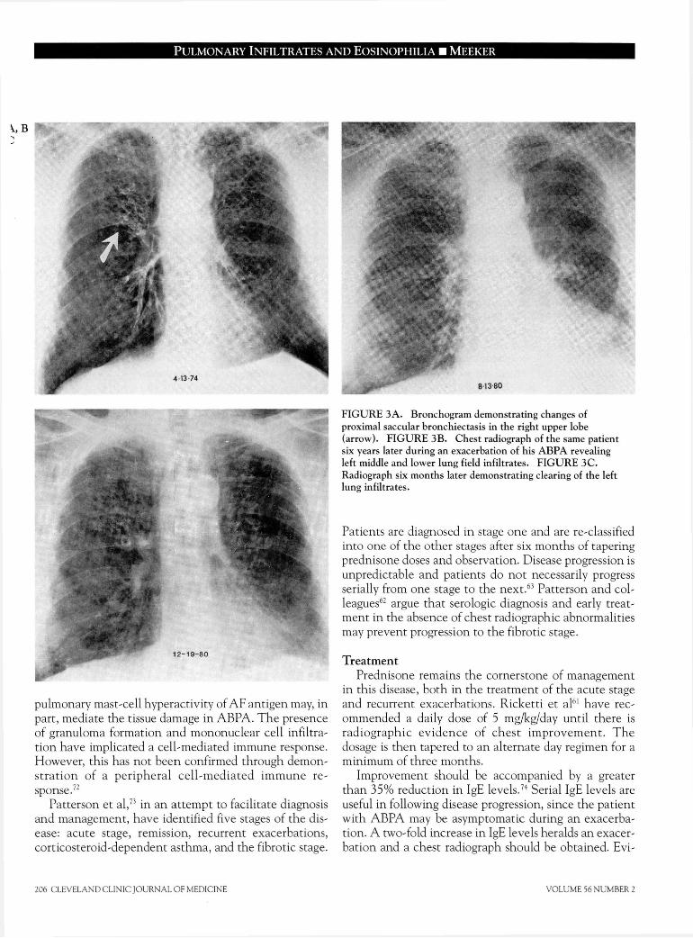
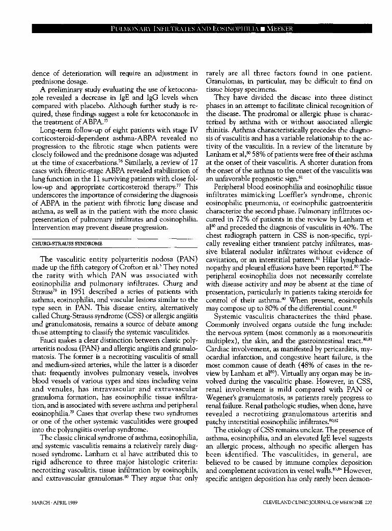
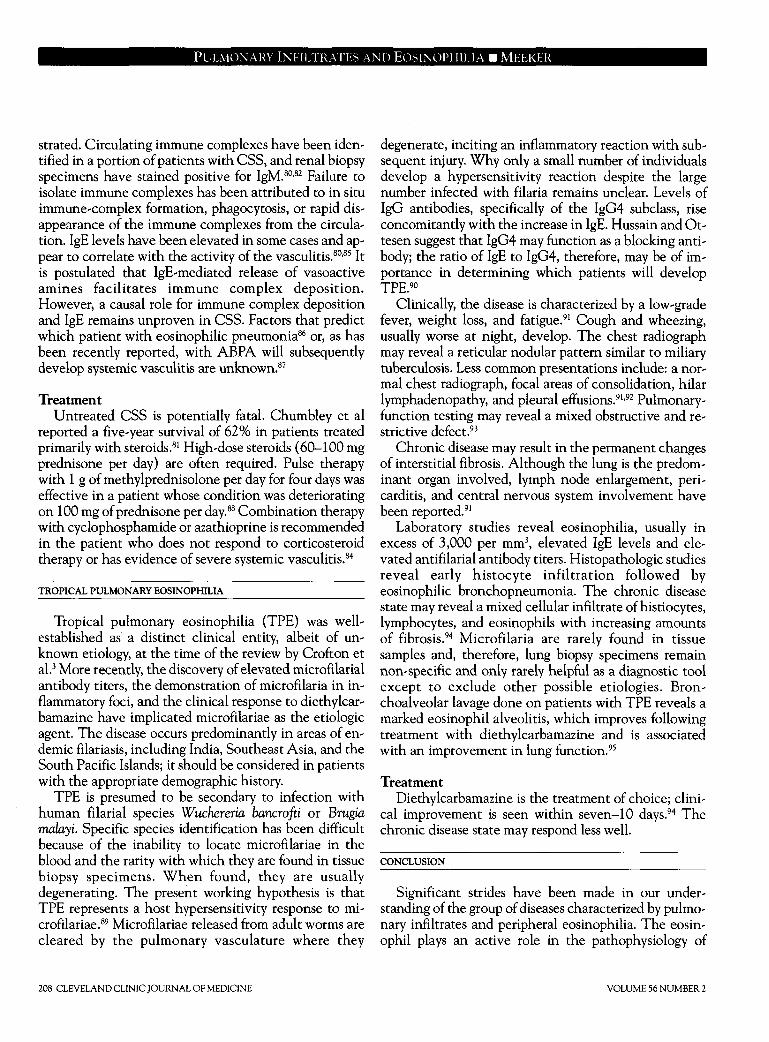
F I G U R E 3 A . Bronchogram demonstrating changes of
proximal saccular bronchiectasis in the right upper lobe
(arrow). F I G U R E 3B. Chest radiograph of the same patient
six years later during an exacerbation of his A B P A revealing
left middle and lower lung field infiltrates. F I G U R E 3C .
Radiograph six months later demonstrating clearing of the left
lung infiltrates.
Patients are diagnosed in stage one and are re-classified
into one of the other stages after six months of tapering
prednisone doses and observation. Disease progression is
unpredictable and patients do not necessarily progress
serially from one stage to the next.63 Patterson and col-
leagues62 argue that serologic diagnosis and early treat-
ment in the absence of chest radiographic abnormalities
may prevent progression to the fibrotic stage.
Treatment
Prednisone remains the cornerstone of management
in this disease, both in the treatment of the acute stage
and recurrent exacerbations. Ricketti et al61 have rec-
ommended a daily dose of 5 mg/kg/day until there is
radiographic evidence of chest improvement. The
dosage is then tapered to an alternate day regimen for a
minimum of three months.
Improvement should be accompanied by a greater
than 35% reduction in IgE levels.74 Serial IgE levels are
useful in following disease progression, since the patient
with ABPA may be asymptomatic during an exacerba-
tion. A two-fold increase in IgE levels heralds an exacer-
bation and a chest radiograph should be obtained. Evi-
VOLUME 56 NUMBER 2
PULMONARY INFILTRATES AND EOSINOPHILIA • MELKER
dence of deterioration will require an adjustment in prednisone dosage.
A preliminary study evaluating the use of ketocona' zole revealed a decrease in IgE and IgG levels when compared with placebo. Although further study is re-quired, these findings suggest a role for ketoconazole in the treatment of ABPA.75
Long-term follow-up of eight patients with stage IV corticosteroid-dependent asthma-ABPA revealed no progression to the fibrotic stage when patients were closely followed and the prednisone dosage was adjusted at the time of exacerbations.76 Similarly, a review of 17 cases with fibrotic-stage ABPA revealed stabilization of lung function in the 11 surviving patients with close fol-low-up and appropriate corticosteroid therapy.77 This underscores the importance of considering the diagnosis of ABPA in the patient with fibrotic lung disease and asthma, as well as in the patient with the more classic presentation of pulmonary infiltrates and eosinophilia. Intervention may prevent disease progression.
CHURG-STRAUSS SYNDROME
The vasculitic entity polyarteritis nodosa (PAN) made up the fifth category of Crofton et al.3 They noted the rarity with which PAN was associated with eosinophilia and pulmonary infiltrates. Churg and Strauss78 in 1951 described a series of patients with asthma, eosinophilia, and vascular lesions similar to the type seen in PAN. This disease entity, alternatively called Churg-Strauss syndrome (CSS) or allergic angiitis and granulomatosis, remains a source of debate among those attempting to classify the systemic vasculitides.
Fauci makes a clear distinction between classic poly-arteritis nodosa (PAN) and allergic angiitis and granulo-matosis. The former is a necrotizing vasculitis of small and medium-sized arteries, while the latter is a disorder that: frequently involves pulmonary vessels, involves blood vessels of various types and sizes including veins and venules, has intravascular and extravascular granuloma formation, has eosinophilic tissue infiltra-tion, and is associated with severe asthma and peripheral eosinophilia.79 Cases that overlap these two syndromes or one of the other systemic vasculitides were grouped into the polyangiitis overlap syndrome.
The classic clinical syndrome of asthma, eosinophilia, and systemic vasculitis remains a relatively rarely diag-nosed syndrome. Lanham et al have attributed this to rigid adherence to three major histologic criteria: necrotizing vasculitis, tissue infiltration by eosinophils, and extravascular granulomas.80 They argue that only
MARCH • APRIL 1989
rarely are all three factors found in one patient. Granulomas, in particular, may be difficult to find on tissue biopsy specimens.
They have divided the disease into three distinct phases in an attempt to facilitate clinical recognition of the disease. The prodromal or allergic phase is charac-terized by asthma with or without associated allergic rhinitis. Asthma characteristically precedes the diagno-sis of vasculitis and has a variable relationship to the ac-tivity of the vasculitis. In a review of the literature by Lanham et al,80 58% of patients were free of their asthma at the onset of their vasculitis. A shorter duration from the onset of the asthma to the onset of the vasculitis was an unfavorable prognostic sign.81
Peripheral blood eosinophilia and eosinophilic tissue infiltrates mimicking Loeffler's syndrome, chronic eosinophilic pneumonia, or eosinophilic gastroenteritis characterize the second phase. Pulmonary infiltrates oc-curred in 72% of patients in the review by Lanham et al80 and preceded the diagnosis of vasculitis in 40%. The chest radiograph pattern in CSS is non-specific, typi-cally revealing either transient patchy infiltrates, mas-sive bilateral nodular infiltrates without evidence of cavitation, or an interstitial pattern.81 Hilar lymphade-nopathy and pleural effusions have been reported.80 The peripheral eosinophilia does not necessarily correlate with disease activity and may be absent at the time of presentation, particularly in patients taking steroids for control of their asthma.80 When present, eosinophils may compose up to 80% of the differential count.81
Systemic vasculitis characterizes the third phase. Commonly involved organs outside the lung include: the nervous system (most commonly as a mononeuritis multiplex), the skin, and the gastrointestinal tract.80,81
Cardiac involvement, as manifested by pericarditis, my-ocardial infarction, and congestive heart failure, is the most common cause of death (48% of cases in the re-view by Lanham et al80). Virtually any organ may be in-volved during the vasculitic phase. However, in CSS, renal involvement is mild compared with PAN or Wegener's granulomatosis, as patients rarely progress to renal failure. Renal pathologic studies, when done, have revealed a necrotizing granulomatous arteritis and patchy interstitial eosinophilic infiltrates.80,82
The etiology of CSS remains unclear. The presence of asthma, eosinophilia, and an elevated IgE level suggests an allergic process, although no specific allergen has been identified. The vasculitides, in general, are believed to be caused by immune complex deposition and complement activation in vessel walls.83,84 However, specific antigen deposition has only rarely been demon-
CLEVELAND CLINIC JOURNAL OF MEDICINE 207
PULMONARY INFILTRATES AND EOSINOPHILIA • MEEKER
strated. Circulating immune complexes have been iden-
tified in a portion of patients with CSS, and renal biopsy
specimens have stained positive for IgM.80,82 Failure to
isolate immune complexes has been attributed to in situ
immune-complex formation, phagocytosis, or rapid dis-
appearance of the immune complexes from the circula-
tion. IgE levels have been elevated in some cases and ap-
pear to correlate with the activity of the vasculitis.80,85 It
is postulated that IgE-mediated release of vasoactive
amines facilitates immune complex deposition.
However, a causal role for immune complex deposition
and IgE remains unproven in CSS. Factors that predict
which patient with eosinophilic pneumonia86 or, as has
been recently reported, with ABPA will subsequently
develop systemic vasculitis are unknown.87
Treatment
Untreated CSS is potentially fatal. Chumbley et al
reported a five-year survival of 62% in patients treated
primarily with steroids.81 High-dose steroids (60-100 mg
prednisone per day) are often required. Pulse therapy
with 1 g of methylprednisolone per day for four days was
effective in a patient whose condition was deteriorating
on 100 mg of prednisone per day.88 Combination therapy
with cyclophosphamide or azathioprine is recommended
in the patient who does not respond to corticosteroid
therapy or has evidence of severe systemic vasculitis.84
TROPICAL PULMONARY EOSINOPHILIA
Tropical pulmonary eosinophilia (TPE) was well-
established as a distinct clinical entity, albeit of un-
known etiology, at the time of the review by Crofton et
al.3 More recently, the discovery of elevated microfilarial
antibody titers, the demonstration of microfilaria in in-
flammatory foci, and the clinical response to diethylcar-
bamazine have implicated microfilariae as the etiologic
agent. The disease occurs predominantly in areas of en-
demic filariasis, including India, Southeast Asia, and the
South Pacific Islands; it should be considered in patients
with the appropriate demographic history.
TPE is presumed to be secondary to infection with
human filarial species Wuchereria bancrofti or Brugia malayi. Specific species identification has been difficult
because of the inability to locate microfilariae in the
blood and the rarity with which they are found in tissue
biopsy specimens. When found, they are usually
degenerating. The present working hypothesis is that
TPE represents a host hypersensitivity response to mi-
crofilariae.89 Microfilariae released from adult worms are
cleared by the pulmonary vasculature where they
degenerate, inciting an inflammatory reaction with sub-
sequent injury. Why only a small number of individuals
develop a hypersensitivity reaction despite the large
number infected with filaria remains unclear. Levels of
IgG antibodies, specifically of the IgG4 subclass, rise
concomitantly with the increase in IgE. Hussain and Ot-
tesen suggest that IgG4 may function as a blocking anti-
body; the ratio of IgE to IgG4, therefore, may be of im-
portance in determining which patients will develop
TPE.90
Clinically, the disease is characterized by a low-grade
fever, weight loss, and fatigue.91 Cough and wheezing,
usually worse at night, develop. The chest radiograph
may reveal a reticular nodular pattern similar to miliary
tuberculosis. Less common presentations include: a nor-
mal chest radiograph, focal areas of consolidation, hilar
lymphadenopathy, and pleural effusions.91,92 Pulmonary-
function testing may reveal a mixed obstructive and re-
strictive defect.93
Chronic disease may result in the permanent changes
of interstitial fibrosis. Although the lung is the predom-
inant organ involved, lymph node enlargement, peri-
carditis, and central nervous system involvement have
been reported.91
Laboratory studies reveal eosinophilia, usually in
excess of 3,000 per mm3, elevated IgE levels and ele-
vated antifilarial antibody titers. Histopathologic studies
reveal early histocyte infiltration followed by
eosinophilic bronchopneumonia. The chronic disease
state may reveal a mixed cellular infiltrate of histiocytes,
lymphocytes, and eosinophils with increasing amounts
of fibrosis.94 Microfilaria are rarely found in tissue
samples and, therefore, lung biopsy specimens remain
non-specific and only rarely helpful as a diagnostic tool
except to exclude other possible etiologies. Bron-
choalveolar lavage done on patients with TPE reveals a
marked eosinophil alveolitis, which improves following
treatment with diethylcarbamazine and is associated
with an improvement in lung function.95
Treatment
Diethylcarbamazine is the treatment of choice; clini-
cal improvement is seen within seven-10 days.94 The
chronic disease state may respond less well.
CONCLUSION
Significant strides have been made in our under-
standing of the group of diseases characterized by pulmo-
nary infiltrates and peripheral eosinophilia. The eosin-
ophil plays an active role in the pathophysiology of
208 CLEVELAND CLINIC JOURNAL OF MEDICINE VOLUME 56 NUMBER 2
PULMONARY INFILTRATES AND EOSINOPHILIA • MELKER
these diseases. Specific causes for tropical pulmonary eosinophilia and many causes of Loeffler's syndrome have been determined. Marked progress has been made in our understanding of allergic bronchopulmonary aspergillosis.
Despite these advances, several syndromes can only be defined clinically, and significant overlap persists be-tween syndromes such as chronic eosinophilic pneu-monia, the hypereosinophilic syndrome, and the Churg-Strauss syndrome. The original classification by Crofton et al3 remains useful, if only to remind one of the overlap between the syndromes.
At present it is impossible to predict which patients may progress from a more benign disorder of isolated pulmonary eosinophilia to the multi-organ system in-
REFERENCES
1. Hirsch JG, Hirsch BI. Paul Ehrlich and the discovery of the eosinophil.
[In] Mahmoud AAF, Austen KF, eds. The Eosinophil in Health and
Disease. New York, Grune and Stratton, 1980, pp 3-23.
2. Reeder WH, Goodrich BE. Pulmonary infiltration with eosinophilia
(PIE syndrome). Ann Intern Med 1952; 36:1217-1240.
3. Crofton JW, Livingstone JL, Oswald NC, Roberts ATM. Pulmonary
eosinophilia. Thorax 1952; 7:1-35.
4- Liebow AA, Carrington CB. The eosinophilic pneumonias. Medicine
1969;48:251-285.
5. Schatz M, Wasserman S, Patterson R. Eosinophils and immunologic
lung disease. Med Clin N Am 1981;65:1055-1071.
6. Schatz M, Wasserman S, Patterson R, The eosinophil and the lung.
Arch Intern Med 1982; 142:1515-1519.
7. Wykoff RF. Eosinophilia. South Med J 1986; 79:608-612.
8. Slungaard A, Ascensao ], Zanjani E, Jacob HS. Pulmonary carcinoma
with eosinophilia: demonstration of a tumor-derived eosinophilopoietic
fector. N Engl J Med 1983; 309:778-781.
9. Dawes PT, Smith DH, Scott DL. Massive eosinophilia in rheumatoid
arthritis: report of four cases. Clin Rheumatol 1986: 5:62-65.
10. Lombard CM, Tazelaar HD, Krasne DL. Pulmonary eosinophilia in
coccidioidal infections. Chest 1987; 91:734-736.
11. Peterson MW, Monick M, Hunninghake GW. Prognostic role of
eosinophils in pulmonary fibrosis. Chest 1987; 92:51-56.
12. Davis BW, Fells GA, Sun X-H, Gadek JE, Venet A, Crystal RG.
Eosinophil-mediated injury to lung parenchymal cells and interstitial
matrix: a possible role for eosinophils in chronic inflammatory disorders
of the lower respiratory tract. J Clin Invest 1984; 74:269-278.
13. HällgrenR, SamuelssonT, Venge P, Modig J. Eosinophil activation in
the lung is related to lung damage in adult respiratory distress syndrome.
Am Rev Respir Dis 1987; 135:639-642.
14. Gleich GJ. Current understanding of eosinophil function. Hosp Prac
1988; 23:137-160.
15. Weller PF. Eosinophilia. J Allergy Clin Immunol 1984; 73:1-10.
16. Gleich GJ, Loegering DA. Immunobiology of eosinophils. Ann Rev
Immunol 1984; 2:429-459.
17. Venge P, Häkansson L, Peterson CGB. Eosinophil activation in aller-
gic disease. Int Arch Allergy Appl Immunol 1987; 82:333-337.
18. Hastie AT, Loegering DA, Gleich GJ, Kueppers F. The effect of
purified human eosinophil major basic protein on mammalian ciliary
activity. Am Rev Respir Dis 1987; 135:848-853.
19. Frigas E, Loegering DA, Gleich GJ. Cytoxic effects of the guinea pig
eosinophil major basic protein on tracheal epithelium. Lab Invest 1980;
42:35-43.
20. Frigas E, Loegering DA, Solly GO, Farrow GM, Gleich GJ. Elevated
volvement found in CSS or HES. Early diagnosis and
close follow-up may prevent permanent organ system
dysfunction.
Corticosteroids remain the primary treatment modal-
ity for most of these diseases. Cytotoxic agents have a
role in treating the patient with multi-organ system in-
volvement. Diagnosis, management, and classification
of the PIE syndromes continues to be hampered by our
incomplete understanding of these syndromes.
ACKNOWLEDGMENT
I would like to thank Muzaffar Ahmad, MD and Herbert Wiedemann,
MD for their review of the manuscript, and Patricia Senor for her technical
assistance.
levels of the eosinophil granule major basic protein in the sputum of
patients with bronchial asthma. Mayo Clin Proc 1981; 56:345-353.
21. Enokihara H, Hamaguchi H, Sakamaki H, Hazama S, Saito K, Furusawa
S, Shishido H. Specific production of eosinophil colony stimulating
factor from sensitized T cells from a patient with allergic eosinophilia.
Br J Haematol 1985; 59:85-91.
22. Prin L, Capron M, Gösset P, et al. Eosinophilic lung disease: im-
munological studies of blood and alveolar eosinophils. Clin Exp Im-
munol 1986; 63:249-257.
23. Fukuda T, Dunnette SL, Reed CE, Ackerman SJ, Peters MS, Gleich GJ.
Increased numbers of hypodense eosinophils in the blood of patients
with bronchial asthma. Am Rev Respir Dis 1985; 132:981-985.
24- Horn BR, Robin ED, Theodore J, Van Kessel A. Total eosinophil
counts in the management of bronchial asthma. N Engl J Med 1975;
292:1152-1155.
25. Altman LC, Hill JS, Hairfield WM, Mullarkey MF. Effects of cor-
ticosteroids on eosinophil Chemotaxis and adherence. J Clin Invest
1981; 67:28-36.
26. Koch-Weser J. Beta adrenergic blockade and circulating eosinophils.
Arch Intern Med 1968; 121:255-258.
27. Weller PF. Parasitic pneumonias. [In] Pennington JE. Respiratory In-
fections: Diagnosis and Management. New York, Raven Press, 1983, pp
439-453.
28. Cooper JAD, White DA, Matthay RA. Drug-induced pulmonary dis-
ease. Am Rev Respir Dis 1986; 133:488-505.
29. Cooper JAD, White DA, Matthay RA. Drug-induced pulmonary dis-
ease. Am Rev Respir Dis 1986; 133:321-340.
30. Nader DA, Schillaci RF. Pulmonary infiltrates with eosinophilia due
to naproxen. Chest 1983; 83:280-282.
31. Yousem SA, Lifson JD, Colby TV. Chemotherapy-induced
eosinophilic pneumonia. Chest 1985; 88:103-106.
32. Wang KK, Bowyer BA, Fleming CR, Schroeder KW. Pulmonary in-
filtrates and eosinophilia associated with sulfasalazine. Mayo Clin Proc
1984; 59:343-346.
33. Carrington CB, Addington WW, Goff AM , et al. Chronic
eosinophilic pneumonia. N Engl J Med 1969; 280:787-798.
34- Pearson DJ, Rosenow EC III. Chronic eosinophilic pneumonia
(Carrington's): a followup study. Mayo Clin Proc 1978; 53:73-78.
35. Fox B, Seed WA. Chronic eosinophilic pneumonia. Thorax 1980;
35:570-580.
36. Ivanick MJ, Donohue JF. Chronic eosinophilic pneumonia: a cause of
adult respiratory distress syndrome. South Med J 1986; 79:686-690.
37. Grantham JG, Meadows JA, Gleich GJ. Chronic eosinophilic
pneumonia, evidence for eosinophil degranulation and release of major
basic protein. Am J Med 1986; 80:89-94.
38. Gonzalez EB, Swedo JL, Rajaraman S, Daniels JC, Grant JA.
MARCH • APRIL 1989 CLEVELAND CLINIC JOURNAL OF MEDICINE 209
PULMONARY INFILTRATES AND EOSINOPHILIA • MEEKER
Ultrastructural and immunohistochemical evidence for release of
eosinophilic granules in vivo: cytotoxic potential in chronic
eosinophilic pneumonia. J Allergy Clin Immunol 1986; 79:755-762.
39. Gaensler EA, Carrington CB. Peripheral opacities in chronic
eosinophilic pneumonia: the photographic negative of pulmonary
edema. AJR 1977; 128:1-13.
40. Onitsuka H, Onitsuka S-i, Yokomizo Y, Matsuura K. Computed
tomography of chronic eosinophilic pneumonia. J Comput Assist
Tomogr 1983; 7:1092-1094.
41. Epler GR, McLoud TC, Gaensler EA, Mikus JP, Carrington CB: Nor-
mal chest roentgenograms in chronic diffuse infiltrative lung disease. N
Engl J Med 298:934,1978.
42. Dejaegher P, Derveaux L, Dubois P, Demedts M. Eosinophilic
pneumonia without radiographic pulmonary infiltrates. Chest 1983;
84:637-638.
43. Quinonez GE, Simon GT, Kay JM. Electron microscopy of chronic
eosinophilic pneumonia, Clin Invest Med 1986; 9:238-243.
44. Reynolds HY. Bronchoalveolar lavage. [In] Murray JF, Nadel JA, eds.
Textbook of Respiratory Medicine. Philadelphia, W.B. Saunders, 1988,
pp 597-610.
45. Dejaegher P, Demedts M. Bronchoalveolar lavage in eosinophilic
pneumonia before and during corticosteroid therapy. Am Rev Respir
Dis 1984; 129:631-632.
46. Ogushi F, Ozaki T, Kawano T, Yasuoka S. PGE2 and PGE2a content in
bronchoalveolar lavage fluid obtained from patients with eosinophilic
pneumonia. Chest 1987; 91:204-206.
47. Dines DE. Chronic eosinophilic pneumonia: a roentgenographic diag-
nosis (editorial). Mayo Clin Proc 1978; 53:129-130.
48. Marnocha KE, Maglinte DDT, Kelvin FM, McCune M, Weiser DC,
Strate R. Eosinophilic enteritis associated with chronic eosinophilic
pneumonia. Am J Gastroenterol 1986; 81:1205-1208.
49. Fauci AS, Harley JB, Roberts WC, Ferrans VJ, Gralnick HR, Bjornson
BH The idiopathic hypereosiniophilic syndrome. Ann Intern Med
1982; 97:78-92.
50. Wynn SR, Sachs MI, Keating MU, et al. Idiopathic hypereosinophilic
syndrome in a 5 1/2-month-old infant. J Pediatr 1987; 111 :94-97.
51. Alfaham MA, Ferguson SD, Sihra B, Davies J. The idiopathic hyper-
eosinophilic syndrome. Arch Dis Child 1987; 62:601-613.
52. Parrillo JE, Borer JS, Henry WL, Wolff SM, Fauci AS. The cardiovas-
cular manifestations of the hypereosinophilic syndrome. Am J Med
1979; 67:572-582.
53. Chusid MJ, Dale DC, West BC, Wolff SM. The hypereosinophilic
syndrome: analysis of fourteen cases with review of the literature.
Medicine 1975;54:1-27.
54. O'Shea JJ, Jafife ES, Lane HC, MacDermott RP, Fauci AS. Peripheral
T cell lymphoma presenting as hypereosinophilia with vasculitis. Am J
Med 1987; 82:539-545.
55. Parrillo JE, Fauci AS, Wolff SM. Therapy of the hypereosinophilic
syndrome. Ann Intern Med 1978; 89:167-172.
56. Szefler S], Brenner M, Jusko WJ, Spector SL, Flesher KA, Ellis EF.
Dose- and time-related effect of troleandomycin on methylprednisolone
elimination. Clin Pharmacol Ther 1982; 32:166-171.
57. Edwards D, Wald J A, Dobozin BS, Kirkpatrick CH. Troleandomycin
and methylprednisolone for treatment of the hypereosinophilic
syndrome (letter). N Engl J Med 1987; 317:573-574.
58. Hinson KFW, Moon AJ, Plummer MS. Bronchopulmonary aspergil-
losis. Thorax 1952; 7:317-333.
59. Greenberger PA, Patterson R. Diagnosis and management of allergic
bronchopulmonary aspergillosis. Ann Allergy 1986; 56:444-448.
60. Rosenberg M, Patterson R, Mintzer R, Cooper BJ, Roberts M, Harris
KE. Clinical and immunologic criteria for the diagnosis of allergic
bronchopulmonary aspergillosis. Ann Intern Med 1977; 86:405^114.
61. Ricketti AJ, Greenberger PA, Mintzer RA, Patterson R. Allergic
bronchopulmonary aspergillosis. Chest 1984; 86:773-778.
62. Patterson R, Greenberger PA, Halwig JM, Liotta JL, Roberts M. Aller-
gic bronchopulmonary aspergillosis. Arch Intern Med 1986; 146:916-
918.
63. Mendelson EB, Fisher MR, Mintzer RA, Halwig JM, Greenberger PA.
Roentgenographic and clinical staging of allergic bronchopulmonary
aspergillosis. Chest 1985; 87:334-339.
64. Slavin RG. Allergic bronchopulmonary aspergillosis. Clin Rev Aller-
gy 1985; 3:167-182.
65. Glancy JJ, Elder JL, McAleer R. Allergic bronchopulmonary fungal
disease without clinical asthma. Thorax 1981; 36:345-349.
66. Wang JLF, Patterson R, Rosenberg M, Roberts M, Cooper BJ. Serum
IgE and IgG antibody activity against Aspergillus fumigatus as a diagnos-
tic aid in allergic bronchopulmonary aspergillosis, Am Rev Resp Dis
1978; 117:917-927.
67. Patterson R, Greenberger PA, Ricketti AJ, Roberts M. A radioim-
munoassay index for allergic bronchopulmonary aspergillosis. Ann In-
tern Med 1983; 99:18-22.
68. Akiyama K, Takizawa H, Suzuki M, Miyachi S, Ichinohe M, Yanagihara
Y. Allergic bronchopulmonary aspergillosis due to Aspergillus oryzae. Chest 1987; 91:285-286.
69. Brummund W, Resnick A, FinkJN, Kurpup VP. Aspergillus fumigatus-
specific antibodies in allergic bronchopulmonary aspergillosis and
aspergilloma: evidence for a polyclonal antibody response. J Clin
Microbiol 1987; 25:5-9.
70. Gutt L, Greenberger PA, Liotta JL. Serum IgA antibodies to Aspergil-lus fumigatus in various stages of allergic bronchopulmonary aspergil-
losis. J Allergy Clin Immunol 1986; 78:98-101.
71. Ricketti AJ, Greenberger PA, Pruzansky JJ, Patterson R. Hyperreac-
tivity of mediator-releasing cells from patients with allergic bronchopul-
monary aspergillosis as evidenced by basophil histamine release. J Aller-
gy Clin Immunol 1983; 72:386-392.
72. Slavin RG, Hutcheson PS, Knutsen AP. Participation of cell-mediated
immunity in allergic bronchopulmonary aspergillosis. Int Arch Allergy
Appl Immunol 1987; 83:337-340.
73. Patterson R, Greenberger PA, Radin RC, Roberts M. Allergic
bronchopulmonary aspergillosis: staging as an aid to management. Ann
Intern Med 1982; 96:286-291.
74. Ricketti AJ, Greenberger PA, Patterson R. Serum IgE as an important
aid in management of allergic bronchopulmonary aspergillosis. J Aller-
gy Clin Immunol 1984; 74:68-71.
75. Shale DJ, Faux JA, Lane DJ. Trial of ketoconazole in non-invasive pul-
monary aspergillosis. Thorax 1987; 42:26-31.
76. Patterson R, Greenberger PA, Lee TM, et al. Prolonged evaluation of
patients with corticosteroid-dependent asthma stage of allergic
bronchopulmonary aspergillosis. J Allergy Clin Immunol 1987;
80:663-668.
77. Lee TM, Greenberger PA, Patterson R, Roberts M, Liotta JL. Stage V
(fibrotic) allergic bronchopulmonary aspergillosis. Arch Intern Med
1987; 147:319-323.
78. Churg J, Strauss L. Allergic granulomatosis, allergic angiitis, and
periarteritis nodosa. Am J Pathol 1951; 27:277-301.
79. Fauci AS. Vasculitis. J Allergy Clin Immunol 1983; 72:211-223.
80. Lanham JG, Elkon KB, Pusey CD, Hughes GR. Systemic vasculitis
with asthma and eosinophilia: a clinical approach to the Churg-Strauss
syndrome. Medicine 1984; 63:65-81.
81. Chumbley LC, Harrison EG Jr, DeRemee RA. Allergic granulomatosis
and angiitis (Churg-Strauss syndrome). Mayo Clin Proc 1977; 52:477-
484.
82. Koss MN, Antonovych T, Hochholzer L. Allergic granulomatosis
(Churg-Strauss syndrome). Am J Surg Pathol 1981; 5:21-28.
83. Lie JT. The classification of vasculitis and a reappraisal of allergic
granulomatosis and angiitis (Churg-Strauss syndrome). M Sinai J Med
(NY) 1986; 53:429-439.
84. Leavitt RY, Fauci AS. Pulmonary vasculitis. Am Rev Respir Dis 1986;
134:149-166.
85. Sale S, Patterson R. Recurrent Churg-Strauss vasculitis with exoph-
thalmos, hearing loss, nasal obstruction, amyloid deposits, hyperim-
munoglobulinemia E, and circulating immune complexes. Arch Intern
Med 1981; 141:1363-1365.
86. Cogen FC, Mayock RL, Zweiman B. Chronic eosinophilic pneumonia
followed by polyarteritis nodosa complicating the course of bronchial
asthma: report of a case. J Allergy Clin Immunol 1977; 60:377-382.
87. Stephens M, Reynolds S, Gibbs AR, Davies B. Allergic bronchopul-
monary aspergillosis progressing to allergic granulomatosis and angiitis
(Churg-Strauss syndrome). Am Rev Respir Dis 1988; 137:1226-1228.
88. MacFadyen R, Tron V, Keshmiri M, Road JD. Allergic angiitis of
210 CLEVELAND CLINIC JOURNAL OF MEDICINE VOLUME 56 NUMBER 2
PULMONARY INFILTRATES AND EOSINOPHILIA • MELKER
Churg and Strauss Syndrome: response to pulse methylprednisolone.
Chest 1987; 91:629-631.
89. Ottesen EA, Neva FA, Paranjape RS, Tripathy SP, Thiruvengadam KV,
Beaven MA. Specific allergic sensitisation to filarial antigens in tropi-
cal eosinophilia Syndrome. Lancet 1979;1:1158-1161.
90. Hussain R, Ottesen EA. IgE responses in human filariasis. IV. Parallel
antigen recognition by IgE and IgG4 subclass antibodies. J Immunol
1986; 135:1859-1863.
91. Spry CJF, Kumaraswami V. Tropical eosinophilic. Sem Hematol 1982;
19:107-115.
92. Jain VK, Beniwal OP. Unusual presentation of tropical pulmonary
eosinophilia. Thorax 1984; 39:634—635.
93. Chhabra SK, Gaur SN. Airway hyperreactivity in tropical pulmonary
eosinophilia. Chest 1988; 93:1105-1106.
94- Neva FA, Ottesen EA. Tropical (filarial) eosinophilia. N Engl J Med
1978; 298:1129-1131.
95. Pinkston P, Vijayan VK, Nutman TB, et al. Acute tropical pulmonary
eosinophilia: characterization of the lower respiratory tract inflamma-
tion and its response to therapy. J Clin Invest 1987; 80:216-225.
MARCH • APRIL 1989 CLEVELAND CLINIC JOURNAL OF MEDICINE 211