Department of Woman and Child Health, and Department of Pathology and Cytology, Karolinska Institute, Stockholm, Sweden
Pyelonephritis provokes growth retardation and apoptosis in infant ratrenal cortex. Childhood pyelonephritis is a common cause of renal corticalscarring and hypoplastic kidneys. To understand the mechanisms under-lying the cortical lesions, urinary tract infection was induced in three-week-old rats by an intravesical infusion of E. coli, type 06 K13 HI, a ratnephropathogenic strain. Four days after infection, histopathologicalexamination showed marked infiltration of leukocytes in the medullarytissue adjoining the calyces and pelvis. In the cortex, signs of inflammationwere found only in the cortical zone adjacent to the pelvis. No cellsindicative of inflammation were observed in other parts of the cortex.Immunohistochemistry for endogenous proliferating cell nuclear antigen(PCNA) demonstrated a marked decrease in immunoreactivity in proxi-mal tubular (PT) cells. The mitotic response of PT cells, assessed by3H-thymidine autoradiography, showed a highly significant decrease dur-ing the first four days after induction of the infection. Four days afterinfection, a transient increase in apoptotic cells was observed in corticalcells outside the inflammatory areas. No increase in apoptotic cells wasdetected in the cortex 10 days after infection. Only a few apoptotic cellswere detected in the control kidneys. In conclusion, the data indicate thatinhibition of cell proliferation and enhancement of apoptosis may con-tribute to the renal parenchymal loss after childhood pyelonephritis.
The natural history of pyelonephritis is age-dependent [1], andin infants this disease may, if untreated, lead to permanent loss ofrenal parenchyma and functional disturbances within a few days[2—4]. In adults, pyelonephritis is a much less fulminant disease.Follow-up studies have indicated that in children older than threeyears, pyelonephritis only rarely result in loss of renal parenchyma[5]. Vesicoureteral reflux predisposes to pyelonephritis and largerefluxes are common in infants [61, but retrospective studiesindicate that the age-dependent effects of pyelonephritis can onlypartially be attributed to the existence of vesicoureteral reflux [2,5, 71.
Pyelonephritic lesions appear radiologically as loss of renalcortical parenchyma or as generalized renal hypoplasia [8—10].Kidney growth is very rapid during infancy [11], and growth of theinfant rat kidney is accompanied by programmed cell death [12].Programmed cell death, which is extensive in the developingkidney, is believed to be necessary for the structural organizationof nephrons [13, 14]. Taken together, these clinical and experi-
Received for publication October 18, 1996and in revised form February 4, 1997Accepted for publication February 6, 1997
mental observations have prompted us to study the extent towhich induction of pyelonephritis in infant rats may influencegrowth and apoptosis. This study shows that the induction ofpyelonephritis in weaning rats causes both inhibition of cellproliferation and enhanced apoptosis. We observed these pro-cesses even in renal tissue that had no apparent signs of inflam-mation. It is suggested that these age-dependent phenomena maypartially explain why pyelonephritis in infancy is associated withsuch a serious natural history.
Methods
Experimental pyelonephritis
The studies were performed on 20- and 40-day-old femaleSprague-Dawley rats. In each group 3 to 10 rats were examined.The rats were fed laboratory standard rat chow (B & B, Sollen-tuna, Sweden) and received tap water ad libitum. Induction ofinfection was performed as previously described [15]. Duringether anesthesia, the bladder was catheterized with a double-lumen silver cannula that allowed continuous recordings of blad-der pressure during infusion. A saline solution containing Esche-richia coli type 06 K13 Hi (I0 bacteria/ml) was infused into thebladder at a constant infusion rate (0.6 mi/mm). In control ratssaline was injected into the bladder at the same infusion rate.
To verify that the bacterial solution reached and infected thekidneys, a separate group of rats was sacrificed 1, 4 and 10 daysafter the intravesical infection. The kidneys were removed understerile conditions, minced and cultured with a standard microbi-ological technique. Cultures from both kidneys were performed 1,4 and 10 days after infection. Cultures were positive for E. coli inall the kidneys from infected rats.
Fixation and tissue processing for histopathology and
immunohistochemisty
The aorta was cannulated via a midline incision and bothkidneys were perfused, first with Ringer's solution and then with4% formalin. The formalin-fixed kidneys were dehydrated inethanol and xylene in a vacuum infiltration processor (Histolab,Goteborg, Sweden) and embedded in paraffin. For light micros-copy, 2 m longitudinal-coronal sections were cut. For his-topathological examination, the sections were stained with hema-toxylin-eosin and Ladewig's trichrome.
1855
1856 Serlachius et al: Pyelonephritis provokes growth retardation
Immunohistochemistiy for ED-i and TGF-/31
The formalin-fixed, paraffin-embedded sections were deparaf-finized, stripped of proteins by incubation with 0.075% protease-(type XIV, protease E; Sigma, Stockholm, Sweden). Endogenousperoxidase was inactivated by covering the sections with 0.75%H202 for 30 minutes. The slides were covered with 5% BSA for 15minutes and incubated at RT with one of the following antibodies:ED-i (1:100; Serotec, Oxford, UK) for 60 minutes, which is amouse monoclonal IgG antibody that recognizes cytoplasmicantigens in macrophages, or a polyclonal antibody againstTGF-/31 (1:500; Santa Cruz Biotechnology, CA, USA) overnight.Next, either a biotinylated rabbit anti-mouse antibody (1:200,Dakopatts AB, Alvsjö, Sweden) for ED-i or a swine-anti-rabbitantibody (1:300, Dakopatts AB) for TGF-j31 was used as second-ary antibody for 30 minutes at RT. The slides were rinsed andcovered with avidin-biotin complex (Strept ABComplex; Dako-patts AB) for 30 minutes at RT. The sites of peroxidase activitywere visualized by incubation in DAB for three minutes at RT.The sections were counterstained with Gills hematoxylin.
In situ detection of apoptosis
Apoptosis can be identified in histological sections by highlycharacteristic morphological changes in cell structure and byend-labeling of endonuclease cleaved DNA [161. The DNAfragmentation associated with apoptosis was detected in situ bythe use of a commercial kit (ApopTag; Oncor, Gaithersburg, MD,USA). With this method, residues of digoxigenin-nucleotide arefirst catalytically added to the DNA ends by terminal deoxyribo-nucleotidyl transferase (TdT). A peroxidase-linked anti-digoxige-nm antibody is then added, and the localized peroxidase generatesa color signal in apoptotic cells from chromogenic substrate(diaminobenzidine, DAB; bioGenex, San Ramon, CA, USA).
Briefly, the formalin-fixed, paraffin-embedded sections weredeparaffinized and stripped of proteins by incubation with 0.075%protease (type XIV, pronase E; Sigma) at room temperature (RT)for five minutes and then washed in distilled water (DW).According to manufacturer's recommendations, endogenous per-oxidase was inactivated by covering the sections with 2% H202 forfive minutes. After preincubation with and equilibration buffer,the samples were incubated with TdT in reaction buffer (contain-ing digoxigenin-nucleotides) at 37°C for 60 minutes. The reactionwas terminated by using stop buffer. The tissue sections wererinsed with PBS, covered with anti-digoxigenin-peroxidase for 30minutes at RT, washed in PBS and stained with DAB for sixminutes at RT. They were counterstained with hematoxylin andeosin. Positive controls obtained from newborn rat kidneys wererun in parallel. Slides were examined by light microscopy forperoxidase staining.
In order to quantify the amount of apoptotic proximal tubulecells, the numbers of in situ-labeled apoptotic cells in proximaltubules were counted in a double-blind fashion. In each kidney 10fields in area IV (Fig. 1A) were examined at a magnification ofx200 with an Olympus PH-2 microscope. The apoptotic index wasderived by taking the mean number of positive-stained nuclei permicroscopic field. In each group, 6 to 9 rats were studied and bothkidneys were evaluated in each rat.
Propidium iodide labeling
Propidium iodide labeling was performed according to Coles,Burne and Raff with a few modifications [12]. The paraffin-
embedded sections were deparaffinized and incubated with 4g/ml propidium iodide (Sigma) and 100 ig/ml RNase (Boe-hringer-Mannheim, Bromma, Sweden) in PBS for 30 minutes at37°C. The slides were washed in PBS, mounted in Immu-mount(Shandon, Pittsburgh, PA, USA) and examined in a NikonMicrophot-FXA fluorescence microscope. Apoptotic nuclei inpropidium-iodide-labeled sections were smaller and more brightlystained than normal nuclei. In each kidney section 10 microscopicfields in area IV (Fig. 1A) were examined at magnification X 200.The apoptotic index was derived by taking the mean number ofpositive-stained nuclei per microscopic field. At least 5 rats wereexamined in each group.
PCNA in situ detection of proliferating cells
The formalin-fixed, paraffin-embedded sections were deparaf-finized in xylene, dehydrated in ethanol and washed in DW. Thesections were covered with 1% Nonidet P40 (Kebo, Spânga,Sweden), rinsed in DW and incubated in acetone for five minutes.Endogenous peroxidase was inactivated by covering the sectionswith 0.75% H202 for 30 minutes. The slides were rinsed with PBSand incubated in PBS with 1% BSA for 45 minutes. The sectionswere incubated at RT overnight with a monoclonal antibodyagainst PCNA (Dakopatts AB), diluted 1:500 in PBS with 1%BSA. After a PBS wash, the slides were incubated at RT for 30minutes with a biotinylated-rabbit-anti-mouse antibody (Dako-patts AB) diluted 1:200 in PBS with 1% BSA. The slides wererinsed and covered with horseradish peroxidase (Strept ABcomplex; Dakopatts AB) for 30 minutes at RT. The sites ofperoxidase activity were visualized by incubation in DAB (Dako-patts AB) for three to six minutes at RT. The sections werecounterstained with hematoxylin and eosin. Slides were examinedby light microscopy for peroxidase staining.
PCNA, a cofactor for DNA-polymerase a-, plays an importantrole in DNA replication during cellular proliferation [17]. NuclearPCNA expression is highest during the transition from the Gi toS-phase of the cell cycle [18]. The nuclear immunofluorescencewith anti-PCNA antibodies will therefore display a wide range ofintensities. As in previous studies, only nuclei in S-phase (intenseperoxidase staining throughout the nucleoplasm) were counted inthe quantitative evaluation [17, 19].
Determination of cell proliferation with 3H-thymidineincorporation
The rats were injected with 3H-thymidine (Amersham, UK) 1Ci/g body wt i.p. and one hour later were anesthetized withmactin 80 mg/kg i.p. (Byk Gulden, Constanz, Germany). Thekidneys were rapidly removed, fixed in 4% paraformaldehyde fornot less than 24 hours and then placed in 10% sucrose phospha-tase buffer overnight at 4°C. They were sectioned (5 m thick) ina cryostat and thawed onto glass slides pretreated with chroma-lun-gelatin. The sections were dipped in photographic emulsion(Kodak NTB2) and exposed for three to four weeks at 4°C. Afterexposure, the preparations were developed in Kodak D 19, fixedin sodium thiosulphate, stained with hematoxylin and eosin andthen mounted.
Determination of the labeling index of cell nuclei
The labeling index for PCNA and 3H-thymidine (LI % =labeled nuclei per 100 proximal tubule cells) was determined bycounting 2000 to 3000 proximal tubule cells in cortical area IV
Serlachius et al: Pyelonephritis provokes growth retardation 1857
(Fig. 1A) on each kidney section. In each group, 3 to 8 rats werestudied and both kidneys were evaluated in each rat.
Statistical analysis
Values are expressed as mean SEM. Statistical comparisonsbetween two groups were performed with the Student's t-test forunpaired data. Comparisons between several groups were done byanalysis of variance. A value of P < 0.05 was considered signifi-cant.
ResultsThe effects of pyelonephritis on histopathology, apoptosis and
cell proliferation was evaluated in weaning, 20-day-old rats. Todescribe the extent of the inflammatory process, the kidney hasbeen divided into five regions: (I) inner medullary area adjoiningthe tip of the calyx; (II) outer medullary area adjacent to thepelvis; (III) cortical area adjacent to the pelvis; (IV) cortical areaadjoining area III; (V) remaining parts of the cortex (Fig 1A).One day after infection, polymorphonuclear neutrophils (PMNL)were present in the renal pelvis and in the epithelium lining thepelvis and along the papilla. Slight infiltration by polymorphonu-clear neutrophils could also be seen in the interstitial tissue justbeneath the epithelium lining the sides of the papilla, but noinflammatory cells in the collecting ducts.
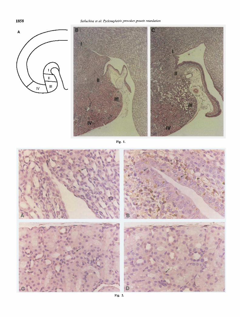
Four days after infection, the infiltration by neutrophils hadincreased in the epithelium and subepithelial tissue along thepapilla, in the inner medulla adjoining the calyces (I) and in theouter medulla adjacent to the pelvis (II). In these areas, tubularatrophy and slight fibrosis had developed beneath the epithelium.In some of the kidneys the inflammation had spread to the cortexadjacent to the pelvis (III; Fig. 1C). No increase in inflammatorycells were detected in the other parts of the cortex (IV, V). Theouter medulla and central parts of the papilla were also devoid ofinflammatory cells. To further investigate the infiltration of in-flammatory cells, the sections were immunostained to ED-i, anantibody which recognizes macrophages. Four days after induc-tion of infection immunolabeling to ED-i was increased in area I,II and III (Fig. 2 A, B). No increase in ED-i positive cells wasseen in area IV (Fig. 1 C, D).
Ten days after infection more monocytic cells than PMNL wereobserved in localized areas of inflammation and the most markedsigns of inflammation were visible in the areas adjacent to thecalyces (I) and the pelvis (II, III). In these areas, fibrosis andtubular atrophy had increased. Casts of polymorphonuclear neu-trophils were observed in the lumen of some tubules and collect-ing ducts in the papilla and medulla. Scattered areas of inflam-mation and fibrosis were observed in other parts of the cortex (IV,V).
Apoptotic nuclei were located by in situ end-labeling of cleavedDNA in the infected kidneys 1, 4 and 10 days after induction ofthe infection and in the age-matched control kidneys. The inci-dence of apoptotic cells was low in the control kidneys. One dayafter infection, in situ labeled cells was detected in the inflamma-tory areas in the epithelium lining the pelvis and along the papilla.No significant increase in apoptotic cells was observed in thenon-inflamed area IV (control 0.075 0.048 vs. infected 0.20.00, N = 3 to 4, NS).
Four days after induction of infection an increase in in situlabeled cells was observed both in areas with and without inflam-mation. In the inflammatory areas, an increase in apoptotic cellswas detected in the medullary (I, II) and cortical tissues (III)
adjacent to the calyces and pelvis. Although renal cells andinflammatory cells could both contribute to the apoptotic cellpopulation in the areas with inflammation, it was not possible toquantify them separately because apoptosis, regardless of cellorigin, is morphologically similar [20].
Interestingly, a marked increase in apoptotic cells was also seenin the cortical parenchyma outside the inflammatory areas, pre-dominantly in area IV. Figure 3 shows the distribution of insitu-labeled apoptotic cells in sections from control (Fig. 3A) andinfected (Fig. 3 B, C) kidneys in area IV, four days after infection.Note that this area is devoid of inflammatory cells. Most of theapoptotic cells were detected individually in tubular epithelialcells, but apoptotic nuclei were also found in the interstitial area.The apoptotic cells exhibited classical morphological features ofapoptosis, with cell shrinkage and the appearance of apoptoticbodies (Fig. 3C). In area IV, the average number of apoptoticproximal tubule cells per microscopic view was increased in theinfected kidneys (control 0.11 0.04 vs. infected 1.21 0.18,Nii to 13, P < 0.001; Fig. 3D). To confirm the effect of pyelone-phritis on apoptosis in area IV, the kidney sections were labeledwith propidium iodide and examined in a fluorescence micro-scope. The apoptotic cells were smaller, more brightly stainedthan normal nuclei and were often fragmented 12. The apoptoticindex was significantly increased in the infected kidneys (control0.18 0.04 vs. infected 1.66 0.24, N = 6 to 7, P < 0.001). Tendays after infection, no increase in apoptotic cells was detected incortical area IV (control 0.06 0.022 vs. infected 0.05 0.022,N = 10, NS).
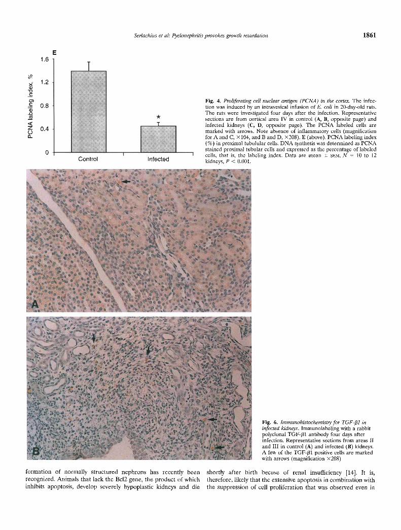
To determine whether pyelonephritis had any localized effecton cell proliferation, immunohistochemical detection of prolifer-ating cell nuclear antigen was performed four days after theinfection. Figure 4 shows cortical PCNA staining in representativekidney sections from the control (Fig. 4 A, B) and infected (Fig.4 C, D) rats. In the infected kidneys, a marked decrease in PCNAstaining was observed in cortical area IV. No increase in inflam-matory cells were present in this area. In infected rats, the PCNAlabeling index of proximal tubule cells was significantly lower thanin control rats (control 1.39 0.15 vs. infected 0.45 0.06, N =10 to 12, P < 0.001; Fig. 4E).
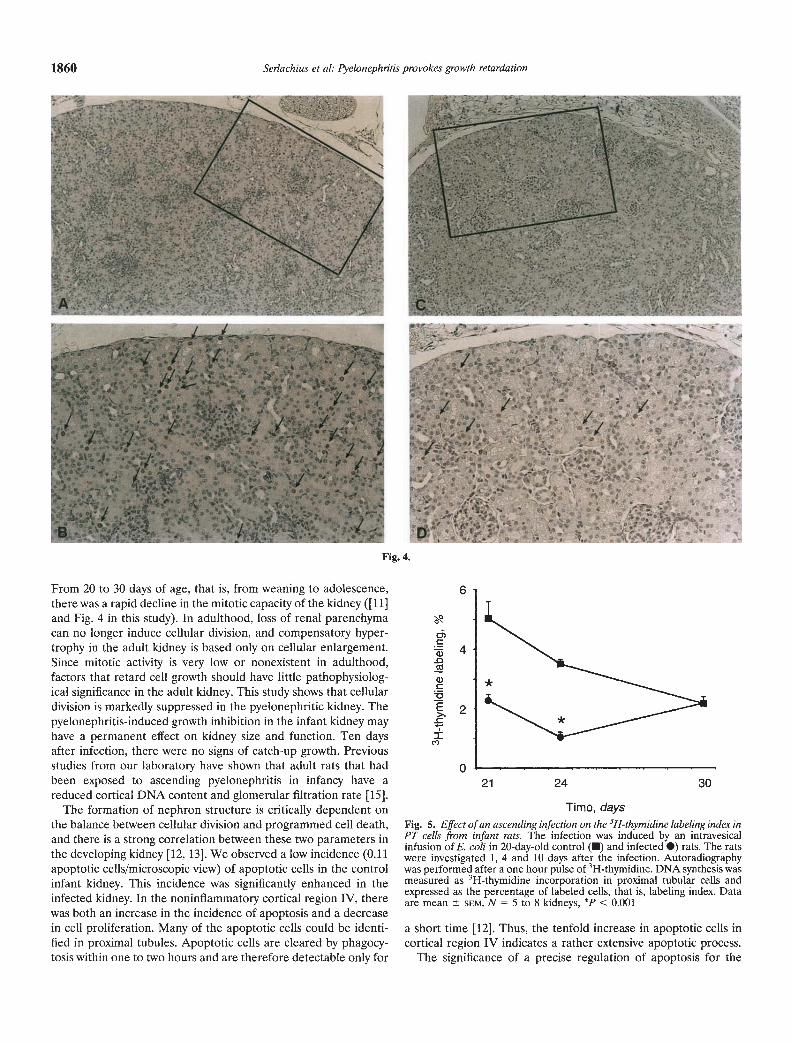
To further evaluate the effect of pyelonephritis on cell prolif-eration in the cortex, DNA synthesis was measured as 3H-thymidine incorporation in proximal tubule cells and expressed asthe percentage of labeled cells, LI. In this protocol we counted thesuperficial cortex at the two poles of the kidney, that is, an areathat roughly corresponding to area IV. Area V was not included.The rats were studied 1, 4 and 10 days after infection, that is, whenthey were 21-, 24- and 30-days-old. In control rats between 21 and30 days of age there was a rapid decline in the rate of cellproliferation. Therefore, kidneys from age-matched control ratswere always used for comparisons. One day after the infection wasinduced, LI was significantly decreased in infected compared tocontrol rats (control 5.0 0.56% vs. infected 2.3 0.31%, N = 5to 8, P < 0.001). After four days the LI remained decreased(control 3.5%±0.i5% vs. infected 1.0 0.16%, N = 6 to 8, P <0.001). Ten days after induction of the infection the LI was thesame as in control rats (control 2.2 0.10% vs. infected 2.20.2%, N = 5 to 7, NS; Fig. 5). In control studies, we found thatneither catheterization nor saline infusion affected the prolifera-tive rate of the PT cells. The results obtained with the PCNAmethod and the 3H-thymidine autoradiography were concordant.
In adult six-week-old rats, the rate of proliferation was very low.
1858 Serlachius et al: Pyelonephritis provokes growth retardation
Fig. 1.
Fig. 2.
A
4
SVtI-
$- 1
- "- 0.-''- -
b o-�- ',+-
'—-ted, --Cr.
I -' bj.as 1J -M • V
-Jr
(C-'
I
S4
I. %,. -c t.tif
•%Ia,
,'ii.eA.
-1i
*
Control Infected
Fig. 3. Location of apoptotic cells using in situ-end labeling. The infectionwas induced by an intravesical infusion of E. coli in 20-day-old rats. Therats were investigated four days after the infection. Representative corticalsections are from area IV in control (A) and infected rats (B, C). Arrowsindicate apoptotic nuclei in tubules. Note the absence of inflammatorycells (magnification for A and B >< 104, and C X208.) D. The number of insitu-labeled cells in proximal tubules. Apoptosis was quantified as de-scribed in the text and was expressed as the apoptotic index, that is, themean number of apoptotic cells per microscope field (X200) in area IV.Data are mean SEM, N = 11 to 13 kidneys, P < 0.001.
Fig. 1. (opposite page) A. Schematic drawing of a longitudinal-coronal ratkidney section. The areas showing inflammatory and cellular responses areindicated as follows: (I) inner medullary area adjoining the tip of the calyx;(II) outer medullary area adjacent to the pelvis; (III) cortical area adjacentto the pelvis; (IV) cortical area adjoining area III; (V) remaining parts ofthe cortex. B, C. Histopathological examination. Areas I to 1V areindicated on representative kidney sections from control (B) and infected(C) rats four days after infection (magnification x42).Fig. 2. (opposite page) Immunolabeling to ED-i Immunolabeling to ED-i,a mouse monoclonal IgG anti-rat macrophage antibody, four days afterinduction of infection. Area 11 is from control (A) and infected (B) kidneys.Area IV is from control (C) and infected (D) kidneys. Arrows indicateED-i labeled cells (magnification X417).
The model used for infection mimicks the human ascendingurinary tract infection [151. With this model it was demonstratedthat pyelonephritis may evoke a cellular response in tissue thatdoes not show any inflammatory changes. This response involvedtwo parameters that are of critical importance for the infantkidney, cell growth and apoptosis.
The E. coli strain used in this study is pathogenic to the rat.Within one to four days after the infection, there was an infiltra-tion of polymorphonuclear neutrophils in the pelvis and the calyxareas typical of an acute inflammatory reaction. Four days afterthe infection there were little or no signs of infection in the cortex.Yet, there were extensive signs of growth retardation and en-hanced apoptosis in cortical areas adjacent to the regions withinflammation, indicating that the presence of bacteria may, in anautocrine or paracrine fashion, have a direct effect on fundamen-tal cell processes.
The infection was induced in weaning, 20-day-old rats. At thatage, kidney growth is still mainly based on cell proliferation [21].
Serlachius et al: Pyelonephritis provokes growth retardation 1859
D1.5
>(ci)
CC)
00.00
0
3H-thymidine LI in 40-day-old rats is 0.066 0.01% (N = 4).Pyelonephritis was induced in adult rats and four days later thekidneys were removed and examined, as described above. Nomeasurable decline was detected in the proliferative rate (0.0510.01%, N = 5, NS).
TGF-131 is an important modulator of renal growth. To inves-tigate the role of TGF-pl in the pyelonephritic process, immuno-histochemistry for TGF-j31 was performed four days after induc-tion of infection. An increased immunolabeling to TGF-131 wasfound in areas with pronounced inflammation and fibrosis. Figure6 shows typical immunostaining for TGF-f31 in areas II and III incontrol (Fig. 6A) and infected (Fig. 6B) kidneys. However, noincrease in immunostaining for TGF-131 was observed in corticalarea IV.
Discussion
—I
T5I __ciI e !3- .1a;
C'I. -, ; "r4, .----- Iaa,•1 at -
C -
c.
1860 Serlachius et at: Pyelonephthis provokes growth retardation
Fig. 4.
From 20 to 30 days of age, that is, from weaning to adolescence,there was a rapid decline in the mitotic capacity of the kidney ([11]and Fig. 4 in this study). In adulthood, loss of renal parenchymacan no longer induce cellular division, and compensatory hyper-trophy in the adult kidney is based only on cellular enlargement.Since mitotic activity is very low or nonexistent in adulthood,factors that retard cell growth should have little pathophysiolog-ical significance in the adult kidney. This study shows that cellulardivision is markedly suppressed in the pyelonephritic kidney. Thepyelonephritis-induced growth inhibition in the infant kidney mayhave a permanent effect on kidney size and function. Ten daysafter infection, there were no signs of catch-up growth. Previousstudies from our laboratory have shown that adult rats that hadbeen exposed to ascending pyelonephritis in infancy have areduced cortical DNA content and glomerular filtration rate [151.
The formation of nephron structure is critically dependent onthe balance between cellular division and programmed cell death,and there is a strong correlation between these two parameters inthe developing kidney [12, 13]. We observed a low incidence (0.11apoptotic cells/microscopic view) of apoptotic cells in the controlinfant kidney. This incidence was significantly enhanced in theinfected kidney. In the noninflammatory cortical region IV, therewas both an increase in the incidence of apoptosis and a decreasein cell proliferation. Many of the apoptotic cells could be identi-fied in proximal tubules. Apoptotic cells are cleared by phagocy-tosis within one to two hours and are therefore detectable only for
Time, daysFig. S. Effect of an ascending infection on the 3H-thymidine labeling index inPT cells from infant rats. The infection was induced by an intravesicalinfusion of E. coli in 20-day-old control (•) and infected •) rats. The ratswere investigated 1, 4 and 10 days after the infection. Autoradiographywas performed after a one hour pulse of 3H-thymidine. DNA synthesis wasmeasured as 3H-thymidine incorporation in proximal tubular cells andexpressed as the percentage of labeled cells, that is, labeling index. Dataare mean SEM, N = 5 to 8 kidneys, *p < 0.001
a short time [12]. Thus, the tenfold increase in apoptotic cells incortical region IV indicates a rather extensive apoptotic process.
The significance of a precise regulation of apoptosis for the
c,)
ci)
cci
ci)C
E>.-c
C')
6
4
2
021 24 30
*
Serlachius et al: Pyelonephritis provokes growth retardation 1861
E1.6
>- 1.2a)1,C
Fig. 4. Proliferating cell nuclear antigen (PCNA) in the cortex. The infec-. 0.8 tion was induced by an intravesical infusion of E. coli in 20-day-old rats.The rats were investigated four days after the infection. Representativesections are from cortical area IV in control (A, B, opposite page) andinfected kidneys (C, D, opposite page). The PCNA labeled cells are
o 0.4 marked with arrows. Note absence of inflammatory cells (magnificationfor A and C, x104, and B and D, x208). E (above). PCNA labeling index(%) in proximal tubulular cells. DNA synthesis was determined as PCNAstained proximal tubular cells and expressed as the percentage of labeled0 cells that is the labeling index. Data are mean SEM, N 10 to 12
Control Infectedkidneys, P < 0.001.
Fig. 6. Immunohistochemistty for TGF-!31 ininfected kidneys. Immunolabeling with a rabbitpolyclonal TGF-/31 antibody four days afterinfection. Representative sections from areas IIand III in control (A) and infected (B) kidneys.A few of the TGF-/31 positive cells are markedwith arrows (magnification X208)
formation of normally structured nephrons has recently been shortly after birth becuse of renal insufficiency [14]. It is,recognized. Animals that lack the Bc12 gene, the product of which therefore, likely that the extensive apoptosis in combination withinhibits apoptosis, develop severely hypoplastic kidneys and die the suppression of cell proliferation that was observed even in
1862 Serlachius et al: Pyelonephritis provokes growth retardation
the noninflammatory areas, may cause a remodeling of the renalstructure that can result in hypoplasia or scarring.
The signal that induces apoptosis and inhibits cell proliferationin infant proximal tubular cells remains to be elucidated. Theseprocesses occurred in renal tissue that had no apparent signs ofinflammation. This raises the question of whether toxic metabo-lites released by bacteria or inflammatory cells cause corticaldamage in an autocrine or a paracrine fashion. The growth-inhibiting effect of pyelonephritis could be caused by a localcytokine production. TGF-J31 is a cytokine that has been shown toinhibit cell proliferation [22—231 and induce apoptosis in a varietyof cell types [24]. Several studies have demonstrated a closeassociation between elevated levels of TGF-pl and the develop-ment of interstitial fibrosis [25, 26]. We found increased immu-nostaining of TGF-131 in the pyelonephritic kidney. The immuno-labeling to TGF-f31 was detected in areas with pronouncedinflammation and fibrosis. However, no increase of TGF-pl wasobserved in the noninflammatory cortical region IV. These find-ings imply that TGF-131 may be involved in the development ofscars, but is not responsible for the growth retardation in thenoninflammatoty areas. A transient increase in mRNA expressionfor IL-i, IL-6 and TNFcs has been reported in the mouse kidneywithin the first three days after induction of pyelonephritis [27]. Itwill be an important topic for future studies to investigate the roleof these cytokines in the pyelonephritic process.
The cellular mechanisms underlying the persistent parenchymaldamage after childhood pyelonephritis have received little atten-tion to date. Our study indicates that pyelonephritis retardskidney growth by inhibiting DNA synthesis and inducing apopto-sis in renal cortical cells. This may explain why pyelonephritiscauses more damage in the infant with rapidly growing kidneysthan in the adult kidney that has a much lower mitotic capacity.
Acknowledgments
This study was supported by grants from the Swedish Medical ResearchCouncil (grant no. 03644) and from Tielmans Foundation, Clas Groschi-nsky, the Swedish Society of Medical Sciences, Mutual Group LifeInsurance Company, the Samariten Foundation and the Swedish Societyfor Medical Research. We thank Thi Hanh Luh and Beatrice Sandvik fortechnical assistance.
Reprint requests to Eva Serlachius, M.D., St. Göran s Children's Hospital,112 81 Stockholm, Sweden. E-mail: [email protected].
1. JODAL U: The natural history of bacteriuria in childhood. Infect DisClin NAm 1:713—729, 1987
2. WINBERG J, BOLLGREN I, KALLENIUS G, MOLLBY R, SVENSSON SB:Clinical pyelonephritis and focal renal scarring. A selected review ofpathogenesis, prevention and prognosis. Pediatr Clin N Am 29:801—814, i982
3. SMELLIE J, RANSLEY P, NORMAND I, PRESCOD N, EDWARDS D:Development of new renal scars: A collaborative study. Br Med J290:1957—1960, 1985
4. JACOBSON SH, EIOF 0, LINS L-E, WIKSTAD I, WINBERG J: Long-termprognosis of post-infectious renal scarring in relation to radiologicalfindings in childhood—A 27-year follow-up. Pediatr Nephrol 6:19—24,1992
5. BERG UB, JOHANSSON SB: Age as a main determinant of renalfunctional damage in urinary tract infection. Arch Dis Child 58:963—969, 1983
6. BAILEY RR: The relationship of vesico-ureteric reflux to urinary tractinfection and chronic pyelonephritis—reflux nephropathy. Clin Neph-rol 1:132—141, 1973
7. DITCHFIELD MR, DE CAMPO JF, NOLAN TM, COOK Di, GRIMw00D K,POWELL HR, SLOANE R, CAHILL S: Risk factors in the development ofearly renal cortical defects in children with urinary tract infection.Am J Radiol 162:1393—1397, 1994
8. BERG U: Long-term follow-up of renal morphology and function inchildren with recurrent pyelonephritis. J Urol 48:1715—1720, 1992
9. CLAESSON I, JACOBSSON B, JODAL U, WINBERG J: Compensatorykidney growth in children with urinary tract infection and unilateralscarring: An epidemiological study. Kidney mt 20:759—764, 1981
10. HELLSTROM M, JACOBSSON B, JODAL U, WINBERG J, 0oEr' A: Renalgrowth after neonatal urinary tract infection. Pediatr Nephrol 1:269—275, 1987
11. CELSI G, JAKOBSSON B, APERIA A: Influence of age on compensatoryrenal growth in rats. Pediatr Res 20:347—350, 1986
12. COLES HSR, BURNE JF, RAFF MC: Large-scale normal cell death inthe developing rat kidney and its reduction by epidermal growthfactor. Development 118:777-784, 1993
13. SAVILL J: Apoptosis and the kidney. JAm Soc Nephrol 5:12—21, 199414. VETS DJ, SORENSON CM, SHUTFER JR, KORSMEYER SJ: Bcl-2-deficient
15. HANNERZ L, CEL5I G, EIa.OF A-C, OLLING S, WIKSTAD I, APERIA A:Ascending pyelonephritis in young rats retards kidney growth. Kidneymt 35:1133—1137, 1989
16. GAVRJELI Y, SHERMAN Y, BEN-SASSON SA: Identification of pro-grammed cell death in situ via specific labeling of nuclear DNAfragmentation. J Cell Biol 119:493—501, 1992
17. DIETRICH DR: Toxicological and pathological applications of prolif-erating cell nuclear antigen (PCNA), a novel endogenous marker forcell proliferation. Crit Rev Toxicol 23:77—109, 1993
18. TAJtAHASUI T, CAviNess VS JR: PCNA-binding to DNA at the GuStransition in proliferating cells of the developing cerebral wall. J Neu-rocytol 22:1096—1102, 1993
19. CONNOLLY KM, BOGDANFFY MS: Evaluation of proliferating cellnuclear antigen (PCNA) as an endogenous marker of cell prolifera-tion in rat liver: A dual-stain comparison with 5-bromo-2'-deoxyuri-dine. J Histochem Cytochem 41:1—6, 1993
20. COHEN JJ: Apoptosis. Immunol Today 4:126—130, 199321. SANDS J, DOBBING J, GRATRIX CA: Cell number and cell size: Organ
growth and development and the control of catch-up growth in rats.Lancet 8:503—505, 1979
22. REDDY KB, HOWE PH: Transforming growth factor /31-mediatedinhibition of smooth muscle cell proliferation is associated with a lateGI cell cycle arrest. J Cell Physiol 156:48—55, 1993
23. THORESEN GH, REFSNES M, CHRISTOFFERSEN T: Inhibition of hepa-tocyte DNA synthesis by transforming growth factor /31 and cyclicAMP: Effect immediately before the GuS border. Cancer Res 52:3598—3603, 1992
24. OBERFIAMMAR FA, PAvELKA M, SHARMA S, TIEFENBACHER R, PUR-CHIO AF, BURSCH W, SCHULTE-HERMANN R: Induction of apoptosis in
cultured hepatocytes and in regressing liver by transforming growthfactor-Bi. Proc Natl Acad Sci USA 89:5408—5412, 1992
25. TAMAKI K, OKUDA S, IWAMOTO AFT, NAKAYAMA M: TGF-(31 inglomeruloscierosis and interstitial fibrosis of adiamycin nephropathy.Kidney mt 45:525—536, 1994
26. RODEMAN HP, BINDER A, BURGER A, GUVEN N, LOFFLER H,BAMBERG M: The underlying cellular mechanism of fibrosis. Kidneymt 49(Suppl 54):S32—S36, 1996
27. RUGO HS, O'HANLEY P, BISHOP AG, PEARCE MK, ABRAMS is,HOWARD M, O'GARRA AO: Local cytokine production in a murinemodel of Escherichia coli pyelonephritis. J Clin Invest 89:1032—1039,1992