Regulation of the heat shocktranscriptional response: cross talkbetween a family of heat shock factors,molecular chaperones,and negative regulatorsRichard I. Morimoto1
Department of Biochemistry, Molecular Biology, and Cell Biology, Rice Institute for Biomedical Research, NorthwesternUniversity, Evanston, Illinois 60208 USA
Our cells and tissues are challenged constantly by expo-sure to extreme conditions that cause acute and chronicstress. Consequently, survival has necessitated the evo-lution of stress response networks to detect, monitor,and respond to environmental changes (Morimoto et al.1990, 1994a; Baeuerle 1995; Baeuerle and Baltimore1996; Feige et al. 1996; Morimoto and Santoro 1998).Prolonged exposure to stress interferes with efficient op-erations of the cell, with negative consequences on thebiochemical properties of proteins that, under ideal con-ditions, exist in thermodynamically stable states. Instressed environments, proteins can unfold, misfold, oraggregate. Therefore, the changing demands on the qual-ity control of protein biogenesis, challenges protein ho-meostasis, for which the heat shock response, throughthe elevated synthesis of molecular chaperones and pro-teases, repairs protein damage and assists in the recoveryof the cell.
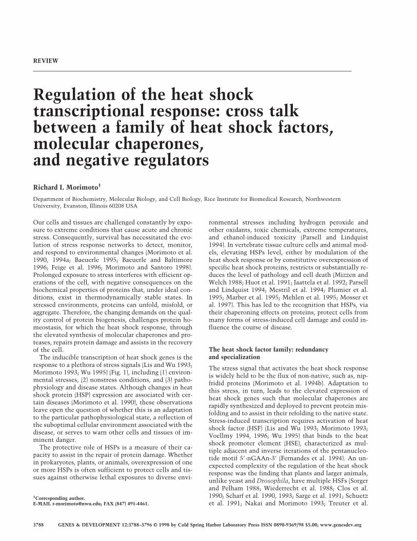
The inducible transcription of heat shock genes is theresponse to a plethora of stress signals (Lis and Wu 1993;Morimoto 1993; Wu 1995) (Fig. 1), including (1) environ-mental stresses, (2) nonstress conditions, and (3) patho-physiology and disease states. Although changes in heatshock protein (HSP) expression are associated with cer-tain diseases (Morimoto et al. 1990), these observationsleave open the question of whether this is an adaptationto the particular pathophysiological state, a reflection ofthe suboptimal cellular environment associated with thedisease, or serves to warn other cells and tissues of im-minent danger.
The protective role of HSPs is a measure of their ca-pacity to assist in the repair of protein damage. Whetherin prokaryotes, plants, or animals, overexpression of oneor more HSPs is often sufficient to protect cells and tis-sues against otherwise lethal exposures to diverse envi-
ronmental stresses including hydrogen peroxide andother oxidants, toxic chemicals, extreme temperatures,and ethanol-induced toxicity (Parsell and Lindquist1994). In vertebrate tissue culture cells and animal mod-els, elevating HSPs level, either by modulation of theheat shock response or by constitutive overexpression ofspecific heat shock proteins, restricts or substantially re-duces the level of pathology and cell death (Mizzen andWelch 1988; Huot et al. 1991; Jaattela et al. 1992; Parselland Lindquist 1994; Mestril et al. 1994; Plumier et al.1995; Marber et al. 1995; Mehlen et al. 1995; Mosser etal. 1997). This has led to the recognition that HSPs, viatheir chaperoning effects on proteins, protect cells frommany forms of stress-induced cell damage and could in-fluence the course of disease.
The heat shock factor family: redundancyand specialization
The stress signal that activates the heat shock responseis widely held to be the flux of non-native, such as, nip-fridid proteins (Morimoto et al. 1994b). Adaptation tothis stress, in turn, leads to the elevated expression ofheat shock genes such that molecular chaperones arerapidly synthesized and deployed to prevent protein mis-folding and to assist in their refolding to the native state.Stress-induced transcription requires activation of heatshock factor (HSF) (Lis and Wu 1993; Morimoto 1993;Voellmy 1994, 1996; Wu 1995) that binds to the heatshock promoter element (HSE), characterized as mul-tiple adjacent and inverse iterations of the pentanucleo-tide motif 58-nGAAn-38 (Fernandes et al. 1994). An un-expected complexity of the regulation of the heat shockresponse was the finding that plants and larger animals,unlike yeast and Drosophila, have multiple HSFs (Sorgerand Pelham 1988; Wiederrecht et al. 1988; Clos et al.1990; Scharf et al. 1990, 1993; Sarge et al. 1991; Schuetzet al. 1991; Nakai and Morimoto 1993; Treuter et al.
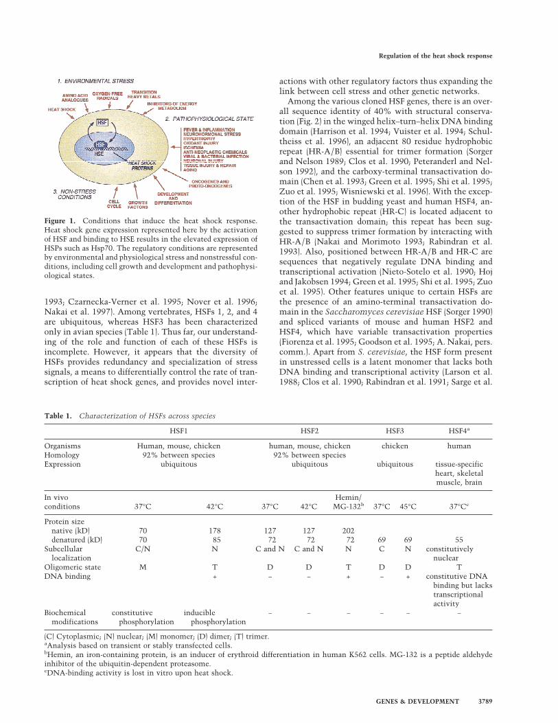
1993; Czarnecka-Verner et al. 1995; Nover et al. 1996;Nakai et al. 1997). Among vertebrates, HSFs 1, 2, and 4are ubiquitous, whereas HSF3 has been characterizedonly in avian species (Table 1). Thus far, our understand-ing of the role and function of each of these HSFs isincomplete. However, it appears that the diversity ofHSFs provides redundancy and specialization of stresssignals, a means to differentially control the rate of tran-scription of heat shock genes, and provides novel inter-
actions with other regulatory factors thus expanding thelink between cell stress and other genetic networks.
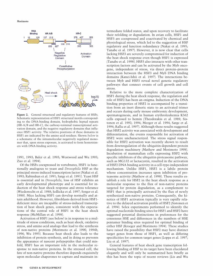
Among the various cloned HSF genes, there is an over-all sequence identity of 40% with structural conserva-tion (Fig. 2) in the winged helix–turn–helix DNA bindingdomain (Harrison et al. 1994; Vuister et al. 1994; Schul-theiss et al. 1996), an adjacent 80 residue hydrophobicrepeat (HR-A/B) essential for trimer formation (Sorgerand Nelson 1989; Clos et al. 1990; Peteranderl and Nel-son 1992), and the carboxy-terminal transactivation do-main (Chen et al. 1993; Green et al. 1995; Shi et al. 1995;Zuo et al. 1995; Wisniewski et al. 1996). With the excep-tion of the HSF in budding yeast and human HSF4, an-other hydrophobic repeat (HR-C) is located adjacent tothe transactivation domain; this repeat has been sug-gested to suppress trimer formation by interacting withHR-A/B (Nakai and Morimoto 1993; Rabindran et al.1993). Also, positioned between HR-A/B and HR-C aresequences that negatively regulate DNA binding andtranscriptional activation (Nieto-Sotelo et al. 1990; Hojand Jakobsen 1994; Green et al. 1995; Shi et al. 1995; Zuoet al. 1995). Other features unique to certain HSFs arethe presence of an amino-terminal transactivation do-main in the Saccharomyces cerevisiae HSF (Sorger 1990)and spliced variants of mouse and human HSF2 andHSF4, which have variable transactivation properties(Fiorenza et al. 1995; Goodson et al. 1995; A. Nakai, pers.comm.). Apart from S. cerevisiae, the HSF form presentin unstressed cells is a latent monomer that lacks bothDNA binding and transcriptional activity (Larson et al.1988; Clos et al. 1990; Rabindran et al. 1991; Sarge et al.
Oligomeric state M T D D T D D TDNA binding + − − + − + constitutive DNA
binding but lackstranscriptionalactivity
Biochemicalmodifications
constitutivephosphorylation
induciblephosphorylation
− − − − − −
(C) Cytoplasmic; (N) nuclear; (M) monomer; (D) dimer; (T) trimer.aAnalysis based on transient or stably transfected cells.bHemin, an iron-containing protein, is an inducer of erythroid differentiation in human K562 cells. MG-132 is a peptide aldehydeinhibitor of the ubiquitin-dependent proteasome.cDNA-binding activity is lost in vitro upon heat shock.
Figure 1. Conditions that induce the heat shock response.Heat shock gene expression represented here by the activationof HSF and binding to HSE results in the elevated expression ofHSPs such as Hsp70. The regulatory conditions are representedby environmental and physiological stress and nonstressful con-ditions, including cell growth and development and pathophysi-ological states.
Regulation of the heat shock response
GENES & DEVELOPMENT 3789
1991, 1993; Baler et al. 1993; Westwood and Wu 1993;Zuo et al. 1994).
Of the HSFs coexpressed in vertebrates, HSF1 is func-tionally analogous to yeast and Drosophila HSF as theprincipal stress-induced transcription factor (Nakai et al.1993; Rabindran et al. 1991; Sarge et al. 1991). Yeast HSFis essential and in Drosophila, loss of HSF exhibits anearly developmental phenotype and is essential for in-duction of the heat shock response and stress tolerance(Wiederrecht et al. 1988; Jedlicka et al. 1997; Sorger et al.1988). Mice lacking HSF1 can develop normally and at-tain adulthood. However, fibroblasts derived from HSF1-deficient mice are incapable of stress-induced transcrip-tion of heat shock genes, consistent with the expecta-tions of the central role of HSF1 in the heat shockresponse (McMillan et al. 1998).
Activation of HSF1 (see below) is in response to a mul-titude of stress conditions, such as heat shock, oxidativestress, and amino acid analogs that lead to the synthesisof non-native proteins (Morimoto et al. 1990, 1994b,1996; Wu 1995). Because heat shock also leads to theinhibition of protein synthesis, and in doing so preventsthe appearance of nascent polypeptides that could mis-fold, HSF1 has an important role in the molecular re-sponse to non-native proteins. In the stressed cell, thefate of non-native proteins therefore depends exquisitelyupon molecular chaperones to capture and maintain in-
termediate folded states, and upon recovery to facilitatetheir refolding or degradation. In avian cells, HSF1 andHSF3 are coexpressed and coactivated by chemical andphysiological stress, which led to the suggestion of HSFregulatory and function redundancy (Nakai et al. 1995;Tanabe et al. 1997). However, it is now clear that cellslacking HSF3 are severely compromised for induction ofthe heat shock response even though HSF1 is expressed(Tanabe et al. 1998). HSF3 also interacts with other tran-scription factors and can be activated by the Myb onco-gene, independent of stress, via direct protein–proteininteraction between the HSF3 and Myb DNA bindingdomains (Kanei-Ishii et al. 1997). The interactions be-tween Myb and HSF3 reveal novel genetic regulatorypathways that connect events of cell growth and cellstress.
Relative to the more complete characterization ofHSF1 during the heat shock response, the regulation androle of HSF2 has been an enigma. Induction of the DNAbinding properties of HSF2 is accompanied by a transi-tion from an inert dimeric state to an activated trimerand occurs during early mouse embryonic development,spermatogenesis, and in human erythroleukemia K562cells exposed to hemin (Theodorakis et al. 1989; Sis-tonen et al. 1992; 1994; Mezger et al. 1994; Sarge et al.1994; Rallu et al. 1997). Although these results suggestedthat HSF2 activity was associated with development anddifferentiation, the events responsible for activation ofHSF2 were uncharacterized. The stress signal respon-sible for HSF2 activation was recently shown to resultfrom downregulation of the ubiquitin-dependent proteindegradation machinery (Mathew and Morimoto 1998).Incubation of mammalian cells expressing HSF2 withspecific inhibitors of the ubiquitin-proteasome pathway,such as MG132 or lactacystin, resulted in the activationof HSF2 DNA binding activity in a cell-type-independentmechanism. Unlike HSF1, HSF2 is a labile proteinwhose concentration increases upon inhibition of pro-teasome activity (Mathew et al. 1998). These results es-tablish a role for HSF2 in the heat shock response as amolecular response to the flux of non-native proteinstargeted for protein degradation, as a complement toHSF1 that is principally activated by the flux of newlysynthesized non-native proteins. Consequently, the ki-netics of HSF1 activation typically is very rapidly rela-tive to the delayed activation profile of HSF2 (Sistonen etal. 1994). Selex experiments performed to identify theoptimal nucleotide binding sites for HSF1 and HSF2 havesuggested potential distinctions in preferences for theconsensus HSE and differences in the numbers of HSEpentamer binding sites required for optimal binding ofeither HSF (Kroeger and Morimoto 1994). These studieshave raised the possibility that HSF2 may have distincttarget genes from those of HSF1, as well as differingspecificities for common target genes (Leppa et al. 1997;Liu et al. 1997).
General features of heat shock gene transcription fol-lowing binding of HSF to its target have been elucidatedelegantly and will only be summarized here briefly asthis has been the topic of recent reviews (Lis and Wu
Figure 2. General structural and regulatory features of HSFs.Schematic representation of HSF1 structural motifs correspond-ing to the DNA-binding domain, hydrophobic heptad repeats(HR-A/B and HR-C), the carboxy-terminal transcriptional acti-vation domain, and the negative regulatory domains that influ-ence HSF1 activity. The relative positions of these domains inHSF1 are indicated by the amino acid residues. Shown below isa schematic of the intramolecular negatively regulated mono-mer that, upon stress exposure, is activated to form homotrim-ers with DNA-binding activity.
Morimoto
3790 GENES & DEVELOPMENT
1993; Wu 1995). The inducible binding of HSF leads tochanges in the organization of chromatin structure lo-calized to the 5∼-flanking regions of heat shock genes(Wu 1980, 1984; Giardina et al. 1992). Although HSFassociates with components of the chromatin remodel-ing machinery (Becker and Wu 1992; Tsukiyama et al.1994; Brown and Kingston 1997) and the basal transcrip-tional machinery, association specifically with TBP alsohas been detected (Mason and Lis 1997). Such interac-tions may be critical for the release of the paused RNApolymerase II that represents a pre-initiated nascenttranscript that elongates in a stress-dependent manner toyield inducible heat shock mRNAs (Gilmour and Lis1986; Rougvie and Lis 1988).
Who detects stress?
Does HSF directly sense its biochemical environment?Although heat shock has been the typical stress condi-tion employed, HSF trimer formation cannot solely be atemperature-regulated event because the majority ofstressors have their effect in cells grown at 37°C. Yet,there exists a substantial amount of data indicating thatHSF translated in vitro in reticulocyte lysates can beactivated by heat shock and that purified DrosophilaHSF or recombinant mouse and human HSF1 can ac-quire DNA binding upon in vitro heat shock (Mosser etal. 1990; Goodson and Sarge 1995; Larson et al. 1995;Zhong et al. 1998). Likewise, in vitro translated HSF1 orpurified HSF can be activated by low pH (pH 6.5) or sa-licylate (Mosser et al. 1990; Zhong et al. 1998). As salic-ylate is an organic acid, its effect on HSF activity couldbe analogous to the activating effects of low pH, perhapsacting on HSF1 conformation. Although these resultssuggest that the intrinsic properties of HSF can be modu-lated by its biochemical environment, they do not ex-clude a role for other negative regulators that may func-tion to keep HSF in the repressed state.
Is activation of HSF1 a titrated phenomenon such thatsimultaneous or subsequent exposure to multiple stress-ors at subthreshold levels leads to a complete heat shockresponse? This issue addresses whether stress has mul-tiple distinct targets or stress receptors and whetherthese stress signals converge upon common pathways.The temperature for activation of HSF is not an absolute;to illustrate this point, growing HeLa cells at tempera-tures <37°C reduces the temperature required for com-plete activation of HSF1 (Abravaya et al. 1991). Likewise,human HSF1 expressed in Drosophila cells becomes ac-tivated at the temperature of the Drosophila heat shockresponse (37°C) (Clos et al. 1993). Consistent with theseobservations, incubation of mammalian cells with a sub-threshold concentration (for HSF1 activation) of indo-methacin or other nonsteroidal anti-inflammatory drugssensitizes treated cells such that the temperature orstress required for induction of the heat shock responseis reduced (Lee et al. 1995). These observations supportthe notion that many if not all of the inducers of the heatshock response converge on common pathways resultingfrom an imbalance in protein homeostasis.
HSF cycle: feedback from molecular chaperonesand negative regulators
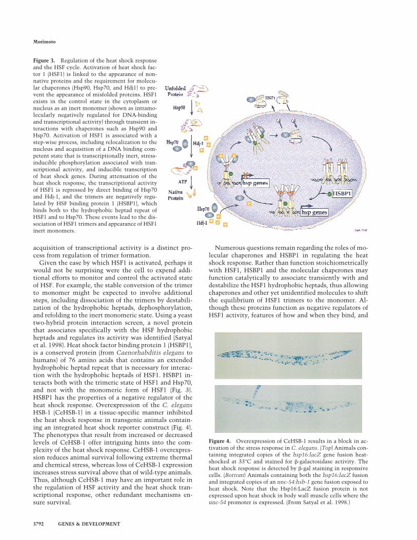
Under normal growth conditions in metazoans, HSF1activity is repressed and exists either in the cytosol ornucleus in an inert monomeric state. Negative regula-tion of the DNA binding and transactivation domainsinvolves intramolecular interactions with the central re-gion of HSF1 and is also influenced by constitutive phos-phorylation at critical serine residues (Knauf et al. 1996;Kline and Morimoto 1997). Molecular chaperones suchas Hsp90 have also been shown to have a role in main-taining HSF1 in an inert state (Ali et al. 1998; Zuo et al.1998). However it remains to be demonstrated how in-teractions with specific chaperones maintains the re-pressed monomeric state or shifts the equilibrium backto the monomer. Heat shock and other stresses causederepression of HSF1, initiating the events that lead tothe appearance of the transcriptionally active trimer. Ac-tivation of HSF1 is a multistep process (Fig. 3) that re-sults in a transcriptionally competent state that is char-acterized typically by hyperphosphorylation at multipleserine residues (Hensold et al. 1990; Jurivich et al. 1992;Sarge et al. 1993; Cotto et al. 1996; Kline and Morimoto1997). Although heat shock and most other stresses con-vert HSF1 to the fully active trimer, sodium salicylatetreatment activates HSF1 to an intermediate trimericstate, which is bound in vivo to the promoter of theHsp70 gene, yet is transcriptionally inert (Jurivich et al.1992). It is intriguing to note that the two DNA bindingcompetent forms of metazoan HSF that differ in tran-scriptional activity correspond respectively to the con-trol and activated forms of S. cerevisiae HSF (Sorger et al.1987). Thus, despite the ancient origins of the heat shockresponse, the regulation of HSF has, along the way, ac-quired additional levels of control.
If the function of the heat shock response is to increasethe levels of heat shock proteins, then what mechanismexists to repress the response? A substantial amount ofgenetic and biochemical evidence from studies in pro-karyotes and eukaryotes support a role for heat shockproteins in the negative regulation of heat shock. Forexample, in yeast, an extragenic suppressor of an Hsp70mutant that exhibits a poor-growth phenotype at 37°Cwas identified as a mutant allele of HSF that had reducedDNA binding activity (Halladay and Craig 1995). Com-plexes of Hsp70 and HSF trimers have been detected dur-ing attenuation of the heat shock transcriptional re-sponse (Abravaya et al. 1992; Baler et al. 1992; Shi et al.1998) and consistent with a regulatory role for chaper-ones in the heat shock response, overexpression ofHsp70 or Hdj-1/Hsp40 in the absence of stress preventsthe inducible transcription of heat shock genes (Mosseret al. 1993; Rabindran et al. 1994; Shi et al. 1998). Al-though the mechanism by which Hsp70 and Hdj1/Hsp40repress HSF1 has not been fully elucidated, these mo-lecular chaperones bind directly to the HSF1 transacti-vation domain (Fig. 3). The Hsp70 chaperones and co-chaperones alone are insufficient to prevent the appear-ance of HSF1 trimers; these results demonstrate that the
Regulation of the heat shock response
GENES & DEVELOPMENT 3791
acquisition of transcriptional activity is a distinct pro-cess from regulation of trimer formation.
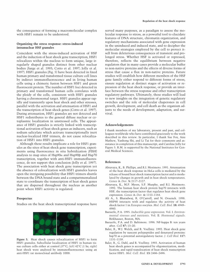
Given the ease by which HSF1 is activated, perhaps itwould not be surprising were the cell to expend addi-tional efforts to monitor and control the activated stateof HSF. For example, the stable conversion of the trimerto monomer might be expected to involve additionalsteps, including dissociation of the trimers by destabili-zation of the hydrophobic heptads, dephosphorylation,and refolding to the inert monomeric state. Using a yeasttwo-hybrid protein interaction screen, a novel proteinthat associates specifically with the HSF hydrophobicheptads and regulates its activity was identified (Satyalet al. 1998). Heat shock factor binding protein 1 (HSBP1),is a conserved protein (from Caenorhabditis elegans tohumans) of 76 amino acids that contains an extendedhydrophobic heptad repeat that is necessary for interac-tion with the hydrophobic heptads of HSF1. HSBP1 in-teracts both with the trimeric state of HSF1 and Hsp70,and not with the monomeric form of HSF1 (Fig. 3).HSBP1 has the properties of a negative regulator of theheat shock response. Overexpression of the C. elegansHSB-1 (CeHSB-1) in a tissue-specific manner inhibitedthe heat shock response in transgenic animals contain-ing an integrated heat shock reporter construct (Fig. 4).The phenotypes that result from increased or decreasedlevels of CeHSB-1 offer intriguing hints into the com-plexity of the heat shock response. CeHSB-1 overexpres-sion reduces animal survival following extreme thermaland chemical stress, whereas loss of CeHSB-1 expressionincreases stress survival above that of wild-type animals.Thus, although CeHSB-1 may have an important role inthe regulation of HSF activity and the heat shock tran-scriptional response, other redundant mechanisms en-sure survival.
Numerous questions remain regarding the roles of mo-lecular chaperones and HSBP1 in regulating the heatshock response. Rather than function stoichiometricallywith HSF1, HSBP1 and the molecular chaperones mayfunction catalytically to associate transiently with anddestabilize the HSF1 hydrophobic heptads, thus allowingchaperones and other yet unidentified molecules to shiftthe equilibrium of HSF1 trimers to the monomer. Al-though these proteins function as negative regulators ofHSF1 activity, features of how and when they bind, and
Figure 4. Overexpression of CeHSB-1 results in a block in ac-tivation of the stress response in C. elegans. (Top) Animals con-taining integrated copies of the hsp16:lacZ gene fusion heat-shocked at 33°C and stained for b-galactosidase activity. Theheat shock response is detected by b-gal staining in responsivecells. (Bottom) Animals containing both the hsp16:lacZ fusionand integrated copies of an unc-54:hsb-1 gene fusion exposed toheat shock. Note that the Hsp16:LacZ fusion protein is notexpressed upon heat shock in body wall muscle cells where theunc-54 promoter is expressed. (From Satyal et al. 1998.)
Figure 3. Regulation of the heat shock responseand the HSF cycle. Activation of heat shock fac-tor 1 (HSF1) is linked to the appearance of non-native proteins and the requirement for molecu-lar chaperones (Hsp90, Hsp70, and Hdj1) to pre-vent the appearance of misfolded proteins. HSF1exists in the control state in the cytoplasm ornucleus as an inert monomer (shown as intramo-lecularly negatively regulated for DNA-bindingand transcriptional activity) through transient in-teractions with chaperones such as Hsp90 andHsp70. Activation of HSF1 is associated with astep-wise process, including relocalization to thenucleus and acquisition of a DNA binding com-petent state that is transcriptionally inert, stress-inducible phosphorylation associated with tran-scriptional activity, and inducible transcriptionof heat shock genes. During attenuation of theheat shock response, the transcriptional activityof HSF1 is repressed by direct binding of Hsp70and Hdj-1, and the trimers are negatively regu-lated by HSF binding protein 1 (HSBP1), whichbinds both to the hydrophobic heptad repeat ofHSF1 and to Hsp70. These events lead to the dis-sociation of HSF1 trimers and appearance of HSF1inert monomers.
Morimoto
3792 GENES & DEVELOPMENT
the consequence of forming a macromolecular complexwith HSF1 remain to be understood.
Organizing the stress response: stress-inducedintranuclear HSF granules
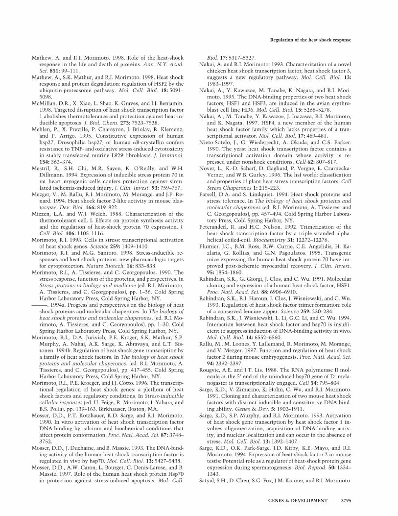
Coincident with the stress-induced activation of HSFand the induction of heat shock gene transcription, HSF1relocalizes within the nucleus to form unique, large ir-regularly shaped granules distinct from other nuclearbodies (Sarge et al. 1993; Cotto et al. 1997; Jolly et al.1997). HSF1 granules (Fig. 5) have been detected in fixedhuman primary and transformed tissue culture cell linesby indirect immunofluorescence and in living humancells using a chimeric fusion between HSF1 and greenfluorescent protein. The number of HSF1 foci detected inprimary and transformed human cells correlates withthe ploidy of the cells, consistent with HSF1 granuleshaving a chromosomal target. HSF1 granules appear rap-idly and transiently upon heat shock and other stresses,parallel with the activation and attenuation of HSF1 andthe transcription of heat shock genes (Cotto et al. 1997).During attenuation, HSF1 granules are not detected andHSF1 redistributes to the general diffuse nuclear or cy-toplasmic localization in unstressed cells. The appear-ance of HSF1 granules is strictly linked with transcrip-tional activation of heat shock genes as inducers, such assodium salicylate which activate transcriptionally inertnuclear-localized HSF trimers, do not cause relocaliza-tion of HSF1 into HSF1 granules.
Although these results implicate a role for HSF1 gran-ules as the sites of heat shock gene transcription, experi-ments using fluorescence in situ hybridization (FISH)analysis to map sites of Hsp90a and Hsp90b and Hsp70transcription, together with anti-HSF1 immunofluores-cence, do not support this conclusion (Jolly et al. 1997).The association with heat shock gene transcription yetthe absence of colocalization with HSF1 granules leavesopen the intriguing possibility that HSF1 trimers shuttlebetween the DNA bound state and a compartmentalizedstate to coordinate the transcription of heat shock genesthat are dispersed throughout the nucleus as anotherpoint where HSF1 activity is regulated.
Prospectus
Studies on the heat shock transcriptional response have
served many purposes, as a paradigm to assess the mo-lecular response to stress, as a powerful tool to elucidatefeatures of DNA structure, chromatin organization, andregulatory mechanisms associated with gene expressionin the uninduced and induced state, and to decipher themolecular strategies employed by the cell to protect it-self from deleterious consequences of transient and pro-longed stress. Whether HSF is activated or repressed,therefore, reflects the equilibrium between negativeregulators that in many cases provide a molecular bufferto non-native proteins and the effects of diverse forms ofstress that cause a flux in non-native proteins. Futurestudies will establish how different members of the HSFgene family either respond to different forms of stress,ensure regulation at distinct stages of activation or re-pression of the heat shock response, or provide an inter-face between the stress response and other transcriptionregulatory pathways. Ultimately, these studies will leadto new insights on the integration of genetic regulatoryswitches and the role of molecular chaperones in cellgrowth, development, and cell death as the organism ad-justs to the needs of development, adaptation, and sur-vival.
Acknowledgements
I thank members of my laboratory, present and past, and col-leagues worldwide who have contributed graciously to the workdescribed in this review. In particular, I thank Sue Fox, AnuMathew, Yanhong Shi, and Li Tai for their comments and as-sistance in completion of this manuscript, and Caroline Jolly forFigure 5. R.M. is supported by the National Institutes for Gen-eral Medical Sciences.
References
Abravaya, K., B. Phillips, and R.I. Morimoto. 1991. Attenuationof the heat shock response in HeLa cells is mediated by therelease of bound heat shock transcription factor and is modu-lated by changes in growth and in heat shock temperatures.Genes & Dev. 5: 2117–2127.
Abravaya, K., M.P. Myers, S.P. Murphy, and R.I. Morimoto.1992. The human heat shock protein hsp70 interacts withHSF, the transcription factor that regulates heat shock geneexpression. Genes & Dev. 6: 1153–1164.
Ali A., S. Bharadwaj, R. O’Carroll, and N. Ovsenek. 1998.HSP90 interacts with and regulates the activity of heatshock factor 1 in Xenopus oocytes. Mol. Cell. Biol. 18: 4949–4960.
Baeuerle, P.A. 1995. Inducible gene expression. Vol. I. Environ-mental stresses and nutrients. Vol. II. Hormonal signals.Birkhauser, Boston, MA.
Baeuerle, P.A. and D. Baltimore. 1996. NF-kappa B: ten yearsafter. Cell 87: 13–20.
Baler, R., W.J. Welch, and R. Voellmy. 1992. Heat shock generegulation by nascent polypeptides and denatured proteins:hsp70 as a potential autoregulatory factor. J. Cell Biol. 117:1151–1159.
Baler, R., G. Dahl, and R. Voellmy. 1993. Activation of humanheat shock genes is accompanied by oligomerization, modi-fication, and rapid translocation of heat shock transcriptionfactor HSF1. Mol. Cell. Biol. 13: 2486–2496.
Figure 5. Heat shock causes relocalization of HSF1 to formHSF1 granules. Subcellular localization of HSF1 in human tis-sue culture cells either at control (37°C; left) 42°C (1 hr; right)heat shock were analyzed by immunofluorescence using ananti-HSF1 rat monoclonal antibody 10H8.
Regulation of the heat shock response
GENES & DEVELOPMENT 3793
Becker, P.B. and C. Wu. 1992. Cell-free system for assembly oftranscriptionally repressed chromatin from Drosophila em-bryos. Mol. Cell. Biol. 12: 2241–2249.
Brown, S.A. and R.E. Kingston. 1997. Disruption of downstreamchromatin directed by a transcriptional activator. Genes &Dev. 11: 3116–3121.
Chen, Y., N.A. Barlev, O. Westergaard, and B.K. Jakobsen. 1993.Identification of the C-terminal activator domain in yeastheat shock factor: Independent control of transient and sus-tained transcriptional activity. EMBO J. 12: 5007–5018.
Clos, J., J.T. Westwood, P.B. Becker, S. Wilson, K. Lambert, andC. Wu. 1990. Molecular cloning and expression of a hexa-meric Drosophila heat shock factor subject to negative regu-lation. Cell 63: 1085–1097.
Clos, J., S. Rabindran, J. Wisniewski, and C. Wu. 1993. Induc-tion temperature of human heat shock factor is repro-grammed in a Drosophila cell environment. Nature 364:252–255.
Cotto, J.J., M. Kline, and R.I. Morimoto. 1996. Activation ofheat shock factor 1 DNA binding precedes stress-inducedserine phosphorylation. Evidence for a multistep pathway ofregulation. J. Biol. Chem. 271: 3355–3358.
Cotto, J., S. Fox, and R.I. Morimoto. 1997. HSF1 granules: Anovel stress-induced nuclear compartment of human cells. J.Cell Sci. 110: 2925–2934.
Czarnecka-Verner, E., C.X. Yuan, P.C. Foxand, and W.B. Gurley.1995. Isolation and characterization of six heat shock tran-scription factor cDNA clones from soybean. Plant Mol. Biol.29: 37–51.
Feige, U. and W. van Eden. 1996. Infection, autoimmunity andautoimmune disease. Stress Inducible Cellular Responses77: 359–373.
Fernandes, M., T. O’Brien, and J.T. Lis. 1994. Structure andregulation of heat shock gene promoters. In The biology ofheat shock proteins and molecular chaperones (ed. R.I. Mo-rimoto, A. Tissieres, and C. Georgopolis), pp. 375–393. ColdSpring Harbor Laboratory Press, Cold Spring Harbor, NY.
Fiorenza, M.T., T. Farkas, M. Dissing, D. Kolding, and V. Zima-rino. 1995. Complex expression of murine heat shock tran-scription factors. Nucleic Acids Res. 23: 467–474.
Giardina, C., M. Perez-Riba, and J.T. Lis. 1992. Promoter melt-ing and TFIID complexes on Drosophila genes in vivo. Genes& Dev. 6: 2190–2200.
Gilmour, D.S. and J.T. Lis. 1986. RNA polymerase II interactswith the promoter region of the noninduced hsp70 gene inDrosophila melanogaster cells. Mol. Cell. Biol. 6: 3984–3989.
Goodson, M.L. and K.D. Sarge. 1995. Heat-inducible DNA bind-ing of purified heat shock transcription factor 1. J. Biol.Chem. 270: 2447–2450.
Green, M., T.J. Schuetz, E.K. Sullivan, and R.E. Kingston. 1995.A heat shock-responsive domain of human HSF1 that regu-lates transcription activation domain function. Mol. Cell.Biol. 15: 3354–3362.
Halladay, J.T. and E.A. Craig. 1995. A heat shock transcriptionfactor with reduced activity suppresses a yeast HSP70 mu-tant. Mol. Cell. Biol. 15: 4890–4897.
Harrison, C.J., A.A. Bohm, and H.C. Nelson. 1994. Crystalstructure of the DNA binding domain of the heat shock tran-scription factor. Science 263: 224–227.
Hensold, J.O., C.R. Hunt, S.K. Calderwood, D.E. Housman, andR.E. Kingston. 1990. DNA binding of heat shock factor to the
heat shock element is insufficient for transcriptional activa-tion in murine erythroleukemia cells. Mol. Cell. Biol.10: 1600–1608.
Hoj, A. and B.K. Jakobsen. 1994. A short element required forturning off heat shock transcription factor: Evidence thatphosphorylation enhances deactivation. EMBO J. 13: 2617–2624.
Huot, J., G. Roy, H. Lambert, P. Chretien, and J. Landry. 1991.Increased survival after treatments with anticancer agents ofChinese hamster cells expressing the human Mr 27000 heatshock protein. Cancer Res. 51: 5245–5252.
Jaattela, M., D. Wissing, P.A. Bauser, and G.C. Li. 1992. Majorheat shock protein hsp70 protects tumor cells from tumornecrosis factor cytotoxicity. EMBO J. 11: 3507–3512.
Jedlicka, P., M.A. Mortin, and C. Wu. 1997. Multiple functionsof Drosophila heat shock transcription factor in vivo. EMBOJ. 16: 2452–2462.
Jolly, C., R. Morimoto, M. Robert-Nicoud, and C. Vourc’h.1997. HSF1 transcription factor concentrates in nuclear fociduring heat shock: Relationship with transcription sites. J.Cell Sci. 110: 2935–2941.
Jurivich, D.A., L. Sistonen, R.A. Kroes, and R.I. Morimoto. 1992.Effect of sodium salicylate on the human heat shock re-sponse. Science 255: 1243–1245.
Kanei-Ishii, C., J. Tanikawa, A. Nakai, R.I. Morimoto, and S.Ishii. 1997. Activation of heat shock transcription factor 3 byc-Myb in the absence of cellular stress. Science 277: 246–248.
Kline, M.P. and R.I. Morimoto. 1997. Repression of the heatshock factor 1 transcriptional activation domain is modu-lated by constitutive phosphorylation. Mol. Cell. Biol.17: 2107–2115.
Knauf, U., E.M. Newton, J. Kyriakis, and R.E. Kingston. 1996.Repression of human heat shock factor 1 activity at controltemperature by phosphorylation. Genes & Dev. 10: 2782–2793.
Kroeger, P.E. and R.I. Morimoto. 1994. Selection of new HSF1and HSF2 DNA-binding sites reveals difference in trimercooperativity. Mol. Cell. Biol. 14: 7592–7603.
Larson, J.S., T.J. Schuetz, and R.E. Kingston. 1988. Activation invitro of sequence-specific DNA binding by a human regula-tory factor. Nature 335: 372–375.
———. 1995. In vitro activation of purified human heat shockfactor by heat. Biochemistry 34: 1902–1911.
Lee, B.S., J. Chen, C. Angelidis, D.A. Jurivich, and R.I. Mori-moto. 1995. Pharmacological modulation of heat shock fac-tor 1 by antiinflammatory drugs results in protection againststress-induced cellular damage. Proc. Natl. Acad. Sci. 92:7207–7211.
Leppa, S., L. Pirkkala, S.C. Chow, J.E. Eriksson, and L. Sistonen.1997. Thioredoxin is transcriptionally induced upon activa-tion of heat shock factor 2. J. Biol. Chem. 272: 30400–30404.
Lis, J. and C. Wu. 1993. Protein traffic on the heat shock pro-moter: Parking, stalling, and trucking along. Cell 74: 1–4.
Liu, X.D., P.C. Liu, N. Santoro, and D.J. Thiele. 1997. Conser-vation of a stress response: human heat shock transcriptionfactors functionally substitute for yeast HSF. EMBO J. 16:6466–6477.
Marber, M.S., R. Mestril, S.H. Chi, M.R. Sayen, D.M. Yellon,and W.H. Dillmann. 1995. Overexpression of the rat induc-ible 70-kD heat stress protein in a transgenic mouse in-creases the resistance of the heart to ischemic injury. J. Clin.Invest. 95: 1446–1456.
Mason, P.B., Jr. and J.T. Lis. 1997. Cooperative and competitiveprotein interactions at the hsp70 promoter. J. Biol. Chem.272: 33227–33233.
Morimoto
3794 GENES & DEVELOPMENT
Mathew, A. and R.I. Morimoto. 1998. Role of the heat-shockresponse in the life and death of proteins. Ann. N.Y. Acad.Sci. 851: 99–111.
Mathew, A., S.K. Mathur, and R.I. Morimoto. 1998. Heat shockresponse and protein degradation: regulation of HSF2 by theubiquitin-proteasome pathway. Mol. Cell. Biol. 18: 5091–5098.
McMillan, D.R., X. Xiao, L. Shao, K. Graves, and I.J. Benjamin.1998. Targeted disruption of heat shock transcription factor1 abolishes thermotolerance and protection against heat-in-ducible apoptosis. J. Biol. Chem. 273: 7523–7528.
Mehlen, P., X. Preville, P. Chareyron, J. Briolay, R. Klemenz,and P. Arrigo. 1995. Constitutive expression of humanhsp27, Drosophilia hsp27, or human aB-crystallin confersresistance to TNF- and oxidative stress-induced cytotoxicityin stably transfected murine L929 fibroblasts. J. Immunol.154: 363–374.
Mestril, R., S.H. Chi, M.R. Sayen, K. O’Reilly, and W.H.Dillmann. 1994. Expression of inducible stress protein 70 inrat heart myogenic cells confers protection against simu-lated ischemia-induced injury. J. Clin. Invest. 93: 759–767.
Mezger, V., M. Rallu, R.I. Morimoto, M. Morange, and J.P. Re-nard. 1994. Heat shock factor 2-like activity in mouse blas-tocysts. Dev. Biol. 166: 819–822.
Mizzen, L.A. and W.J. Welch. 1988. Characterization of thethermotolerant cell. I. Effects on protein synthesis activityand the regulation of heat-shock protein 70 expression. J.Cell. Biol. 106: 1105–1116.
Morimoto, R.I. and M.G. Santoro. 1998. Stress-inducible re-sponses and heat shock proteins: new pharmacologic targetsfor cytoprotection. Nature Biotech. 16: 833–838.
Morimoto, R.I., A. Tissieres, and C. Georgopoulos. 1990. Thestress response, function of the proteins, and perspectives. InStress proteins in biology and medicine (ed. R.I. Morimoto,A. Tissieres, and C. Georgopoulos), pp. 1–36. Cold SpringHarbor Laboratory Press, Cold Spring Harbor, NY.
———. 1994a. Progress and perspectives on the biology of heatshock proteins and molecular chaperones. In The biology ofheat shock proteins and molecular chaperones, (ed. R.I. Mo-rimoto, A. Tissieres, and C. Georgopoulos), pp. 1–30. ColdSpring Harbor Laboratory Press, Cold Spring Harbor, NY.
Morimoto, R.I., D.A. Jurivich, P.E. Kroger, S.K. Mathur, S.P.Murphy, A. Nakai, A.K. Sarge, K. Abravaya, and L.T. Sis-tonen. 1994b. Regulation of heat shock gene transcription bya family of heat shock factors. In The biology of heat shockproteins and molecular chaperones. (ed. R.I. Morimoto, A.Tissieres, and C. Georgopoulos), pp. 417–455. Cold SpringHarbor Laboratory Press, Cold Spring Harbor, NY.
Morimoto, R.I., P.E. Kroeger, and J.J. Cotto. 1996. The transcrip-tional regulation of heat shock genes: a plethora of heatshock factors and regulatory conditions. In Stress-induciblecellular responses (ed. U. Feige, R. Morimoto, I. Yahara, andB.S. Polla), pp. 139–163. Birkhauser, Boston, MA.
Mosser, D.D., P.T. Kotzbauer, K.D. Sarge, and R.I. Morimoto.1990. In vitro activation of heat shock transcription factorDNA-binding by calcium and biochemical conditions thataffect protein conformation. Proc. Natl. Acad. Sci. 87: 3748–3752.
Mosser, D.D., J. Duchaine, and B. Massie. 1993. The DNA-bind-ing activity of the human heat shock transcription factor isregulated in vivo by hsp70. Mol. Cell. Biol. 13: 5427–5438.
Mosser, D.D., A.W. Caron, L. Bourget, C. Denis-Larose, and B.Massie. 1997. Role of the human heat shock protein Hsp70in protection against stress-induced apoptosis. Mol. Cell.
Biol. 17: 5317–5327.Nakai, A. and R.I. Morimoto. 1993. Characterization of a novel
chicken heat shock transcription factor, heat shock factor 3,suggests a new regulatory pathway. Mol. Cell. Biol. 13:1983–1997.
Nakai, A., Y. Kawazoe, M. Tanabe, K. Nagata, and R.I. Mori-moto. 1995. The DNA-binding properties of two heat shockfactors, HSF1 and HSF3, are induced in the avian erythro-blast cell line HD6. Mol. Cell. Biol. 15: 5268–5278.
Nakai, A., M. Tanabe, Y. Kawazoe, J. Inazawa, R.I. Morimoto,and K. Nagata. 1997. HSF4, a new member of the humanheat shock factor family which lacks properties of a tran-scriptional activator. Mol. Cell. Biol. 17: 469–481.
Nieto-Sotelo, J., G. Wiederrecht, A. Okuda, and C.S. Parker.1990. The yeast heat shock transcription factor contains atranscriptional activation domain whose activity is re-pressed under nonshock conditions. Cell 62: 807–817.
Nover, L., K.-D. Scharf, D. Gagliard, P. Vergne, E. Czarnecka-Verner, and W.B. Gurley. 1996. The hsf world: classificationand properties of plant heat stress transcription factors. CellStress Chaperones 1: 215–223.
Parsell, D.A. and S. Lindquist. 1994. Heat shock proteins andstress tolerence. In The biology of heat shock proteins andmolecular chaperones (ed. R.I. Morimoto, A. Tissieres, andC. Georgopoulos), pp. 457–494. Cold Spring Harbor Labora-tory Press, Cold Spring Harbor, NY.
Peteranderl, R. and H.C. Nelson. 1992. Trimerization of theheat shock transcription factor by a triple-stranded alpha-helical coiled-coil. Biochemistry 31: 12272–12276.
Plumier, J.C., B.M. Ross, R.W. Currie, C.E. Angelidis, H. Ka-zlaris, G. Kollias, and G.N. Pagoulatos. 1995. Transgenicmice expressing the human heat shock protein 70 have im-proved post-ischemic myocardial recovery. J. Clin. Invest.95: 1854–1860.
Rabindran, S.K., G. Giorgi, J. Clos, and C. Wu. 1991. Molecularcloning and expression of a human heat shock factor, HSF1.Proc. Natl. Acad. Sci. 88: 6906–6910.
Rabindran, S.K., R.I. Haroun, J. Clos, J. Wisniewski, and C. Wu.1993. Regulation of heat shock factor trimer formation: roleof a conserved leucine zipper. Science 259: 230–234.
Rabindran, S.K., J. Wisniewski, L. Li, G.C. Li, and C. Wu. 1994.Interaction between heat shock factor and hsp70 is insuffi-cient to suppress induction of DNA-binding activity in vivo.Mol. Cell. Biol. 14: 6552–6560.
Rallu, M., M. Loones, Y. Lallemand, R. Morimoto, M. Morange,and V. Mezger. 1997. Function and regulation of heat shockfactor 2 during mouse embryogenesis. Proc. Natl. Acad. Sci.94: 2392–2397.
Rougvie, A.E. and J.T. Lis. 1988. The RNA polymerase II mol-ecule at the 58 end of the uninduced hsp70 gene of D. mela-nogaster is transcriptionally engaged. Cell 54: 795–804.
Sarge, K.D., V. Zimarino, K. Holm, C. Wu, and R.I. Morimoto.1991. Cloning and characterization of two mouse heat shockfactors with distinct inducible and constitutive DNA-bind-ing ability. Genes & Dev. 5: 1902–1911.
Sarge, K.D., S.P. Murphy, and R.I. Morimoto. 1993. Activationof heat shock gene transcription by heat shock factor 1 in-volves oligomerization, acquisition of DNA-binding activ-ity, and nuclear localization and can occur in the absence ofstress. Mol. Cell. Biol. 13: 1392–1407.
Sarge, K.D., O.K. Park-Sarge, J.D. Kirby, K.E. Mayo, and R.I.Morimoto. 1994. Expression of heat shock factor 2 in mousetestis: Potential role as a regulator of heat-shock protein geneexpression during spermatogenesis. Biol. Reprod. 50: 1334–1343.
Satyal, S.H., D. Chen, S.G. Fox, J.M. Kramer, and R.I. Morimoto.
Regulation of the heat shock response
GENES & DEVELOPMENT 3795
1998. Negative regulation of the heat shock transcriptionalresponse by HSBP1. Genes & Dev. 12: 1962–1974.
Scharf, K.D., S. Rose, W. Zott, L. Nover, and F. Schoffl. 1990.Three tomato genes code for heat stress transcription factorswith a region of remarkable homology to the DNA-bindingdomain of the yeast HSF. EMBO J. 9: 4495–4501.
Scharf, K.D., S. Rose, J. Thierfelder, and L. Nover. 1993. TwocDNAs for tomato heat stress transcription factors. PlantPhysiol. 102: 1355–1356.
Schuetz, T.J., G.J. Gallo, L. Sheldon, P. Tempst, and R.E. Kings-ton. 1991. Isolation of a cDNA for HSF2: evidence for twoheat shock factor genes in humans. Proc. Natl. Acad. Sci.88: 6911–6915.
Schultheiss, J., O. Kunert, U. Gase, K.D. Scharf, L. Nover, andH. Ruterjans. 1996. Solution structure of the DNA-bindingdomain of the tomato heat stress transcription factor HSF24.Eur. J. Biochem. 236: 911–921.
Shi, Y., P.E. Kroeger, and R.I. Morimoto. 1995. The carboxyl-terminal transactivation domain of heat shock factor 1 isnegatively regulated and stress responsive. Mol. Cell. Biol.15: 4309–4318.
Shi, Y., D.D. Mosser, and R.I. Morimoto. 1998. Molecular chap-erones as HSF1-specific transcriptional repressors. Genes &Dev. 12: 654–666.
Sistonen, L., K.D. Sarge, B. Phillips, K. Abravaya, and R.I. Mo-rimoto. 1992. Activation of heat shock factor 2 during he-min-induced differentiation of human erythroleukemiacells. Mol. Cell. Biol. 12: 4104–4111.
Sistonen, L., K.D. Sarge, and R.I. Morimoto. 1994. Human heatshock factors 1 and 2 are differentially activated and cansynergistically induce hsp70 gene transcription. Mol. Cell.Biol. 14: 2087–2099.
Sorger, P.K. and H.C. Nelson. 1989. Trimerization of a yeasttranscriptional activator via a coiled-coil motif. Cell 59:807–813.
Sorger, P.K. and H.R. Pelham. 1988. Yeast heat shock factor isan essential DNA-binding protein that exhibits tempera-ture-dependent phosphorylation. Cell 54: 855–864.
Sorger, P.K., M.J. Lewis, and H.R. Pelham. 1987. Heat shockfactor is regulated differently in yeast and HeLa cells. Nature329: 81–84.
Tanabe, M., Y. Kawazoe, S. Takeda, R.I. Morimoto, K. Nagata,and A. Nakai. 1998. Disruption of the HSF3 gene results inthe severe reduction of heat shock gene expression and lossof thermotolerance. EMBO J. 17: 1750–1758.
Tanabe, M., A. Nakai, Y. Kawazoe, and K. Nagata. 1997. Dif-ferent thresholds in the responses of two heat shock tran-scription factors, HSF1 and HSF3. J. Biol. Chem. 272: 15389–15395.
Theodorakis, N.G., D.J. Zand, P.T. Kotzbauer, G.T. Williams,and R.I. Morimoto. 1989. Hemin-induced transcriptional ac-tivation of the HSP70 gene during erythroid maturation inK562 cells is due to a heat shock factor-mediated stress re-sponse. Mol. Cell. Biol. 9: 3166–3173.
Treuter, E., L. Nover, K. Ohme, and K.D. Scharf. 1993. Promoterspecificity and deletion analysis of three tomato heat stresstranscription factors. Mol. & Gen. Genet. 240: 113–125.
Tsukiyama, T., P.B. Becker, and C. Wu. 1994. ATP-dependentnucleosome disruption at a heat-shock promoter mediatedby binding of GAGA transcription factor. Nature 367: 525–532.
Voellmy, R. 1994. Transduction of the stress signal and mecha-nisms of transcriptional regulation of heat shock/stress pro-
———. 1996. Sensing stress and responding to stress. In Stress-inducible cellular responses (ed. U. Feige, R.I. Morimoto, I.Yahara, and B.S. Polla), pp. 121–137. Birkhauser, Boston,MA.
Vuister, G.W., S.J. Kim, C. Wu, and A. Bax. 1994. NMR evidencefor similarities between the DNA-binding regions of Dro-sophila melanogaster heat shock factor and the helix-turn-helix and HNF-3/forkhead families of transcription factors.Biochemistry 33: 10–16.
Westwood, J.T. and C. Wu. 1993. Activation of Drosophila heatshock factor: conformational change associated with amonomer-to-trimer transition. Mol. Cell. Biol. 13: 3481–3486.
Wiederrecht, G., D. Seto, and C.S. Parker. 1988. Isolation of thegene encoding the S. cerevisiae heat shock transcription fac-tor. Cell 54: 841–853.
Wisniewski, J., A. Orosz, R. Allada, and C. Wu. 1996. The C-terminal region of Drosophila heat shock factor (HSF) con-tains a constitutively functional transactivation domain.Nucleic Acids Res. 24: 367–374.
Wu, C. 1980. The 58 ends of Drosophila heat shock genes inchromatin are hypersensitive to DNase I. Nature 286: 854–860.
———. 1984. Two protein-binding sites in chromatin impli-cated in the activation of heat-shock genes. Nature 309:229–234.
Wu, C., J. Clos, G. Giorgi, R.I. Haroun, S.-J. Kim, S.K. Rabin-dran, J.T. Westwood, J. Wisniewski, and G. Yim. 1994. Struc-ture and regulation of heat shock transcription factor. In Thebiology of heat shock proteins and molecular chaperones(ed. R.I. Morimoto, A. Tissieres, and C. Georgopoulos), pp.395–416. Cold Spring Harbor Laboratory Press, Cold SpringHarbor, NY.
Zhong, M., A. Orosz, and C. Wu. 1998. Direct sensing of heatand oxidation by Drosophila heat shock transcription factor.Mol. Cell 2: 101–108.
Zuo, J., R. Baler, G. Dahl, and R. Voellmy. 1994. Activation ofthe DNA-binding ability of human heat shock transcriptionfactor 1 may involve the transition from an intramolecularto an intermolecular triple-stranded coiled-coil structure.Mol. Cell. Biol. 14: 7557–7568.
Zuo, J., D. Rungger, and R. Voellmy. 1995. Multiple layers ofregulation of human heat shock transcription factor 1. Mol.Cell. Biol. 15: 4319–4330.
Zou, J., Y. Guo, T. Guettouche, D.F. Smith, and R. Voellmy.1998. Repression of heat shock transcription factor HSF1activation by HSP90 (HSP90 complex) that forms a stress-sensitive complex with HSF1. Cell 94: 471–480.
![[VI]. Post-Transcriptional Processing and Post-Transcriptional Control of Gene Expression](https://static.documents.pub/doc/80x56/56815a87550346895dc7f921/vi-post-transcriptional-processing-and-post-transcriptional-control-of-gene.jpg)


