International Journal of Clinical and Experimental Medical Sciences 2016; 2(3): 31-39 http://www.sciencepublishinggroup.com/j/ijcems doi: 10.11648/j.ijcems.20160203.11 ISSN: 2469-8024 (Print); ISSN: 2469-8032 (Online) Research Article Suprascapular Nerve: Anatomical and Clinical Study El Sayed Aly Mohamed Metwally 1, * , Rasha Mohamed Elshenety 1 , Bahaa Ahmed Motawea 2 1 Department of Anatomy and Embryology, Faculty of Medicine, Alexandria University, Alexandria, Egypt 2 Orthopaedic Department, Faculty of Medicine, Alexandria University, Alexandria, Egypt Email address: [email protected] (E. S. A. M. Metwally) * Corresponding author To cite this article: El Sayed Aly Mohamed Metwally, Rasha Mohamed Elshenety, Bahaa Ahmed Motawea. Suprascapular Nerve: Anatomical and Clinical Study. International Journal of Clinical and Experimental Medical Sciences. Vol. 2, No. 3, 2016, pp. 31-39. doi: 10.11648/j.ijcems.20160203.11 Received: March 23, 2016; Accepted: March 30, 2016; Published: April 25, 2016 Abstract: The suprascapular nerve arises from the upper trunk (Erb’s point) of the brachial plexus in the posterior triangle of the neck.This research was conducted to study the anatomy of the suprascapular nerve in the scapular region and its relation to both suprascapular and spinoglenoid notches. This data is very important in suprascapular nerve block and suprascapular nerve surgical decompression. Dissection of 20 scapular regions of 10 formalin preserved male cadavers was done. Also thirty three adult patients; 26 males and 7 females suffering from vague shoulder pain subjected to suprascapular nerve surgical decompression. In all cadaveric specimens, careful dissection and anatomical study of suprascapular nerve regarding its course, distribution and relations was carried out. Origin of the nerve was demonstrated from upper trunk of the brachial plexus. Passage of the nerve through a narrow medial compartment of supraglenoid canal in all cases has been identified. Measurements of two important diameters relevant to suprascapular notch were also reported. The transverse scapular ligament was identified to be of uniform thickness. In the clinical study of all cases with suprascapular nerve entrapment regardless its etiology whether idiopathic or not, conservative therapy by means of the exercise was of limited value especially for the motor affection. All of the cases were subjected to surgical maneuverer to decompress the nerve. It is concluded that the anatomical findings allow better choice of the surgical procedure, more precise surgical dissection, better results and fewer complications. Keywords: Suprascapular Nerve, Suprascapular Notch, Spinoglenoid Notch, Suprascapular Nerve Entrapment, Suprascapular Nerve Block 1. Introduction The suprascapular nerve arises from the upper trunk (Erb’s point) of the brachial plexus in the posterior triangle of the neck. Its root value is C5&6. The suprascapular nerve and vessels reach the suprascapular notch of the upper border of scapula. The suprascapular nerve enters the supraspinous fossa by passing through the suprascapular foramen which is formed by the suprascapular notch and the transverse scapular ligament. 1 The suprascapular nerve supplies sensory fibers to about 70% of the shoulder joint, including the superior and posterosuperior regions of the shoulder and the acromioclavicular joints. 2 The suprascapular nerve is liable to be compressed where it crosses osteofibrous canals at the suprascapular and at the spinoglenoid notches. 3 Compression by tumors and ganglion cysts, traction injuries, direct trauma such as fracture of scapula and variations in anatomical course of the nerve have been reported as the causes of suprascapular nerve entrapment. 4-6 Suprascapular nerve entrapment is characterized by pain in the posterolateral region of the shoulder, atrophy of the infraspinatus and supraspinatus muscles and weakness of the arm’s external rotation and abduction. 1–2% of shoulder pain is caused by the entrapment of the suprascapular nerve, and therefore can be easily overlooked in the differential diagnoses of shoulder discomfort. 7 The diagnosis of the entrapment
Transcript
International Journal of Clinical and Experimental Medical Sciences 2016; 2(3): 31-39
http://www.sciencepublishinggroup.com/j/ijcems
doi: 10.11648/j.ijcems.20160203.11
ISSN: 2469-8024 (Print); ISSN: 2469-8032 (Online)
Research Article
Suprascapular Nerve: Anatomical and Clinical Study
El Sayed Aly Mohamed Metwally1, *
, Rasha Mohamed Elshenety1, Bahaa Ahmed Motawea
2
1Department of Anatomy and Embryology, Faculty of Medicine, Alexandria University, Alexandria, Egypt 2Orthopaedic Department, Faculty of Medicine, Alexandria University, Alexandria, Egypt
Email address: [email protected] (E. S. A. M. Metwally) *Corresponding author
To cite this article: El Sayed Aly Mohamed Metwally, Rasha Mohamed Elshenety, Bahaa Ahmed Motawea. Suprascapular Nerve: Anatomical and Clinical Study.
International Journal of Clinical and Experimental Medical Sciences. Vol. 2, No. 3, 2016, pp. 31-39. doi: 10.11648/j.ijcems.20160203.11
Received: March 23, 2016; Accepted: March 30, 2016; Published: April 25, 2016
Abstract: The suprascapular nerve arises from the upper trunk (Erb’s point) of the brachial plexus in the posterior triangle of
the neck.This research was conducted to study the anatomy of the suprascapular nerve in the scapular region and its relation to
both suprascapular and spinoglenoid notches. This data is very important in suprascapular nerve block and suprascapular nerve
surgical decompression. Dissection of 20 scapular regions of 10 formalin preserved male cadavers was done. Also thirty three
adult patients; 26 males and 7 females suffering from vague shoulder pain subjected to suprascapular nerve surgical
decompression. In all cadaveric specimens, careful dissection and anatomical study of suprascapular nerve regarding its course,
distribution and relations was carried out. Origin of the nerve was demonstrated from upper trunk of the brachial plexus. Passage
of the nerve through a narrow medial compartment of supraglenoid canal in all cases has been identified. Measurements of two
important diameters relevant to suprascapular notch were also reported. The transverse scapular ligament was identified to be of
uniform thickness. In the clinical study of all cases with suprascapular nerve entrapment regardless its etiology whether
idiopathic or not, conservative therapy by means of the exercise was of limited value especially for the motor affection. All of the
cases were subjected to surgical maneuverer to decompress the nerve. It is concluded that the anatomical findings allow better
choice of the surgical procedure, more precise surgical dissection, better results and fewer complications.
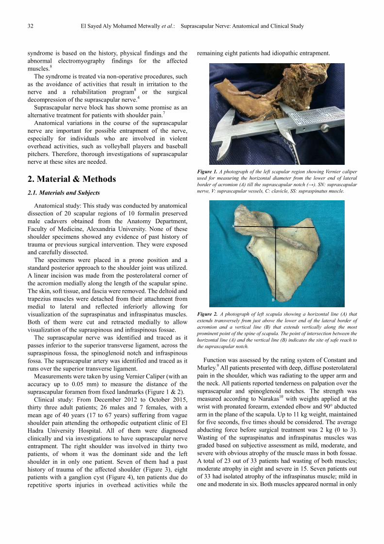
Figure 2. A photograph of left scapula showing a horizontal line (A) that
extends transversely from just above the lower end of the lateral border of
acromion and a vertical line (B) that extends vertically along the most
prominent point of the spine of scapula. The point of intersection between the
horizontal line (A) and the vertical line (B) indicates the site of safe reach to
the suprascapular notch.
Function was assessed by the rating system of Constant and
Murley.9 All patients presented with deep, diffuse posterolateral
pain in the shoulder, which was radiating to the upper arm and
the neck. All patients reported tenderness on palpation over the
suprascapular and spinoglenoid notches. The strength was
measured according to Narakas10
with weights applied at the
wrist with pronated forearm, extended elbow and 90° abducted
arm in the plane of the scapula. Up to 11 kg weight, maintained
for five seconds, five times should be considered. The average
abducting force before surgical treatment was 2 kg (0 to 3).
Wasting of the supraspinatus and infraspinatus muscles was
graded based on subjective assessment as mild, moderate, and
severe with obvious atrophy of the muscle mass in both fossae.
A total of 23 out of 33 patients had wasting of both muscles;
moderate atrophy in eight and severe in 15. Seven patients out
of 33 had isolated atrophy of the infraspinatus muscle; mild in
one and moderate in six. Both muscles appeared normal in only
International Journal of Clinical and Experimental Medical Sciences 2016; 2(3): 31-39 33
three patients.
The diagnosis was confirmed by electrophysiological
studies in all 33 patients. In 23 patients signs of nerve
degeneration were found. Isolated denervation of the
infraspinatus muscle was seen in only seven patients while the
remaining three it was inconclusive. In all patients plain
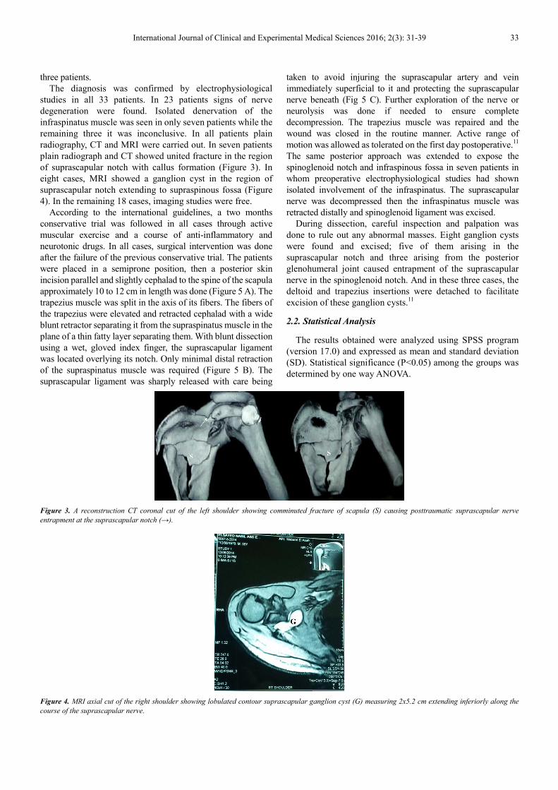
radiography, CT and MRI were carried out. In seven patients
plain radiograph and CT showed united fracture in the region
of suprascapular notch with callus formation (Figure 3). In
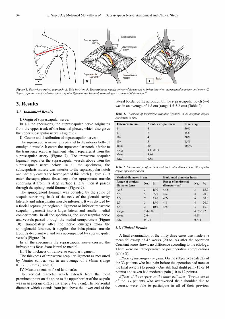
eight cases, MRI showed a ganglion cyst in the region of
suprascapular notch extending to supraspinous fossa (Figure
4). In the remaining 18 cases, imaging studies were free.
According to the international guidelines, a two months
conservative trial was followed in all cases through active
muscular exercise and a course of anti-inflammatory and
neurotonic drugs. In all cases, surgical intervention was done
after the failure of the previous conservative trial. The patients
were placed in a semiprone position, then a posterior skin
incision parallel and slightly cephalad to the spine of the scapula
approximately 10 to 12 cm in length was done (Figure 5 A). The
trapezius muscle was split in the axis of its fibers. The fibers of
the trapezius were elevated and retracted cephalad with a wide
blunt retractor separating it from the supraspinatus muscle in the
plane of a thin fatty layer separating them. With blunt dissection
using a wet, gloved index finger, the suprascapular ligament
was located overlying its notch. Only minimal distal retraction
of the supraspinatus muscle was required (Figure 5 B). The
suprascapular ligament was sharply released with care being
taken to avoid injuring the suprascapular artery and vein
immediately superficial to it and protecting the suprascapular
nerve beneath (Fig 5 C). Further exploration of the nerve or
neurolysis was done if needed to ensure complete
decompression. The trapezius muscle was repaired and the
wound was closed in the routine manner. Active range of
motion was allowed as tolerated on the first day postoperative.11
The same posterior approach was extended to expose the
spinoglenoid notch and infraspinous fossa in seven patients in
whom preoperative electrophysiological studies had shown
isolated involvement of the infraspinatus. The suprascapular
nerve was decompressed then the infraspinatus muscle was
retracted distally and spinoglenoid ligament was excised.
During dissection, careful inspection and palpation was
done to rule out any abnormal masses. Eight ganglion cysts
were found and excised; five of them arising in the
suprascapular notch and three arising from the posterior
glenohumeral joint caused entrapment of the suprascapular
nerve in the spinoglenoid notch. And in these three cases, the
deltoid and trapezius insertions were detached to facilitate
excision of these ganglion cysts.11
2.2. Statistical Analysis
The results obtained were analyzed using SPSS program
(version 17.0) and expressed as mean and standard deviation
(SD). Statistical significance (P<0.05) among the groups was
determined by one way ANOVA.
Figure 3. A reconstruction CT coronal cut of the left shoulder showing comminuted fracture of scapula (S) causing posttraumatic suprascapular nerve
entrapment at the suprascapular notch (→).
Figure 4. MRI axial cut of the right shoulder showing lobulated contour suprascapular ganglion cyst (G) measuring 2x5.2 cm extending inferiorly along the
course of the suprascapular nerve.
34 El Sayed Aly Mohamed Metwally et al.: Suprascapular Nerve: Anatomical and Clinical Study
Figure 5. Posterior surgical approach. A, Skin incision. B, Supraspinatus muscle retracted downward to bring into view suprascapular artery and nerve. C,
Suprascapular artery and transverse scapular ligament are isolated, permitting easy removal of ligament. 11
3. Results
3.1. Anatomical Results
I. Origin of suprascapular nerve:
In all the specimens, the suprascapular nerve originates
from the upper trunk of the brachial plexus, which also gives
the upper subscapular nerve. (Figure 6)
II. Course and distribution of suprascapular nerve:
The suprascapular nerve runs parallel to the inferior belly of
omohyoid muscle. It enters the suprascapular notch inferior to
the transverse scapular ligament which separates it from the
suprascapular artery (Figure 7). The transverse scapular
ligament separates the suprascapular vessels above from the
suprascapualr nerve below. In all the specimens, the
subscapularis muscle was anterior to the suprascapular notch
and partially covers the lower part of this notch (Figure 7). It
enters the supraspinous fossa deep to the supraspinatus muscle,
supplying it from its deep surface (Fig 8) then it passes
through the spinoglenoid foramen (Figure 9).
The spinoglenoid foramen was bounded by the spine of
scapula superiorly, back of the neck of the glenoid cavity
laterally and infraspinatus muscle inferiorly. It was divided by
a fascial septum (spinoglenoid ligament or inferior transverse
scapular ligament) into a larger lateral and smaller medial
compartments. In all the specimens, the suprascapular nerve
and vessels passed through the medial compartment (Figure
10). Immediately after the nerve emerges from the
spinoglenoid foramen, it supplies the infraspinatus muscle
from its deep surface and was accompanied by suprascapular
vessels (Figure 10).
In all the specimens the suprascapular nerve crossed the
infraspinous fossa from lateral to medial.
III. The thickness of transverse scapular ligament:
The thickness of transverse scapular ligament as measured
by Vernier caliber, was in an average of 9.84mm (range
8.11-11.3 mm) (Table 1).
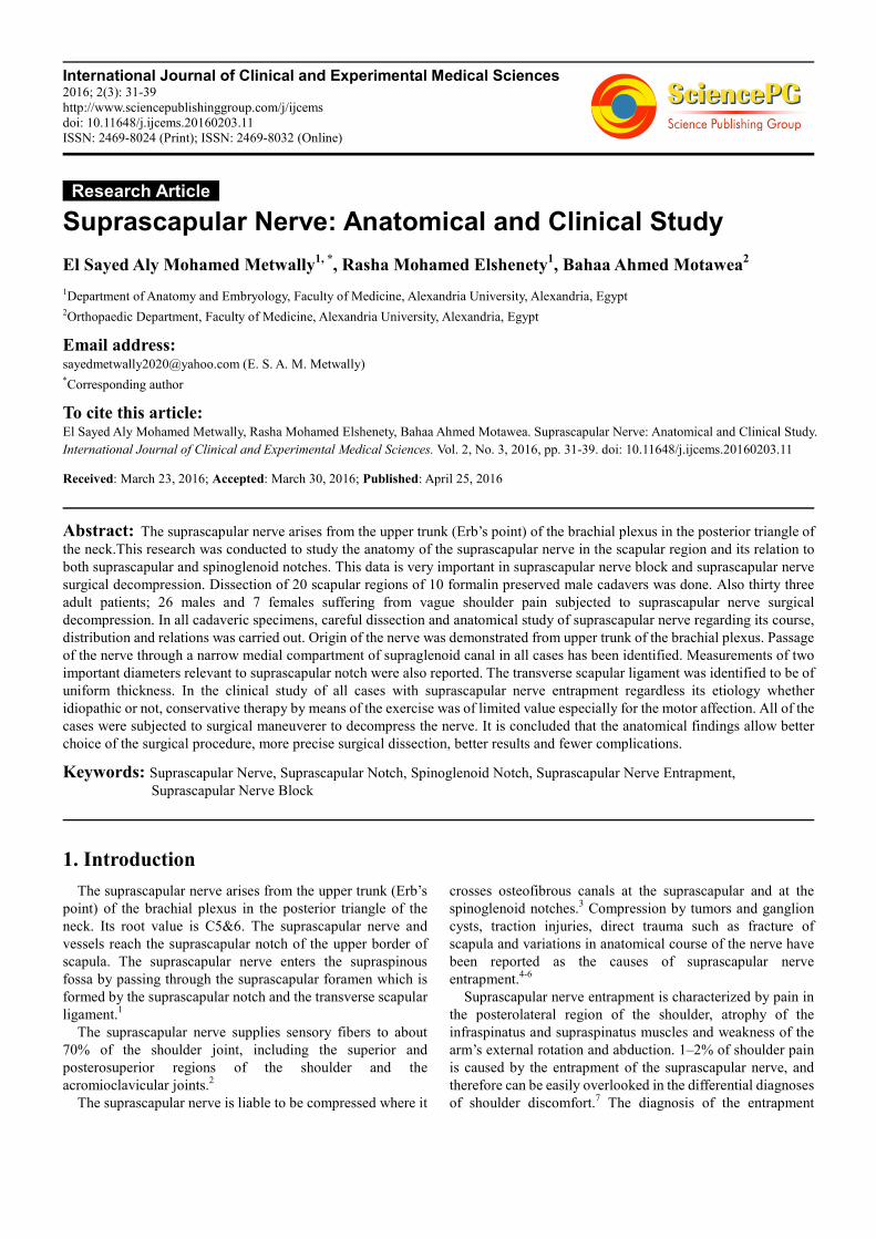
IV. Measurements to fixed landmarks:
The vertical diameter which extends from the most
prominent point on the spine to the upper border of the scapula
was in an average of 2.5 cm (range 2.4-2.8 cm). The horizontal
diameter which extends from just above the lower end of the
lateral border of the acromion till the suprascapular notch (→)
was in an average of 4.8 cm (range 4.5-5.2 cm) (Table 2).
Table 1. Thickness of transverse scapular ligament in 20 scapular region
specimens in mm.
Thickness in mm Number of specimens Percentage
8- 6 30%
9- 7 35%
10- 4 20%
11+ 3 15%
Total 20 100%
Range 8.11-11.3
Mean 9.84
S.D. 0.88
Table 2. Measurements of vertical and horizontal diameters in 20 scapular
region specimens in cm.
Vertical diameter in cm Horizontal diameter in cm
Range of vertical
diameter (cm) No. %
Range of horizontal
diameter (cm) No. %
<2.5 3 15.0 <4.6 3 15.0
2.5- 5 25.0 4.6- 4 20.0
2.6- 7 35.0 4.7- 6 30.0
2.7- 3 15.0 4.8- 4 20.0
2.8+ 2 10.0 4.9+ 3 15.0
Range 2.4-2.88 4.52-5.22
Mean 2.64 4.68
S.D. 0.123 0.811
3.2. Clinical Results
A final examination of the thirty three cases was made at a
mean follow-up of 42 weeks (20 to 94) after the operation
Constant score shows, no difference according to the etiology.
There were no intraoperative or postoperative complications
(table 3).
Effects of the surgery on pain: On the subjective scale, 25 of
the 33 patients who had pain before the operation had none at
the final review (15 points). One still had slight pain (13 or 14
points) and seven had moderate pain (10 to 12 points).
Effects of the surgery on the daily activities: Twenty seven
of the 33 patients who overexerted their shoulder due to
overuse, were able to participate in all of their previous
International Journal of Clinical and Experimental Medical Sciences 2016; 2(3): 31-39 35
activities without restriction, the remaining 6 patients were not
able to return to their previous work.
Effects of the surgery on range of movement: In 25 patients
there was a pain free range including flexion, abduction,
external and internal rotation while the remaining eight
patients had mild pain with movements.
Effects of the surgery on force of abduction: The mean force
of abduction which was attained at follow-up was 6 kg (mean
1.5 to 10). For comparison, the mean abduction force in the
contralateral unaffected shoulder was 7 kg (3 to 11) (fig 11).
Effects of the surgery on muscle wasting: Of the 25 patients
who had wasting of both muscles, 13 had full recovery of
muscle bulk, in seven the atrophy became less marked and in
five there was no noticeable atrophy of the supraspinatus
while that of infraspinatus became less marked. Isolated
atrophy of infraspinatus, which was found in seven patients
before surgery, persisted in four.
Effect of time lapse: Patients who were operated on within
six months of the onset of symptoms, showed better recovery
than those who had surgery after a longer interval.
Table 3. Clinical evaluation of the studied patients pre and post operative.
Pre operative Post operative
p No. % No. %
Pain
0.0001* No 8 24.2 25 75.8
Mild 18 54.5 7 21.2
Moderate 7 21.2 1 3.0
Daily activities
0.0021*
Normal activities 6 18.2 0 0.0
Overexerted due to
overuse 27 81.8 0 0.0
Return to previous work 0 0.0 27 81.8
Not able to work 0 0.0 6 18.2
Range of motion
0.001* No pain on motion 9 27.3 25 75.8
Pain at flexion abduction,
external and internal 24 72.7 8 24.2
Muscle wasting
0.0136*
No wasting 8 24.2 17 51.5
With wasting 25 75.8 5 15.2
Atrophy 0 0.0 7 21.2
Persisted 0 0.0 4 12.1
Force of abduction
0.254
After 6 months of follow up
Range 1.5-10.0
Mean±S.D. 6.22±3.68
In contralateral unaffected side
Range 3-11
Mean±S.D. 7.1±3.11
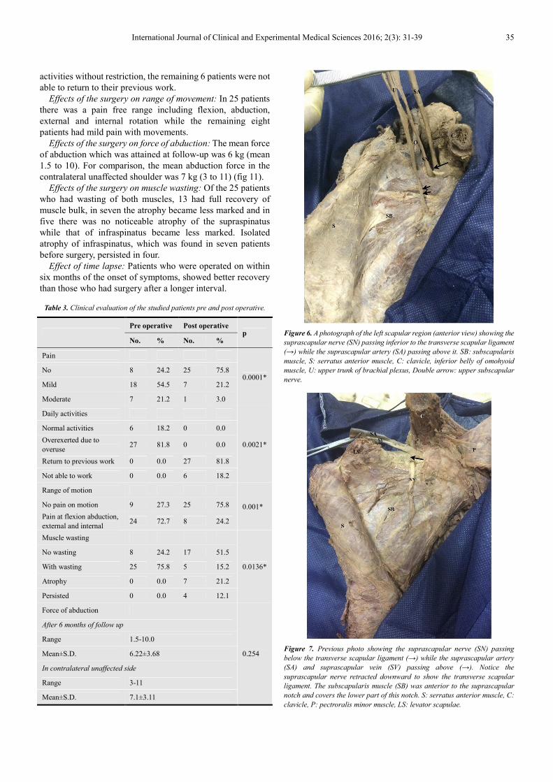
Figure 6. A photograph of the left scapular region (anterior view) showing the
suprascapular nerve (SN) passing inferior to the transverse scapular ligament
(→) while the suprascapular artery (SA) passing above it. SB: subscapularis
Figure 7. Previous photo showing the suprascapular nerve (SN) passing
below the transverse scapular ligament (→) while the suprascapular artery
(SA) and suprascapular vein (SV) passing above (→). Notice the
suprascapular nerve retracted downward to show the transverse scapular
ligament. The subscapularis muscle (SB) was anterior to the suprascapular
notch and covers the lower part of this notch. S: serratus anterior muscle, C:
clavicle, P: pectroralis minor muscle, LS: levator scapulae.
36 El Sayed Aly Mohamed Metwally et al.: Suprascapular Nerve: Anatomical and Clinical Study
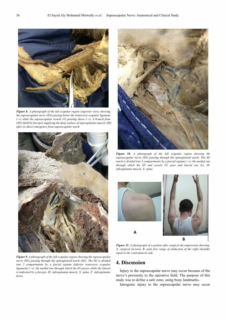
Figure 8. A photograph of the left scapular region (superior view) showing
the suprascapular nerve (SN) passing below the transverse scapular ligament
(→) while the suprascapular vessels (V) passing above (→). A branch from
(SN) (hold by forceps) supplying the deep surface of supraspinatus muscle (SS)
after its direct emergence from suprascapular notch.
Figure 9. A photograph of the left scapular region showing the suprascapular
nerve (SN) passing through the spinoglenoid notch (SG). The SG is divided
into 2 compartments by a fascial septum (inferior transverse scapular
ligament) (→); the medial one through which the SN passes while the lateral
is indicated by a forceps. IS: infraspinatus muscle, S: spine, F: infraspinatus
fossa.
Figure 10. A photograph of the left scapular region showing the
suprascapular nerve (SN) passing through the spinoglenoid notch. The SG
notch is divided into 2 compartments by a fascial septum (→); the medial one
through which the SN and vessels (V) pass and lateral one (L). IS:
infraspinatus muscle, S: spine.
Figure 11. A photograph of a patient after surgical decompression showing:
A- surgical incision, B- pain free range of abduction of the right shoulder
equal to the contralateral side.
4. Discussion
Injury to the suprascapular nerve may occur because of the
nerve’s proximity to the operative field. The purpose of this
study was to define a safe zone, using bony landmarks.
Iatrogenic injury to the suprascapular nerve may occur
International Journal of Clinical and Experimental Medical Sciences 2016; 2(3): 31-39 37
during arthroscopic repair of chronic, massive rotator cuff
tears.7-9
Prior anatomic studies of the suprascapular nerve have
attempted to define a safe zone to avoid injury during
arthroscopic transglenoid Bankart repairs, SLAP repairs, and
open surgical procedures.3, 7, 8
In the present study, the suprascapular nerve originated
from the upper trunk of the brachial plexus, which also gives
upper subscapular nerve, a finding which was in agreement
with Tasaki et al.12
In the present study, the suprascapular nerve runs parallel to
the inferior belly of omohyoid muscle. It enters the
suprascapular notch inferior to the transverse scapular
ligament which separates it from suprascapular artery.
The transverse scapular ligament separates the
suprascapular vessels above from the suprascapular nerve
below. The subscapularis muscle lies anterior to the
suprascapular notch and covers the lower part of this notch.
However, Polguj et al.13
found that the suprascapular notch
was bounded by the fascia of the supraspinatus muscle, which
is attached to the superior margin of the scapula as well as to
the superior transverse scapular ligament.
In a similar study done by Bayramoglu et al.14
the
suprascapular ligament was divided into two parts (anterior
and posterior) which differs from the results of the present
study that shows the ligament is only one mass.
Polguj et al.13
in their study on suprascapular nerve found
that the suprascapular nerve and vein pass below the ligament
in 61.3% of the specimens which differs from current findings
in which all the specimens had the suprascapular vessels
above and the suprascapular nerve below the ligament.
Bayramoglu et al.15
found that hypertrophy of the
subscapularis muscle might be an etiologic factor for
suprascapular nerve entrapment at the suprascapular notch.
This is in agreement with the results of the present study that
showed the subscapularis muscle forming the anterior
boundary of suprascapular notch.
Warner et al.16
in their study on the suprascapular nerve
stated that it runs parallel to the inferior belly of omohyoid
muscle, which is similar to the findings of the present study.
In the present study, the suprascapular nerve enters the
supraspinous fossa deep to the supraspinatus muscle,
supplying it from its deep surface, then it passes through the
spinoglenoid foramen which is bounded by the spine of
scapula superiorly, back of the neck of the glenoid cavity
laterally and infraspinatus muscle inferiorly. It is also divided
by a fascial septum (spinoglenoid ligament or inferior
transverse scapular ligament) into a larger lateral and smaller
medial compartments.
Plancher et al.17
classified spinoglenoid ligament into two
types; type 1 (a thin indistinct band of tissue) and type 2 (a
well formed ligament), a finding which is not apparent in the
present study. The ligament was seen as being of uniform
thickness and cannot be differentiated into two parts as
previously stated.
In all specimens of the present study the suprascapular
nerve and vessels passes through the medial compartment.
Immediately after emerging from the spinoglenoid foramen,
the suprascapular nerve supplies the infraspinatus muscle
from its deep surface and was accompanied by suprascapular
vessels. In all cadaveric specimens, the suprascapular nerve
crosses the infraspinous fossa from lateral to medial.
Ghodadra et al.18
in their study stated that the suprascapular
nerve release is often performed for compression neuropathy
and to release pressure on the nerve associated with
arthroscopic labral repair. This report describes a novel
arthroscopic technique for decompression of the suprascapular
nerve at the suprascapular notch or spinoglenoid notch
through a subacromial approach. Through the subacromial
space, spinoglenoid notch cysts can be visualized between the
supraspinatus and infraspinatus at the base of the scapular
spine this approach is explained by the finding of the present
study that the spinoglenoid foramen is bounded by the spine of
scapula superiorly, back of the neck of the glenoid cavity
laterally and infraspinatus muscle inferiorly.
In the present study, the thickness of transverse scapular
ligament as measured was in an average of 9.84 mm (range:
8.11-11.3 mm). The vertical diameter which extends from the
most prominent point on the spine to the upper border of
scapula was in an average of 2.5 cm (range 2.4-2.8 cm). The
horizontal diameter which extends from just above the lower
end of the lateral border of acromion till the suprascapular
notch was in an average of 4.8 cm (range 4.5-5.2 cm)
Ruotolo et al.19
in their study concluded that the thickness
thickness of transverse scapular ligament was 7 -8mm,
afinding which is near to the present study.
Ide et al.20
classified the ligament as either ligament- or
membrane-type depending on its thickness. The width of the
ligament and its maximal distance to bone and nerve were
measured. Among the 115 specimens, the ligament was absent
in 21 cases (18.3%), the ligament-type was present in 25
(21.7%), and the membrane-type was present in 69 (60.0%).
The ligament width varied from 1.8 to 9.0 mm (mean, 5.4 mm),
the maximal distance from ligament to bone varied from 3.0 to
11.1 mm (mean, 5.7 mm), and the distance from ligament to
the nerve was from 0.1 to 7.0 mm (mean, 3.1 mm). As there
was considerable variation in the distances from the ligament
to the bone and nerve, the ligament may play a role in paralysis
of the infraspinatus muscle, depending on these distances.
These findings are different from the present study and this
could be explained by the small sample size of the present
study.
Warner et al.16
evaluated the limits within which lateral
mobilization of chronic massive retracted rotator cuff tears can
be performed during open procedures without risking
neurovascular injury. Even with delineation of these safe
zones, however, iatrogenic injury to the suprascapular nerve
during open rotator cuff repair and clavicle excision has been
reported.
Knowledge of the distance from the suprascapular notch to
the lateral border of the acromion may be used to establish a
safe zone for anterolateral portal placement. Bigliani et al.,21
described a safe zone enabling surgeons to avoid the
suprascapular nerve during arthroscopic Bankart repair and
38 El Sayed Aly Mohamed Metwally et al.: Suprascapular Nerve: Anatomical and Clinical Study
open surgical procedures. This safe zone, located in the
posterior glenoid neck, measured 2 cm in diameter at the level
of the supraglenoid tubercle and 1cm in diameter at the level
of the scapular spine. A finding which is near the findings of
the present study and the meauresments of the present study
are easy to perform.
Woolf et al.22
described the safety of the superior-medial
portal, citing a mean distance of 2.42 cm from the
suprascapular nerve. While this measurement is helpful in
determining a safe distance from the suprascapular nerve to
this single portal, many surgeons employ multiple
arthroscopic portals for rotator cuff repairs.
Shishido and Kikuchi23
described a safe zone for avoiding
suprascapular nerve injury in open dissection of the posterior
shoulder joint and arthroscopic procedures for Bankart repair
in which blind drilling is involved.
In the present study of the 25 patients who had wasting of
both muscles, 13 had full recovery of muscle bulk, in seven the
atrophy became less marked and in the remaining five patients
there was no noticeable atrophy of the supraspinatus while that
of infraspinatus became less marked. Isolated atrophy of
infraspinatus, which was found in seven patients before
surgery, persisted in four of them.
25 of the 33 patients who had pain before the operation had
none at the final review, One still had slight pain and seven had
moderate pain. 27 of the 33 patients who overexerted their
shoulder due to overuse, were able to participate in all of their
previous activities without restriction, the remaining 6 patients
were not able to return to their previous work. In 25 patients
there was a pain free range including flexion, abduction,
external and internal rotation while the remaining eight
patients had mild pain with movements. Patients who were
operated on within six months of the onset of symptoms,
showed better recovery than those who had surgery after a
longer interval.
Researchers24-30
found that if there is evidence of muscular
atrophy or severe pain, not controlled by drugs, the operation
should not be delayed beyond three months.
Martin et al.31
stated that, the patients who were operated on
within six months of the onset of symptoms, showed better
recovery than those who had surgery after a longer interval
and that operative excision is indicated in patients whose
symptoms result from compression of the nerve by a ganglion.
In the present study, regardless of the cause of suprascapular
nerve compression the delay in operation within 6 months had
better recovery.
Black and Lombardo32
in their study found that if the patient
experiences a decrease in pain or increase in strength, and the
electrophysiological findings show improvement during the
preoperative period, the operation should be postponed or
cancelled. When this occurs, the patient will usually recover
spontaneously, particularly if the entrapment follows trauma.
5. Conclusion
Overviewing the results of the present study, careful
investigation should be done as compression of the
suprascapular nerve may be at the suprascapular notch or
spinoglenoid notch or it may be idiopathic. Patients who were
operated upon within six months of the onset of symptoms,
showed better recovery than those who had surgery after a
longer interval and that operative excision is indicated in
patients whose symptoms result from compression of the
nerve by a ganglion.
Conflict of Interest
None declared.
References
[1] Standring S. Gray's Anatomy: The Anatomical Basis of Clinical Practice. 40th ed. London: Longmans, Green and Co, 2008: 1123–1124.
[2] Neeta VK. Clinical anatomy 2nd edition. Superficial muscles of the back and scapular region. 2012; 13: 111-112.
[3] Ritchie ED, Tong D, Chung F, Norris AM, Miniaci A, Vairavanathan SD. Suprascapular nerve block for postoperative pain relief in arthroscopic surgery: a new modality. Anesth Analg. 1997; 84: 1306–1312.
[4] Brown DE, James DC, Roy S. Pain relief by suprascapular nerve block in glenohumeral arthritis. Scand J Rheumatol. 1988; 17: 411–415.
[5] Green S, Buchbinder R, Glazier R, Forbes A. Systematic review of randomised controlled trials of interventions for painful shoulder: selection criteria, outcome assessment, and efficacy. BMJ. 1998; 316: 354–360.
[6] Van der Heijden GJ, van der Windt DA, Kleijnen J, Koes BW, Bouter LM. Steroid injections for shoulder disorders: a systematic review of randomized clinical trials. Br J Gen Pract. 1996; 46: 309–316.
[7] Van der Winddt DA, van der Heijden GJ, Scholten RJ, Koes
BW, Bouter LM. The efficacy of non-steroidal
anti-inflammatory drugs (NSAIDS) for shoulder complaints. A
International Journal of Clinical and Experimental Medical Sciences 2016; 2(3): 31-39 39
[13] Polguj M, Jędrzejewski K, Majos A, Topol M. Variations in bifid superior transverse scapular ligament as a possible factor of suprascapular entrapment: an anatomical study. Int Orthop. 2012; 36(10): 2095–2100.
[14] Bayramoğlu A, Demiryürek D, Tüccar E, et al. Variations in anatomy at the suprascapular notch possibly causing suprascapular nerve entrapment: an anatomical study. Knee Surg Sports Traumatol Arthrosc. 2003; 11(6): 393-398.
[15] Bayramoglu A, Demiryurek D, Erbil M, Aktekin M, Tetik O, Doral M N. Hypertrophy of the subscapularis muscle might be an etiologic factor for suprascapular nerve entrapment at the suprascapular notch. Neuroanatomy. 2002; 1: 5-6.
[16] Warner J P; Krushell R J; Masquelet A; Gerber C. Anatomy and relationships of the suprascapular nerve: anatomical constraints to mobilization of the supraspinatus and infraspinatus muscles in the management of massive rotator-cuff tears. J Bone Joint Surg Am. 1992; 74 (1): 36-45.
[17] Plancher KD, Peterson RK, Johnston JC, Luke TA. The spinoglenoid ligament. Anatomy, morphology, and histological findings J Bone Joint Surg Am. 2005; 87: 361-365.
[18] Ghodadra N, Nho SJ, Verma NN, et al. Arthroscopic decompression of the suprascapular nerve at the spinoglenoid notch and suprascapular notch through the subacromial space. Arthroscopy. 2009; 25: 439-445.
[19] Ruotolo CL, Fow JE, Nottage WM. The supraspinatus footprint: an anatomic study of the supraspinatus insertion. Arthroscopy. 2004; 20(3): 246-249.
[20] Ide J, Maeda S, Takagi K. Does the inferior transverse scapular ligament cause distal suprascapular nerve entrapment? An anatomic and morphologic study. J Shoulder Elbow Surg. 2003; 12(3): 253-255.
[21] Bigliani L. U, Dalsey R. M., McCann P. D., April E. W. An anatomical study of the suprascapular nerve. Arthroscopy. 1990; l6; (4), 301–305.
[22] Woolf S. K., Guttmann D., Karch M. M., Graham R. D, Reid B., Lubowitz J. H. The superior-medial shoulder arthroscopy portal is safe. Arthroscopy. 2007: 23: 247–250.
[23] Shishido H and Kikuchi S. Injury of the suprascapular nerve in shoulder surgery: an anatomic study. Journal of Shoulder &Elbow Surgery. 2001; 10(4): 372–376.
[24] Vastam¨aki M, G¨oransson H. Suprascapular nerve entrapment. Clin Orthop. 1993; 297: 135-143.
[25] Emery P, Wedderburn L, Grahame R. Suprascapular nerve block for shoulder pain in rheumatoid arthritis. BMJ. 1989; 299: 1079–1080.
[26] Post M, Mayer J. Suprascapular nerve entrapment: diagnosis and treatment. Clin Orthop. 1987; 223: 126-136.
[27] Callahan JD, Scully TB, Scott A, Shapiro SA, Worth RM. Supra- scapular nerve entrapment: a series of 27 cases. J Neurosurg. 1991; 74: 893-896.
[28] Ferretti A, Cerullo G, Russo G. Suprascapular neuropathy in volley-ball players. J Bone Joint Surg [Am] 1987; 69-A: 260-263.
[29] Ringel SP, Treihaft M, Carry M, Fisher R, Jacobs P. Suprascapular neuropathy in pitchers. Am J Sports Med. 1990; 18: 80-86.
[30] Fritz RC, Helms CA, Steinbach LS, Genant HK. Suprascapular nerve entrapment: evaluation with MR imaging. Radiology. 1992; 182: 437-444.
[31] Martin SD, Warren RF, Martin TL, et al. Suprascapular neuropathy: results of non-operative treatment. J Bone Joint Surg [Am]. 1997; 79-A: 1159-1165.
[32] Black KP, Lombardo JA. Suprascapular nerve injuries with isolated paralysis of the infraspinatus. Am J Sports Med. 1990; 18: 225-228.