research papers IUCrJ (2018). 5, 309–324 https://doi.org/10.1107/S2052252518003317 309 IUCrJ ISSN 2052-2525 CHEMISTRY j CRYSTENG Received 11 December 2017 Accepted 26 February 2018 Edited by L. R. MacGillivray, University of Iowa, USA Keywords: triamterene; pK a analysis; crystal engineering; liquid-assisted grinding; pharma- ceutical salt solvates; hydrogen bonding; motif analysis; stable duplex structures. CCDC references: 1579683; 1579684; 1579685; 1579686; 1579687; 1579688; 1579689 Supporting information: this article has supporting information at www.iucrj.org Structural studies of crystalline forms of triamterene with carboxylic acid, GRAS and API molecules Abida Rehman, a Amit Delori, b * David S. Hughes a * and William Jones a a Department of Chemistry, University of Cambridge, Lensfield Road, Cambridge, Cambridgeshire CB2 1EW, England, and b Strathclyde Institute of Pharmacy and Biomedical Sciences (SIPBS), University of Strathclyde, 161 Cathedral Street, Glasgow G4 0RE, Scotland. *Correspondence e-mail: [email protected], [email protected]Pharmaceutical salt solvates (dimethyl sulfoxide, DMSO) of the drug triamterene with the coformers acetic, succinic, adipic, pimelic, azelaic and nicotinic acid and ibuprofen are prepared by liquid-assisted grinding and solvent-evaporative crystallization. The modified pK a rule as proposed by Cruz-Cabeza [(2012). CrystEngComm, 14, 6362–6365] is in close agreement with the results of this study. All adducts were characterized by X-ray diffraction and thermal analytical techniques, including single-crystal X-ray diffraction, powder X-ray diffraction, differential scanning calorimetry and thermal gravimetric analysis. Hydrogen-bonded motifs combined to form a variety of extended tapes and sheets. Analysis of the crystal structures showed that all adducts existed as salt solvates and contained the aminopyridinium–carboxylate heterodimer, except for the solvate containing triamterene, ibuprofen and DMSO, as a result of the presence of a strong and stable hemitriamterenium duplex. A search of the Cambridge Structural Database (CSD 5.36, Version 1.18) to determine the frequency of occurrence of the putative supramolecular synthons found in this study showed good agreement with previous work. 1. Introduction The compound 2,4,7-triamino-6-phenylpteridine (C 12 H 11 N 7 ), named triamterene (1), is a potassium-sparing diuretic and modest inhibitor of dihydrofolate reductase. It is marketed under the name of Dyrenium (Well Spring Pharmaceutical Corporation, Concordia Pharmaceutical Inc., Carilion Mate- rials Management and GlaxoSmithKline Inc.) for the treat- ment of oedema (water retention) and, when combined with hydrochlorthiazide as Maxzide (Mylan Pharmaceuticals) and Dyazide (Cardinal Health and SmithKline Beecham), for the treatment of hypertension (high blood pressure). This drug has poor solubility in water and a low bioavail- ability (45 mg ml 1 ) (Dittert et al., 1964), and is therefore an excellent candidate for a cocrystal or salt screen (Thakuria et al., 2013; Duggirala et al., 2016). A recent study (Ma et al., 2013) found two polymorphs of the salt of 1 with cucurbit[7]uril (CB[7]). The powder of the CB[7] salt forms a stable complex with 1 in aqueous solution with improved dissolution and solubility by a factor of 1.6 > 1 in 0.1 M hydrochloric acid. The increased solubility and consequent oral bioavailability of 1 in this case was attributed to the formation of a hydrophilic cylinder composed of two hydro- philic portals containing carbonyl functional groups, in which two molecules of 1 are encapsulated to form a stable complex. As an alternative approach, adduct (cocrystal or salt) forma- tion with carboxylic acids (Bhatt et al., 2009), Generally Regarded As Safe (GRAS) molecules (Delori et al., 2008;
Structural studies of crystalline forms oftriamterene with carboxylic acid, GRAS and APImolecules
Abida Rehman,a Amit Delori,b* David S. Hughesa* and William Jonesa
aDepartment of Chemistry, University of Cambridge, Lensfield Road, Cambridge, Cambridgeshire CB2 1EW, England, andbStrathclyde Institute of Pharmacy and Biomedical Sciences (SIPBS), University of Strathclyde, 161 Cathedral Street,
the M7 motif, also with graph-set notation R22ð8Þ. DMSO
interacts with two molecules of 1 via two N—H� � �O hydrogen
bonds, in addition to an intermolecular N—H� � �N bond
between adjacent molecules of 1, which creates the M5 motif
with R23ð8Þ graph-set notation. Motifs M7, M6, M3, M6 and M7
combine to form TM16, and M5, M4 and M5 combine to form
TM17 (see Fig. 4a for details).
These tape motifs create a supramolecular tape of 1 mole-
cules (containing additional acid and solvent molecules) that is
research papers
IUCrJ (2018). 5, 309–324 Abida Rehman et al. � Crystalline salt solvates of triamterene with carboxylic acid 313
Figure 3(a) Motifs M2, M1 and M2 (tape motif TM15) and motif M3 observed in1. (b) The view along c illustrating the relationship between adjacenttapes using the M3 motif (side view). (c) Packing diagram of the steppedhydrogen-bonded sheets.
interlinked to an adjacent tape of 1 through (C—H� � �O) weak
hydrogen bonds that create voids in the crystal structure (Fig.
4a). Around each void, hydrogen-bonded molecules of 1
create A and B cyclic networks. In the A cyclic network, two
DMSO molecules occupy the void; however, in the B network,
the acid molecules protrude into the void.
The expansion of these combined (A and B) cyclic networks
in two dimensions results in the formation of a hydrogen-
bonded sheet formed by stepped tapes similar to that seen in 1
and shown in Fig. 4(b) using a space-filling representation.
3.3. Salt solvate of triamterene with succinic acid and DMSO(1b�DMSO)
1b�DMSO crystallizes from DMSO solution as a salt solvate
in the monoclinic P21/c space group (see Table 1). The
asymmetric unit contains one molecule each of 1, b and
DMSO (1:1:1). The asymmetric unit of 1b�DMSO is shown in
Fig. S4 and the hydrogen bonds are given in Table S7. It can be
seen from the asymmetric unit that only one of the two
carboxylic acid groups of b is deprotonated and the H atom is
transferred to the most basic atom (N1) of 1; the other
carboxyl group is involved in an intramolecular O—H� � �O�
hydrogen bond and is therefore not available for deproton-
ation.
The crystal structure of 1b�DMSO arises from the formation
of two types of hydrogen-bonded network, namely A, which is
a ten-membered cyclic hydrogen-bonded network, and B, a
six-membered cyclic hydrogen-bonded network similar to that
seen in 1a�DMSO. Furthermore, the A network involves two
sets of three molecules (trimers) of 1 linked by the M8 motif
that interact with two b molecules through the M7 motif
present in 1a�DMSO (see Fig. 5). Two DMSO molecules act as
a bridge between 1 and b creating the M9 motif and
completing the ten-membered cyclic network. The cavity
created by the A network is filled by two phenyl rings of 1 and,
if these phenyl rings are flipped away from the cavity, voids of
12.7 � 9.0 A are created. In cyclic network B, two single
molecules of 1 connect with two molecules of b, creating two
M7 motifs, and the two DMSO molecules protrude into the
cavity formed by 1, with b acting as a bridge, joined to 1 by
hydrogen bonds and completing the six-membered cyclic
network (Fig. 5). We note that in the crystal structure the
cyclic networks are made by 1, b and solvent molecules
(Fig. 5), as opposed to only 1 and a in 1a�DMSO. Motifs M7,
M10, M8 and M9 combine to create the tape motif TM18, M8
and M9 form TM19, and M7, M10 and M8 form TM20, and so
complete the hydrogen-bond description of the tape structure
found in 1b�DMSO.
Further expansion of the hydrogen-bonded tape structure
in the second dimension results in a sheet structure in which
rows of M8 motifs of 1 are interconnected through carboxylic
research papers
314 Abida Rehman et al. � Crystalline salt solvates of triamterene with carboxylic acid IUCrJ (2018). 5, 309–324
Figure 4(a) Motifs M7, M6, M3, M6 and M7 (TM16), and M5, M4 and M5 (TM17)found in the crystal structure of 1a�DMSO. (b) The host–guest network inthe form of stepped tapes forming a sheet structure.
Figure 5The cyclic hydrogen-bonded networks A and B of the crystal structure of1b�DMSO.
Figure 6Motifs M7, M10, M8 and M9 (TM18), M8 and M9 (TM19), and M7, M10and M8 (TM20) observed in the hydrogen-bonded sheets of 1b�DMSO.
acid and solvent molecules by weak hydrogen bonds (see
Fig. 6 for details).
3.4. Salt solvate of triamterene with adipic acid and DMSO(1c�DMSO)
Crystallization of 1 and c from DMSO generated a 2:1 salt
(21+�c2�) together with two molecules of DMSO in the triclinic
space group P�11 (see Table 1); c acts as a proton donor by the
deprotonation of both protons of the carboxylic acids. The
asymmetric unit is shown in Fig. S5 and contains two mole-
cules of 1, one of c and two of DMSO (2:1:2). One DMSO
molecule is found to be disordered over two sites with occu-
pancies of 0.7 and 0.3. For the purposes of the crystal structure
refinement, geometric constraints and a common isotropic
displacement parameter for the non-H atoms were applied to
both. A list of hydrogen bonds for 1c�DMSO is given in
Table S8.
The crystal structure is a host–guest assembly, with the
framework created by molecules of 1 and c, as shown in Figs.
7(a) and 7(b). The interaction between 1 and c is through the
M7 motif seen in earlier structures, while the molecules of 1
interlink through M3 and M4 motifs (also present in
1a�DMSO) to form two sets of three molecules (trimers)
which link to form sheets. The trimers (tapes) also connect
with each other through two acid molecules by hydrogen
bonding. This results in the formation of an eight-membered
cyclic host network with a cavity of 10.0 � 15.3 A containing
the two molecules of DMSO, as shown in Fig. 7(b).
Motifs M5, M4 and M5 (TM16), and M7, M6, M3, M6 and
M7 (TM17) create the hydrogen-bonded network structure
(see Fig. 8). Such host–guest assemblies are well known in the
field of supramolecular chemistry (Bhatt et al., 2009; Galcera et
al., 2012).
3.5. Salt solvate of triamterene with pimelic acid (1d�DMSO)
The molecular adduct of 1d�DMSO crystallizes in the
triclinic P�11 space group (see Table 1) as a salt between 1 and d
in which the acid is found to be doubly deprotonated, trans-
ferring protons to two separate molecules of the most basic
atom (N1) of 1. The asymmetric unit of the crystal structure is
shown in Fig. S6 and contains four molecules of 1 (A, B, C and
D), two molecules of d (E and F) and two molecules of DMSO
(G). Hydrogen-bond details for 1d�DMSO are given in
Table S9.
In the crystal structure, molecules of 1 are connected via
N—H� � �N hydrogen bonds joined by M8 motifs to form
trimers. The acid molecules act as spacers between the trimers
joined by the M7 motif and create a zigzag crinkled supra-
molecular sheet (Fig. 9a), as seen in 1b�DMSO (see Fig. 6). As
noted earlier, this tape is fundamentally different from the
supramolecular tape found in 1c�DMSO (see Fig. 7) since, in
this case, motifs M3 and M4 are not involved in creating a
trimer of 1 molecules.
Expansion of the supramolecular tape in the second
dimension results in a cyclic eight-membered host network,
creating a cavity of 14.9 � 9.1 A (Fig. 9a). This cavity is filled
research papers
IUCrJ (2018). 5, 309–324 Abida Rehman et al. � Crystalline salt solvates of triamterene with carboxylic acid 315
Figure 7(a) The eight-membered hydrogen-bonded cyclic host network with acavity of 15.3 � 10.0 A dimensions and (b) the cavity filled with twoDMSO molecules as guests in 1c�DMSO.
Figure 8Motifs M5, M4 and M5 (TM16), and M7, M6, M3, M6 and M7 (TM17)found in the hydrogen-bonded sheets of 1c�DMSO.
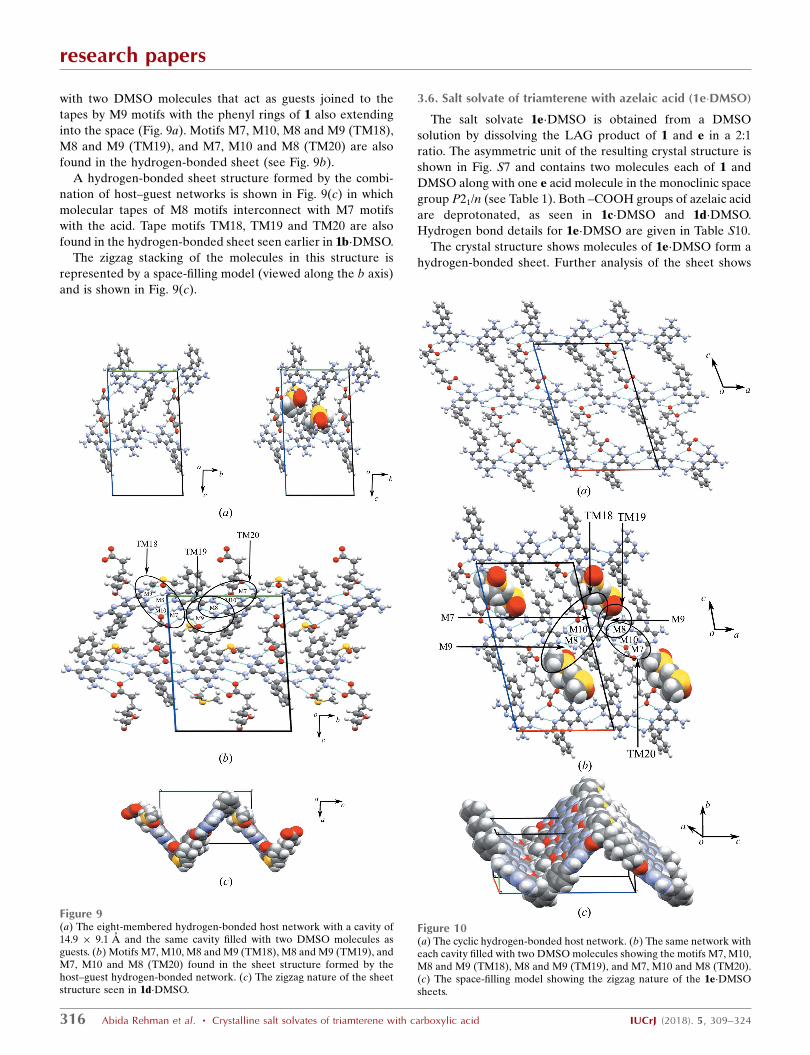
with two DMSO molecules that act as guests joined to the
tapes by M9 motifs with the phenyl rings of 1 also extending
into the space (Fig. 9a). Motifs M7, M10, M8 and M9 (TM18),
M8 and M9 (TM19), and M7, M10 and M8 (TM20) are also
found in the hydrogen-bonded sheet (see Fig. 9b).
A hydrogen-bonded sheet structure formed by the combi-
nation of host–guest networks is shown in Fig. 9(c) in which
molecular tapes of M8 motifs interconnect with M7 motifs
with the acid. Tape motifs TM18, TM19 and TM20 are also
found in the hydrogen-bonded sheet seen earlier in 1b�DMSO.
The zigzag stacking of the molecules in this structure is
represented by a space-filling model (viewed along the b axis)
and is shown in Fig. 9(c).
3.6. Salt solvate of triamterene with azelaic acid (1e�DMSO)
The salt solvate 1e�DMSO is obtained from a DMSO
solution by dissolving the LAG product of 1 and e in a 2:1
ratio. The asymmetric unit of the resulting crystal structure is
shown in Fig. S7 and contains two molecules each of 1 and
DMSO along with one e acid molecule in the monoclinic space
group P21/n (see Table 1). Both –COOH groups of azelaic acid
are deprotonated, as seen in 1c�DMSO and 1d�DMSO.
Hydrogen bond details for 1e�DMSO are given in Table S10.
The crystal structure shows molecules of 1e�DMSO form a
hydrogen-bonded sheet. Further analysis of the sheet shows
research papers
316 Abida Rehman et al. � Crystalline salt solvates of triamterene with carboxylic acid IUCrJ (2018). 5, 309–324
Figure 10(a) The cyclic hydrogen-bonded host network. (b) The same network witheach cavity filled with two DMSO molecules showing the motifs M7, M10,M8 and M9 (TM18), M8 and M9 (TM19), and M7, M10 and M8 (TM20).(c) The space-filling model showing the zigzag nature of the 1e�DMSOsheets.
Figure 9(a) The eight-membered hydrogen-bonded host network with a cavity of14.9 � 9.1 A and the same cavity filled with two DMSO molecules asguests. (b) Motifs M7, M10, M8 and M9 (TM18), M8 and M9 (TM19), andM7, M10 and M8 (TM20) found in the sheet structure formed by thehost–guest hydrogen-bonded network. (c) The zigzag nature of the sheetstructure seen in 1d�DMSO.
that molecules of 1 are joined by M8 motifs to form tapes, as
shown in Fig. 10(a). These tapes are held together by mole-
cules of azelaic acid through M7 motifs utilizing charge-
and M9 (TM18), M8 and M9 (TM19), and M7, M10 and M8
(TM20) motifs create the hydrogen-bonded network. This
association creates an eight-membered cyclic network with
voids of 15 � 9.3 A which are filled by two phenyl rings of 1
molecules along with the two DMSO molecules that act as
guests (see Fig. 10b).
Stacking of these cyclic networks (viewed along the c axis)
results in the zigzag hydrogen-bonded sheet structure shown
in Fig. 10(c), and these sheets, in turn, stack using van der
Waals forces to create the complete three-dimensional crystal
structure.
3.7. Salt solvate of triamterene with nicotinic acid(1f�DMSO)
The salt obtained by dissolving a 2:1 LAG (DMSO) sample
of 1 and f crystallizes as a DMSO solvate in the monoclinic
space group P21/n (see Table 1). The resulting asymmetric unit
of the crystal structure is shown in Fig. S8 and contains five
molecules: two molecules of 1, one molecule of f and two
DMSO molecules. In this case, one of the molecules of 1 is
protonated (A) while the other remains neutral (B). A list of
hydrogen bonds for 1f�DMSO is included in Table S11.
The crystal structure shows that in the supramolecular tape
formed between 1, f and DMSO, A and B molecules are
connected by M8 motifs in an alternating AB–AB fashion to
produce a basic structural ribbon of 1 molecules to which f
molecules are attached through the common M7 motif
involving N—H� � �O and N+—H� � �O� hydrogen bonds.
DMSO molecules are connected through M9 motifs, as shown
in Fig. 11(a). Motifs M7, M10, M8 and M9 (TM18), M8 and M9
(TM19), and M7, M10 and M8 (TM20) create the hydrogen-
bonded tape seen in Fig. 11(a).
Extension of this tape in the second dimension, allows
connection between adjacent tapes using weak C—H� � �O
hydrogen bonds to produce the hydrogen-bonded sheet shown
in Fig. 11(b). Further analysis reveals that the tapes of 1
molecules are stacked in a staggered fashion with the hydro-
phobic phenyl groups arranged at a maximum distance from
each other (Fig. 12a). Essentially, A molecules interact with
the –COO� group of nicotinic acid using the M7 motif and an
N—H� � �N hydrogen bond occurs between the N atom of
nicotinic acid and the 2-amino group of a B molecule with an
H� � �N distance of 2.30 A, as shown in Fig. 12(b).
In the third dimension, molecules are arranged as parallel
zigzag stacked sheets, as seen in the space-filling diagram
shown in Fig. 13.
3.8. Salt solvate of triamterene with ibuprofen (1g�DMSO)
The salt obtained between 1 and g crystallizes as a DMSO
solvate in the triclinic space group P�11, as shown in Table 1. It is
obtained by dissolving a mixture of 1 and g in a 2:1 ratio in
DMSO. The asymmetric unit is shown in Fig. S9 and contains
four molecules in total: two molecules of 1 along with one each
of g and DMSO. As in 1f�DMSO, one of the molecules of 1 is
research papers
IUCrJ (2018). 5, 309–324 Abida Rehman et al. � Crystalline salt solvates of triamterene with carboxylic acid 317
Figure 12(a) Stacking of molecules of 1 to form double hydrogen-bonded chainsthrough (b) nicotinic acid linkages in 1f�DMSO.
Figure 11(a) The motifs M7, M10, M8 and M9 (TM18), M8 and M9 (TM19), andM7, M10 and M8 (TM20) that create the hydrogen-bonded tape and (b)sheet that forms part of the crystal structure of 1f�DMSO.
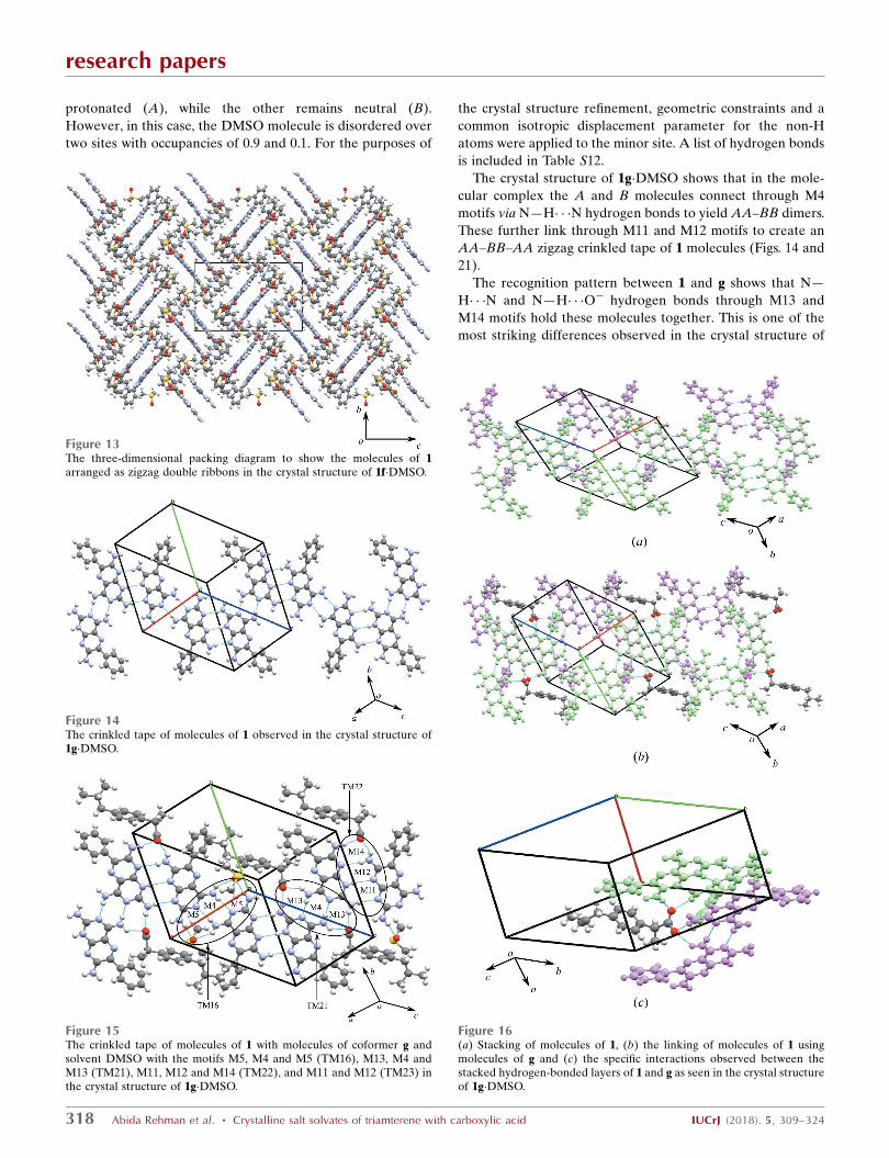
protonated (A), while the other remains neutral (B).
However, in this case, the DMSO molecule is disordered over
two sites with occupancies of 0.9 and 0.1. For the purposes of
the crystal structure refinement, geometric constraints and a
common isotropic displacement parameter for the non-H
atoms were applied to the minor site. A list of hydrogen bonds
is included in Table S12.
The crystal structure of 1g�DMSO shows that in the mole-
cular complex the A and B molecules connect through M4
motifs via N—H� � �N hydrogen bonds to yield AA–BB dimers.
These further link through M11 and M12 motifs to create an
AA–BB–AA zigzag crinkled tape of 1 molecules (Figs. 14 and
21).
The recognition pattern between 1 and g shows that N—
H� � �N and N—H� � �O� hydrogen bonds through M13 and
M14 motifs hold these molecules together. This is one of the
most striking differences observed in the crystal structure of
research papers
318 Abida Rehman et al. � Crystalline salt solvates of triamterene with carboxylic acid IUCrJ (2018). 5, 309–324
Figure 15The crinkled tape of molecules of 1 with molecules of coformer g andsolvent DMSO with the motifs M5, M4 and M5 (TM16), M13, M4 andM13 (TM21), M11, M12 and M14 (TM22), and M11 and M12 (TM23) inthe crystal structure of 1g�DMSO.
Figure 13The three-dimensional packing diagram to show the molecules of 1arranged as zigzag double ribbons in the crystal structure of 1f�DMSO.
Figure 14The crinkled tape of molecules of 1 observed in the crystal structure of1g�DMSO.
Figure 16(a) Stacking of molecules of 1, (b) the linking of molecules of 1 usingmolecules of g and (c) the specific interactions observed between thestacked hydrogen-bonded layers of 1 and g as seen in the crystal structureof 1g�DMSO.
1g�DMSO in comparison with all adducts described above. In
the previous cases, the M7 motif is found between molecules
of 1 and all previous coformers containing the –COOH group
of a–f. However, in this case, the M7 motif is absent since
molecules of 1 combine to form the M11 motif, as shown in
Fig. 15. At the other end of the combined motif, two molecules
of 1 combine with one O atom of the carboxyl group to form
the M14 motif. The DMSO molecules interconnect with 1
through the M5 motif, as observed in 1a�DMSO and
1c�DMSO. The motifs M5, M4 and M5 (TM16), M13, M4 and
M13 (TM21), M11, M12 and M14 (TM22), and M11 and M12
(TM23) make up the hydrogen-bonded tape structure seen in
1g�DMSO (see Fig. 15 for details).
Further analysis reveals staggered
stacking of the tapes of 1 molecules with
the hydrophobic phenyl groups
arranged at a maximum distance from
each other (Fig. 16a). The stacks of 1
connect with each other through g
molecules involving motifs M13 and
M14, which results in the formation of
pseudo-cyclic networks (as shown in
Fig. 16b and highlighted in Fig. 16c).
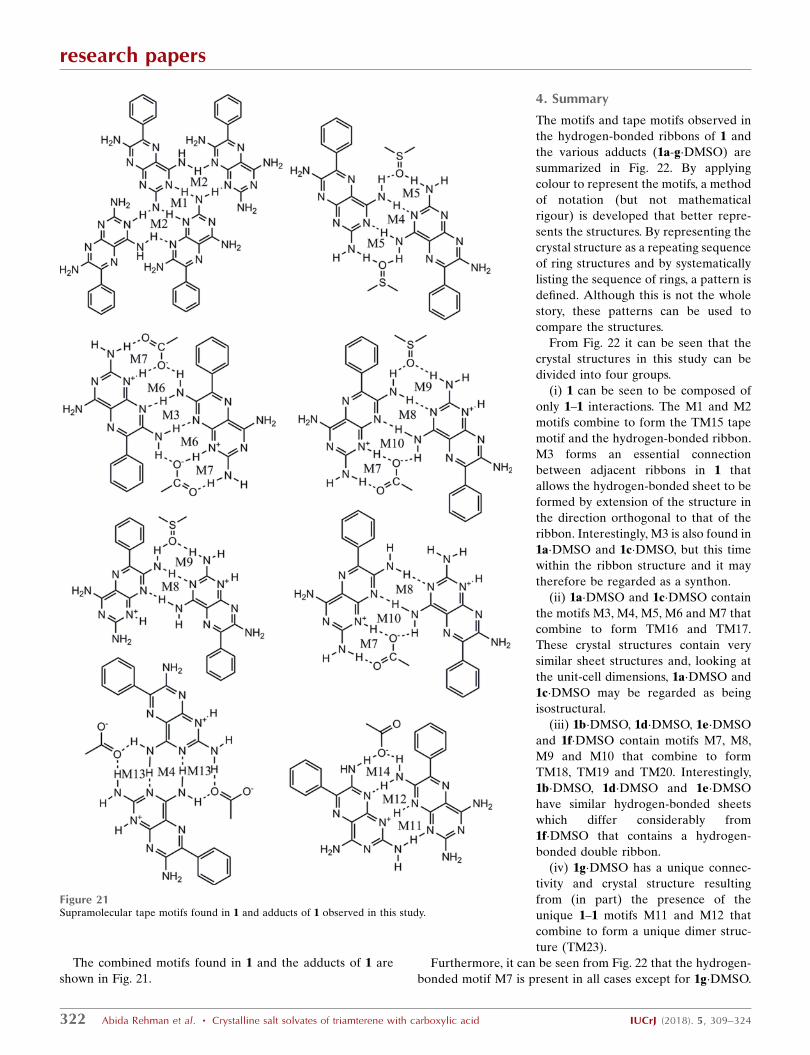
3.9. Analysis of the hydrogen-bondedmotifs in 1 and the reported adducts(1a–g)
Overall, 14 different motifs have been
observed in 1 and the reported adducts
1a–g�DMSO (see Figs. 17 and 19). It is
recognized that the standard graph-set
notation Gad(n) introduced by Etter
(1991) and described in Bernstein et al.
(1995) is not sufficient to accurately
describe the interactions found in this
study, since various ways of forming the
same graph set are possible depending
upon the specific functional groups
involved.
As a result, the concepts of motif and
supramolecular synthon have been
developed and are used throughout this
discussion. Additionally, combined
(tape) motifs are identified because of
the combination of individual motifs in
different structural arrangements.
A survey of the CSD (Version 5.36;
ConQuest Version 1.18) was conducted
in order to better understand the like-
lihood of these motifs and putative
supramolecular synthons occurring in
other adducts similar in molecular
structure to those found in this study.
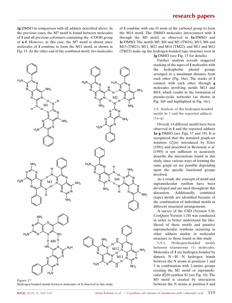
3.9.1. Hydrogen-bonded motifsbetween triamterene (1) molecules.Molecules of 1 are hydrogen bonded by
dimeric N—H� � �N hydrogen bonds
between the N atoms at positions 1 and
3 in combination with 2-amino groups
creating the M1 motif or supramole-
cular R22 8ð Þ synthon S1 (see Fig. 18). The
M3 motif is created by interaction
between the N atoms at position 8 and
research papers
IUCrJ (2018). 5, 309–324 Abida Rehman et al. � Crystalline salt solvates of triamterene with carboxylic acid 319
Figure 17Hydrogen-bonded motifs between molecules of 1 observed in this study.
the 7-amino groups of molecules of 1 on adjacent tapes and
may be described as the supramolecular R22 8ð Þ synthon S1. The
other homodimeric motifs only found in the salt solvates
between molecules of 1 are M4 and M8 and it is clear from Fig.
17 that they all originate by utilizing different positions of the
1 molecule, but also that they may all be represented by the
supramolecular R22 8ð Þ synthon S1.
The M2 motif joins three molecules of 1 utilizing the N
atoms at positions 1, 3 and 8, and the 2- and 4-amino groups,
and creates the supramolecular R33ð10) synthon S2 (see
Fig. 18).
The M11 and M12 motifs are unique in this study and exist
between two molecules of 1 in 1g�DMSO, one of which is
protonated (A) while the other is neutral (B). M11 and M12
both possess N+—H� � �N and N—H� � �N hydrogen bonds and
are represented by the supramolecular
R22 8ð Þ synthon S3 (Fig. 18).
Although they have the same graph-
set descriptor and indeed the same
hydrogen bonds, they represent
different supramolecular synthons since
they utilize different positions on the
molecules of 1 involved in their crea-
tion.
The seven motifs found between 1
molecules reported here are shown in
Fig. 17.
A CSD analysis undertaken by
Delori, Suresh & Pedireddi (2013)
revealed that molecules containing the
N1—C1—NH2 functionality had a high
propensity to form supramolecular
synthon S1. In the present study, a
search of the CSD shows that, in the
solid state, 52.97% (989 out of 1867) of
the crystal structures containing this
functionality do form supramolecular
synthon S1. Indeed, the largest number
of motifs (M1, M3, M4 and M8)
between molecules of 1 in this study are
represented by this supramolecular
synthon (see Fig. 18). Supramolecular
synthon S2 is extremely rare and found
in only two structures (0.21%) out of
the 918 possible crystal structures in the
CSD. Finally, the supramolecular
synthon S3 is represented by 1.96% (25
structures out of a possible 1277)
suitable crystal structures in the CSD.
The various putative supramolecular
synthons described in this section are
shown in Fig. 18.
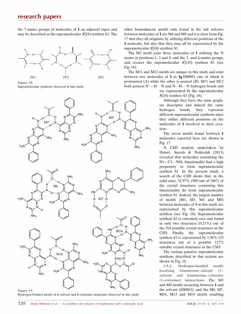
3.9.2. Hydrogen-bonded motifsinvolving triamterene–solvent (1–solvent) and triamterene–coformer(1–coformer) interactions. The M5
and M9 motifs occurring between 1 and
the solvent (DMSO), and the M6, M7,
M10, M13 and M14 motifs resulting
research papers
320 Abida Rehman et al. � Crystalline salt solvates of triamterene with carboxylic acid IUCrJ (2018). 5, 309–324
Figure 19Hydrogen-bonded motifs of 1–solvent and 1–coformer molecules observed in this study.
Figure 18Supramolecular synthons observed in this study.
from the interaction between 1 and the coformers (a–g) are
shown in Fig. 19.
M5 involves the interaction of three molecules including the
O atom of DMSO using two N—H� � �O hydrogen bonds with
the 2- and 4-amino groups of different molecules of 1 and a
further N—H� � �N hydrogen bond between the N3 atom and
the 4-amino group of adjacent molecules of 1. M5 may be
represented by the supramolecular R23 8ð Þ synthon S4 as seen in
Fig. 20 and although M9 is also represented by the supramo-
lecular R23 8ð Þ synthon S4, it clearly differs from M5 in the
position of one of the amino groups on the molecule of 1.
The M6 and M10 motifs seen in Fig. 19 and represented by
the supramolecular R23 8ð Þ synthon S5 in Fig. 20 are formed
through charge-assisted N+—H� � �O�and neutral N—H� � �N
hydrogen bonds between 1 and the carboxyl group of the acid.
Both of these motifs utilize the protonated N atom of 1 at
position 1 and the N atom at position 8, but the N—H� � �N
hydrogen bond has a different composition in each motif,
involving the 7-amino group for M6 and the 4-amino group for
M10.
The unique M13 motif or supramolecular R23 8ð Þ synthon S6
(see Fig. 20) is only found in 1g�DMSO. This motif results from
an N—H� � �N hydrogen bond between atom N3 and the 4-
amino group, along with two N—H� � �O hydrogen bonds
involving the 2-amino group of one molecule of 1 and the 4-
amino group of the other. Similarly, the unique M14 motif or
tape motifs and 7 supramolecular synthons are reported and
their structural relationships explored. The anomalous beha-
viour of 1 g�DMSO is explained by the inability of ibuprofen
to disrupt the stable hemitriamterenium duplex.
Acknowledgements
DSH would like to thank the Department of Chemistry at the
University of Cambridge for granting the visiting status that
allowed him to develop many of the ideas proposed in this
paper. The authors would like to acknowledge Dr John E.
Davies for collecting and processing the single-crystal X-ray
data used in this study. Professor Joel Bernstein is also
thanked for helpful correspondence concerning motif analysis
and graph sets and Dr Neil Feeder for helpful discussions on
motif and synthon searches of the CSD.
Funding information
AR would like to thank the Islamic Development Bank, in
collaboration with the Saudi Arabia and Cambridge
Commonwealth Trust, for the award of a studentship to study
at the University of Cambridge. AD would like to thank the
Pfizer Institute for Pharmaceutical Materials Science for
funding.
References
Aitipamula, S., Chow, P. S. & Tan, R. B. H. (2014). CrystEngComm,16, 3451–3465.
Aitipamula, S., Vangala, V. R., Chow, P. S. & Tan, R. B. H. (2012).Cryst. Growth Des. 12, 5858–5863.
Aitipamula, S., Wong, A. B. H., Chow, P. S. & Tan, R. B. H. (2012).CrystEngComm, 14, 8515–8524.
research papers
IUCrJ (2018). 5, 309–324 Abida Rehman et al. � Crystalline salt solvates of triamterene with carboxylic acid 323
Figure 22Summary of hydrogen-bond motifs found in this study. Green represents 1–1 interactions, blue represents 1–solvent interactions, red represents 1–coformer interactions and purple represents solvent–1–coformer interactions.
Allen, F. H., Samuel Motherwell, W. D., Raithby, P. R., Shields, G. P. &Taylor, R. (1999). New J. Chem. 23, 25–34.
Altomare, A., Cascarano, G., Giacovazzo, C. & Guagliardii, A.(1993). J. Appl. Cryst. 26, 343–350.
Arhangelskis, M., Lloyd, G. O. & Jones, W. (2012). CrystEngComm,14, 5203–5208.
Bathori, N. B., Lemmerer, A., Venter, G. A., Bourne, S. A. & Caira,M. R. (2011). Cryst. Growth Des. 11, 75–87.
Bernstein, J., Davis, R. E., Shimoni, L. & Chang, N.-L. (1995). Angew.Chem. Int. Ed. Engl. 34, 1555–1573.
Bhatt, P. M., Azim, Y., Thakur, T. S. & Desiraju, G. R. (2009). Cryst.Growth Des. 9, 951–957.
Bruker (2001). SADABS. Bruker AXS Inc., Madison, Wisconsin,USA.
Bruker (2007). SAINT. Bruker AXS Inc., Madison, Wisconsin, USA.Bucar, D. K., Elliott, J. A., Eddleston, M. D., Cockcroft, J. K. & Jones,
W. (2015). Angew. Chem. 127, 251–255.Bucar, D. K., Filip, S., Arhangelskis, M., Lloyd, G. O. & Jones, W.
(2013). CrystEngComm, 15, 6289–6291.Cheney, M. L., Shan, N., Healey, E. R., Hanna, M., Wojtas, L.,
Zaworotko, M. J., Sava, V., Song, S. J. & Sanchez-Ramos, J. R.(2010). Cryst. Growth Des. 10, 394–405.
Cheney, M. L., Weyna, D. R., Shan, N., Hanna, M., Wojtas, L. &Zaworotko, M. J. (2010). Cryst. Growth Des. 10, 4401–4413.
Childs, S. L., Stahly, G. P. & Park, A. (2007). Mol. Pharm. 4, 323–338.Cruz-Cabeza, A. J. (2012). CrystEngComm, 14, 6362–6365.Delori, A., Galek, P. T. A., Pidcock, E. & Jones, W. (2012). Chem. Eur.
J. 18, 6835–6846.Delori, A., Galek, P. T. A., Pidcock, E., Patni, M. & Jones, W. (2013).
CrystEngComm, 15, 2916–2928.Delori, A., Maclure, P., Bhardwaj, R. M., Johnston, A., Florence, A. J.,
Sutcliffe, O. B. & Oswald, I. D. H. (2014). CrystEngComm, 16,5827–5831.
Delori, A., Suresh, E. & Pedireddi, V. R. (2008). Chem. Eur. J. 14,6967–6977.
Delori, A., Suresh, E. & Pedireddi, V. R. (2013). CrystEngComm, 15,4811–4815.
Delori, A., Urquhart, A. J. & Oswald, I. D. H. (2016). CrystEng-Comm, 18, 5360–5364.
Desiraju, G. R. (1995). Angew. Chem. Int. Ed. Engl. 34, 2311–2327.Dittert, L. W., Higuchi, T. & Reese, D. R. (1964). J. Pharm. Sci. 53,
1325–1328.Duggirala, N. K., Perry, M. L., Almarsson, O., Orn, & Zaworotko, M.
J. (2016). Chem. Commun. 52, 640–655.Etter, M. C. (1991). J. Phys. Chem. 95, 4601–4610.Etter, M. C. & Reutzel, S. M. (1991). J. Am. Chem. Soc. 113, 2586–
2598.Etter, M. C., Urbanczyk-Lipkowska, Z., Zia-Ebrahimi, M. &
Panunto, T. W. (1990). J. Am. Chem. Soc. 112, 8415–8426.Fabian, L. (2009). Cryst. Growth Des. 9, 1436–1443.Friscic, T. & Jones, W. (2010). J. Pharm. Pharmacol. 62, 1547–1559.Galcera, J., Friscic, T., Hejczyk, K. E., Fabian, L., Clarke, S. M., Day,
G. M., Molins, E. & Jones, W. (2012). CrystEngComm, 14, 7898–7906.
Galek, P. T. A., Fabian, L. & Allen, F. H. (2010). CrystEngComm, 12,2091–2099.
Galek, P. T. A., Fabian, L., Motherwell, W. D. S., Allen, F. H. &Feeder, N. (2007). Acta Cryst. B63, 768–782.
Grobelny, P., Mukherjee, A. & Desiraju, G. R. (2011). CrystEng-Comm, 13, 4358–4364.
Groom, C. R., Bruno, I. J., Lightfoot, M. P & Ward, S. C. (2016). Acta.Cryst. B72, 171–179.
Grothe, E., Meekes, H., Vlieg, E., ter Horst, J. H. & de Gelder, R.(2016). Cryst. Growth Des. 16, 3237–3243.
Hilal, S. H., Karickhoff, S. W. & Carreira, L. A. (1995). Quant. Struct.-Act. Relat. 14, 348–355.
Huang, Y. T., Zhang, B. W., Gao, Y., Zhang, J. J. & Shi, L. M. (2014). J.Pharm. Sci. 103, 2330–2337.
Hughes, D. S., Delori, A., Rehman, A. & Jones, W. (2017). Chem.Cent. J. 11, 63–82.
Li, A. Y., Xu, L. L., Chen, J. M. & Lu, T. B. (2015). Cryst. Growth Des.15, 3785–3791.
Lu, J. & Rohani, S. (2010). J. Pharm. Sci. 99, 4042–4047.Ma, W. J., Chen, J. M., Jiang, L., Yao, J. & Lu, T. B. (2013). Mol.
Pharm. 10, 4698–4705.Macrae, C. F., Bruno, I. J., Chisholm, J. A., Edgington, P. R., McCabe,
P., Pidcock, E., Rodriguez-Monge, L., Taylor, R., van de Streek, J. &Wood, P. A. (2008). J. Appl. Cryst. 41, 466–470.
Molcanov, K. & Kojic-Prodic, B. (2010). CrystEngComm, 12, 925–939.Pallipurath, A., Skelton, J. M., Delori, A., Duffy, C., Erxleben, A. &
Jones, W. (2015). CrystEngComm, 17, 7684–7692.Perumalla, S. R., Pedireddi, V. R. & Sun, C. C. (2013). Cryst. Growth
Des. 13, 429–432.Sanphui, P., Goud, N. R., Khandavilli, U. B. R., Bhanoth, S. & Nangia,
A. (2011). Chem. Commun. 47, 5013–5015.Schwalbe, C. H. & Williams, G. J. B. (1987). Acta Cryst. C43, 1097–
1100.Sheldrick, G. M. (2015). Acta Cryst. C71, 3–8.Sowa, M., Slepokura, K. & Matczak-Jon, E. (2012). Acta Cryst. C68,
262–265.Spek, A. L. (2009). Acta Cryst. D65, 148–155.Stahl, P. H. & Wermuth, C. G. (2008). In Handbook of Pharmaceutical
Salts: Properties, Selection, and Use. Weinheim: Wiley.Thakuria, R., Delori, A., Jones, W., Lipert, M. P., Roy, L. &
Rodrıguez-Hornedo, N. (2013). Int. J. Pharm. 453, 101–125.
Trask, A. V., Motherwell, W. D. S. & Jones, W. (2005). Cryst. GrowthDes. 5, 1013–1021.
Trask, A. V., Motherwell, W. D. S. & Jones, W. (2006). Int. J. Pharm.320, 114–123.
Tutughamiarso, M. & Bolte, M. (2007). Private communication(refcode: FITZAJ01). CCDC, Cambridge, England.
Varughese, S., Azim, Y. & Desiraju, G. R. (2010). J. Pharm. Sci. 99,3743–3753.
Wood, P. A., Feeder, N., Furlow, M., Galek, P. T. A., Groom, C. R. &Pidcock, E. (2014). CrystEngComm, 16, 5839–5848.
Yan, D. P., Patel, B., Delori, A., Jones, W. & Duan, X. (2013). Cryst.Growth Des. 13, 333–340.
research papers
324 Abida Rehman et al. � Crystalline salt solvates of triamterene with carboxylic acid IUCrJ (2018). 5, 309–324