Role o Regulating C A dissertation in pa Dep of Gut Commensal Micro Colonic Sensory-Related S By Mònica Aguilera Pujabet artial fulfilment of the requirements for the d Philosophy Neuroscience Doctoral Programme partment of Cell Biology, Physiology and Immuno Neuroscience Institute Universitat Autònoma de Barcelona Advisor Dr. Vicente Martínez Perea Bellaterra, Barcelona 2014 obiota Systems degree of Doctor of ology
Transcript
Role of Gut Commensal Microbiota
Regulating Colonic Sensory
A dissertation in partial fulfilment of the requirements for the degree of Doctor
Department of Cell Biology, Physiology and Immunology
Role of Gut Commensal Microbiota
Regulating Colonic Sensory-Related Systems
By
Mònica Aguilera Pujabet
A dissertation in partial fulfilment of the requirements for the degree of Doctor
Philosophy
Neuroscience Doctoral Programme
Department of Cell Biology, Physiology and Immunology
Neuroscience Institute
Universitat Autònoma de Barcelona
Advisor
Dr. Vicente Martínez Perea
Bellaterra, Barcelona
2014
Role of Gut Commensal Microbiota
Related Systems
A dissertation in partial fulfilment of the requirements for the degree of Doctor of
Department of Cell Biology, Physiology and Immunology
VICENTE MARTÍNEZ PEREA,
Associate Professor of Physiology at the Department of Cell Biology,
Physiology and Immunology; Universitat Autònoma de Barcelona.
I hereby certify that the thesis entitled “Role of gut commensal microbiota
regulating colonic sensory-related systems”, submitted by MÒNICA AGUILERA
PUJABET in partial fulfillment of the requirements for the degree of Doctor of
Philosophy, was carried out under my supervision and I authorize the submission to
undertake its oral defense.
In witness whereof, I hereby sign this document.
Bellaterra, Barcelona, September 2014
Vicente Martínez Perea, DVM, PhD
Ph.D. Advisor
This work has been funded by the Spanish “Ministerio de Ciencia e Innovación”, project
BFU2009-08229 and the FPI program (reference BES-2010-037699; personal support to Mònica
Aguilera Pujabet).
Experience is simply the name we give our mistakes
Oscar Wilde (1854 – 1900)
Irish writer and poet
There's Plenty of Room at the Bottom
Richard P. Feynman (1918 – 1988)
American theoretical physicist
AGRAÏMENTS
Han passat 5 anys des de que vaig entrar el departament i no me n’he adonat de com ha
passat el temps. Hi ha hagut estones de tot però m’emporto un molt bon record i molta
experiència guanyada, en tots el sentits.
En primer lloc, voldria agrair al Vicente, el meu director de tesi, l’ajuda donada i la
confiança que ha anat dipositant en mi al llarg del temps. He après molt al teu costat i estic molt
contenta de què hagis estat tu el meu director de tesi. El treball és en gran part mèrit teu. Gràcies.
A la Patri, pel seminari de fisiologia de segon de carrera parlant dels ratolins, tenies raó, i
per haver-me introduït al departament i al món de l’animal de laboratori.
A Silvia Melgar, por tu amabilidad, ayuda, enseñanza y confiança. La estància en Cork fué
más agradable en parte gracias a ti. Tack!
No voldria deixar de mencionar al personal del Fisiologia de la Facultat de Veterinària, ja
que en menor o major mesura, també heu contribuït en aquesta tesi. Als professors Ester, Marcel i
Maite. No vull oblidar els tècnics i administratius Antonio i David, pels riures (no cal que diguem
gràcies a qui... ☺), l’ajuda i la bona cara en tot aquest temps. La següent persona, i no podria ser
una altra, és l’Emma. Gràcies pels riures, consells, ajuda i confidències i també pel “¿qué haces?”
està clar! I pels riures que ens has causat, que no són pocs.
I ara ja venen el becaris/residents. En general em coneixeu bastant i ja sabeu què penso,
com ho penso i perquè ho penso. No caldria que ho deixés per escrit, però com a bona tradició,
què menys que deixar plasmades quatre paraules d’agraïment. Cronològicament, gràcies Sandra,
sempre seràs la compi de FISH i per l’amistat trobada. Al Joan Antoni perquè sempre seràs el meu
“sènior” preferit i per l’ajuda em vas donar des del principi i que has mantingut. A l’Elena Tàpia,
por lo compartido dentro y fuera de fisio y lo que queda. Al Joan Burgueño, per la sinceritat,
l’ajuda i les converses al 127. Fes-me cas en alguna cosa! Al Sergio, perquè ets un tros de pa i per
les converses del despatx que tinc més presents del que et penses. Al Javi, per ser el meu “júnior”,
per l’ajuda donada, les converses de despatx i perquè espero que em facis cas i no siguis tant
pessimista! A la Míriam, per ser companya de foscor del 143, cosa que ha ajudat a descobrir una
amistat. A la Marina, perquè ets de les persones que més em coneixen i sempre seràs la meva
CAP. Confia en tu. A la Noe, perquè ets un sol i transmets més del què tu creus. I , finalment, tot i
haver treballat o coincidit menys, no voldria deixar de mencionar al Paco, l’Eva, l’Elena Eyre, la
Claudia, la Diana, el Jakub i l’Asun.
Finalment, vull agrair a la meva família, i sobretot a la Montse i al Marc. Ja sabeu que sou
pilars de la meva vida. Gràcies per aguantar-me, estar allà i fer-me feliç.
Mònica Aguilera Pujabet
Bellaterra, Setembre de 2014
TABLE OF CONTENTS Abbreviations .............................................................................................................................. 13
Gut commensal microbiota (GCM) is a key component of gastrointestinal homeostasis.
Functional gastrointestinal disorders (mainly irritable bowel syndrome, IBS) and inflammatory bowel diseases (IBD) have been related to states of altered GCM (dysbiosis). Simultaneously, IBS and IBD
patients show local states of abnormal immune activation with altered motor and sensory responses.
In particular, in IBS patients sensory alterations lead to characteristic states of visceral hypersensitivity.
The exact causal role of GCM remains unclear, but the presence of dysbiosis and the positive effects of
antibiotics and some probiotics suggest a key role for the microbiota.
The present work explores the potential role of gut microbiota affecting visceral pain-related
sensory systems within the gut and the effects on nociceptive responses. For this purpose, states of real (spontaneous adaptive microbial changes, antibiotic treatment-derived microbial changes) or
simulated (direct stimulation of host-bacterial interaction systems) colonic dysbiosis were generated in
rats and mice. Colonic microbiota (luminal and wall-adhered) was characterized by fluorescent in situ
hybridization (FISH) and qPCR. The immune status of the colon and bacterial-host interactions were
determined assessing the expression (RT-qPCR) of pro- and anti-inflammatory cytokines; antimicrobial
peptides, integrins and Toll-like receptors (TLRs), the production of secretory IgA (s-IgA), the presence
of histopathological alterations and the state of the mucous barrier. Simultaneously, changes in
sensory related markers were also assessed. Changes in viscerosensitivity were determined in
conscious mice using the Writhing test or following the intracolonic administration of capsaicin.
Overall, antibiotics-induced alterations of the GCM, but not spontaneous changes associated
to environmental adaptation, generated a state of local immune activation within the colon. This state
was characterized by selective up- and down-regulation of pro- and anti-inflammatory cytokines and host-bacterial interaction markers and changes in the amounts of s-IgA. Similar immune response was
observed when a dysbiotic state was simulated in rats by the direct stimulation of colonic TLR4 with
bacterial lipopolysaccharides (LPS) or TLR7 with the selective agonist imiquimod. Although these
changes, and regardless the model considered, no macroscopical or microscopical signs of colonic
inflammation were detected. In both, mice and rats, real or simulated colonic dysbiotic states were
also associated to a local modulation of sensory-related markers (endocannabinoid, serotonergic, opioid and vanilloid systems), with specific treatment-related up- and down-regulatory responses (RT-
qPCR and immunohistochemistry). These variations at the molecular level translated in functional
changes as it relates to visceral pain-related responses. In mice with antibiotic-induced dysbiosis,
visceral pain responses assessed using the Writhing test or the intracolonic administration of capsaicin
were significantly attenuated when compared to non-dysbiotic animals; thus suggesting a hypoalgesic state. Moreover, colonic contractility assessed in vitro (organ bath) was also altered in dysbiotic mice,
indicating a state of increased colonic motility.
Generally, results obtained show that during states of dysbiosis of the GCM there is a complex
host response that implies a local immune activation, probably directed towards the reshaping of the
microbiota. Data obtained shows that the microbiota is able to influence gut sensory systems and that
these changes translate at a functional level in the modulation of visceral pain, eliciting, at least in the
present experimental conditions, analgesic-like responses. Similar underlying mechanisms might be responsible for the beneficial effects observed in IBD and, particularly, in IBS patients during antibiotic
treatments or during the use of certain bacterial strains as probiotics. Further studies should address
the characterization of the specific bacterial groups implicated in these effects. These results highlight
the importance of the microbiota as pathogenic factor in gastrointestinal disorders and its potential as
a therapeutic approach.
17
RESUMEN
La microbiota comensal del intestino se considera un factor clave en la homeostasis
gastrointestinal. Las alteraciones funcionales gastrointestinales (síndrome del intestino irritable, SII) y las
enfermedades inflamatorias intestinales (EII) se han relacionado con alteraciones de la microbiota comensal
(disbiosis). Estos pacientes muestran una activación inmune local anormal, con respuestas motoras y
sensoriales alteradas, que en el SII se traducen en estados de hipersensibilidad visceral. El papel causal de la
microbiota no se conoce con exactitud, pero la presencia de disbiosis y los efectos positivos asociados al
tratamiento con antibióticos o ciertos probióticos sugieren un papel destacado.
Este trabajo explora la importancia de la microbiota intestinal modulando los sistemas sensoriales
intestinales relacionados con el dolor visceral y sus efectos en respuestas nociceptivas viscerales. Para ello,
se ha trabajado con ratas y ratones en los cuales se ha inducido un estado real (cambios adaptativos
espontáneos, cambios inducidos por tratamiento con antibióticos) o simulado (estimulación directa de
sistemas de interacción hospedador-microbiota) de disbiosis cólica. La microbiota (luminal y adherida al
epitelio) se caracterizó usando hibridación in situ con sondas fluorescentes (FISH) y qPCR. La respuesta
inmune local y los mecanismos de interacción hospedador-microbiota se valoraron determinando cambios
en la expresión génica (RT-qPCR) de citoquinas, péptidos antimicrobianos, integrinas y receptores de tipo
Toll (TLR), la producción de IgA secretora, alteraciones histopatológicas y el estado de la barrera de moco.
Simultáneamente, se evaluaron cambios en la expresión de marcadores sensoriales. La sensibilidad visceral
se valoró mediante el test de Writhing o la administración intracólica de capsaicina.
La disbiosis cólica inducida por antibióticos, pero no la observada durante un proceso de adaptación
espontánea al ambiente, se asoció a un estado de activación inmune local caracterizado por una regulación
selectiva (tanto al alza como a la baja) de citoquinas pro-inflamatorias y de marcadores de interacción
hospedador-microbiota y por cambios en los niveles luminales de IgA. Respuestas similares se observaron
cuando se simuló al simular un estado de disbiosis mediante la estimulación local del TLR4 (lipopolisacárido)
o del TLR7 (imiquimod). En ningún caso se observaron signos macroscópicos o microscópicos de colitis.
Tanto en la rata como en el ratón, los estados de disbiosis cólica, real o simulada, se asociaron a una
modulación local de la expresión de marcadores sensoriales (sistemas endocanabinoide, serotonérgico,
opioide y vaniloide). Se observaron tanto regulaciones al alza como a la baja (RT-qPCR/inmunohistoquímica)
dependiendo del modelo de disbiosis y del marcador sensorial considerado. Estos cambios moleculares se
tradujeron en cambios funcionales relacionados con respuestas nociceptivas viscerales. Así, en ratones con
disbiosis cólica inducida con antibióticos, las repuestas de dolor visceral determinadas con el test de
Writhing o tras la administración intracólica de capsaicina mostraron una atenuación significativa con
respecto a las observadas en animales control, sugiriendo un estado de hipoalgesia. Estos animales
mostraron además una contractilidad cólica alterada (baño de órganos), indicativa de un estado de
hipermotilidad.
Estos resultados muestran que en estados de disbiosis intestinal se produce una activación inmune
local, dirigida, probablemente, a la restauración de la composición de la microbiota. Se observa que la
microbiota es capaz de modular la actividad de los sistemas sensoriales intestinales, generando cambios
funcionales que se traducen, en las condiciones experimentales presentes, en una modificación de las
respuestas de dolor visceral compatible con un estado de tipo analgésico. Mecanismos similares podrían
explicar los efectos beneficiosos asociados al tratamiento con antibióticos o al uso de probióticos
observados en pacientes con SII o EII. Estudios posteriores deberían centrarse en la caracterización de los
grupos bacterianos específicamente responsables de estos efectos. Estos resultados muestran la
importancia de la microbiota como factor patogénico en las alteraciones gastrointestinales y su interés
como aproximación terapéutica para las mismas.
19
RESUM
La microbiota comensal de l’intestí es considera un factor clau en la homeòstasis gastrointestinal.
Les alteracions funcionals gastrointestinals (la síndrome de l’intestí irritable, SII) i la malaltia inflamatòria
intestinal (MII) s’han relacionat amb alteracions de la microbiota comensal (disbiosi). Els pacients d’aquestes
malalties presenten una activació del sistema immune local anormal, amb respostes motores i sensorial
alterades, que, en el SII, es tradueixen en un estat d’hipersensibilitat visceral. El paper causal de la
microbiota no es coneix amb exactitud, però la presència de disbiosi i els efectes positius associats al
tractament amb determinats antibiòtics o probiòtics en suggereixen un paper destacat.
Aquest treball explora la importància de la microbiota intestinal modulant els sistemes sensorials
del mateix relacionats amb el dolor visceral i els efectes en respostes nociceptives viscerals. Per aquest
motiu, s’ha treballat amb rates i ratolins, en els quals se’ls ha induït un estat real (per canvis adaptatius
espontanis, o pel tractament amb antibiòtics) o simulat (estimulació directa de sistemes d’interacció hoste-
microbiota) de disbiosi colònica. La microbiota (luminal i adherida a l’epiteli) es va caracteritzar mitjançant
hibridació in situ fluorescent (FISH)i qPCR. La resposta immune local i els mecanismes d’interacció hoste-
microbiota es van valorar determinant canvis en l’expressió gènica (RT-qPCR) de citocines, pèptids
antimicrobians, integrines i receptors de tipo-toll (TLR), la producció d’IgA secretada, avaluant alteracions
histopatològiques i l’estat de la barrera del moc intestinal. Simultàniament, es van valorar canvis en
l’expressió de marcadors sensorials. Finalment, la sensibilitat visceral es va determinar mitjançant el test de
Writhing o l’administració intracolònica de capsaïcina.
La disbiosi colònica induïda amb antibiòtics, però no la observada durant processos d’adaptació
espontània a l’ambient, es va associar a un estat d’activació immune local caracteritzat per una regulació
selectiva (tant a l’alça com a la baixa) de citocines pro-inflamatòries i de marcadors d’interacció hoste-
microbiota i, per canvis en els nivells luminals d’IgA. Respostes similars es van detectar al simular un estat de
disbiosi mitjançant l’estimulació local del TLR4 (amb lipopolisacàrid) o del TLR7 (amb imiquimod). En cap cas
es van observar signes macroscòpics/microscòpics de colitis. Tant en la rata com en el ratolí, els estats de
disbiosi colònica, real o simulada, s’associen a una modulació local de l’expressió de marcadors sensorials
(principalment dels sistemes endocanabinoide, serotoninèrgic, opioide i vaniloide). Depenent del model de
disbiosi i del marcador sensorial considerat, aquesta modulació va implicar canvis, tant a l’alça com a la
baixa (RT-qPCR/immunohistoquímica). Els canvis moleculars es van traduir en canvis funcionals relacionats
amb respostes nociceptives viscerals. Tanmateix, en ratolins amb disbiosi colònica induïda amb antibiòtics,
les respostes de dolor visceral, determinades amb el test de Writhing o amb l’administració intracolònica de
capsaïcina, van mostrar una atenuació significativa respecte a les mostrades pels animals control, suggerint
un estat d’hipoalgèsia. A més a més, aquests animals van mostrar la contractilitat colònica alterada (amb
bany d’òrgans), assenyalant un estat d’hipermotilitat.
Aquests resultats assenyalen que en estats de disbiosi intestinal es produeix un activació immune
local, dirigida, probablement, a la restauració de la composició de la microbiota comensal. Sembla que la
microbiota es capaç de modular l’activitat dels sistemes sensorials intestinals, generant canvis funcionals
que es tradueixen, en les condicions experimentals presents, en una modificació de les respostes de dolor
visceral compatible amb un estat analgèsic. Mecanismes similars podrien explicar els efectes beneficiosos
associats al tractament amb antibiòtics o a l’ús de probiòtics descrits en pacients amb SII o MII. Els estudis
posteriors s’haurien de centrar en la caracterització dels grups bacterians responsables d’aquests efectes. Els
resultats presentats evidencien la importància de la microbiota com a factor patogènic o curatiu en les
alteracions gastrointestinals i el seu interès com aproximació terapèutica per a les mateixes.
INTRODUCTION
Introduction
23
1. THE GASTROINTESTINAL TRACT
The gastrointestinal (GI) tract is a continuous tubular structure that goes from the mouth
to the anus. The intestinal region is composed of the small (duodenum, jejunum and ileum) and
the large (cecum, colon and rectum) intestine. Main intestinal functions include transport of the
food bolus, enzymatic digestion, absorption of water/electrolytes/nutrients and protection against
the external environment (barrier function). As it relates, in particular, to the large intestine, its
primary function is to dehydrate and store fecal materials. In this work, we have focused on the
large intestine and, particularly, in the ceco-colonic region.
1.1. STRUCTURAL ORGANIZATION
From a structural (histological) point of view, the intestine is formed by four tissue layers
(from the inner luminal part to the outside): mucosa (epithelium, lamina propria and muscularis
mucosae), submucosa (with blood and lymphatic vessels), muscularis propria (composed of two
smooth muscle layers, the inner circular layer and the outer longitudinal layer) and serosa
(covering layer of connective tissue) (Figure 1). A particular characteristic of the gastrointestinal
tract is the presence of an intrinsic nervous system (enteric nervous system, ENS). Within the
intestine, the ENS presents a clear distribution in two neuronal plexuses localized within the
submucosa (submucosal plexus) and between the circular and longitudinal smooth muscle layers
(myenteric plexus) (Figure 1).
Figure 1. Histological structure of the rat colon.
LumenMucus layer
Laminapropia
Longitudinalmuscle
Serosa
Myentericplexus
Introduction
24
1.2. EPITHELIAL CELL TYPES
Histologically, the epithelial lining of the large intestine is organized into multiple crypts
associated with a flat luminal surface. The mucosa is composed of tubular glands (Lieberkühn
glands) and the crypts are lined by different epithelial cells types (columnar absorptive
Peptidoglycan Zymosan Triacyl and diacyl lipopeptides(cell-surface lipoproteins) Atypical LPS(bacteria) Phospholipomannan (fungi) Glycosylphosphatidylinositol (protozoa) Envelope protein (virus)
Cell surface: Apical in villi and crypts; Apical and basolateral in lymphoid-associated tissue. Low expression in adult ileum or colon, mainly in crypts.
Chemokine and cytokine production. Protect from apoptosis Involved in AMPs ZO1 redistribution and TFF3 expression Preserved tight junction structure Increased TFF3 expression RELA phosphorylation.
TLR 3 Viral RNA ds DNA Poly(I:C)
Intracellular: Endosomes Neurons and glial cells of the myenteric and submucous plexus. DRG IEC: Basolateral in ileum and colon; top of colonic crypts.
Cell surface: (Apical in Terminal ileum; basolateral in colon (low); basolateral in ileal crypts; basolateral in colon (low); apical in active Crohn’s disease (ileum and colon). Intracellular (in fetal small intestine) Neurons and glial cells of the myenteric and submucous plexus. DRG
Cell growth. Chemokine and cytokine production. Phagocytosis and translocation of bacteria, and uptake of micro particles by M cells; Expression leads to increased TNF production, apoptosis and NF-κB activation; lack of expression leads to decreased TNF production and protects against NEC
TLR 5 Flagellin (bacteria) Gram + and -.
Cell surface: Basolateral in ileum and colon; apical in FAE (small intestine). Intracellular in colon
Preliminary observations suggest that specific bacterial strains might modulate the
endogenous expression of several mediators implicated in viscerosensitivity. For instance, a
specific strain of Lactobacillus acidophilus given as a probiotic was able to modulate the content of
CB2 and MOR in the gut and to reduce visceral pain responses in rats.10 These results agree with
the present observations showing that spontaneous adaptive variations of the GCM imply changes
in the intestinal expression of receptors implicated in sensory/nociceptive-related mechanisms. In
the present study we did not correlate changes in receptors expression with any particular strain
of bacteria, but with large spontaneous changes in gut commensal microbiota. From our
observations, spontaneous fluctuations of Bacteroides spp, Bifidobacterium spp. and Lactobacillus
spp strains correlate with changes in sensory-related markers. Although no clear cause-effect
relationship can be inferred, these observations suggest that such bacterial strains might be
(directly or indirectly) implicated in the changes observed in sensory-related markers. Previous
studies have pointed towards two main genera of bacteria as having beneficial effects in gut
homeostasis, including the modulation of visceral pain-related responses: Lactobacillus spp and
Bifidobacterium spp.6–8,10,27,29,30 In our conditions, spontaneous changes in these bacterial groups
correlated with variations in TRPV3 and MOR expression. This partially agrees with the previous
reports in which gut microbiota was related with the content of MOR and CB2 receptors in the gut,
leading to a state of analgesia-like.10 Interestingly, expression changes were observed in both pro-
nociceptive (TRPV3 and PAR-2) and anti-nociceptive (MOR and CB2) systems. In all cases, a down-
regulation was observed in the adaptation process from barrier to conventional conditions. This
might suggest differences in pain responses between animals maintained in barrier or standard
conditions or during the adaptation process from one environment to the other; although this
remains to be demonstrated. Supporting this view, large alterations of gut microbiota, associated
to the treatment with antibiotics or by adaptation to a standard, non-sterile environment, was
Chapter 1
67
associated to altered visceral pain responses in mice.32,33 Moreover, mice data suggest also that
GCM is fundamental for the development of inflammatory pain.34 Therefore, it is feasible to
assume that microbial changes may result in alterations in visceral pain responses.
It is important to remind that the receptor modulation was initially determined at the
gene expression level (mRNA). To further determine if expression changes could have
consequences at the protein level we further assessed the expression of CB1/CB2 and MOR in
colonic tissues using immunohistochemistry. In all cases CB1, CB2 and MOR immunoreactivity
were identified throughout the colon. Tissue distribution observed for either receptor was in
accordance to that previously described for the colon in mice, rats and pigs.35–37 Overall, no
differences among groups in immunoreactivity were observed for none of the receptors assessed.
Nevertheless, a more detailed analysis of immunoreactivity in the myenteric plexus indicated
lower protein content in ganglionic structures in conventional-breed-and-maintained animals
compared with the barrier-breed-and-maintained group; in agreement with the mRNA expression
results. This reinforces the view that these changes might translate into functional differences in
CB2- and MOR-mediated responses.
As mentioned, the mechanisms through which GCM influences the expression of neuro-
immune mediators remain largely unknown. Extensive work has demonstrated that the microbiota
interacts with the internal milieu through specific bacterial recognition systems. These systems,
with TLRs as main exponent, recognize bacterial components throughout specific epithelial
receptors.9,20–23 We previously showed that spontaneous adaptive variation of the gut microbiota
are associated to minor changes in bacterial recognition systems, in particular TLR2 and TLR4
expression; and that the expression of these TLRs had no clear correlation with the microbiota.12
The present results agree with these observations and show that spontaneous changes of GCM
are not enough to directly modify the expression of TLR2, 4, 5 or 7. It can be speculated that
profound changes in the microbiota, including the appearance of pathogenic strains, can be
necessary to significantly alter host-microbial interactions, leading to changes in the expression of
TLRs.
Overall, the present observations support the view that GCM is a dynamic system able to
experience environmental-related adaptive changes. Moreover data obtained suggest that the
microbiota is able to interact with the host leading to the modulation of endogenous regulatory
systems. This might be part of the underlying mechanisms mediating the beneficial effects of
Chapter 1
68
certain probiotics on gastrointestinal disorders. The present data directly implicate large
spontaneous changes in gut commensal microbiota with the modulation of endogenous systems
potentially implicated in sensory responses arising from the gut. These observations warrant
further studies assessing how spontaneous or directed changes in gut commensal flora affect
neural functions within the gut from a functional, morphological and molecular point of view.
Chapter 1
69
ACKNOWLEDGEMENTS
We thank Emma Martínez and Antonio Acosta for their technical support in different
stages of the study. This work was supported by grants BFU2009-08229 from the Spanish
Ministerio de Ciencia e Innovación and 2009SGR-708 from the Generalitat de Catalunya. M.
Aguilera personal support from the FPI program (BES-2010-037699 - Spanish Ministerio de Ciencia
e Innovación).
Chapter 1
70
REFERENCES
1 Camp JG, Kanther M, Semova I, Rawls JF. Patterns and scales in gastrointestinal microbial ecology. Gastroenterology 2009;136:1989–2002.
2. Kelly D, Mulder IE. Microbiome and immunological interactions. Nutr Rev 2012;70 Suppl 1:S18–30.
3. Collins SM, Bercik P. The relationship between intestinal microbiota and the central nervous system in normal gastrointestinal function and disease. Gastroenterology 2009;136:2003–14.
5. Xavier RJ, Podolsky DK. Unravelling the pathogenesis of inflammatory bowel disease. Nature 2007;448:427–434.
6. Diop L, Guillou S, Durand H. Probiotic food supplement reduces stress-induced gastrointestinal symptoms in volunteers: a double-blind, placebo-controlled, randomized trial. Nutr Res 2008;28:1–5.
7. Eutamene H, Lamine F, Chabo C, Theodorou V, Rochat F, Bergonzelli GE, et al. Synergy between Lactobacillus paracasei and its bacterial products to counteract stress-induced gut permeability and sensitivity increase in rats. J Nutr 2007;137:1901–1907.
8. Fan Y, Chen S, Yu Y, Si J, Liu B. A probiotic treatment containing Lactobacillus, Bifidobacterium and Enterococcus improves IBS symptoms in an open label trial. J Zhejiang Univ Sci B 2006;7:987–991.
9. Honda K, Takeda K. Regulatory mechanisms of immune responses to intestinal bacteria. Mucosal Immunol 2009;2:187–196.
10. Rousseaux C, Thuru X, Gelot A, Barnich N, Neut C, Dubuquoy L, et al. Lactobacillus acidophilus modulates intestinal pain and induces opioid and cannabinoid receptors. Nat Med 2007;13:35–37.
11. Verdú EF, Bercik P, Verma-Gandhu M, Huang X-X, Blennerhassett P, Jackson W, et al. Specific probiotic therapy attenuates antibiotic induced visceral hypersensitivity in mice. Gut 2006;55:182–190.
12. Terán-Ventura E, Roca M, Martin MT, Abarca ML, Martinez V, Vergara P. Characterization of housing-related spontaneous variations of gut microbiota and expression of toll-like receptors 2 and 4 in rats. Microb Ecol 2010;60:691–702.
13. Campbell JH, Foster CM, Vishnivetskaya T, Campbell AG, Yang ZK, Wymore A, et al. Host genetic and environmental effects on mouse intestinal microbiota. ISME J 2012;6:2033–44.
14. Ma BW, Bokulich NA, Castillo PA, Kananurak A, Underwood MA, Mills DA, et al. Routine habitat change: a source of unrecognized transient alteration of intestinal microbiota in laboratory mice. PLoS One 2012;7:e47416.
15. Blackshaw LA, Brierley SM, Hughes PA. TRP channels: new targets for visceral pain. Gut 2010;59:126–135.
16. Brusberg M, Arvidsson S, Kang D, Larsson H, Lindström E, Martinez V. CB1 receptors mediate the analgesic effects of cannabinoids on colorectal distension-induced visceral pain in rodents. J Neurosci 2009;29:1554–1564.
17. Bueno L. Protease activated receptor 2: a new target for IBS treatment. Eur Rev Med Pharmacol Sci 2008;12 Suppl 1:95–102.
18. Davis MP. Drug management of visceral pain: concepts from basic research. Pain Res Treat 2012;2012:265605.
Chapter 1
71
19. Izzo AA, Sharkey KA. Cannabinoids and the gut: new developments and emerging concepts. Pharmacol Ther 2010;126:21–38.
20. Cario E. Therapeutic impact of toll-like receptors on inflammatory bowel diseases: a multiple-edged sword. Inflamm Bowel Dis 2008;14:411–421.
21. Carvalho FA, Aitken JD, Vijay-Kumar M, Gewirtz AT. Toll-like receptor-gut microbiota interactions: perturb at your own risk! Annu Rev Physiol 2012;74:177–198.
22. Marques R, Boneca IG. Expression and functional importance of innate immune receptors by intestinal epithelial cells. Cell Mol Life Sci 2011;68:3661–3673.
23. Rakoff-Nahoum S, Paglino J, Eslami-Varzaneh F, Edberg S, Medzhitov R. Recognition of commensal microflora by toll-like receptors is required for intestinal homeostasis. Cell 2004;118:229–241.
24. Harmsen HJM, Raangs GC, He T, Degener JE, Welling GW. Extensive set of 16S rRNA-based probes for detection of bacteria in human feces. Appl Environ Microbiol 2002;68:2982–2990.
25. Selinummi J, Seppälä J, Yli-Harja O, Puhakka JA. Software for quantification of labeled bacteria from digital microscope images by automated image analysis. Biotechniques 2005;39:859–863.
26. Livak KJ, Schmittgen TD. Analysis of relative gene expression data using real-time quantitative PCR and the 2(-Delta Delta C(T)) Method. Methods 2001;25:402–408.
27. Dinoto A, Suksomcheep A, Ishizuka S, Kimura H, Hanada S, Kamagata Y, et al. Modulation of rat cecal microbiota by administration of raffinose and encapsulated Bifidobacterium breve. Appl Environ Microbiol 2006;72:784–792.
28. Kajander K, Hatakka K, Poussa T, Färkkilä M, Korpela R. A probiotic mixture alleviates symptoms in irritable bowel syndrome patients: a controlled 6-month intervention. Aliment Pharmacol Ther 2005;22:387–394.
29. Whelan K. Probiotics and prebiotics in the management of irritable bowel syndrome: a review of recent clinical trials and systematic reviews. Curr Opin Clin Nutr Metab Care 2011;14:581–587.
30. Wildt S, Munck LK, Vinter-Jensen L, Hanse BF, Nordgaard-Lassen I, Christensen S, et al. Probiotic treatment of collagenous colitis: a randomized, double-blind, placebo-controlled trial with Lactobacillus acidophilus and Bifidobacterium animalis subsp. Lactis. Inflamm Bowel Dis 2006;12:395–401.
31. Williams EA, Stimpson J, Wang D, Plummer S, Garaiova I, Barker ME, et al. Clinical trial: a multistrain probiotic preparation significantly reduces symptoms of irritable bowel syndrome in a double-blind placebo-controlled study. Aliment Pharmacol Ther 2009;29:97–103.
32. Veerappan GR, Betteridge J, Young PE. Probiotics for the treatment of inflammatory bowel disease. Curr Gastroenterol Rep 2012;14:324–333.
33. Aguilera M, Vergara P, Martínez V. Stress and antibiotics alter luminal and wall-adhered microbiota and enhance the local expression of visceral sensory-related systems in mice. Neurogastroenterol Motil 2013;25:e515–529.
34. Verdu EF, Collins SM. Irritable bowel syndrome and probiotics: from rationale to clinical use. Curr Opin Gastroenterol 2005;21:697–701.
35. Amaral FA, Sachs D, Costa V V, Fagundes CT, Cisalpino D, Cunha TM, et al. Commensal microbiota is fundamental for the development of inflammatory pain. Proc Natl Acad Sci U S A 2008;105:2193–2197.
Chapter 1
72
36. Coutts AA, Irving AJ, Mackie K, Pertwee RG, Anavi-Goffer S. Localisation of cannabinoid CB(1) receptor immunoreactivity in the guinea pig and rat myenteric plexus. J Comp Neurol 2002;448:410–422.
37.Holzer P. Opioid receptors in the gastrointestinal tract. Regul Pept 2009;155:11–27.
38. Wright KL, Duncan M, Sharkey KA. Cannabinoid CB2 receptors in the gastrointestinal tract: a regulatory system in states of inflammation. Br J Pharmacol 2008;153:263–270.
39. Zwielehner J, Lassl C, Hippe B, Pointner A, Switzeny OJ, Remely M, et al. Changes in human fecal microbiota due to chemotherapy analyzed by TaqMan-PCR, 454 sequencing and PCR-DGGE fingerprinting. PLoS One 2011;6:e28654.
40. Haarman M, Knol J. Quantitative real-time PCR analysis of fecal Lactobacillus species in infants receiving a prebiotic infant formula. Appl Environ Microbiol 2006;72:2359–2365.
CHAPTER 2
STIMULATION OF COLONIC TOLL-LIKE RECEPTORS LEADS TO A
LOCAL IMMUNE AND SENSORY-RELATED ACTIVATION WITH MINOR
CHANGES IN THE COMMENSAL MICROBIOTA IN RATS
M. Aguileraa,c, J. Plaa, V. Martíneza,b,c
aDepartment of Cell Biology, Physiology and Immunology, Universitat Autònoma de Barcelona,
Barcelona, Spain.
bCentro de Investigación Biomédica en Red de Enfermedades Hepáticas y Digestivas (CIBERehd),
Instituto de Salud Carlos III, Spain.
cNeuroscience Institute, Universitat Autònoma de Barcelona, Barcelona, Spain.
Chapter 2
75
ABSTRACT
Toll-like receptors (TLRs) participate in microbial recognition within the gut. Dysbiotic
states might generate an imbalance in TLR-mediated signaling leading to exaggerated immune
responses and the development of intestinal inflammation and sensory motor alterations. We
characterized local neuro-immune responses and changes in sensory-related systems associated
to the selective stimulation of colonic TLR4 (LPS, E. coli O5:B55) or TLR7 (imiquimod) in rats.
A time-related (single vs. 5-day repeated treatment) and TLR-specific response was
observed. Overall, LPS-mediated stimulation of TLR4 lead to higher responses in magnitude;
further enhanced during a 5-day repeated treatment vs. a single treatment. Stimulation of TLR4
lead to significant up-regulation of inflammatory markers with changes in host-bacterial
interaction systems, including up-regulation of TLRs, integrins and antimicrobial peptides, and an
increase in the ratio of secretory-IgA-coated bacteria. Sensory-related markers (cannabinoid
receptors, TRPV1/3/4 and CGRP) were also up-regulated by LPS. Imiquimod had only marginal
effects. Luminal microbiota was not affected, although LPS enhanced epithelial adherence for
Clostridia/Bifidobacteria. No histopathological alterations consisting with the induction of
inflammation were observed.
Stimulation of colonic TLRs by microbial-related luminal factors elicits a TLR-specific
immune activation, without inflammatory-like structural changes, and modulates the expression
of sensory-related systems. Changes observed might be part of the host’s response to an
alteration of the microbiome, with the objective of regaining a balance within the commensal
microbiota, avoiding excessive immune responses and the development of inflammation. Similar
mechanisms might explain the beneficial/detrimental effects described for different bacterial
strains and might represent a basis for the mechanism of action of probiotics.
Keywords: gut commensal microbiota, toll-like receptors, imiquimod, lipopolysaccharide,
(TRPV3) and 4 (TRPV4), µ opioid receptor (MOR), calcitonin gene-related peptide (CGRP) and
proteinase activated receptor 2 (PAR-2). Each symbol represents an individual animal; the
horizontal lines with errors represent the mean±SEM. *, ***: P<0.05 or P<0.001 vs.
corresponding vehicle group. #: P<0.05 vs. single treatment with LPS. St: single treatment; Rt:
5-day repeated treatment. IMQ: imiquimod.
DISCUSSION
Changes in the composition of the commensal microbiota with alterations in host-
microbial interactions and/or in local neuro-immune responses have been suggested as key
factors for the development in inflammatory and functional alterations within the gastrointestinal
tract. In this study, we directly stimulated components of the innate immune system related to
bacterial recognition (namely TLR4 and 7) in order to characterize changes in neuro-immune
mechanisms within the gut elicited by the interaction with microbial components. We found that
the direct stimulation of TLR4 and 7 leads to a time-related, TLR-specific local activation of the
Chapter 2
91
immune system and a modulation of host-bacterial interaction and sensory-related systems, with
minor changes in the commensal microbiota, per se.
Within the gut, TLR4 and TLR7 have been implicated in the genesis of host responses to
the microbiota. To simulate a state of altered microbiota, leading to an over-stimulation of these
receptors, we administered locally selective agonists, LPS and imiquimod, which are likely to
activate TLR4- or TLR7-dependent signaling cascades, respectively, in a selective manner. Indeed,
we observed a local immune activation, as indicated by the up-regulation of pro- and anti-
inflammatory cytokines. These changes occurred, however, in the absence of structural (macro or
microscopical) alterations consistent with the induction of inflammation. This is coherent with
previous reports in which similar procedures lead to an immune activation in the absence of overt
inflammation. 30–32 The only structural alteration observed was a transitory change in goblet cells,
with higher ratios of mixed mucins, after a single treatment with LPS. 33 However, this seems to be
quickly compensated by the host, as no similar alterations were found after repeated exposure, in
agreement with that previously reported. 32A reason for the lack of clear inflammation could be
the duration of the treatment. Since immune responses were enhanced with the repeated
treatment, it is feasible to speculate that longer-lasting treatments (simulating a more sustained
state of dysbiosis) could lead to an overt state of colitis. Overall, LPS was more effective than
imiquimod up-regulating pro-inflammatory cytokines. This agrees with the described effect of
bacterial LPS altering gut homeostasis and leading to systemic responses after local
administration. 32 Interestingly, and particularly for LPS, the up-regulation of pro-inflammatory
cytokines coincided with an up-regulation of IL-10, the main anti-inflammatory cytokine. This
indicates the onset of compensatory mechanisms to a pro-inflammatory state and might explain
the absence of an over inflammatory response.
Activation of TLR4 and 7 was also associated with changes in host-bacterial interaction
systems, including the self-regulation of TLRs. Again, as mentioned above, responses were more
evident for the LPS-mediated stimulation of TLR4 and enhanced during the repeated treatment.
Alterations in TLR4 signaling have been linked to changes in IgA levels. Here, in particular, we
observed diminished levels of free luminal secretory-IgA during the repeated treatment with LPS,
thus agreeing with data showing that systemic LPS decreased intestinal s-IgA levels. 34 However, an
increase in the counts of luminal IgA-coated bacteria were observed during repeated treatment
with either LPS or imiquimod. The relationship between free s-IgA and IgA-coated bacteria is not
Chapter 2
92
clear. Numerous environmental factors (diet, microbial components,…) and inflammation per se
can modify the number of IgA-coated bacteria. 29,35 It is known that commensal bacteria stimulates
the production and secretion of s-IgA and that s-IgA-coated bacteria are unable to adhere to or
penetrate the mucosal barrier and are efficiently eliminated from the host. 36 Therefore, an
increase in the ratio of s-IgA-coated bacteria might represent a defensive response of the host in a
state of dysbiosis and might suggest an increased secretory activity, although the low levels of free
luminal s-IgA detected. This coincided also with an increase in the expression of antimicrobial
peptides, seen particularly during repeated LPS treatment. This is consistent with previous studies
which described that bacterial ligands, including LPS, and direct TLR activation are sufficient to
trigger the expression of antimicrobial peptides. 2,37,38 All together, these changes might reflect
and active response of the host towards the increased TLR-mediated signaling, which might be
interpreted as a dysbiotic state with the need of the activation of microbiota-controlling
mechanisms.
These changes are also coherent with the presence of alterations in host-bacterial
interaction systems. Sings of dysbiosis, indicated by the increased TLR-mediated signaling,
modulated the expression of molecules related to epithelium-bacteria interactions that can act as
bacterial receptors, such as integrins, facilitating bacterial aggregation and attachment. 39,40
Integrins, particularly β2, can also act as modulators or TLR-mediated signaling, avoiding
exaggerated responses due to excessive TLR stimulation. 41 Overall, the changes described suggest
the generation of a host response directed towards the reshaping of the microbiota, the control of
the immune response and, although not assessed here, the prevention of bacterial translocation.
In agreement, work in animal models of colitis has shown that LPS and imiquimod ameliorate
colitis by enhancing the expression of cytokines and antimicrobial peptides. 31,42,43
Although the host responses described above, luminal microbiota was not affected during
TLR4/7 stimulation. However, repeated treatment with LPS seemed to favor bacterial (Clostridia
and Bifidobacteria) attachment to the colonic wall. This is consistent with the expression changes
in TLRs and integrins, as described above, which might act as factors modulating (favoring)
bacterial attachment. Bacterial adherence to the intestinal epithelium is important because
adhered microbiota is the one directly interacting with the host’s bacterial recognition systems
and, therefore, generating beneficial or harmful responses within the gut.44,45 Increased bacterial
adherence seem to be important to maintain mucosal inflammation and is commonly observed in
Chapter 2
93
states of immune activation, such as during intestinal inflammation or in functional
gastrointestinal diseases. 23,46,47
In addition to an immune modulation, stimulation of TLR-dependent signaling cascades
lead also to a modulation of sensory-related systems within the colon. The existence of a crosstalk
between the immune and the enteric nervous system is widely accepted. Together with previous
observations, our data clearly supports the existence of a functional axis connecting the
microbiota, the immune systems and the enteric nervous system. 5,11,12 Our results show that the
selective stimulation of TLR4, but not TLR7, resulted in an up-regulation of several sensory-related
markers directly implicated in viscerosensitivity. Recently, a potential role for TLR4 directly
modulating visceral sensitivity has been proposed, suggesting that TLR4-dependent cytokines
production might be necessary for the development of stress-induced visceral hypersensitivity.48–51
Results obtained here might represent an insight into the pathways leading to TLR4-mediated
altered visceral sensitivity, which might implicate modulation of cannabinoid and TRPV receptors
and CGRP content. In addition, these observations further support a direct action of microbiota
regulating sensory systems within the gut, as observed in states of dysbiosis, during spontaneous
adaptive changes of the microbiota or during the administration of certain probiotic strains. 11–14
These observations warrant follow up studies, outside the original scope of the present work,
assessing the potential functional consequences of the changes observed here in sensory markers.
A functional translation of the present molecular observations is supported, for instance, by data
indicating that LPS activates visceral afferents and can generate states of visceral hyperalgesia
both in animals and humans. 48–52
In summary, we found that simulation of a dysbiotic state with altered microbial-derived
signaling to the host by the selective stimulation of colonic TLR4 and 7 induces a local immune
activation, alters host-bacterial interactions and leads a modulation of sensory-related systems.
Responses observed were TLR- and time-dependent, with enhanced responses associated to the
repetitive LPS-mediated stimulation of TLR4; thus suggesting that prolonged changes in mainly
LPS-producing Gram negative bacteria (signaling through TLR4) might have the major impact in gut
homeostasis. Overall, changes observed might represent a part of the host’s response to an
alteration of the microbiome, with the objective of regaining a balance within the commensal
microbiota, avoiding excessive immune responses and the development of active (structural)
inflammation.
Chapter 2
94
ACKNOWLEDGMENTS
Martínez and A. Acosta are thanked for their technical assistance. This study was
supported by grants BFU2009-08229 and BES-2010-037699 (FPI program; M.A. personal support)
from the Ministerio de Ciencia e Innovación (Spain) and 2009SGR708 from the Generalitat de
Catalunya.
The authors do not have any conflict of interest.
Chapter 2
95
REFERENCES
1. Garrett WS, Gordon JI, Glimcher LH. Homeostasis and inflammation in the intestine. Cell 2010;140:859–870.
2. Vaishnava S, Yamamoto M, Severson KM, Ruhn KA, Yu X, Koren O, et al. The antibacterial lectin RegIIIgamma promotes the spatial segregation of microbiota and host in the intestine. Science 2011;334:255–258.
3. Hooper L V, Macpherson AJ. Immune adaptations that maintain homeostasis with the intestinal microbiota. Nat Rev Immunol 2010;10:159–169.
4. Koboziev I, Reinoso Webb C, Furr KL, Grisham MB. Role of the enteric microbiota in intestinal homeostasis and inflammation. Free Radic Biol Med 2014;68:122–133.
5. Costes LM, Boeckxstaens GE, de Jonge WJ, Cailotto C. Neural networks in intestinal immunoregulation. Organogenesis 2013;9:216–223.
6. Collins SM, Surette M, Bercik P. The interplay between the intestinal microbiota and the brain. Nat Rev Microbiol 2012;10:735–742.
7. Kamada N, Chen GY, Inohara N, Núñez G. Control of pathogens and pathobionts by the gut microbiota. Nat Immunol 2013;14:685–690.
8. Artis D. Epithelial-cell recognition of commensal bacteria and maintenance of immune homeostasis in the gut. Nat Rev Immunol 2008;8:411–420.
9. Lavelle EC, Murphy C, O’Neill LAJ, Creagh EM. The role of TLRs, NLRs, and RLRs in mucosal innate immunity and homeostasis. Mucosal Immunol 2010;3:17–28.
11. Aguilera M, Vergara P, Martínez V. Environment-related adaptive changes of gut commensal microbiota do not alter colonic toll-like receptors but modulate the local expression of sensory-related systems in rats. Microb Ecol 2013;66:232–243.
12. Aguilera M, Vergara P, Martínez V. Stress and antibiotics alter luminal and wall-adhered microbiota and enhance the local expression of visceral sensory-related systems in mice. Neurogastroenterol Motil 2013;25:e515–529.
13. Verdú EF, Bercik P, Verma-Gandhu M, Huang X-X, Blennerhassett P, Jackson W, et al. Specific probiotic therapy attenuates antibiotic induced visceral hypersensitivity in mice. Gut 2006;55:182–190.
14. Rousseaux C, Thuru X, Gelot A, Barnich N, Neut C, Dubuquoy L, et al. Lactobacillus acidophilus modulates intestinal pain and induces opioid and cannabinoid receptors. Nat Med 2007;13:35–37.
15. Distrutti E, Cipriani S, Mencarelli A, Renga B, Fiorucci S. Probiotics VSL#3 protect against development of visceral pain in murine model of irritable bowel syndrome. PLoS One 2013;8:e63893.
16. Agostini S, Goubern M, Tondereau V, Salvador-Cartier C, Bezirard V, Lévèque M, et al. A marketed fermented dairy product containing Bifidobacterium lactis CNCM I-2494 suppresses gut hypersensitivity and colonic barrier disruption induced by acute stress in rats. Neurogastroenterol Motil 2012;24:376–e172.
17. Johnson AC, Greenwood-Van Meerveld B, McRorie J. Effects of Bifidobacterium infantis 35624 on post-inflammatory visceral hypersensitivity in the rat. Dig Dis Sci 2011;56:3179–3186.
18. Kamdar K, Nguyen V, DePaolo RW. Toll-like receptor signaling and regulation of intestinal immunity. Virulence 2013;4:207–212.
Chapter 2
96
19. Gómez-Llorente C, Muñoz S, Gil A. Role of Toll-like receptors in the development of immunotolerance mediated by probiotics. Proc Nutr Soc 2010;69:381–389.
20. Abreu MT. Toll-like receptor signalling in the intestinal epithelium: how bacterial recognition shapes intestinal function. Nat Rev Immunol 2010;10:131–144.
21. Barajon I, Serrao G, Arnaboldi F, Opizzi E, Ripamonti G, Balsari A, et al. Toll-like receptors 3, 4, and 7 are expressed in the enteric nervous system and dorsal root ganglia. J Histochem Cytochem 2009;57:1013–1023.
22. Cario E, Podolsky DK. Differential alteration in intestinal epithelial cell expression of toll-like receptor 3 (TLR3) and TLR4 in inflammatory bowel disease. Infect Immun 2000;68:7010–7017.
23. Terán-Ventura E, Aguilera M, Vergara P, Martínez V. Specific changes of gut commensal microbiota and TLRs during indomethacin-induced acute intestinal inflammation in rats. J Crohns Colitis 2014; 8:1043-1054.
24. Igarashi H, Ohno K, Maeda S, Kanemoto H, Fukushima K, Uchida K, et al. Expression profiling of pattern recognition receptors and selected cytokines in miniature dachshunds with inflammatory colorectal polyps. Vet Immunol Immunopathol 2014;159:1–10.
25. Brint EK, MacSharry J, Fanning A, Shanahan F, Quigley EMM. Differential expression of toll-like receptors in patients with irritable bowel syndrome. Am J Gastroenterol 2011;106:329–336.
26. Melgar S, Engström K, Jägervall A, Martinez V. Psychological stress reactivates dextran sulfate sodium-induced chronic colitis in mice. Stress 2008;11:348–362.
27. Selinummi J, Seppälä J, Yli-Harja O, Puhakka JA. Software for quantification of labeled bacteria from digital microscope images by automated image analysis. Biotechniques 2005;39:859–63.
28. Livak KJ, Schmittgen TD. Analysis of relative gene expression data using real-time quantitative PCR and the 2(-Delta Delta C(T)) Method. Methods 2001;25:402–408.
29. Massot-Cladera M, Pérez-Berezo T, Franch A, Castell M, Pérez-Cano FJ. Cocoa modulatory effect on rat faecal microbiota and colonic crosstalk. Arch Biochem Biophys 2012;527:105–112.
30. Martinez V, Melgar S. Lack of colonic-inflammation-induced acute visceral hypersensitivity to colorectal distension in Na(v)1.9 knockout mice. Eur J Pain 2008;12:934–944.
31. Im JP, Ye BD, Kim JM, Jung HC, Song IS, Kim JS. Rectal administration of lipopolysaccharide and ovalbumin ameliorates acute murine colitis. Dig Dis Sci 2011;56:2292–2298.
32. Im E, Riegler FM, Pothoulakis C, Rhee SH. Elevated lipopolysaccharide in the colon evokes intestinal inflammation, aggravated in immune modulator-impaired mice. Am J Physiol Gastrointest Liver Physiol 2012;303:G490–497.
33. Chen L, Wang T, Zhang J-Y, Zhang S-F, Liu D-S, Xu D, et al. Toll-like receptor 4 relates to lipopolysaccharide-induced mucus hypersecretion in rat airway. Arch Med Res 2009;40:10–17.
34. Liu C. Changes in intestinal mucosal immune barrier in rats with endotoxemia. World J Gastroenterol 2009;15:5843.
35. Van der Waaij LA, Kroese FGM, Visser A, Nelis GF, Westerveld BD, Jansen PLM, et al. Immunoglobulin coating of faecal bacteria in inflammatory bowel disease. Eur J Gastroenterol Hepatol 2004;16:669–674.
36. Macpherson AJ, McCoy KD, Johansen F-E, Brandtzaeg P. The immune geography of IgA induction and function. Mucosal Immunol 2008;1:11–22.
37. Brandl K, Plitas G, Schnabl B, DeMatteo RP, Pamer EG. MyD88-mediated signals induce the bactericidal lectin RegIII gamma and protect mice against intestinal Listeria monocytogenes infection. J Exp Med 2007;204:1891–1900.
Chapter 2
97
38. Kinnebrew MA, Ubeda C, Zenewicz LA, Smith N, Flavell RA, Pamer EG. Bacterial flagellin stimulates Toll-like receptor 5-dependent defense against vancomycin-resistant Enterococcus infection. J Infect Dis 2010;201:534–543.
39. Lacy-Hulbert A, Smith AM, Tissire H, Barry M, Crowley D, Bronson RT, et al. Ulcerative colitis and autoimmunity induced by loss of myeloid alphav integrins. Proc Natl Acad Sci U S A 2007;104:15823–15828.
40. Sinclair JF, Dean-nystrom EA, Brien ADO. The Established Intimin Receptor Tir and the Putative Eucaryotic Intimin Receptors Nucleolin and  1 Integrin Localize at or near the Site of Enterohemorrhagic Escherichia coli O157 : H7 Adherence to Enterocytes In Vivo 2006;74:1255–1265.
41. Yee NK, Hamerman J a. β(2) integrins inhibit TLR responses by regulating NF-κB pathway and p38 MAPK activation. Eur J Immunol 2013;43:779–792.
42. Sainathan SK, Bishnupuri KS, Aden K, Luo Q, Houchen CW, Anant S, et al. Toll-like receptor-7 ligand Imiquimod induces type I interferon and antimicrobial peptides to ameliorate dextran sodium sulfate-induced acute colitis. Inflamm Bowel Dis 2012;18:955–967.
43. Saito K, Katakura K, Suzuki R, Suzuki T, Ohira H. Modulating Toll-like receptor 4 signaling pathway protects mice from experimental colitis. Fukushima J Med Sci 2013;59:81–88.
44. Lu L, Walker WA. Pathologic and physiologic interactions of bacteria with the gastrointestinal epithelium. Am J Clin Nutr 2001;73:1124S–1130.
45. Zoetendal EG, von Wright A, Vilpponen-Salmela T, Ben-Amor K, Akkermans ADL, de Vos WM. Mucosa-associated bacteria in the human gastrointestinal tract are uniformly distributed along the colon and differ from the community recovered from feces. Appl Environ Microbiol 2002;68:3401–3407.
46. Swidsinski A, Loening-Baucke V, Lochs H, Hale L-P. Spatial organization of bacterial flora in normal and inflamed intestine: a fluorescence in situ hybridization study in mice. World J Gastroenterol 2005;11:1131–1140.
47. Carroll IM, Ringel-Kulka T, Keku TO, Chang Y-H, Packey CD, Sartor RB, et al. Molecular analysis of the luminal- and mucosal-associated intestinal microbiota in diarrhea-predominant irritable bowel syndrome. Am J Physiol Gastrointest Liver Physiol 2011;301:G799–807.
48. Tramullas M, Finger BC, Moloney RD, Golubeva A V, Moloney G, Dinan TG, et al. Toll-like receptor 4 regulates chronic stress-induced visceral pain in mice. Biol Psychiatry 2014;76:340–348.
49. Meseguer V, Alpizar YA, Luis E, Tajada S, Denlinger B, Fajardo O, et al. TRPA1 channels mediate acute neurogenic inflammation and pain produced by bacterial endotoxins. Nat Commun 2014;5:3125.
50. Sauer R-S, Hackel D, Morschel L, Sahlbach H, Wang Y, Mousa SA, et al. Toll like receptor (TLR)-4 as a regulator of peripheral endogenous opioid-mediated analgesia in inflammation. Mol Pain 2014;10:10.
51. Chen Z-Y, Zhang X-W, Yu L, Hua R, Zhao X-P, Qin X, et al. Spinal toll-like receptor 4-mediated signalling pathway contributes to visceral hypersensitivity induced by neonatal colonic irritation in rats. Eur J Pain 2014; doi: 10.1002/ejp.534..
52. Benson S, Kattoor J, Wegner A, Hammes F, Reidick D, Grigoleit J-S, et al. Acute experimental endotoxemia induces visceral hypersensitivity and altered pain evaluation in healthy humans. Pain 2012;153:794–799.
CHAPTER 3
STRESS AND ANTIBIOTICS ALTER LUMINAL AND WALL-ADHERED
MICROBIOTA AND ENHANCE THE LOCAL EXPRESSION OF VISCERAL
SENSORY-RELATED SYSTEMS IN MICE
M. Aguileraa,c, P. Vergaraa,b,c, V. Martíneza,b,c
aDepartment of Cell Biology, Physiology and Immunology, Universitat Autònoma de Barcelona,
Barcelona, Spain.
bCentro de Investigación Biomédica en Red de Enfermedades Hepáticas y Digestivas (CIBERehd),
Instituto de Salud Carlos III, Spain.
cNeuroscience Institute, Universitat Autònoma de Barcelona, Barcelona, Spain.
Chapter 3
101
ABSTRACT
Stress leads to altered gastrointestinal neuro-immune responses. We characterized the
interaction between stress and gut commensal microbiota and their role modulating colonic
responses to stress, the induction of inflammation, the expression of sensory-related markers and
visceral sensitivity. C57BL/6 female mice were treated (7 days, PO) with non-absorbable-broad
spectrum antibiotics (bacitracin/neomycin, 0.4 mg/mouse/day). Simultaneously, mice were
subjected to a 1 h/day (7 days) session of psychological stress (water avoidance stress, WAS).
Luminal and wall-adhered microbiota were characterized by fluorescent in situ hybridization.
Cannabinoid receptors 1 and 2 (CB1/2), tryptophan hydroxylase 1 and 2 (TPH1/2) and
inflammatory markers were quantified by RT-qPCR and Secretory-IgA (s-IgA) by ELISA. Visceral
sensitivity was assessed after the intracolonic administration of capsaicin. Antibiotics did not affect
the defecatory and endocrine responses to stress. However, antibiotics diminished by 2.5-folds
total bacterial counts, induced a specific dysbiosis and favored bacterial wall adherence.
Combining antibiotics and stress resulted in further reductions in bacterial counts and a dysbiosis,
with enhanced bacterial wall adherence. Luminal s-IgA levels were enhanced in dysbiotic mice.
Nevertheless, no alterations consistent with the induction of colonic inflammation were observed.
Dysbiosis up-regualted CB2 expression and WAS up-regulated both CB2 and TPH1 expression. In
dysbiotic mice, visceral pain-related responses were reduced. Manipulations of the commensal
microbiota and the interaction host-microbiota are able to modulate the local expression of
neuro-immune-endocrine systems within the colon, leading to a modulation of visceral sensitivity.
These mechanisms might contribute to the pathogenic and protective roles of microbiota in
gastrointestinal homeostasis.
Key words: endocannabinoid system; gut commensal microbiota; intestinal dysbiosis;
secretory-IgA; serotonergic system; visceral pain
Chapter 3
102
INTRODUCTION
Functional gastrointestinal disorders, represented mainly by irritable bowel syndrome
(IBS), are among the most prevalent gastrointestinal alterations in the western population.
Alterations in bowel habits, abdominal pain and discomfort, believed to reflect increased visceral
sensitivity, are hallmarks of IBS.1 Symptoms in IBS fluctuate over time in intensity and character,
but the mechanisms underlying these cycles are unclear. Several factors, including stress,
intestinal infection, drugs and diet have been reported to exacerbate symptomatology, and might
be key components of the pathophysiology of the disease.2,3 A growing body of evidence suggests
that IBS pathogenesis is likely dependent on the interaction between local immune reactions
within the intestinal wall and environmental factors in genetically susceptible individuals. In
particular, stress and perturbations of the gut commensal microbiota have been recognized as two
potential factors contributing to the onset, maintenance and exacerbation of both functional and
inflammatory gastrointestinal disorders.4,5 Indeed, stressful life events or depression are risk
factors for the onset or relapse of intestinal inflammation and for symptoms presentation in IBS
patients. Similarly, growing evidences suggest that IBS patients have a dysbiotic intestinal
microbiota.4,6 Despite these evidences, the exact role of gut microbiota and stress, individually or
as interactive factors, in the pathophysiology of IBS remains largely unknown.
In this study, we characterized the interaction between stress and microbiota and their
potential role modulating functional colonic responses to stress and the induction of
inflammatory-like changes in mice. First, we assessed the effects of repetitive psychological stress
(water avoidance stress, WAS) and antibiotic treatment, individually or in combination, on the
composition of ceco-colonic commensal microbiota and the induction of inflammatory-like
changes in the colon. In the same animals, endocrine and colonic motor responses to stress were
assessed simultaneously. To characterize the ceco-colonic microbiota we determined changes in
both luminal and wall (epithelium)-adhered microbiota. The assessment of inflammatory
responses was based on inflammatory markers, histological evaluation of the colon and
quantification of luminal secretory-IgA (s-IgA). s-IgA is considered the main anti-inflammatory
immunoglobulin of the mucosal intestinal immune system regulating the number, composition
and functions of luminal bacteria.7,8 Moreover, we also determined changes in relevant systems
that have been involved in sensory responses within the colon, with particular relevance to IBS,
namely the endocannabinoid and the serotonergic systems. For this, colonic expression of
Chapter 3
103
cannabinoid receptors type 1 and 2 (CB1 and CB2) and activity of the serotonergic system [density
of enterochromaffin cells and expression of the tryptophan hydroxylase isoform 1 and 2 (TPH1 and
TPH2)] were characterized in the same animals. Finally, to determine if these alterations translate
into functional changes in visceral sensitivity we tested visceral pain-related responses in animals
treated with antibiotics, with or without the addition of stress. For this, we assessed the presence
of visceral pain-related behaviours associated to the intracolonic administration of capsaicin, as
previously described.9,10
MATERIALS AND METHODS
ANIMALS
Female C57Bl/6 mice, 6 week-old (Charles River Laboratories, Lyon, France) were used.
Upon arrival, animals were acclimatized for a 1-week period prior to any experimentation. All
animals were maintained in standard conditions in an environmentally controlled room (20-22ºC,
12 h light:dark cycle), with food and water ad libitum. All procedures were approved by the Ethical
Committee of the Universitat Autònoma de Barcelona (protocols 1099 and 1101) and the
Generalitat de Catalunya (protocols 5645 and 5646).
ANTIBIOTIC TREATMENT
Animals received a mixture of non-absorbable, broad spectrum antibiotics containing
Bacitracin A (Vetranal™; Sigma-Aldrich, Barcelona, Spain) and Neomycin (Neomycin trisulfate salt
hydrate; Sigma-Aldrich). Amphotericin B (Sigma-Aldrich) was added to prevent yeast overgrowth.
Animals were dosed by oral gavage with 0.3 mL of the antibiotic/antifungal mixture, during 7
consecutive days. In addition, the same antibiotic/antifungal mixture was added to the drinking
water during the same period of time. This protocol ensured a minimum dose of 0.4 mg for
bacitracin and neomycin and 0.1 mg for amphotericin B (per mouse and day). Vehicle-treated
animals received vehicle (deionised water) by oral gavage (0.3 mL) and normal drinking water
during the same period of time. Water consumption, assessed on a daily basis during the
treatment period, was similar across groups (data not shown). Similar treatment protocols have
Chapter 3
104
been followed previously in comparable studies in mice and rats, demonstrating the induction of
significant changes of the commensal microbiota.11-13
2. Elsenbruch S. Abdominal pain in Irritable Bowel Syndrome: a review of putative psychological, neural and neuro-immune mechanisms. Brain Behav Immun 2011; 25:386-394.
3. Barbara G, Cremon C, De Giorgio R, Dothel G, Zecchi L, Bellacosa L, et al. Mechanisms underlying visceral hypersensitivity in irritable bowel syndrome. Curr Gastroenterol Rep 2011; 13:308-315.
4. O'Mahony SM, Marchesi JR, Scully P, Codling C, Ceolho AM, Quigley EM, et al. Early life stress alters behavior, immunity, and microbiota in rats: implications for irritable bowel syndrome and psychiatric illnesses. Biol Psychiatry 2009; 65:263-267.
5. Bercik P, Collins SM, Verdu EF. Microbes and the gut-brain axis. Neurogastroenterol Motil 2012; 24:405-413. .
6. DuPont AW, DuPont HL. The intestinal microbiota and chronic disorders of the gut. Nat Rev Gastroenterol Hepatol 2011; 8:523-531.
7. Mantis NJ, Rol N, Corthesy B. Secretory IgA's complex roles in immunity and mucosal homeostasis in the gut. Mucosal Immunol 2011; 4:603-611.
8. Hansen J, Gulati A, Sartor RB. The role of mucosal immunity and host genetics in defining intestinal commensal bacteria. Curr Opin Gastroenterol 2010; 26:564-571.
9. van der Waaij D, Berghuis-de Vries JM, Korthals Altes C. Oral dose and faecal concentration of antibiotics during antibiotic decontamination in mice and in a patient. J Hyg (Lond) 1974; 73:197-203.
10. Verdu EF, Bercik P, Verma-Gandhu M, Huang XX, Blennerhassett P, Jackson W, et al. Specific probiotic therapy attenuates antibiotic induced visceral hypersensitivity in mice. Gut 2006; 55:182-190.
11. Bailey MT, Dowd SE, Galley JD, Hufnagle AR, Allen RG, Lyte M. Exposure to a social stressor alters the structure of the intestinal microbiota: Implications for stressor-induced immunomodulation. Brain Behav Immun 2011; 25:397-407.
12. Larsson MH, Miketa A, Martinez V. Lack of interaction between psychological stress and DSS-induced colitis affecting colonic sensitivity during colorectal distension in mice. Stress 2009; 12:434-444.
13. Melgar S, Engstrom K, Jagervall A, Martinez V. Psychological stress reactivates dextran sulfate sodium-induced chronic colitis in mice. Stress 2008; 11:348-362.
14. Laird JM, Martinez-Caro L, Garcia-Nicas E, Cervero F. A new model of visceral pain and referred hyperalgesia in the mouse. Pain 2001; 92:335-342.
15. Teran-Ventura E, Roca M, Martin MT, Abarca ML, Martinez V, Vergara P. Characterization of housing-related spontaneous variations of gut microbiota and expression of toll-like receptors 2 and 4 in rats. Microb Ecol 2010; 60:691-702.
16. Salzman NH, de Jong H, Paterson Y, Harmsen HJ, Welling GW, Bos NA. Analysis of 16S libraries of mouse gastrointestinal microflora reveals a large new group of mouse intestinal bacteria. Microbiology 2002; 148:3651-3660.
17. Selinummi J, Seppala J, Yli-Harja O, Puhakka JA. Software for quantification of labeled bacteria from digital microscope images by automated image analysis. BioTechniques 2005; 39:859-863.
Chapter 3
127
18. Pelissier MA, Vasquez N, Balamurugan R, Pereira E, Dossou-Yovo F, Suau A, et al. Metronidazole effects on microbiota and mucus layer thickness in the rat gut. FEMS Microbiol Ecol 2010; 73:601-610.
19. Livak KJ, Schmittgen TD. Analysis of relative gene expression data using real-time quantitative PCR and the 2(-Delta Delta C(T)) Method. Methods 2001; 25:402-408.
20. Swidsinski A, Loening-Baucke V, Theissig F, Engelhardt H, Bengmark S, Koch S, et al. Comparative study of the intestinal mucus barrier in normal and inflamed colon. Gut 2007; 56:343-350.
21. Amador-Arjona A, Delgado-Morales R, Belda X, Gagliano H, Gallego X, Keck ME, et al. Susceptibility to stress in transgenic mice overexpressing TrkC, a model of panic disorder. J Psychiatr Res 2010; 44:157-167.
22. Lupp C, Robertson ML, Wickham ME, Sekirov I, Champion OL, Gaynor EC, et al. Host-mediated inflammation disrupts the intestinal microbiota and promotes the overgrowth of Enterobacteriaceae. Cell Host Microbe 2007; 2:204.
23. Swidsinski A, Sydora BC, Doerffel Y, Loening-Baucke V, Vaneechoutte M, Lupicki M, et al. Viscosity gradient within the mucus layer determines the mucosal barrier function and the spatial organization of the intestinal microbiota. Inflamm Bowel Dis 2007; 13:963-970.
24. Zoetendal EG, Mackie RI. Molecular methods in microbial ecology. In: Tannock GW, editor. Probiotics and prebiotics: Scientific aspects. 1st ed. Dunedin, New Zealand: Caister Academic Press, University of Otago; 2005. pp. 1-24.
25. Larauche M, Gourcerol G, Million M, Adelson DW, Tache Y. Repeated psychological stress-induced alterations of visceral sensitivity and colonic motor functions in mice: influence of surgery and postoperative single housing on visceromotor responses. Stress 2010; 13:343-354.
26. Derrien M, Collado MC, Ben-Amor K, Salminen S, de Vos WM. The Mucin degrader Akkermansia muciniphila is an abundant resident of the human intestinal tract. Appl Environ Microbiol 2008; 74:1646-1648.
27. Joossens M, Huys G, Cnockaert M, De Preter V, Verbeke K, Rutgeerts P, et al. Dysbiosis of the faecal microbiota in patients with Crohn's disease and their unaffected relatives. Gut 2011; 60:631-637.
28. O'Malley D, Julio-Pieper M, Gibney SM, Dinan TG, Cryan JF. Distinct alterations in colonic morphology and physiology in two rat models of enhanced stress-induced anxiety and depression-like behaviour. Stress 2010; 13:114-122.
29. Martinez V, Wang L, Rivier J, Grigoriadis D, Tache Y. Central CRF, urocortins and stress increase colonic transit via CRF1 receptors while activation of CRF2 receptors delays gastric transit in mice. J Physiol 2004; 556:221-234.
30. Soderholm JD, Perdue MH. Stress and gastrointestinal tract. II. Stress and intestinal barrier function. Am J Physiol Gastrointest Liver Physiol 2001; 280:G7-G13.
31. Caso JR, Leza JC, Menchen L. The effects of physical and psychological stress on the gastro-intestinal tract: lessons from animal models. Curr Mol Med 2008; 8:299-312.
32. Bergstrom KS, Guttman JA, Rumi M, Ma C, Bouzari S, Khan MA, et al. Modulation of intestinal goblet cell function during infection by an attaching and effacing bacterial pathogen. Infect Immun 2008; 76:796-811.
33. Van den Abbeele P, Van de Wiele T, Verstraete W, Possemiers S. The host selects mucosal and luminal associations of coevolved gut microorganisms: a novel concept. FEMS Microbiol Rev 2011; 35:681-704.
Chapter 3
128
34. Salzman NH. Microbiota-immune system interaction: an uneasy alliance. Curr Opin Microbiol 2011; 14:99-105.
35. Bishara J, Peled N, Pitlik S, Samra Z. Mortality of patients with antibiotic-associated diarrhoea: the impact of Clostridium difficile. J Hosp Infect 2008; 68:308-314.
36. Buffie CG, Jarchum I, Equinda M, Lipuma L, Gobourne A, Viale A, et al. Profound alterations of intestinal microbiota following a single dose of clindamycin results in sustained susceptibility to Clostridium difficile-induced colitis. Infect Immun 2012; 80:62-73.
37. Chang JY, Antonopoulos DA, Kalra A, Tonelli A, Khalife WT, Schmidt TM, et al. Decreased diversity of the fecal Microbiome in recurrent Clostridium difficile-associated diarrhea. J Infect Dis 2008; 197:435-438.
38. Videla S, Vilaseca J, Guarner F, Salas A, Treserra F, Crespo E, et al. Role of intestinal microflora in chronic inflammation and ulceration of the rat colon. Gut 1994; 35:1090-1097.
39. Schultsz C, Van Den Berg FM, Ten Kate FW, Tytgat GN, Dankert J. The intestinal mucus layer from patients with inflammatory bowel disease harbors high numbers of bacteria compared with controls. Gastroenterology 1999; 117:1089-1097.
40. Soderholm JD, Yang PC, Ceponis P, Vohra A, Riddell R, Sherman PM, et al. Chronic stress induces mast cell-dependent bacterial adherence and initiates mucosal inflammation in rat intestine. Gastroenterology 2002; 123:1099-1108.
41. Guarner F, Malagelada JR. Role of bacteria in experimental colitis. Best Pract Res Clin Gastroenterol 2003; 17:793-804.
42. Reeves AE, Theriot CM, Bergin IL, Huffnagle GB, Schloss PD, Young VB. The interplay between microbiome dynamics and pathogen dynamics in a murine model of Clostridium difficile Infection. Gut Microbes 2011; 2:145-158.
43. Tlaskalova-Hogenova H, Stepankova R, Kozakova H, Hudcovic T, Vannucci L, Tuckova L, et al. The role of gut microbiota (commensal bacteria) and the mucosal barrier in the pathogenesis of inflammatory and autoimmune diseases and cancer: contribution of germ-free and gnotobiotic animal models of human diseases. Cell Mol Immunol 2011; 8:110-120.
44. Martinez-Medina M, Aldeguer X, Gonzalez-Huix F, Acero D, Garcia-Gil LJ. Abnormal microbiota composition in the ileocolonic mucosa of Crohn's disease patients as revealed by polymerase chain reaction-denaturing gradient gel electrophoresis. Inflamm Bowel Dis 2006; 12:1136-1145.
45. Sudo N, Chida Y, Aiba Y, Sonoda J, Oyama N, Yu XN, et al. Postnatal microbial colonization programs the hypothalamic-pituitary-adrenal system for stress response in mice. J Physiol 2004; 558:263-275.
46. Bravo JA, Forsythe P, Chew MV, Escaravage E, Savignac HM, Dinan TG, et al. Ingestion of Lactobacillus strain regulates emotional behavior and central GABA receptor expression in a mouse via the vagus nerve. Proc Natl Acad Sci U S A 2011; 108:16050-16055.
47. Gareau MG, Wine E, Rodrigues DM, Cho JH, Whary MT, Philpott DJ, et al. Bacterial infection causes stress-induced memory dysfunction in mice. Gut 2011; 60:307-317.
48. Rousseaux C, Thuru X, Gelot A, Barnich N, Neut C, Dubuquoy L, et al. Lactobacillus acidophilus modulates intestinal pain and induces opioid and cannabinoid receptors. Nat Med 2007; 13:35-37.
49. Amaral FA, Sachs D, Costa VV, Fagundes CT, Cisalpino D, Cunha TM, et al. Commensal microbiota is fundamental for the development of inflammatory pain. Proc Natl Acad Sci U S A 2008; 105:2193-2197.
Chapter 3
129
50. Brusberg M, Arvidsson S, Kang D, Larsson H, Lindstrom E, Martinez V. CB1 receptors mediate the analgesic effects of cannabinoids on colorectal distension-induced visceral pain in rodents. J Neurosci 2009; 29:1554-1564.
51. Cremon C, Carini G, Wang B, Vasina V, Cogliandro RF, De Giorgio R, et al. Intestinal serotonin release, sensory neuron activation, and abdominal pain in irritable bowel syndrome. Am J Gastroenterol 2011; 106:1290-1298.
52. Yuce B, Kemmer M, Qian G, Muller M, Sibaev A, Li Y, et al. Cannabinoid 1 receptors modulate intestinal sensory and motor function in rat. Neurogastroenterol Motil 2010; 22:672-e205.
53. Zoppi S, Madrigal JL, Perez-Nievas BG, Marin-Jimenez I, Caso JR, Alou L, et al. Endogenous Cannabinoid System Regulates Intestinal Barrier Function in Vivo through Cannabinoid Type 1 Receptor Activation. Am J Physiol Gastrointest Liver Physiol 2011; 302:G565-571.
54. Muccioli GG, Naslain D, Backhed F, Reigstad CS, Lambert DM, Delzenne NM, et al. The endocannabinoid system links gut microbiota to adipogenesis. Mol Syst Biol 2010; 6:392.
55. Julio-Pieper M, O'Mahony CM, Clarke G, Bravo JA, Dinan TG, Cryan JF. Chronic stress-induced alterations in mouse colonic 5-HT and defecation responses are strain dependent. Stress 2012; 15:218-226.
56. Bertrand PP, Barajas-Espinosa A, Neshat S, Bertrand RL, Lomax AE. Analysis of real-time serotonin (5-HT) availability during experimental colitis in mouse. Am J Physiol Gastrointest Liver Physiol 2010; 298:G446-455.
57. Wheatcroft J, Wakelin D, Smith A, Mahoney CR, Mawe G, Spiller R. Enterochromaffin cell hyperplasia and decreased serotonin transporter in a mouse model of postinfectious bowel dysfunction. Neurogastroenterol Motil 2005; 17:863-870.
58. Reber SO. Stress and animal models of inflammatory bowel disease--an update on the role of the hypothalamo-pituitary-adrenal axis. Psychoneuroendocrinology 2012; 37:1-19.
59. Santos J, Yang PC, Soderholm JD, Benjamin M, Perdue MH. Role of mast cells in chronic stress induced colonic epithelial barrier dysfunction in the rat. Gut 2001; 48:630-636.
60. Tsuruta T, Inoue R, Iwanaga T, Hara H, Yajima T. Development of a method for the identification of S-IgA-coated bacterial composition in mouse and human feces. Biosci Biotechnol Biochem 2010; 74:968-973.
61. Mathias A, Corthesy B. N-Glycans on secretory component: mediators of the interaction between secretory IgA and gram-positive commensals sustaining intestinal homeostasis. Gut Microbes 2011; 2:287-293.
62. Duerkop BA, Vaishnava S, Hooper LV. Immune responses to the microbiota at the intestinal mucosal surface. Immunity 2009; 31:368-376.
Figure 8. Effects of antibiotic treatment on colonic contractility assessed in vitro: basal
contractility; contractile responses to NO-synthase inhibition with LNNA; Concentration-
response curves to cholinergic stimulation with carbachol (CCh) and corresponding EC50s.
Data are mean±SEM, n=5-6 per group, each point represents an individual animal (except for
the concentration-response curves, where only mean±SEM is shown). *: P<0.05 vs. vehicle.
DISCUSSION
In the present study, we show that a 2-week treatment with antibiotics generates a
moderate dysbiotic state, with increments in total bacterial counts, in mice. These changes imply a
modulation of both host-bacterial interaction systems and local neuro-immune systems; leading to
functional alterations revealed as changes in colonic sensitivity and motor activity.
The 2-week antibiotic treatment caused a specific dysbiosis accompanied with an
enlargement of the cecum, although no signs of inflammation were observed, as previously
described.17,25,26 Overall, this dysbiotic state is similar to that described in previous studies in mice
and indicates antibiotic-induced temporal and spatial changes in the GCM composition.17,25,27 In
the present conditions, total bacterial load was increased in antibiotic-treated animals, suggesting
Chapter 4
154
that the treatment favored the expansion of antibiotic-resistant bacterial groups. This contrasts
with other studies indicating that antibiotics alter bacterial community richness decreasing overall
bacterial density.24–26 In the same way, similar antibiotic treatment might cause different microbial
changes depending upon the doses administered, the basal microbial composition, the strain and
commercial breeder of the animals used or the environmental conditions. Specifically, short- and
long-term antibiotic treatments seem to generate different states of dysbiosis25,27which can be
related to different responses of the host.
In our conditions, luminal ceco-colonic dysbiosis was characterized by increments in
Bacteroides spp, C. coccoides (cluster XIVa) and Lactobacillus-Enterococcus spp. and reductions in
Bifidobacterium spp. counts. All these bacteria have been implicated in both GI physiology and
pathology.16,19,24,28–30 Recent evidences suggest a key role of some bacteria in immune functions
(mainly segmented filamentous bacteria, SFB, and Clostridia from the clusters XIVa and VI) which
are, in normal conditions, in direct contact to the host and, therefore, directly influencing host
immune responses.17,31–33 In agreement with this, we found a large proportion of Clostridia, SFB,
and Enterobacteria adhered to the colonic epithelium. However, despite the luminal dysbiosis
observed, antibiotics did not modify the rate of attachment, in agreement to that previously
reported during long-term antibiotic treatment in mice.26 Nevertheless, these results contrast with
data obtained during short-term antibiotic treatment, which suggested a facilitation in bacterial
attachment.17 These differences further enfatize the importance of the duration of the antibitotic
exposure in the microbial and functional responses elicited. Interestingly, a change in bacterial
morphology for epithelium-attached Clostridia was observed in antibiotic-treated animals. While
in control conditions Clostridia showed a predominant bacillary shape after antibiotics treatment,
a higher proportion of coccoidal Clostridia was observed in close contact to the colonic epithelium.
This agrees with the key role given to C. coccoides in immune activation within the gut.31 Together
with the increase in the ratio of luminal Clostridium spp. observed in antibiotic-treated animals,
these observations further suggest that antibiotics favored the proliferation of some bacterial
groups, particularly C. coccoides.
In antibiotic-treated mice, changes in markers related to host-bacterial interactions were
detected. TLRs are primary sensors of luminal bacteria and key components in hos-bacterial
interactions. In our conditions, there was a type-specific modulation of TLRs expression, with an
up-regulation of TLR4 and 7 and a down-regulation of TLR5. Changes in TLR5 and TLR7 might be
Chapter 4
155
difficult to correlate with specific microbial changes because they recognize components of a wide
variety of ligands from both Gram positive and negative bacteria.34 On the other hand, TLR4 is
mainly activated by lipopolysaccharides (LPS) from Gram negative bacteria.34 Therefore, in our
conditions, the slight TLR4 up-regulation observed might represent and adaptive response to the
proliferation of Bacteroides spp. In fact, similar up-regulation of TLR4 was observed in estates of
colitis characterized by increases in the counts of gram-negative bacteria (Bacteroides spp.)35 and,
on the contrary, a TLR4 down-regulation was detected when Bacteroides spp. counts were
reduced.36
Other markers assessed indicate more extended changes in the local innate immune
system. These included changes in the mucosal barrier, the secretion of sIgA and the up-regulation
of AMPs and pro-inflammatory cytokines. Overall, these changes indicate an activation of the
innate immune system, which might be related to the dysbiotic state. Antibiotic-treated mice
showed similar morphological (cecal enlargement, changes in goblet cells) and innate immune-
related responses as those observed in other models of antibiotic-induced dysbiosis, in germ free
animals or in different models of intestinal inflammation.25,37,38 These changes might represent a
general adaptive pattern of the host in response to alterations in the composition of the
microbiota and, therefore, in host-bacterial interactions.
In addition, markers involved in colonic sensitivity and secretomotor responses were also
affected during the antibiotic treatment, thus indicating that local adaptive processes to microbial
modifications might take place at multiple levels and affect various regulatory systems. From the
markers assessed, CB1 and MOR showed a down-regulation while CB2 had a tendency to be up-
regulated. Similarly to that observed here, during spontaneous adaptive changes of the microbiota
or during the administration of probiotics, both the endocannabinoid and opioid systems were
modulated.17,19,39,40 However, the changes observed contrast with those after short (1-week)
treatment with the same antibiotic regime, in which CB2 was up-regulated, without changes in
CB1 expression.17 These differences are likely to relate to the different duration of the antibiotic
treatment, reflecting time-related variations (1 wk vs. 2 wk) in the adaptive process of sensory
mechanisms. In any case, these observations indicate that modulation of cannabinoid and opioid
pathways might be important in host-bacterial interactions and might mediate neural-related
functional changes associated to alterations of the GCM. Moreover, gene expression changes are
translated at the protein level, since CB1, CB2 and MOR immunoreactivity in the enteric nervous
Chapter 4
156
system was also modified in the same direction as the gene expression. Furthermore, these
changes seem to have functional significance, since visceral pain-related responses were also
affected in antibiotic-treated animals. Indeed, visceral pain-related responses elicited by
intraperitoneal or direct intracolonic chemical stimulation were attenuated by 40% in antibiotic-
treated mice, thus suggesting that treatment with antibiotics can generate an analgesic-like state
within the gut. Cannabinoid receptors and MOR are directly involved in visceral pain, eliciting
analgesic responses.39,41 Therefore, the down-regulation of MOR and CB1 contrasts with the
analgesic-like state observed, an effect likely compensated by the moderate up-regulation of CB2,
which has been implicated in pain modulation in states of inflammation and immune activation.42
Additionally, NGF has been implicated in the sensitization of visceral afferents leading to the
development of hypersensitivity.43 Therefore, NGF down-regulation might also contribute to the
analgesic-like responses observed here.
Recent evidences have linked activation of TLRs, particularly TLR-4, with changes in
nociception.44,45 In the present study, although changes in TLRs expression were relatively minor,
we observed correlations between TLR expression (in particular TLR-7) and nociceptive markers
(CB2, MOR and TRPV1/3). This furthers supports the possibility that TLRs act as transducers of
microbial-generated signals generating local changes in neuro-immune systems and leading to a
modulation of viscerosensitivity. Moreover, several studies linking the gut microbiota with visceral
sensitivity, have shown that increments in the Lactobacilli family (during probiotic treatment or
during states of dysbiosis) are associated to visceral analgesic-like states.16,17,19,30 In our conditions,
we can speculate that the moderate increment in Lactobacillus spp. counts observed in antibiotic-
treated animals might be important in the observed visceral pain-related responses. Overall these
observations might have relevance in IBS patients in which dysbiosis coexists with alterations in
visceral sensitivity (visceral hyperalgesia) and this state ameliorates during antibiotic treatment
and with administration of certain probiotics.13,46–49 Although further studies are needed, we can
speculate that a similar modulation of sensory-related systems to that described here might
operate during antibiotic/probiotic treatments in IBS, leading to an improvement in visceral
sensitivity.
The microbiota and microbial-derived products are factors that also affect gastrointestinal
motility.50,51 In the present conditions, basal colonic contractility was increased in dysbiotic mice.
This, together with the observed increased responses to carbachol and NO-synthase inhibition
Chapter 4
157
suggests that during dysbiosis enhanced colonic motility represents an imbalance between
excitatory (mainly cholinergic) and inhibitory (mainly NO-dependent) systems. Several bacterial
metabolic products, such as hydrogen sulphide production or short chain fatty acids, might act as
mediators modulating colonic motility. Similarly, it has been suggested that direct TLR4-dependet
host-bacterial interactions enhance motility through a neutrally-mediated effect.50 Overall, these
observations suggest that altered gut microbiota might be responsible, at least in part, for the
colonic motor disturbances observed in IBS patients and might help to explain the beneficial
effects observed after antibiotic or probiotic treatments.49,52,53
In summary, the results presented here indicate that during states of dysbiosis there is a
local neuro-immune adaptive response, likely associated to changes in host-bacterial interactions,
which leads to functional alterations manifested as changes in viscerosensitivity and motor activity
within the colon. According to previous observations, we can speculate that proliferation of C.
coccoides, Lactobacillus–Enterococcus spp. and Bacteroides spp. and reduction in Bifidobacterium
spp. counts might be significant for the molecular and functional changes observed. Similar
microbial-dependent modulatory actions, might explain the beneficial effects associated to the
use of antibiotics or probiotic bacteria in IBS patients.
Chapter 4
158
ACKNOWLEDGMENTS
E. Martínez and A. Acosta are thanked for their technical assistance. Dr. I. Badiola is
thanked for their help in the obtention of reference bacterial strains for PCR quantification. Dr. J.
Grootjans is thanked for kindly sharing the metha-carnoy protocol.
This study was supported by grants BFU2009-08229 and BES-2010-037699 (FPI program;
M. A. personal support) from the Ministerio de Ciencia e Innovación (Spain) and 2009SGR708 from
the Generalitat de Catalunya.
The authors don’t have any conflict of interest.
Chapter 4
159
REFERENCES
1. Sood R, Law GR, Ford AC. Diagnosis of IBS: symptoms, symptom-based criteria, biomarkers or “psychomarkers”? Nat Rev Gastroenterol Hepatol 2014; (in press; doi: 10.1038/nrgastro.2014.127).
2. Simrén M. IBS with intestinal microbial dysbiosis: a new and clinically relevant subgroup? Gut 2014; (in press; doi: 10.1136/gutjnl-2013-306434).
3. O’Hara AM, Shanahan F. The gut flora as a forgotten organ. EMBO Rep 2006;7:688–693.
4. Cerf-Bensussan N, Gaboriau-Routhiau V. The immune system and the gut microbiota: friends or foes? Nat Rev Immunol 2010;10:735–744.
5. Artis D. Epithelial-cell recognition of commensal bacteria and maintenance of immune homeostasis in the gut. Nat Rev Immunol 2008;8:411–420.
6. Collins SM, Bercik P. The relationship between intestinal microbiota and the central nervous system in normal gastrointestinal function and disease. Gastroenterology 2009;136:2003–2014.
7. De Palma G, Collins SM, Bercik P, Verdu EF. The microbiota-gut-brain axis in gastrointestinal disorders: stressed bugs, stressed brain or both? J Physiol 2014;592:2989–2997.
8. Koboziev I, Reinoso Webb C, Furr KL, Grisham MB. Role of the enteric microbiota in intestinal homeostasis and inflammation. Free Radic Biol Med 2014;68:122–133.
9. Duerkop BA, Vaishnava S, Hooper L V. Immune responses to the microbiota at the intestinal mucosal surface. Immunity 2009;31:368–376.
10. Kamada N, Chen GY, Inohara N, Núñez G. Control of pathogens and pathobionts by the gut microbiota. Nat Immunol 2013;14:685–690.
11. Foxx-Orenstein AE, Chey WD. Manipulation of the Gut Microbiota as a Novel Treatment Strategy for Gastrointestinal Disorders. Am J Gastroenterol Suppl 2012;1:41–46.
12. Jalanka-Tuovinen J, Salojärvi J, Salonen A, Immonen O, Garsed K, Kelly FM, et al. Faecal microbiota composition and host-microbe cross-talk following gastroenteritis and in postinfectious irritable bowel syndrome. Gut 2013; (in press; doi: 10.1136/gutjnl-2013-305994).
13. Simrén M, Barbara G, Flint HJ, Spiegel BMR, Spiller RC, Vanner S, et al. Intestinal microbiota in functional bowel disorders: a Rome foundation report. Gut 2013;62:159–176.
14. Söderholm JD, Yang P-C, Ceponis P, Vohra A, Riddell R, Sherman PM, et al. Chronic stress induces mast cell-dependent bacterial adherence and initiates mucosal inflammation in rat intestine. Gastroenterology 2002;123:1099–1108.
15. Verdú EF, Collins SM. Irritable bowel syndrome and probiotics: from rationale to clinical use. Curr Opin Gastroenterol 2005;21:697–701.
16. Verdú EF, Bercik P, Verma-Gandhu M, Huang X-X, Blennerhassett P, Jackson W, et al. Specific probiotic therapy attenuates antibiotic induced visceral hypersensitivity in mice. Gut 2006;55:182–190.
17. Aguilera M, Vergara P, Martinez V. Stress and antibiotics alter luminal and wall-adhered microbiota and enhance the local expression of visceral sensory-related systems in mice. Neurogastroenterol Motil 2013;25:e515–529.
Chapter 4
160
18. Barbara G, Stanghellini V, Brandi G, Cremon C, Di Nardo G, De Giorgio R, et al. Interactions between commensal bacteria and gut sensorimotor function in health and disease. Am J Gastroenterol 2005;100:2560–2568.
19. Rousseaux C, Thuru X, Gelot A, Barnich N, Neut C, Dubuquoy L, et al. Lactobacillus acidophilus modulates intestinal pain and induces opioid and cannabinoid receptors. Nat Med 2007;13:35–37.
20. Collins S, Verdé E, Denou E, Bercik P. The role of pathogenic microbes and commensal bacteria in irritable bowel syndrome. Dig Dis 2009;27 Suppl 1:85–89.
21. Goto Y, Ivanov II. Intestinal epithelial cells as mediators of the commensal-host immune crosstalk. Immunol Cell Biol 2013;91:204–214.
23. Saulnier DM, Ringel Y, Heyman MB, Foster JA, Bercik P, Shulman RJ, et al. The intestinal microbiome, probiotics and prebiotics in neurogastroenterology. Gut Microbes 2013;4:17–27.
24. Wlodarska M, Willing B, Keeney KM, Menendez A, Bergstrom KS, Gill N, et al. Antibiotic treatment alters the colonic mucus layer and predisposes the host to exacerbated Citrobacter rodentium-induced colitis. Infect Immun 2011;79:1536–1545.
25. Hill DA, Hoffmann C, Abt MC, Du Y, Kobuley D, Kirn TJ, et al. Metagenomic analyses reveal antibiotic-induced temporal and spatial changes in intestinal microbiota with associated alterations in immune cell homeostasis. Mucosal Immunol 2010;3:148–158.
26. Puhl NJ, Uwiera RRE, Yanke LJ, Selinger LB, Inglis GD. Antibiotics conspicuously affect community profiles and richness, but not the density of bacterial cells associated with mucosa in the large and small intestines of mice. Anaerobe 2012;18:67–75.
27. Croswell A, Amir E, Teggatz P, Barman M, Salzman NH. Prolonged impact of antibiotics on intestinal microbial ecology and susceptibility to enteric Salmonella infection. Infect Immun 2009;77:2741–2753.
28. Dinan TG, Cryan JF. Regulation of the stress response by the gut microbiota: implications for psychoneuroendocrinology. Psychoneuroendocrinology 2012;37:1369–1378.
29. Barbara G, De Giorgio R, Stanghellini V, Cremon C, Corinaldesi R. A role for inflammation in irritable bowel syndrome? Gut 2002;51 Suppl 1:i41–44.
30. Xu D, Gao J, Gillilland M, Wu X, Song I, Kao JY, et al. Rifaximin alters intestinal bacteria and prevents stress-induced gut inflammation and visceral hyperalgesia in rats. Gastroenterology 2014;146:484–496.
31. Barnes MJ, Powrie F. Immunology. The gut’s Clostridium cocktail. Science 2011;331:289–290.
32. Koboziev I, Reinoso Webb C, Furr KL, Grisham MB. Role of the enteric microbiota in intestinal homeostasis and inflammation. Free Radic Biol Med 2013;68C:122–133.
33. Swidsinski A, Loening-Baucke V, Lochs H, Hale L-P. Spatial organization of bacterial flora in normal and inflamed intestine: a fluorescence in situ hybridization study in mice. World J Gastroenterol 2005;11:1131–1140.
34. Abreu MT. Toll-like receptor signalling in the intestinal epithelium: how bacterial recognition shapes intestinal function. Nat Rev Immunol 2010;10:131–144.
35. Terán-Ventura E, Aguilera M, Vergara P, Martínez V. Specific changes of gut commensal microbiota and TLRs during indomethacin-induced acute intestinal inflammation in rats. J Crohns Colitis 2014;8:1043-1054.
Chapter 4
161
36. Terán-Ventura E, Roca M, Martin MT, Abarca ML, Martinez V, Vergara P. Characterization of housing-related spontaneous variations of gut microbiota and expression of toll-like receptors 2 and 4 in rats. Microb Ecol 2010;60:691–702.
37. Reikvam DH, Erofeev A, Sandvik A, Grcic V, Jahnsen FL, Gaustad P, et al. Depletion of murine intestinal microbiota: effects on gut mucosa and epithelial gene expression. PLoS One 2011;6:e17996.
38. Deplancke B, Gaskins HR. Microbial modulation of innate defense: goblet cells and the intestinal mucus layer. Am J Clin Nutr 2001;73:1131S–1141S.
39. Boué J, Basso L, Cenac N, Blanpied C, Rolli-Derkinderen M, Neunlist M, et al. Endogenous regulation of visceral pain via production of opioids by colitogenic CD4(+) T cells in mice. Gastroenterology 2014;146:166–175.
40. Distrutti E, Cipriani S, Mencarelli A, Renga B, Fiorucci S. Probiotics VSL#3 protect against development of visceral pain in murine model of irritable bowel syndrome. PLoS One 2013;8:e63893.
41. Brusberg M, Arvidsson S, Kang D, Larsson H, Lindström E, Martinez V. CB1 receptors mediate the analgesic effects of cannabinoids on colorectal distension-induced visceral pain in rodents. J Neurosci 2009;29:1554–1564.
42. Wright KL, Duncan M, Sharkey KA. Cannabinoid CB2 receptors in the gastrointestinal tract: a regulatory system in states of inflammation. Br J Pharmacol 2008;153:263–270.
43. Jardí F, Fernández-Blanco JA, Martinez V, Vergara P. Plasticity of Dorsal Root Ganglion Neurons in a Rat Model of Post-Infectious Gut Dysfunction: Potential Implication of Nerve Growth Factor. Scand J Gastroenterol 2014; (in press).
44. Meseguer V, Alpizar YA, Luis E, Tajada S, Denlinger B, Fajardo O, et al. TRPA1 channels mediate acute neurogenic inflammation and pain produced by bacterial endotoxins. Nat Commun 2014;5:3125.
45. Tramullas M, Finger BC, Moloney RD, Golubeva A V, Moloney G, Dinan TG, et al. Toll-like receptor 4 regulates chronic stress-induced visceral pain in mice. Biol Psychiatry 2014;76:340–348.
46. Cash BD. Emerging role of probiotics and antimicrobials in the management of irritable bowel syndrome. Curr Med Res Opin 2014;30:1405–1415.
47. Guglielmetti S, Mora D, Gschwender M, Popp K. Randomised clinical trial: Bifidobacterium bifidum MIMBb75 significantly alleviates irritable bowel syndrome and improves quality of life--a double-blind, placebo-controlled study. Aliment Pharmacol Ther 2011;33:1123–1132.
48. Koo HL, DuPont HL. Rifaximin: a unique gastrointestinal-selective antibiotic for enteric diseases. Curr Opin Gastroenterol 2010;26:17–25.
49. Pimentel M, Chow EJ, Lin HC. Eradication of small intestinal bacterial overgrowth reduces symptoms of irritable bowel syndrome. Am J Gastroenterol 2000;95:3503–3506.
50. Anitha M, Vijay-Kumar M, Sitaraman S V, Gewirtz AT, Srinivasan S. Gut microbial products regulate murine gastrointestinal motility via Toll-like receptor 4 signaling. Gastroenterology 2012;143:1006–1016.
51. Bär F, Von Koschitzky H, Roblick U, Bruch HP, Schulze L, Sonnenborn U, et al. Cell-free supernatants of Escherichia coli Nissle 1917 modulate human colonic motility: evidence from an in vitro organ bath study. Neurogastroenterol Motil 2009;21:559–66.
52. Guyonnet D, Chassany O, Ducrotte P, Picard C, Mouret M, Mercier C-H, et al. Effect of a fermented milk containing Bifidobacterium animalis DN-173 010 on the health-related quality
Chapter 4
162
of life and symptoms in irritable bowel syndrome in adults in primary care: a multicentre, randomized, double-blind, controlled trial. Aliment Pharmacol Ther 2007;26:475–486.
53. Quigley EMM. Bacteria: a new player in gastrointestinal motility disorders--infections, bacterial overgrowth, and probiotics. Gastroenterol Clin North Am 2007;36:735–748.
54. Van der Waaij D, Berghuis-de Vries JM, Korthals Altes C. Oral dose and faecal concentration of antibiotics during antibiotic decontamination in mice and in a patient. J Hyg (Lond) 1974;73:197–203.
55. Aguilera M, Vergara P, Martinez V. Environment-related adaptive changes of gut commensal microbiota do not alter colonic toll-like receptors but modulate the local expression of sensory-related systems in rats. Microb Ecol 2013;66:232–243.
56. Furet J-P, Firmesse O, Gourmelon M, Bridonneau C, Tap J, Mondot S, et al. Comparative assessment of human and farm animal faecal microbiota using real-time quantitative PCR. FEMS Microbiol Ecol 2009;68:351–362.
57. Zwielehner J, Lassl C, Hippe B, Pointner A, Switzeny OJ, Remely M, et al. Changes in human fecal microbiota due to chemotherapy analyzed by TaqMan-PCR, 454 sequencing and PCR-DGGE fingerprinting. PLoS One 2011;6:e28654.
58. Livak KJ, Schmittgen TD. Analysis of relative gene expression data using real-time quantitative PCR and the 2(-Delta Delta C(T)) Method. Methods 2001;25:402–408.
59. Martinez V, Thakur S, Mogil JS, Taché Y, Mayer E a. Differential effects of chemical and mechanical colonic irritation on behavioral pain response to intraperitoneal acetic acid in mice. Pain 1999;81:179–186.
60. Harmsen HJM, Raangs GC, He T, Degener JE, Welling GW. Extensive set of 16S rRNA-based probes for detection of bacteria in human feces. Appl Environ Microbiol 2002;68:2982–2990.
61. Haarman M, Knol J. Quantitative real-time PCR analysis of fecal Lactobacillus species in infants receiving a prebiotic infant formula. Appl Environ Microbiol 2006;72:2359–2365.
62. Salzman NH, de Jong H, Paterson Y, Harmsen HJM, Welling GW, Bos NA. Analysis of 16S libraries of mouse gastrointestinal microflora reveals a large new group of mouse intestinal bacteria. Microbiology 2002;148:3651–3660.
63. Salzman NH, Hung K, Haribhai D, Chu H, Karlsson-Sjöberg J, Amir E, et al. Enteric defensins are essential regulators of intestinal microbial ecology. Nat Immunol 2010;11:76–83.
GENERAL DISCUSSION
General discussion
165
During the last years, the microbial compartment has been described as a key factor
contributing to both intestinal health and disease states. Imbalances between the microbial, the
immune and the nervous systems, which are supposed to maintain the GI milieu, seem to be
causative factors of gut diseases. In particular, alterations in the normal composition of the GCM
(dysbiosis) have been suggested to be a pathogenic factor in functional GI disorders, specifically in
IBS. IBS is considered a multifactorial disorder comprising genetic susceptibilities, chronic stress,
enteric infections, deregulations of the brain-gut axis, altered intestinal immune function, motility
and visceral sensitivity and an altered microbial composition (Longstreth et al., 2006; Khan and
Chang, 2010; Simrén et al., 2013). The cause-effect relationship among these factors remains
unknown and it is not clear if the dysbiotic state described in IBS patients is a cause or a
consequence or how the dysbiotic state contributes to the pathophysiology of the disease.
In order to gain insight into the potential role of microbiota in IBS, and particularly its
implication in visceral pain-related sensory alterations we assessed the expression of colonic
sensory systems and changes in visceral pain in different situations of colonic dysbiosis in rodents.
Moreover, to further explore the mechanisms involved, we also studied dysbiosis-associated
changes in the local innate immune system and in host-bacterial interaction systems.
Composition of the gut commensal microbiota is similar in rats and mice and follows the
same architecture described in humans
Although it was not a primary objective of this work, the studies performed confirm
previous data showing that Gram positive Firmicutes, specifically the groups belonging to the
Clostridium clusters XIVa and XIVb, followed by the Gram negative Bacteroides spp., represent the
vast majority of the GCM, either in rats or mice. These groups represented, overall, by 90% of the
total bacterial counts, in agreement with previous reports (Dinoto et al., 2006; Terán-Ventura et
al., 2010; Hildebrand et al., 2013). Other groups assessed (Verrucobacteria, Enterobacteria,
Lactobacilli and Bifidobacteria) were scarce or even in the limits of detection. Despite the presence
of relative and specific fluctuations in the microbial composition due to the different treatments
(e.g. antibiotics) or environmental conditions, the described common architecture of the
commensals, in terms of bacterial families, were stable in all animals. Interestingly, from a relative
General discussion
166
point of view, the groups less represented (Enterobacteria, Lactobacilli and Bifidobacteria) were,
overall, the more susceptible to change in the course of dysbiosis.
Taking into account the recently introduced concept of enterotypes, this general structure
of the GCM follows the same pattern as that described in humans (Eckburg et al., 2005;
Arumugam et al., 2011). This reinforces the translational validity of animal studies as predictors of
human responses related to the gut microbiota.
Spontaneous housing-related changes in commensal microbiota are not enough to
change the expression of colonic TLR-dependent host-bacterial interaction systems but influence
the expression of colonic nociceptive markers
We have previously described that the microbial compartment, although its stability, is a
dynamic system that can be influenced by the housing environmental conditions (Terán-Ventura
et al., 2010). Following that approach, we characterized how spontaneous environmental-related
adaptive changes might affect sensory-relayed systems within the gut. In agreement with previous
observations, the adaptation from a barrier environment (high degree of hygiene with reduced
bacterial charge) to a conventional environment (lower degree of hygiene with increased bacterial
charge) presented a change in the microbiota towards the characteristics of those animals born
and breed in conventional conditions. Main adaptive changes included specific variations in
Bifidobacterium spp., Bacteroides spp. and Lactobacillus-Enterococcus spp. groups, without
changes in total bacterial counts. In these animals, TLRs expression was not affected, thus
suggesting that the microbial changes were not enough to alter host-bacterial interaction systems,
at least as it relates to TLRs expression. However, in the same animals the expression of different
sensory-related markers, namely TRPV3, MOR and CB2, was down-regulated, approaching the
situation reflected in animals breed in conventional conditions. Although the changes observed
were limited, this represents a clear indication that the microbiota can affect sensory mechanisms
within the gut. Nevertheless, we wanted to explore dysbiotic states in which the microbial changes
were associated to an immune activation (at least as it relates to host-bacterial interaction
systems) to be more closely related to the situations described in IBS.
General discussion
167
Simulation of a dysbiotic state with altered TLR4- and 7-dependent signaling produces a
local immune activation without changes in the microbial composition
With the intention of generating a microbial-related local immune response we mimetized
a state of dysbiosis with altered (increased) host-bacterial interactions by means of a direct
stimulation of colonic TLRs. Specifically, we simulated a dysbiosis by over-stimulation of TLR4 and
7, using selective ligands for these receptors, bacterial lipopolysaccharide (LPS) and imiquimod
(IMQ), respectively (Poltorak et al., 1998; Lee et al., 2003; Abreu, 2010).
As expected, direct stimulation of TLRs elicited a local response in the host that was TLR-
and duration of treatment-related. Overall, responses observed were minor after a single
treatment but increased in magnitude and in the number of parameters affected during a 5-day
repeated treatment. This is consistent with the idea that a sustained (chronic) dysbiosis will elicit
stronger responses that just a short, transitory, microbial change. In fact, in the same experimental
conditions, no changes were observed when the same parameters were evaluated 5-h after TLRs
stimulation (data not included). Overall, an immune activation characterized by an up-regulation
of pro- and anti-inflammatory cytokines was observed. This agrees with the responses described
using a similar approach in both in vivo and in vitro situations (Gaekwad et al., 2010; Sainathan et
al., 2012; Yue et al., 2012; Gutschow et al., 2013). Moreover, systems directly implicated in host-
bacterial interactions were also up-regulated. These included the expression of integrins,
antimicrobial peptides and the self-regulation of TLRs as well as changes in s-IgA and the ratio of s-
IgA-coated bacteria. All together, these changes are consistent with a state in which host-bacterial
interactions might be increased.
As mentioned, changes observed were also TLR-dependent, with simulation of TLR4 with
LPS leading to more extended effects than those observed after TLR7 stimulation with imiquimod.
Since TLR4 are stimulated by lipopolysaccharides produced by Gram-negative bacteria (Munford,
2008), this might suggest that the presence in the intestine of LPS-producing Gram-negative
bacteria, such as some pathogenic strains of E. coli, might be particularly important eliciting
immune responses in the host. Dysbiotic states involving these bacterial groups might have
relevant pathophysiological implications.
Taking into account that host-bacterial interactions seem to be bidirectional we also
expected to find changes in the microbial composition during TLRs stimulation. However, both
General discussion
168
luminal and wall adhered microbiota were fairly constant, regardless the treatment considered.
Nevertheless, from the present observations we cannot exclude that in conditions in which
different TLRs and/or other PRRs are stimulated the responses elicited by the host might be able
to affect the microbiota. Indeed, it has been suggested that part of the host-bacterial interactions
associated to a dysbiotic state, particularly those generated by the host, are directed towards the
reshaping of the microbiota and the restoration of the eubiotic state (Fung et al., 2014; Kamada
and Núñez, 2014; Peterson and Artis, 2014).
Antibiotic-induced dysbiosis produces a local immune activation
Finally, taking into account that other approaches failed inducing a dysbiotic state, animals
were treated with non-absorbable, broad-spectrum antibiotics. In these conditions, we were able
to observe a real dysbiotic state, with changes in the total bacterial load and the relative
composition of the colonic microbiota, with changes in the counts of specific bacterial groups. The
dysbiosis generated, as it relates to both luminal and wall adhered microbiota, was related to the
duration of the treatment (1-week vs. 2-week) and was consistent with previous studies in rats
and mice (Verdú et al., 2006; Croswell et al., 2009; Hill et al., 2010; Wlodarska et al., 2011).
Moreover, changes in bacterial morphology, particularly for the C. coccoides group (cluster XIVa),
were also observed, thus suggesting that antibiotics might promote the expansion of some
bacterial groups, likely resistant to the antibiotic treatment.
In these conditions, significant changes in innate immune components and host-bacterial
interaction systems were observed. Selective up- and down-regulations were observed depending
upon the component considered. Changes observed affected also the mucus layer, likely
facilitating bacterial adherence and, thus, enhancing bacterial-derived signaling to the host and
subsequent host responses. As discussed above, changes observed suggest the existence of an
overall host response directed towards the reshaping of the microbiota and the restoration of an
eubiotic state.
General discussion
169
Immune activation due to microbial changes does not cause an overt colonic
inflammation
Traditionally, opportunistic pathogens and alterations of the commensal bacteria have
been linked to intestinal inflammation (Sartor and Mazmanian, 2012). Moreover, dysbiosis is a
common observation in IBD and IBS patients and a feature of animals models of these diseases
(Rodríguez-Fandiño et al., 2010; Spiller and Lam, 2011; Chassard et al., 2012; O’Mahony et al.,
2014; Schwab et al., 2014; Terán-Ventura et al., 2014). However, as mentioned, a cause-effect
relationship has not been clearly established. In the present studies, an activation of the local
immune system (more or less general depending upon de model considered) was observed.
However, regardless the model considered (species, mouse vs. rats; or the treatments applied) or
the extent of the local immune activation observed, in no case, macroscopical or microscopical
signs of colonic inflammation were observed. Such a state might be consistent with that described
in IBS patients in which an immune activation is described in the absence of structural alterations
consistent with the existence of inflammation, defined by some authors as an state of low
inflammatory degree (Bercik et al., 2005; Akiho et al., 2010; Collins, 2014; Piche, 2014). These
similarities will reinforce the translational value of the observations derived from animal models of
dysbiosis to the human condition.
Microbiota influences intestinal sensory-related systems implicated in viscerosensitivity
Changes in visceral sensitivity are a common finding in IBS patients; in fact, visceral
hypersensitivity is considered one of the main symptoms of IBS (Sikandar and Dickenson, 2012;
Fukudo, 2013). During the last years some reports suggest that microbiota might influence sensory
mechanisms within the gut leading to altered visceral pain-related responses (Parkes et al., 2008;
Collins et al., 2009; Lee and Lee, 2014; Theodorou et al., 2014). Since IBS patients, together with
other alterations, present also a dysbiotic situation (Rajilić-Stojanović et al., 2011; Jalanka-
Tuovinen et al., 2013; Simrén et al., 2013), it seems feasible that a link between the microbiota
and the manifestations of the disease (such as changes in viscerosensitivity) should exist. To gain
insight in this aspect, a main objective of this work was to assess changes in colonic sensory-
related systems in the different dysbiotic states generated.
General discussion
170
In all cases, the perturbation of the microbiota (either spontaneous alterations during an
adaptive process or alterations induced with antibiotics) or the stimulation of host-bacterial
interactions systems (in the absence of a manifest dysbiotic state) were associated to changes in
the local expression of sensory-related markers. Interestingly, both antinociceptive systems (such
as the endocannabinoid and the opioid systems) and pro-nociceptive systems (such as TRPV) were
affected. However, the pattern of change was not constant and selective up- or down-regulations
(with higher or lower immunorreactivity at the level of the ENS) were observed depending upon
the experimental situation considered. From these observations, it is difficult to speculate on the
final functional consequences, if any, of these changes. Simultaneous up- and down-regulation of
pro- and anti-nociceptive systems might be a consequence of the redundancy in regulatory
mechanisms and might suggest a fine tuning in pain-related responses, with functional
consequences depending upon the balance between proalgesic and analgesic mechanisms. In any
case, these observations strongly support the existence of a modulation of sensory systems during
states of dysbiosis (real or perceived by the host). Moreover, changes in NGF expression, observed
in some of the conditions studied, further support the existence of a neuronal remodeling, likely
affecting sensory pathways. Neurotrophins, and particularly NGF, have been implicated in the
remodeling of neural pathways in states of inflammation and immune activation; and have been
associated with changes in nociceptive responses (Barreau et al., 2004; Stanzel et al., 2008;
Hoffman et al., 2011; Tsang et al., 2012; Jardí et al., 2014; Lewin et al., 2014).
From the present studies, we cannot define the mechanisms underlying these changes in
sensory markers. Nevertheless, we can speculate that direct interaction of microbial components
with the host (for instance via TLRs) might be important. Observations in other systems have
shown that stimulation of TLRs initiates complex signaling cascades that lead to the local release of
neuro-immune mediators (Kraneveld et al., 2008; Rodríguez-Fandiño et al., 2010; Hughes et al.,
2013; Assas et al., 2014). In these conditions, the remodeling of sensory mechanisms is likely to
occur, as described for states of inflammation (Bueno and Fioramonti, 2002; Fernández-Blanco et
al., 2011; Demir et al., 2013). Furthermore, TLRs, an in particular TLR4, have been directly
associated with the modulation of visceral pain arising from the gut (Meseguer et al., 2014; Sauer
et al., 2014; Tramullas et al., 2014), further establishing a direct link between microbiota and
sensory mechanisms.
General discussion
171
Some in vitro and in vivo studies have linked specific bacterial strains with potential actions
(beneficial or harmful) within the gut. In our studies, it is difficult to establish a direct link between
a particular bacterial group and the effects observed in sensory systems. Although some of the
changes observed in sensory markers correlated with variations in the counts of some specific
bacterial groups, a cause-effect relationship cannot be established. Since our approach was based
on relatively large variations of the GCM as a whole, the changes observed are likely to result from
the interaction of multiple bacterial groups. Nevertheless, bacterial groups present in low
percentages, such as the Lactobacilli-Enterococci group, seem to be the ones more implicated with
changes in the expression of nociceptive markers. However, the largest commensal group, the
Clostridia cluster XIVa group, also showed correlations with nociceptive markers. Future studies
assessing particular bacterial groups/strains, used as a probiotic treatment, and/or axenic animals
might clarify these aspects and delimitate the exact components of the GCM with the ability to
affect sensory mechanism within the gut.
Microbial-induced changes in sensory systems translate into attenuated visceral pain-
related responses and altered colonic contractility
Mice with antibiotic-induced dysbiosis showed attenuated visceral pain-related responses
in two models of chemically-induced visceral pain, the Writhing test and the intracolonic
administration of capsaicin. These observations give a functional significance to the molecular
changes in sensory-related systems discussed above. Overall, these data agree with previous
reports showing that visceral pain responses were modified in states of dysbiosis or by the
treatment with certain probiotic strains (Verdú et al., 2006; Rousseaux et al., 2007; Amaral et al.,
2008; O’Mahony et al., 2014).
Similar attenuation in visceral pain-related responses were observed after a 1-week or 2-
week antibiotic treatment. However, the dysbiotic state generated and the changes in sensory-
related markers were not identical in both situations (1-week vs. 2-week treatment). This might
indicates that the functional effects observed do not result neither from the modulation of a
particular sensory mechanisms nor from changes in a particular bacterial group. Indeed, the
antinociceptive responses are likely to reflect the balance of simultaneous changes in several
sensory systems (mainly endocannabinoid, opioid and vanilloid) associated to multiple combined
General discussion
172
changes of the GCM. As mentioned above, both analgesic (endocannabinoids and opioids) and
proalgesic (vanilloid) systems were modulated during antibiotics-induced dysbiosis, further
indicating that the final analgesic-like responses observed should results from a balance between
mechanism promoting and containing pain.
Antibiotic-induced dysbiosis in mice was also associated to changes in colonic contractility,
as assessed in vitro in an organ bath system. In these conditions, spontaneous smooth muscle
activity and responses to the cholinergic stimulation were increased in dysbiotic animals, thus
suggesting an increased excitatory state that, in in vivo conditions, should results in increased
colonic motility. Indeed, previous studies implicated cholinergic-dependent mechanisms in the
motility effects associated to bacterial-derived products (Guyonnet et al., 2007; Anitha et al.,
2012; Reigstad and Kashyap, 2013). However, the NO-dependent inhibitory component was also
increased in dysbiotic animals. This might represent a compensatory mechanism to the state of
hyperactivity described above. In fact, colonic motility seem to be normal in animals with
antibiotic-induced dysbiosis, at least as we have observed taking fecal pellet output as a measure
of colonic motor activity. However, differences in the experimental protocols (mainly the duration
of the treatment with antibiotics) should be taking into account.
In a state of antibiotics-induced dysbiosis endocrine and colonic motor responses to
stress are not altered
Recent evidences support the existence of a brain-gut-microbiota axis. This is evidenced by
studies indicating that the gut microbiota can influence CNS functions and might participate in
neural pathologies (Rhee et al., 2009; Dinan and Cryan, 2012; Foster and McVey Neufeld, 2013;
Montiel-Castro et al., 2013). Among other effect, the microbiota could be able to modulate the
activity of the hypothalamic-pituitary-adrenal (HPA) axis and, through this, modulate the
responses to stress (Grenham et al., 2011). As discussed along this work, both stress and
microbiota are significant pathogenic components of IBS (Taché et al., 2004; Lutgendorff et al.,
2008; Gulewitsch et al., 2013; Jalanka-Tuovinen et al., 2013; Kennedy et al., 2014), therefore their
interaction might be important determining the characteristics and course of the disease.
To gain insight into the interplay stress-microbiota we combined an state of antibiotic-
induced dysbiosis with psychological stress (repeated water avoidance stress) in mice.
General discussion
173
Psychological stress, per se, induced a characteristic dysbiotic state, different to that associated to
antibiotics, thus suggesting and activation of the brain-to-gut signaling results in a microbial
modulation. Stress also modulated colonic sensory-related systems, an effect that was further
enhanced by the treatment with antibiotics, resulting, in particular, in an up-regulation of CB2
receptors. These interactions were also evidenced at a functional level, leading to a modulation of
visceral pain-related responses. As expected from previous works, stress increased visceral
sensitivity (Bradesi et al., 2002; Posserud et al., 2004) and the addition of antibiotics prevented
these effect. Overall, this might suggest an interaction between luminal microbial-derived and
stress-derived CNS signals modifying sensory mechanism within the gut. However, despite these
interactions, the endocrine (as it relates to activation of the HPA axis) and colonic motor responses
to stress were not affected by antibiotics.
Overall, these observations support the existence of a brain-gut-microbiota axis and
reinforce the view that stress and the gut microbiota should be regarded as interacting factors in
the pathophysiology of functional GI disorders.
Figure 6 integrates the findings of the present studies with literature data and shows the
main components of the brain-gut-microbiota-axis, highlighting some of the mechanisms through
which intestinal microbiota could affect visceral pain-related responses. Microbiota and microbial-
derived products are likely to interact with host bacterial recognition systems (such as TLRs)
leading to a signaling cascade that will lead to a local immune activation. This implies the
generation of a host response involving a series of mechanisms (secretion of IgA, modulation of
AMPs and integrins expression, changes in the mucus layer, etc.) likely directed towards the
reshaping of the microbiota. In these conditions, TLR and immune signals might influence sensory
neural mechanisms within the gut, et the level of the ENS and the extrinsic innervation. Sensory
systems affected seem to involve, at least but not exclusively, the endocannabinoid, opioid, and
vanilloid systems. In this conditions, bidirectional communication with the CNS, will lead to an
altered perception of sensory signals and an altered manifestation of pain. Changes in pain
responses observed here suggest, in fact, that during antibiotic-induced dysbiosis this circuit is
remodeled as to elicit analgesic responses against, at least, the chemical noxious stimulation of
the colon. This might represent a protective mechanism of the body to avoid enhanced pain
responses in states of alteration of the gastrointestinal homeostasis.
General discussion
174
Figure 6. Schematic representation of some of the putative mechanisms leading to microbial-
mediated changes in visceral sensitivity. Microbiota and microbial-derived products interact
with host bacterial recognition systems (such as TLRs) (1) generating to a signaling cascade
(2) that will lead to a local immune activation. TLRs and immune signals (2) might influence
sensory neural mechanisms within the gut, depending upon the ENS (3) and the extrinsic
innervation. In this conditions, bidirectional communication with the CNS (4), will lead to an
altered perception of sensory signals and an altered manifestation of pain (5).
Colonic neuro-endocrine related systems: 5-HT (serotonin) Enteroendocrine cellsCannabinoid OpioidVanilloid Serotonergic
In summary, the results obtained in this work give a general overview of the impact of
GCM on intestinal neuro-immune interactions, with emphasis in sensory-related mechanisms,
from a molecular to a functional point of view. We have shown that the GCM microbiota is a
dynamic organ directly implicated in host pathophysiological neuro-immune responses. Dysbiotic
states (including altered signaling from microbial recognition systems) induce a local immune
activation, compatible with the recently described state of low-grade inflammation in IBS patients,
modulate the expression of sensory-related markers involved in visceral pain perception and lead
to altered functional responses in visceral pain. Given the fact that the microbiota is altered in
functional GI disorders, and particularly in IBS patients, and that altered visceral sensitivity
(hypersensitivity) is a major finding in the same subjects, the present data further support the
view that the GCM represents a highly attractive intervention target for the treatment of IBS.
CONCLUSIONS
Conclusions
179
1. In rats, environment-related spontaneous adaptive changes of the colonic commensal
microbiota do not affect the expression of toll-like receptor-dependent host-bacterial
interaction systems, at list as it relates to Toll-like receptors 2, 4, 5 and 7; but down-regulate
colonic sensory-related systems; particularly mu-opioid receptors; and to a lesser extend the
cannabinoid receptor type 2 and the protease-activate receptor 2.
2. In rats, the simulation of a colonic dysbiotic state by direct stimulation of colonic Toll-like
receptors 4 and 7 induces a time-related and toll-like receptor-specific host response directed
towards the modulation of the microbiota. This response includes a local immune activation
characterized by an increase in the counts of IgA-coated bacteria and an up-regulation of
antimicrobial peptides, integrins, pro- and anti-inflammatory cytokines and toll-like receptors.
3. In rats, the stimulation of colonic Toll-like receptor 4, but not Toll-like receptor 7, leads to a
local modulation of sensory-related systems characterized by an up-regulation of cannabinoid
receptors type 1 and 2 and transient receptor potential vanilloid types 1, 3 and 4.
4. In mice, repeated psychological stress generates a colonic dysbiotic state characterized by an
increment in the luminal counts of Clostridium cluster XIVa and a decrease in luminal and
wall-adhered Verrucobacteria.
5. In mice, endocrine and colonic motor responses to repeated psychological stress are not
altered during states of antibiotic-induced dysbiosis; revealing a normal functionality of the
gut-brain-gut axis.
6. In mice, antibiotic-induced colonic dysbiosis depends upon the duration of the treatment. A
1-week antibiotic exposure reduces total bacterial load, with a combined increase in
Lactobacillus-Enterococcus spp. and Enterobacteria and a reduction in Clostridium cluster
XIVa counts. A 2-week antibiotic exposure increases total bacterial load, with a combined
reduction in Bacteroides spp. and an increase in Bifidobacterium spp. and Clostridium
coccoides counts.
7. In mice, bacterial adherence to the colonic epithelium is favored during a 1-week, but not a 2-
week, antibiotic treatment. The addition of psychological stress further enhances Bacteroides
spp. attachment while reducing the adherence of Enterobacteria.
Conclusions
180
8. In mice, antibiotic-induced colonic dysbiosis modulates the local immune system; with an
increase in luminal levels of s-IgA and a selective up- or down-regulation of antimicrobial
peptides and toll-like receptors.
9. In mice, antibiotic-induced colonic dysbiosis leads to the local modulation of sensory related
systems. Specifically, an up-regulation of the cannabinoid receptor type 2 and a down-
regulation of the cannabinoid receptor type 1 and mu-opioid receptors are observed. These
changes translate into attenuated visceral pain-related responses and enhanced colonic
contractility.
10. Neither antibiotic-induced dysbiosis in mice nor the simulation of a dysbiotic state using
selective ligands for toll-like receptor 4 or 7 in rats cause macroscopical or microscopical signs
of colonic inflammation.
CONCLUSIONES
Conclusiones
183
1. En la rata, los cambios espontáneos de la microbiota comensal del colon asociados a la
adaptación a nuevas condiciones ambientales no afectan la expresión de los sistemas de
interacción hospedador-microbiota dependientes de receptores de tipo Toll, al menos en lo
que se refiere a los subtipos 2, 4, 5 y 7; pero implican una regulación a la baja de la expresión
cólica de sistemas relacionados con funciones sensoriales; principalmente de los receptores
opioides de tipo mu, y en menor grado de los receptores canabinoides de tipo 2 y del
receptor activado por proteasas de tipo 2.
2. En la rata, la simulación de un estado de disbiosis cólica mediante la estimulación directa de
los receptores de tipo Toll 4 y 7 induce una respuesta dirigida al control de la microbiota que
depende tanto del receptor estimulado como de la duración de dicha estimulación. Esta
respuesta incluye una activación local del sistema inmune caracterizada por un aumento en
los contajes de bacterias cubiertas de IgA y una regulación al alza de la expresión de péptidos
antimicrobianos, integrinas, citoquinas pro- y anti-inflamatorias y de los propios receptores
de tipo Toll.
3. En la rata, la estimulación del receptor de tipo Toll 4 del colon, pero no del 7, produce una
modulación de los sistemas sensoriales locales; caracterizada por una regulación al alza de los
receptores canabinoides de tipo 1 y 2 y de los receptores de potencial transitorio V1, V3 y V4.
4. En el ratón, el estrés psicológico repetido genera un estado de disbiosis cólica caracterizado
por un incremento en los recuentos luminales de Clostridium grupo XIVa y una disminución
en los recuentos luminales y la adherencia epitelial del grupo Verrucobacteria.
5. En el ratón, la respuestas endocrinas y motoras del colon asociadas al estrés psicológico
repetido no se ven alteradas durante estados de disbiosis inducidos con antibióticos;
mostrando una funcionalidad normal del eje intestino-cerebro-intestino.
6. En el ratón, la disbiosis cólica inducida por el tratamiento con antibióticos depende de la
duración del mismo. La administración de antibióticos durante 1 semana reduce la carga
bacteriana total, con una reducción en los contajes de Clostridium cláster XIVa y un aumento
simultáneo en los recuentos de Lactobacillus-Enterococcus spp. y Enterobacteria. La
administración de antibióticos durante 2 semanas aumenta la carga bacteriana total, con un
aumento en los recuentos de Bifidobacterium spp. y Clostridium coccoides y una reducción
simultánea en los recuentos de Bacteroides spp.
Conclusiones
184
7. En el ratón, la adherencia bacteriana al epitelio del colon aumenta tras el tratamiento con
antibióticos durante 1 semana, pero no durante 2 semanas. La adición de estrés psicológico al
tratamiento con antibióticos facilita la adherencia de Bacteroides spp. y reduce la adherencia
del grupo Enterobacteria.
8. En el ratón, la disbiosis cólica inducida con antibióticos modula el sistema inmune local;
observándose un aumento en la secreción de IgA y una regulación selectiva al alza o a la baja
de péptidos antimicrobianos y de receptores de tipo Toll.
9. En el ratón, la disbiosis cólica inducida con antibióticos resulta en una modulación de la
expresión de los sistemas sensoriales locales; regulando al alza la expresión del receptor
canabinoide de tipo 2 y a la baja la expresión del receptor canabinoide de tipo 1 y de los
receptores opioides de tipo mu. Estos cambios moleculares se traducen a nivel funcional en
respuestas de dolor visceral atenuadas y en un aumento de la contractilidad cólica.
10. Ni la disbiosis cólica inducida con antibióticos en el ratón, ni su simulación por estimulación
local de los receptores de tipo Toll 4 o 7 con ligandos selectivos en la rata causa signos
macroscópicos o microscópicos de inflamación en el colon.
REFERENCES
187
Abreu MT (2010) Toll-like receptor signalling in the intestinal epithelium: how bacterial recognition shapes intestinal function. Nat Rev Immunol 10:131–144.
Akiho H, Ihara E, Nakamura K (2010) Low-grade inflammation plays a pivotal role in gastrointestinal dysfunction in irritable bowel syndrome. World J Gastrointest Pathophysiol 1:97–105.
Albiger B, Dahlberg S, Henriques-Normark B, Normark S (2007) Role of the innate immune system in host defence against bacterial infections: focus on the Toll-like receptors. J Intern Med 261:511–528.
Alhouayek M, Muccioli GG (2012) The endocannabinoid system in inflammatory bowel diseases: from pathophysiology to therapeutic opportunity. Trends Mol Med 18:615–625.
Amaral FA, Sachs D, Costa V V, Fagundes CT, Cisalpino D, Cunha TM, Ferreira SH, Cunha FQ, Silva TA, Nicoli JR, Vieira LQ, Souza DG, Teixeira MM (2008) Commensal microbiota is fundamental for the development of inflammatory pain. Proc Natl Acad Sci U S A 105:2193–2197.
Anitha M, Vijay-Kumar M, Sitaraman S V, Gewirtz AT, Srinivasan S (2012) Gut microbial products regulate murine gastrointestinal motility via Toll-like receptor 4 signaling. Gastroenterology 143:1006–1016.
Arumugam M et al. (2011) Enterotypes of the human gut microbiome. Nature 473:174–180.
Assas BM, Miyan JA, Pennock JL (2014) Cross-talk between neural and immune receptors provides a potential mechanism of homeostatic regulation in the gut mucosa. Mucosal Immunol. doi: 10.1038/mi.2014.80.
Atreya R, Neurath MF (2008) New therapeutic strategies for treatment of inflammatory bowel disease. Mucosal Immunol 1:175–182.
Bailey MT, Dowd SE, Galley JD, Hufnagle AR, Allen RG, Lyte M (2011) Exposure to a social stressor alters the structure of the intestinal microbiota: implications for stressor-induced immunomodulation. Brain Behav Immun 25:397–407.
Bailey MT, Engler H, Sheridan JF (2006) Stress induces the translocation of cutaneous and gastrointestinal microflora to secondary lymphoid organs of C57BL/6 mice. J Neuroimmunol 171:29–37.
Barajon I, Serrao G, Arnaboldi F, Opizzi E, Ripamonti G, Balsari A, Rumio C (2009) Toll-like receptors 3, 4, and 7 are expressed in the enteric nervous system and dorsal root ganglia. J Histochem Cytochem 57:1013–1023.
Barker N (2014) Adult intestinal stem cells: critical drivers of epithelial homeostasis and regeneration. Nat Rev Mol Cell Biol 15:19–33.
Barnes MJ, Powrie F (2011) Immunology. The gut’s Clostridium cocktail. Science 331:289–290.
Barnett AM, Roy NC, McNabb WC, Cookson AL (2012) The interactions between endogenous bacteria, dietary components and the mucus layer of the large bowel. Food Funct 3:690–699.
Barreau F, Cartier C, Ferrier L, Fioramonti J, Bueno L (2004) Nerve growth factor mediates alterations of colonic sensitivity and mucosal barrier induced by neonatal stress in rats. Gastroenterology 127:524–534.
Beaulieu JF (1999) Integrins and human intestinal cell functions. Front Biosci 4:D310–321.
Bemark M, Boysen P, Lycke NY (2012) Induction of gut IgA production through T cell-dependent and T cell-independent pathways. Ann N Y Acad Sci 1247:97–116.
Bercik P, Verdu EF, Collins SM (2005) Is irritable bowel syndrome a low-grade inflammatory bowel disease? Gastroenterol Clin North Am 34:235–245.
Billiet T, Rutgeerts P, Ferrante M, Van Assche G, Vermeire S (2014) Targeting TNF-α for the treatment of inflammatory bowel disease. Expert Opin Biol Ther 14:75–101.
Blackshaw LA (2014a) TRPs in visceral sensory pathways. Br J Pharmacol. 171(10):2528-2536.
Blackshaw LA (2014b) Transient receptor potential cation channels in visceral sensory pathways. Br J Pharmacol 171:2528–2536.
Blasius AL, Beutler B (2010) Intracellular Toll-like Receptors. Immunity 32:305–315.
Boesmans W, Owsianik G, Tack J, Voets T, Vanden Berghe P (2011) TRP channels in neurogastroenterology: opportunities for therapeutic intervention. Br J Pharmacol 162:18–37.
Bradesi S, Eutamene H, Garcia-Villar R, Fioramonti J, Buéno L (2002) Acute and chronic stress differently affect visceral sensitivity to rectal distension in female rats. Neurogastroenterol Motil 14:75–82.
Brint EK, MacSharry J, Fanning A, Shanahan F, Quigley EMM (2011) Differential expression of toll-like receptors in patients with irritable bowel syndrome. Am J Gastroenterol 106:329–336.
Brusberg M, Arvidsson S, Kang D, Larsson H, Lindström E, Martinez V (2009) CB1 receptors mediate the analgesic effects of cannabinoids on colorectal distension-induced visceral pain in rodents. J Neurosci 29:1554–1564.
Bueno L, Fioramonti J (2002) Visceral perception: inflammatory and non-inflammatory mediators. Gut 51:i19–i23.
Bulmer DC, Grundy D (2011) Achieving translation in models of visceral pain. Curr Opin Pharmacol 11:575–581.
Camilleri M (2009) Serotonin in the gastrointestinal tract. Curr Opin Endocrinol Diabetes Obes 16:53–59.
Camilleri M, Ford MJ (1998) Review article: colonic sensorimotor physiology in health, and its alteration in constipation and diarrhoeal disorders. Aliment Pharmacol Ther 12:287–302.
Cario E (2008) Therapeutic impact of toll-like receptors on inflammatory bowel diseases: a multiple-edged sword. Inflamm Bowel Dis 14:411–421.
Chang F-Y (2014) Irritable bowel syndrome: the evolution of multi-dimensional looking and multidisciplinary treatments. World J Gastroenterol 20:2499–2514.
Chassard C, Dapoigny M, Scott KP, Crouzet L, Del’homme C, Marquet P, Martin JC, Pickering G, Ardid D, Eschalier A, Dubray C, Flint HJ, Bernalier-Donadille A (2012) Functional dysbiosis within the gut microbiota of patients with constipated-irritable bowel syndrome. Aliment Pharmacol Ther 35:828–838.
189
Chen J, Diacovo TG, Grenache DG, Santoro SA, Zutter MM (2002) The alpha(2) integrin subunit-deficient mouse: a multifaceted phenotype including defects of branching morphogenesis and hemostasis. Am J Pathol 161:337–344.
Chow JYC, Li ZJ, Wu WKK, Cho CH (2013) Cathelicidin a potential therapeutic peptide for gastrointestinal inflammation and cancer. World J Gastroenterol 19:2731–2735.
Clark MA, Hirst BH, Jepson MA (1998) M-cell surface beta1 integrin expression and invasin-mediated targeting of Yersinia pseudotuberculosis to mouse Peyer’s patch M cells. Infect Immun 66:1237–1243.
Clevers HC, Bevins CL (2013) Paneth cells: maestros of the small intestinal crypts. Annu Rev Physiol 75:289–311.
Collins S, Verdu E, Denou E, Bercik P (2009) The role of pathogenic microbes and commensal bacteria in irritable bowel syndrome. Dig Dis 27 Suppl 1:85–89.
Collins SM (2014) A role for the gut microbiota in IBS. Nat Rev Gastroenterol Hepatol 11:497–505.
Collins SM, Bercik P (2009) The relationship between intestinal microbiota and the central nervous system in normal gastrointestinal function and disease. Gastroenterology 136:2003–2014.
Collins SM, Surette M, Bercik P (2012) The interplay between the intestinal microbiota and the brain. Nat Rev Microbiol 10:735–742.
Cremon C, Carini G, Wang B, Vasina V, Cogliandro RF, De Giorgio R, Stanghellini V, Grundy D, Tonini M, De Ponti F, Corinaldesi R, Barbara G (2011) Intestinal serotonin release, sensory neuron activation, and abdominal pain in irritable bowel syndrome. Am J Gastroenterol 106:1290–1298.
Critchley-Thorne RJ, Stagg AJ, Vassaux G (2006) Recombinant Escherichia coli expressing invasin targets the Peyer’s patches: the basis for a bacterial formulation for oral vaccination. Mol Ther 14:183–191.
Crosnier C, Stamataki D, Lewis J (2006) Organizing cell renewal in the intestine: stem cells, signals and combinatorial control. Nat Rev Genet 7:349–359.
Croswell A, Amir E, Teggatz P, Barman M, Salzman NH (2009) Prolonged impact of antibiotics on intestinal microbial ecology and susceptibility to enteric Salmonella infection. Infect Immun 77:2741–2753.
Da Re S, Valle J, Charbonnel N, Beloin C, Latour-Lambert P, Faure P, Turlin E, Le Bouguénec C, Renauld-Mongénie G, Forestier C, Ghigo J-M (2013) Identification of commensal Escherichia coli genes involved in biofilm resistance to pathogen colonization. PLoS One 8:e61628.
Dai C, Zheng C-Q, Jiang M, Ma X-Y, Jiang L-J (2013) Probiotics and irritable bowel syndrome. World J Gastroenterol 19:5973–5980.
De Jonge WJ (2013) The Gut’s Little Brain in Control of Intestinal Immunity. ISRN Gastroenterol 2013:630159.
De Palma G, Nadal I, Medina M, Donat E, Ribes-Koninckx C, Calabuig M, Sanz Y (2010) Intestinal dysbiosis and reduced immunoglobulin-coated bacteria associated with coeliac disease in children. BMC Microbiol 10:63.
De Petrocellis L, Di Marzo V (2009) Role of endocannabinoids and endovanilloids in Ca2+ signalling. Cell Calcium 45:611–624.
190
De Petrocellis L, Orlando P, Moriello AS, Aviello G, Stott C, Izzo AA, Di Marzo V (2012) Cannabinoid actions at TRPV channels: effects on TRPV3 and TRPV4 and their potential relevance to gastrointestinal inflammation. Acta Physiol (Oxf) 204:255–266.
Demir IE, Schäfer K-H, Tieftrunk E, Friess H, Ceyhan GO (2013) Neural plasticity in the gastrointestinal tract: chronic inflammation, neurotrophic signals, and hypersensitivity. Acta Neuropathol 125:491–509.
Derrien M, Van Baarlen P, Hooiveld G, Norin E, Müller M, de Vos WM (2011) Modulation of Mucosal Immune Response, Tolerance, and Proliferation in Mice Colonized by the Mucin-Degrader Akkermansia muciniphila. Front Microbiol 2:166.
Derrien M, Vaughan EE, Plugge CM, de Vos WM (2004) Akkermansia muciniphila gen. nov., sp. nov., a human intestinal mucin-degrading bacterium. Int J Syst Evol Microbiol 54:1469–1476.
Dinan TG, Cryan JF (2012) Regulation of the stress response by the gut microbiota: implications for psychoneuroendocrinology. Psychoneuroendocrinology 37:1369–1378.
Dinoto A, Suksomcheep A, Ishizuka S, Kimura H, Hanada S, Kamagata Y, Asano K, Tomita F, Yokota A (2006) Modulation of rat cecal microbiota by administration of raffinose and encapsulated Bifidobacterium breve. Appl Environ Microbiol 72:784–792.
Eckburg PB, Bik EM, Bernstein CN, Purdom E, Dethlefsen L, Sargent M, Gill SR, Nelson KE, Relman DA (2005) Diversity of the human intestinal microbial flora. Science 308:1635–1638.
Fernández-Blanco JA, Barbosa S, Sánchez de Medina F, Martínez V, Vergara P (2011) Persistent epithelial barrier alterations in a rat model of postinfectious gut dysfunction. Neurogastroenterol Motil 23:e523–533.
Foster JA, McVey Neufeld K-A (2013) Gut-brain axis: how the microbiome influences anxiety and depression. Trends Neurosci 36:305–312.
Fukudo S (2013) Stress and visceral pain: focusing on irritable bowel syndrome. Pain 154 Suppl :S63–70.
Fukushima K, Sasaki I, Ogawa H, Naito H, Funayama Y, Matsuno S (1999) Colonization of microflora in mice: mucosal defense against luminal bacteria. J Gastroenterol 34:54–60.
Fung TC, Artis D, Sonnenberg GF (2014) Anatomical localization of commensal bacteria in immune cell homeostasis and disease. Immunol Rev 260:35–49.
Gaekwad J, Zhang Y, Zhang W, Reeves J, Wolfert MA, Boons G-J (2010) Differential induction of innate immune responses by synthetic lipid a derivatives. J Biol Chem 285:29375–29386.
Gallo RL, Hooper L V (2012) Epithelial antimicrobial defence of the skin and intestine. Nat Rev Immunol 12:503–516.
Geremia A, Biancheri P, Allan P, Corazza GR, Di Sabatino A, Di A (2013) Innate and adaptive immunity in inflammatory bowel disease. Autoimmun Rev 13(1):3-10. .
Gershon MD, Tack J (2007) The serotonin signaling system: from basic understanding to drug development for functional GI disorders. Gastroenterology 132:397–414.
Ghia J-E, Li N, Wang H, Collins M, Deng Y, El-Sharkawy RT, Côté F, Mallet J, Khan WI (2009) Serotonin has a key role in pathogenesis of experimental colitis. Gastroenterology 137:1649–1660.
191
Gionchetti P, Rizzello F, Lammers K-M, Morselli C, Sollazzi L, Davies S, Tambasco R, Calabrese C, Campieri M (2006) Antibiotics and probiotics in treatment of inflammatory bowel disease. World J Gastroenterol 12:3306–3313.
Gómez-Llorente C, Muñoz S, Gil A (2010) Role of Toll-like receptors in the development of immunotolerance mediated by probiotics. Proc Nutr Soc 69:381–389.
Grangette C (2012) Bifidobacteria and subsets of dendritic cells: friendly players in immune regulation! Gut 61:331–332.
Gray AC, Coupar IM, White PJ (2006) Comparison of opioid receptor distributions in the rat ileum. Life Sci 78:1610–1616.
Grenham S, Clarke G, Cryan JF, Dinan TG (2011) Brain-gut-microbe communication in health and disease. Front Physiol 2:94.
Gulewitsch MD, Enck P, Schwille-Kiuntke J, Weimer K, Schlarb AA (2013) Mental Strain and Chronic Stress among University Students with Symptoms of Irritable Bowel Syndrome. Gastroenterol Res Pract 2013:206574.
Gutschow M V, Hughey JJ, Ruggero NA, Bajar BT, Valle SD, Covert MW (2013) Single-cell and population NF-κB dynamic responses depend on lipopolysaccharide preparation. PLoS One 8:e53222.
Guyonnet D, Chassany O, Ducrotte P, Picard C, Mouret M, Mercier C-H, Matuchansky C (2007) Effect of a fermented milk containing Bifidobacterium animalis DN-173 010 on the health-related quality of life and symptoms in irritable bowel syndrome in adults in primary care: a multicentre, randomized, double-blind, controlled trial. Aliment Pharmacol Ther 26:475–486.
Harmsen HJM, Elfferich P, Schut F, Welling GW (1999) A 16S rRNA-targeted Probe for Detection of Lactobacilli and Enterococci in Faecal Samples by Fluorescent In Situ Hybridization. Microb Ecol Heal Dis 11:3-12.
Hildebrand F, Nguyen TLA, Brinkman B, Yunta RG, Cauwe B, Vandenabeele P, Liston A, Raes J (2013) Inflammation-associated enterotypes, host genotype, cage and inter-individual effects drive gut microbiota variation in common laboratory mice. Genome Biol 14:R4.
Hill DA, Hoffmann C, Abt MC, Du Y, Kobuley D, Kirn TJ, Bushman FD, Artis D (2010) Metagenomic analyses reveal antibiotic-induced temporal and spatial changes in intestinal microbiota with associated alterations in immune cell homeostasis. Mucosal Immunol 3:148–158.
Ho S, Pothoulakis C, Koon HW (2013) Antimicrobial peptides and colitis. Curr Pharm Des 19:40–47.
Hoffman EM, Zhang Z, Anderson MB, Schechter R, Miller KE (2011) Potential mechanisms for hypoalgesia induced by anti-nerve growth factor immunoglobulin are identified using autoimmune nerve growth factor deprivation. Neuroscience 193:452–465.
Holschneider DP, Bradesi S, Mayer EA (2011) The role of experimental models in developing new treatments for irritable bowel syndrome. Expert Rev Gastroenterol Hepatol 5:43–57.
Holzer P (2008) TRPV1: a new target for treatment of visceral pain in IBS? Gut 57:882–884.
Holzer P (2009) Opioid receptors in the gastrointestinal tract. Regul Pept 155:11–17.
Hong S, Fan J, Kemmerer ES, Evans S, Li Y, Wiley JW (2009) Reciprocal changes in vanilloid (TRPV1) and endocannabinoid (CB1) receptors contribute to visceral hyperalgesia in the water avoidance stressed rat. Gut 58:202–210.
192
Hughes P a, Zola H, Penttila IA, Blackshaw LA, Andrews JM, Krumbiegel D (2013) Immune activation in irritable bowel syndrome: can neuroimmune interactions explain symptoms? Am J Gastroenterol 108:1066–1074.
Hungin APS, Mulligan C, Pot B, Whorwell P, Agréus L, Fracasso P, Lionis C, Mendive J, Philippart de Foy J-M, Rubin G, Winchester C, de Wit N (2013) Systematic review: probiotics in the management of lower gastrointestinal symptoms in clinical practice -- an evidence-based international guide. Aliment Pharmacol Ther 38:864–886.
Hutchinson MR, Lewis SS, Coats BD, Skyba DA, Crysdale NY, Berkelhammer DL, Brzeski A, Northcutt A, Vietz CM, Judd CM, Maier SF, Watkins LR, Johnson KW (2009) Reduction of opioid withdrawal and potentiation of acute opioid analgesia by systemic AV411 (ibudilast). Brain Behav Immun 23:240–250.
Ivanov II, Honda K (2012) Intestinal commensal microbes as immune modulators. Cell Host Microbe 12:496–508.
Izzo AA, Sharkey KA (2010) Cannabinoids and the gut: new developments and emerging concepts. Pharmacol Ther 126:21–38.
Jalanka-Tuovinen J, Salojärvi J, Salonen A, Immonen O, Garsed K, Kelly FM, Zaitoun A, Palva A, Spiller RC, de Vos WM (2013) Faecal microbiota composition and host-microbe cross-talk following gastroenteritis and in postinfectious irritable bowel syndrome. Gut. doi: 10.1136/gutjnl-2013-305994.
Jardí F, Fernández-Blanco JA, Martínez V, Vergara P (2014) Plasticity of Dorsal Root Ganglion Neurons in a Rat Model of Post-Infectious Gut Dysfunction: Potential Implication of Nerve Growth Factor. Scand J Gastroenterol (in press).
Johansson ME V (2014) Mucus Layers in Inflammatory Bowel Disease. Inflamm Bowel Dis. (in press)
Johansson ME V, Larsson JMH, Hansson GC (2011) The two mucus layers of colon are organized by the MUC2 mucin, whereas the outer layer is a legislator of host-microbial interactions. Proc Natl Acad Sci U S A 108 Suppl :4659–4665.
Jones RCW, Gebhart GF (2004) Models of visceral pain: colorectal distension (CRD). Curr Protoc Pharmacol Chapter 5:Unit 5.36.
Julio-Pieper M, O’Mahony CM, Clarke G, Bravo JA, Dinan TG, Cryan JF (2012) Chronic stress-induced alterations in mouse colonic 5-HT and defecation responses are strain dependent. Stress 15:218–226.
Kamada N, Chen GY, Inohara N, Núñez G (2013) Control of pathogens and pathobionts by the gut microbiota. Nat Immunol 14:685–690.
Kamada N, Núñez G (2014) Regulation of the immune system by the resident intestinal bacteria. Gastroenterology 146:1477–1488.
Kamdar K, Nguyen V, DePaolo RW (2013) Toll-like receptor signaling and regulation of intestinal immunity. Virulence 4:207–212.
Katakura K, Lee J, Rachmilewitz D, Li G, Eckmann L, Raz E (2005) Toll-like receptor 9-induced type I IFN protects mice from experimental colitis. J Clin Invest 115:695–702.
Kennedy PJ, Cryan JF, Quigley EMM, Dinan TG, Clarke G (2014) A sustained hypothalamic-pituitary-adrenal axis response to acute psychosocial stress in irritable bowel syndrome. Psychol Med 44:3123–3134.
193
Khan S, Chang L (2010) Diagnosis and management of IBS. Nat Rev Gastroenterol Hepatol 7:565–581.
Kidd M, Gustafsson BI, Drozdov I, Modlin IM (2009) IL1beta- and LPS-induced serotonin secretion is increased in EC cells derived from Crohn’s disease. Neurogastroenterol Motil 21:439–450.
Kim YS, Ho SB (2010) Intestinal goblet cells and mucins in health and disease: recent insights and progress. Curr Gastroenterol Rep 12:319–330.
Kobayashi T, Matsuoka K, Sheikh SZ, Elloumi HZ, Kamada N, Hisamatsu T, Hansen JJ, Doty KR, Pope SD, Smale ST, Hibi T, Rothman PB, Kashiwada M, Plevy SE (2011) NFIL3 is a regulator of IL-12 p40 in macrophages and mucosal immunity. J Immunol 186:4649–4655.
Kraneveld AD, Rijnierse A, Nijkamp FP, Garssen J (2008) Neuro-immune interactions in inflammatory bowel disease and irritable bowel syndrome: future therapeutic targets. Eur J Pharmacol 585:361–374.
Krimi RB, Kotelevets L, Dubuquoy L, Plaisancié P, Walker F, Lehy T, Desreumaux P, Van Seuningen I, Chastre E, Forgue-Lafitte M-E, Marie J-C (2008) Resistin-like molecule beta regulates intestinal mucous secretion and curtails TNBS-induced colitis in mice. Inflamm Bowel Dis 14:931–941.
Kuiken SD, Tytgat GN, Boeckxstaens GE (2005) Review article: drugs interfering with visceral sensitivity for the treatment of functional gastrointestinal disorders--the clinical evidence. Aliment Pharmacol Ther 21:633–651.
Kuner R (2010) Central mechanisms of pathological pain. Nat Med 16:1258–1266.
Laird JM, Martinez-Caro L, Garcia-Nicas E, Cervero F (2001) A new model of visceral pain and referred hyperalgesia in the mouse. Pain 92:335–342.
Larauche M, Mulak A, Kim YS, Labus J, Million M, Taché Y (2012) Visceral analgesia induced by acute and repeated water avoidance stress in rats: sex difference in opioid involvement. Neurogastroenterol Motil 24:1031–e547.
Larauche M, Mulak A, Taché Y (2011) Stress-related alterations of visceral sensation: animal models for irritable bowel syndrome study. J Neurogastroenterol Motil 17:213–234.
Lavelle EC, Murphy C, O’Neill LAJ, Creagh EM (2010) The role of TLRs, NLRs, and RLRs in mucosal innate immunity and homeostasis. Mucosal Immunol 3:17–28.
Lee J, Chuang T-H, Redecke V, She L, Pitha PM, Carson DA, Raz E, Cottam HB (2003) Molecular basis for the immunostimulatory activity of guanine nucleoside analogs: activation of Toll-like receptor 7. Proc Natl Acad Sci U S A 100:6646–6651.
Lee KN, Lee OY (2014) Intestinal microbiota in pathophysiology and management of irritable bowel syndrome. World J Gastroenterol 20:8886–8897.
Leulier F, Lemaitre B (2008) Toll-like receptors--taking an evolutionary approach. Nat Rev Genet 9:165–178.
Lewin GR, Lechner SG, Smith ESJ (2014) Nerve growth factor and nociception: from experimental embryology to new analgesic therapy. Handb Exp Pharmacol 220:251–282.
Lindén SK, Bierne H, Sabet C, Png CW, Florin TH, McGuckin MA, Cossart P (2008) Listeria monocytogenes internalins bind to the human intestinal mucin MUC2. Arch Microbiol 190:101–104.
Lopetuso LR, Scaldaferri F, Petito V, Gasbarrini A (2013) Commensal Clostridia: leading players in the maintenance of gut homeostasis. Gut Pathog 5:23.
Ludigs K, Parfenov V, Du Pasquier RA, Guarda G (2012) Type I IFN-mediated regulation of IL-1 production in inflammatory disorders. Cell Mol Life Sci 69:3395–3418.
Lutgendorff F, Akkermans LMA, Söderholm JD (2008) The role of microbiota and probiotics in stress-induced gastro-intestinal damage. Curr Mol Med 8:282–298.
Mantani Y, Nishida M, Yuasa H, Yamamoto K, Takahara E-I, Omotehara T, Udayanga KGS, Kawano J, Yokoyama T, Hoshi N, Kitagawa H (2014) Ultrastructural and histochemical study on the Paneth cells in the rat ascending colon. Anat Rec (Hoboken) 297:1462–1471.
Margolis KG, Stevanovic K, Li Z, Yang QM, Oravecz T, Zambrowicz B, Jhaver KG, Diacou A, Gershon MD (2014) Pharmacological reduction of mucosal but not neuronal serotonin opposes inflammation in mouse intestine. Gut 63:928–937.
Martínez V, Thakur S, Mogil JS, Taché Y, Mayer E a (1999) Differential effects of chemical and mechanical colonic irritation on behavioral pain response to intraperitoneal acetic acid in mice. Pain 81:179–186.
Martinez-Medina M, Denizot J, Dreux N, Robin F, Billard E, Bonnet R, Darfeuille-Michaud A, Barnich N (2014) Western diet induces dysbiosis with increased E coli in CEABAC10 mice, alters host barrier function favouring AIEC colonisation. Gut 63:116–124.
Maruya M, Kawamoto S, Kato LM, Fagarasan S (2013) Impaired selection of IgA and intestinal dysbiosis associated with PD-1-deficiency. Gut Microbes 4:165–171.
Matsuo K, Ota H, Akamatsu T, Sugiyama A, Katsuyama T (1997) Histochemistry of the surface mucous gel layer of the human colon. Gut 40:782–789.
Maynard CL, Weaver CT (2008) Diversity in the contribution of interleukin-10 to T-cell-mediated immune regulation. Immunol Rev 226:219–233.
Melgar S, Shanahan F (2010) Inflammatory bowel disease—from mechanisms to treatment strategies. Autoimmunity 43:463–477.
Meseguer V et al. (2014) TRPA1 channels mediate acute neurogenic inflammation and pain produced by bacterial endotoxins. Nat Commun 5:3125.
Montiel-Castro AJ, González-Cervantes RM, Bravo-Ruiseco G, Pacheco-López G (2013) The microbiota-gut-brain axis: neurobehavioral correlates, health and sociality. Front Integr Neurosci 7:70.
Moran GW, Leslie FC, Levison SE, Worthington J, McLaughlin JT (2008) Enteroendocrine cells: neglected players in gastrointestinal disorders? Therap Adv Gastroenterol 1:51–60.
Munford RS (2008) Sensing gram-negative bacterial lipopolysaccharides: a human disease determinant? Infect Immun 76:454–465.
Muniz LR, Knosp C, Yeretssian G (2012) Intestinal antimicrobial peptides during homeostasis, infection, and disease. Front Immunol 3:310.
195
Nilius B, Mahieu F (2006) A road map for TR(I)Ps. Mol Cell 22:297–307.
O’Mahony SM, Felice VD, Nally K, Savignac HM, Claesson MJ, Scully P, Woznicki J, Hyland NP, Shanahan F, Quigley EM, Marchesi JR, O’Toole PW, Dinan TG, Cryan JF (2014) Disturbance of the Gut microbiota in early-life selectively affects visceral pain in adulthood without impacting cognitive or anxiety-related behaviors in male rats. Neuroscience 277C:885–901.
Ohman L, Simrén M (2010) Pathogenesis of IBS: role of inflammation, immunity and neuroimmune interactions. Nat Rev Gastroenterol Hepatol 7:163–173.
Ordás I, Eckmann L, Talamini M, Baumgart DC, Sandborn WJ (2012) Ulcerative colitis. Lancet 380:1606–1619.
Ostaff MJ, Stange EF, Wehkamp J (2013) Antimicrobial peptides and gut microbiota in homeostasis and pathology. EMBO Mol Med 5:1465–1483.
Pabst O (2012) New concepts in the generation and functions of IgA. Nat Rev Immunol 12:821–832.
Parkes GC, Brostoff J, Whelan K, Sanderson JD (2008) Gastrointestinal microbiota in irritable bowel syndrome: their role in its pathogenesis and treatment. Am J Gastroenterol 103:1557–1567.
Paul G, Khare V, Gasche C (2012) Inflamed gut mucosa: downstream of interleukin-10. Eur J Clin Invest 42:95–109.
Peluso I, Pallone F, Monteleone G (2006) Interleukin-12 and Th1 immune response in Crohn’s disease: pathogenetic relevance and therapeutic implication. World J Gastroenterol 12:5606–5610.
Peterson LW, Artis D (2014) Intestinal epithelial cells: regulators of barrier function and immune homeostasis. Nat Rev Immunol 14:141–153.
Petrella C, Agostini S, Alema’ GS, Casolini P, Carpino F, Giuli C, Improta G, Linari G, Petrozza V, Broccardo M (2010) Cannabinoid agonist WIN55,212 in vitro inhibits interleukin-6 (IL-6) and monocyte chemo-attractant protein-1 (MCP-1) release by rat pancreatic acini and in vivo induces dual effects on the course of acute pancreatitis. Neurogastroenterol Motil 22:1248–56, e323.
Philippe D, Chakass D, Thuru X, Zerbib P, Tsicopoulos A, Geboes K, Bulois P, Breisse M, Vorng H, Gay J, Colombel J-F, Desreumaux P, Chamaillard M (2006) Mu opioid receptor expression is increased in inflammatory bowel diseases: implications for homeostatic intestinal inflammation. Gut 55:815–823.
Phillis BD, Martin CM, Kang D, Larsson H, Lindström EA, Martinez V, Blackshaw LA (2009) Role of TRPV1 in high-threshold rat colonic splanchnic afferents is revealed by inflammation. Neurosci Lett 459:57–61.
Piche T (2014) Tight junctions and IBS--the link between epithelial permeability, low-grade inflammation, and symptom generation? Neurogastroenterol Motil 26:296–302.
Pol O, Alameda F, Puig MM (2001) Inflammation enhances mu-opioid receptor transcription and expression in mice intestine. Mol Pharmacol 60:894–899.
Poltorak A, He X, Smirnova I, Liu MY, Van Huffel C, Du X, Birdwell D, Alejos E, Silva M, Galanos C, Freudenberg M, Ricciardi-Castagnoli P, Layton B, Beutler B (1998) Defective LPS signaling in C3H/HeJ and C57BL/10ScCr mice: mutations in Tlr4 gene. Science 282:2085–2088.
196
Posserud I, Agerforz P, Ekman R, Björnsson ES, Abrahamsson H, Simrén M (2004) Altered visceral perceptual and neuroendocrine response in patients with irritable bowel syndrome during mental stress. Gut 53:1102–1108.
Qin J et al. (2010) A human gut microbial gene catalogue established by metagenomic sequencing. Nature 464:59–65.
Quigley EMM (2011) Therapies aimed at the gut microbiota and inflammation: antibiotics, prebiotics, probiotics, synbiotics, anti-inflammatory therapies. Gastroenterol Clin North Am 40:207–222.
Rajilić-Stojanović M, Biagi E, Heilig HGHJ, Kajander K, Kekkonen RA, Tims S, de Vos WM (2011) Global and deep molecular analysis of microbiota signatures in fecal samples from patients with irritable bowel syndrome. Gastroenterology 141:1792–1801.
Ratcliffe EM (2011) Molecular development of the extrinsic sensory innervation of the gastrointestinal tract. Auton Neurosci 161:1–5.
Reigstad CS, Kashyap PC (2013) Beyond phylotyping: understanding the impact of gut microbiota on host biology. Neurogastroenterol Motil 25:358–372.
Rescigno M (2011) The intestinal epithelial barrier in the control of homeostasis and immunity. Trends Immunol 32:256–264.
Rhee SH, Pothoulakis C, Mayer EA (2009) Principles and clinical implications of the brain-gut-enteric microbiota axis. Nat Rev Gastroenterol Hepatol 6:306–314.
Ringel Y, Williams RE, Kalilani L, Cook SF (2009) Prevalence, characteristics, and impact of bloating symptoms in patients with irritable bowel syndrome. Clin Gastroenterol Hepatol 7:68–72.
Rodríguez-Fandiño O, Hernández-Ruiz J, Schmulson M (2010) From cytokines to toll-like receptors and beyond - current knowledge and future research needs in irritable bowel syndrome. J Neurogastroenterol Motil 16:363–373.
Round JL, Mazmanian SK (2010) Inducible Foxp3+ regulatory T-cell development by a commensal bacterium of the intestinal microbiota. Proc Natl Acad Sci U S A 107:12204–12209.
Rousseaux C, Thuru X, Gelot A, Barnich N, Neut C, Dubuquoy L, Dubuquoy C, Merour E, Geboes K, Chamaillard M, Ouwehand A, Leyer G, Carcano D, Colombel J-F, Ardid D, Desreumaux P (2007) Lactobacillus acidophilus modulates intestinal pain and induces opioid and cannabinoid receptors. Nat Med 13:35–37.
Sainathan SK, Bishnupuri KS, Aden K, Luo Q, Houchen CW, Anant S, Dieckgraefe BK (2012) Toll-like receptor-7 ligand Imiquimod induces type I interferon and antimicrobial peptides to ameliorate dextran sodium sulfate-induced acute colitis. Inflamm Bowel Dis 18:955–967.
Saito K, Katakura K, Suzuki R, Suzuki T, Ohira H (2013) Modulating Toll-like receptor 4 signaling pathway protects mice from experimental colitis. Fukushima J Med Sci 59:81–88.
Salzman NH, Underwood M a, Bevins CL (2007) Paneth cells, defensins, and the commensal microbiota: a hypothesis on intimate interplay at the intestinal mucosa. Semin Immunol 19:70–83.
Sanders KM (2008) Regulation of smooth muscle excitation and contraction. Neurogastroenterol Motil 20 Suppl 1:39–53.
197
Sarna SK (2010) Colonic Motility: From Bench Side to Bedside [2010] - PubMed - NCBI. San Rafael Morgan Claypool Life Sci Available at: http://www.ncbi.nlm.nih.gov/pubmed/21452445.
Sartor RB, Mazmanian SK (2012) Intestinal Microbes in Inflammatory Bowel Diseases. Am J Gastroenterol Suppl 1:15–21.
Sauer R-S, Hackel D, Morschel L, Sahlbach H, Wang Y, Mousa SA, Roewer N, Brack A, Rittner HL (2014) Toll like receptor (TLR)-4 as a regulator of peripheral endogenous opioid-mediated analgesia in inflammation. Mol Pain 10:10.
Saulnier DM, Ringel Y, Heyman MB, Foster JA, Bercik P, Shulman RJ, Versalovic J, Verdu EF, Dinan TG, Hecht G, Guarner F (2013) The intestinal microbiome, probiotics and prebiotics in neurogastroenterology. Gut Microbes 4:17–27.
Schwab C, Berry D, Rauch I, Rennisch I, Ramesmayer J, Hainzl E, Heider S, Decker T, Kenner L, Müller M, Strobl B, Wagner M, Schleper C, Loy A, Urich T (2014) Longitudinal study of murine microbiota activity and interactions with the host during acute inflammation and recovery. ISME J 8(5):1101-1114.
Sekirov I, Russell SL, Antunes LCM, Finlay BB (2010) Gut microbiota in health and disease. Physiol Rev 90:859–904.
Shah N, Kammermeier J, Elawad M, Glocker E-O (2012) Interleukin-10 and interleukin-10-receptor defects in inflammatory bowel disease. Curr Allergy Asthma Rep 12:373–379.
Sharma R, Schumacher U, Ronaasen V, Coates M (1995) Rat intestinal mucosal responses to a microbial flora and different diets. Gut 36:209–214.
Shibolet O, Podolsky DK (2007) TLRs in the Gut. IV. Negative regulation of Toll-like receptors and intestinal homeostasis: addition by subtraction. Am J Physiol Gastrointest Liver Physiol 292:G1469–1473.
Sikandar S, Dickenson AH (2012) Visceral pain: the ins and outs, the ups and downs. Curr Opin Support Palliat Care 6:17–26.
Simrén M, Barbara G, Flint HJ, Spiegel BMR, Spiller RC, Vanner S, Verdu EF, Whorwell PJ, Zoetendal EG (2013) Intestinal microbiota in functional bowel disorders: a Rome foundation report. Gut 62:159–176.
Sommer F, Bäckhed F (2013) The gut microbiota--masters of host development and physiology. Nat Rev Microbiol 11:227–238.
Spencer NJ (2001) Control of migrating motor activity in the colon. Curr Opin Pharmacol 1:604–610.
Spiller R, Aziz Q, Creed F, Emmanuel A, Houghton L, Hungin P, Jones R, Kumar D, Rubin G, Trudgill N, Whorwell P (2007) Guidelines on the irritable bowel syndrome: mechanisms and practical management. Gut 56:1770–1798.
Spiller R, Lam C (2011) The shifting interface between IBS and IBD. Curr Opin Pharmacol 11:586–592.
Stanzel RDP, Lourenssen S, Blennerhassett MG (2008) Inflammation causes expression of NGF in epithelial cells of the rat colon. Exp Neurol 211:203–213.
Storr MA, Yüce B, Andrews CN, Sharkey KA (2008) The role of the endocannabinoid system in the pathophysiology and treatment of irritable bowel syndrome. Neurogastroenterol Motil 20:857–868.
198
Strugnell RA, Wijburg OLC (2010) The role of secretory antibodies in infection immunity. Nat Rev Microbiol 8:656–667.
Swidsinski A, Loening-Baucke V, Lochs H, Hale L-P (2005) Spatial organization of bacterial flora in normal and inflamed intestine: a fluorescence in situ hybridization study in mice. World J Gastroenterol 11:1131–1140.
Taché Y, Martinez V, Wang L, Million M (2004) CRF1 receptor signaling pathways are involved in stress-related alterations of colonic function and viscerosensitivity: implications for irritable bowel syndrome. Br J Pharmacol 141:1321–1330.
Terán-Ventura E, Aguilera M, Vergara P, Martínez V (2014) Specific changes of gut commensal microbiota and TLRs during indomethacin-induced acute intestinal inflammation in rats. J Crohns Colitis 8(9):1043-1054.
Terán-Ventura E, Roca M, Martin MT, Abarca ML, Martinez V, Vergara P (2010) Characterization of housing-related spontaneous variations of gut microbiota and expression of toll-like receptors 2 and 4 in rats. Microb Ecol 60:691–702.
Theodorou V, Belgnaoui AA, Agostini S, Eutamene H (2014) Effect of commensals and probiotics on visceral sensitivity and pain in irritable bowel syndrome. Gut Microbes 5(3).
Tramullas M, Finger BC, Moloney RD, Golubeva A V, Moloney G, Dinan TG, Cryan JF (2014) Toll-like receptor 4 regulates chronic stress-induced visceral pain in mice. Biol Psychiatry 76:340–348.
Treuting PM, Dintzis SM (2012) Comparative Anatomy and Histology. Elsevier. Chapter 12, p177-192.
Tsang SW, Zhao M, Wu J, Sung JJY, Bian Z-X (2012) Nerve growth factor-mediated neuronal plasticity in spinal cord contributes to neonatal maternal separation-induced visceral hypersensitivity in rats. Eur J Pain 16:463–472.
Tschurtschenthaler M, Wang J, Fricke C, Fritz TMJ, Niederreiter L, Adolph TE, Sarcevic E, Künzel S, Offner FA, Kalinke U, Baines JF, Tilg H, Kaser A (2014) Type I interferon signalling in the intestinal epithelium affects Paneth cells, microbial ecology and epithelial regeneration. Gut. doi: 10.1136/gutjnl-2013-305863.
Turroni F, Ventura M, Buttó LF, Duranti S, O’Toole PW, Motherway MO, van Sinderen D (2014) Molecular dialogue between the human gut microbiota and the host: a Lactobacillus and Bifidobacterium perspective. Cell Mol Life Sci 71:183–203.
Ueda T, Yamada T, Ugawa S, Ishida Y, Shimada S (2009) TRPV3, a thermosensitive channel is expressed in mouse distal colon epithelium. Biochem Biophys Res Commun 383:130–134.
Uematsu S, Fujimoto K (2010) The innate immune system in the intestine. Microbiol Immunol 54:645–657.
Vaishnava S, Yamamoto M, Severson KM, Ruhn KA, Yu X, Koren O, Ley R, Wakeland EK, Hooper L V (2011) The antibacterial lectin RegIIIgamma promotes the spatial segregation of microbiota and host in the intestine. Science 334:255–258.
Van der Waaij LA, Kroese FGM, Visser A, Nelis GF, Westerveld BD, Jansen PLM, Hunter JO (2004) Immunoglobulin coating of faecal bacteria in inflammatory bowel disease. Eur J Gastroenterol Hepatol 16:669–674.
Van der Waaij LA, Limburg PC, Mesander G, van der Waaij D (1996) In vivo IgA coating of anaerobic bacteria in human faeces. Gut 38:348–354.
199
Venkatachalam K, Montell C (2007) TRP channels. Annu Rev Biochem 76:387–417.
Verdú EF, Bercik P, Verma-Gandhu M, Huang X-X, Blennerhassett P, Jackson W, Mao Y, Wang L, Rochat F, Collins SM (2006) Specific probiotic therapy attenuates antibiotic induced visceral hypersensitivity in mice. Gut 55:182–190.
Vergnolle N (2014) TRPV4: new therapeutic target for inflammatory bowel diseases. Biochem Pharmacol 89:157–161.
Vicario M, Alonso C, Guilarte M, Serra J, Martínez C, González-Castro AM, Lobo B, Antolín M, Andreu AL, García-Arumí E, Casellas M, Saperas E, Malagelada JR, Azpiroz F, Santos J (2012) Chronic psychosocial stress induces reversible mitochondrial damage and corticotropin-releasing factor receptor type-1 upregulation in the rat intestine and IBS-like gut dysfunction. Psychoneuroendocrinology 37:65–77.
Vijay-Kumar M, Aitken JD, Gewirtz AT (2008) Toll like receptor-5: protecting the gut from enteric microbes. Semin Immunopathol 30:11–21.
Waldner MJ, Neurath MF (2014) Master regulator of intestinal disease: IL-6 in chronic inflammation and cancer development. Semin Immunol 26(1):75-9..
Wlodarska M, Willing B, Keeney KM, Menendez A, Bergstrom KS, Gill N, Russell SL, Vallance BA, Finlay BB (2011) Antibiotic treatment alters the colonic mucus layer and predisposes the host to exacerbated Citrobacter rodentium-induced colitis. Infect Immun 79:1536–1545.
Wright K, Rooney N, Feeney M, Tate J, Robertson D, Welham M, Ward S (2005) Differential expression of cannabinoid receptors in the human colon: cannabinoids promote epithelial wound healing. Gastroenterology 129:437–453.
Wright KL, Duncan M, Sharkey KA (2008) Cannabinoid CB2 receptors in the gastrointestinal tract: a regulatory system in states of inflammation. Br J Pharmacol 153:263–270.
Yu LC-H, Shih Y-A, Wu L-L, Lin Y-D, Kuo W-T, Peng W-H, Lu K-S, Wei S-C, Ni Y-H (2014) Enteric dysbiosis promotes antibiotic-resistant bacterial infection: systemic dissemination of resistant and commensal bacteria through epithelial transcytosis. Am J Physiol Gastrointest Liver Physiol. pii: ajpgi.00070.2014.
Yue C, Ma B, Zhao Y, Li Q, Li J (2012) Lipopolysaccharide-induced bacterial translocation is intestine site-specific and associates with intestinal mucosal inflammation. Inflammation 35:1880–1888.
Zoppi S, Madrigal JLM, Pérez-Nievas BG, Marín-Jiménez I, Caso JR, Alou L, García-Bueno B, Colón A, Manzanares J, Gómez-Lus ML, Menchén L, Leza JC (2012) Endogenous cannabinoid system regulates intestinal barrier function in vivo through cannabinoid type 1 receptor activation. Am J Physiol Gastrointest Liver Physiol 302:G565–71.
APENDICCES
PUBLICATIONS DERIVED FROM THIS THESIS
Papers:
Aguilera M, Vergara P, Martínez V. Stress and antibiotics alter luminal and wall-adhered
microbiota and enhance the local expression of visceral sensory-related systems in mice.
Neurogastroenterol Motil. 2013; 25(8): e515-29.
Aguilera M, Vergara P, Martínez V. Environment-related adaptive changes of gut commensal
microbiota do not alter colonic toll-like receptors but modulate the local expression of sensory-
related systems in rats. Microb Ecol. 2013; 66(1): 232-43.
Aguilera M, Cerdà-Cuéllar M, Martínez V. Antibiotic-induced dysbiosis alters host-bacterial
interactions and leads to colonic sensory and motor changes in mice. Gut Microbes (Submitted).
Abstracts:
Aguilera M, Vergara P, Martínez V. Antibiotic-induced dysbiosis of the gut commensal microbiota
does not affect the endocrine and colonic motor responses to stress in mice. Gastroenterology 2011;
140 (Sppl 1): S-372.
Digestive Disease Week (DDW). Chicago (USA), 2011. Poster
Aguilera M, Estévez J, Vergara P, Martinez V. Stress- and antibiotic-induced dysbiosis of gut
commensal microbiota (GCM) disrupts the mucus layer and promotes bacterial wall adherence in
mice.
15th International Congress of Mucosal Immunology. Paris (France), 2011. Poster
Aguilera M, Estévez J, Vergara P, Martinez V. Stimulation of colonic toll-like receptor 7 induces a
local immune response without histological signs of inflammation or changes in bacterial wall
adherence in rats. Gastroenterology 2012; 142 (Sppl 1): S-677.
Digestive Disease Week (DDW). San Diego (USA), 2012. Poster
Aguilera M, Vergara P, Martinez V. Environment-related adaptive changes of gut commensal
microbiota do not alter colonic toll-like-dependent bacterial recognition systems but modulate the
local expression of sensory-related systems. Gastroenterology 2012; 142 (Sppl 1): S-676.
Digestive Disease Week (DDW). San Diego (USA), 2012. Poster
Aguilera M, Vergara P, Martinez V. Stress- and antibiotic-induced colonic dysbiosis results in
increased bacterial wall adherence and enhanced local expression of visceral sensory-related
systems in mice. Gastroenterology 2012; 142 (Sppl 1): S-555.
Digestive Disease Week (DDW). San Diego (USA), 2012. Poster
Aguilera M, Estévez J, Vergara P, Martínez V. Stimulation of colonic Toll-like receptors induces a
local immune response without histological signs of inflammation or changes in bacterial wall
adherence in rats. Neurogastroenterol Motil 2012; 24 (Sppl 2): 43–190
International Neurogastroenterology and Motility Meeting. Bologna (Italy), 2012. Poster
Aguilera M, Estévez J, Martínez V. LPS-dependent stimulation of colonic Toll-like receptors (TLRs)
enhances the intestinal endocannabinoid system in the absence of inflammatory-like responses or
changes in bacterial wall adherence in rats. Gut 2012; 61 (S3): A175.
United European Gastroenterology Week (UEGW). Amsterdam (Holland), 2012. Poster
Aguilera M, Vergara P, Martínez V. Visceral pain-related responses are attenuated in mice with
and antibiotic treatment, individually or in combina-
tion, on the composition of ceco-colonic commensal
microbiota and the induction of inflammatory-like
changes in the colon. In the same animals, endocrine
and colonic motor responses to stress were assessed
simultaneously. To characterize the ceco-colonic
microbiota, we determined changes in both luminal
and wall (epithelium)-adhered microbiota. The assess-
ment of inflammatory responses was based on
inflammatory markers, histological evaluation of the
colon, and quantification of luminal secretory-IgA
(s-IgA). s-IgA is considered the main anti-inflamma-
tory immunoglobulin of the mucosal intestinal
immune system regulating the number, composition,
and functions of luminal bacteria.7,8 Moreover, we
also determined changes in relevant systems that
have been involved in sensory responses within the
colon, with particular relevance to IBS, namely the
endocannabinoid and the serotonergic systems. For
this, colonic expression of cannabinoid receptors type
1 and 2 (CB1 and CB2) and activity of the serotoner-
gic system [density of enterochromaffin cells (EC) and
expression of the tryptophan hydroxylase isoform 1
and 2 (TPH1 and TPH2)] were characterized in the
same animals. Finally, to determine if these altera-
tions translate into functional changes in visceral
sensitivity, we tested visceral pain-related responses
in animals treated with antibiotics, with or without
the addition of stress. For this, we assessed the
presence of visceral pain-related behaviors associated
with the intracolonic administration of capsaicin, as
previously described.9,10
MATERIALS AND METHODS
Animals
Female C57BL/6N mice, 6 weeks old (Charles River Laboratories,Lyon, France) were used. Upon arrival, animals were acclimatizedfor a 1-week period prior to any experimentation. All animals weremaintained in standard conditions in an environmentally controlledroom (20–22 °C, 12 h light : dark cycle), with food and water ad
libitum. All procedures were approved by the Ethical Committee ofthe Universitat Aut�onoma de Barcelona (protocols 1099 and 1101)and the Generalitat de Catalunya (protocols 5645 and 5646).
Antibiotic treatment
Animals received a mixture of non-absorbable, broad spectrumantibiotics containing Bacitracin A (VetranalTM; Sigma-Aldrich,Barcelona, Spain), and Neomycin (Neomycin trisulfate salthydrate; Sigma-Aldrich). Amphotericin B (Sigma-Aldrich) wasadded to prevent yeast overgrowth. Animals were dosed by oralgavage with 0.3 mL of the antibiotic/antifungal mixture, duringseven consecutive days. In addition, the same antibiotic/antifun-gal mixture was added to the drinking water during the sameperiod of time. This protocol ensured a minimum dose of 0.4 mgfor bacitracin and neomycin and 0.1 mg for amphotericin B (permouse and day). Vehicle-treated animals received vehicle (deion-ized water) by oral gavage (0.3 mL) and normal drinking waterduring the same period of time. Water consumption, assessed on adaily basis during the treatment period, was similar across groups(data not shown). Similar treatment protocols have been followedpreviously in comparable studies in mice and rats, demonstratingthe induction of significant changes of the commensal microbi-ota.11–13
Repetitive psychological stress (WAS)
Water avoidance stress was performed following previous protocolsdescribed by us.14,15 Animals were placed on a platform (4 cmdiameter, 6 cm height) located in the center of a standard plastic cage(530 9 280 9 155 mm) filled with tap water (18–20 °C) to about1 cm below the edge of the platform. Stress sessions lasted for 1 hand were repeated on seven consecutive days. Control animals weremaintained in their home cages. All procedures were performed inthe morning (finishing no later than 12:00 h) to minimize anyinfluence of circadian rhythms. Fecal pellet output during the 1-hsession of WAS/non-stress was used as a marker of stress.
Behavioral responses to intracoloniccapsaicin-evoked visceral pain
Spontaneous visceral pain-related behaviors induced by intraco-lonic capsaicin were assessed following previously describedprotocols, with minor modifications.9,10 Mice were anesthetizedwith isoflurane (Isoflo; Esteve, Barcelona, Spain) and capsaicin(0.05 mL per mice, 0.1% in ethanol : Tween 80 : saline; 1 : 1 : 8,v : v : v; Sigma-Aldrich) was administered intracolonically (about
M. Aguilera et al. Neurogastroenterology and Motility
4 cm from the anus) with a rounded tip plastic cannula (length7.5 cm, diameter 0.61 mm). Petroleum jelly was applied on theperianal area to avoid stimulation of somatic areas throughcontact with capsaicin. Animals were placed in plastic cages(20 9 20 9 14 cm) and, after recovering consciousness, visceralpain-related behaviors were assessed during a 30-min period. Painbehaviors were visually assessed by two independent researchers.Behaviors assessed included: licking of the abdomen, stretching ofabdomen, squashing the abdomen to the floor, and abdominalretractions. For each animal, the number of behaviors for the 30-min observation time was determined as the mean of thequantification performed by the two observers.
Experimental protocols
Mice (n = 24) were randomly divided into four experimentalgroups (n = 6 each): (i) vehicle-treated non-stressed mice; (ii)vehicle-treated stressed mice; (iii) antibiotic-treated non-stressedmice; and (iv) antibiotic-treated stressed mice. Animals weretreated with antibiotics or vehicle for a period of 7 days, asdescribed above. In addition, from day 2 to 8, animals weresubjected to a 1 h per day session of psychological stress (WAS) ormaintained in their home cages (control). On day 8, immediatelyafter the last session of stress, animals were euthanized and blood,tissue (ceco-colonic region), and fecal samples were obtained.
In a second experiment, mice (n = 20) were divided into thesame experimental groups and followed the same treatments(n = 5 per group). At the end of treatments, visceral pain-relatedresponses to intracolonic capsaicin were assessed as describedabove. In this case, at the end of the procedure, animals wereeuthanized and weight of body organs was assessed (see samplescollection).
Samples collection
Immediately after the last stress session, mice were deeplyanesthetized with isoflurane (Isoflo; Esteve) and euthanatized byexsanguination through intracardiac puncture followed by cervi-cal dislocation. Thereafter, a medial laparotomy was performed,the ceco-colonic region localized and the cecum and colondissected. Afterward, ceco-colonic fecal contents and a tissuesample from the proximal colon were collected and frozenimmediately in liquid nitrogen. Frozen samples were stored at�80 �C until analysis. At the same time, tissue samples of theproximal and middle colon (about 1.5 cm each) were collectedand fixed overnight in Carnoy fixative (ethanol : chloro-form : glacial acetic acid, 6 : 3 : 1, v : v : v) or in 4% parafor-maldehyde. After an overnight fixing, tissues were paraffinembedded and 5-lm-thick sections were obtained. In addition,the adrenal glands, the thymus, and the spleen were dissectedand weighed. Serum was obtained by centrifugation of bloodsamples (15 min, 2465 g, 4°°C) and maintained at �80°°C untilanalysis. In animals used to assess visceral sensitivity, atnecropsy, only the weight of body organs was assessed (cecum,adrenal glands, thymus, and spleen).
Bacterial identification by fluorescence in situhybridization
For fluorescence in situ hybridization (FISH), oligonucleotideprobes consisted in a single-strain DNA covalently linked with aCy3 (carbocyanine) reactive fluorescent dye at the 5′ end (Biomers,Ulm/Donau, Germany and Tib Molbiol, Mannheim, Germany).
Probes used were as follows: EUB 338 (5′GCTGCCTCCCGTAGGAGT3′) to total Bacteria; NON 338 (5′ACATCCTACGGGAGGC3′) to non-bacteria (negative control); BAC 303 (5′CAATGTGGGGGACCTT3′) to Bacteroides spp.; EREC 482 (5′GCTTCTTAGT-CAGGTACCG3′) to Clostridium Cluster XIVa; LAB 158 (5′GGTATTAGCACCTGTTTCCA3′) to Lactobacillus spp. and Entero-
Fecal samples of ceco-colonic content were used to charac-terize luminal commensal microbiota. In situ hybridization ofbacteria in the luminal content was performed on glass slides,as previously described.16,17 Samples were hybridized in a darkmoist chamber (for 3 h) by addition of 100 lL hybridizationbuffer (20 mmol L�1 Tris-HCl, 0.9 mol L�1 NaCl, 0.1% SDS atpH 7.2) with the corresponding Cy3-labeled oligonucleotideprobe (concentration 5 ng lL�1). Treatments with formamide orlysozyme and hybridization temperatures were used as describedto achieve the optimal stringency. After hybridization, theslides were rinsed in a pre-warmed washing buffer(20 mmol L�1 Tris-HCl, 0.9 mol L�1 NaCl at pH 7.2) for30 min and then cleaned with miliQ water to remove unboundprobes. Washed slides were air dried and mounted withVectashield-DAPI (Vector Laboratoires, Orton Southgate, Peter-borough, UK). The fluorescent stain 4′,6-diamidino-2-phenylin-dole (DAPI), that binds strongly to DNA, served as a controlsignal in all samples. Hybridized slides were viewed under oilimmersion, using a Carl Zeiss Axioskop 40 FL epifluorescencemicroscope (Carl Zeiss, Jena, Germany) equipped with a digitalcamera (Zeiss AxioCam MRm) for obtaining digital images(Zeiss AxioVision Release 4.8.1). For quantification of bacteria,20 randomly selected fields were photographed, the number ofhybridized cells counted using the CellC software18, and themean value obtained. All procedures were performed on codedslides to avoid bias.
Hybridization of tissue samples was performed following,with minor modifications, methods described by Pelissier et al.19
Sections from Carnoy-fixed tissues were deparaffinized, rehy-drated, post-fixed in 4% paraformaldehyde and washed. Hybrid-ization conditions used were, essentially, as described above forluminal bacteria, but tissue samples were incubated for 16 hwith the hybridization buffer. In hybridized tissue samples, 20randomly selected fields were photographed. Analysis of theimages was performed manually by two independent researcherswho observed the pictures and localized hybridized bacteriawithin the mucus layer or attached to the epithelial surface.A coincidence between the two observers in bacterial location inat least 15% of the pictures observed (at least three of 20) wasrequired to decide that there was bacterial attachment to theepithelium. All procedures were performed on coded slides toavoid bias.
mRNA analysis
Total RNA was extracted from frozen tissue samples using TRIreagent with Ribopure Kit (Ambion/Applied biosystems, FosterCity, CA, USA). Thereafter, a two-step quantitative real-time PCR(RT-qPCR) was performed. RNA samples were converted intocDNA using a High Capacity cDNA Reverse Transcription Kit(Applied Biosystems). Only a consistent 260/280 ratio (between 1.8and 2) found with NanoDrop (ND-1000 spectrophotometer, Nano-Drop Technologies, Wilmington, DE, USA) was accepted toperform a TaqMan quantitative RT-qPCR. TaqMan geneexpressions assays (hydrolysis probes) for CB1 receptors(Mm01212171_s1), CB2 receptors (Mm00438286_m1), interleukin
Volume 25, Number 8, August 2013 Stress and antibiotics modulate colonic immune and sensory-related systems
6 (IL-6) (Mm00446190_m1), tumor necrosis factor a (TNFa;m00443258_m1), TPH1 (Mm00493794_m1), and TPH2 (Mm00557715_m1) were used (Applied Bio-systems).b-2-microglobulin (Mm00437762_m1)wasused as endog-enous reference gene.
The PCR reaction mixture was incubated on the ABI 7900 HTSequence Detection System (Applied Biosystems). All samples, aswell as the negative controls, were assayed in triplicates. RQManager 1.2 software was used to obtain the cycle threshold foreach sample; thereafter, all data were analyzed with the compar-ative Ct method (2�ΔΔCt) with the vehicle–non-stressed groupserving as the calibrator.20
Quantification of secretory immunoglobulin A
Luminal s-IgA was measured in fresh homogenates of cecalcontents (diluted in PBS 19) using a commercial double-antibodysandwich ELISA, following manufacturers’ instructions (MBS564073; MyBiosource, San Diego, CA, USA).
Histology
For histological examination, hematoxylin–eosin-stained sectionsfrom the colon were obtained following standard procedures.A histopathological score (ranging from 0, normal, to 12, maximalalterations) was assigned to each animal. Specifically, parametersscored included: epithelial structure (0: normal; 1: mild alterationsof the villi; 2: local villi destruction and/or fusion; 3: generalizedvilli destruction and/or fusion), structure of the crypts (0: normal;1: mild alterations of the crypts; 2: local destruction of the crypts;3: generalized destruction of the crypts), presence of edema (0:normal; 1: mild local edema in submucosa and/or lamina propria;2: moderate diffuse edema in submucosa and/or lamina propria; 3:severe generalized edema in submucosa and/or lamina propria),and presence of inflammatory infiltrate (0: normal; 1: mildlocalized infiltrate; 2: mild generalized infiltrate; 3: severe gener-alized infiltrate). Scoring was performed on coded slides by twoindependent researchers.
The mucous layer was assessed in Carnoy-fixed samples ofcolonic tissue. Thickness of the mucous layer was measured in10 different fields, for triplicate, in representative regionscovering, at least, 20% of the epithelial surface.21 All measure-ments were performed on coded slides by two independentinvestigators using the Zeiss AxioVision Release 4.8.1 software.Moreover, tissue sections were also stained with Alcian Blue pH2.5/Periodic Acid Schiff (AB 2.5/PAS kit; Bio-Optica, Milano,Italy) to specifically stain neutral (pink) and acidic (blue) mucins.Thereafter, colonic goblet cells were counted in 20 longitudi-nally oriented villus-crypt units. Length of the villus-crypt unitwas also determined to obtain goblet cells density (number ofcells mm�1).
Immunohistochemistry
Immunohistochemistry was used to detect serotonin (5-HT) andMouse Mast Cell Protease I (MMCP-I) in colonic tissue. Theprimary antibodies included a rabbit polyclonal anti-5-HT(1:20000; RA20080; Neuromics, Edina, MN, USA) and a sheeppolyclonal anti-MMCP-I (1:500; MS-RM8; Moredun Scientific,Penicuik, Midlothian, Scotland). The secondary antibodies usedwere a biotinylated polyclonal swine anti-rabbit IgG (1:200; E0353; DakoCytomation, Glostrup, Denmark) or a polyclonalrabbit anti-sheep IgG-B (1:200; SC-2776, Santa Cruz Biotechnol-
ogy, Santa Cruz, CA, USA), as appropriate. Antigen retrieval forserotonin was achieved by microwave processing of the slides in10 mmol L�1 citrate buffer. Quenching of endogenous peroxidasewas performed by 1-h incubation with 5% H2O2 in distilledwater. Detection was performed with avidin/peroxidase kit(Vectastatin ABC kit; Vector Laboratories). Antigen–antibodycomplexes were reveled with 3-3′-diaminobenzidine (SK-4100DAB; Vector Laboratories). Specificity of the staining wasconfirmed by omission of the primary antibody.
For quantification, immunopositive cells were counted at highpower field (hpf; 4009 magnification) in 10 microscope fields,randomly selected, in duplicate, for each tissue sample. Whenassessing serotonin immunoreactivity, immunopositive cells,likely corresponding to EC, were counted in the mucosa. Whenassessing MMCP-I immunoreactivity, immunopositive cells,corresponding to mucosal mast cells, were counted in the mucosaand submucosa. All cell counting was performed on coded slidesto avoid bias.
Plasma corticosterone and haptoglobin
Plasma corticosterone levels were determined by double-antibodyRIA. The characteristics of the antibody and the basic RIAprocedure had been described previously.22 In brief, 125I-cortico-sterone-carboximethyloxime-tyrosine-methyl ester (ICN-Biolink2000, Barcelona, Spain), synthetic corticosterone (Sigma-Aldrich),as the standard, and an antibody raised in rabbits againstcorticosterone-carboximethyloxime-BSA were used. All sampleswere run in the same assay to avoid interassay variability. Theintraassay coefficient of variation was less than 8% and thesensitivity was 0.1 lg dL�1.
Plasma concentrations of the acute-phase protein haptoglobinwere determined using a commercial ELISA kit, followingmanufacturer’s instructions (sensitivity; 0.005 mg�1; intraassayvariability: 5.3–6.3%; interassay variability: 4.1–5.7%; “PHA-SE”TM Haptoglobin Assay; Tridelta Development Limited,Maynooth, County Kildare, Ireland).
Statistical analysis
Data are expressed as mean � SEM. A robust analysis (oneiteration) was used to obtain mean � SEM for RT-qPCR data.Data were analyzed by one-way ANOVA or a non-parametric ANOVA
(visceral pain data), followed, when necessary, by a Student–Neuwman–Keuls multiple comparisons test. Data were consid-ered statistically significant when P < 0.05. All statistical analy-ses were performed using GraphPad Prism 4 (GraphPad Software,La Jolla, CA, USA).
RESULTS
Functional and endocrine responses to repetitivepsychological stress (WAS)
In vehicle-treated mice maintained in non-stressful
conditions, pellet output was low and not affected by
the antibiotic treatment (mean value for the 7 days of
Figure 1 Functional and endocrine responses to repetitive water avoidance stress (WAS, 1 h day�1 for 7 days) in mice. (A) Mean fecal pellet output
during the time of stress, across the 7 days of treatment. *P < 0.05 vs non-stressed groups. (B) Mean fecal pellet output for the seven WAS
sessions applied. (C) Plasma levels of corticosterone at the end of the last stress session. *P < 0.05 vs non-stressed groups. (D) Weight of the adrenal
glands. *P < 0.05 vs vehicle–non-stressed group. In all cases data are mean � SEM, n = 6 per group (except for the weight of the adrenal glands,
Volume 25, Number 8, August 2013 Stress and antibiotics modulate colonic immune and sensory-related systems
could be observed, but no treatment-related incidence
could be established. Final histopathological scores
were similar in all experimental groups (Fig. 2A).
Nevertheless, total histopathological scores assigned
to the antibiotic–WAS group were relatively high
compared with other groups; however, no statistical
significance was reached [F(3.19) = 2.090; P = 0.135].
Increased scores in this group were mainly associated
with a worsening in the epithelial structure with
increased desquamation and scant alterations in some
of the crypts [F(3.19) = 3.116; P = 0.048 antibiotic-
treated–non-stressed vs antibiotic-treated–WAS]. No
differences among groups were found in the length of
the colonic crypts.
A
B
C
D
Figure 2 Ceco-colonic histopathology and immune-related parameters at the time of necropsy in the different experimental groups. (A) (top row):
Histopathological evaluation: weight of the cecum (left panel); relative weight of the colon (middle panel), and colonic histopathological scores (right
panel). Data are mean � SEM of 5–11 animals per group. Because of technical problems the weight of the cecum in the vehicle–WAS group was only
assessed for five animals; and histopathological scores were not determined in one animal of the antibiotic-treated–WAS group. *P < 0.05 vs vehicle–non-stressed group. (B) (second row): Local and systemic inflammatory markers: colonic expression of IL-6 (left panel) and TNFa (middle panel) and
plasma levels of haptoglobin (right panel). Each point represents an individual animal; the horizontal bar with errors represents de mean � SEM;
n = 6 per group. (C) (third row): Relative weight (% of total bodyweight) of the thymus (left panel) and the spleen (right panel). Data are mean � SEM
of 11 animals per group. (D) (bottom row): Luminal secretory-IgA (s-IgA) in the different experimental groups. Data are mean � SEM of six animals
ND, Not detected (below 106 cells mL�1); FISH, fluorescent in situ hybridization; DAPI, 4′,6-diamidino-2-phenylindole; WAS, water avoidance stress.
*P < 0.05 vs vehicle–non-stressed or vehicle–WAS groups.†P < 0.05 vs antibiotic–non-stressed group.‡P < 0.05 vs vehicle–non-stressed group.§Data represent mean � SEM from six animals per group.
Table 1 Effect of stress and/or antibiotics on the colonic mucous layer
M. Aguilera et al. Neurogastroenterology and Motility
aiming the prevention of local and systemic inflam-
mation and bacterial translocation. Multiple factors
ranging from the species/strain used to the intensity of
the stressors applied or the microbial environment
might contribute to the final immune response to a
dysbiotic state. Systematic studies addressing these
aspects will be necessary to determine the relative
contribution of these factors to the final responses
observed within the gut.
In summary, the current study shows that gut
commensal microbiota and stress are likely to act as
interactive components in the maintenance of gut
homeostasis and in the development of gut pathophys-
iology. Changes observed here suggest that microbiota
and stress are able to selectively modulate gut sensory
mechanisms, in the absence of obvious structural or
biochemical alterations compatible with the presence
of intestinal inflammation. Nevertheless, a mucosal
immune response, characterized by increased s-IgA
production, could be observed. Moreover, the treat-
ment with antibiotics was associated with a reduction
in stress-induced visceral hypersensitivity, thus sug-
gesting that microbiota, influencing sensory-related
systems within the gut, is able to modulate visceral
pain arising from the intestine. Overall, these data
support the potential involvement of stress and gut
microbiota in the alterations observed in patients with
functional gastrointestinal disorders, characterized by
secretomotor and sensory alterations in the absence of
structural changes. These observations warrant further
studies dissecting the pathways altered by stress and
gut microbes and the associated functional changes.
Our observations support the view that the beneficial
effect of certain bacterial strains, used as probiotics,
might be associated with the modulation of the
activity of endogenous sensory-related systems, such
as the endocannabinoid system.
ACKNOWLEDGMENTS
E. Mart�ınez and A. Acosta are thanked for their technicalassistance.
FUNDING
This study was supported by grants BFU2009-08229 and BES-2010-037699 (FPI program; M. A. personal support) from theMinisterio de Ciencia e Innovacion (Spain) and 2009SGR708 fromthe Generalitat de Catalunya.
DISCLOSURE
The authors do not have any conflict of interest.
AUTHOR CONTRIBUTION
MA and VM designed and performed experiments, analyzed data,and wrote the manuscript; PV participated in the discussion of thedata; All authors approved the final version of the manuscript.
2 Elsenbruch S. Abdominal pain in Irri-table Bowel Syndrome: a review ofputative psychological, neural andneuro-immune mechanisms. BrainBehav Immun 2011; 25: 386–94.
3 Barbara G, Cremon C, De Giorgio Ret al. Mechanisms underlying vis-ceral hypersensitivity in irritablebowel syndrome. Curr Gastroenterol
Rep 2011; 13: 308–15.4 O’Mahony SM, Marchesi JR, Scully P
et al. Early life stress alters behavior,immunity, and microbiota in rats:implications for irritable bowelsyndrome and psychiatric illnesses.Biol Psychiatry 2009; 65: 263–7.
5 Bercik P, Collins SM, Verdu EF.Microbes and the gut-brain axis.
Neurogastroenterol Motil 2012; 24:405–13.
6 DuPont AW, DuPont HL. The intes-tinal microbiota and chronic disordersof the gut. Nat Rev Gastroenterol
Hepatol 2011; 8: 523–31.7 Mantis NJ, Rol N, Corthesy B. Secre-
tory IgA’s complex roles in immunityand mucosal homeostasis in the gut.Mucosal Immunol 2011; 4: 603–11.
8 Hansen J, Gulati A, Sartor RB. Therole of mucosal immunity and hostgenetics in defining intestinal com-mensal bacteria. Curr Opin Gastro-
enterol 2010; 26: 564–71.9 Gonzalez-Cano R, Merlos M, Baeyens
JM, Cendan CM. Sigma1 receptors areinvolved in the visceral pain inducedby intracolonic administration of cap-saicin in mice. Anesthesiology 2013;118: 691–700.
10 Laird JM, Martinez-Caro L, Garcia-Nicas E, Cervero F. A new model ofvisceral pain and referred hyperalgesiain the mouse. Pain 2001; 92: 335–42.
11 van der Waaij D, Berghuis-de VriesJM, Korthals Altes C. Oral dose andfaecal concentration of antibioticsduring antibiotic decontamination inmice and in a patient. J Hyg 1974; 73:197–203.
12 Verdu EF, Bercik P, Verma-GandhuMet al. Specific probiotic therapy atten-uates antibiotic induced visceralhypersensitivity in mice. Gut 2006;55: 182–90.
13 Bailey MT, Dowd SE, Galley JD,Hufnagle AR, Allen RG, Lyte M.Exposure to a social stressor altersthe structure of the intestinalmicrobiota: implications for stres-sor-induced immunomodulation.Brain Behav Immun 2011; 25:397–407.
14 Larsson MH, Miketa A, Martinez V.Lack of interaction between psycho-logical stress and DSS-induced colitisaffecting colonic sensitivity duringcolorectal distension in mice. Stress2009; 12: 434–44.
16 Teran-Ventura E, Roca M, MartinMT, Abarca ML, Martinez V, VergaraP. Characterization of housing-related spontaneous variations of gutmicrobiota and expression of toll-likereceptors 2 and 4 in rats. Microb Ecol
2010; 60: 691–702.17 Salzman NH, de Jong H, Paterson Y,
Harmsen HJ, Welling GW, Bos NA.Analysis of 16S libraries of mousegastrointestinal microflora reveals alarge new group of mouse intestinalbacteria. Microbiology 2002; 148:3651–60.
18 Selinummi J, Seppala J, Yli-Harja O,Puhakka JA. Software for quantifica-tion of labeled bacteria from digitalmicroscope images by automatedimage analysis. Biotechniques 2005;39: 859–63.
19 Pelissier MA, Vasquez N, Balamur-ugan R et al. Metronidazole effectson microbiota and mucus layer thick-ness in the rat gut. FEMS Microbiol
Ecol 2010; 73: 601–10.20 Livak KJ, Schmittgen TD. Analysis of
relative gene expression data usingreal-time quantitative PCR and the 2(-Delta Delta C(T)) Method. Methods2001; 25: 402–8.
21 Swidsinski A, Loening-Baucke V,Theissig F et al. Comparative studyof the intestinal mucus barrier innormal and inflamed colon. Gut
2007; 56: 343–50.22 Amador-Arjona A, Delgado-Morales
R, Belda X et al. Susceptibility tostress in transgenic mice overexpress-ing TrkC, a model of panic disorder. JPsychiatr Res 2010; 44: 157–67.
23 Lupp C, Robertson ML, WickhamMEet al. Host-mediated inflammationdisrupts the intestinal microbiota andpromotes the overgrowth of Entero-bacteriaceae. Cell Host Microbe 2007;2: 204.
24 Swidsinski A, Sydora BC, Doerffel Yet al. Viscosity gradient within themucus layer determines the mucosalbarrier function and the spatial orga-nization of the intestinal microbiota.Inflamm Bowel Dis 2007; 13:963–70.
25 Zoetendal EG, Mackie RI. Molecularmethods in microbial ecology. In:Tannock GW, ed. Probiotics and
Prebiotics: Scientific Aspects, 1stedn. Dunedin, New Zealand: Caister
Academic Press, University of Otago,2005: 1–24.
26 Larauche M, Gourcerol G, Million M,Adelson DW, Tache Y. Repeated psy-chological stress-induced alterationsof visceral sensitivity and colonicmotor functions in mice: influenceof surgery and postoperative singlehousing on visceromotor responses.Stress 2010; 13: 343–54.
27 Derrien M, Collado MC, Ben-AmorK, Salminen S, de Vos WM. TheMucin degrader Akkermansia mucin-iphila is an abundant resident of thehuman intestinal tract. Appl Environ
Microbiol 2008; 74: 1646–8.28 Joossens M, Huys G, Cnockaert M
et al. Dysbiosis of the faecal microbi-ota in patients with Crohn’s diseaseand their unaffected relatives. Gut
2011; 60: 631–7.29 O’Malley D, Julio-Pieper M, Gibney
SM, Dinan TG, Cryan JF. Distinctalterations in colonic morphologyand physiology in two rat models ofenhanced stress-induced anxiety anddepression-like behaviour. Stress2010; 13: 114–22.
30 Martinez V, Wang L, Rivier J, Grigor-iadis D, Tache Y. Central CRF uro-cortins and stress increase colonictransit via CRF1 receptors while acti-vation of CRF2 receptors delays gas-tric transit in mice. J Physiol 2004;556: 221–34.
31 Soderholm JD, Perdue MH. Stress andgastrointestinal tract. II. Stress andintestinal barrier function. Am JPhysiol Gastrointest Liver Physiol
2001; 280: G7–13.32 Caso JR, Leza JC, Menchen L. The
effects of physical and psychologicalstress on the gastro-intestinal tract:lessons from animal models. Curr
Mol Med 2008; 8: 299–312.33 Bergstrom KS, Guttman JA, Rumi M
et al. Modulation of intestinal gobletcell function during infection by anattaching and effacing bacterial path-ogen. Infect Immun 2008; 76: 796–811.
34 Van den Abbeele P, Van de Wiele T,Verstraete W, Possemiers S. The hostselects mucosal and luminal associa-tions of coevolved gut microorgan-isms: a novel concept. FEMSMicrobiol Rev 2011; 35: 681–704.
36 Bishara J, Peled N, Pitlik S, Samra Z.Mortality of patients with antibiotic-
associated diarrhoea: the impact ofClostridium difficile. J Hosp Infect
2008; 68: 308–14.37 Buffie CG, Jarchum I, Equinda M
et al. Profound alterations of intesti-nal microbiota following a single doseof clindamycin results in sustainedsusceptibility to Clostridium diffi-cile-induced colitis. Infect Immun
2012; 80: 62–73.38 Chang JY, Antonopoulos DA, Kalra A
et al. Decreased diversity of the fecalMicrobiome in recurrent Clostridiumdifficile-associated diarrhea. J Infect
Dis 2008; 197: 435–8.39 Videla S, Vilaseca J, Guarner F et al.
Role of intestinal microflora inchronic inflammation and ulcerationof the rat colon. Gut 1994; 35:1090–7.
40 Schultsz C, Van Den Berg FM, TenKate FW, Tytgat GN, Dankert J. Theintestinal mucus layer from patientswith inflammatory bowel disease har-bors high numbers of bacteria com-pared with controls.Gastroenterology
1999; 117: 1089–97.41 Soderholm JD, Yang PC, Ceponis P
et al. Chronic stress induces mastcell-dependent bacterial adherenceand initiates mucosal inflammationin rat intestine. Gastroenterology
2002; 123: 1099–108.42 Guarner F, Malagelada JR. Role of
bacteria in experimental colitis. BestPract Res Clin Gastroenterol 2003;17: 793–804.
43 Reeves AE, Theriot CM, Bergin IL,Huffnagle GB, Schloss PD, Young VB.The interplay between microbiomedynamics and pathogen dynamics ina murine model of Clostridium diffi-cile Infection. Gut Microbes 2011; 2:145–58.
44 Tlaskalova-Hogenova H, StepankovaR, Kozakova H et al. The role ofgut microbiota (commensal bacteria)and the mucosal barrier in thepathogenesis of inflammatory andautoimmune diseases and cancer:contribution of germ-free and gnoto-biotic animal models of humandiseases. Cell Mol Immunol 2011; 8:110–20.
45 Martinez-Medina M, Aldeguer X,Gonzalez-Huix F, Acero D, Garcia-Gil LJ. Abnormal microbiota compo-sition in the ileocolonic mucosa ofCrohn’s disease patients as revealedby polymerase chain reaction-dena-turing gradient gel electrophoresis.Inflamm Bowel Dis 2006; 12: 1136–45.
M. Aguilera et al. Neurogastroenterology and Motility
46 Sudo N, Chida Y, Aiba Y et al. Post-natal microbial colonization pro-grams the hypothalamic-pituitary-adrenal system for stress response inmice. J Physiol 2004; 558: 263–75.
47 Bravo JA, Forsythe P, Chew MV et al.
Ingestion of Lactobacillus strain reg-ulates emotional behavior and centralGABA receptor expression in a mousevia the vagus nerve. Proc Natl Acad
Sci USA 2011; 108: 16050–5.48 Gareau MG, Wine E, Rodrigues DM
et al. Bacterial infection causesstress-induced memory dysfunctionin mice. Gut 2011; 60: 307–17.
49 Rousseaux C, Thuru X, Gelot A et al.Lactobacillus acidophilus modulatesintestinal pain and induces opioid andcannabinoid receptors. Nat Med2007; 13: 35–7.
50 Amaral FA, Sachs D, Costa VV et al.
Commensal microbiota is fundamen-tal for the development of inflamma-tory pain. Proc Natl Acad Sci USA
2008; 105: 2193–7.51 Brusberg M, Arvidsson S, Kang D,
Larsson H, Lindstrom E, Martinez V.CB1 receptors mediate the analgesiceffects of cannabinoids on colorectaldistension-induced visceral pain inrodents. J Neurosci 2009; 29: 1554–64.
52 Cremon C, Carini G, Wang B et al.
Intestinal serotonin release, sensoryneuronactivation, andabdominalpainin irritable bowel syndrome. Am
J Gastroenterol 2011; 106: 1290–8.53 Yuce B, Kemmer M, Qian G et al.
Cannabinoid 1 receptors modulate
intestinal sensory and motor functionin rat. Neurogastroenterol Motil
BG et al. Endogenous cannabinoidsystem regulates intestinal barrierfunction in vivo through cannabinoidtype 1 receptor activation. AmJ Physiol Gastrointest Liver Physiol
2011; 302: G565–71.55 Muccioli GG, Naslain D, Backhed F
et al. The endocannabinoid systemlinks gut microbiota to adipogenesis.Mol Syst Biol 2010; 6: 392.
56 Julio-Pieper M, O’Mahony CM,Clarke G, Bravo JA, Dinan TG, CryanJF. Chronic stress-induced alterationsin mouse colonic 5-HT and defeca-tion responses are strain dependent.Stress 2012; 15: 218–26.
57 Bertrand PP, Barajas-Espinosa A, Ne-shat S, Bertrand RL, Lomax AE. Anal-ysis of real-time serotonin (5-HT)availability during experimental coli-tis in mouse. Am J Physiol Gastroin-
test Liver Physiol 2010; 298: G446–55.58 Wheatcroft J, Wakelin D, Smith A,
Mahoney CR, Mawe G, Spiller R.Enterochromaffin cell hyperplasiaand decreased serotonin transporterin a mouse model of postinfectiousbowel dysfunction. Neurogastroen-
60 van den Wijngaard RM, Welting O,Bulmer DC et al. Possible role forTRPV1 in neomycin-induced inhibi-tion of visceral hypersensitivity inrat. Neurogastroenterol Motil 2009;21: 863–e60.
61 Reber SO. Stress and animal modelsof inflammatory bowel disease–anupdate on the role of the hypothal-amo-pituitary-adrenal axis. Psycho-neuroendocrinology 2012; 37: 1–19.
62 Santos J, Yang PC, Soderholm JD,Benjamin M, Perdue MH. Role ofmast cells in chronic stress inducedcolonic epithelial barrier dysfunctionin the rat. Gut 2001; 48: 630–6.
63 Tsuruta T, Inoue R, Iwanaga T, HaraH, Yajima T. Development of amethod for the identification ofS-IgA-coated bacterial compositionin mouse and human feces. BiosciBiotechnol Biochem 2010; 74:968–73.
64 Mathias A, Corthesy B. N-Glycans onsecretory component: mediators ofthe interaction between secretoryIgA and gram-positive commensalssustaining intestinal homeostasis.Gut Microbes 2011; 2: 287–93.
65 Duerkop BA, Vaishnava S, HooperLV. Immune responses to the micro-biota at the intestinal mucosal sur-face. Immunity 2009; 31: 368–76.
Volume 25, Number 8, August 2013 Stress and antibiotics modulate colonic immune and sensory-related systems
HOST MICROBE INTERACTIONS
Environment-Related Adaptive Changes of Gut CommensalMicrobiota Do not Alter Colonic Toll-Like Receptorsbut Modulate the Local Expression of Sensory-RelatedSystems in Rats
M. Aguilera & P. Vergara & V. Martínez
Received: 19 December 2012 /Accepted: 26 April 2013 /Published online: 12 May 2013# Springer Science+Business Media New York 2013
Abstract Pathogenic and protective roles have been attrib-uted to gut commensal microbiota (GCM) in gastrointestinalinflammatory and functional disorders. We have shown thatthe adaptation to a new environment implies specificchanges in the composition of GCM. Here we assessed ifenvironment-related adaptive changes of GCM modulatethe expression of colonic Toll-like receptors (TLRs) andsensory-related systems in rats. Adult male SD rats weremaintained under different environmental conditions:barrier-breed-and-maintained, barrier-breed adapted to con-ventional conditions or conventional-breed-and-maintained.Fluorescent in situ hybridization and real-time quantitativePCR (qPCR) were used to characterize luminal ceco-colonicmicrobiota. Colonic expression of TLR2, TLR4, TLR5, andTLR7, cannabinoid receptors (CB1/CB2), μ-opioid receptor(MOR), transient receptor potential vanilloid (TRPV1,TRPV3, and TRPV4), protease-activated receptor 2 (PAR-2), and calcitonin gene-related peptide were quantified byRT-qPCR. CB1, CB2 and MOR expression, was evaluated
also by immunohistochemistry. In rats, housing-related en-vironmental conditions induce specific changes of GCM,without impact on the expression of TLR-dependent bacte-rial recognition systems. Expression of sensory-relatedmarkers (MOR, TRPV3, PAR-2, and CB2) decreased withthe adaptation to a conventional environment, correlatingwith changes in Bacteroides spp., Lactobacillus spp., andBifidobacterium spp. counts. This suggests an interactionbetween GCM and visceral sensory mechanisms, whichmight be part of the mechanisms underlying the beneficialeffects of some bacterial groups on functional and inflam-matory gastrointestinal disorders.
Introduction
Gut commensal microbiota (GCM) is a dynamic microbio-logical system comprised by a large number of bacterialspecies [6]. Numerous evidences support a critical role forcommensal bacteria in the maintenance of gut homeostasis.For instance, GCM appears to be of crucial importance inthe modulation of local immune responses [23] and, throughthese mechanisms, to be a pathogenic component of inflam-matory and functional gastrointestinal alterations, such asinflammatory bowel disease (IBD) and irritable bowel syn-drome (IBS). Indeed several reports suggest that GCMcomposition is altered in both IBD and IBS patients [10,25, 39]. Therefore, modulation of the commensal microbiota,mainly through the administration of probiotics and/or pre-biotics, has become an attractive, and largely explored, ap-proach for the treatment of these alterations. This is based onnumerous studies, both in animals and humans, suggestingthat specific microbial agents might modulate immune re-sponses, local epithelial functions, motor activity, afferent
M. Aguilera : P. Vergara :V. MartínezDepartment of Cell Biology, Physiology and Immunology,Universitat Autònoma de Barcelona, Barcelona, Spain
P. Vergara :V. MartínezCentro de Investigación Biomédica en Red de EnfermedadesHepáticas y Digestivas (CIBERehd), Instituto de Salud Carlos III,Madrid, Spain
M. Aguilera : P. Vergara :V. MartínezNeuroscience Institute, Universitat Autònoma de Barcelona,Barcelona, Spain
V. Martínez (*)Edifici V, Unitat de Fisiologia, Universitat Autònoma deBarcelona, 08193 Bellaterra, Barcelona, Spaine-mail: [email protected]
sensory information associated to visceral sensitivity, andeven central nervous system responses associated to intestinalstimuli [10, 14–16, 20]. However, the mechanisms mediatingthese effects remain largely unknown.
Recent data derived from animal models of IBS evaluat-ing sensory information arising from the gut suggest thatGCM might influence neuronal and/or neuro-immunemechanisms within the gut [10]. In mice and rats, modi-fications of the intestinal commensal microbiota resultin changes in nociceptive responses that have beenassociated to alterations in neuro-immune mediators im-plicated in sensory mechanisms, including pain re-sponses, within the gut [29, 33]. We have recentlyshown that GCM has spontaneous, environmental-related, adaptive changes. In particular, animals breedunder barrier conditions, when moved to standard hous-ing conditions, present an adaptive shift in their GCMtowards the characteristic of the new environment [31].Similar environmental-related changes have been alsodescribed in mice [7, 26]. The significance of thesechanges is not clear but might implicate alterations ingut homeostasis, leading to altered functional responsesand/or the development of pathophysiological states.
Based on these observations, we aimed to correlateenvironmental-related adaptive changes in GCM withchanges in neuro-immune sensory systems within thegut. For this, we characterized spontaneous changes ofceco-colonic commensal microbiota, using fluorescencein situ hybridization (FISH), in rats born and bred underdifferent environmental conditions. FISH results wereconfirmed by real-time quantitative PCR (qPCR). Inthe same animals the gene expression of cannabinoidreceptors (CB1 and CB2), transient receptor potentialvanilloid channels (TRPV1, TRPV3 and TRPV4),protease-activated receptor 2 (PAR-2), μ-opioid recep-tors (MOR) and calcitonine-gene related peptide(CGRP) was assessed also by RT-qPCR. Moreover, theintestinal expression of cannabinoid receptors (CB1 andCB2) and MOR was also evaluated by immunohisto-chemistry. All of these receptors/mediators participate insensory and secretomotor responses within the gut andhave been implicated in both inflammatory and func-tional gastrointestinal alterations [3–5, 12, 21].
The interaction between gut microbiota and the hostis known to be mediated through pattern recognitionreceptors (PRR). Toll-like receptors (TLRs) are amongthe best characterized PRRs; they are largely expressedwithin the gut and are key components mediating bac-teria–host interactions and microbial recognition [8, 9,27, 28]. Therefore, we also assessed if environmental-related changes in the luminal GCM affect the localexpression of TLRs, specifically the TLR subtypes 2,4, 5, and 7 (TLR2, TLR4, TLR5, and TLR7).
Materials and Methods
Animals
Four 6-week-old and four 9-week-old male OFASprague–Dawley rats (specific pathogen free) bred andmaintained in a barrier-protected area with all mate-rials, water, food, and bedding sterilized before enter-ing the barrier were obtained from Charles RiverLaboratories (Lyon, France). The original microbiotainoculated in these animals (as provided by the breed-er) consisted of Bacteroides distasonis, Lactobacillusacidophilus , Lactobacil lus sal ivaris , Schaedlerfusiform-shaped bacterium, three strains of CRLfusiform-shaped bacterium, CRL mouse spirochete,Escherichia coli (non-hemolytic), Streptococcusfaecalis (group D), and Enterococcus spp. In addition,four 9-week-old male OFA Sprague–Dawley rats bredin conventional conditions in the Animal Facility of theUniversitat Autònoma de Barcelona were used. Thisconventional colony was established in 1994 fromOFA Sprague–Dawley rats from Charles River Labora-tories (Lyon, France) and has been appropriately cross-bred in order to maintain genetic stability. When inconventional conditions, water, food, and bedding weregiven to animals as facilitated by the commercial pro-vider, without any further treatment.
All procedures were approved by the Ethical Committeeof the Universitat Autònoma de Barcelona and theGeneralitat de Catalunya.
Experimental Groups and Sample Collection
Three experimental groups were defined: (1) 9-week-old barrier-bred rats (barrier-breed-and-maintainedgroup; n=4), (2) 9-week-old rats born and bred underconventional conditions (conventional-breed-and-maintained group; n=4), and (3) 6-week-old barrierrats maintained under conventional conditions at theanimal facility of the UAB for 3 weeks (barrier-breedadapted to conventional conditions group; n=4). Allanimals were 9 weeks old at the time of testing. Toavoid potential time-related changes in microbiota, allexperimental groups were processed simultaneously.
Animals were euthanized by CO2 inhalation followedby a thoracotomy. Thereafter, the abdominal cavity wasopened, the ceco-colonic region was localized, andfecal content and tissue samples were collected andfrozen immediately with liquid nitrogen. All sampleswere stored at −80 °C until analysis. For immunohis-tochemistry analysis, a 3-cm segment of the mid-portion of the colon was removed and fixed overnightwith 4 % paraformaldehyde.
Environment-Related Adaptive Changes of Gut Microbiota 233
Enumeration of Bacteria Using Fluorescence In SituHybridization
For FISH, general methods previously reported werefollowed [18, 31]. Oligonucleotide probes consisted of asingle-strain DNA covalently linked with Cy3 at the 5′ end(see Table 1 for details of the probes used). All probes wereobtained from Tib MolBiol or Biomers.
Frozen fecal contents (about 0.5 g) were thawed, and4.5 ml of Millipore filtered phosphate-buffered saline(PBS) 1×, at pH 7.2, and three to five glass beads (3 mmin diameter) were added and homogenized on a vortex for3 min. The suspension obtained was then centrifuged for1 min at 700 g. One milliliter of the supernatant was col-lected and fixed overnight (4 °C) in 3 ml of freshly prepared4 % paraformaldehyde and stored at −20 °C until analyzed.
At the time of analysis, samples were diluted in PBS 1×and spotted on pre-cleaned, gelatin-coated [0.1 % gelatin,0.01 % KCr(SO4)2] Teflon-printed slides (ten wells, diame-ter 8 mm; K-11, Knittel Gläss), dried at room temperature,and dehydrated in 96 % ethanol (10 min). Samples werehybridized in a dark moist chamber by addition of 100 μlhybridization buffer (20 mM Tris–HCl, 0.9 M NaCl, 0.1 %SDS at pH 7.2) preheated with the corresponding Cy3-labeled oligonucleotide probe (with a final concentrationof 10 ng/μl) in each well. Hybridization temperatures andduration were used as previously described to achieve opti-mal stringency [31]. Lysozyme treatment (37 °C, 90 min)
was performed prior to the hybridization process for detec-tion of Lactobacillus spp.
After hybridization, the slides were rinsed in a pre-warmed washing buffer (20 mM Tris–HCl, 0.9 M NaCl atpH 7.2) for 30 min, at each hybridization temperature, andthen cleaned with miliQ water to remove unbound probes.Washed slides were air-dried and mounted with Vectashield®MountingMedia with-DAPI (H-1200; Vector Laboratories) tostain the chromosomes as a control signal.
Hybridized slides were viewed under oil immersion usinga Carl Zeiss Axioskop 40 FL epifluorescence microscope(filter for Cy3) equipped with a digital camera ZeissAxioCam MRm for obtaining digital images (ZeissAxioVision Release 4.8.1; Carl Zeiss Microscopy). Forquantification of bacteria, 20 randomly selected fields werephotographed and the number of hybridized cells countedusing the CellC software [30].
Relative Quantification of Bacteria Using Real-TimeQuantitative PCR
Total DNA was isolated from frozen ceco-colonic con-tent using QIAamp® DNA Stool Mini Kit (Qiagen),following the manufacturer’s instructions. Thereafter,DNA was quantified using the NanoDrop ND-1000spectrophotometer (NanoDrop Technologies), diluted toequal concentrations with sterile deionised water, andstored at −20 °C until analysis.
Table 1 Primers (FISH) and probes (qPCR) used for quantification of bacterial 16S rRNA genes
[18, 31] Forward AG AGG AAG GTC CCC [40]Reverse GC TAC TTG GCT GGT T
Probe CA TTG ACC AAT ATTCCT CAC TGC TGC
Bifidobacterium spp BIF 164 CATCCGGCATTACCACCC
[18, 31] Forward CG TGC TTA ACA CAT GCA A [40]Reverse CAC CCG TTT CCA GGA G
Probe TCA CGC ATT ACTCAC CCG TTC G
Clostridium cluster XIVa EREC 482 GCTTCTTAGTCAGGTACCG
[18, 31] Forward A GTG GGG AAT ATT [40]Reverse T TGA GTT TCA TTC TTG C
Probe AATGACGGTACCTGA
Lactobacillus spp LAB 158 GGTATTAGCACCTGTTTCCA
[18, 31] Forward TGG ATG CCT TGG CAC TAG GA [17]Reverse AAA TCT CCG GAT CA
A AGC TTA CTT AT
Probe TAT TAG TTC CGT CCT TCA TC
234 M. Aguilera et al.
The relative abundance of bacteria was measured using16S rRNA gene-targeting hydrolysis probes (CustomTaqMan assays; Applied Biosystems; see Table 1 for detailsof the different probes used). Amplifications were carriedout in a final volume of 20 μL [1 μL of assay (primers18 μM and probe 5 μM), 10 μL of TaqMan UniversalMaster Mix II, 5 μL of miliQ water, and 4 μL of DNA(40 ng for Bifidobacterium spp., Clostridium spp., andBacteroides spp. and 4 ng for Lactobacillus spp.)]. Ampli-fication program was 95 °C during 10 min, followed by 40cycles at 95 °C for 30 s and 60 °C for 1 min. For Lactoba-cillus spp., the amplification program was increased to 50cycles. All samples and the negative controls were assayedfor triplicate. The barrier-breed-and-maintained groupserved as the calibrator. To determine the relative bacterialcontent, an expression value of 1 was assigned to thebarrier-breed-and-maintained group, and expression levelsin the other groups were referred to it. In these conditions,relative expression values above 1 imply an increase inbacterial counts in relation to the barrier-breed-and-maintained group. Conversely, relative expression valuesbelow 1 imply a decrease in bacterial counts in relation tothe barrier-breed-and-maintained group.
Gene Expression Using Quantitative Reverse Transcription-PCR
Total RNAwas extracted from frozen tissue samples using TRIreagent with Ribopure Kit (AM1924; Ambion/AppliedBiosystems); thereafter, a two-step RT-PCR was performed.RNA samples were converted to cDNA using a high-capacitycDNA reverse transcription kit (4368814; AppliedBiosystems). Only a consistent 260/280 ratio (between 1.8and 2) found with NanoDrop ND-1000 spectrophotometer(NanoDrop Technologies) was accepted to perform quantita-tive real-time PCR using hydrolysis probes. TaqMan geneexpression assays for CB1 receptors (Rn00562880_m1), CB2receptors (Rn00571953_m1), MOR (Rn01430371_m1),TRPV1 (Rn00583117_m1), TRPV3 (Rn01460303_m1),TRPV4 (Rn00576745_m1), PAR-2 (Rn00588089_m1),CGRP (Rn01511354_m1), TLR2 (Rn02133647_s1), TLR4(Rn00569848_m1), TLR5 (Rn01411671_s1), and TLR7(Rn01771083_s1) were used (Applied Biosystems). β-Actin(Rn00667869_m1) was used as endogenous housekeeping andthe barrier-breed-and-maintained group served as the calibrator.
The PCR reaction mixture was transferred to aMicroAmp optical 96-well reaction plate and incubat-ed on the ABI 7900 HT Sequence Detection System(Applied Biosystems). RQ Manager 1.2 software wasused to obtain the cycle threshold for each sample;thereafter, all data were analyzed with the comparativeCt method (2−ΔΔCt) [24]. All samples and the nega-tive controls were assayed for triplicate.
Immunohistochemistry
Paraffin-embedded tissue sections (5 μm in thickness) weredeparaffinized and rehydrated with a battery gradient ofalcohols. Antigen retrieval for CB1 receptor and MORwas achieved by processing the slides in a microwave with10 mM of citrate solution. Epitope retrieval for CB2 recep-tor was performed using a pressure cooker (at full pressure,for 3 min) in Tris–EDTA solution buffer. Quenching ofendogenous peroxidase was performed by 1 h of incubationwith 5 % H2O2 in distilled water. Primary antibodies includ-ed a rabbit polyclonal anti-CB1 (1:100; rabbit polyclonal tocannabinoid receptor 1, ab23703; Abcam), a rabbit poly-clonal anti-CB2 [1:100; rabbit polyclonal to cannabinoidreceptor 2 (H-60), sc-25494; Santa Cruz Biotechnology],and a rabbit polyclonal anti-MOR (1:2,500; rabbit polyclon-al to mu opioid receptor AB1580; Chemicon/Millipore).The secondary antibody used was a biotinylated polyclonalswine anti-rabbit IgG (E 0353; DakoCytomation). Detectionwas performed with avidin/peroxidase kit (Vectastain EliteABC kit, PK-6100; Vector Laboratories); antigen–antibodycomplexes were reveled with 3-3′-diaminobenzidine (SK-4100 DAB; Vector Laboratories), with the same time expo-sure per antibody, and sections were counterstained withhematoxylin. Specificity of staining was confirmed by omis-sion of the primary antibody.
Quantification of Immune-Positive Signal in the MyentericPlexus
For CB2, immunopositive cells were counted in 30, ran-domly selected, myenteric ganglia, in duplicate, for eachtissue sample. Cells were considered to be immunopositiveif they expressed more labeling than the background levelsseen in the negative controls.
MOR immunoreactive myenteric ganglia were quantifiedby means of gray density using ImageJ software (NIHImage, USA). A minimum of 15 myenteric ganglia persample were evaluated. Images were taken with the samelight intensity, and control negative signals were used todetermine the positive gray threshold. All counting wasperformed on coded slides to avoid any bias.
Statistical Analysis
Data are expressed as mean ± SEM. A robust analysis(one iteration) was used to obtain mean ± SEM forqPCR data. Comparison between multiple groups wereperformed using one-way analysis of variance (one-wayANOVA), followed when necessary by Student–New-man–Keuls multiple comparisons test. Correlation be-tween parameters was assessed by linear regression andPearson’s analysis. In all cases, results were considered
Environment-Related Adaptive Changes of Gut Microbiota 235
as statistically significant when P<0.05. All statisticalanalysis and graphs were performed using GraphPadPrism 4 (GraphPad Software, La Jolla, CA, USA).
Results
Characterization of Intestinal (Ceco-colonic) CommensalMicrobiota by FISH
Total mean bacterial counts, as determined using DAPIstaining and EUB 338 hybridization, oscillated between5.3×109 and 2.2×10¹⁰ cells/ml and were comparable in allexperimental groups and within the margins previously de-scribed [13, 31]. However, FISH analysis revealed signifi-cant differences among groups in the commensal microbiotacomposition (Table 2; Figs. 1 and 2).
Among Gram-negative bacteria, the counts ofEnterobacteriaceae (ENT-D probe) were scarce (in manycases, less than 1 % of the flora quantified), regardless ofthe group considered. In contrast, Bacteroides spp. group(BAC 303 probe) was relatively abundant, and counts weresignificantly higher in the barrier-breed-and-maintained andthe conventional-breed-and-maintained groups when com-pared with the barrier-breed adapted to conventional condi-tions group (Table 2; Figs. 1 and 2).
Overall, Gram-positive bacteria represented more than80 % of the total FISH-quantified microbiota. In particular,Clostridium coccoides–Eubacterium rectale group (Clos-tridium cluster XIVa, EREC 482 probe) accounted for thelargest bacterial population in all experimental groups.Counts for Lactobacillus spp. and Enterococcus spp. (LAB158 probe) and Bifidobacterium spp. (BIF 164 probe) werehigher in fecal samples from the conventional-breed-and-maintained group when compared with the barrier-breed-
and-maintained and barrier-breed adapted to conventionalconditions groups, which showed clear transitional changestowards adaptation to conventional conditions (Table 2;Figs. 1 and 2).
Characterization of Intestinal (Ceco-colonic) CommensalMicrobiota by qPCR
qPCR was used to confirm FISH results. The bacterialgroups assessed (Bifidobacterium spp., Lactobacillus spp.,Bacteroides spp., and Clostridium cluster XIVa) weredetected in all fecal samples. However, relative expressionlevels (indicative of the bacterial density) were differentamong bacterial and among experimental groups. Overall,the relative abundance for the different bacterial groupsfollowed the same pattern obtained using FISH (Fig. 2),revealing the same changes associated to the environ-mental conditions.
Colonic Expression of TLRs
TLRs expression was similar across groups, regardless ofthe environmental conditions considered. Overall, colonicexpression of TLR4 and TLR 5 was similar across groupsand higher (by six-fold) than the expression of TLR2 orTLR7. Relative expression levels were TLR4 ~ TLR5 >TLR2 ~ TLR7 (Fig. 3).
Colonic Expression of Sensory-Related Markersand Correlation with Bacterial Counts
The most abundantly expressed sensory-related markerwithin the colon was PAR-2, with a minimum of ten-foldhigher expression than other markers assessed, regardless ofthe experimental group considered. In all cases, CGRP
Table 2 Bacterial counts in the different experimental groups as determined by FISH
Barrier-breed-and-maintained(×108 cells/ml)
Barrier-breed adapted to conventionalconditions (×108 cells/ml)
Conventional-breed-and-maintained(×108 cells/ml)
Total cellsa 171.1±21.5 113.1±34.9 138.0±37.4
Enterobacteria 0.7±0.09 0.7±0.3 1.28±0.2
Bacteroides spp. 6.4±2.2 0.428±0.84& 2.77±0.7
Bifidobacteriumspp.
0.2±0.06 0.27±0.17 1.8±0.6
Clostridiumcluster XIVa
35.7±11.2 24.8±5.4 45.3±20
Lactobacillus spp. 0.3±0.1** 1.28±0.38* 3.4±0.8
Data are mean ± SEM, n=4 per group. Because of technical problems, one animal of the barrier-breed-and-maintained group was not analyzed forBifidobacterium sppa Total fecal microbial counts as determined by DAPI staining
*P<0.05 vs. conventional-breed-and-maintained group; **P<0.01 vs. conventional-breed-and-maintained group; &P<0.05 vs. barrier-breed-and-maintained group (ANOVA)
236 M. Aguilera et al.
was undetectable. Other markers assessed, althoughdetected in all samples analyzed, showed, in general,low levels of expression.
Expression of CB2 (P=0.1), PAR-2 (P=0.07), TRPV3(P=0.03), and MOR (P=0.01) was higher in the barrier-breed-and-maintained group when compared to theconventional-breed-and-maintained group, showing inter-mediate levels of expression in the barrier-breed adapted toconventional conditions group (Fig. 4). Expression levels ofCB1 and TRPV1 and 4 were similar across groups.
Expression levels of MOR and TRPV3 correlatedwith changes in the composition of the luminalmicrobiota. In particular, expression levels of TRPV3showed a negative correlation with the counts ofBifidobacterium spp. (P=0.01; r2=0.55), and Lactoba-cillus spp. (P=0.02; r2=0.45) (Fig. 5). Similarly,Enterobacteria counts also tended to correlate in a neg-ative manner with TRPV3 mRNA (P=0.07; r2=0.27).On the other hand, expression levels of MOR showed aclear tendency to positively correlate with the counts ofBacteroides spp. (P=0.07; r2=0.28) and to negatively
correlate with Lactobacillus spp. counts (P=0.07; r2=0.27; Fig. 5). Similar correlations were determined whenusing relative bacterial abundance determined by qPCR(data not shown).
Localization and Quantification of Cannabinoid and μOpioid Receptors by Immunohistochemistry
CB2 immunoreactivity was observed in the luminal surface ofthe epithelial microvilli and within ganglionic cells of themyenteric plexus. Very few immunoreactive cells were ob-served in the submucous plexus. Intense CB2 immunoreactivitywas observed in Peyer’s patches and also in some unidentifiedcells distributed through the submucosa, likely corresponding toimmune cells. The smooth muscle of the blood vessels wallsshowed intense immunoreactivity. Overall, similar patterns ofdistribution and staining intensities were observed in all exper-imental groups (Fig. 6). Nevertheless, CB2-immunoreactiveganglionic cell density within the myenteric plexus had a cleartrend to be lower in the conventional-breed-and-maintainedgroup compared with the other experimental groups, althoughstatistical significance was not achieved (P=0.053; Fig. 6).
Immunostaining for CB1 receptors was found in epithelialcells, mainly within the apical surface of the microvilli. In thesubmucosa, a fainter positive staining was seen in the smoothmuscle of the blood vessel walls. No staining was observed inthe muscle layers. Cells within ganglia of the myenteric plexusalso appeared immunostained, while within the submucous plex-us immunoreactive cells were rarely seen. Within the myentericplexus, the density of immunoreactive cells was low: CB1–immunoreactive cells oscillated between zero and twocells/ganglion and was similar among all experimental groups(data not shown). Peyer’s patches showed moderate staining inwell-defined cells. Similar distribution and staining intensitieswere observed in all experimental groups (data not shown).
MOR immunoreactivity was mainly found within theganglia of both myenteric and submucosal plexuses. Someepithelial cells as well as unidentified cells in the laminapropia also appeared stained (Fig. 6). Immunostaining in-tensity within the myenteric plexus was higher in thebarrier-breed-and-maintained group and showed a clear ten-dency to decrease with the adaptation to conventional hous-ing conditions (Fig. 6).
In all cases, absence of the primary antibody resulted in acomplete loss of immunoreactivity, thus confirming thespecificity of staining.
Discussion
During the recent years, there has been an increasing interest inthe role of GCM in the maintenance of gastrointestinal homeo-stasis and its potential implication in pathophysiological
Figure 1 Relative composition of the ceco-colonic microbiota, asquantified by FISH, in the different experimental groups. Data repre-sent relative abundance (%) of the different bacterial groups character-ized by FISH (Bacteroides spp., Enterobacteriaceae, Bifidobacteriumspp., Lactobacillus–Enterococcus spp., and Clostridium cluster XIVagroup). Relative percent composition was calculated taking as 100 %the total counts of the bacterial groups assessed. Barrier barrier-breed-and-maintained group, Adapted barrier-breed adapted to conventionalconditions group, Conventional conventional-breed-and-maintainedgroup (see Table 1 for exact cell counts)
Environment-Related Adaptive Changes of Gut Microbiota 237
conditions. Here we present, for the first time, evidence thatspontaneous variations in gut microbiota, related to environ-mental adaptation, are associated with changes in the expres-sion of secretomotor and sensory-related markers within thegut (mainly the opioid, vanilloid, and endocannabinoid sys-tems), without alterations in TLRs expression.
We show that the environmental conditions ofbreeding/housing of the animals determine, at least par-tially, the composition of the GCM, as previouslyshown for both rats and mice [7, 26, 31]. Moreover,
in agreement with our previous observations [31], re-sults obtained indicate that the relative composition ofthe ceco-colonic microbiota in rats varies spontaneouslywith changes in the environmental conditions. Changesin the composition of GCM observed here are in con-cordance with those previously described by us in sim-ilar experimental conditions and using animals of thesame genetic background [31]. Main microbial changesassociated to the adaptation from a barrier to a conventionalenvironment implied a loss of Bacteroides spp. with a
Figure 2 Comparison of thececo-colonic microbiota, asquantified by FISH (leftcolumn) and qPCR (rightcolumn), in the same animals.FISH quantification (leftcolumn): data are media(interquartile range) ± SD foreach bacterial group analyzed(n=4 per group). Because oftechnical problem, one animalof the barrier group was notanalyzed for Bifidobacteriumspp. *P<0.05 vs. theconventional group; **P<0.01vs. the conventional group;#P<0.05 vs. the barriergroup. qPCR quantification(right column): each pointrepresents an individualanimal. The horizontal linewith errors represent themean ± SEM (n=4 pergroup). Because of technicalproblems, one animal of thebarrier group was notanalyzed for Bifidobacteriumspp. and Clostridium spp.and one animal of theadapted group was notanalyzed for Bacteroides spp.*P<0.05 vs. the conventionalgroup; ***P<0.001 vs. theconventional group. Barrierbarrier-breed-and-maintainedgroup, Adapted barrier-breedadapted to conventionalconditions group,Conventional conventional-breed-and-maintained group
238 M. Aguilera et al.
simultaneous increase in the counts of Lactobacillus–Entero-coccus spp. and Bifidobacterium spp. Quantitative changes inbacterial composition across groups were further confirmedusing qPCR. Overall, relative bacterial abundances assessedby qPCR or direct bacterial quantifications by FISH wereequivalent. Since qPCR is a more sensitive technique thanFISH, these similarities reinforce the value of our FISH dataand indicate that the results described reflect a consistentchange in microbial composition.
Numerous reports have implicated GCM in the develop-ment of immune responses within the gut and identifiedbacterial flora as a significant component of the pathogene-sis of several intestinal disorders, including inflammatorybowel disease and irritable bowel syndrome [10, 23, 25, 39].Following these observations, several studies suggest thatmicrobiota might influence immune- and neurally-mediated
responses within the gut. In particular, several lines ofevidence suggest that bacterial flora is implicated in themodulation of sensory mechanisms arising from the gutand, therefore, in changes in visceral pain-related responses.First, clinical and preclinical studies have shown that ad-ministration of certain probiotics can prevent abdominalsymptoms in IBS and IBD patients (pain and discomfort,bloating, or altered bowel habits) [14, 16, 22, 32, 35–37].Second, probiotic-like treatments reduced visceral pain-related responses in animal models of the disease [15, 29].Despite the clinical interest of these observations, the mech-anisms mediating these beneficial effects remain largelyunknown. Animal data suggest, at least, a microbial-dependent modulation of the opioid and the endocannabinoidsystems, both mediating analgesic responses within the gut [4,12, 21], as a potential underlying mechanism [29, 33]. Here
Figure 3 mRNA expression ofTLR2, TLR4, TLR5, and TLR7in colonic tissue samples of thedifferent experimental groups.Barrier barrier-breed-and-maintained group, Adaptedbarrier-breed adapted toconventional conditions group,Conventional conventional-breed-and-maintained group.Data are mean ± SEM of fouranimals per group
Figure 4 mRNA expression ofMOR, TRPV3, PAR-2, andCB2 in colonic tissue samplesof the different experimentalgroups. Data are mean ± SEMof four animals per group.*P<0.05 vs. barrier;#P<0.05 vs. other groups.Barrier barrier-breed-and-maintained group, Adaptedbarrier-breed adapted toconventional conditions group,Conventional conventional-breed-and-maintained group
Environment-Related Adaptive Changes of Gut Microbiota 239
we show that, in rats, spontaneous adaptive variations ofGCM, associated to environmental changes, lead to a modu-lation of the expression of sensory-related systems within thegut, including the opioid, the endocannabinoid, and thevanilloid (TRPV) systems. Altogether these data support arole for microbiota affecting sensory-related gastrointestinalregulatory mechanisms.
Preliminary observations suggest that specific bacterialstrains might modulate the endogenous expression of sev-eral mediators implicated in viscero-sensitivity. For in-stance, a specific strain of L. acidophilus given as aprobiotic was able to modulate the content of CB2 andMOR in the gut and to reduce visceral pain responses inrats [29]. These results agree with the present observationsshowing that spontaneous adaptive variations of the GCMimply changes in the intestinal expression of receptors im-plicated in sensory/nociceptive-related mechanisms. In thepresent study, we did not correlate changes in receptorexpression with any particular strain of bacteria, but withlarge spontaneous changes in gut commensal microbiota.From our observations, spontaneous fluctuations ofBacteroides spp., Bifidobacterium spp., and Lactobacillusspp. strains correlate with changes in sensory-relatedmarkers. Although no clear cause–effect relationship canbe inferred, these observations suggest that such bacterialstrains might be (directly or indirectly) implicated in thechanges observed in sensory-related markers. Previous stud-ies have pointed towards two main genera of bacteria ashaving beneficial effects in gut homeostasis, including themodulation of visceral pain-related responses: Lactobacillusspp. and Bifidobacterium spp. [14–16, 22, 29, 36, 37]. Inour conditions, spontaneous changes in these bacterialgroups correlated with variations in TRPV3 and MOR
expression. This partially agrees with previous reports inwhich gut microbiota was related with the content of MORand CB2 receptors in the gut, leading to an analgesia-likestate [29]. Interestingly, expression changes were observedin both pro-nociceptive (TRPV3 and PAR-2) and anti-nociceptive (MOR and CB2) systems. In all cases, down-regulation was observed in the adaptation process frombarrier to conventional conditions. This might suggest dif-ferences in pain responses between animals maintained inbarrier or standard conditions or during the adaptation pro-cess from one environment to the other, although this re-mains to be demonstrated. Supporting this view, largealterations of gut microbiota, associated to the treatmentwith antibiotics or by adaptation to a standard, non-sterileenvironment, were associated to altered visceral pain re-sponses in mice [1, 34]. Moreover, mice data suggest alsothat GCM is fundamental for the development of inflamma-tory pain [2]. Therefore, it is feasible to assume that microbialchanges may result in alterations in visceral pain responses.
It is important to remind that receptor modulation wasinitially determined at the gene expression level (mRNA).To further determine if expression changes could have con-sequences at the protein level, we further assessed the ex-pression of CB1/CB2 and MOR in colonic tissues usingimmunohistochemistry. In all cases CB1, CB2, and MORimmunoreactivity were identified throughout the colon. Tis-sue distribution observed for either receptor was in accor-dance to that previously described for the colon in mice,rats, and pigs [11, 19, 38]. Overall, no differences amonggroups in immunoreactivity were observed for none of thereceptors assessed. Nevertheless, a more detailed analysis ofimmunoreactivity in the myenteric plexus indicated lowerprotein content in ganglionic structures in conventional-
Figure 5 Correlation betweenthe relative expression ofsensory-related markers andbacterial counts. Top MOR;bottom TRPV3 for eitherBacteroides spp. (BAC),Lactobacillus–Enterococcusspp. (LAB), or Bifidobacteriumspp. (BIF). Each pointrepresents an individual animal.Broken lines represent the 95 %confidence interval
240 M. Aguilera et al.
breed-and-maintained animals compared with the barrier-breed-and-maintained group, in agreement with the mRNAexpression results. This reinforces the view that thesechanges might translate into functional differences in CB2-and MOR-mediated responses.
As mentioned, the mechanisms through which GCM in-fluences the expression of neuro-immune mediators remainlargely unknown. Extensive work has demonstrated that themicrobiota interacts with the internal milieu through specificbacterial recognition systems. These systems, with TLRs asmain exponent, recognize bacterial components throughoutspecific epithelial receptors [8, 9, 20, 27, 28]. We previouslyshowed that spontaneous adaptive variation of the gutmicrobiota is associated to minor changes in bacterial rec-ognition systems, in particular TLR2 and TLR4 expression,
and that the expression of these TLRs had no clear correla-tion with the microbiota [31]. The present results agree withthese observations and show that spontaneous changes ofGCM are not enough to directly modify the expression ofTLR2, TLR4, TLR5, or TLR7. It can be speculated thatprofound changes in the microbiota, including the appear-ance of pathogenic strains, can be necessary to significantlyalter host–microbe interactions, leading to changes in theexpression of TLRs.
Overall, the present observations support the view thatGCM is a dynamic system able to experience environmental-related adaptive changes. Moreover, data obtained suggest thatthe microbiota is able to interact with the host, leading to themodulation of endogenous regulatory systems. This might bepart of the underlying mechanisms mediating the beneficial
Figure 6 Expression of MOR and CB2 assessed by immunohistochem-istry. Representative images (×200) showing the localization of MOR (A,a) and CB2 receptor (B, b) immunoreactivity in themyenteric plexus of therat colon. Upper panels (A, B) correspond to a barrier-breed-and-maintained animal; lower panels (a, b) correspond to a conventional-breed-and-maintained animal. Arrows indicate myenteric ganglia. Bargraphs at the bottom show the quantification of MOR-like
immunoreactivity in the myenteric ganglia and the number of CB2-immu-noreactive cells within the myenteric plexus in the different experimentalgroups. Data are mean ± SEM (n=4 per group). Number on top of columnscorrespond to P values (ANOVA) vs. barrier for MOR and vs. conven-tional for CB2. Barrier barrier-breed-and-maintained group, Adapted bar-rier-breed adapted to conventional conditions group, Conventionalconventional-breed-and-maintained group
Environment-Related Adaptive Changes of Gut Microbiota 241
effects of certain probiotics on gastrointestinal disorders. Thepresent data directly implicate large spontaneous changes ingut commensal microbiota with the modulation of endogenoussystems potentially implicated in sensory responses arisingfrom the gut. These observations warrant further studiesassessing how spontaneous or directed changes in gut com-mensal flora affect neural functions within the gut from afunctional, morphological, and molecular point of view.
Acknowledgments We thank Emma Martínez and Antonio Acostafor their technical support in different stages of the study. This workwas supported by grants BFU2009-08229 from the Spanish Ministeriode Ciencia e Innovación and 2009SGR-708 from the Generalitat deCatalunya. M. Aguilera received personal support from the FPI pro-gram (BES-2010-037699—SpanishMinisterio de Ciencia e Innovación).
References
1. Aguilera M, Vergara P, Martinez V (2013) Stress and antibiotics alterluminal andwall-adheredmicrobiota and enhance the local expressionof visceral sensory-related systems in mice. Neurogastroenterology &Motility, in press. doi:10.1111/nmo.12154
2. Amaral FA, Sachs D, Costa VV, Fagundes CT, Cisalpino D, CunhaTM, Ferreira SH, Cunha FQ, Silva TA, Nicoli JR, Vieira LQ,Souza DG, Teixeira MM (2008) Commensal microbiota is funda-mental for the development of inflammatory pain. Proc Natl AcadSci U S A 105:2193–2197
3. Blackshaw LA, Brierley SM, Hughes PA (2010) TRP channels:new targets for visceral pain. Gut 59:126–135
4. Brusberg M, Arvidsson S, Kang D, Larsson H, Lindstrom E,Martinez V (2009) CB1 receptors mediate the analgesic effectsof cannabinoids on colorectal distension-induced visceral pain inrodents. J Neurosci 29:1554–1564
5. Bueno L (2008) Protease activated receptor 2: a new target for IBStreatment. Eur Rev Med Pharmacol Sci 12(Suppl 1):95–102
6. Camp JG, Kanther M, Semova I, Rawls JF (2009) Patterns andscales in gastrointestinal microbial ecology. Gastroenterology136:1989–2002
7. Campbell JH, Foster CM, Vishnivetskaya T, Campbell AG, YangZK, Wymore A, Palumbo AV, Chesler EJ, Podar M (2012) Hostgenetic and environmental effects on mouse intestinal microbiota.ISME J 6:2033–2044
8. Cario E (2008) Therapeutic impact of toll-like receptors on inflam-matory bowel diseases: a multiple-edged sword. Inflamm BowelDis 14:411–421
9. Carvalho FA, Aitken JD, Vijay-Kumar M, Gewirtz AT (2012) Toll-like receptor-gut microbiota interactions: perturb at your own risk!Annu Rev Physiol 74:177–198
10. Collins SM, Bercik P (2009) The relationship between intestinalmicrobiota and the central nervous system in normal gastrointesti-nal function and disease. Gastroenterology 136:2003–2014
11. Coutts AA, Irving AJ, Mackie K, Pertwee RG, Anavi-Goffer S (2002)Localisation of cannabinoid CB(1) receptor immunoreactivity in theguinea pig and rat myenteric plexus. J Comp Neurol 448:410–422
12. Davis MP (2012) Drug management of visceral pain: conceptsfrom basic research. Pain Res Treat 2012:265605
13. Dinoto A, Suksomcheep A, Ishizuka S, Kimura H, Hanada S,Kamagata Y, Asano K, Tomita F, Yokota A (2006) Modulation ofrat cecal microbiota by administration of raffinose and encapsulatedBifidobacterium breve. Appl Environ Microbiol 72:784–792
14. Diop L, Guillou S, Durand H (2008) Probiotic food supplementreduces stress-induced gastrointestinal symptoms in volunteers: adouble-blind, placebo-controlled, randomized trial. Nutr Res 28:1–5
15. Eutamene H, Lamine F, Chabo C, Theodorou V, Rochat F,Bergonzelli GE, Corthesy-Theulaz I, Fioramonti J, Bueno L(2007) Synergy between Lactobacillus paracasei and its bacterialproducts to counteract stress-induced gut permeability and sensi-tivity increase in rats. J Nutr 137:1901–1907
16. Fan YJ, Chen SJ, Yu YC, Si JM, Liu B (2006) A probiotic treatmentcontaining Lactobacillus, Bifidobacterium and Enterococcus improvesIBS symptoms in an open label trial. J Zhejiang Univ Sci B 7:987–991
17. Haarman M, Knol J (2006) Quantitative real-time PCR analysis offecal Lactobacillus species in infants receiving a prebiotic infantformula. Appl Environ Microbiol 72:2359–2365
18. Harmsen HJ, Raangs GC, He T, Degener JE, Welling GW (2002)Extensive set of 16S rRNA-based probes for detection of bacteriain human feces. Appl Environ Microbiol 68:2982–2990
19. Holzer P (2009) Opioid receptors in the gastrointestinal tract.Regul Pept 155:11–17
20. Honda K, Takeda K (2009) Regulatory mechanisms of immuneresponses to intestinal bacteria. Mucosal Immunol 2:187–196
21. Izzo AA, Sharkey KA (2010) Cannabinoids and the gut: newdevelopments and emerging concepts. Pharmacol Ther 126:21–38
22. Kajander K, Hatakka K, Poussa T, Farkkila M, Korpela R (2005)A probiotic mixture alleviates symptoms in irritable bowel syn-drome patients: a controlled 6-month intervention. AlimentPharmacol Ther 22:387–394
23. Kelly D, Mulder IE (2012) Microbiome and immunological in-teractions. Nutr Rev 70(Suppl 1):S18–S30
24. Livak KJ, Schmittgen TD (2001) Analysis of relative gene expres-sion data using real-time quantitative PCR and the 2(−Delta DeltaC(T)) method. Methods 25:402–408
25. Looijer-van Langen MA, Dieleman LA (2009) Prebiotics in chron-ic intestinal inflammation. Inflamm Bowel Dis 15:454–462
26. Ma BW, Bokulich NA, Castillo PA, Kananurak A, UnderwoodMA, Mills DA, Bevins CL (2012) Routine habitat change: a sourceof unrecognized transient alteration of intestinal microbiota inlaboratory mice. PLoS One 7:e47416
27. Marques R, Boneca IG (2011) Expression and functional impor-tance of innate immune receptors by intestinal epithelial cells. CellMol Life Sci 68:3661–3673
28. Rakoff-Nahoum S, Paglino J, Eslami-Varzaneh F, Edberg S,Medzhitov R (2004) Recognition of commensal microflora by toll-like receptors is required for intestinal homeostasis. Cell 118:229–241
29. Rousseaux C, Thuru X, Gelot A, Barnich N, Neut C, Dubuquoy L,Dubuquoy C, Merour E, Geboes K, Chamaillard M, Ouwehand A,Leyer G, Carcano D, Colombel JF, Ardid D, Desreumaux P (2007)Lactobacillus acidophilus modulates intestinal pain and inducesopioid and cannabinoid receptors. Nat Med 13:35–37
30. Selinummi J, Seppala J, Yli-Harja O, Puhakka JA (2005) Softwarefor quantification of labeled bacteria from digital microscope im-ages by automated image analysis. Biotechniques 39:859–863
31. Teran-Ventura E, Roca M, Martin MT, Abarca ML, Martinez V,Vergara P (2010) Characterization of housing-related spontaneousvariations of gut microbiota and expression of toll-like receptors 2and 4 in rats. Microb Ecol 60:691–702
32. Veerappan GR, Betteridge J, Young PE (2012) Probiotics for thetreatment of inflammatory bowel disease. Curr Gastroenterol Rep14:324–333
33. Verdu EF, Bercik P, Verma-Gandhu M, Huang XX, BlennerhassettP, Jackson W, Mao Y, Wang L, Rochat F, Collins SM (2006)Specific probiotic therapy attenuates antibiotic induced visceralhypersensitivity in mice. Gut 55:182–190
34. Verdu EF, Collins SM (2005) Irritable bowel syndrome andprobiotics: from rationale to clinical use. Curr Opin Gastroenterol21:697–701
35. Whelan K (2011) Probiotics and prebiotics in the management ofirritable bowel syndrome: a review of recent clinical trials andsystematic reviews. Curr Opin Clin Nutr Metab Care 14:581–587
36. Wildt S, Munck LK, Vinter-Jensen L, Hanse BF, Nordgaard-Lassen I, Christensen S, Avnstroem S, Rasmussen SN, Rumessen JJ(2006) Probiotic treatment of collagenous colitis: a randomized,double-blind, placebo-controlled trial with Lactobacillus acidophilusand Bifidobacterium animalis subsp. lactis. Inflamm Bowel Dis12:395–401
37. Williams EA, Stimpson J, Wang D, Plummer S, Garaiova I, BarkerME, Corfe BM (2009) Clinical trial: a multistrain probiotic prepara-tion significantly reduces symptoms of irritable bowel syndrome in a
38. Wright KL, Duncan M, Sharkey KA (2008) CannabinoidCB2 receptors in the gastrointestinal tract: a regulatory sys-tem in states of inflammation. Br J Pharmacol 153:263–270
40. Zwielehner J, Lassl C, Hippe B, Pointner A, Switzeny OJ, RemelyM, Kitzweger E, Ruckser R, Haslberger AG (2011) Changes inhuman fecal microbiota due to chemotherapy analyzed byTaqMan-PCR, 454 sequencing and PCR-DGGE fingerprinting.PLoS One 6:e28654
Environment-Related Adaptive Changes of Gut Microbiota 243


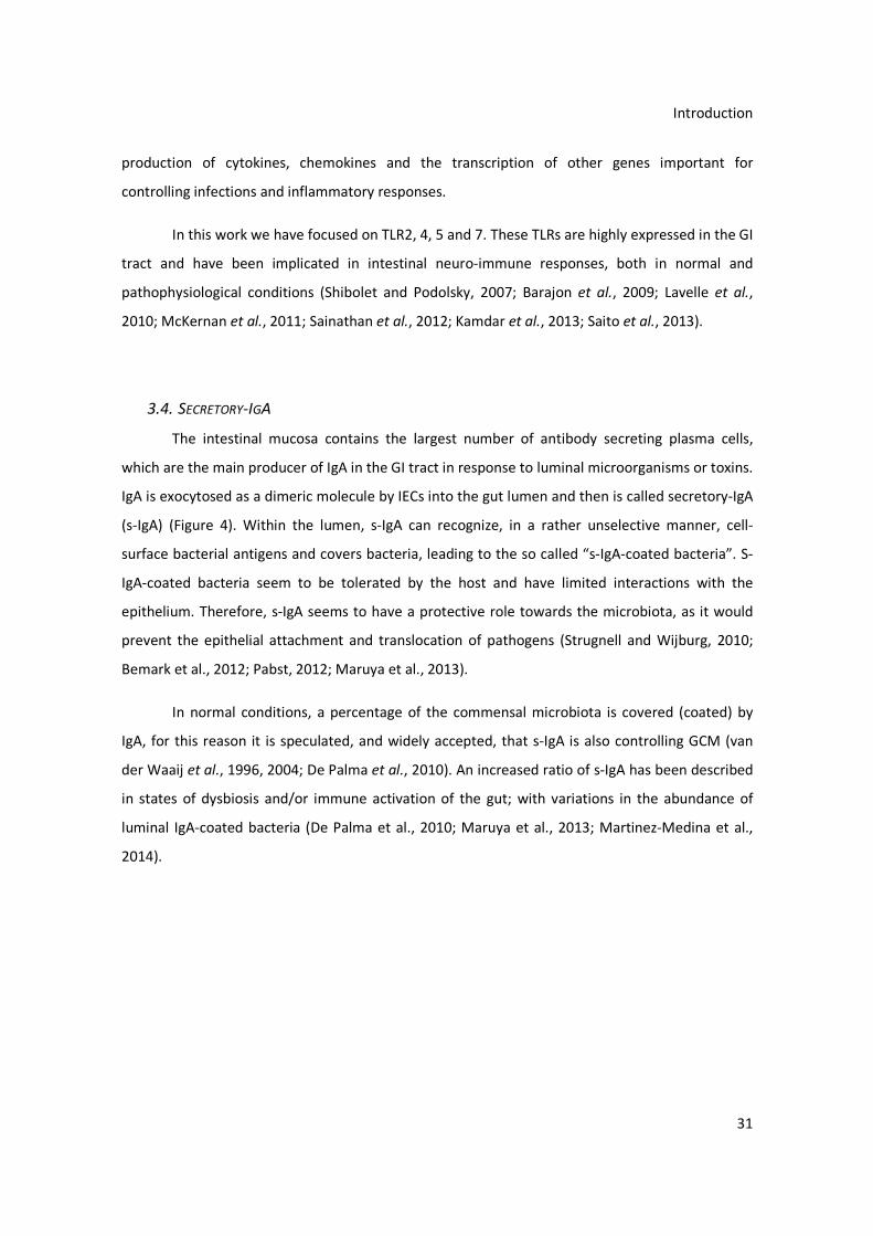


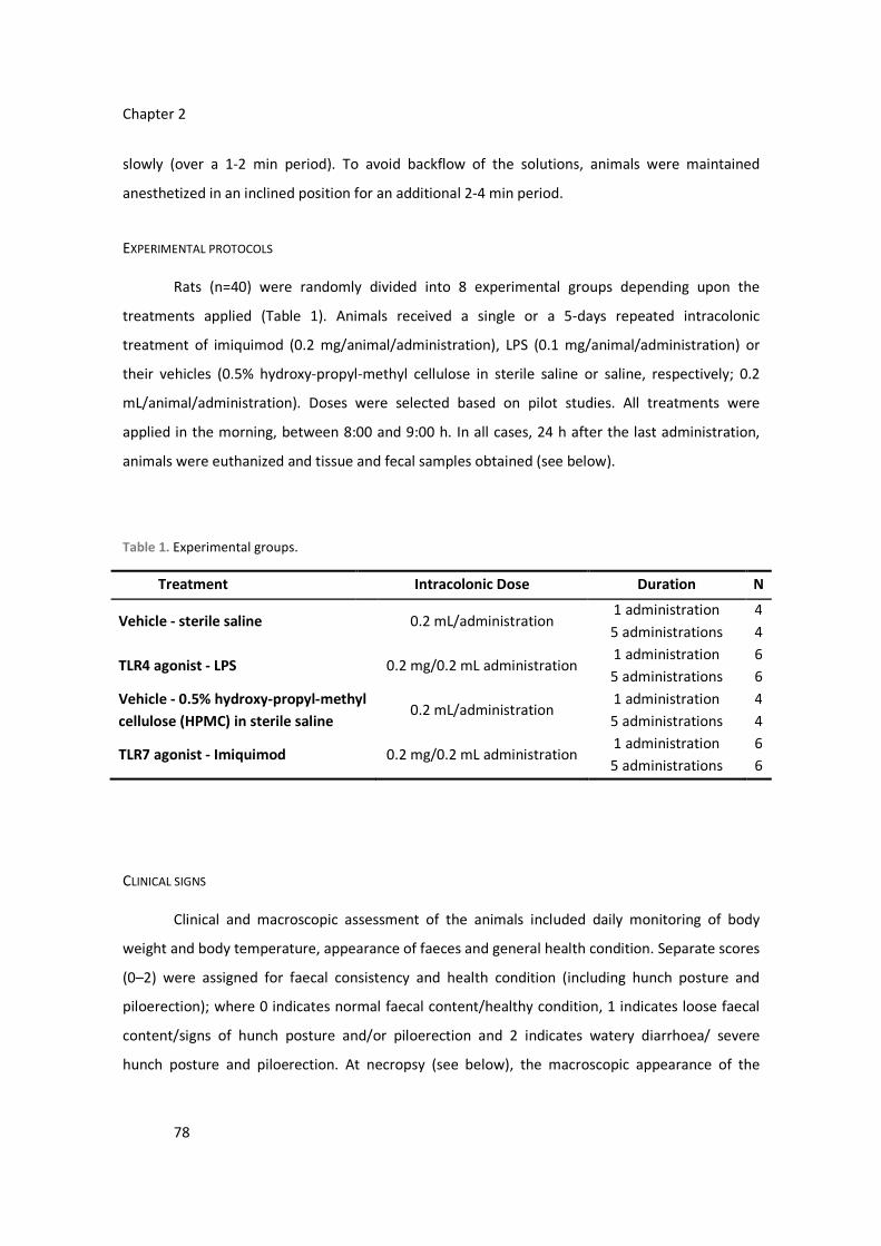
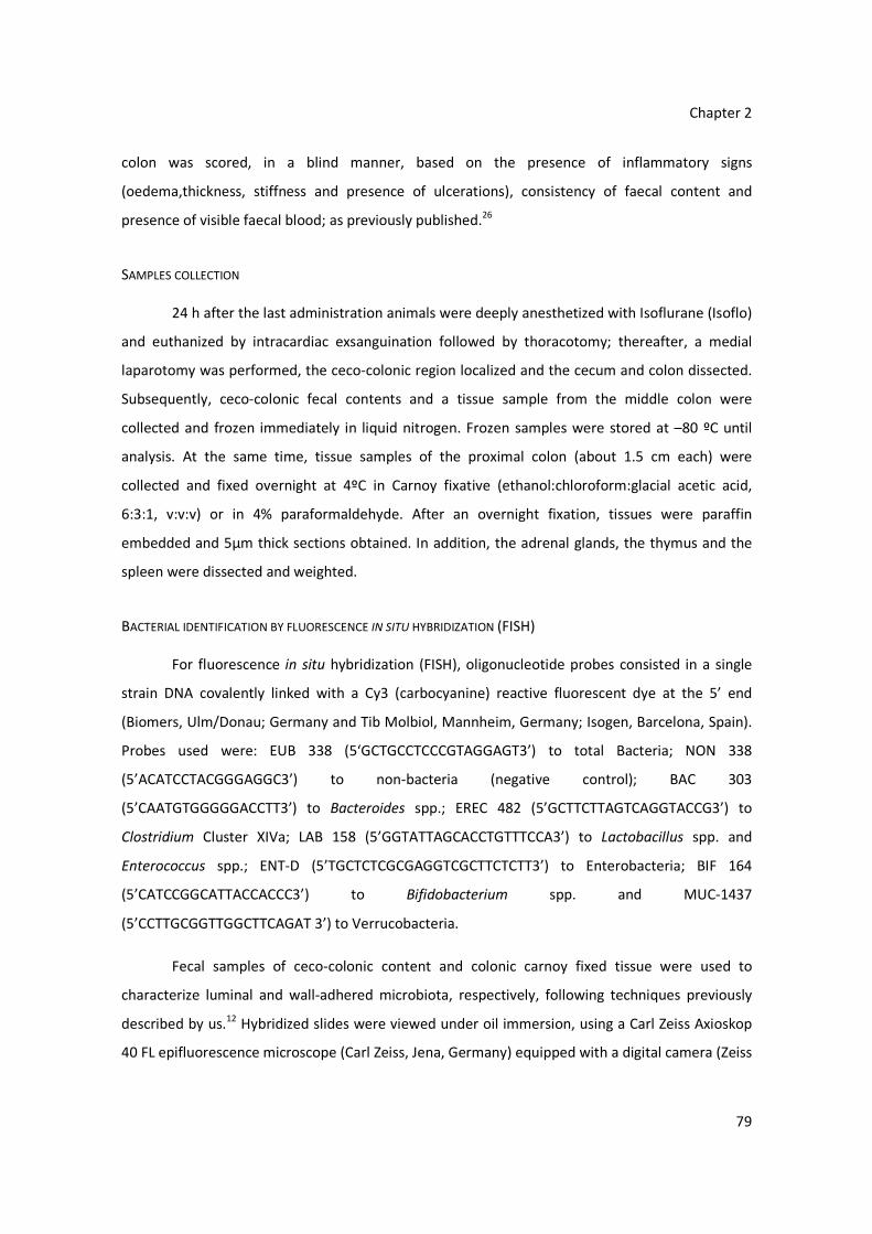


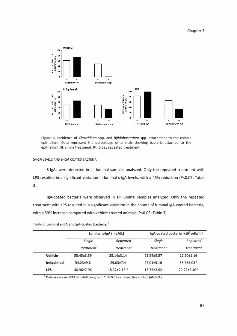


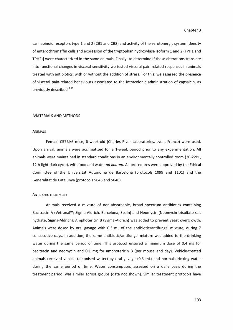


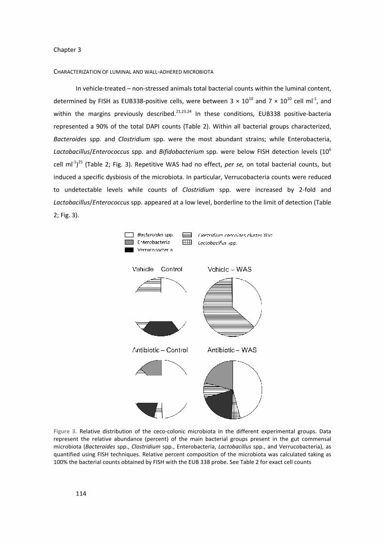

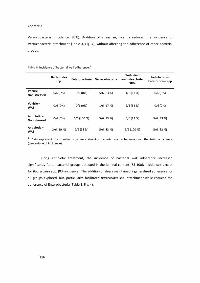


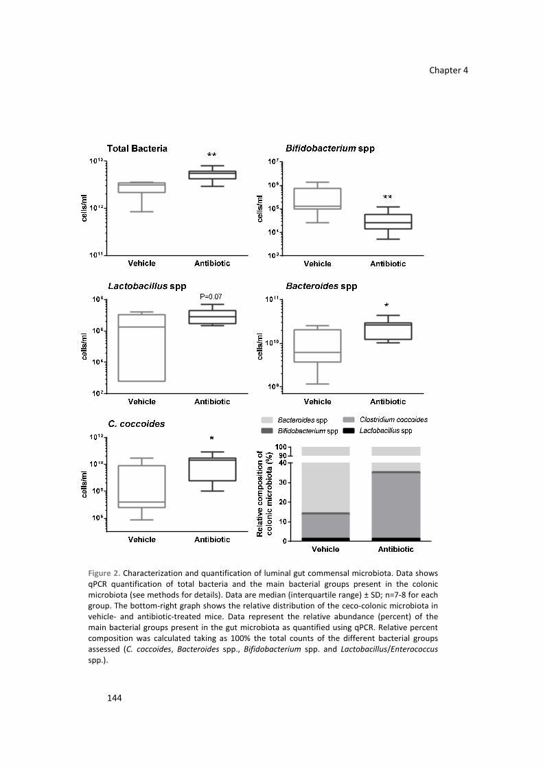
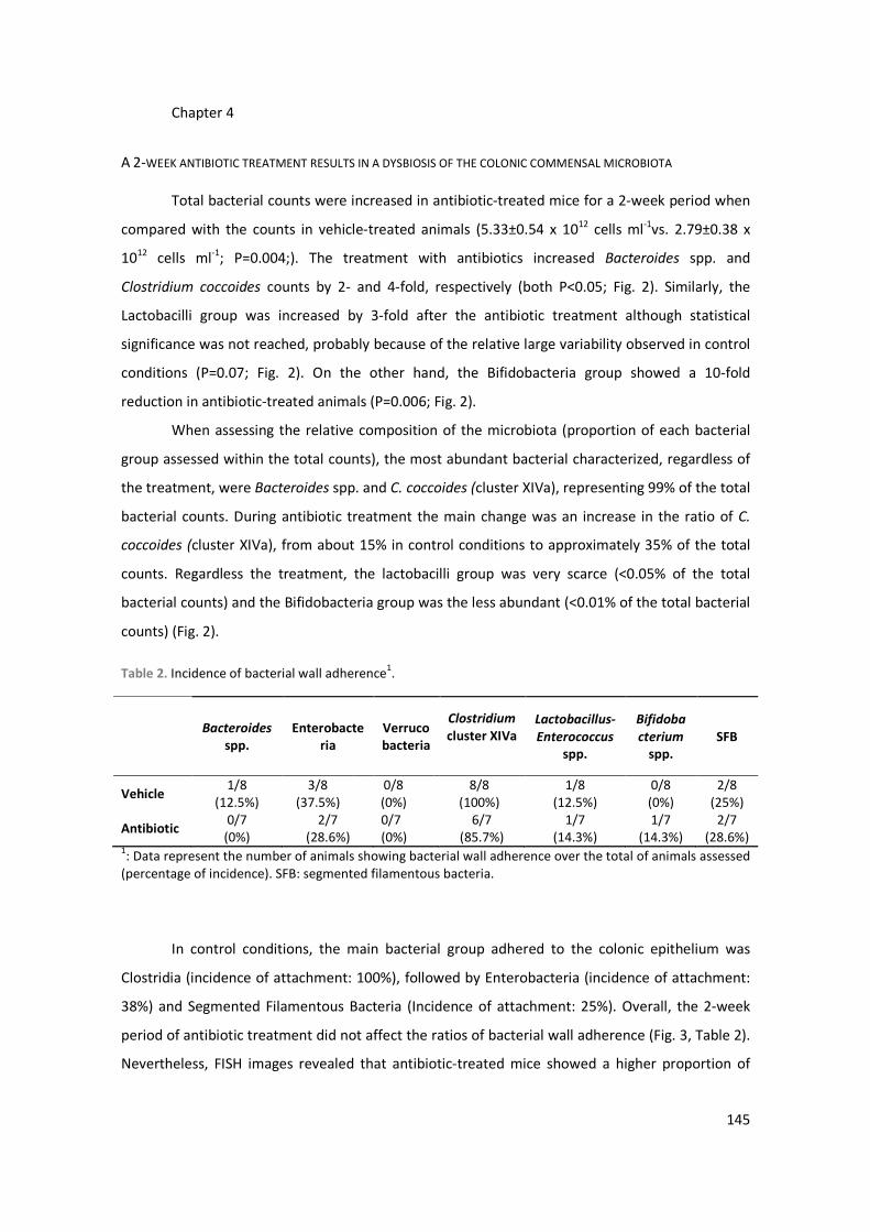


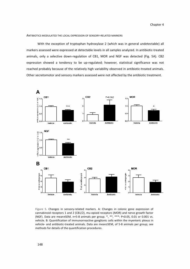


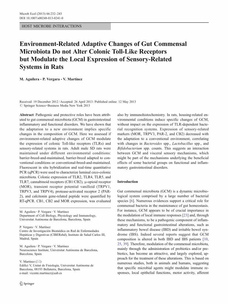

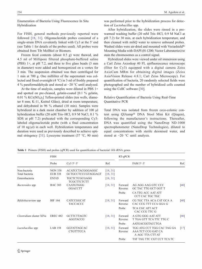


![Untitled-4 [] · ˘ ˘ ˇˆ ˙ ˘ ˘ ˝ ˛ ˘ ˇ ˇ˚˘ ˇ ˝ ˘ ˜˘ ! ˇ˘ ˇ˘ ˘ ˛ ˇ ˝˘ ˚ ˘ ˚ " ˘ ˇ # $ ˇ ˘ ˝ !!! ˇ !˘ ˇ ˝ " "ˇ ˇ ˛ ˝˜ ˆ % ˚˛ ˝! ˘ˆ](https://static.documents.pub/doc/80x56/5f2ad62a9e3f9d18cd6b754e/untitled-4-oe-.jpg)


