[Expand] [Expand] [Expand] [Collapse] SH Lecture - Lymphatic Structure and Organs Contents Introduction This lecture will provide an overview of the lymphoid structure and histology of key cells, vessels, structures and organs lymphoid organs, including the lymph nodes, spleen and thymus, as well as extranodal lymphoid tissues including mucosal associated lymphoid tissues (MALT). In this lecture I will go through the structures in sequence from cells through to organs, immunity itself is covered in detail elsewhere in the course. 2017 Lecture - PDF 2017 (/embryology/images/8/8d/SH_Lecture_2017_-_Lymphatic_Structure_and_Organs.pdf) 2017 Laboratory - Support Page (/embryology/index.php/SH_Practical_-_Lymphatic_Structure_and_Organs) Textbook References Lecture Archive UNSW Research Structure Function 1. Cells - blood cells (parenchyma), connective tissue (stroma) 2. Vessels - lymphatic vessels 3. Diffuse - (extra-nodal tissue) nodules, Mucosal Associated Lymphoid Tissues (MALT) 4. Nodes - (historic, "glands") 5. Organs - thymus, spleen 1. Immune - “monitor” of body surfaces, internal fluids 2. Extracellular fluid - returns interstitial fluid to circulation 3. Gastrointestinal tract - carries fat and fat-soluble vitamins (/embryology/index.php/File:Lymphatic-system-overview.jpg) Blood Cells Blood Cell Development (/embryology/index.php/File:SHsmall.jpg)
Transcript
[Expand] [Expand] [Expand]
[Collapse]
SH Lecture - Lymphatic Structure and OrgansContents
IntroductionThis lecture will provide an overview of the lymphoid structure and histology of key cells, vessels, structuresand organs lymphoid organs, including the lymph nodes, spleen and thymus, as well as extranodal lymphoidtissues including mucosal associated lymphoid tissues (MALT).
In this lecture I will go through the structures in sequence from cells through to organs, immunity itself is covered in detail elsewhere in the course.
2017 Lecture - PDF 2017 (/embryology/images/8/8d/SH_Lecture_2017_-_Lymphatic_Structure_and_Organs.pdf) 2017 Laboratory - Support Page(/embryology/index.php/SH_Practical_-_Lymphatic_Structure_and_Organs)
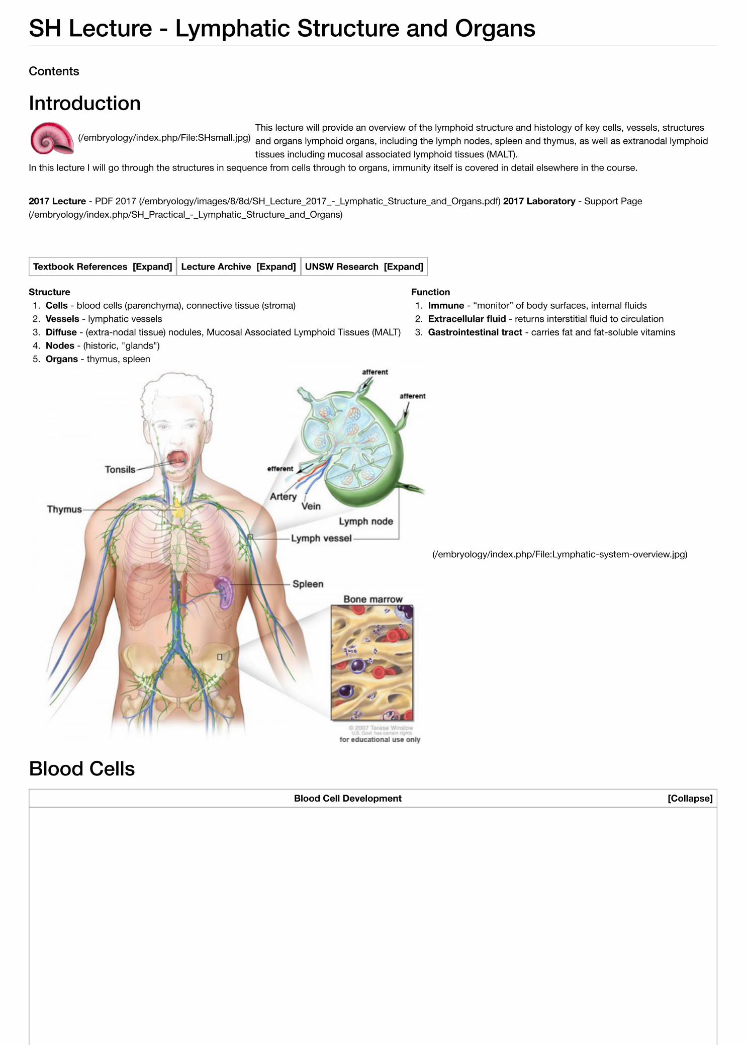
Two Blood Cell Systems1. Mononuclear Phagocytic System - circulating monocytes of peripheral blood and non-circulating (fixed) tissue macrophages found throughout the body.2. Lymphoid System - lymphocytes, three major types of T, B, and NK.
Lymphoid Organs
Central - (primary) Lymphocytes develop from precursor cells in bone marrow and thymus. (see blood marrow image)Peripheral - (secondary) Lymphocytes respond to antigen lymph nodes or spleen.
Blood Cells
1. Mononuclear Phagocytic System
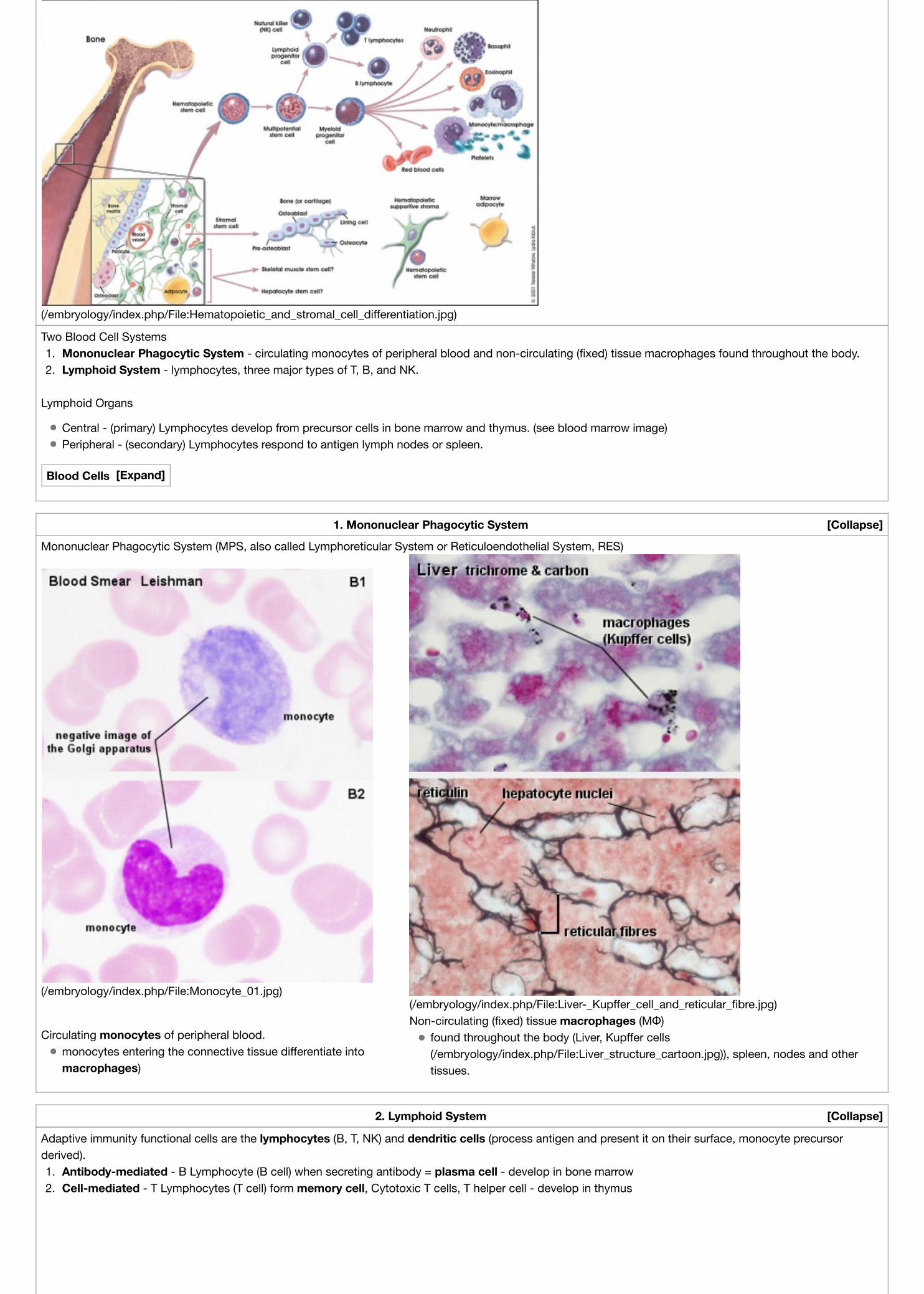
Mononuclear Phagocytic System (MPS, also called Lymphoreticular System or Reticuloendothelial System, RES)
Circulating monocytes of peripheral blood.monocytes entering the connective tissue differentiate intomacrophages)
Non-circulating (fixed) tissue macrophages (MΦ)found throughout the body (Liver, Kupffer cells(/embryology/index.php/File:Liver_structure_cartoon.jpg)), spleen, nodes and othertissues.
2. Lymphoid System
Adaptive immunity functional cells are the lymphocytes (B, T, NK) and dendritic cells (process antigen and present it on their surface, monocyte precursorderived).1. Antibody-mediated - B Lymphocyte (B cell) when secreting antibody = plasma cell - develop in bone marrow2. Cell-mediated - T Lymphocytes (T cell) form memory cell, Cytotoxic T cells, T helper cell - develop in thymus
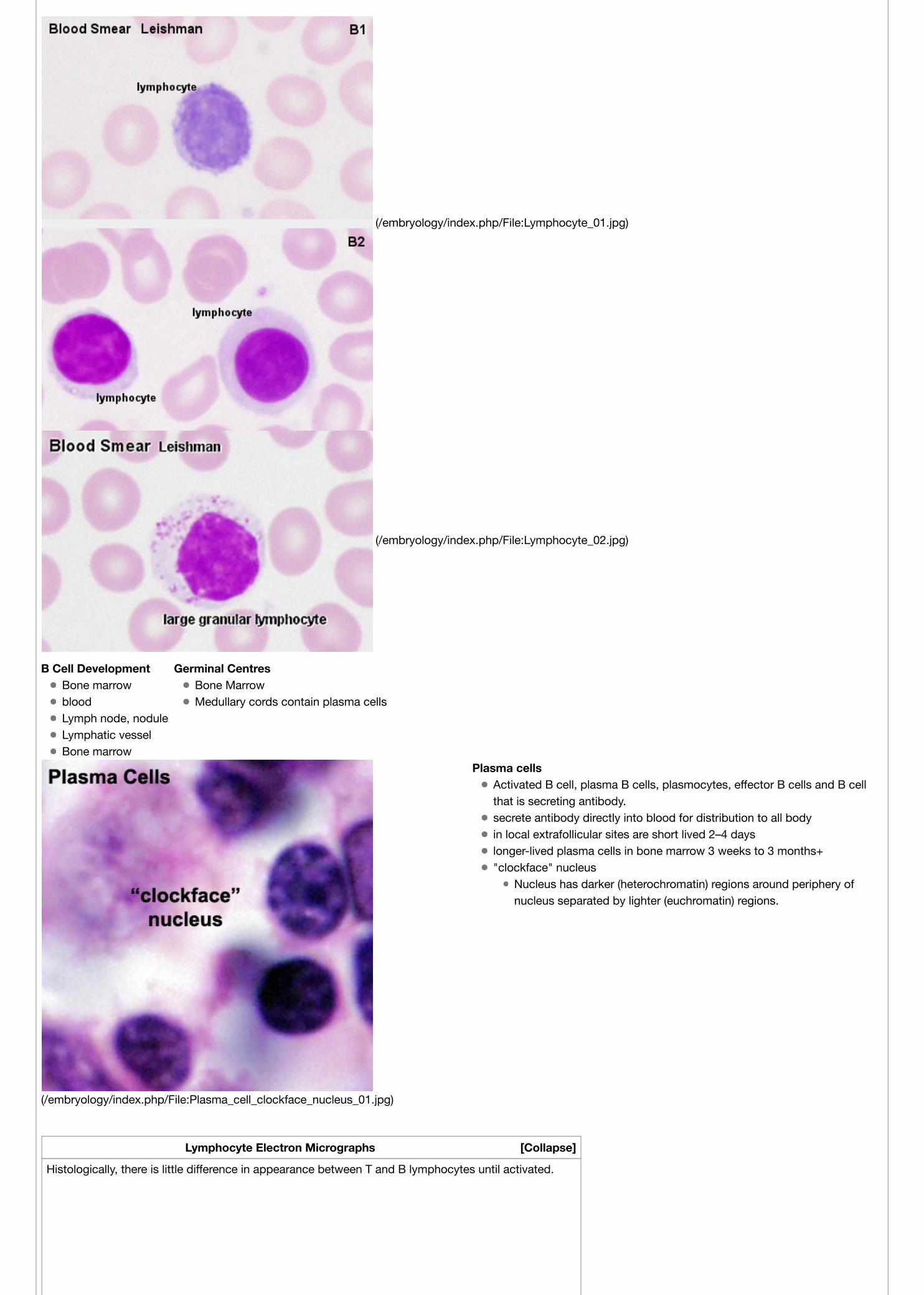
Plasma cellsActivated B cell, plasma B cells, plasmocytes, effector B cells and B cellthat is secreting antibody.secrete antibody directly into blood for distribution to all bodyin local extrafollicular sites are short lived 2–4 dayslonger-lived plasma cells in bone marrow 3 weeks to 3 months+"clockface" nucleus
Nucleus has darker (heterochromatin) regions around periphery ofnucleus separated by lighter (euchromatin) regions.
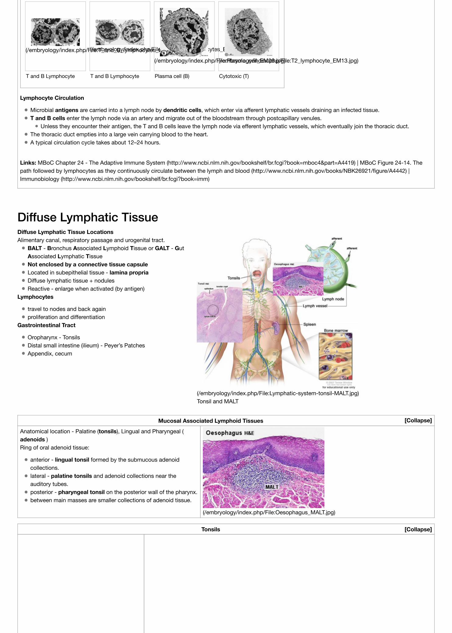
Lymphocyte Electron Micrographs
Histologically, there is little difference in appearance between T and B lymphocytes until activated.
Microbial antigens are carried into a lymph node by dendritic cells, which enter via afferent lymphatic vessels draining an infected tissue.T and B cells enter the lymph node via an artery and migrate out of the bloodstream through postcapillary venules.
Unless they encounter their antigen, the T and B cells leave the lymph node via efferent lymphatic vessels, which eventually join the thoracic duct.The thoracic duct empties into a large vein carrying blood to the heart.A typical circulation cycle takes about 12–24 hours.
Links: MBoC Chapter 24 - The Adaptive Immune System (http://www.ncbi.nlm.nih.gov/bookshelf/br.fcgi?book=mboc4&part=A4419) | MBoC Figure 24-14. Thepath followed by lymphocytes as they continuously circulate between the lymph and blood (http://www.ncbi.nlm.nih.gov/books/NBK26921/figure/A4442) |Immunobiology (http://www.ncbi.nlm.nih.gov/bookshelf/br.fcgi?book=imm)
BALT - Bronchus Associated Lymphoid Tissue or GALT - GutAssociated Lymphatic TissueNot enclosed by a connective tissue capsuleLocated in subepithelial tissue - lamina propriaDiffuse lymphatic tissue + nodulesReactive - enlarge when activated (by antigen)
Lymphocytes
travel to nodes and back againproliferation and differentiation
Gastrointestinal Tract
Oropharynx - TonsilsDistal small intestine (ilieum) - Peyer’s PatchesAppendix, cecum
(/embryology/index.php/File:Lymphatic-system-tonsil-MALT.jpg)Tonsil and MALT
Mucosal Associated Lymphoid Tissues
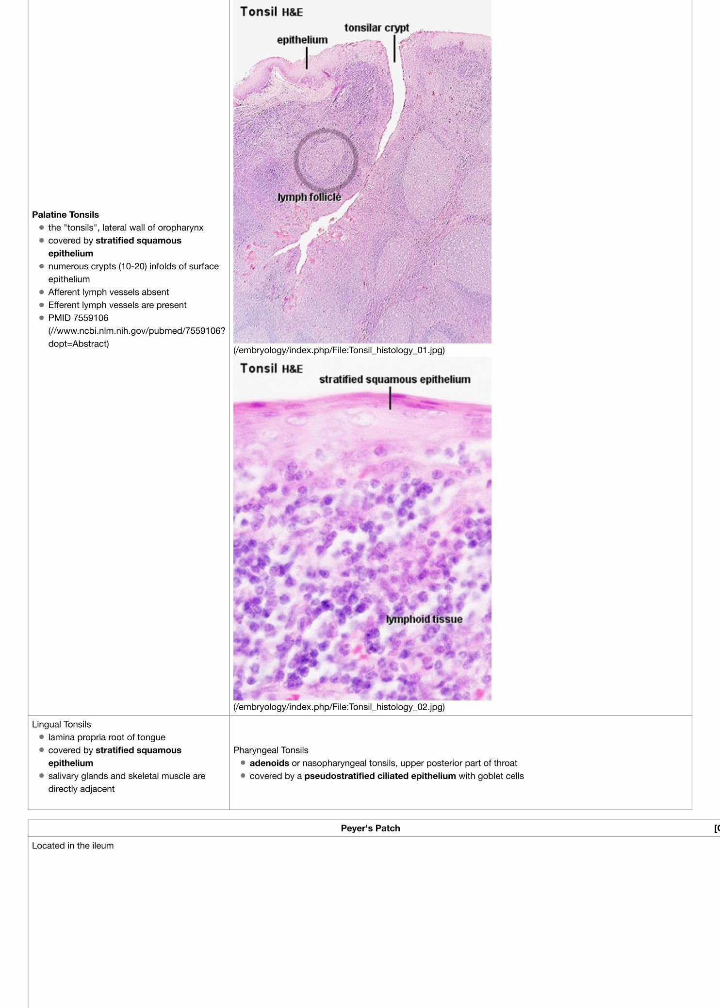
Anatomical location - Palatine (tonsils), Lingual and Pharyngeal (adenoids )Ring of oral adenoid tissue:
anterior - lingual tonsil formed by the submucous adenoidcollections.lateral - palatine tonsils and adenoid collections near theauditory tubes.posterior - pharyngeal tonsil on the posterior wall of the pharynx.between main masses are smaller collections of adenoid tissue.
Lingual Tonsilslamina propria root of tonguecovered by stratified squamousepitheliumsalivary glands and skeletal muscle aredirectly adjacent
Pharyngeal Tonsilsadenoids or nasopharyngeal tonsils, upper posterior part of throatcovered by a pseudostratified ciliated epithelium with goblet cells
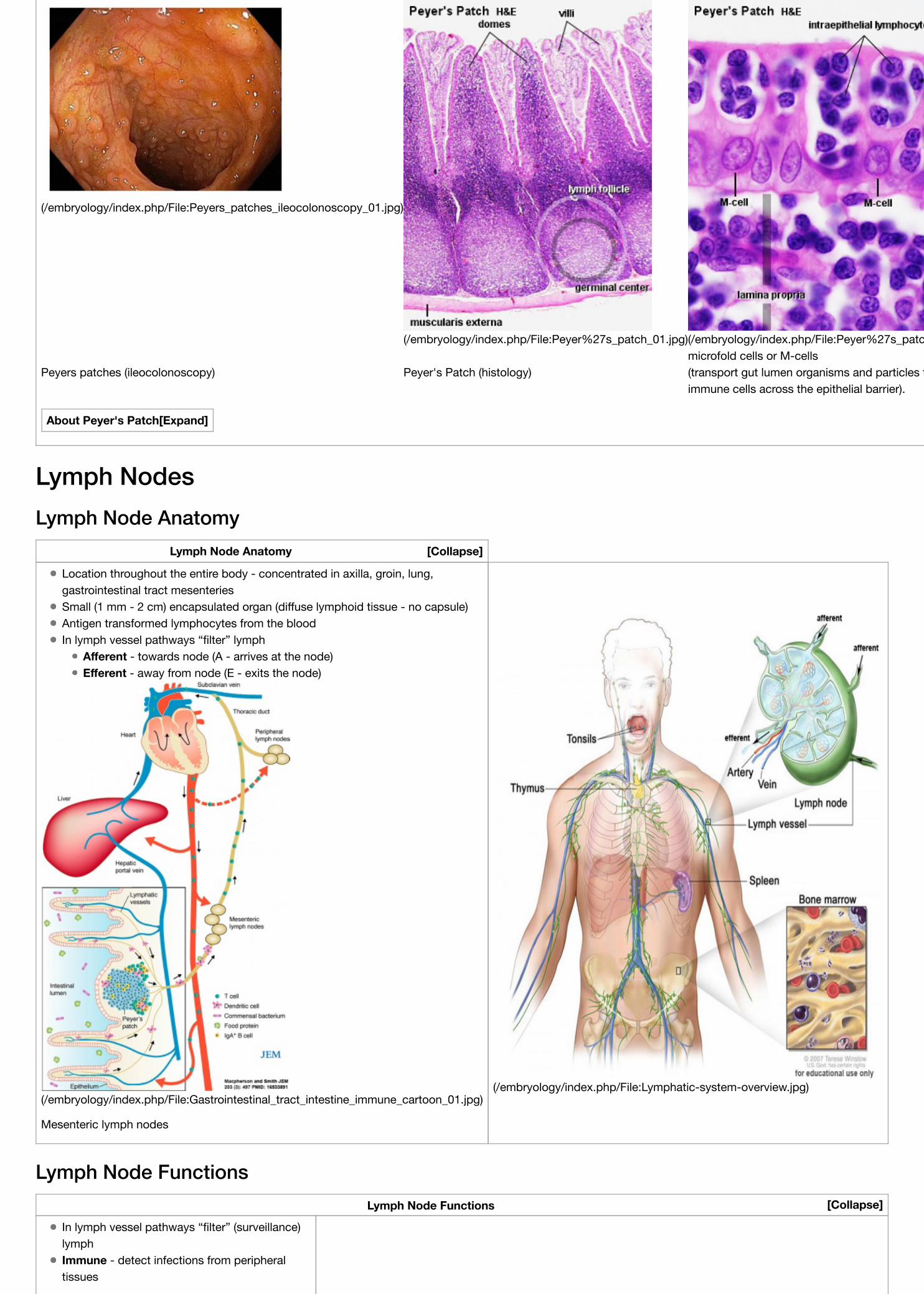
Peyers patches (ileocolonoscopy) Peyer's Patch (histology)microfold cells or M-cells(transport gut lumen organisms and particles toimmune cells across the epithelial barrier).
About Peyer's Patch
Lymph NodesLymph Node Anatomy
Lymph Node Anatomy
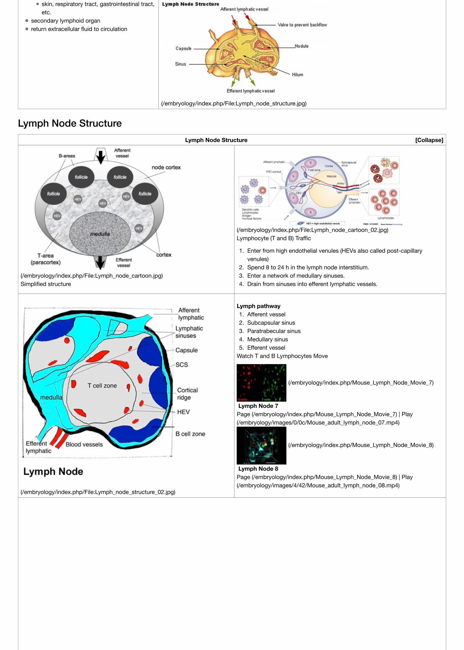
Location throughout the entire body - concentrated in axilla, groin, lung,gastrointestinal tract mesenteriesSmall (1 mm - 2 cm) encapsulated organ (diffuse lymphoid tissue - no capsule)Antigen transformed lymphocytes from the bloodIn lymph vessel pathways “filter” lymph
Afferent - towards node (A - arrives at the node)Efferent - away from node (E - exits the node)
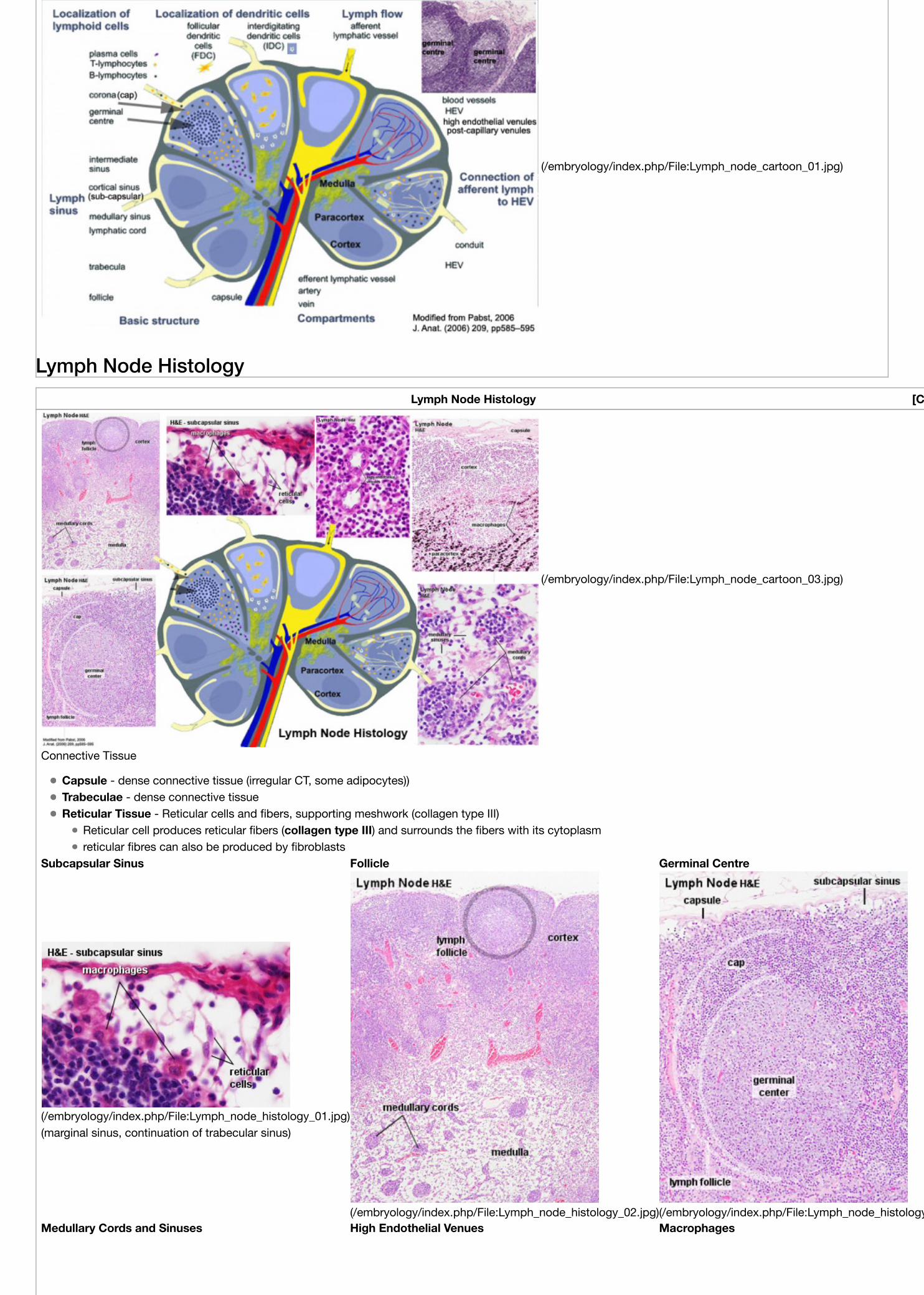
Capsule - dense connective tissue (irregular CT, some adipocytes))Trabeculae - dense connective tissueReticular Tissue - Reticular cells and fibers, supporting meshwork (collagen type III)
Reticular cell produces reticular fibers (collagen type III) and surrounds the fibers with its cytoplasmreticular fibres can also be produced by fibroblasts
Subcapsular Sinus Follicle Germinal Centre
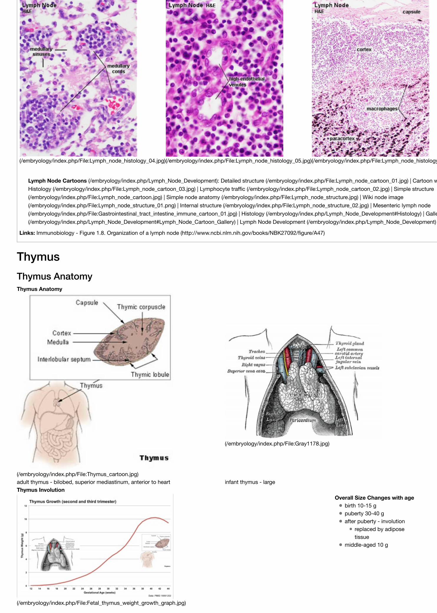
(/embryology/index.php/File:Lymph_node_histology_01.jpg)(marginal sinus, continuation of trabecular sinus)
(/embryology/index.php/File:Lymph_node_histology_02.jpg)(/embryology/index.php/File:Lymph_node_histology_03.jpg)Medullary Cords and Sinuses High Endothelial Venues Macrophages
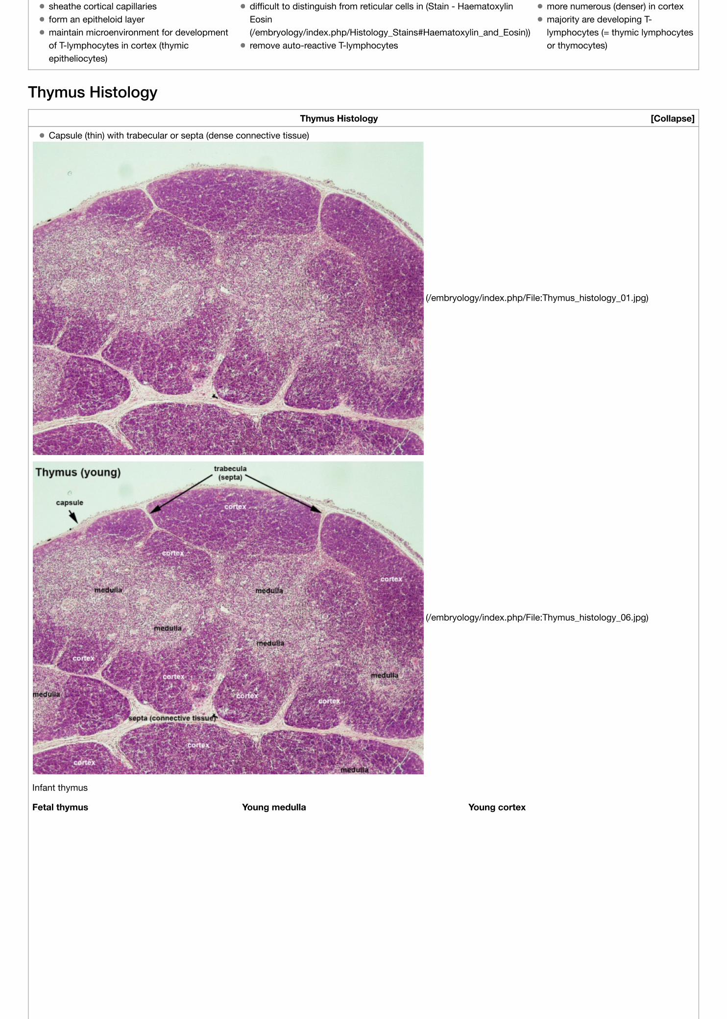
specialised thymus microenvironments allow the production ofself-tolerant T-cells (T lymphocytes) from immature precursors.
immature precursors enter the thymusdifferentiate and undergo selection by thymic epithelial cell(TEC) subtypesmature release into circulation of these cells
destruction of cells that recognise self antigensT-cells kill infected and oncogenic cells
T Cells maturation within the thymus
1. T cell progenitors enter the thymus at the cortex/medullaborder via post–capillary venules
2. migrate toward the capsule in response to chemokine signalling.3. cortex - thymocytes undergo positive selection by cTECs then
migrate to the medulla4. medulla - thymocytes are screened for reactivity to tissue-
restricted self antigens expressed by mTECs.5. Mature T cells exit the thymus via blood or lymphatic vessels in
response to a sphingosine-1-phosphate (S1P) gradient.
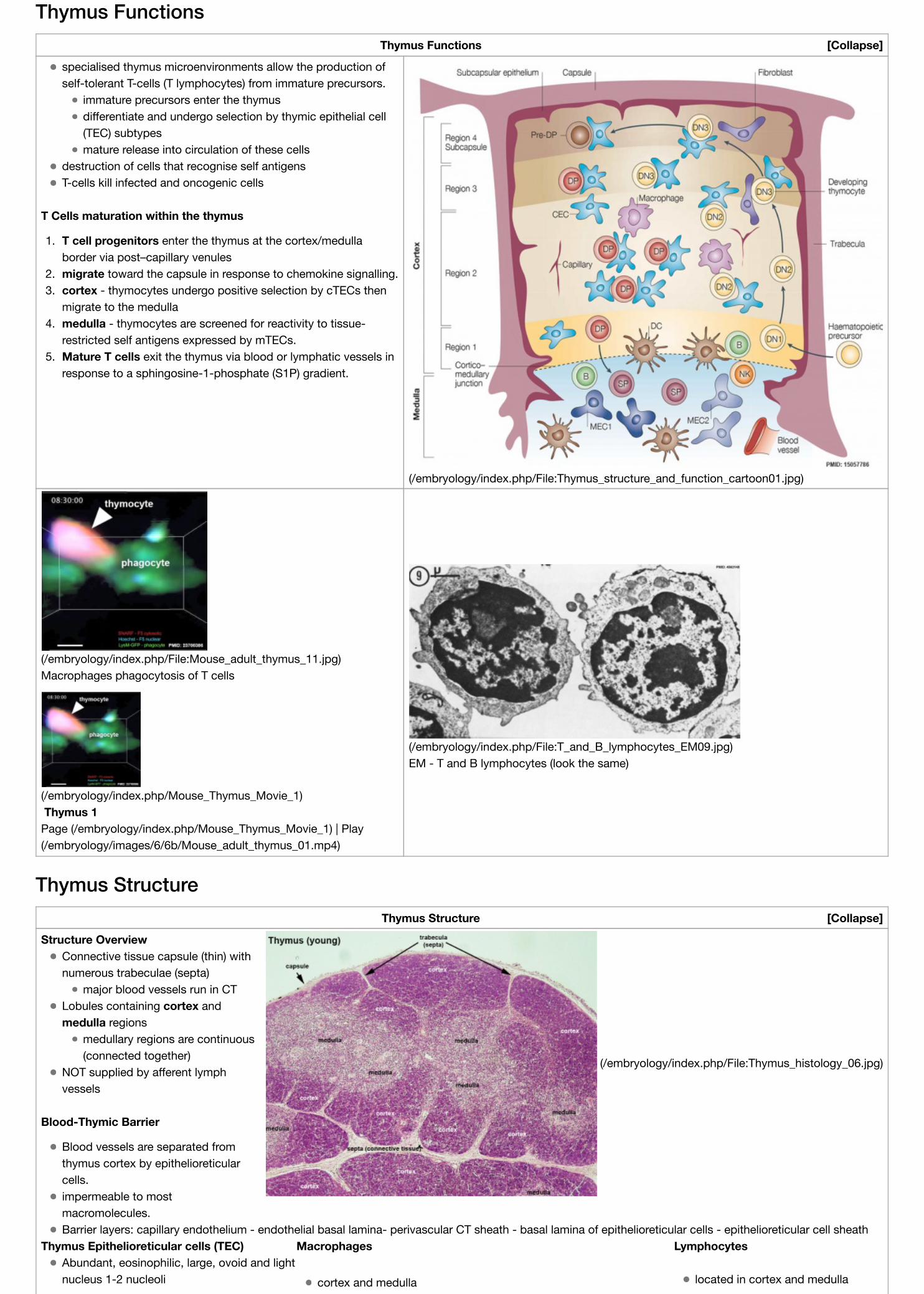
major blood vessels run in CTLobules containing cortex andmedulla regions
medullary regions are continuous(connected together)
NOT supplied by afferent lymphvessels
Blood-Thymic Barrier
Blood vessels are separated fromthymus cortex by epithelioreticularcells.impermeable to mostmacromolecules.Barrier layers: capillary endothelium - endothelial basal lamina- perivascular CT sheath - basal lamina of epithelioreticular cells - epithelioreticular cell sheath
Thymus Epithelioreticular cells (TEC) Macrophages LymphocytesAbundant, eosinophilic, large, ovoid and lightnucleus 1-2 nucleoli cortex and medulla located in cortex and medulla
sheathe cortical capillariesform an epitheloid layermaintain microenvironment for developmentof T-lymphocytes in cortex (thymicepitheliocytes)
difficult to distinguish from reticular cells in (Stain - HaematoxylinEosin(/embryology/index.php/Histology_Stains#Haematoxylin_and_Eosin))remove auto-reactive T-lymphocytes
more numerous (denser) in cortexmajority are developing T-lymphocytes (= thymic lymphocytesor thymocytes)
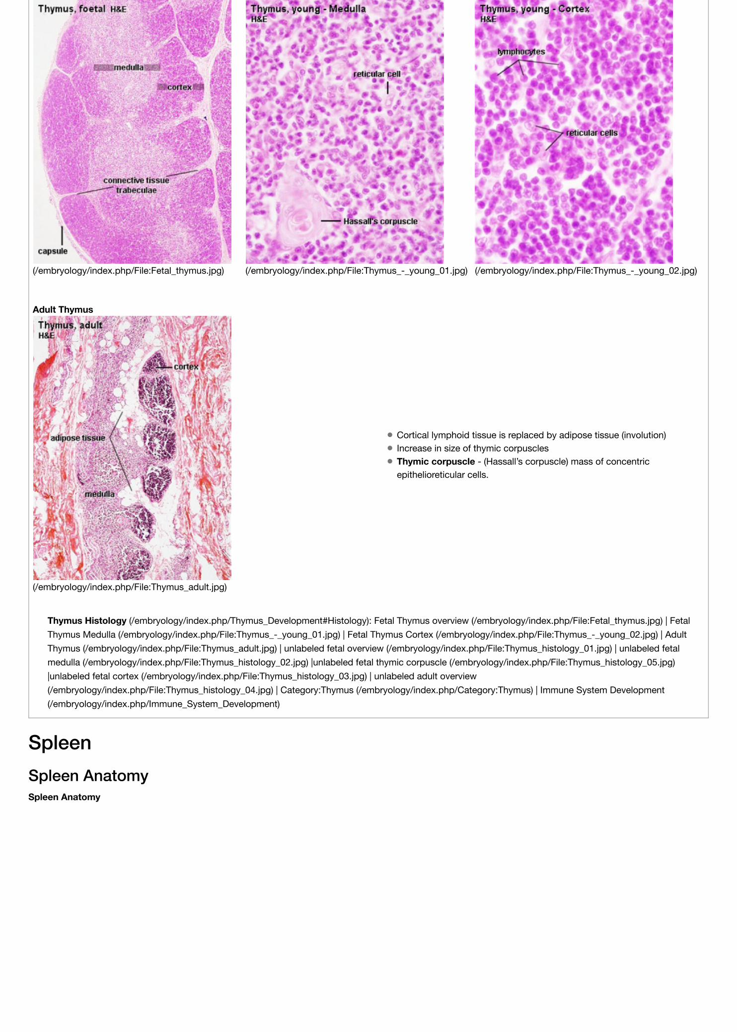
Thymus HistologyThymus Histology
Capsule (thin) with trabecular or septa (dense connective tissue)
Cortical lymphoid tissue is replaced by adipose tissue (involution)Increase in size of thymic corpusclesThymic corpuscle - (Hassall’s corpuscle) mass of concentricepithelioreticular cells.
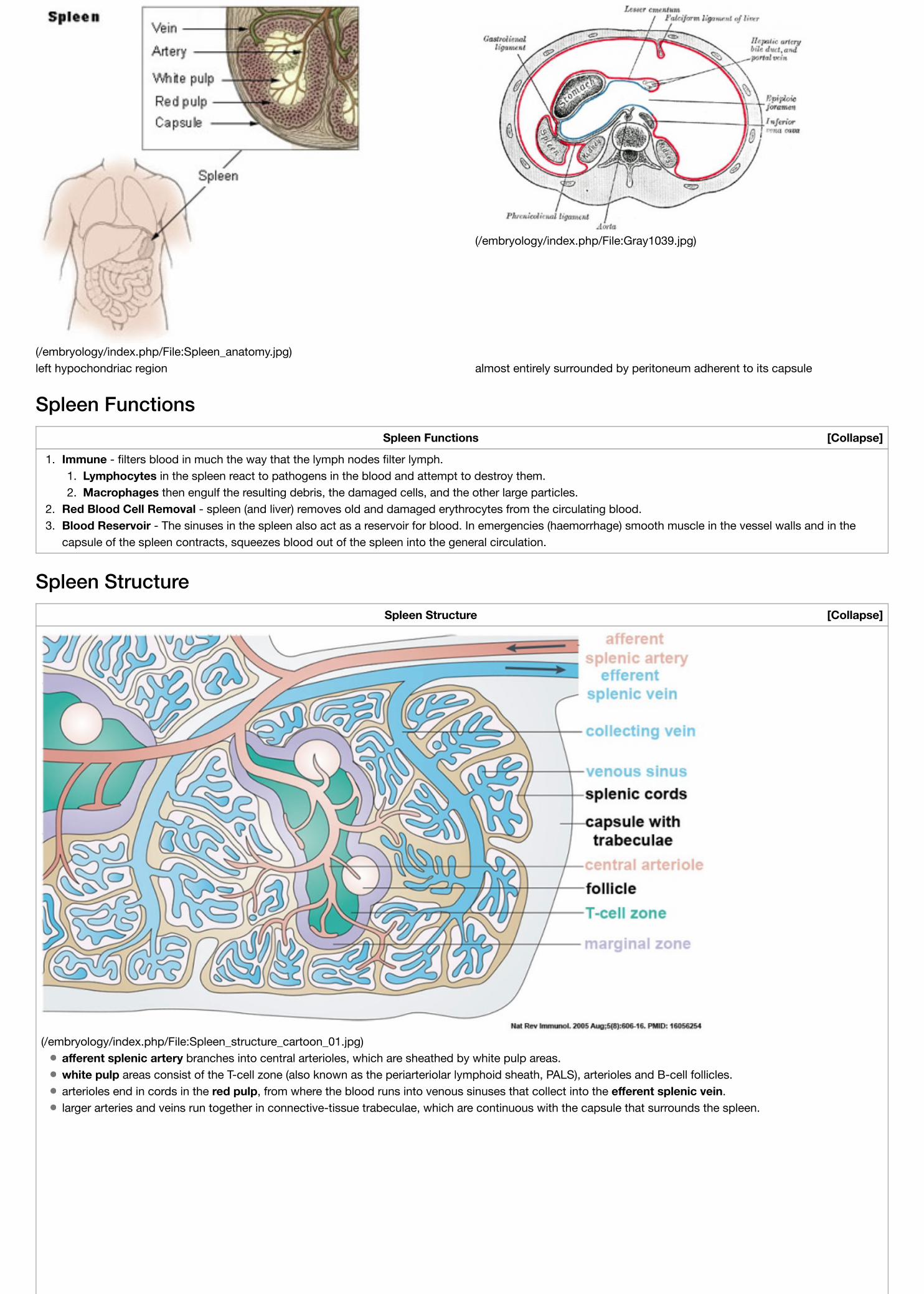
left hypochondriac region almost entirely surrounded by peritoneum adherent to its capsule
Spleen FunctionsSpleen Functions
1. Immune - filters blood in much the way that the lymph nodes filter lymph.1. Lymphocytes in the spleen react to pathogens in the blood and attempt to destroy them.2. Macrophages then engulf the resulting debris, the damaged cells, and the other large particles.
2. Red Blood Cell Removal - spleen (and liver) removes old and damaged erythrocytes from the circulating blood.3. Blood Reservoir - The sinuses in the spleen also act as a reservoir for blood. In emergencies (haemorrhage) smooth muscle in the vessel walls and in the
capsule of the spleen contracts, squeezes blood out of the spleen into the general circulation.
Spleen StructureSpleen Structure
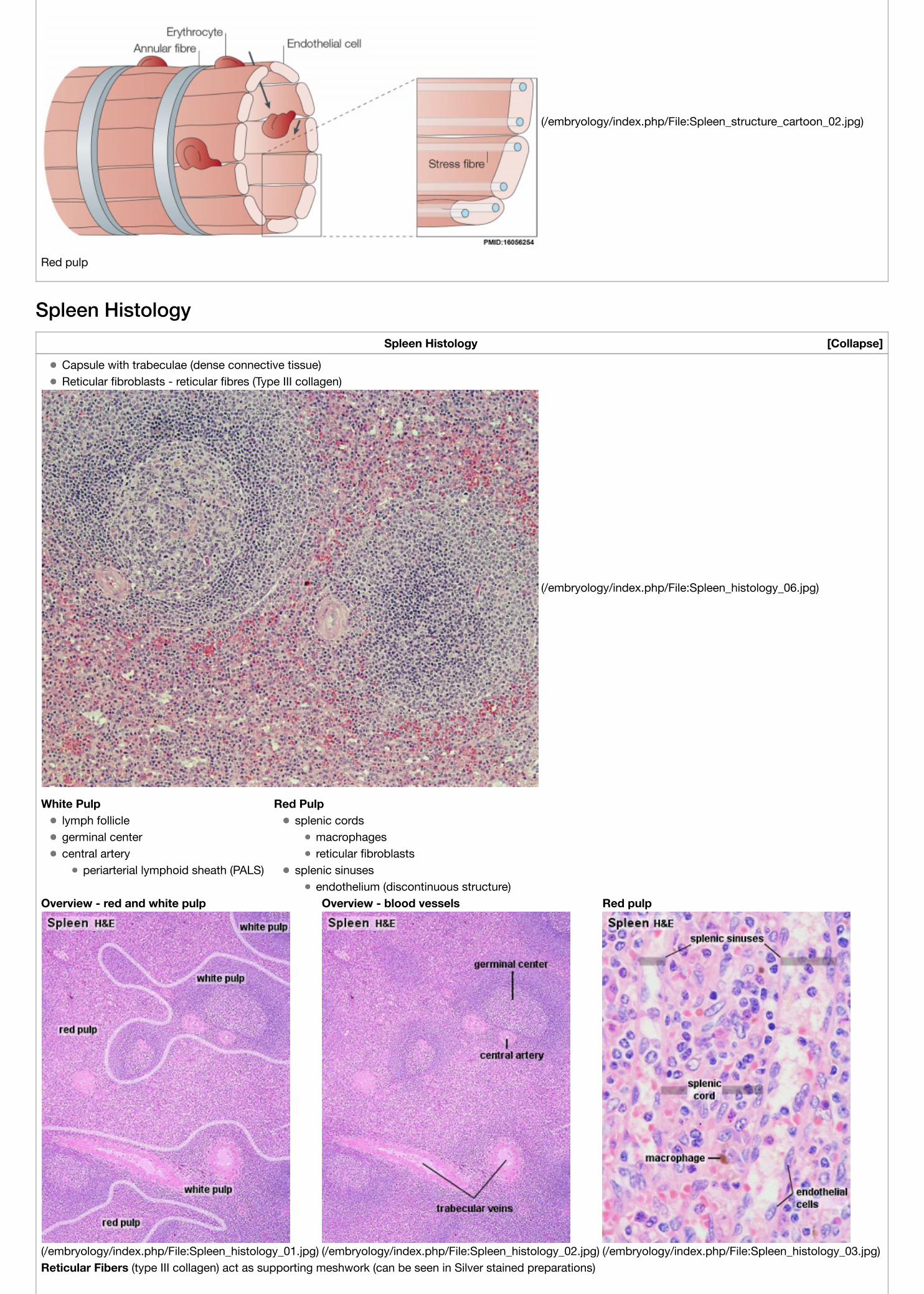
(/embryology/index.php/File:Spleen_structure_cartoon_01.jpg)afferent splenic artery branches into central arterioles, which are sheathed by white pulp areas.white pulp areas consist of the T-cell zone (also known as the periarteriolar lymphoid sheath, PALS), arterioles and B-cell follicles.arterioles end in cords in the red pulp, from where the blood runs into venous sinuses that collect into the efferent splenic vein.larger arteries and veins run together in connective-tissue trabeculae, which are continuous with the capsule that surrounds the spleen.
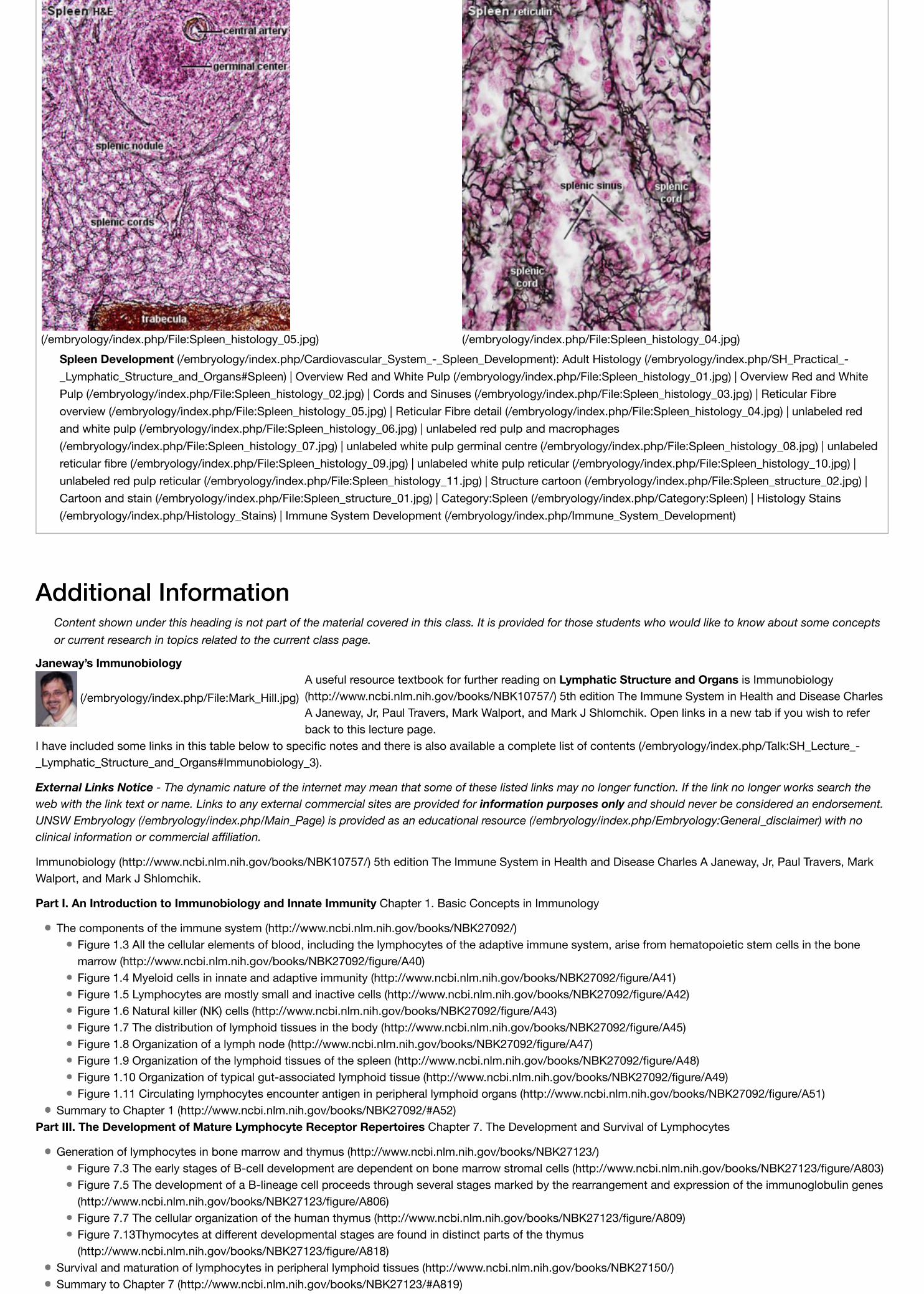
Overview - red and white pulp Overview - blood vessels Red pulp
(/embryology/index.php/File:Spleen_histology_01.jpg) (/embryology/index.php/File:Spleen_histology_02.jpg) (/embryology/index.php/File:Spleen_histology_03.jpg)Reticular Fibers (type III collagen) act as supporting meshwork (can be seen in Silver stained preparations)
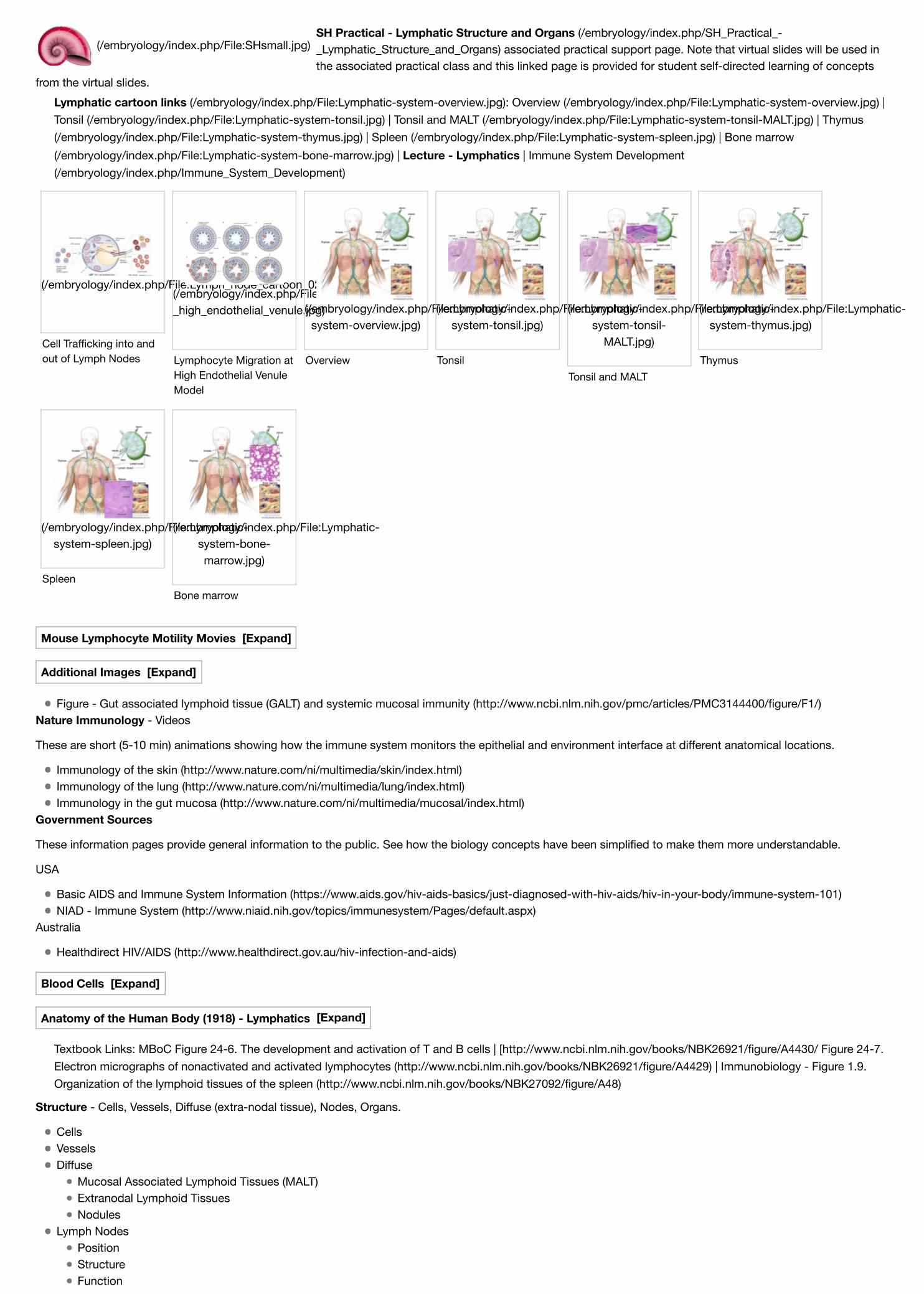
(/embryology/index.php/File:Spleen_histology_05.jpg) (/embryology/index.php/File:Spleen_histology_04.jpg)Spleen Development (/embryology/index.php/Cardiovascular_System_-_Spleen_Development): Adult Histology (/embryology/index.php/SH_Practical_-_Lymphatic_Structure_and_Organs#Spleen) | Overview Red and White Pulp (/embryology/index.php/File:Spleen_histology_01.jpg) | Overview Red and WhitePulp (/embryology/index.php/File:Spleen_histology_02.jpg) | Cords and Sinuses (/embryology/index.php/File:Spleen_histology_03.jpg) | Reticular Fibreoverview (/embryology/index.php/File:Spleen_histology_05.jpg) | Reticular Fibre detail (/embryology/index.php/File:Spleen_histology_04.jpg) | unlabeled redand white pulp (/embryology/index.php/File:Spleen_histology_06.jpg) | unlabeled red pulp and macrophages(/embryology/index.php/File:Spleen_histology_07.jpg) | unlabeled white pulp germinal centre (/embryology/index.php/File:Spleen_histology_08.jpg) | unlabeledreticular fibre (/embryology/index.php/File:Spleen_histology_09.jpg) | unlabeled white pulp reticular (/embryology/index.php/File:Spleen_histology_10.jpg) |unlabeled red pulp reticular (/embryology/index.php/File:Spleen_histology_11.jpg) | Structure cartoon (/embryology/index.php/File:Spleen_structure_02.jpg) |Cartoon and stain (/embryology/index.php/File:Spleen_structure_01.jpg) | Category:Spleen (/embryology/index.php/Category:Spleen) | Histology Stains(/embryology/index.php/Histology_Stains) | Immune System Development (/embryology/index.php/Immune_System_Development)
Additional InformationContent shown under this heading is not part of the material covered in this class. It is provided for those students who would like to know about some conceptsor current research in topics related to the current class page.
Janeway’s Immunobiology A useful resource textbook for further reading on Lymphatic Structure and Organs is Immunobiology(http://www.ncbi.nlm.nih.gov/books/NBK10757/) 5th edition The Immune System in Health and Disease CharlesA Janeway, Jr, Paul Travers, Mark Walport, and Mark J Shlomchik. Open links in a new tab if you wish to referback to this lecture page.
I have included some links in this table below to specific notes and there is also available a complete list of contents (/embryology/index.php/Talk:SH_Lecture_-_Lymphatic_Structure_and_Organs#Immunobiology_3).
External Links Notice - The dynamic nature of the internet may mean that some of these listed links may no longer function. If the link no longer works search theweb with the link text or name. Links to any external commercial sites are provided for information purposes only and should never be considered an endorsement.UNSW Embryology (/embryology/index.php/Main_Page) is provided as an educational resource (/embryology/index.php/Embryology:General_disclaimer) with noclinical information or commercial affiliation.
Immunobiology (http://www.ncbi.nlm.nih.gov/books/NBK10757/) 5th edition The Immune System in Health and Disease Charles A Janeway, Jr, Paul Travers, MarkWalport, and Mark J Shlomchik.
Part I. An Introduction to Immunobiology and Innate Immunity Chapter 1. Basic Concepts in Immunology
The components of the immune system (http://www.ncbi.nlm.nih.gov/books/NBK27092/)Figure 1.3 All the cellular elements of blood, including the lymphocytes of the adaptive immune system, arise from hematopoietic stem cells in the bonemarrow (http://www.ncbi.nlm.nih.gov/books/NBK27092/figure/A40)Figure 1.4 Myeloid cells in innate and adaptive immunity (http://www.ncbi.nlm.nih.gov/books/NBK27092/figure/A41)Figure 1.5 Lymphocytes are mostly small and inactive cells (http://www.ncbi.nlm.nih.gov/books/NBK27092/figure/A42)Figure 1.6 Natural killer (NK) cells (http://www.ncbi.nlm.nih.gov/books/NBK27092/figure/A43)Figure 1.7 The distribution of lymphoid tissues in the body (http://www.ncbi.nlm.nih.gov/books/NBK27092/figure/A45)Figure 1.8 Organization of a lymph node (http://www.ncbi.nlm.nih.gov/books/NBK27092/figure/A47)Figure 1.9 Organization of the lymphoid tissues of the spleen (http://www.ncbi.nlm.nih.gov/books/NBK27092/figure/A48)Figure 1.10 Organization of typical gut-associated lymphoid tissue (http://www.ncbi.nlm.nih.gov/books/NBK27092/figure/A49)Figure 1.11 Circulating lymphocytes encounter antigen in peripheral lymphoid organs (http://www.ncbi.nlm.nih.gov/books/NBK27092/figure/A51)
Summary to Chapter 1 (http://www.ncbi.nlm.nih.gov/books/NBK27092/#A52)Part III. The Development of Mature Lymphocyte Receptor Repertoires Chapter 7. The Development and Survival of Lymphocytes
Generation of lymphocytes in bone marrow and thymus (http://www.ncbi.nlm.nih.gov/books/NBK27123/)Figure 7.3 The early stages of B-cell development are dependent on bone marrow stromal cells (http://www.ncbi.nlm.nih.gov/books/NBK27123/figure/A803)Figure 7.5 The development of a B-lineage cell proceeds through several stages marked by the rearrangement and expression of the immunoglobulin genes(http://www.ncbi.nlm.nih.gov/books/NBK27123/figure/A806)Figure 7.7 The cellular organization of the human thymus (http://www.ncbi.nlm.nih.gov/books/NBK27123/figure/A809)Figure 7.13Thymocytes at different developmental stages are found in distinct parts of the thymus(http://www.ncbi.nlm.nih.gov/books/NBK27123/figure/A818)
Survival and maturation of lymphocytes in peripheral lymphoid tissues (http://www.ncbi.nlm.nih.gov/books/NBK27150/)Summary to Chapter 7 (http://www.ncbi.nlm.nih.gov/books/NBK27123/#A819)
SH Practical - Lymphatic Structure and Organs (/embryology/index.php/SH_Practical_-_Lymphatic_Structure_and_Organs) associated practical support page. Note that virtual slides will be used inthe associated practical class and this linked page is provided for student self-directed learning of concepts
from the virtual slides.Lymphatic cartoon links (/embryology/index.php/File:Lymphatic-system-overview.jpg): Overview (/embryology/index.php/File:Lymphatic-system-overview.jpg) |Tonsil (/embryology/index.php/File:Lymphatic-system-tonsil.jpg) | Tonsil and MALT (/embryology/index.php/File:Lymphatic-system-tonsil-MALT.jpg) | Thymus(/embryology/index.php/File:Lymphatic-system-thymus.jpg) | Spleen (/embryology/index.php/File:Lymphatic-system-spleen.jpg) | Bone marrow(/embryology/index.php/File:Lymphatic-system-bone-marrow.jpg) | Lecture - Lymphatics | Immune System Development(/embryology/index.php/Immune_System_Development)
Figure - Gut associated lymphoid tissue (GALT) and systemic mucosal immunity (http://www.ncbi.nlm.nih.gov/pmc/articles/PMC3144400/figure/F1/)Nature Immunology - Videos
These are short (5-10 min) animations showing how the immune system monitors the epithelial and environment interface at different anatomical locations.
Immunology of the skin (http://www.nature.com/ni/multimedia/skin/index.html)Immunology of the lung (http://www.nature.com/ni/multimedia/lung/index.html)Immunology in the gut mucosa (http://www.nature.com/ni/multimedia/mucosal/index.html)
Government Sources
These information pages provide general information to the public. See how the biology concepts have been simplified to make them more understandable.
USA
Basic AIDS and Immune System Information (https://www.aids.gov/hiv-aids-basics/just-diagnosed-with-hiv-aids/hiv-in-your-body/immune-system-101)NIAD - Immune System (http://www.niaid.nih.gov/topics/immunesystem/Pages/default.aspx)
Textbook Links: MBoC Figure 24-6. The development and activation of T and B cells | [http://www.ncbi.nlm.nih.gov/books/NBK26921/figure/A4430/ Figure 24-7.Electron micrographs of nonactivated and activated lymphocytes (http://www.ncbi.nlm.nih.gov/books/NBK26921/figure/A4429) | Immunobiology - Figure 1.9.Organization of the lymphoid tissues of the spleen (http://www.ncbi.nlm.nih.gov/books/NBK27092/figure/A48)
TermsA few key terms associated with the Lymphoid system.
Immune Development (/embryology/index.php/Immune_System_Development)
adenoid - (Greek " +-oeides = in form of) in the form of a gland, glandular; the pharyngeal tonsil.afferent lymph - vessel carrying lymph towards a node.acquired immune deficiency syndrome - (AIDS) note this is now better described as "advanced HIV disease", decrease in the number of CD4 T cells. (More?Immunobiology - AIDS (https://www.ncbi.nlm.nih.gov/books/NBK27126/))anastomose - joining of two tubes or structures together.Antibody mediated immunity - the immune function of plasma cells (active B lymphocytes) secreting antibody which binds antigen.antibodies - mammals have five classes (IgA, IgD, IgE, IgG, and IgM)antigen - any substance that is recognised by the immune system and stimulates antibody production.appendix - is a gut-associated lymphoid tissue (GALT) located at the beginning of the colon. The anatomy is as a finger-like structure that arises from thececum. The length (2.5-13 cm) is longer in both infants and children and also has more abundant lymphatic tissue in early life. The wall structure is similar to thesmall intestine (though with no villi), nor plicae circularis. Lymph nodules surround the lumen of the gastrointestinal tract and extend from the mucosa into thesubmucosa.B cell - (B-cell, B lymphocyte) historically named after a structure called the bursa of Fabricius in birds, a source of antibody-producing lymphocytes. Theseimmune cells develop in the bone marrow. (More? Electron micrographs of nonactivate and activated lymphocytes (/embryology/index.php/SH_Practical_-_Lymphatic_Structure_and_Organs#Lymphocyte_Electron_Micrographs))B lymphocyte - (B cell, B-cell)BALT - (Bronchus Associated Lymphoid Tissue) immune tissue associated with the respiratory tract.band cell - (band neutrophil or stab cell) seen in bone marrow smear, a cell undergoing granulopoiesis, derived from a metamyelocyte, and leading to a maturegranulocyte. Also occasionally seen in circulating blood.cecum - (caecum, Latin, caecus = "blind") within the gastrointestinal tract a pouch that connects the ileum with the ascending colon of the large intestine.cell - has a specific cell biology definition, but is often used instead of "lymphocyte" when describing B and T cells.cell-mediated immunity - the immune function of T lymphocytes. (More? Immunobiology - T Cell-Mediated Immunity(https://www.ncbi.nlm.nih.gov/books/NBK10762))central tolerance - in thymus mediated by cortical epithelial cells, medullary epithelial cells and thymic DCs, involves deletion of self reactive thymocytes (Tcell)."clockface" - a term used to describe the appearance of plasma cell nuclei due to the clumping of the chromatin at the nucleus periphery. More clearly seen intissue plasma cells that the bone marrow smear, where they are sometimes confused with the basophilic erythroblasts. Image - plasma cell(/embryology/index.php/File:Plasma_cell_clockface_nucleus_01.jpg)CD - (cluster of differentiation) identifies immunological surface markers on cells.CD4+ - (T helper cells) refers to T lymphocytes that express CD4 (glycoprotein of the immunoglobulin superfamily) on their surface. These cells can be infectedby human immunodeficiency virus (HIV).CD8+ - (cytotoxic T cells) refers to T lymphocytes that express CD8 (glycoprotein of the immunoglobulin superfamily) on their surface."clockface" - a term used to describe the appearance of plasma cell nuclei due to the clumping of the chromatin at the nucleus periphery. More clearly seen intissue plasma cells that the bone marrow smear, where they are sometimes confused with the basophilic erythroblasts.cords of Billroth - spleen cellular columns located in red pulp. surrounded by splenic sinusoids. Cords contain reticular cells, macrophages, lymphocytes,plasma cells and erythrocytes.cortex - outer layer, used in association with medulla (innner layer or core) a general description that can be applied to describing an organ with a layeredstructure.dendritic cell - (DC, antigen-presenting cell, APC) cells that present antigens and induce a primary immune response in resting naïve T lymphocytes. Originatefrom the same common progenitor as monocytes (PMID 20193011 (//www.ncbi.nlm.nih.gov/pubmed/20193011?dopt=Abstract)). In 2011 Ralph M. Steinmanreceived half the Nobel Prize (http://www.nobelprize.org/nobel_prizes/medicine/laureates/2011/) half of the award to to Ralph M. Steinman for his discovery ofthe dendritic cell and its role in adaptive immunity.Effector cells - the immune functioning (active) B and T lymphocytes.Efferent lymph - vessel carrying lymph away from a node.fibroblastic reticular cell - (FRC) specialized myofibroblasts that form the structural mesenchymal network "sponge" within lymphoid tissue, through which Tcells, B cells, dendritic cells (DCs), plasma cells and macrophages move and interact.GALT - Gut Associated Lymphatic Tissue consisting of Peyer’s patches, isolated lymphoid follicles and mesenteric lymph nodes.haemopoiesis (hemopoiesis) formation of blood cells.Hassall's corpuscle - thymic corpuscle.high endothelial venule - (HEV) the specialised post-capillary venous region that enables blood lymphocytes to enter a lymph node. These specialised post-capillary venules enables blood lymphocytes to enter a lymph node. Their endothelial cells express ligands that bind lymphocytes, aiding their adhesion andsubsequent transmigration into the lymph node.humoral immune response - production of antibody by plasma cells derived from B lymphocytes (B cells).IEL - Intraepithelial Lymphocyte are T lymphocytes located in the gastrointestinal tract epithelium. Natural IELs (previously ‘type b’ IELs) acquire activatedphenotype during development in the thymus in the presence of self antigens. Induced IELs (previously ‘type a’ IELs) progeny of conventional T cells activatedpost-thymically in response to peripheral antigens.IgA - the main class of antibody in secretions (saliva, tears, milk, and respiratory and intestinal secretions).IgD - the immunoglobulin B cell starts to produce as a cell-surface molecule after leaving the bone marrow.IgE - bind Fc receptors (surface of mast cells in tissues and basophils in the blood) release of potent pro inflammatory molecules mediators of allergic reactions.IgG - the major class of immunoglobulin in the blood.IgM - the first class of antibody made by a developing B cell, which may switch to making other classes of antibody.immunodeficiency - when one or more components of the immune system is defective. (More? Immunobiology - immunodeficiency(http://www.ncbi.nlm.nih.gov/entrez/query.fcgi?cmd=Search&db=books&rid=imm.section.1494))immunoglobulin - (antibody, Ab) protein produced by plasma cells.intraepithelial lymphocyte (IEL) immune cells residing in the gastrointestinal tract epithelium. image - Intraepithelial lymphocyte differentiation(/embryology/index.php/File:Intraepithelial_lymphocyte_differentiation_01.jpg)involution - in the thymus refers to the replacement, mainly in the cortex, of cells by adipose tissue. (More? PubMed- thymus involution(http://www.ncbi.nlm.nih.gov/entrez/query.fcgi?db=PubMed&cmd=Search&term=thymus+involution&doptcmdl=Books)) | Cancer Medicine - Thymomas and
Thymic Tumors (http://www.ncbi.nlm.nih.gov/entrez/query.fcgi?cmd=Search&db=books&rid=cmed6.section.23856#23857))Kupffer cells - stellate macrophage cells located in the liver sinusoids, named after Karl Wilhelm von Kupffer (1829 - 1902) a German anatomist who originallyidentified these cells. (More? Liver Development (/embryology/index.php/Gastrointestinal_Tract_-_Liver_Development))lacteal - term used to describe the lymphatic vessels of the small intestine.lamina propria - a layer of loose connective tissue found underneath an epithelium, together with the epithelium described as mucosa.Langerhans cell - (LC, dendritic cell) Antigen-presenting immune cell found mainly in the basal/suprabasal layers of adult skin and mucosa. Cells lie in thebasal/suprabasal layers of stratified epidermal and mucosal tissues. First in the innate antiviral immune defines and can migrate to lymph nodes and induce a Tcell–mediated adaptive immune response. (More? Integumentary (/embryology/index.php/Integumentary_System_Development#Langerhans_Cells) | ImmuneSystem Development (/embryology/index.php/Immune_System_Development))Leukocyte - (Greek, lukos = clear, white) white blood cell.lingual - related to the tongue.lymph node - connective tissue encapsulated lymphoid organ (1mm - 2cm in size), positioned in the pathway of lymph vessels. (More? Lymph NodeDevelopment (/embryology/index.php/Lymph_Node_Development))lymphangion - the functional unit of a lymph vessel that lies between two semilunar (half moon-shaped) valves.M cell - (microfold cell) found in the follicle-associated epithelium of the Peyer's patch. Function to transport gut lumen organisms and particles to immune cellsacross the epithelial barrier.macrophage - a large highly motile white blood cell which engulfs foreign material (bacteria etc) and both degenerating cells and cell fragments. Differentiatesfrom a monocyte and found in many different tissues and locations. Current theory suggests tissue macrophage is also derived from resident stem cellpopulation in many tissues. More? Immunobiology - Defects in phagocytic cells are associated with persistence of bacterial infection(http://www.ncbi.nlm.nih.gov/books/bv.fcgi?rid=imm.figgrp.1508))MALT - Mucosa Associated Lymphoid Tissuemedulla - inner layer or core, used in association with cortex (outer layer) a general description that can be applied to describing an organ with a layeredstructure.Memory Cell - effector T cell (lymphocyte)mesenteric lymph nodes - Part of GALT as well as being involved in gut-draining. image - mesenteric lymph nodes(/embryology/index.php/File:Gastrointestinal_tract_intestine_immune_cartoon_01.jpg)Mononuclear Phagocytic System - (MPS, Lymphoreticular System, Reticuloendothelial System, RES) Consists of circulating monocytes in the peripheral bloodand non-circulating (fixed) tissue macrophages (MΦ) located in tissues and organs.negative selection - T cells bearing autoreactive T cell antigen receptors (TCRs) are eliminated during their development in the thymus, protects againstautoimmunity.normoblast - seen in bone marrow smear, a developing erythroblast (red blood cell) that still retains a nucleus.nude mice - (nu/nu) mice which are congenitally hairless and athymic, therefore they do not reject tissue and tumor grafts.parenchyma - (Greek = enkeim "to pour in") cells forming the functional cells of an organ or tissue. These cells carry out the function of the organ at a cellularlevel, and are not the structural cells, connective tissue, extracellular matrix (stromal).periarterial lymphoid sheath - (PALS) in the spleen the white pulp that surrounds the central arteries. (T-lymphocytes,macrophages and plasma cells)Plasma Cell - active B cell (lymphocyte) which is secreting antibody. Located in either bone marrow or peripheral lymphoid tissues, these cells have andincreased cytoplasmic volume (due to increase rough endoplasmic reticulum) in comparison to the inactive (non-secreting) lymphocyte.red pulp - spleen region, organized as cell cords (splenic cords, cords of Billroth) and vascular sinuses.regulatory T cells - (Tregs) maintain self tolerance and suppress pathological immune responses by control of immune response to non-self antigens.sentinel lymph node - the hypothetical first lymph node or group of nodes reached by metastasizing cancer cells from a primary tumour.splenic sinusoids - enlarged spleen capillary spaces located in red pulp and surrounding cords of Billroth.stroma - (Greek = "a cover, table-cloth, bedding") tissue forming the framework/support of an organ or tissue. That is the structural cells which form connectivetissue and secrete extracellular matrix, rather than the functional cells (parenchymal). All organs can therefore be functionally divided into these 2 components,stromal/parenchymal.Subcapsular sinus (=marginal sinus) space lying under the connective tissue capsule which receives lymph from afferent lymphatic vessels.T cell - (T-cell, T lymphocyte) named after thymus, where they develop, the active cell is responsible for cell-mediated immunity (killer T cells and helper T cells).Cells express T-cell receptor on surface and directly kill virally or bacterially infected cells. These cells can themselves be infected by HIV. (More? Electronmicrographs of nonactivate and activated lymphocytes (/embryology/index.php/SH_Practical_-_Lymphatic_Structure_and_Organs#Lymphocyte_Electron_Micrographs))T cell activation - (T lymphocyte activation)The activation process begins with T-cells searching for and encountering antigen-bearing dendritic cells withinlymph nodes.Thymic corpuscle (=Hassall's corpuscle) a mass of concentric epithelioreticular cells found in the thymus. The number present and size tend to increase withthymus age. (see classical description of Hammar, J. A. 1903 Zur Histogenese und Involution der Thymusdriise. Anat. Anz., 27: 1909 Fiinfzig JahreThymusforschung. Ergebn. Anat. Entwickl-gesch. 19: 1-274.)thymic epitheliocytes - reticular cells located in the thymus cortex that ensheathe the cortical capillaries, creating and maintain the microenvironmentnecessary for the development of T-lymphocytes in the cortex.T helper cells - (helper T-cells) (T cells, CD4+) refers to T lymphocytes that when mature express CD4 (glycoprotein of the immunoglobulin superfamily) on theirsurface.T lymphocyte - (T cell, T-cell).thymus - an immune/endocrine (thymic hormone) organ involved in the maturation of T lymphocytes (T-cells). Thymus Development(/embryology/index.php/Thymus_Development)tonsils - lymph nodules embedded in the mucus membranes located at the back of the mouth and top of the throat. The overlying epithelium helps identify thelocation.vermiform appendix - see appendix, anatomical region containing gut-associated lymphoid tissue located within the gastrointestinal tract at the beginning ofthe colon. The anatomy is as a finger-like structure that arises from the cecum. The length (2.5-13 cm) is longer in both infants and children and also has moreabundant lymphatic tissue in early life. The wall structure is similar to the small intestine (though with no villi), nor plicae circularis. Lymph nodules surround thelumen of the gastrointestinal tract and extend from the mucosa into the submucosa.VDJ recombination - (variable, diversity and joining gene segments) genetic recombination event that occurs in immune cell maturation in primary lymphoidorgans, B cells ((bone marrow) and T cells (thymus).white pulp - (Malpighian bodies of the spleen, splenic lymphoid nodules) spleen lymphoid region, organized as lymphoid sheaths with both T-cell and B-cellcompartments, around the branching arterial vessels (resembles lymph node structure).
A (/embryology/index.php/A) | B (/embryology/index.php/B) | C (/embryology/index.php/C) | D (/embryology/index.php/D) | E (/embryology/index.php/E) | F(/embryology/index.php/F) | G (/embryology/index.php/G) | H (/embryology/index.php/H) | I (/embryology/index.php/I) | J (/embryology/index.php/J) | K(/embryology/index.php/K) | L (/embryology/index.php/L) | M (/embryology/index.php/M) | N (/embryology/index.php/N) | O (/embryology/index.php/O) | P(/embryology/index.php/P) | Q (/embryology/index.php/Q) | R (/embryology/index.php/R) | S (/embryology/index.php/S) | T (/embryology/index.php/T) | U(/embryology/index.php/U) | V (/embryology/index.php/V) | W (/embryology/index.php/W) | X (/embryology/index.php/X) | Y (/embryology/index.php/Y) | Z(/embryology/index.php/Z) | Numbers (/embryology/index.php/Numbers) | Symbols (/embryology/index.php/Symbols)
Cite this page: Hill, M.A. 2017 Embryology SH Lecture - Lymphatic Structure and Organs. Retrieved February 26, 2017, fromhttps://embryology.med.unsw.edu.au/embryology/index.php/SH_Lecture_-_Lymphatic_Structure_and_Organs(https://embryology.med.unsw.edu.au/embryology/index.php/SH_Lecture_-_Lymphatic_Structure_and_Organs)
What Links Here? (http://php.med.unsw.edu.au/embryology/index.php?title=Special:WhatLinksHere/SH_Lecture_-_Lymphatic_Structure_and_Organs)