84
SKELETAL MATURATION Bhaskar.k PG
| Date post: | 21-Apr-2017 |
| Category: |
Science |
| Upload: | bhaskarkinthada |
| View: | 67 times |
| Download: | 3 times |
SKELETAL MATURATION
Bhaskar.k PG
CONTENTS• Introduction• Growth spurts• Various methods to determine the skeletal age - Hand and wrist radiographs - Tooth mineralisation - Cervical vertebrae - Mid palatal suture - Frontal sinus - Harmones related to skeletal maturation

• Conclusion • References

INTRODUCTION• Growth factor is critical variable in orthodontic
treatment• Ricketts said that to take advantage of growth, we
must have some idea of its amount and direction.• Julian mentions it also need to know when the major
growth increments are likely to occur.

Developmental status of the child judged by:• Sexual maturation characterstics• Chronological age• Biologic age- Dental development - Skeletal development - morphologic age - onset of puberty
• Skeletal maturation - degree of development of ossification in bone. It is more closely related to sexual maturity than to stature.
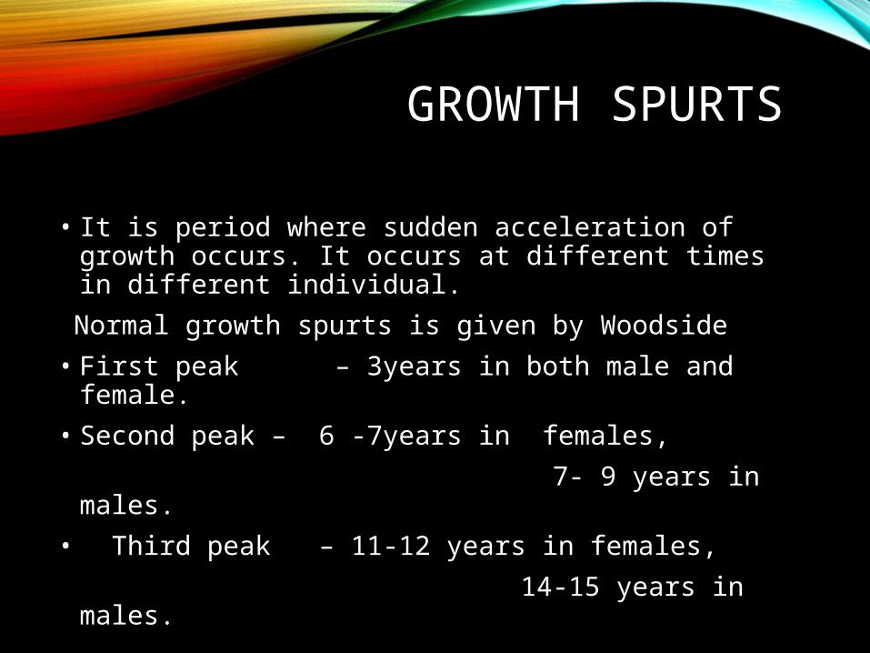
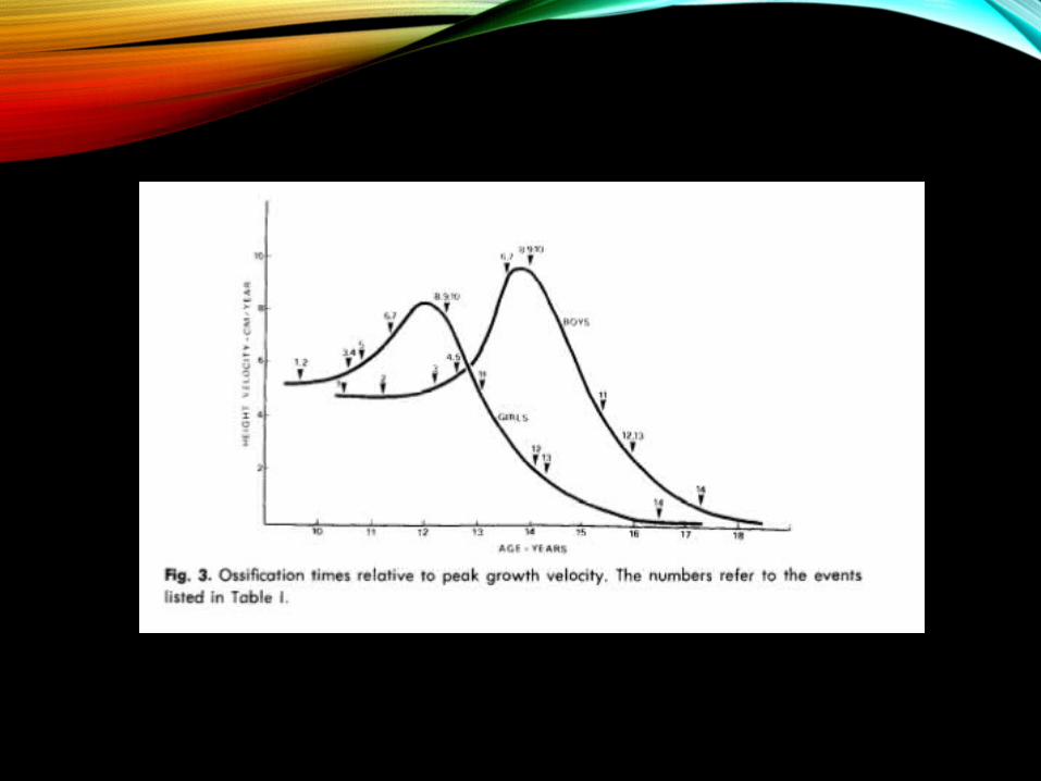
GROWTH SPURTS
• It is period where sudden acceleration of growth occurs. It occurs at different times in different individual.
Normal growth spurts is given by Woodside• First peak – 3years in both male and female.• Second peak – 6 -7years in females, 7- 9 years in males.• Third peak – 11-12 years in females, 14-15 years in males.

• WOODSIDE - pubertal growth spurt is important period for the orthodontics
• PROFFIT - juvenile growth spurt is more prominent in girls than pubertal growth spurt

VARIOUS METHODS TO DETERMINE THE SKELETAL
AGEHand and wrist radiographs• Standard method for the evaluation of the skeletal
age. • Easily identifiable maturity indicators.• Reliable source of maturation process. • Serves as a useful diagnostic aid.

Anatomy of skeleton of the hand• Phalanges – 14 in no.• Metacarpals – 5 • Carpals - 8

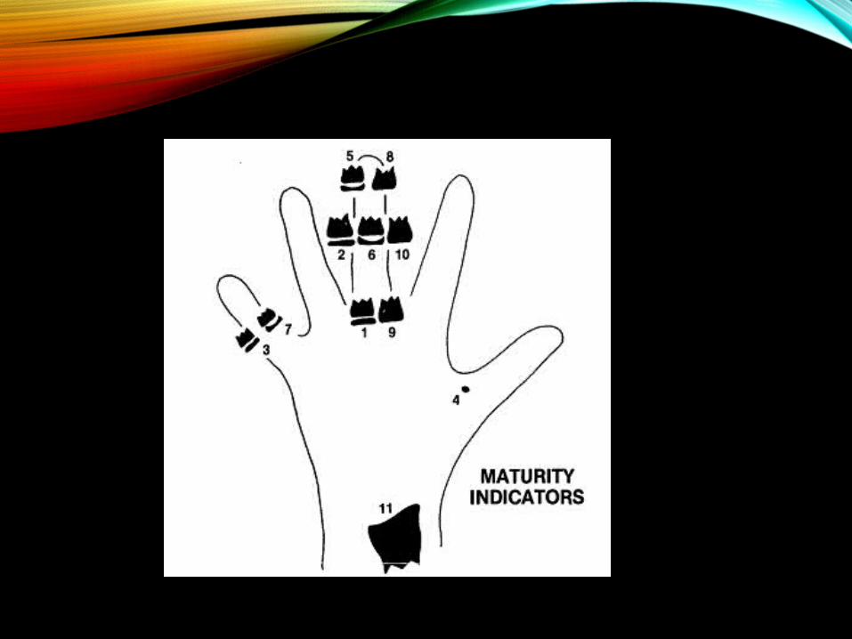
GRAVE & BROWN , AJO 1976

FIRST STAGE• PP2 = -Stage
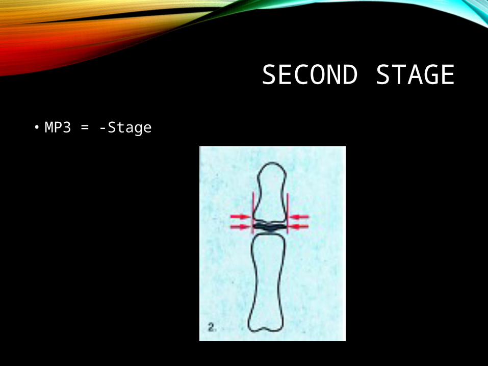
SECOND STAGE• MP3 = -Stage

THIRD STAGE• H1, pisi, and R
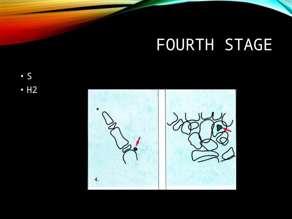
FOURTH STAGE• S• H2
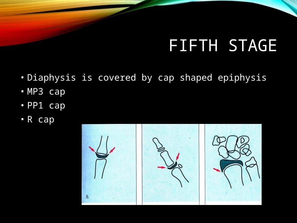
FIFTH STAGE• Diaphysis is covered by cap shaped epiphysis• MP3 cap• PP1 cap • R cap

SIXTH STAGE• DP3u• End of pubertal growth

SEVENTH STAGE• PP3u
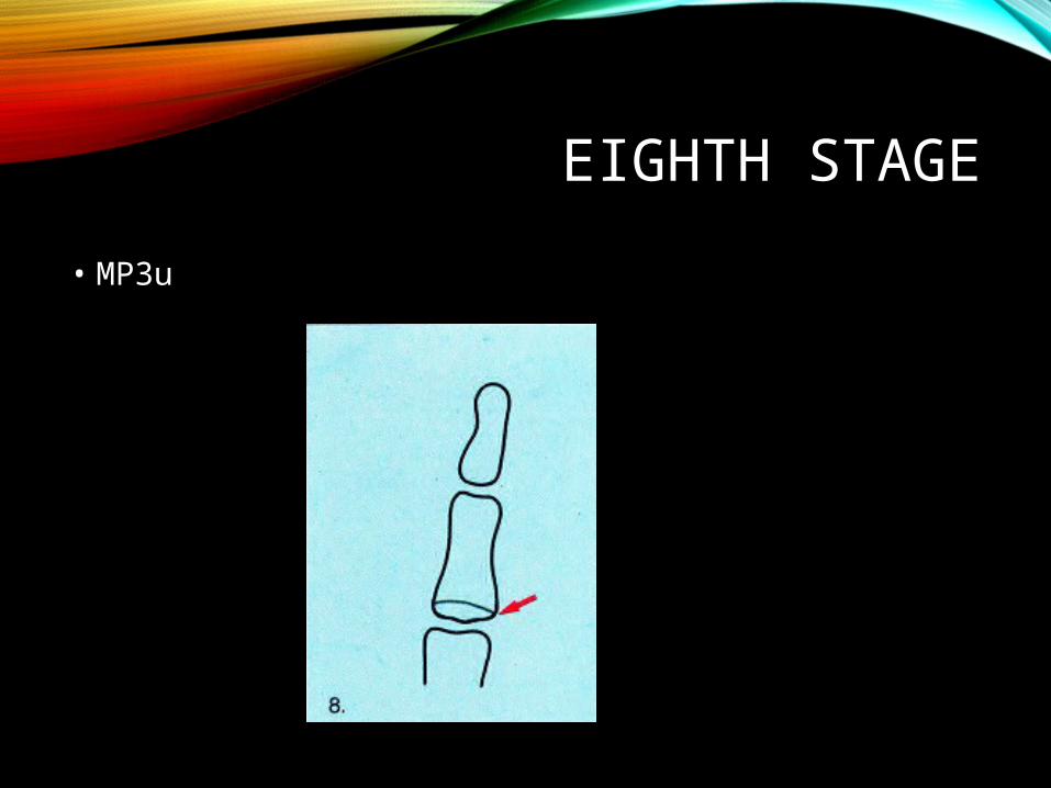
EIGHTH STAGE• MP3u

NINTH STAGE• Ru• Skeletal growth finished

CLINICAL IMPORTANCE• Treatment involving the application of extraoral
traction should be commenced prior to ossification of the hamate-2 or sesamoid.
• Complete band placement should take place at the time of peak growth velocity
• retention phase of treatment.

JULIAN SINGER AO 1980
• 1. To enable the clinician to rapidly and with some degree of reliability utilize the hand and wrist film to determine the maturation status.
• 2. Examine several other stages of the growth which
could be significance in treatment, exclusive puberal growth spurt.

EARLY

PREPUBERTAL
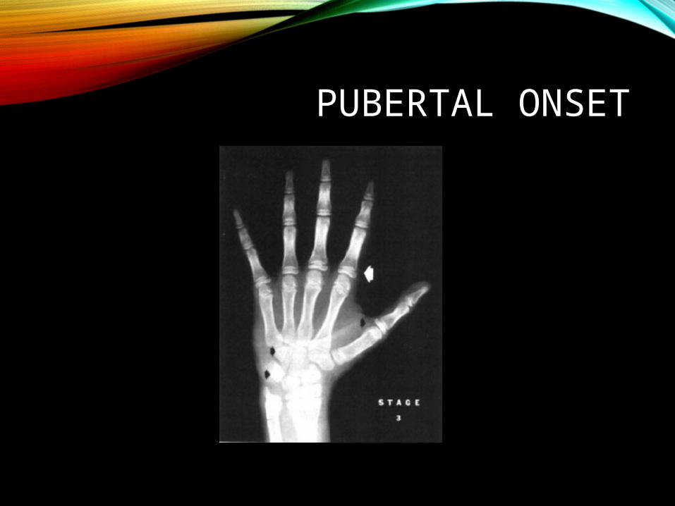
PUBERTAL ONSET

PUBERTAL

PUBERTAL DECELERATION

GROWTH COMPLETION• No remaining growth sites

Mainly concerned with stage 2 & 5.• Stage 2 represents that period prior to the adolescent
growth spurt during which significant amounts of mandibular growth are possible.
• Stage 5 represents that period of growth when orthodontic treatment might be completed and the pt is in retention therapy.
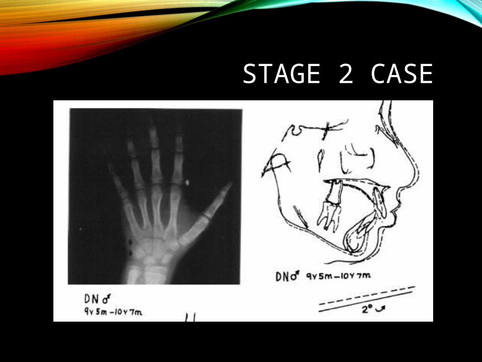
STAGE 2 CASE

STAGE 5 CASE


• Prepubertal period - during which Cl.II correction could be effectively achieved
• Pubertal spurt may be short in female.• Stage 5-period of residual growth during which post
treatment changes could occur

• Hunter- stated that dimension Ar- Po increased at a rate of 2.73mm per yr in females and 3.58mm per yr in males during the puberal growth period.

FISHMAN AO 1982


HAGG & TARANGER1982
• A longitudinal study- 212 children- from birth to childhood.
• Examinations include- standing height - tooth emergence - pubertal development - radiograph of right hand and
wrist

• Skeletal deveolpment• Two new epiphyseal stages: - MP3-FG - R-IJ
Hagg & Taranger, AJODO 1982

• The ossification of the ulnar sesamoid (S)- not a reliable indicator of the beginning of the pubertal growth in either sex.
• In clinical context - if S is not attained, neither is PHV.• If S is just attained, most individuals are in the
acceleration period of the pubertal growth spurt.• According to Bjork, the pubertal growth spurt ends
with the complete fusion of the third distal phalanx (DP3-I).

RELATIONSHIP OF ULNAR SESAMOID BONE AND MAXIMUM MANDIBULAR
GROWTH VELOCITY• Several studies were done and observed that- sesamoid bone precedes peak mandibular velocity
by - 0.72 yrs in males - 1.09 yrs in females• Mean age of appearance of sesamoid bone in females
precedes mean age in males by 2.34yrs
Pileski et al..,April 1973
THE RELATIONSHIP BETWEEN TOOTH MINERALIZATION AND EARLY
RADIOGRAPHIC EVIDENCE OF ULNAR SESAMOID
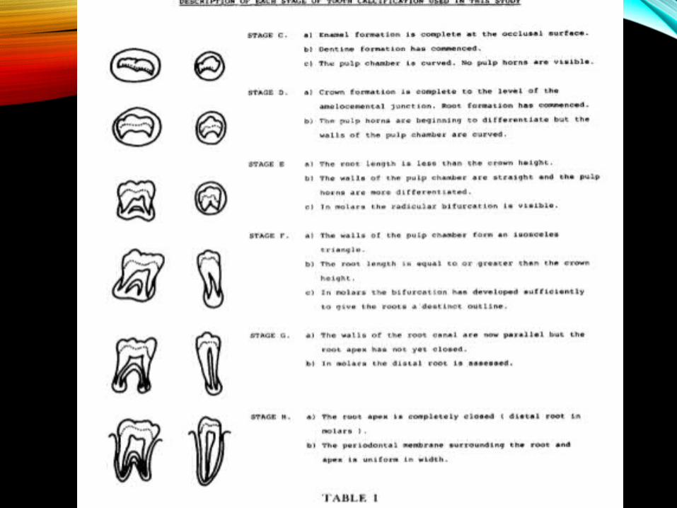
• Demirjain, Goldstein and Tanner- gave description of each stage of tooth clcification.
• Ossification of adductor sesamoid commences two yrs earlier among girls when compared with boys at mean age of 11.3yrs
• Lewis and Garn – no close relationship• Close relationship b/w calcification stage G of
mandibular canine and calcification of sesamoid

MANDIBULARCANINE CALCIFICATION
Coutinho, AJODO 1993
• They concluded that the initiation of pubertal growth spurt relates with stage F of canine calcification.
• canine calcification stage G- relates to MP3cap,S,PP5cap.

• Stage G occurs approximately around 1 yrs before PHV in boys but only 5 months before PHV in girls.(coincides with MP3cap,PP5cap.S)
• The intermediate stage between stage F and stage G should be used to identify the early stages of pubertal growth spurt.
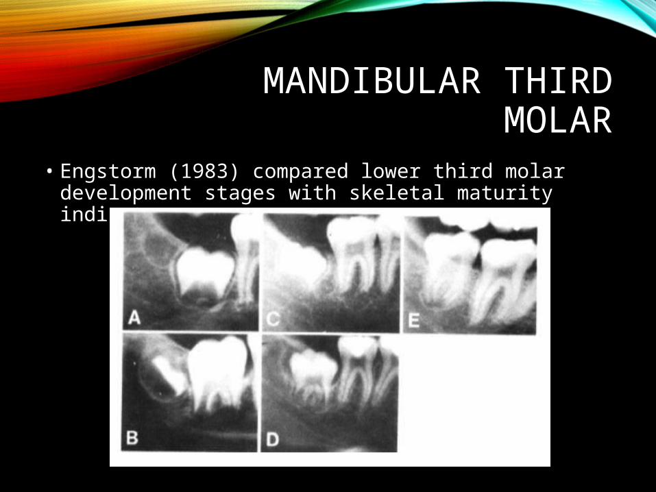
MANDIBULAR THIRD MOLAR
• Engstorm (1983) compared lower third molar development stages with skeletal maturity indicators.

• PP2=• MP3 cap• DP3 u• Ru:

CERVICAL VERTEBRAE MATURATION
• Lamparski (1972)
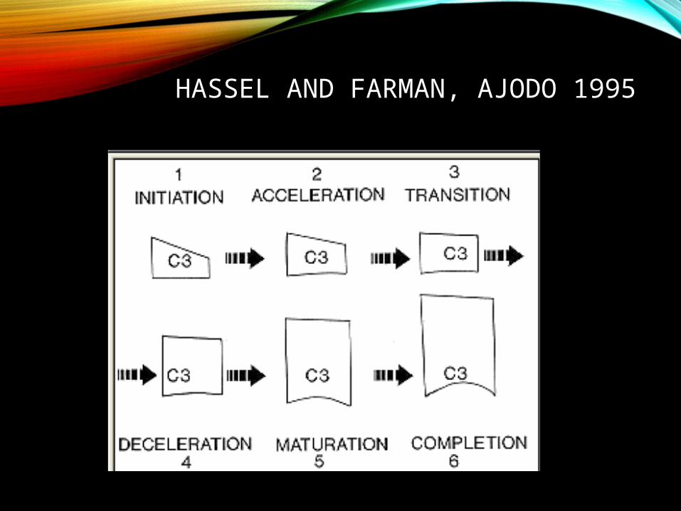
HASSEL AND FARMAN, AJODO 1995

NEW CVM• Baccetti et al (AO 2002) came with new version of
CVM.
• Lampraski method was modified because:• Make it easier & applicable to majority• To use minimal no. of vertebral bodies
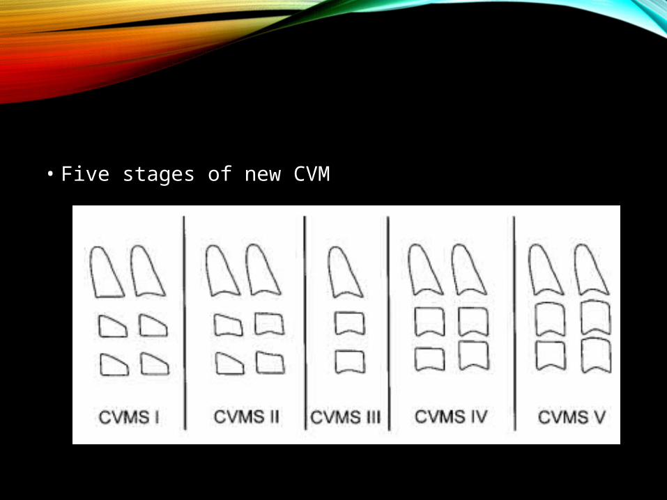
• Five stages of new CVM
A COMPARISON OF MODIFIED MP3STAGES & THE CERVICAL VERTEBRAE AS MATURITY
INDICATORS RAJAGOPAL AND KANSAL,
JCO JULY 2002
• Aim: determine whether the 6 modified MP3 stages (Hagg & Taranger) could be correlated with the 6 stages of CVMI’s (Hassel & farman).
• Evaluate the feasibility of recording MP3 stages using the standard dental x-ray.

• Chapman – first use the IOPA film to evaluate ossification of the ulnar sesamoid.
• Abdel kadar applied this idea to recording MP3 stages.

• MP3-F stage & CVMI 1

• MP3 – FG stage & CVMI 2

• MP3 - G stage & CVMI 3

• MP3 – H stage &CVMI 4
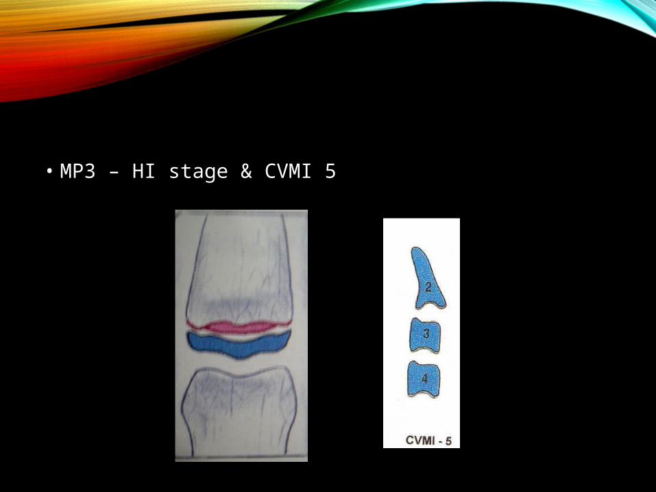
• MP3 – HI stage & CVMI 5

• MP3 – I stage & CVMI - 6
Modified MP3 stages using periapical X-ray film can be an accurate & simple growth indicator. Advantage of modified MP3
• Lower radiation exposure• High degree of clarity on radiograph• Easily identifiable stages of development• Close correlation to the six stages of CVMI• Less equipment
MATURATIONAL EVALUATION OF
OSSIFICATION OF THE MID PALATAL SUTURE
• positive correlation b/w adolescent maturation development and the approximation of the mid palatal suture.
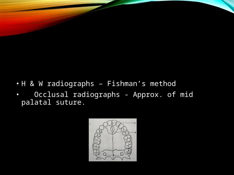
• H & W radiographs – Fishman’s method• Occlusal radiographs - Approx. of mid palatal suture.

• Increase in sutural approx. as SMI stages progressed.• SMI 1&2(PP3&MP3= less sutural approximation• After SMI 9(PP5u)- significant increase in the sutural
approximation• No significant difference b/w sexes.

Significant correlation b/w maturational development &beginning of ossification.
• At SMI 3(MP5)-• AT SMI 4-7 -• At SMI 9 (PP3u) –• At SMI 11 (Ru) -

• If orthopedic expansion forces are applied to physically open an approx. suture – best to accomplish this before SMI 9.
• Ideal time to initiate orthopedic expansion is during the SMI 1- 4 / early maturational stage.
• Mid palatal approx. occurs more posterior.

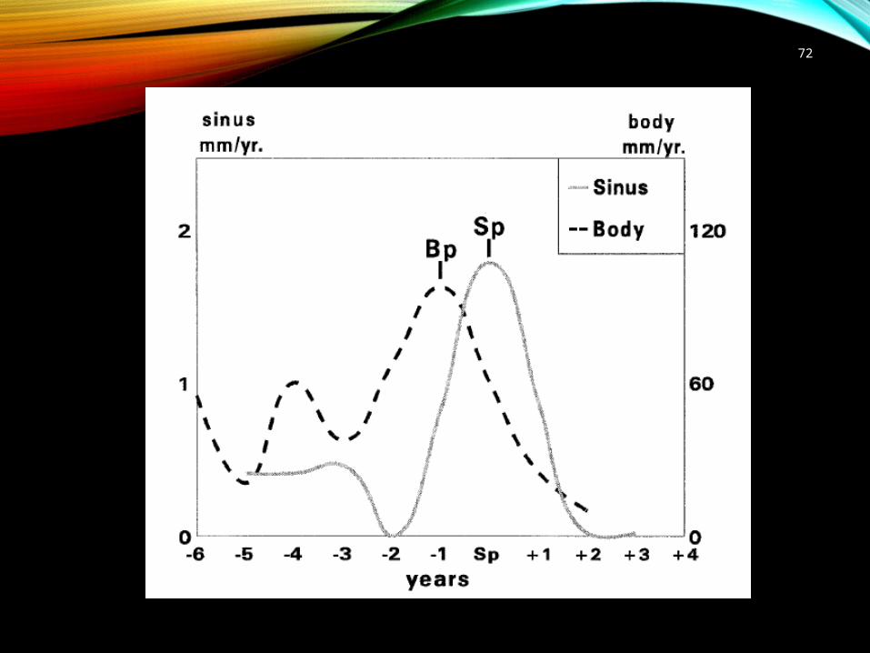
The frontal sinus- indicator for somatic maturity at puberty
Sabine Ruf AJODO 1991
Sh
Si
72

FRONTAL SINUS GROWTH VELOCITY (SV)
THRESHOLD VALUE T (1.3 MM/YR.)
Sinus data Prediction
Sv > T. Bp passed by approximately 1.4 yrs
Sv < T and Age <I5.1 yrs Bp not yet reached or reached less than 1.4 yrs. before the end of the observation interval
Sv < T and Age >15.1 yrs Bp passed by more than 1.4 yrs. with respect to the beginning of the observation interval
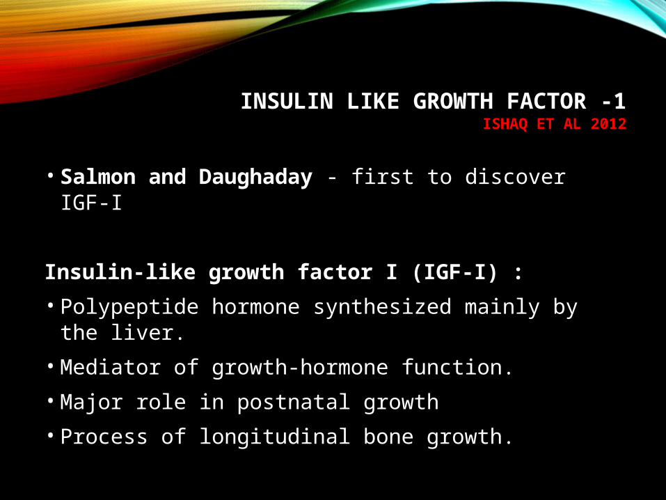
INSULIN LIKE GROWTH FACTOR -1ISHAQ ET AL 2012
• Salmon and Daughaday - first to discover IGF-I
Insulin-like growth factor I (IGF-I) :• Polypeptide hormone synthesized mainly by the liver.• Mediator of growth-hormone function. • Major role in postnatal growth • Process of longitudinal bone growth.

•Peak IGF-I concentrations reaches 1 year before puberty.•Earlier in girls than boys, •Peak IGF-I concentrations declines after puberty.

SERUM PTHRPZAHID HUSSAIN ET AL AJODO 2013
PTHrP is produced by cells at the periarticular ends of bones and acts on nearby chondrocytes bearing PTH/PTHrP receptors to keep them proliferating and delay their differentiation.
Inference :
• Peak serum PTHrP – late pubertal growth spurt

SERUM DHEAS• Stimulation of pitutary and hypothalamus• Stimulate growth and proliferation of epiphyseal
cartilage and potentiates the action of GH.• Enhance bone deposition
InferenceFirst peak – 6-8yrsSecond peak – 11yrs in females - 13yrs in males

CONCLUSION
• With reference to the studies related to the each of the maturity indicators, we can conclude that tooth calcification alone cannot be used for assessing maturity and the remaining growth potential.
• Hence correlation of the various indicators are necessary to plan treatment accordingly for any individual patient, whether to treat orthodontically, orthopaedically (or) surgically.

REFERENCES• 1. Hassel B, Farman A G.”Skeletal maturation
evaluation using cervical vertebrae” Am J Orthod, 1995; 107:58-61
• 2. Julian Singer “ Physiologic timing of orthondic treatment”. Angle Orthod, 1980; 50:320-333.
• 3.Hagg U,Taranger J “ Maturational indicators and the pubertal growth spurt”. Am J Orthod, 1982; 88:299-309.
• 4.Revelo B,Fishman LS,” Maturational evaluation of ossification of midpalatal suture”. Am J Orthod, 1994;105:288-292

• 5. Fishman L S,” Radiographic evaluation of skeletal maturation”. Angle Orthod ; 1982; 52:89-111
• 6. A comparison of modified MP3stages & the cervical vertebrae as maturity indicators - Rajgopal and kansal ,Jco 2002
• 7. Tooth mineralization as an indicator of the pubertal growth spurt. Am J Orthod, Jan 1980, Chertkow.
• 8. Sabine Ruf. Frontal sinus development as maturity indicator at puberty? Am J Orthod Dentofac Orthop 1996 110 476-82

• 9. Mohammed Zahid Hussain,a Ashok Kumar Talapaneni,b Mandava Prasad,c and Ramalingam Krishnand ;Serum PTHrP level as a biomarker in assessing skeletal maturation during circumpubertal development. Am J Orthod Dentofacial Orthop2013;143:515-21
• 10. Ramy Abdul Rahman Ishaq,a Sanaa Abou Zeid Soliman,b Manal Yehya Foda,b and Mona Mohamed Salah Fayed;Insulin-like growth factor I: A biologic maturation indicator; Am J Orthod Dentofacial Orthop 2012;142:654-61
• 11. Relationship between mandibular canine calcification stages and skeletal maturity – Am J Ortho Dentofac Orthop 1993; 104: 262-8.


![Skeletal maturation of the cervical vertebrae: association ... · Stage 1 or 2 of cervical vertebra maturation than individuals with Class I malocclusion (OR = 2.1 [CI 95%, 1.33-3.18]).](https://static.documents.pub/doc/80x56/5f8957bb6dc74c641762d7f3/skeletal-maturation-of-the-cervical-vertebrae-association-stage-1-or-2-of-cervical.jpg)


