9. Polymers under Deformation 175410. Polymer Fiber Spinning 1755
VI. Concluding Remarks 1756VII. Acknowledgments 1757VIII. References 1757
I. Introduction
A. Aim and Scope
This review will cover small-angle X-ray scatteringof polymers, especially with the use of synchrotronradiation. By nature, small-angle X-ray scattering(SAXS) probes relatively large-scale structures, incontrast to wide-angle X-ray diffraction (WAXD) thatdeals mainly with the atomic structure of crystals.SAXS includes not only the diffraction of large latticespacing, of the order of tens, hundreds, or eventhousands of interatomic distances, but also thescattering by perturbed or nonperiodic structures ofamorphous and mesormorphic materials. While thefundamental relation describing WAXD remains tobe the Bragg equation, nλ ) 2d sin(θ/2) [with θ beingthe scattering angle (we note that θ is used in thisreview instead of 2θ in order to be consistent withlight scattering); λ is the X-ray wavelength; and d isthe lattice spacing], the scattering (or diffraction) ofsemicrystalline or amorphous materials is oftendescribed in terms of electron density at point x, F(x),in reciprocal or Fourier space. Polymer chains canform semicrystalline, mesormorphic, or amorphousstructures. Therefore, a proper structure and mor-phology analysis of many polymers requires informa-tion using a combination of SAXS and WAXD, eventhough the WAXD measurements can appear in thenormal SAXS q range because of larger latticespacing. Here, q [)(4π/λ)sin(θ/2)] is the magnitudeof the scattering wave vector.
Polymers are molecules made up of many monomersegments. If all the monomer segments are of thesame type, these are homopolymers. If the segmentsare made up of two (or more) monomer types, theseare copolymers. Copolymers can have star, random,graft, block, and branched architectures. Polymerscan also be neutral or charged macromolecules, suchas biopolymers. In fact, a vast number of SAXSexperiments were reported on biological macro-molecules in solution, before synchrotron facilitieshad become available. However, as time-resolvedprotein folding and protein dynamics are covered
elsewhere in this special issue, the review on thebiological systems will be cursory in nature, empha-sizing mainly on the similarity between the oftencomplex biopolymers and the simpler synthetic poly-mers. Russell1 reviewed the topic of synchrotronSAXS studies for polymers with extensive theoriesand references. The present review will emphasizeexperiments performed after his review. Some funda-mental equations are, nevertheless, introduced forthe convenience of the reader.
In this review, we have intentionally missed twoexciting recent developments related to the techniqueof synchrotron SAXS. One is the microfocus synchro-tron SAXS technique, and the other is the coherentscattering or photon correlation studies using high-intensity SAXS (for example, Sector 8 at the APS willbe devoted for this purpose) to investigate dynamicprocesses in polymers. Each of these subjects is underrapid development, which may deserve an indepen-dent review later. The scope of this article is mainly
intended to cover the more “conventional” applica-tions of synchrotron SAXS.
B. Short History
Wilhelm Conrad Rontgen received the first NobelPrize in Physics for discovering X-rays. P. Krishna-murti reported the first SAXS observations on amor-phous materials including colloidal solutions andliquid mixtures in the late 1920s.2 In the next 40years or so it was not a trivial task to make SAXSmeasurements using conventional X-ray sources,especially on polymer solutions. The primary diffi-culty is associated with the fact that the scatteringmeasurements have to be done so very close to theincident X-ray beam. For example, with Cu KRradiation of 0.154 nm and a spacing d of 10 nm, thescattering angle is 0.88° (or a Bragg angle of 0.44°).With a period of 100 nm, θ ) 0.088°. These days, wedeal with hierarchical structures, often having lengthscales in the micrometer-size range. With d ) 1000nm, θ ) 0.0088° (or 32 s). At θ ) 32 s or 0.16 mrad,corresponding to making a scattering measurement16 mm away from the incident beam with a samplelocated 100 m away from the detector. This wouldbe a difficult task by any measure. Thus, the keypoint in any SAXS experimental setup is to try toreduce the background or stray X-rays at smallscattering angles (so-called the minimization of para-sitic scattering).
Over the years, there have been several designs onX-ray collimation systems. These are the four pinhole(or slit) system, the Kratky collimation system, andfor very small scattering angles the use of Bonse-Hart channel-cut crystals. We shall refer to theirdesigns for later discussions. It is suffice to mentionthat the descriptions and the original references havebeen presented in several excellent monographs.3-7
With synchrotron X-rays, the high power densityand small beam divergence of the incident X-raybeam permit design of time-resolved SAXS/WAXDexperiments8 and the use of very small specimens.Furthermore, the tunability of X-ray wavelength overa range near the K or L edge of an element has madeanomalous SAXS/WAXD a feasible approach forstructure and morphology investigations of a specificelement in the presence of other elements.9 Withmore complex systems, SAXS/WAXD experiments areoften coupled with other physical techniques, suchas laser light scattering, Raman spectroscopy, ther-mal analysis, Fourier transform infrared spectros-copy, and different forms of rheological techniques.One may also perturb the system by suddenly chang-ing the thermodynamic parameters, such as temper-ature jump, pressure jump, or rapid mixing ofmaterials using the stop-flow approach or a mixingcell. The coupling of additional variables could helpin the interpretation of SAXS/WAXD results of morecomplex polymer systems.
C. Light, X-rays, and Neutrons
The principles of scattering by light, X-rays, andneutrons are the same, being related to the inter-actions of radiation with matter. For X-rays and light,
Benjamin Chu obtained his B.S. degree, magna cum laude, from St.Norbert College and his Ph.D. degree in Physical Chemistry from CornellUniversity. He was a postdoctoral student with the late Professor PeterJ. W. Debye for four years before he started his academic career at theUniversity of Kansas. In 1968, he moved to the State University of NewYork at Stony Brook, where he is now a Distinguished Professor.
Benjamin Hsiao obtained his B.S. degree from National Taiwan Universityand his Ph.D. degree from the University of Connecticut. He was aPostdoctoral Fellow with Professors Richard S. Stein and H. HenningWinter at the University of Massachusetts, Amherst, from 1987 to 1989.He was a Staff Scientist and then a Senior Scientist with DuPont Fibersand DuPont Central Research & Development, respectively, from 1989to 1997. He was also an Adjunct Associate Professor, Materials Science,University of Delaware, from 1994 to 1997. In 1997 he moved to theState University of New York at Stony Brook, where he is currently anAssociate Professor in the Chemistry Department.
1728 Chemical Reviews, 2001, Vol. 101, No. 6 Chu and Hsiao
the electromagnetic radiation differs in the wave-length, with visible light in the 350-700 nm rangeand X-rays varying from ca. 0.01 to ca. 0.2 nm. Thereis no intrinsic limitation over the wavelength in theelectromagnetic spectrum from very short wave-length X-rays (in the γ-ray region) to the infraredregion, except for the range between long-wavelengthX-rays of about 3 nm and the vacuum ultravioletregion where the radiation is intensely absorbed byall materials and therefore rendering the scatteringby such probing radiation difficult to deal with. Forneutrons, the wavelength is on the order of 0.2-1.0nm, but neutrons obey the de Broglie relation with λ) h/p, h and p being Planck’s constant and themomentum, respectively. Due to the fact that thescattering by light, X-rays, and neutrons depends,respectively, on the differences in the refractive index(or dielectric constant), the electron density, and thescattering length (a nuclear property), the datatreatment can be different because of the intrinsicproperty of each radiation and its specific interactionswith matter are different. Nevertheless, we shallremember the similarity and the difference in dealingwith three different but complementary types ofradiation. Recently, we reviewed this subject else-where,9 and the interested readers are encouragedto read this review.
II. X-ray Instrumentation
A. Conventional versus Synchrotron X-raySources
With conventional X-ray sources, the beam diver-gence and the limit of using essentially one wave-length (often in the form of a copper or molybdenumtarget) for each setting have practically made SAXSexperiments a relatively difficult technique to under-take. These restrictions, however, have been removedwith the advance of synchrotron X-rays, rejuvenatingSAXS as a very useful technique for applications inbiology, chemistry, physics, materials science, andengineering. Furthermore, it should not be over-looked that advances in the X-ray linear position-sensitive detectors and X-ray area detectors, togetherwith multilayer monochromators that can also act asfocusing mirrors, have made SAXS a viable techniqueeven with conventional X-ray sources. Polymer chainsseldom form single crystals. In the amorphous orsemicrystalline state, the multilayer monochromatorcan provide adequate energy resolution on the order
of 0.1-1% and can increase the power density of theincident X-ray beam by a factor of 10-100, whencompared with conventional crystal monochromatorsthat have energy resolutions often better than 0.01%,though mostly not essential for SAXS of polymers.
A summary on the advantages of synchrotronX-rays (from a bending magnet source) is listed inTable 1. It is suffice to state that the small beamdivergence and the high brilliance of synchrotronX-rays permit experiments with small sample speci-mens as well as time-resolved and anomalous SAXSmeasurements, resulting in a much-expanded viewon synchrotron SAXS applications. Aside from theinformative and excellent review by Russell,1 reviewson selected topics related to SAXS have appeared.9-16
The advantages of using synchrotron X-rays from aninsertion device, such as an undulator or a wiggler,perhaps are more than those from a bending magnetsource. For example, the X-ray brilliance (a measureof the number of photons per second per solid angleper source area per unit bandwidth) from an undu-lator source can be several orders of magnitudehigher than that from a bending magnet source. Thecharacteristics of the X-rays from an undulator or awiggler can be found in the Handbook of SynchrotronRadiation as listed in ref 1.
B. Synchrotron SAXS BeamlinesKoch10 reviewed the state of the art for synchrotron
SAXS in 1988. Bras and Ryan8 subsequently listedthe synchrotron SAXS beamlines up to 1996, includ-ing three in Daresbury, two at the Photon Factory,three at APS, one at SPring 8, one at SSRL, four atESRF, four in Hamburg, two in Lure, one at NSLS,and one at ELECTRA, for a total of 20 synchrotronSAXS beamlines. Their Table 18 also listed the sourceof information for these beamlines. To our knowledge,NSLS has at least two more SAXS beamlines andChina, Korea, and Taiwan have one each. Thus, thetotal number of synchrotron beamlines capable ofSAXS experiments is increasing. Recently, DESY(Hamburg) is planning an X-ray-free electron laser(FEL) facility that can generate X-ray intensity evenseveral orders of magnitude higher than the undu-lator beamlines. A dedicated SAXS station in thisFEL facility, to our knowledge, is being planned.
The natural divergence of the X-ray beam gener-ated from a synchrotron source (bending magnet,wiggler, or undulator) can be considered as the lowestnatural limit for angular resolution of the instru-
Table 1. Advantages of Synchrotron X-rays (from a bending magnet source) for SAXS and WAXD
advantages applications
1. high intensity time-resolved experimentssmall specimenbetter counting statistics
3. well-defined energy4. tunability of wavelength anomalous SAXS and WAXD
atom selectivity5. pulsed source
∼107/s, each ns wide6. polarization of beam
Small-Angle X-ray Scattering of Polymers Chemical Reviews, 2001, Vol. 101, No. 6 1729
ment. Thus, without touching the beam, one mayexpect the highest spatial resolution that can beobtained based on the incident beam divergencealone. This is, of course, not practical as mostbeamlines involve a long source-to-detector distance(in the range of tens of meters). As the detectordimension is limited, the beam size has to be reduced.The process of the beam reduction, inevitably, willproduce parasitic scattering. In section II.B, we willbriefly describe several means to minimize suchundesired stray scattering. Riekel et al. recentlydemonstrated the SAXS capability from a microfocusbeamline at ESRF (ID13).17-20 The smallest achiev-able beam size is about 1 µm. This is a greataccomplishment which can be attributed to theexcellent arrangement of the collimation and focusingcomponents. It is worth mentioning here that thesmaller beam size is often balanced by a larger beamdivergence in order to retain sufficient intensity.
Most synchrotron SAXS beamlines tend to use theslit collimation system for ease of operation in chang-ing the beam dimensions because all the slit move-ments have to be operated by remote control. Themore stringent requirement involves moving the slitsinside the vacuum path because the number ofwindows should be reduced to a minimum in orderto minimize the amount of parasitic scattering. Thisis a constant battle in instrumentation design forsynchrotron SAXS experiments. It is noted that thehigh power density of the incident X-ray beam willbe completely wasted if the parasitic backgroundscattering were to increase proportionately. For theease in alignment and its ability to change theincident beam dimensions, the slit collimation systemand the corresponding complementary collimationsystems are presented. The purpose of the presenta-tion on the collimation systems is for the reader tohave some idea on how a SAXS instrument isdesigned and constructed. It is by no means meantto be exhaustive, and only selected references areused.
C. Collimation Systems
1. Pinhole Collimator
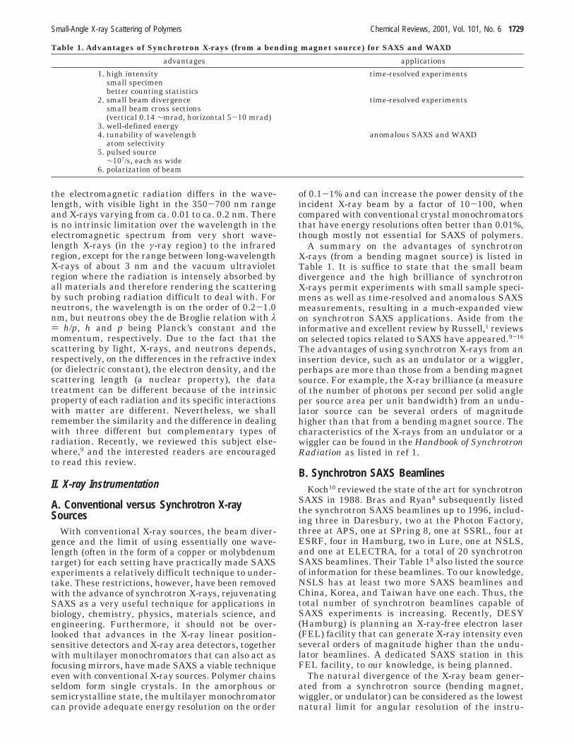
The synchrotron radiation source in combinationwith a focusing mirror offers only directional colli-mation. In contrast, the pinhole (or slit) collimationsystem offers symmetric beam cross-section and hasthe ability to investigate anisotropic scattering pat-terns.21 The pinhole (or slit) collimation system alsomakes the desmearing effect less serious. Over theyears, the pinhole geometry has been discussedextensively.4,22-38 It is important to note that manydesigns of the slit system use the geometry shownschematically in Figure 1 (reproduced from ref 4).The optical elements denote a double-crystal mono-chromator and a focusing mirror. A set of beam-defining slits is used to limit the incident beam cross-section and to take out unwanted background. Thescatter slits, as shown in Figure 1, are used to followthe incident beam profile and to act as guard slits,especially for the set near the sample. It is importantto note that although the cone produced by parasitic
scattering is governed by the two sets of scatter slits,fluctuations caused by the positions of the X-raysource, the beam defining slits, and the opticalelements would increase the amount of parasiticscattering, especially for the slit set near the sample.Consequently, depending on the stability of all thecomponents before the scatter slits, the beam stophas to be made larger and the expected parasiticscattering contribution should be higher than onewould normally expect from theoretical computationsalone.
The slit collimation system also suffers from havingtoo many adjustable parameters. Each blade, inprinciple, should have a translational stage and threeangular adjustments in order to locate the desirededge at the proper position. As shown in Figure 1,the two sets of scatter slits have four blades, requir-ing a total of 32 adjustments. Clearly, such anarrangement would make proper alignment a diffi-cult practical task. Thus, many requirements of theslit alignment have to be designed into the systemduring construction. Nevertheless, it would remaina formidable task for a slit collimation system toreach the design limit in terms of parasitic scattering.
The quality of the blade can also influence theeffectiveness of the collimation. The blade should bemade of materials with heavy atomic number (goodX-ray absorber), low fluorescent scattering back-ground, and machinability, such as tantalum andtungsten (lead is too soft to be machined). In prin-ciple, any materials placed in the X-ray beam willalways cause scattering (sometimes diffraction) and/or reflection, which then lead to unwanted parasiticbackground. The problem of scattering can be mini-mized by increasing the homogeneity of the bladematerial, and the problem of reflection can be elimi-nated by using a tilted smooth surface.
On the basis of the same principle as slit collima-tion, a pinhole collimation system immediately re-duces the number of adjustable parameters requiredto align such a collimator. If one were to have thetapered pinholes (the smaller opening always facesthe incident beam to minimize the reflection) alignedperpendicular to the propagation axis of the incidentbeam, only four translational stages to move the twopinholes in line with the incident X-ray beam arerequired. To avoid uncertainties due to fluctuationsin the position of the X-ray source, the defining slits,and the optical elements, a third guard pinhole isadded to the pinhole collimation system.21 With threepinholes, the collimation system is self-sufficient and
Figure 1. Schematic diagram of a slit collimation systemfor focused synchrotron X-rays. (Reprinted with permissionfrom ref 4. Copyright 1982 Academic Press.)
1730 Chemical Reviews, 2001, Vol. 101, No. 6 Chu and Hsiao
should be relatively independent of the fluctuationsdiscussed above.
Figure 2 shows a schematic diagram with three-pinhole collimation geometry.21 The first and secondpinholes define the incident beam. The third guardpinhole blocks the parasitic scattering due to the edgescattering from the second pinhole. It is noted thatdue to the intrinsic divergence of the synchrotronbeam, the incident flux is mainly controlled by thesize of the first pinhole. To achieve a high angularresolution and low parasitic scattering, the guardpinhole must be designed and placed in such a waythat it is close to but does not touch the incidentbeam.
The smallest angle one could reach without aserious parasitic scattering problem, θs, can be de-scribed by the relation
where ds denotes a finite size for the detector elementand d3 [)(d1 + d2)l2/l1 + d2] depends on l1, l2, d1, andd2. There is always a compromise between theincident intensity and the angular resolution. Herewe refer the incident intensity as the total flux ofX-rays reaching the sample. Clearly the total fluxdepends not only on the angular divergence of theincident beam, but also on its beam cross section. Inthe third-generation synchrotron X-ray sources, it isthe brilliance that has an edge over this concern. Thedesign of the pinhole collimation system depends alsoon the space limitation, especially in the length ofthe instrumentation setup. For example, for the X3A2beamline at NSLS, we chose l1 ) l2 ) 609 mm (sothat the total length was sufficiently short for easytransportation of the entire collimator between StonyBrook and NSLS by car) and d1 ) d2 ) 0.3 mm.Therefore, d3 should be slightly larger than 0.9 mm.We used ls ≈ l3 ) 1030 mm and d3 ) 1.0 mm withthe extra 0.1 mm as a compromise in alignmentuncertainty and edge asymmetry. Theoretically, sucha pinhole collimation geometry should yield a θs of1.62 mrad for a CCD-based detector with ds ) 0.135mm and a θs of 1.58 mrad for a Braun detector withds ) 0.046 mm. It should be noted that the differencesbetween Figures 1 and 2 are as follows. (1) Figure 1has a focused X-ray beam, while Figure 2 does not.Thus, for a synchrotron X-ray beam with very smallbeam divergence, the choice of scatter slits (Figure1) or of d1 and d2 depends on the incident beam
profile. (2) Figure 1 has two sets of scatter slits, whileFigure 2 has a guard pinhole, in addition to the twodefining pinholes. The guard pinhole is clearlyredundant if the X-ray source position remains fixed.Unfortunately, in practice, fluctuations of the X-raysource position should be taken into account. Thus,the use of an additional guard pinhole is highlyrecommended.
2. Kratky Block Collimator39
Among the collimation systems proposed to achievelow parasitic scattering in SAXS, the concept of ablock-collimation system, as first proposed byKratky,40,41 has stood the test of time because of itssimplicity in construction and ease of operation. Amore detailed description of the Kratky camera,including the compact camera, has been reported.42,43
Another advantage of the Kratky block collimator,as shown in Figure 3, is that there is no guard slitrequired. In comparison with Figure 2, the Kratkyblock-collimation system can be made shorter thana comparable pinhole collimation system by a factorcorresponding to that of l2. It also differs from theslit collimation system of Figure 1 because theincident X-ray beam is defined by the Kratky colli-mator, where small fluctuations of the X-ray sourceprovide very little contribution to the parasitic scat-tering. Furthermore, the Kratky block collimator isbest with a slit geometry that coincides with theunequal divergence of the synchrotron X-ray beam.Consequently, a larger total flux from the use of alonger slit length can be accommodated. However, forthe same reason, it is not usually used for experi-ments with anisotropic samples where the scatteringfrom x-z directions is different.
Figure 2. Schematic diagram of a pinhole collimationgeometry. (Reprinted with permission from ref 21. Copy-right 1994.)
Figure 3. Section along the y direction of the synchrotronbeam perpendicular to the x,z plane. Vertical z1 scale ismultifold stretched compared to the horizontal y1 axis.Variable entrance slit S and blocks B1 (middle block) andB2 (bridge) are perpendicular to the x1,z1 plane. In the planeof registration R, the primary beam does not have atriangular (vertical) beam profile. A Gossip-shaped inten-sity beam profile is presented schematically to emphasizethat the synchrotron radiation (as denoted by the dottedarea) is highly collimated. M0 is the distance from themaximum intensity to the plane H. The Gossip-shapedbeam has fairly symmetrical long tails, mainly due toscattering by the edges k1 and k2. O1 and O2 represent thetop surface of block B1 and the bottom surface of block B2,respectively. The small-angle X-ray diffractometer (SAXD)dimensions are a ) 16.7 mm, b ) 402.0 mm, c is variablefrom ∼0.2 to 1.5 m. The subscript 1 denotes the coordinatesystem x1,y1,z1 for the SAXD, where x,y,z denotes thecoordinate system for the synchrotron primary beam. Wewant x||x1, z||z1, and the tilt y1 with angle δ between y andy1 using x as the rotation axis. (Reprinted with permissionfrom ref 39. Copyright 1987.)
θs )d2 + d3
2l2+
d3
2l3+
ds
2l3(1)
Small-Angle X-ray Scattering of Polymers Chemical Reviews, 2001, Vol. 101, No. 6 1731
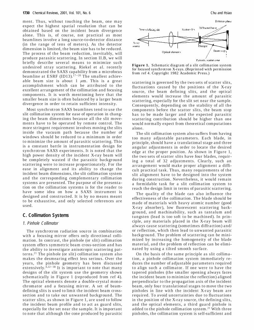
3. Bonse−Hart Channel-Cut Crystals Optics44-46
In a small-angle X-ray scattering experiment, thelarger the inhomogeneity size in a system, the longerthe X-ray wavelength or the smaller the scatteringangle, i.e., the smaller the q value, is needed in orderto determine the structures. A Kratky block-colli-mation system could usually reach a q value of about0.04 nm-1. This q value corresponds to an X-raywavelength λ of 0.154 nm and a scattering angle ofabout 1 mrad. On the other hand, routine laser lightscattering can cover a q range from 0.004 to 0.036nm-1. With a specially designed instrument, e.g., byusing a prism cell,47 the smallest accessible q valuecould be on the order of 0.0008 nm-1. With synchro-tron X-rays from an insertion device (such as awiggler or an undulator), the intrinsic beam diver-gence is very small. Thus, by using long distancesbetween the scatter slits (Figure 1) or the definingpinholes (Figure 2), it is expected that θs down to tensof µrad should be readily accessible. The use ofBonse-Hart X-ray optics is reserved only for specialoccasions since the scattered intensity at each scat-tering angle has to be measured separately. It canbe used with conventional X-ray sources, and a high-flux/high-temperature setup has been reported.48 Noparallel methods for simultaneous measurements ofthe scattered intensity at many scattering angleshave yet been devised.
In the 1940s, Fankuchen and Jellinek49 andothers50-52 proposed to use one crystal to mono-chromatize the X-ray beam and another one as ananalyzer for a SAXS instrument. However, theangular resolution was not sufficiently good if onewere to reflect the X-rays only once in each of thecrystals. In 1966, Bonse and Hart53,54 increased thenumber of reflections in each of the crystals with acorresponding dramatic increase in the angularresolution.
There have been considerable interests in furtherdevelopment of the Bonse-Hart instrument,55-68
particularly in synchrotron X-ray facilities, becauseof its potential applications to many current topicsin materials research with length scales in themesoscopic micrometer-size range. It should also benoted that many materials studies deal with hightemperatures and involve temperature scanning,jumping, quenching, and annealing experiments. Byusing Super Invar as the basic building material forthe posts, supports, and the microscrews, the thermalexpansion effect could be minimized.45 As the Bonse-Hart optics directly measure the small angles nearthe incident beam, the measurement device needs tobe kept extremely stable over a range of tempera-tures.
Figure 4 shows a schematic diagram of the instru-ment setup46 used in the X3A2 beamline at theNSLS. Two silicon crystals with channel cuts alongthe (111) reflection plane were used. The samecrystals could accept an X-ray beam at different X-raywavelengths, such as λ ) 0.06573 or 0.154 nm. Eachcrystal had rocking, tilting, and translational adjust-ments. The microscrews were located 4.875 in. apartand had a resolution of 250 µm per revolution. Theactuator (Klinger BM25-PP) had a resolution of 0.15
µm per step. With a distance of 3.75 in. from the pivotin the first crystal and 4.75 in. from the pivot in thesecond crystal, the angular resolutions were about0.33 arc s for the first crystal and about 0.26 arc sfor the second crystal. Due to the high penetrationpower of the shorter X-ray wavelength, higher orderharmonic wavelengths from the synchrotron X-raysmade attenuation of the incident X-ray more difficult.The distortion in the SAXS profile produced by higherharmonics should be considered.
4. USAXS
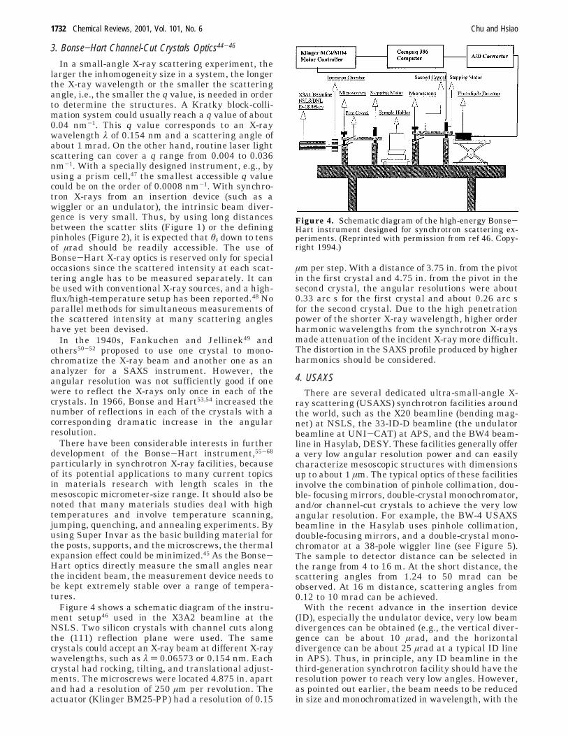
There are several dedicated ultra-small-angle X-ray scattering (USAXS) synchrotron facilities aroundthe world, such as the X20 beamline (bending mag-net) at NSLS, the 33-ID-D beamline (the undulatorbeamline at UNI-CAT) at APS, and the BW4 beam-line in Hasylab, DESY. These facilities generally offera very low angular resolution power and can easilycharacterize mesoscopic structures with dimensionsup to about 1 µm. The typical optics of these facilitiesinvolve the combination of pinhole collimation, dou-ble- focusing mirrors, double-crystal monochromator,and/or channel-cut crystals to achieve the very lowangular resolution. For example, the BW-4 USAXSbeamline in the Hasylab uses pinhole collimation,double-focusing mirrors, and a double-crystal mono-chromator at a 38-pole wiggler line (see Figure 5).The sample to detector distance can be selected inthe range from 4 to 16 m. At the short distance, thescattering angles from 1.24 to 50 mrad can beobserved. At 16 m distance, scattering angles from0.12 to 10 mrad can be achieved.
With the recent advance in the insertion device(ID), especially the undulator device, very low beamdivergences can be obtained (e.g., the vertical diver-gence can be about 10 µrad, and the horizontaldivergence can be about 25 µrad at a typical ID linein APS). Thus, in principle, any ID beamline in thethird-generation synchrotron facility should have theresolution power to reach very low angles. However,as pointed out earlier, the beam needs to be reducedin size and monochromatized in wavelength, with the
Figure 4. Schematic diagram of the high-energy Bonse-Hart instrument designed for synchrotron scattering ex-periments. (Reprinted with permission from ref 46. Copy-right 1994.)
1732 Chemical Reviews, 2001, Vol. 101, No. 6 Chu and Hsiao
required optical components needed to be used in thedownstream of the ID in order to specify the incidentbeam. A careless design of an individual opticalcomponent can significantly increase the parasiticscattering of the beam downward and destroy the lowangular region of the beamline.
D. Detection SystemsWith the high brilliance of synchrotron X-rays, the
development of detector technology is still laggingbehind. It is evident that close to 10 years after thereview by Russell,1 X-ray detectors for short-timetime-resolved measurements have not yet met themany requirements demanded for fast point counters,linear position-sensitive detectors, and CCD areadetectors.
Each detector has its own strengths and weak-nesses. The selection depends on the particularapplication and the specifications of the detector aswell as the X-ray wavelength. Generally, it is es-sential to consider the quantum efficiency, spatialresolution, temporal resolution, energy resolution,linearity, dynamic range, uniformity of response,count-rate limitations, and background noise of adetector at a specific X-ray wavelength in order toproperly match the detector for a specific application.Russell1 reviewed the gas-filled detectors, photodiodearrays, charged-coupled devices (CCDs), electro-optical devices, and channel electron multipliers. Inthis review, only a very brief discussion is presented,with emphasis on more recent advances.
1. Point DetectorsThere are a variety of point detectors available for
use in SAXS/WAXD experiments ranging fromsingle-channel proportional counters, scintillationcounters, to photodiode detectors. While the technol-ogy for proportional counters and scintillation countershas remained essentially unchanged, the more in-teresting development deals with the use of photo-diode detectors68,69 as an X-ray photon counter and,at high-count rates, as an analogue device to measurethe photocurrent that is proportional to the X-rayintensity.69
For X-ray photon counting and correlation, thephotodiode should be cooled to low temperatures(normally about -10 °C) in order to reduce thermalnoise. The output signal could be amplified, discrimi-nated, and counted by a scalar/timer or correlated
by a digital correlator. At λ ) 0.0643 nm, the darkcount was negligible after discrimination.68 A cooledphotodiode with energy-dispersive capability togetherwith photon counting in the ac mode by means ofmultichannel analysis and with current measure-ments in the dc mode is available commercially.68
Such a cooled photodiode detector with a Be windowand thermal isolation can reduce the dark current(count) by a factor of about 100, thus making weakersignal measurements feasible. The large dynamicrange of the photodiode detector makes it suitableas a convenient point detector for X-rays. Never-theless, for low count-rate experiments, the pro-portional and scintillation counters remain theappropriate detectors, especially when one wants toachieve a predetermined signal-to-noise ratio over arange of q where the intensity is a strong function ofq. The recent development of a new kind of silicondrift detector demonstrates that very high count-rateperformance (1 000 000 cps maximum input countrate, 400 000 cps output count rate) can be achievedby the point detector system.
2. Linear Position-Sensitive Detectors70
The use of linear position-sensitive detectors(LPSDs) increases the efficiency of SAXS/WAXDexperiments drastically and makes time-resolvedmeasurements feasible. Russell1 reviewed the posi-tion-sensitive proportional counters. In particular,the Gabriel-type detectors72-75 are very suitable forrecording SAXS of extremely weak scatterers suchas dilute polymer solutions. The general principle ofthis detector is simple. A proportional detector con-sists essentially of a cell with detecting gas (Ar/CO2or Xe/CO2) and a thin (10 µm) central metal wire(Au-coated Cu wire). A high voltage is appliedbetween the wire and the body of the detector. Whenan ionization event occurs in the gas, photoelectronsthat are produced by the ionization are acceleratedin the wire. In the vicinity of the wire, a Townsendavalanche occurs and provides a large amplification.A preamplifier picks up this electric signal, which iscorrelated with the energy of the incident event(intensity) and the position of the avalanche. Unfor-tunately, wire/gas detectors, which are quantumlimited, have relatively low maximum count rates(e106 X-ray photons per second), and further incre-mental improvements in wire/gas detectors are dif-ficult to achieve. For many strong scatterers such ascrystalline polymers, one often has to attenuate theincident synchrotron X-ray beam in order to protectthe detector and thereby defeats one of the uniqueadvantages of synchrotron X-rays.
Two types of photodiode array (PDA) detectorshave been used with some success: (1) directlyexposed X-ray PDAs71-77 and (2) phosphor scintilla-tors coupled to PDAs to convert X-ray photons tovisible photons.78 The first type has a higher gainthan the second one71 but has a narrower detectableX-ray energy range (2-20 keV) and suffers radiationdamage resulting in a permanent rise in dark countnoise and a decrease in sensitivity. Thus, its use hasnot been very popular. The second approach is quitefeasible. For time-resolved studies, it is important to
Figure 5. Schematic diagram of the USAXS beamline(BW4) in the Hasylab, DESY. (Diagram reproduced fromthe beamline description of BW4 at Hasylab, DESY.)
Small-Angle X-ray Scattering of Polymers Chemical Reviews, 2001, Vol. 101, No. 6 1733
select an appropriate phosphor with sufficiently shortlifetimes, not only for the conversion of X-ray photonsto visible photons, but also for the conversion againto visible photons inside the intensifier via fiberoptics for acceptance by the photodiode array. ThePDA-LPSDs have good spatial resolution, excellentlinearity of response to the X-ray intensity, and stablepixel uniformity. All the instrument parameters canbe calibrated, and the PDA-LPSDs are relatively lowcost, when compared with CCD X-ray area detectors,and can be used for fairly fast (etens of ms per frame)time-resolved SAXS/WAXD experiments of symmet-ric systems or anisotropic systems that have beenspatially averaged over all orientations. One draw-back has been that the PDA-LPSDs have a limitedspatial range, e.g., normally an effective length ofonly 25 mm for a photodiode array of about 1000pixels is used.69 Furthermore, its dynamic range isoften limited.
3. Area Detectors
For weak signals, the Gabriel-type 2-dimensionalmultiwire proportional chamber (2D MWPC) areadetectors generally have a low dark count rate thatcannot be surpassed and can provide precise digitalcounting information on the X-ray intensity.73-75 Ina typical 2D MWPC area detector configuration, theanode consists of a central plane of metal wires,located between two planes of cathode wires. Eachelement of the cathode wires is connected to a singledelay line. The signal pick up on this delay line thusis correlated with the energy and the position of theincident event along the cathode plane. Two anodeplanes are arranged perpendicular to each other.Such geometry can thus be used to resolve theposition of the event in two dimensions. In theconventional design, all the wires (anode and cath-odes) are prealigned in a straight-line fashion witha uniform spacing of about 0.5 mm apart. Theentrance window is made of carbon composite whichis used to withstand high gas pressures (∼6 atm). Amuch more advanced MWPC detector has recentlybeen developed for a very high count rate and veryfast read-out performance.8
The imaging plate,79 either coupled directly witha scanner or operating separately from a scanner,offers the best compromise for routine operations. Ithas a large dynamic range, excellent spatial rangeand revolution, low background, and reasonablelinearity. It is robust, and the operating cost isrelatively low, especially for multiple users. However,it cannot be used easily for time-resolved experiments(its typical read-out time is 300 s, and the handlingtime is 150 s) and measurements of absolute intensityare difficult to achieve because the exposed imagingplate decays slowly and depends partly on the timeperiod after exposure and before processing.
CCD X-ray area detectors have made incrementaladvances in recent years and are commercially avail-able. X-rays could be detected directly by CCDs.However, the chip size is usually too small (≈25-50mm on each side) for convenient applications. Moreimportantly, the CCD would suffer radiation damageby direct exposure to X-rays, leading to an increase
in the dark count rate80-82 and a limited dynamicrange because each X-ray photon could producehundreds of electron-hole pairs due to the highenergy of the X-ray radiation.83 Converting X-rays tovisible light photons by means of an appropriatephosphor could avoid these problems but wouldintroduce statistical noise.84 After conversion, theimage on the phosphor can be transferred to the CCDby using a lens system,85 fiber-optic coupling,86 animage intensifier and a lens system,87-89 or an imageintensifier and fiber-optic coupling.90 Figure 6 showsa schematic comparison of the collection efficiencybetween a fiber-optic-coupled CCD detector and alens-coupled CCD detector.91 In a lens-coupled CCDdetector, one can compensate for the light loss byusing an image intensifier. However, this approachwould increase the complexity of the instrument,decrease the dynamic range and the linearity due tolimitations of the image intensifier, and result in ahigh background noise. Thus, commercial CCD X-rayarea detectors now use the format of the configura-tion shown in the top of Figure 6. Large CCD chips(up to 4k pixels) or multiple CCD chips (often fourchips with 1k pixels per chip), together with anappropriate fiber-optic taper, can provide a largeenough spatial range and high enough spatial resolu-tion for most purposes. However, in the time-resolvedmode, a compromise has to be made in selecting theappropriate dynamic and spatial ranges as well asspatial resolution in order to achieve a reasonablenumber of frames per second.
Finally, it is noted that even with the popularityof CCD X-ray area detectors, further improvementson gas/wire counters should not be ignored. Forexample, a new type of centroid-finding method forposition-sensitive detectors has allowed higher countrates and good spatial resolution.92 A curved micro-strip gas counter has been designed for synchrotronradiation time-resolved SAXS/WAXD experimentswith count rates of up to 1 MHz per channel and achannel width of 0.4 mm.93 Time-resolved SAXS/WAXD experiments allowing two-dimensional pat-terns to be recorded with exposure times as short as
Figure 6. Fiber-optic-coupled CCDE detectors (top) havea significantly higher collection efficiency than comparablelens-coupled CCD detectors (bottom). The fiber bundle,with its larger light-capturing area, maintains the incidentpattern of illumination and delivers it to the CCD. (Re-printed with permission from ref 91. Copyright 1988.)
1734 Chemical Reviews, 2001, Vol. 101, No. 6 Chu and Hsiao
40 ms have been achieved.94 Thus, it is expected thateven with the conventional approach, time-resolvedmeasurements down to the millisecond range can beachieved.
III. Environment Controls for in-Situ orTime-Resolved Measurements
Recently, a comprehensive review on the subjectof combining synchrotron SAXS with different ex-perimental techniques has been made by Bras andRyan.8 In this section, we will briefly review the morerecent development of some unique sample chambersfor the combined techniques. We will place lessemphasis on the instrumentation design aspects butmore emphasis on the materials research (section V).
A. Temperature ChambersTypical temperature chambers used for synchro-
tron SAXS measurements are in the transmissionmode, employing some sorts of X-ray windows madeof beryllium or Kapton films. These chambers can bemodified directly from the hot stages for opticalmicroscopy, which usually have good heating andcooling capability at moderate rates (1-100 °C/min).These chambers are suitable for routine polymerresearch. For time-resolved phase-transition study,such as isothermal crystallization, the moderatecooling rate may not be fast enough to allow the studyof rapid structural changes. Several different cellshave been designed for this purpose. For example, adual-chamber temperature-jump unit was designedand constructed by Chu and co-workers.95 The sampleswere initially equilibrated at one temperature (suchas above the melting point, T1) and were quicklyjumped to a different temperature (T2) for measure-ments using a pneumatic piston. The typical coolingrate during the jump was about 300 °C/min duringthe initial 95% of the temperature drop, which wassimilar to the fastest cooling rate used in DSC. Thetotal time for the sample to reach the measurementtemperature was 10-60 s, depending on the samplemass (several grams) and the thermal conditions. Fora faster cooling rate experiment, one can consider theuse of less sample mass, better thermal insulation,or even the use of stop flow.96 For example, a rapidtemperature-jump (T-J) device was designed for thekinetic SAXS measurements from solutions of bio-logical macromolecular systems using a stop flowgeometry.96 The dead time of this apparatus was onlya few hundred milliseconds. The structural changethus can be monitored in a time scale of less than asecond. This type of measurement, however, is lim-ited to small sample mass and the availability ofdetectors with fast time-resolution capability (inmillisecond). For rapid heating experiments, one canconsider the use of laser heating such as the ap-paratus designed by Hiragi et al.97 The schematicdiagram of the dual-chamber temperature-jump unitequipped with simultaneous SAXS/WAXD setup isillustrated in Figure 7.
On the basis of our experience in the SAXS beam-lines (X3A2 and X27C) at NSLS, most kineticsstudies of crystallization and phase transition in
polymers have often been carried out with the dual-chamber temperature-jump device because the re-quired resolution times are usually in seconds or evenminutes. These resolution times can be easily accom-modated by most detection systems. Examples forstudies of phase transition in crystalline polymers,polymer blends, block copolymers, etc., will be dis-cussed later. Recently, several commercial hot stages,which can also perform the function of thermalanalysis such as differential scanning calorimetry(DSC) or differential thermal analysis (DTA),98 havebeen used in time-resolved X-ray scattering experi-ments. Thus, in-situ thermal analysis and SAXS/WAXD measurements can be carried out simulta-neously. These combined techniques have becomequite routine in the synchrotron community thesedays.
B. Pressure CellsExtraordinarily high pressures have been known
to strong-arm elements to yield exotic new materials.The best-known example is the high-pressure manu-facture of synthetic diamond (from graphite), whichis already a near billion-dollar industry. While therehas been much interest dealing with the kinetics ofphase transitions in polymers, it has been mostdifficult, if not impossible, to follow the morphologicaldevelopment from early times. This stems either fromthe inability to heat or cool a device rapidly enoughor from the poor thermal conductivity of polymers.Phase transitions can also be brought about by rapidchanges in pressure at a constant temperature. Themajor limitation up to this point has been the needto have exceptionally small volumes to produce auniform pressure field. With very small scatteringvolumes, one can use thermally equilibrated high-pressure cells (e.g., diamond anvil cells) to generatesufficient pressure and to provide very rapid pressurechanges. Thus, one has the potential of investigatingthe kinetics of relatively simple phase transitionssuch as the demixing of homopolymers, the micro-phase separation of block copolymers, and crystal-lization under high pressures.99-103 In addition, morecomplex processes such as reaction-driven phaseseparation under pressure, as in the case ofinterpenetrating and semi-interpenetrating polymernetworks, can also be studied.
In general, there are two types of pressure mea-surements that are of interest to the polymer com-munity. One is the hydrostatic pressure experimentfor the study of phase transformation in the solid
Figure 7. Schematic diagram of the dual-chamber temp-erature-jump unit equipped with simultaneous SAXS/WAXD setup constructed in our laboratory.
Small-Angle X-ray Scattering of Polymers Chemical Reviews, 2001, Vol. 101, No. 6 1735
state (higher pressure); the other is the high-pressurestudy of polymers in liquid or solutions (lower pres-sure). The design and construction of the first typeof high-pressure cell for SAXS experiments has beenillustrated by Lorenzen et al.100 This cell couldoperate up to 10 kbar and 300 °C using silicon oil asa pressure medium and diamond windows. Anotherapparatus, which has been used for pressure-jumpexperiments, was described by Steinhart et al.101 Inthis apparatus, pressure from 1 atm up to 0.35 GPawas produced by a motor-driven, piston-type genera-tor. This cell used two beryllium windows with a lowbackground scattering that was suitable for SAXSmeasurements. Pressure-jump experiments with aresolution time of 5 ms were demonstrated. Presslet al. designed a compact high-pressure cell withoperating pressures to 3 kbar and temperaturesbetween -20 and +80 °C.102 The cell was designedto investigate the biological systems of interest,especially lipid-water dispersions. They have carriedout a pressure-dependent SAXS study of phospho-lipid-water dispersion at constant temperature.Maeda et al. used a similar high-pressure cell tostudy the pressure-dependent polymorph of a liquidcrystalline polyester.103 They found that the forma-tion of the crystal polymorph is substantially acceler-ated by the hydrostatic pressure and the heat treat-ment. Seto et al.104 designed a high-pressure solutionSAXS cell to study the pressure-induced phasetransition in a ternary microemulsion consisting ofdioctyl sulfosuccinate sodium salt (AOT), water, andn-decane. A pressure-jump experiment from the high-pressure lamellar phase to the low-pressure phasewas carried out using this apparatus. A differentdesign of high-pressure solution cell was also illus-trated by Kato et al.105 They used two syntheticdiamond windows (maximum pressure 700 MPa),which allowed an accurate solvent background cor-rection for quantitative analysis of the data. Thepressure-induced phase transition in liquid crystalmembranes by water106 and the change of internaldynamics of DNA in water107 were carried out byGouner and co-workers. They used a high-pressuresolution cell not only suitable for water but also fordifferent solvents.
The use of supercritical solvents for polymer syn-thesis and processing has gained momentum forhigh-pressure studies of polymers. The properties ofvarying supercritical solvents including water, CO2,and CF3H have been recently characterized by Moritaand co-workers using synchrotron SAXS.108 Theyfound that the behavior in the long-range inhomo-geneity of water in the supercritical state was indiscord with the ordinary behaviors for other com-pounds such as CO2 and CF3H. The use of CO2 is ofparticular interest to the polymer community becauseof its environmental friendliness. Synchrotron SAXSis an ideal technique to study the phase behavior ofpolymers in supercritical CO2. Several unique super-critical CO2 SAXS experiments have been carried outrecently. For example, the aggregation behavior ofdilute poly(1,1-dihydroperfluorooctyl acrylate-b-vinylacetate) diblock copolymer in supercritical CO2 wascarried out by Chu and co-workers.109 In the isother-
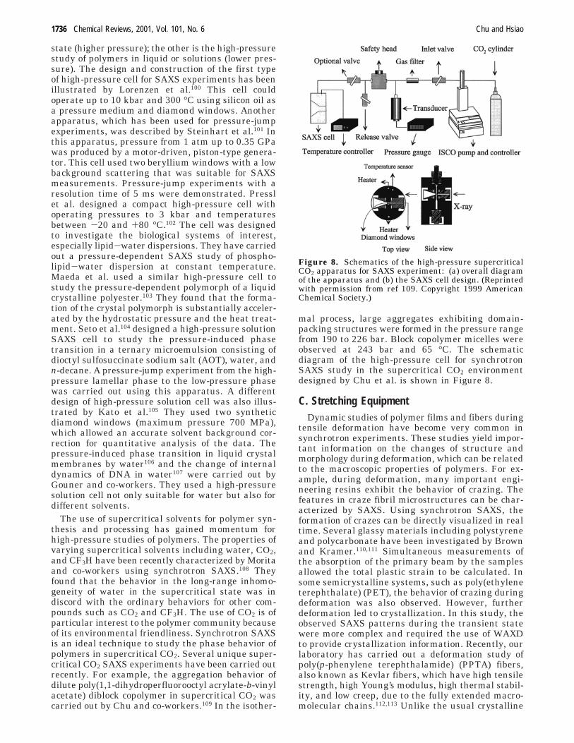
mal process, large aggregates exhibiting domain-packing structures were formed in the pressure rangefrom 190 to 226 bar. Block copolymer micelles wereobserved at 243 bar and 65 °C. The schematicdiagram of the high-pressure cell for synchrotronSAXS study in the supercritical CO2 environmentdesigned by Chu et al. is shown in Figure 8.
C. Stretching EquipmentDynamic studies of polymer films and fibers during
tensile deformation have become very common insynchrotron experiments. These studies yield impor-tant information on the changes of structure andmorphology during deformation, which can be relatedto the macroscopic properties of polymers. For ex-ample, during deformation, many important engi-neering resins exhibit the behavior of crazing. Thefeatures in craze fibril microstructures can be char-acterized by SAXS. Using synchrotron SAXS, theformation of crazes can be directly visualized in realtime. Several glassy materials including polystyreneand polycarbonate have been investigated by Brownand Kramer.110,111 Simultaneous measurements ofthe absorption of the primary beam by the samplesallowed the total plastic strain to be calculated. Insome semicrystalline systems, such as poly(ethyleneterephthalate) (PET), the behavior of crazing duringdeformation was also observed. However, furtherdeformation led to crystallization. In this study, theobserved SAXS patterns during the transient statewere more complex and required the use of WAXDto provide crystallization information. Recently, ourlaboratory has carried out a deformation study ofpoly(p-phenylene terephthalamide) (PPTA) fibers,also known as Kevlar fibers, which have high tensilestrength, high Young’s modulus, high thermal stabil-ity, and low creep, due to the fully extended macro-molecular chains.112,113 Unlike the usual crystalline
Figure 8. Schematics of the high-pressure supercriticalCO2 apparatus for SAXS experiment: (a) overall diagramof the apparatus and (b) the SAXS cell design. (Reprintedwith permission from ref 109. Copyright 1999 AmericanChemical Society.)
1736 Chemical Reviews, 2001, Vol. 101, No. 6 Chu and Hsiao
polymers, the chains in Kevlar are highly extended,which can form the so-called paracrystalline or meso-morphic structure with poor lateral order in thenoncrystalline zone. The combination of SAXS andWAXD has proven to be very useful to study thechanges of lattice structure, morphology, and macro-scopic properties during deformation.
The deformation experiment involves the use ofsome kind of commercial or custom-built tensilestretching devices. The major requirement of thedevice is that it should provide symmetrical stretch-ing, which guarantees that the focused X-rays canilluminate the same position on the sample duringdeformation. Otherwise, the sample detection posi-tion will be changed continuously, which can lead touncertainties in conclusion.
In our laboratory we modified a tabletop non-symmetrical stretching device to provide symmetricaldeformation.112 The modification can be briefly de-scribed as follows. The tensile stretching apparatuswas a modified version of model 4410 from InstronInc. and had a load capacity of 500 N. The maximaldistance between the two grips was about 460 mm.Sample could be heated to a temperature up to 300°C with a custom-designed sample chamber. Thestretching speed could be adjusted from 0.2 to 1000mm/min. The modified stretching unit adopted acustom-built vertical translational stage, which pro-vided translational motion opposite to the pro-grammed stretching with the same speed.
Recently, a fully integrated stretching system hasbeen developed by Fuller and co-workers,114 whichallowed the study of polymer deformation via simul-taneous SAXS/WAXD and stress-strain techniques.2D SAXS/WAXD images were collected using twoCCD-based area X-ray detectors, which providedvideo signal outputs. A video extensometer addition-ally provided sample strain and cross-section dataduring deformation. All three video signals wereprocessed by a Synoptics i860 processor-based videoframegrabber, which was capable of collecting dataat a rate of 40 ms per frame. A strain gauge was usedto reveal the mechanical yielding behavior of thesample. An electronic trigger mechanism was equippedto provide accurate synchronization of the X-ray datawith sample dimensional changes and loading infor-mation. This integrated stretching system has beendemonstrated to be a useful tool for the study ofpolymer deformation at rates relevant to practicalprocessing.
D. Fiber Spinning EquipmentIn polymer processing, the stake in understanding
the fiber spinning technology is very high becausethe variations of the processing parameters candirectly affect the properties of the final products. Forfundamental studies of polymer physics, the spinningprocess also provides an important mean to look intothe initial stage of crystallization during elongationalflow. Very interesting results have recently beenseen in the low-speed melt spinning of severalpolymers,115-121 where SAXS signals persistentlyoccurred before WAXD. This observation suggestedthat density fluctuations occurred prior to crystal-
lization or might even be a precursor to crystalliza-tion.116 In our opinion, this observation can also beattributed to an instrumentation artifact, which willbe commented on later in section V.B.5. Below wewill briefly describe two types of apparatus for fibermelt spinning and for fiber solution (gel) spinning.
The first fiber melt spinning device was built byZachmann and co-workers in the A2 Polymer line atHASYLAB, DESY.115 This melt spinning equipmentconsisted of a 20-mm single-screw extruder attachedto a metering pump. The extruder and the meteringpump were mounted on a horizontal platform thatcould be translated in the vertical direction with theuse of a precise stepper-motor drive system (Figure9). Distances ranging from 28 to 87.5 cm from thespinneret could be examined on the spinline usingthis apparatus. The main beam had a rectangularshape with the long axis normal to the fiber. Theextruded fiber was taken up on a 19 cm diametergodet roll after passing through a ceramic guide usedto minimize fiber movement and vibration. A verysimilar fiber spinning (film extrusion) apparatus wasalso constructed in the Daresbury Laboratory byRyan and co-workers116 to study the initial stages ofpolymer crystallization.
At Stony Brook, we also designed and constructeda portable melt spinning apparatus which consistedof a 3/4 in. Independent Laboratory single-screwextruder (C. W. Brabender Instruments Inc., NJ) anda custom-built vertical lifter with about 1.2 m ofdisplacement (Applied Automation Research Corp.,FL). The maximum extrusion temperature was about325 °C. The extruder was mounted on a horizontalplatform that could be translated in the verticaldirection by computer control. The extruded fiber was
Figure 9. On-line simultaneous SAXS/WAXD melt spin-ning apparatus at the A1 beamline in Hasylab, DESY. Thedetection system contains two imaging plates. (Reprintedwith permission from ref 115. Copyright 1993.)
Small-Angle X-ray Scattering of Polymers Chemical Reviews, 2001, Vol. 101, No. 6 1737
taken up with a portable wind-up device after passingthrough a ceramic guide, which was used to minimizefiber movement and vibration for X-ray detection.Several spinneret dies were available to producemonofilament, multifilament, or film. The typical rateof the mass output from this extruder was 1-7 g/min.Once the fiber spinning commenced, the extrudercould be translated remotely in the vertical direction,allowing measurements to be made along the spinline(20-65 cm from the spinneret, with a precision of 0.5cm). The maximum take-up speed was about 2000m/min. This spinning apparatus was made portablefor transport to different synchrotron facilities.
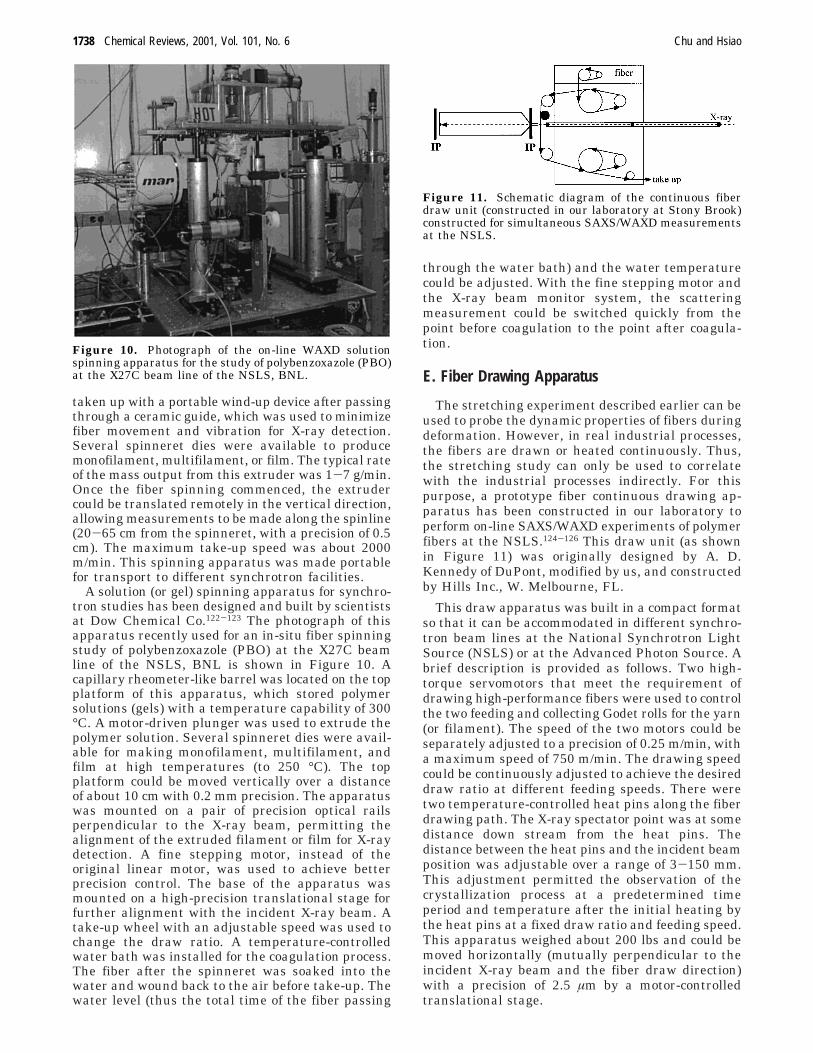
A solution (or gel) spinning apparatus for synchro-tron studies has been designed and built by scientistsat Dow Chemical Co.122-123 The photograph of thisapparatus recently used for an in-situ fiber spinningstudy of polybenzoxazole (PBO) at the X27C beamline of the NSLS, BNL is shown in Figure 10. Acapillary rheometer-like barrel was located on the topplatform of this apparatus, which stored polymersolutions (gels) with a temperature capability of 300°C. A motor-driven plunger was used to extrude thepolymer solution. Several spinneret dies were avail-able for making monofilament, multifilament, andfilm at high temperatures (to 250 °C). The topplatform could be moved vertically over a distanceof about 10 cm with 0.2 mm precision. The apparatuswas mounted on a pair of precision optical railsperpendicular to the X-ray beam, permitting thealignment of the extruded filament or film for X-raydetection. A fine stepping motor, instead of theoriginal linear motor, was used to achieve betterprecision control. The base of the apparatus wasmounted on a high-precision translational stage forfurther alignment with the incident X-ray beam. Atake-up wheel with an adjustable speed was used tochange the draw ratio. A temperature-controlledwater bath was installed for the coagulation process.The fiber after the spinneret was soaked into thewater and wound back to the air before take-up. Thewater level (thus the total time of the fiber passing
through the water bath) and the water temperaturecould be adjusted. With the fine stepping motor andthe X-ray beam monitor system, the scatteringmeasurement could be switched quickly from thepoint before coagulation to the point after coagula-tion.
E. Fiber Drawing Apparatus
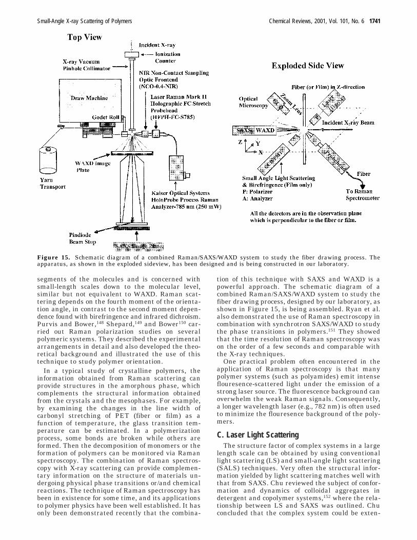
The stretching experiment described earlier can beused to probe the dynamic properties of fibers duringdeformation. However, in real industrial processes,the fibers are drawn or heated continuously. Thus,the stretching study can only be used to correlatewith the industrial processes indirectly. For thispurpose, a prototype fiber continuous drawing ap-paratus has been constructed in our laboratory toperform on-line SAXS/WAXD experiments of polymerfibers at the NSLS.124-126 This draw unit (as shownin Figure 11) was originally designed by A. D.Kennedy of DuPont, modified by us, and constructedby Hills Inc., W. Melbourne, FL.
This draw apparatus was built in a compact formatso that it can be accommodated in different synchro-tron beam lines at the National Synchrotron LightSource (NSLS) or at the Advanced Photon Source. Abrief description is provided as follows. Two high-torque servomotors that meet the requirement ofdrawing high-performance fibers were used to controlthe two feeding and collecting Godet rolls for the yarn(or filament). The speed of the two motors could beseparately adjusted to a precision of 0.25 m/min, witha maximum speed of 750 m/min. The drawing speedcould be continuously adjusted to achieve the desireddraw ratio at different feeding speeds. There weretwo temperature-controlled heat pins along the fiberdrawing path. The X-ray spectator point was at somedistance down stream from the heat pins. Thedistance between the heat pins and the incident beamposition was adjustable over a range of 3-150 mm.This adjustment permitted the observation of thecrystallization process at a predetermined timeperiod and temperature after the initial heating bythe heat pins at a fixed draw ratio and feeding speed.This apparatus weighed about 200 lbs and could bemoved horizontally (mutually perpendicular to theincident X-ray beam and the fiber draw direction)with a precision of 2.5 µm by a motor-controlledtranslational stage.
Figure 10. Photograph of the on-line WAXD solutionspinning apparatus for the study of polybenzoxazole (PBO)at the X27C beam line of the NSLS, BNL.
Figure 11. Schematic diagram of the continuous fiberdraw unit (constructed in our laboratory at Stony Brook)constructed for simultaneous SAXS/WAXD measurementsat the NSLS.
1738 Chemical Reviews, 2001, Vol. 101, No. 6 Chu and Hsiao
F. Shear Apparatus
It has been demonstrated that the phenomena offlow-induced molecular orientation,127 mixing/demix-ing,128 and order-disorder transitions in block co-polymers at large strains129,130 can be investigated inreal-time by synchrotron SAXS. The use of shear flowis often deployed because of its easy accessibility. Theelongation flow, which is considered a stronger flow,has not been used extensively because of experimen-tal difficulties. Generally speaking, there are twotypes of shear deformations that are of interest: (1)steady-state shear and (2) oscillatory shear. For thesestudies, several X-ray-modified rheometers have beendemonstrated involving different types of sheargeometry and different operational modes: parallel-plate shear apparatus, Couette flow shear apparatus,and modified rheometric solids analyzer (RSA-SL)rheometer.
The X-ray modification for the parallel-plate shearapparatus has been described by Burghardt and co-workers.131 The schematic diagram of the shear stagewith a similar X-ray modification made by us isshown in Figure 12, which can be described asfollows. The Linkam Cambridge shear system (CSS)450 was a high-temperature shearing stage oftenused to examine viscous liquid materials for in-situoptical microscopic studies. The modification for theX-ray detection involved the use of Kapton windowsand the special construction of the rotating plateswith small openings (see Figure 12). The sample wasplaced in a gap between two parallel X-ray windows.Shearing took place by rotating one of the disks usinga precision stepper motor as the other disk remainsfixed. The gap between the windows was adjustable,ranging from 10 to 2500 µm, using a second steppermotor. Each of the two quartz windows was in closethermal contact with a silver block heater thatcontrolled the sample temperature. The sampletemperature could be controlled from ambient condi-tions to 450 °C. To protect the motors and electroniccomponents from heat damage, the shear cell waswater-cooled.
For the oscillatory shear experiment, a Rheomet-rics RSA-SL system has been modified in our labora-tory (Figure 13). The modification included theincorporation of two vacuum paths (before and afterthe sample) and several X-ray windows (e.g., Kaptonfilms) along the beam path. One section of thevacuum path was in a cone shape having a maximumscattering angle (θ) of about 30°. The maximumoperating temperature for the environment chamberwas about 500 °C (using hot air). This apparatus wassuitable for the studies of stress-induced crystalliza-tion of (fully or lightly) cross-linked polymer melts,without the use of a sandwich shear cell. Theexperimental procedure for the solid-state elastomerdid not need to use the sample holder, which wasessentially the same as for typical dynamic mechan-ical analysis (DMA). Hamley et al. used a similarapparatus to study large-amplitude shearing on theorientation of cubic phases in gels of block copolymersformed in concentrated solutions129 and on the bi-continuous cubic phase of a block copolymer melt.130
The synchrotron SAXS technique is an importanttool to study polymer melts and solutions during flow.Many examples have been demonstrated by theneutron scattering method but only a few by X-rayscattering. There are significant advantages in usingthe SAXS technique rather than SANS. Very fastdetection time with very small sample sizes (downto length scales of micrometers) can be achieved bysynchrotron SAXS, which is suitable for time-resolved experiments. In contrast, neutron measure-ments usually require a collection time that is 100-1000 times longer.
IV. Combined Techniques
A. Simultaneous SAXS/WAXDThe simultaneous SAXS and WAXD techniques are
perhaps one of the most frequently requested meth-ods for studying the structure and morphology changesin real time during phase transformation. The prin-ciple of this combined method is quite simple. Duringthe experiment, two position-sensitive detectors areplaced in different locations, which should cover a
Figure 12. Schematics of the Linkam Shear stage, show-ing the placement of sample and the rotating and station-ary plates. The synchrotron small-angle X-ray scattering(SAXS) setup with a MARCCD area detector is also shown.
Figure 13. Photograph of a modified Rheometrics RSA-SL rheometer for synchrotron SAXS/WAXD measurements.This instrument was modified by our group at Stony Brook.
Small-Angle X-ray Scattering of Polymers Chemical Reviews, 2001, Vol. 101, No. 6 1739
wide angular range such as ∼100 µrad < θ < 0.5 rad.This means that ca. four orders of magnitude in q()(4π/λ)sin(θ/2)) can be obtained. The positioning ofthe two (or more) detectors depends on the structure,morphology, and orientation of the polymer system,the availability of detectors, and the available spacefor instrumentation. It is generally desired to havea minimal blank space between the SAXS and WAXDangular ranges and to operate the two detectorssynchronously. Many polymer problems can be tack-led by the use of simultaneous SAXS/WAXD tech-niques. These problems could include crystallizationand melting, phase transformation of polymers (suchas ethylene-based copolymers,132-134 iPP,135,136
PEEK,137,138 PEN139), polymerization,140 and the for-mation of colloids and gels,141 just to name a few.
The simultaneous SAXS/WAXD techniques canarbitrarily be divided into two geometrical configura-tions. For isotropic systems, two one-dimensional(1D) position-sensitive detectors (PSDs) can be used.For anisotropic systems (such as dynamic stretchingexperiments), two area detectors are often needed,which sometimes impose challenges in the cameradesign. In 1992, Zachmann and co-workers firstreported the use of simultaneous SAXS/WAXD tostudy the crystallization of polyethylene.133 In 1993,Bras et al. described the instrumentation developedfor performing simultaneous time-resolved SAXS/WAXD experiments using two gas-filled proportionaldetectors.142 Recently, Bras and Ryan reviewed thissubject.8 They stated the importance of these tech-niques for understanding fundamental aspects ofphase transformations as well as the applied fieldsof polymer processing. They also noted that theinstrumentation limits in the detectors and thesample environmental chambers have hindered theexperiments with shorter time resolution. For thispurpose, Bras described the improved design of adedicated station for real-time SAXS/WAXD experi-ments on a bending magnet beamline at theESRF143,144 and the development of a high-count-rate-curved PSD (1 MHz per channel) based on the
microstrip gas counter (MSGC) technology forWAXD.145 Laggner and co-workers also reported thedesign of a dedicated wiggler beamline for SAXS/WAXD measurements at ELETTRA.146 This facilitywas designed specifically for time-resolved (resolutionca. 1 ms) structure studies on gels, liquid crystals,(bio)polymers, amorphous materials, muscles, andproteins in solutions.
For anisotropic systems, simultaneous 2D SAXS/WAXD measurements have been demonstrated byusing two area detectors (MWPC or CCD) both withtime-resolution capability.147 However, this arrange-ment is not widely used because the WAXD imagecollected this way is often in a distorted form havingonly limited values for quantitative data analysis. Ifthe time resolution is not the primary concern, suchas for the steady-state experiments (fiber spinningor fiber drawing), two (or more) imaging plates (IP)can be used as X-ray area detectors. The WAXD IPcontains a central opening which allows the passageof the SAXS signal. Because the typical time resolu-tion of IP is several minutes, this system is onlyapplicable to systems at equilibrium, under steadystate, or at very slow kinetics conditions. One majorchallenge of using IP is that a quantitative compari-son of different images is difficult to carry out sinceeach image has a different background and a slightlydifferent location of the scattering center. In addition,it is difficult to correct the incident beam fluctuationsand the sample absorption. The schematic diagramof using two 1D PSDs to collect simultaneous SAXS/WAXD profiles during fiber drawing at the X27Cbeamline is illustrated in Figure 14. The bottomdiagrams illustrate the pinhole collimation systemand the double-multilayer monochromator.
B. Raman Spectroscopy
Raman scattering is a technique for studyingvibrational spectra using coherent, intense, lightbeams generated by laser devices. Raman spectros-copy yields information on the vibrational modes of
Figure 14. Schematics of simultaneous SAXS/WAXD setup for fiber drawing experiments at the X27C beamline in NSLS.The bottom diagrams illustrate the pinhole collimation system (left) and the double-multilayer monochromator (right).
1740 Chemical Reviews, 2001, Vol. 101, No. 6 Chu and Hsiao
segments of the molecules and is concerned withsmall-length scales down to the molecular level,similar but not equivalent to WAXD. Raman scat-tering depends on the fourth moment of the orienta-tion angle, in contrast to the second moment depen-dence found with birefringence and infrared dichroism.Purvis and Bower,148 Shepard,149 and Bower150 car-ried out Raman polarization studies on severalpolymeric systems. They described the experimentalarrangements in detail and also developed the theo-retical background and illustrated the use of thistechnique to study polymer orientation.
In a typical study of crystalline polymers, theinformation obtained from Raman scattering canprovide structures in the amorphous phase, whichcomplements the structural information obtainedfrom the crystals and the mesophases. For example,by examining the changes in the line width ofcarbonyl stretching of PET (fiber or film) as afunction of temperature, the glass transition tem-perature can be estimated. In a polymerizationprocess, some bonds are broken while others areformed. Then the decomposition of monomers or theformation of polymers can be monitored via Ramanspectroscopy. The combination of Raman spectros-copy with X-ray scattering can provide complemen-tary information on the structure of materials un-dergoing physical phase transitions or/and chemicalreactions. The technique of Raman spectroscopy hasbeen in existence for some time, and its applicationsto polymer physics have been well established. It hasonly been demonstrated recently that the combina-
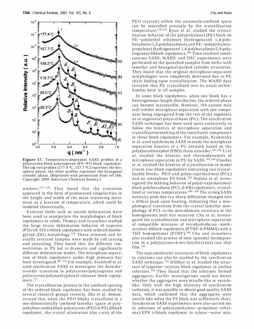
tion of this technique with SAXS and WAXD is apowerful approach. The schematic diagram of acombined Raman/SAXS/WAXD system to study thefiber drawing process, designed by our laboratory, asshown in Figure 15, is being assembled. Ryan et al.also demonstrated the use of Raman spectroscopy incombination with synchrotron SAXS/WAXD to studythe phase transitions in polymers.151 They showedthat the time resolution of Raman spectroscopy wason the order of a few seconds and comparable withthe X-ray techniques.
One practical problem often encountered in theapplication of Raman spectroscopy is that manypolymer systems (such as polyamides) emit intenseflouresence-scattered light under the emission of astrong laser source. The fluorescence background canoverwhelm the weak Raman signals. Consequently,a longer wavelength laser (e.g., 782 nm) is often usedto minimize the flouresence background of the poly-mers.
C. Laser Light ScatteringThe structure factor of complex systems in a large
length scale can be obtained by using conventionallight scattering (LS) and small-angle light scattering(SALS) techniques. Very often the structural infor-mation yielded by light scattering matches well withthat from SAXS. Chu reviewed the subject of confor-mation and dynamics of colloidal aggregates indetergent and copolymer systems,152 where the rela-tionship between LS and SAXS was outlined. Chuconcluded that the complex system could be exten-
Figure 15. Schematic diagram of a combined Raman/SAXS/WAXD system to study the fiber drawing process. Theapparatus, as shown in the exploded sideview, has been designed and is being constructed in our laboratory.
Small-Angle X-ray Scattering of Polymers Chemical Reviews, 2001, Vol. 101, No. 6 1741
sively investigated by combining laser light scatter-ing and synchrotron SAXS with measurements ofrheological properties of the supramolecules. Theinter- and intramolecular interactions involvinghydrophobic-hydrophilic interactions, solvent-non-solvent considerations, and metal coordination couldbe revealed in detail by complementary scatteringtechniques to provide useful information on the size,size distribution, and conformation of the aggregates.Studies of microstructures of the colloidal aggregatesat dilute concentrations became feasible only with theintense synchrotron X-rays. Li et al. demonstratedthe study of the aggregation behavior of polydi-acetylene in THF-PhMe mixed solvents using staticlight scattering (SLS), dynamic light scattering (DLS)and synchrotron SAXS.153 The transition of singlemolecular coils to stretched-chain aggregates couldbe described approximately by the Avrami equationthrough SLS and SAXS experiments. On the basisof semiquantitative comparison of the angular dis-tribution of scattered intensity with a variety oftheoretical scattering form factors, the structure ofthe P4BCMU aggregates was best described byribbonlike unsymmetrical elliptic cylinders.154
Recently, Chu and co-workers designed and con-structed a unique high-pressure fiber-optic light-scattering spectrometer that was capable of investi-gating the molecular dissociation and associationbehavior as well as the polymerization process insupercritical fluids.155 Multiple fiber-optic probes and
graded index microlenses were used to transmit theincident laser beam and to receive the scattered lightfrom the high-pressure cell. With the miniaturizationand flexibility provided by optical fibers, this high-pressure light-scattering cell was modified and couldbe transported to a synchrotron facility for synchro-tron SAXS measurements. The modification involvedthe use of a different set of diamond windows forX-ray transmissions. This spectrometer was usedsuccessfully for the in-situ study of the molecularassociation behavior of a 1,1-dihydroperfluorooctyl-acrylate and vinyl acetate diblock copolymer insupercritical carbon dioxide under pressures up to552 bar. The schematic diagram of the apparatus isillustrated in Figure 7.
Zachmann and co-workers designed and con-structed an apparatus that was capable of measuringSAXS, WAXD, and SALS signals simultaneously.156
They used this apparatus to study the crystallizationof polymers. A schematic diagram of this apparatusis illustrated in Figure 16. By comparing the changesin the integrated intensity from SAXS and WAXDas well as the scattering data from SALS, theyrevealed the mechanism in structural evolution dur-ing secondary crystallization, which involved themechanisms of crystal thickening, formation of newcrystals within the lamellar stacks, and formation ofnew lamellar stacks.
Figure 16. Schematic diagram of a combined laser light scattering/SAXS/WAXD system to study the polymer crystallizationprocess. This apparatus was designed and constructed by Zachmann and co-workers at Hamburg. (Reprinted with permissionfrom ref 156. Copyright 1993.)
1742 Chemical Reviews, 2001, Vol. 101, No. 6 Chu and Hsiao
D. Fourier Transform IR SpectroscopyAbsorption in the infrared wavelength range occurs
by interactions between the incident radiation fieldand the dipoles in the medium. These dipoles maybe either permanent, as in the case of the fluorine-carbon atom dipoles in polyvinylidene fluoride, orinduced as a consequence of unsymmetrical stretch-ing or bending of carbon-carbon bonds, as in the caseof polyethylene. Infrared dichroism (D) arises fromthe anisotropy of the absorption, which is defined asthe ratio of absorbances for the polarization direc-tions parallel and perpendicular in a plane containingthe incident beam. The infrared dichroism is relatedto the orientation function of the dichroic groups inthe polymer chains.
There are many polymer problems in which thecombination of Fourier transform infrared (FTIR)and SAXS can make significant contributions towardthe development of more in-depth understanding.Elwell et al. reviewed the system of segmentedpolyurethane copolymers.157 They concluded that thestructure development during reactive processing ofwater-blown, flexible copoly(urethane-urea) foamscould be well characterized by the combination oftime-resolved FTIR spectroscopy, synchrotron SAXS,and rheology. FTIR spectroscopy monitored themicrophase separation transition (MST) of isocyanatefunctional groups and followed the kinetics associatedwith nucleation and growth. Synchrotron SAXSprobed the kinetics of the reaction-induced phase-separation process, the mechanism, the length scales,and the resultant morphology. Dynamic rheometrymonitored the rheological properties associated withthe evolving morphology. Lee et al. also demonstratedthat the information of segment and domain orienta-tions in poly(butylene succinate) (PBS) and poly-tetramethylene glycol (PTMG) segmental block co-polymers during deformation could be obtained bysynchrotron SAXS and IR dichroic methods.158
Recently, Panick et al. demonstrated that the com-bination of pressure-jumped FTIR and synchrotronSAXS yielded important information about the pres-sure-induced unfolding and refolding of wild-typestaphylococcal nuclease.159,160 FTIR spectroscopy moni-tored the changes in the tertiary and secondarystructures of the protein upon pressurization, whileSAXS gave information about the chain collapse ofthe molecules in solution. The effect of pressure onthe kinetics resulted in a larger positive activationvolume for folding than for unfolding, and this effectled to a significant slowing down of the folding ratewith increasing pressure. These studies indicatedthat the changes in the secondary structure infolding/unfolding reactions of Snase were probablydependent upon the same rate-limiting step as thechanges in the tertiary structure. Similar high-pressure FTIR and SAXS studies were carried outto investigate the denaturation and aggregation ofâ-lactoglobulin and its genetic variants, as well asthe structural development of dipalmitoylphospha-tidylcholine bilayer membranes under pressure.161
The combined techniques of IR and synchrotronSAXS are relatively more difficult to carry out thanthe combined techniques of Raman and SAXS. To
optimize the IR signals, thin and optically clearsamples have to be used. The thin sample oftencompromises the X-ray signals. In contrast, theconcern over the sample thickness is not as severefor Raman study. Ryan et al. used the combinedtechniques of synchrotron SAXS and FTIR to studythe structure development and reaction kineticsduring the polymerization process of segmented blockcopolyurethanes.162,163 This study confirmed that themain driving force for structure development inpolyurethanes was due to phase separation ratherthan hydrogen bonding.
V. Applications
A. Materials-Based Studies
1. Block Copolymers
The SAXS technique offers some unique researchopportunities to study block copolymer systems eitherin the solid state or in solution. The intricate struc-tures and the complex morphologies of block copoly-mers usually show a dimension in the range of 1-100nm that is ideal to be studied by SAXS. Severalcomprehensive reviews of this subject are available(for examples, see refs 164 and165).
A block copolymer can exhibit multiple SAXS peaksdue to its periodic microdomain structure having along-range order. Information on the microdomainmorphology can be obtained from the relative posi-tions of these peaks. They can exhibit specific spatialrelationships depending on the shape of the micro-domain structure. For example, the ratio of the qvalues at the scattering maxima should be 1, 2, 3,4,... for lamellae; 1, x3, x4, x7, x9,... for cylindersin a hexagonal array; 1, x2, x3, x4, x5,... forspheres in a body-centered cubic array, etc. A samplediagram is shown in Figure 17, which illustrates thetemperature-dependent SAXS profiles of a polystyrene-b-polyisoprene (PS-block-PI) block copolymer ob-tained by the Hashimoto group.166
The synchrotron SAXS method adds another di-mension to the study of block copolymers, i.e., therapid kinetics of the phase transition can be followedin real time. For example, Floudas et al. studiedseveral order-to-order transitions (OOT) in poly-(isoprene-b-ethyleneoxide) diblock copolymer involv-ing crystalline lamellar phase (Lc), hexagonalmesophase (Hex), and bicontinuous cubic phase(Gyroid).167,168 They concluded that the transitionsfollowed typical nucleation and growth behavior. Kimet al. combined the rheological characterization withSAXS to investigate the OOT between the Hex andbody-centered cubic (BCC) phases in polystyrene PS-block-PI and PS-block-PI-block-PS systems.169-171
They found that the transition between HEX andBCC was thermally reversible.171 Hamley et al.studied the Lc to gyroid transition in poly(oxyethyl-ene)-poly(oxybutylene) (EB) diblock copolymer andalso reported that the transitions were thermallyreversible.129,130 The dynamics as well as the kineticsof the transition from the lamellar microphase tothe isotropic melt in butadiene-styrene diblock co-polymers were also studied by Ruland and co-
Small-Angle X-ray Scattering of Polymers Chemical Reviews, 2001, Vol. 101, No. 6 1743
workers.172-175 They found that the transitionappeared in the form of pronounced singularities inthe height and width of the main scattering maxi-mum as a function of temperature, which could bemodeled theoretically.
External fields such as tensile deformation havebeen used to manipulate the morphologies of blockcopolymers in solids. Thomas and co-workers studiedthe large strain deformation behavior of isoprene(PI)-rich SIS triblock copolymers with ordered double-gyroid (DG) morphology.176 These oriented and bi-axially textured samples were made by roll castingand annealing. They found that the different con-nectivities in PS led to dramatic and significantlydifferent deformation modes. The microphase separa-tion of block copolymers under high pressure hasbeen investigated.99-103 For example, Steinhoff et al.used synchrotron SAXS to investigate the disorder-to-order transition in polystyrene/polyisoprene andpolystyrene/polymethyphenyl siloxane block copoly-mers.177
The crystallization process in the confined spacingof the ordered block copolymer has been studied byseveral research groups recently. Zhu et al. demon-strated that when the PEO blocks crystallized in aone-dimensionally confined lamellar space of poly-(ethylene oxide)-block-polystyrene (PEO-b-PS) diblockcopolymer, the crystal orientation (the c-axis of the
PEO crystals) within the nanoscale-confined spacecan be controlled precisely by the crystallizationtemperature.178,179 Ryan et al. studied the crystal-lization behavior of the poly(ethylene) (PE) block onPE-poly(ethyl ethylene) (hydrogenated 1,4-poly-butadiene/1,2-polybutadiene) and PE-poly(ethylene-propylene) (hydrogenated 1,4-polybutadiene/1,4-poly-isoprene) diblock copolymers.180 Time-resolved simul-taneous SAXS, WAXD, and DSC experiments wereperformed on the quenched samples from melts withlamellar and hexagonal-packed cylinder structures.They found that the original microphase-separatedmorphologies were completely destroyed due to PEchain folding upon crystallization. The WAXD datarevealed that PE crystallized into its usual orthor-hombic form in all samples.
In some block copolymers, when one block has aheterogeneous length distribution, the ordered phasecan become inaccessible. However, the system maystill exhibit microphase separation with one compo-nent being segregated from the rest of the segmentsas in segmental polyurethane (PU). The synchrotronSAXS technique has been used quite extensively tofollow the kinetics of microphase separation andcrystallization/melting of the constituent componentsin these block copolymers. For example, Krakovskyet al. used synchrotron SAXS to study the microphaseseparation kinetics of a PU network based on thepoly(butadiene)diol (PBD) chain extender.181,182 Li etal. studied the kinetics and thermodynamics ofmicrophase separation in PU by SAXS.183,184 Floudaset al. studied the kinetics of crystallization in modeltriarm star block copolymers containing two crystal-lizable blocks: PEO and poly(ε-caprolactone) (PCL)and an amorphous PS block.185 Nojima et al. inves-tigated the melting behavior of poly(ε-caprolactone)-block-polybutadiene (PCL-b-PB) copolymers, crystal-lized at various temperatures.186-188 The strong SAXSintensity peak due to a sharp diffraction changed intoa diffuse peak upon heating, indicating that a mor-phological transition from the crystal lamellar mor-phology of PCL to the microdomain structure or thehomogeneous melt has occurred. Chu et al. investi-gated the crystallization and microphase separationof compatible mixtures of tetrahydrofuran-meth-acrylate diblock copolymers (PTHF-b-PMMA) with aTHF homopolymer (PTHF).189 Chu and co-workersalso studied the process of slow spinodal decomposi-tion in a poly(styrene-b-tert-butylstyrene) cast thinfilm.190
The supramolecular structures of block copolymersin solutions can also be studied by the synchrotronSAXS technique.191 Hilfiker et al. studied the struc-ture of isoprene-styrene block copolymer in anilinesolutions.192 They found that the solutions formedaggregates. Earlier investigations could not detectwhether the aggregates were micelle-like or vesicle-like. Only with the high intensity of synchrotronradiation, it was possible to obtain good quality SAXSdata, which confirmed that the aggregates werevesicle-like when the PS block was sufficiently short.Synchrotron SAXS experiments were also carried outin solutions of polyoxy(ethylene-propylene-ethyl-ene) EPE triblock copolymer in xylene-water mix-
Figure 17. Temperature-dependent SAXS profiles of apolystyrene-block-polyisoprene (PS-PI) block copolymer.The top two profiles (177.4 °C, 157.7 °C) represent the bcc-sphere phase; the other profiles represent the hexagonalcylinder phase. (Reprinted with permission from ref 166.Copyright 2000 American Chemical Society.)
1744 Chemical Reviews, 2001, Vol. 101, No. 6 Chu and Hsiao
tures and of PEP triblocks in water.185 The copoly-mers were found to form aggregates in selectivesolvents. With SAXS, the internal structure of self-assembled supramolecular systems (micelles andvesicles) was determined. In addition, synchrotronSAXS was also used to examine the chain lengthratio, chain sequence, and total chain length indiblocks and triblocks poly(styrene/tert-butylstyrene)as well as the effects of charge (SPS)/polarity andsolvent quality on the microstructures of polymercolloids.190,193
Recently, the large-scale grain size of the lamellarstructures in styrene-butadiene block copolymershas been characterized by ultra small-angle X-rayscattering (USAXS) measurements.194 All of the blockcopolymer specimens displayed a scattering peak inthe USAXS regime, and the grain size was estimatedusing the spherical form factor. The calculatedPorod’s law constant and the value of the scatteringinvariant were consistent with the scattering mech-anism proposed. The USAXS grain-size estimate wasfurther verified by TEM. Grain size in a givenpolymer was found to be a function of annealingtemperature and time.
2. Ionomers
The unique class of ion-containing polymers (iono-mers) consists of clusters or domains that are gener-ated by ionic interactions of the charged segmentsin the polymer chains. These clusters usually have adimension in the range of 1-4 nm that can becharacterized by the SAXS technique. A comprehen-sive review of the SAXS studies of bulk ionomers hasrecently been made by Chu.195 The high brilliance ofsynchrotron SAXS was found to be particularly usefulto resolve the details of the ionic structure. Forexample, Chu et al. demonstrated that precise syn-chrotron SAXS curves of Na and Zn salts of sul-fonated polystyrene could be obtained by using amodified Kratky block collimation system.196,197 Theresults supported the formation of ion-rich phases byshowing a corresponding glass transition tempera-ture. The ionic domain structures at elevated tem-peratures could be analyzed by using Porod’s law andthe Fourier transformation technique.
Using the synchrotron SAXS technique, the crys-tallization, melting, and dissolution behavior of zincstearate (ZnSt) in ZnSt-filled sulfonated poly(ethylene-co-propylene-co-ethylidene norbornene) (SEPDM) ion-omers was studied in real time by Jackson et al.198
Temperature-dependent SAXS patterns showed thatupon melting some or all of the ZnSt rapidly dissolvedinto the matrix, where ionic aggregates in the neationomer persisted up to 300 °C, which was way abovethe melting point of PE. Inomata et al. used synchro-tron SAXS to study the behavior of isothermalcrystallization of an associated polymer blend, con-sisting of one-end-aminated polystyrene (APS) andone-end-sulfonated poly(ethylene glycol) (SPEG).199
Different sample preparation schemes were found tohave a profound impact on the phase transition andthe kinetics properties of the blend. The differencein the formation of microphases and macrophases inthe molten state was responsible for the difference
in the crystallization rate of the samples, e.g., thecrystallization rate of homopolymer was greater thanthat of solvent-cast sample, which was greater thanthat of freeze-dry sample because the freeze-drysample had the smallest SPEG domains available forcrystallization.
The ionic behavior of certain ionomers can onlyappear in the presence of solvent such as water.These ionomers can be used as proton-exchangemembranes. One example is the system of sulfonatedstyrene-ethylene/butylenestyrene triblock copolymerand sulfonated hydrogenated, random copolymer ofstyrene and butadiene, which has been studied byhigh-resolution SANS and synchrotron SAXS.200
Rabeony et al. investigated the structure develop-ment in these membranes upon swelling in water.200
The dry triblock copolymer exhibited a lamellarstructure with a long spacing of 37 nm but with noevidence of sulfonic acid aggregation. Upon hydra-tion, the long spacing increased to 63 nm whileretaining the lamellar morphology. The dry randomcopolymer exhibited an “ionomer peak” at about 5nm, which increased to 9 nm upon hydration. Theionic behavior of Nafion membranes in water wasalso studied by synchrotron SAXS by Wu et al.201
They demonstrated that the Debye-Bueche correla-tion function approach could be applied to the analy-sis of the SAXS data. This method was less biasedthan the fitting of the SAXS profile with a morerestrictive assumed morphological model. The sizesof the ion/water aggregates and the most probableinteraggregate distance were found to increase withincreasing side chain length. A liquidlike model coulddescribe the spatial arrangement of the ion/wateraggregates.
3. Liquid Crystalline PolymersThe changes of mesomorphic structure of liquid
crystalline polymers in solutions (lyotropic) and inmelts (thermotropic) can also be characterized bysynchrotron SAXS. For example, Perez-Mendez et al.used the simultaneous synchrotron SAXS/WAXDtechnique to study the behavior of cholesteric poly-esters in aqueous solution and their interactions withthe lipid membrane.202 The chiral behavior withliposomes of racemic lipids and enantiomeric lipidswas considered for drug delivery applications. He etal. studied the structure of a self-assembled hydrogen-bonded main chain liquid crystalline polymer by meltmixing two complementary components, A and B,which in their individual states did not exhibit liquidcrystallinity.203 The combined synchrotron SAXS/WAXD/DSC experiments revealed that the polymericsalt had an AABB chain structure and formed asmectic layer with a length of the A-B repeatingunit. Buchner et al. investigated the molecular order-ing, phase transitions, and chain mobility in liquidcrystalline poly(ethylene naphthalene-2,6-dicarboxy-late) and its copolyesters with p-hydroxybenzoic acidusing the combined method of WAXD and DSC.204
Campoy et al. used synchrotron SAXS to study thepolymorphic transformations that occurred with in-creasing temperatures and the corresponding struc-tural changes in blends of nylon 6 and a liquid crystalcopolyester Vectra.205
Small-Angle X-ray Scattering of Polymers Chemical Reviews, 2001, Vol. 101, No. 6 1745
In-situ fiber spinning X-ray scattering experimentsfrom the lyotropic dope of rigid polybenzoxazole(PBO) were carried out by Dow scientists.122,123 Thegeneral scheme for drawing PBO fibers started witha dry-jet wet-spinning process from a 13-15% poly-mer solution in polyphosphoric acid (PPA). The fiberstructure was then developed by coagulation in H2O.The drawing process before coagulation was studiedin detail by in-situ synchrotron WAXD. From thiswork, the effects of line tension, spin draw ratio(SDR), and temperature on the orientation parameterand the microdomain size were determined. Typi-cally, orientation parameters of about 0.95 wereobtained for the uncoagulated monofilament withSDR values of about 20. Coagulation brought aboutthe rapid formation of a crystal solvate with amonoclinic unit cell structure. At the same time, amicrofibrillar microstructure was fixed in the fila-ment by SAXS.206
4. Biopolymers
Most biopolymers exhibit some forms of mesoscopicproperties in aqueous solutions. As the electrondensity contrast under the in vitro environment isweak and the mesoscopic structure can be complex,these systems are ideal to be studied by synchrotronSAXS with well-defined high-intensity beams. Bordasand Koch demonstrated the use of X-ray scatteringand diffraction techniques to study monodipersemacromolecules in solutions, dispersed solids, andpartially ordered systems.207 In this section, severalrecent synchrotron SAXS studies of biopolymers arereviewed.
For complex structures, Salditt et al. used synchro-tron SAXS to study the mutilayered, self-assembled
structures of mixtures of DNA and cationic lipo-somes.208 In these mixtures, DNA was confinedbetween charged lipid bilayers and formed a two-dimensional (2D) smectic ordering. Bouwstra et al.studied structural changes of the lipids in human andhairless mouse stratum corneum after heating thestratum corneum to various temperatures usingsynchrotron SAXS.209 Yu and co-workers used syn-chrotron SAXS to address the question as to howmuscle generates force and produces physical dis-placements at the expense of using the energyreleased by ATP hydrolysis. Skeletal muscle fibersmainly consisted of two types of protein filaments,the myosin-containing filaments and the actin-containing filaments.210,211 Force was generated byinteractions between myosin and actin. Conforma-tional changes were found within the actin-myosincomplex, and significant movement of the filamentswas seen during shortening. A typical SAXS patternof the myosin-containing filaments and the molecularmodeling schematics are illustrated in Figure 18.
In the case of a less complex structure, diffuseSAXS profiles can be detected. Jenkins et al. studiedthe effects of hydrolysis on the structure of threetypes of starch and their subsequent gelation behav-ior.212 By fully fitting the SAXS curves from differentregions within the starch granule, they verified thepreferential destruction of the amorphous phaseduring the hydrolysis process. The structures ofstarch, in fact, are quite complex. Donald et al. usedthe microfocus SAXS technique to scan across thestarch granule and reported the intricate lamellarstructures within the granule.213 Sano et al. reportedthe size and shape of A-protein of tobacco mosaicvirus coat protein (TMVP) and cucumber green
Figure 18. 2D SAXS image from a muscle bundle, under ATP-depleted conditions. The layer lines mainly arise from theactin filament with myosin bound to it (the first layer line is at 365 Å spacing). The corresponding supramolecular structureis illustrated at right. (Reprinted with permission from refs 210 and 211. Copyright 1995.)
1746 Chemical Reviews, 2001, Vol. 101, No. 6 Chu and Hsiao
mottle mosaic virus coat protein (CGMMVP) bysynchrotron SAXS.214 They concluded that in theA-protein structure, the globular core structure wasa subunit which could be modeled as a thin isoscelestriangular prism composed of four globular coresjoined by rather flexible segments. These cores wereprobably related to four helical regions in a subunitand could rearrange their relative positions accordingto external conditions. Barteri et al. used SAXS tostudy the influence of glycerol on the structure andstability of ferric horse heart myoglobin.215 Theyfound that both tertiary and secondary (R-helix)conformations of the protein were influenced byglycerol. Congiu Castellano et al. studied the confor-mational changes of lyophilized samples of apo-,mono-, and diferric-human transferrin by the fractalanalysis of the SAXS data.216 Miles et al. usedsynchrotron SAXS to study 11S soya globulin. Sub-maxima at higher angles were recorded which havenot been seen previously.217 Such detailed informa-tion in the scattering curve is important for struc-tural modeling of this protein. Bonnete et al. usedagarose gels in combination with synchrotron SAXSto follow hen egg white (HEW) lysozyme nucleationand growth.218 The advantage of using gels ratherthan solutions was that the nucleation centers couldbe trapped in the gel network, allowing the time-resolved experiment by SAXS.
To clarify the mechanisms of folding and unfoldingin proteins, several studies of thermal denaturationof proteins have been carried out at low proteinconcentrations as thermal denaturation accompanieda great tendency of aggregation. By using synchro-tron SAXS, Shigeki et al. found that the presence ofrepulsive interparticle interaction between proteinscould maintain solute particles separation to preventfurther aggregation in thermal denaturation pro-cesses of hen egg-white lysozyme (HEWL).219 Aspointed out earlier, the pressure-induced folding/unfolding of wild-type staphylococcal nuclease (SnaseWT) was studied by Panick et al.159,160 They foundthat the pressure-induced denatured state at >3 kbarretained some degree of â-like secondary structure,and the molecules could not be described as a fullyextended random coil.
B. Phenomenon-Based Studies
1. Polymer Solutions and Gels
SAXS is one of the best available techniques tomeasure the compactness and shape of macro-molecules, especially when the size is small, suchas proteins (for examples, Kataoka et al.,220-222
Flanagan et al.,223,224 Nishi et al.,225 and Lattman226).The molecular parameters revealed by solution SAXSinclude the radius of gyration (Rg), the maximumdimension, the shape, and the globularity. Further-more, high-q SAXS profiles will provide informationon the polymer inner structure, which can be cor-related, for example, in the case of proteins, withinteractions between secondary structural elements.The SAXS results thus complement the resultsobtained by other techniques, such as NMR. As aresult, SAXS has been combined with magnetic
birefringence to study the orientational behavior ofmicrotubules assembled in strong magnetic fields(Bras et al.227) and with other techniques to studythe polymer association process (Uversky and Fink228
and Chu et al.109).SAXS of polymers in solution invariably leads to
the protein-folding problem as one of the most chal-lenging research topics of current interest. Earlierresults have emphasized the structural characteriza-tion of the native and equilibrium molten globule.SAXS studies of the equilibrium molten globulestates of cytochrome c, apomyoglobin, and R-lactal-bumin as well as of partially folded fragmentsof nuclease have been reported (Dolgikh et al.,229
Flanagan et al.,223,224 Kataoka et al.,220-222 and Gastet al.230). Recent SAXS studies of the equilibriumunfolding of hen lysozyme at low pH have providedevidence for the formation of a subpopulation ofpartially folded molecules (Chen et al.231). Withsynchrotron X-rays, both stopped-flow (Kihara232) andtemperature-jump (Hiragi et al.97) systems have beendeveloped for kinetic solution SAXS studies. Thekinetic studies indicate a major problem of molecularassociation or aggregation during the protein-foldingprocess (Eliezer et al.233,234). More intense X-raysources are required as the data collection timebecomes too short for fast kinetic studies (Kataokaand Goto235). Eliezer et al. were able to obtain theradius of gyration of sperm whale apomyoglobin 100ms after initiation of folding by rapid dilution of asolution containing 5.6 M urea.233,234 In essence, onlyintegrated SAXS intensity was applied successfullyfor a direct monitoring of fast intramolecular globu-larization during protein folding (Semisotnov etal.236). Equilibrium unfolding of hen egg lysozyme asa function of urea concentration at pH 2.9 has beenstudied by solution SAXS as well as by far and nearUV CD at 222 and 298 nm, respectively (Chen etal.237). By using a combination of time-resolved SAXSand time-resolved tryptophan fluorescence, togetherwith the rate of formation of native molecules usinginterrupted refolding experiments, the structuralcharacteristics of a nonspecifically collapsed state oflysozyme at low pH have been reported.
The analysis of radial autocorrelation functions interms of long-range and short-range correlations hasbeen used to examine the details of SAXS profiles.The type of information that can be extracted on theoverall structure, as well as the internal structureof polymers, makes SAXS a useful probe. In recentyears, while Kratky plots of SAXS data are useful tostudy association-induced folding of globular proteins(Uversky et al.238), attempts have been made tocalculate SAXS profiles using a variety of approaches.These approaches include rigid body motion in crys-talline lysozyme as probed by molecular dynamicssimulation for X-ray diffuse scattering (Hery et al.239)as well as modification of the atomic structure factorof subunits of thiamine diphosphate from the originalcrystallographic model. More importantly, SAXS hasrecently been analyzed with a new method to retrieveconvergent model structures that fit the experimen-tally determined scattering profiles (Chacon et al.240).Instead of attempting to complete the Debye formula
Small-Angle X-ray Scattering of Polymers Chemical Reviews, 2001, Vol. 101, No. 6 1747
for all the possible mass distributions, low-resolutionstructures of proteins in solution are retrieved withan algorithm that efficiently searches the configura-tional space and evolves best-fit bead models basedon X-ray crystal structures in the presence of noise.Approaches that try to take into account the time-evolution of tertiary and secondary structures withknown native and denatured states of proteins canclearly improve the SAXS technique as a powerfultool to investigate the kinetics of protein folding aswell as the interactions with other macromolecularassemblies. While these approaches are often notapplicable to polymer solutions in general, it is notedhere that for synthetic polymers with more complexmolecular architecture, one can then take a similarroute of analysis.
The characteristic length scales determined fromSAXS deal with not only the overall size and shapeof the polymer molecule, but also its internal second-ary structure, if any. The length scales of secondaryand tertiary structures of the protein, as expressedin terms of the hydrodynamic radius (Rh) fromdynamic light scattering (DLS), the radius of gyration(Rg), and the intraparticle scattering factor P(q) fromSAXS and the secondary structure, if any (e.g., fromCD), imply the use of appropriate experimentalsetups in the different techniques which satisfy therequired temporal and angular ranges (e.g., delaytime increment and range in DLS and appropriate qrange in SAXS). For synchrotron SAXS with modernX-ray optics and detection systems, the excess scat-tered intensity, I, can be obtained over a large rangeof q. It is essential to reduce the parasitic scatteringand background so that I(q) at large q values canhave an acceptable signal-to-noise ratio.
The excess scattered intensity from a macromol-ecule can be expressed in terms of a difference in theelectron density, ∆F() F1 - Fo), between that of themacromolecule (F1) and of the solvent (Fo) as well asthe volume (v) of the macromolecule
where F(q) is the normalized form factor (F(0) ) 1)with
and V being the scattering volume of the macromol-ecule. It is noted that solution SAXS only measuresthe electron density distribution averaged in timeover all orientations.
Kataoka and Goto showed an instructive table(Table 2) outlining the analytical methods of solutionSAXS in terms of obtainable parameters and corre-sponding properties.235 It is, of course, understoodthat the data analysis includes corrections for para-sitic scattering, background, attenuation, incident
intensity variation, and desmearing4 before oneproceeds with any determination of the structuralparameters. In particular, the distance distributionfunction P(r) involves a conventional Fourier trans-formation with the main problems being the termi-nation effect due to finite q ranges of the experimentand the remaining background scattering at large qvalues. One must be careful with the Fourier ripples.
Synchrotron SAXS has also been used to investi-gate solutions of native DNA at different ionicstrengths and temperatures. For large DNA frag-ments, the mass per unit length, the radius ofgyration of the cross-section of DNA, and the appar-ent second virial coefficient (A2) could be obtainedfrom the Zimm plot in the rodlike approximation(Barone et al.241). The positive and almost constantA2 value suggested dominating repulsive interactionseven at resolutions of 5-8 nm. The rodlike-wormliketransition temperature was found to increase withincreasing ionic strength.
The ability to mix liquids in microseconds hasexpanded the accessible time scales and ushered ina new era for kinetic studies (Beechem242). Regenfusset al. pioneered the development of a continuous-flowmicrosecond mixing device.243 Microsecond mixingstudies of protein folding have appeared only recently(Takahashi et al.,244,245 Chan et al.246) utilizing aT-mixer version of the original Regenfuss et al.design.243 On the basis of an adaptation of this design,Shasty et al. reported a detailed description of theassembly and operation of a modern microsecondmixing, continuous-flow instrument in which thereaction kinetics were followed by measuring fluo-rescence versus distance downstream from themixer.247 With a mixing time of about 15 µs after itsinitiation and a dead-time of the measurement of 45( 5 µs, the work revealed that the kinetic databecome accessible down to about 50 µs. Kineticstudies over a time scale ranging from ∼50 µs to tensof seconds should enhance our understanding of themechanism of protein folding, as well as a widevariety of other important biological reactions. It wasnoted that shorter time scale measurements (e.g.,laser-based T-jump approach in the nanosecondrange) were also needed to help bridge the gapbetween the reaction rate theory (picoseconds tonanoseconds) and the biological structure/function(∆A per picosecond to microsecond to millisecond tosecond).
In the Shasty et al. capillary mixing apparatus,247
the quartz flow cell had a 250 µm diameter channelthat might be translated into a more demanding needon the quantity of protein required to undertake athorough kinetic study on the protein-folding prob-lem. Furthermore, this arrangement, while excellentfor optical measurements, could not be adapted easilyfor SAXS experiments. The mixer designed by the
Table 2.
method obtainable parameters
Guinier plot ln I(q) vs q2 radius of gyration, molecular weight (or aggregation)Kratky plot q2 I(q) vs q to get peak intensity and persistence lengthP(r) (1/22) ∫I(q)(sin qr/qr)dq dmax (molecular size/shape)I(q) at high q values internal structure
I(q) ) (∆Fv)2F2(q) (2)
F(q) ) ∫ F1(r)e-iqr dV (3)
1748 Chemical Reviews, 2001, Vol. 101, No. 6 Chu and Hsiao
Eaton group246 was more robust and had beenadapted for SAXS measurements, although the mixerconstruction was quite demanding. Recently, Knightet al.248 developed a rapid-mixing microchannel con-tinuous-flow cell that has been adapted successfullyby Sol Gruner at Cornell University for synchrotronSAXS experiments (Pollack et al.249). The Austindesign (Knight et al.248) can be modified to performkinetic studies using either X-rays or lasers withidentical geometrical pathways. In addition, it hasthe potential to reach even shorter reaction times anduses a smaller volume, which can be further reducedin combination with micro X-ray beams available atthe third-generation synchrotron light sources, suchas the Advanced Photon Source.
Small colloidal particles in suspensions as well asmicroemulsions and micelles often have sizes verysuitable for measurements by means of SAXS. Char-acteristics of aerosol-OT reversed micellar formation(Hirai et al.193,250) as well as the structures of micro-capsules (Dobashi et al.251) and of block copolymers(Hilfiker et al.192 and Wu el al252,253) have beeninvestigated by means of SAXS, especially in combi-nation with laser light scattering. SAXS has also beenused to study thermotropic phase transitions (Hiraiet al.254) and the sol-gel process (Gaponov et al.255).
SAXS studies of dilute suspensions of photoniccolloidal crystals were reported by Vos et al.256,257 andMegens et al.258,259 They found that scattering fromdilute suspensions was related to the particle formfactor. The radius, size distribution, and informationabout the internal structure of the particles could bedetermined from the analysis. With the experimen-tally determined particle form factor, structure fac-tors were extracted from the diffraction patterns ofthe colloidal crystals. The crystal structure, latticeparameters, and average orientations were thendetermined. Thus, a combination of SAXS/WAXD canbe used effectively to study the morphology andcrystal structures of colloids. More examples includeda similar SAXS study by Diat et al.,260 crystallizationstudy by Nojima et al.,261 in-situ crystallization studyby de Moor et al.,262 and phase separation kineticsstudy by Li et al.263
A morphological characterization of bicontinuousphase-separated polymer blends and one-phase mi-croemulsions has been reported by Jinnai et al.264 Themorphological similarity of the two systems permit-ted one to use the same approach to carry out theanalysis. Berk developed a theory to analyze thescattering data from bicontinuous microemulsionsand subsequently to generate the three-dimensionalmorphology.265 Chen et al. proposed a peaked spectralfunction with a power law decay at large wavenum-bers.266 By applying this approach (called a modifiedBerk or MB theory) with a three-parameter spectralfunction and the Cahn267 clipping scheme to calculatethe Debye correlation function (Debye el al268) for thescattered intensity, the random wave model couldthen be used to generate the 3D morphologies. Theanalysis was complex, and the experimental demand
for precision and the q range was relatively high,often requiring intensity measurements covering 4-5orders of magnitude. A representation of the 3Dmorphology for a polymer blend and a bicontinuousmicroemulsion is shown in Figure 19.
3. Polymer GelsSAXS and laser light scattering are powerful and
complementary tools to investigate the structure anddynamics of polymer networks in solutions (gels). Thestructural changes in polymer gels can be inducedby pH (Hirai et al.269), temperature (Izumi et al.,270
Figure 19. (A) 3D morphology of the phase-separatedpolymer blend (box size, 22.43 µm3). (B) 3D morphology ofthe one-phase bicontinuous microemulsion at the hydro-phile-lipophile balanced temperature (box size, 8003 Å3).Both are generated from the MB theory with three fittingparameters obtained from the best fit to the correspondingscattering profiles. (Reprinted with permission from ref264. Copyright 1997 American chemical Society.)
Small-Angle X-ray Scattering of Polymers Chemical Reviews, 2001, Vol. 101, No. 6 1749
Liao et al.271), ionic strength, and other physicalparameters.272 Some example studies are listed asfollows.
The level of pH has a profound effect to changestructures in polymer gels. Hirai et al. studied thepH-induced structural change in poly(vinyl alcohol)(PVA) hydrogels cross-linked with poly(acrylic acid)(PAA) by means of synchrotron SAXS.269 The physi-cally cross-linked gel formed by blending of PVA andPAA in an aqueous solution was compared with thechemically cross-linked gel by esterification of PVAwith PAA in the hydrogel state. The chemical cross-linking was found to convert the physical cross-linksinto a folded structure. SAXS results suggested thatthe folded structure exhibited a fractal rough inter-face, which could change into the structure of per-colation clusters at a high pH level. The gelsimmersed in pH 8 showed a remarkable structuralchange accompanying the swelling. The conforma-tional change of the PAA chains, induced by the pHchange, was used to explain the presence of astructural change in an interpenetrating polymernetwork.
Temperature also plays an important role on thestructure in gels. The spinodal decomposition behav-ior in a covalently cross-linked n-isopropylacrylamidegel was studied by Liao et al. using SAXS.271 Follow-ing a temperature jump into the two-phase region,the scattered intensity was found to increase withtime and eventually to become saturated.271 Theearly-stage data could be described by the linearCahn-Hilliard-Cook (CHC) theory.267 At later times,a pinning of the structure was also observed. Thebehavior of a two-stage melting process in dilute gelsof poly(γ-benzyl L-glutamate) was studied by Izumiet al. using synchrotron SAXS.270 DSC heating traceexhibited two endotherms, indicating the melting oftwo types of aggregates. SAXS measurements showedthat the two-stage melting of the aggregates was duefirst to the melting of bundles of three rods and thento that of bundles of two rods. Above the two-stagemelting temperature, the gels transformed into iso-tropic solutions with no aggregation of polymerchains.
The presence of inorganic particulate often inducedgellike structures in polymer solution, which can bestudied by synchrotron SAXS but not light scatteringas the solution is opaque. For example, de Lange etal. used synchrotron SAXS to study the sol-gelbehavior in polymeric SiO2 with SiO2/TiO2, SiO2/ZrO2,and SiO2/Al2O3 binary sols.272 This system was de-signed for ceramic membrane applications. Theaggregation of silica sols was found to obey the tip-to-tip cluster-cluster aggregation model in the initialstages.
Gel can form self-assembled structures. For ex-ample, the sol-gel transition of gellan gum aqueoussolution in the absence of added salts was studiedby Izumi et al.273 using synchrotron SAXS. Resultsindicated that the cloudy gels exhibited three scat-tering peaks, which were in the simple ratio of 1:2:3, suggesting that the junction zones in these gelswere formed by a lamellar structure. These peaksshifted to a lower scattering vector as the content of
the added ions was increased, indicating the forma-tion of larger lamellae. The flow-induced orienta-tional and morphological changes in structured gelswere studied by Vigild et al.274 They investigatedface-centered cubic (fcc) and body-centered cubic(bcc) phases in gels of poly(oxyethylene)-poly(oxy-butylene) diblock copolymers and for the bicontinuouscubic ‘gyroid’ structure in a poly(ethylene-alt-pro-pylene)-poly(dimethylsiloxane) diblock copolymermelt. The orientations of the micellar bcc phases inthe gels and the gyroid structure (belonging to thebcc space group) following large-amplitude shearingwere essentially the same, i.e., directionally orientedcrystals are produced in both cases, in which (111)directions were oriented along the shear direction.A typical SAXS profile of PMMA gel with C12TAcomplex is shown in Figure 20 (Zhou et al.275). Thescattered peaks are indexed by a cubic structure ofPm3n space group. The dotted line represents thecalculated scattering curve based on the connectedunit cell model shown in the inset.
4. Polymer BlendsCommodity polymers can be made useful by mixing
them under appropriate conditions. The key aspectis the structure-property relationship of the materi-als, which can form microphase domains in a phaseseparation process. The understanding of this processcan lead to improved properties.
One common SAXS study is based on the binaryblends, in which one or both components can crystal-lize. The kinetics of crystallization can be followedunder isothermal conditions in real time by simul-taneous SAXS, WAXD, and small-angle light scat-tering (SALS). Stein and co-workers carried out astudy involving blends of linear high-density poly-ethylene (HDPE) and branched linear low-densitypolyethylene (LLDPE).95,276 In this study, variables
Figure 20. SAXS profile of PMMA gel with C12TAcomplex. The scattered peaks are indexed by a cubicstructure of Pm3n space group. The dotted line representsthe calculated scattering curve based on the connected unitcell model shown in the inset. (Reprinted with permissionfrom ref 275. Copyright 2000 American Chemical Society.)
1750 Chemical Reviews, 2001, Vol. 101, No. 6 Chu and Hsiao
such as degree of crystallinity, crystal perfection,lamellar thicknesses, and their distribution, spheru-lite size, and spherulite optical anisotropy have beenfollowed. It was found that the crystallization processexhibited non-Avrami behavior, and the resultantmorphology was controlled by the competitionbetween crystallization and diffusion of the crystal-lizing chains. A further investigation can involvestudies of crystallization where phase separation willoccur in the amorphous phase. This can take placewhen molecular weights of the components and/or thedegree of branching in LLDPE are sufficiently high.Nojima et al. used synchrotron SAXS to study thecrystallization of poly(ε-caprolactone) and poly(ε-caprolactone)/poly(vinyl chloride) blends as wellas poly(ε-caprolactone) and polystyrene oligomerblends.277,278 In their systems, the SAXS curvesindicated a possible coexistence of the crystal regionand the amorphous region throughout the morpho-logical formation in the blend. Cheung et al. studiedthe evolution of crystalline structures in poly(ε-caprolactone)/polycarbonate blends.279 An insertionmechanism was proposed in which the PCL wascrystallized in the amorphous intralamellar phase.Eersels et al. investigated the effect of transreactionprocesses on the morphology of semicrystalline ali-phatic/aromatic polyamide (PA) blends.280 They con-cluded that polyamide copolymers consisted of bothcrystallizable PA 46 and noncrystallizable PA 6Isequences after the transreaction. Hsiao et al. studiedthe crystallization behavior of poly(ether ether ketone)/poly(ether imide) blends,281 and they reported thatPEEK crystallized in densely crystalline lamellarstacks through all stages of primary crystallization,noting that the noncrystalline PEI was almost en-tirely excluded from the stacks at all times duringspherulitic growth. Wang et al. used simultaneousSAXS/WAXD techniques to study the microstructuresof PEEK/PEKK blends at both structural and lamel-lar levels.282 Liu et al. studied the crystallization ofmiscible blends of semicrystalline poly(vinylidenefluoride) (PVF2) and semicrystalline poly(1,4-butylene adipate) (PBA).283,284 They concluded thatthat PBA could crystallize as a thick lamella in thePVF2 interlamellar region and that there was amixed amorphous phase on either side of the PBAlamella, i.e., between the PBA and PVF2 crystallamellae. Yeh et al. studied the effects of misciblepolymer diluents on the development of lamellarmorphology in poly(oxymethylene) (POM) blends.285
They reported that lamellar thickening with time inthe long-time secondary crystallization region wasobserved in neat POM and the blend with 10% low-Tg diluent, while this process was inhibited with thehigh-Tg diluent due to the higher Tg values of theinterlamellar species.
In addition to the crystallization study, severaldifferent types of SAXS experiments have also beencarried out for polymer blends. Vandermarliere et al.studied the morphological changes during melting ofpolycaprolactone and poly(styrene-co-acrylonitrile)blends.286 Wilkson et al. studied the melting, reaction,and recrystallization behavior in a reactive PC-PBTblend.287 They reported that increasing transesteri-
fication resulted in a progressive reduction in themelting and crystallization temperatures and degreeof crystallinity with the development of a mixed-phase glass. Inomata et al. used synchrotron SAXSto study an associated polymer blend consisting ofone-end-aminated polystyrene (APS) and one-end-sulfonated poly(ethylene glycol) (SPEG).199
For blends containing no crystallizable compo-nents, synchrotron SAXS is also useful to character-ize the structure and to correlate with the properties.Okamoto et al. studied plastic deformation (crazingand shear yielding) of high-impact polystyrene/poly-(2,6-dimethyl-1,4-phenylene ether) blends.288 Chu etal. used synchrotron SAXS to study the local concen-tration fluctuations in a poly(2-chlorostyrene) andpolystyrene blend, which was induced by tempera-ture jumps. They concluded that the blends in theimmediate neighborhood of the critical mixing pointcould be used to locate the critical mixing pointprecisely.289-291 Rabeony et al. used a combination ofSAXS, calorimetry, and cloud point measurementsto determine the phase diagram of the poly(cyclo-hexyl acrylate) and poly(2-bromostyrene) blends.292
5. Polymer Crystallization
The combined synchrotron SAXS and WAXD tech-nique is an ideal tool to study the structural andmorphological changes during crystallization andmelting in polymers. The published articles in thisarea are abundant: (poly(aryl ether ketones),293-304
and polyamides333-335). In this section, we will em-phasize some new findings in the field of polymercrystallization from the use of synchrotron SAXS.These findings are as follows.
During isothermal crystallization study, the evolu-tion of a distinct scattering maximum is usuallyobserved. For semicrystalline polymers, these SAXSprofiles can be analyzed via a combination of thecorrelation function γ(r) and the interface distributionfunction g(r).336-339 Using a finite lamellar stacksmodel, morphological variables such as long period(L), lamellar thickness (lc), amorphous layer thick-ness (la), and the scattering invariant Q can becalculated. For the analysis of time-resolved SAXSprofiles, Hsiao and Verma340 devised a practicalapproach to extract morphological variables. Theprinciple of the calculation is based on alternativeexpressions of the Porod law. The approach enablesa continuous estimate of the Porod constant andcorrections for liquid scattering and finite interfacebetween the two phases, from the time-resolved data.Figure 21 shows typical morphological results derivedfrom the SAXS profiles for PE during isothermalcrystallization. It is found that during primarycrystallization, both average long period (L) andlamellar thickness (lc) exhibit a significant decrease.During secondary crystallization, these values showa smaller decrease over a longer period of time, whichis approximately linear with log time.
The decrease in the long period with time duringcrystallization appears to be a universal behavior in
Small-Angle X-ray Scattering of Polymers Chemical Reviews, 2001, Vol. 101, No. 6 1751
semicrystalline polymers. This decrease was firstdocumented by Zachmann and co-workers in PET341
and later by Keller in PS342 and has been reproducedrecently by several other authors in a variety of othersystems (PEEK,293,297-299,303 PET,307-311 PBT,315 eth-ylene-based copolymers,317-324 and PLA, PGA, andPGA-co-PLA325). The observed decrease in long periodwith crystallization time for semicrystalline semi-stiffpolymers is in stark contrast to the reported increasein long spacing for flexible polymers (see, for example,the work of Fischer and Schmidt 343 in polyethylene).The dual-lamellar stack model can best describe thechanges in the long period during isothermal crystal-lization. In this model, primary lamellar stacks areformed first comprising thicker crystalline lamellaewhereas secondary lamellar stacks are formed later.The decrease in the average long spacing is due tothe formation of secondary lamellar stacks betweenthe existing stacks or the insertion of thinner lamel-lae within the existing stacks. It was found that bothsecondary and primary lamellar stacks could undergoa great deal of crystal perfection and rearrangementwith time. In semi-stiff chain polymers such asPBT,315 PET,307-310 PGL,325 and nylon,333 no lamellarthickening process could be observed during theisothermal annealing process. Very often these stiff-chain polymers exhibited a multiple-melting behaviorduring heating in DSC, which was due to theconsequence of sequential melting of thinner second-ary lamellae, thicker primary lamellae, and recrys-tallized crystallites with good lateral crystal perfec-tion. These behaviors have been verified by theSAXS/WAXD measurements during heating of thepolymers. The PEO system exhibited both isothermalthickening and isothermal thinning processes due tothe thermodynamic driving force of integral foldingfrom nonintegral folded chain crystals.327-332 Theeffects of chain branching and polymer blending couldsignificantly retard the thickening process.317-321
For the determination of correct lamellar thickness,we would like to caution one aspect of the correlationfunction method. Briefly, the SAXS data can be“unambiguously” analyzed to obtain an average long
period (sum of the lamellar and amorphous layerthicknesses) and two estimates for the crystal andamorphous thicknesses. The long period (L) can becalculated from the first maximum in the correlationfunction. However, for the estimate of lamellar thick-ness, the correlation function has to be comparedwith the calculation of a model electron densityprofile. The ideal two-phase model is often used forthis purpose, and most authors favor the methodoutlined by Strobl and Schneider.338 Although theanalysis reveals two thicknesses of the constituentphases, it cannot distinguish which one representsthe crystal lamellar thickness. Since it is not possibleto mathematically identify the correct value of thelamellar thickness from the SAXS data alone, infor-mation from other techniques is required for thispurpose.
An interesting observation has been reported dur-ing isothermal crystallization of some quenchedglassy polymers such as PET342-347 and poly(etherketone ketone) (PEKK)348 samples. It was found thatthe SAXS peak appeared before the WAXD crystal-line peaks. It was thought that the SAXS peak wasbrought about by density fluctuations on the orderof 80-200 Å while the WAXD crystalline peaks werebrought about by the three-dimensional crystallineorder. Thus, the appearance of the SAXS peak beforethe WAXD peaks suggested that density fluctuationsmight serve as a precursor to initial crystallization.Using simultaneous SAXS/WAXD methods, Ryanand co-workers observed this behavior during meltcrystallization of iPP349-351 while Hsiao and co-workers also saw this behavior during melt crystal-lization of PBT.315 Imai et al.344-347 and Olmsted etal.351 suggested that density fluctuations in theinduction period might follow the mechanism ofspinodal decomposition. The thermodynamic drivingforce for the spinodal decomposition could be due tothe coupling between the chain conformations, whichinduced a liquid-liquid binodal within the equilib-rium liquid-crystal solid coexistence region. How-ever, a recent study by Hsiao and co-workers352,353
indicated that the observation of SAXS signals before
Figure 21. Changes of long period (L), crystal lamellar thickness (lc), amorphous layer thickness (la), invariant (Q), andcrystallinity (Φc) during isothermal crystallization of PE (at 115 °C). The left diagram illustrates the possible lamellarinsertion mechanism during secondary crystallization.
1752 Chemical Reviews, 2001, Vol. 101, No. 6 Chu and Hsiao
WAXD could be explained simply by the detectionlimits of the different techniques. They reported thatWAXD detected only a minimum level of crystallinityat about 1%, SAXS detected about 0.1%, and lightscattering (LS) detected about 0.01%. In addition, theinitial stages of the crystallization kinetics data fromWAXD, SAXS, and LS during isothermal melt crys-tallization of iPP all could be described by a simpleAvrami equation, which was inconsistent with thespinodal decomposition behavior but more in linewith the nucleation and growth processes. Thus, thedetailed nature on the initial stages of polymercrystallization still awaits further more definitiveexperiments.
6. Chemical Reactions (Polymerization)
During the process of polymerization (in solutionsor in melts) or curing, the electron density of theforming polymer is likely to be larger than thereacting medium. As a result, the scattering contrastis often increased and the process can be monitoredby time-resolved synchrotron SAXS. Several ex-amples are listed as follows.
Ryan and co-workers354-358 studied the in-situpolymerization and microphase separation processesof polyurethanes using a reaction injection molding(RIM) apparatus coupled with synchrotron SAXS.They reported that the microphase separation transi-tion (MST) occurred at a critical conversion of iso-cyanate functional groups and hydrogen bondingfollowed the kinetics associated with the process ofnucleation and growth. Microphase separation gener-ated a nonequilibrium, co-continuous morphologywith a size scale of about 100 Å. Winans et al.359 usedsynchrotron SAXS to carry out reactivity studies oncoals, asphaltenes, and polymers. The objective of thisstudy was to examine changes in the structures ofcoals, asphaltenes, and polymers during thermalreaction. Lyakhov et al.360 used the synchrotronSAXS methods to study the structure of the reactionzone for topochemical transformations and the kinet-ics of phase formation in solid-solid and sol-gelprocesses in situ. Ogasawara et al.361 studied theacid-catalyzed condensation reaction of perfluoro-octyltrimethoxysilane (PFOS) and n-octyltrimeth-oxysilane (OTMS) in ethanol with time-resolvedSAXS. Diffuse SAXS profiles from PFOS and OTMShave been interpreted as from mass fractals withfractal dimension (Df) ) 2 (PFOS) and Df ) 1.7(OTMS). They suggested that those small clusters,such as monomers, dimers, and trimers, were formedin the initial step, while larger clusters were formedin the second step.
7. Polymer Nanocomposites
Nanocomposites are a broad family of materialsconsisting of two or more component phases, at leastone of which has dimensions between 1 and 100 nm.Over the past several years, nanocomposite materialsprepared by dispersing nanometer-scale inorganicparticles or molecular clusters in a soft condensedmatter matrix (such as polymeric or biological tem-plates) have emerged as useful alternatives to con-ventional materials and macroscopic composites. The
nanocomposites can exhibit markedly improved prop-erties when compared with the neat matrix materialsor conventional composites, and they may supplantmore traditional materials for many applicationswhere high strength-to-weight ratios are requiredunder extreme conditions. The state of the nanopar-ticle dispersion can be characterized by synchrotronSAXS in a straightforward fashion.
Vaia et al.362 used synchrotron SAXS to study themesoscopic structure of polymer-layered silicate nano-composites. They reported the presence of ultra-long-range (20-60 nm) mesoscopic order in polyamide six-layered silicate nanocomposites at ultralow loadings(0.8%). These superstructures agreed with theoreticalexcluded volume calculations and predictions basedon modified Onsager free energy functional in com-bination with the Flory-Huggins free energy ofmixing. Barber et al. studied the dispersion of nano-scopic clay particles in thermoplastic polymers suchas polystyrene/sulfonated polystyrene, PET/sulfonat-ed PET, and polypropylene/carboxylated polypro-pylene with SAXS, TEM, and DSC.363 Ionomericcompatibilizers were used to facilitate a homogeneousdispersion of the clay nanoparticles in the polymermatrix. Chaker et al.364 studied the structure ofsilica-polypropylene glycol (PPG) nanocompositeswith weak physical bonds between the organic (PPG)and inorganic (silica) phase, prepared by the sol-gelprocess, with synchrotron SAXS. The shape of themeasured SAXS profiles agreed with that expectedfor scattered intensity produced by fractal aggregatessized between 30 and 90 Å. The correlation length ofthe fractal aggregates was found to decrease and thefractal dimension was found to increase with increas-ing silica content. Becker et al. used SAXS and TEMto investigate the mesoscopic structure of thermo-plastic nanocomposites containing functionalized silicananoparticles.365 The materials were based on athermoplastic matrix of a copolymer of methyl meth-acrylate and 2-hydroxyethyl methacrylate with spheri-cal 10-nm silica particles as fillers. Depending on thesurface modification of the particles, it was possibleto control the aggregation of the filler particles. Beck-Tan et al. studied the dendrimer-based nanocompos-ites by synchrotron SAXS and SANS.366 Nanocom-posites containing a dendritic polymer matrix suchas PAMAM [methyl propenoate-1,2-ethanediaminecopolymer] and copper sulfide inorganic nanoparticleswere prepared. They reported that no perturbationof the dendritic species occurred on complexation, andsecondary supermolecular aggregation was seen innanocomposite solutions (by SAXS).
8. Polymers in Flow
In typical flexible or semi-flexible chain polymers,the stability of the oriented chains during flowdepends on the relaxation behaviors of the polymerchains in the melt. The longer chain molecules takea longer time for relaxation from deformation thanthe shorter ones and thus have a better chance toremain oriented. The orientation of the chain seg-ments can induce crystallization. Hsiao and co-workers132 used synchrotron SAXS to follow thebehavior of orientation-induced crystallization in iPP
Small-Angle X-ray Scattering of Polymers Chemical Reviews, 2001, Vol. 101, No. 6 1753
subcooled melt at 140 °C after step-shear. Twounexpected findings were made: (1) no evidence ofthe ‘shish’ morphology and (2) a significant increasein crystallization rate of unoriented crystals. Themass fraction of unoriented crystals during flowrelaxation was determined by the following proce-dures. The total scattered intensity from SAXS wasdeconvoluted into two contributions from the orientedcrystals and unoriented crystals, respectively. Thescattered intensity arising from the unoriented crys-tals (isotropic) was azimuthal independent (a func-tion of the scattering vector q only), while theintensity arising from the oriented crystals wasazimuthal dependent (functions of q and φ (azimuthalangle)). Typical time evolution profiles of the totalscattered intensity (curve A), the intensity fromunoriented crystals (curve B), and the intensity fromoriented crystals (curve C) after step-shear (shearrate ) 102 s-1, strain ) 1400%, temperature ) 140°C) are shown in Figure 22.
In Figure 22, it is seen that the total scatteredintensity rises rapidly after the application of shearpulse. The initial oriented intensity has a largefraction immediately after shear; the rate of the riseis the fastes, but the magnitude of the rise is thelowest. The unoriented intensity has a low initialvalue, which arises from the amorphous melt. Thisintensity rise, representing the evolution of randomlydistributed crystals, is found to also increase rapidlywith a rate that is only slightly slower than theoriented crystal growth but is still much faster thanthe growth in the quiescent state. This is ratherunexpected as we anticipated that the developmentof unoriented crystals should mirror the process inthe quiescent melt.
On the basis of these observations, Hsiao et al.argued that orientation affects the crystallizationboth thermodynamically and hydrodynamically.367
The thermodynamic effect involves entropy reductionof oriented chains and favors the formation of pri-mary nuclei. The hydrodynamic effect generates thelandscape of orientation in chains, which leads togrowth of a different morphology. They showed that
only the polymer molecules above a “critical orienta-tion molecular weight” (M*) could become orientedat a given shear rate (γY). The M* values at differentshear rates were determined from the area fractionsof the molecular weight distribution of the polymer.The observed M* and shear rate has a relationship,M* ∝ γY-R, with R being an exponent (0.15 for iPP).They further reported that the variation of molecularweight has little influence on the value of M*.368
The molecular orientation of rigid chain polymersduring flow can be directly measured by synchrotronSAXS techniques. Burghardt and co-workers369-372
used SAXS to characterize the molecular orientationin thermotropic liquid crystalline polymers in bothsimple and complex (channel) flows. Extensionalgradients in a slit-contraction flow significantlyincreased molecular orientation. In sharp slit-expan-sion flow, a bimodal molecular orientation state wasseen where one population was aligned in the down-stream direction and the other was perpendicular tothe expected local streamlines. Orientation changesdramatically in contraction and expansion regions,which cannot be explained by changes in superficialvelocity alone. In lyotropic solutions, Burghardt etal.131,373 used X-ray scattering to monitor molecularorientation and investigated the coexistence of hex-agonal and nematic phases in highly concentratedPBG/cresol solutions under shear flow. Beyond athreshold concentration, the diffuse nematic lobe inX-ray scattering patterns was found to be accompa-nied by sharp reflections indicating lateral packingof molecules in a hexagonal arrangement. Relaxationexperiments at high shear rates indicated that thefinal state of the solution was highly sensitive to thepresence of the hexagonal phase during shear. Theyalso reported that X-ray and optical data wereconsistent in showing decreasing orientation in hy-droxypropyl) cellulose (HPC) solutions during relax-ation, but discrepancies were found in relaxation ofPBG solutions.
9. Polymers under Deformation
The deformation mechanism of synthetic polymersand biological fibers (such as spider silks, worm silks,and muscles) has become a major research interestin the polymer community in recent years. To studythe structural changes in the mesoscopic and nano-scopic scales during deformation, synchrotron SAXSis definitely one of the most powerful characterizationtechniques. Several example studies are listed asfollows.
Hsiao and co-workers112,113,124-126,374 carried out on-line structural and morphological studies on Kevlar49, iPP, and nylon fibers during a continuous draw-ing process using simultaneous 2D SAXS and WAXDtechniques. A novel image analysis method was usedto extract quantitative fractions of the crystal, amor-phous, and mesomorphic phases from two-dimen-sional (2D) WAXD patterns. Results showed thatabout 20 wt % in the Kevlar 49 fiber had a mesophasemorphology. SAXS patterns indicated that the super-structure of the Kevlar 49 fiber was fibrillar innature. WAXD results of iPP fibers showed that theR-form crystals were quite defective in the initial
Figure 22. Time evolution SAXS intensity changes duringflow-induced crystallization of iPP by step shear (shear rate) 102 s-1, strain ) 1428%, temperature ) 140 °C): (A)total scattered intensity, (B) scattered intensity from theunoriented crystals, and (C) scattered intensity from theoriented crystals after the step shear. The images cor-respond to SAXS patterns after t ) 300 s.
1754 Chemical Reviews, 2001, Vol. 101, No. 6 Chu and Hsiao
state and converted to the mesomorphic modificationby drawing at room temperatures. They argued thatthe constituents of the mesophase in the drawn iPPfibers are oriented bundles of chains with randomhelical hands which have only partial packing order-ing, similar to the mesophase in Kevlar fibers. Hsiaoet al.124 also investigated the structure and morphol-ogy development during the heat-draw process ofnylon 66 fibers by synchrotron X-ray techniques.They confirmed that the triclinic cell structure per-sisted above the Brill transition temperature (about443 K). With increasing temperature, the unit-celldimension a (dominated by hydrogen bonding) re-mained almost unchanged while b increased and cdecreased (both show a step-change at 403 K, priorto the Brill transition). The constant value of a agreedwith the argument that the hydrogen bonding isrelatively immobile at high temperatures prior tomelting. Mass fractions of crystal, mesormorphic, andamorphous phases during continuous drawing ofpartially crystallized iPP fibers as a function of drawratio at room temperature is shown in Figure 23
Lee et al.375 investigated the microphase morphol-ogy during the deformation of the segmented blockcopolymers with synchrotron SAXS and FTIR meth-ods. Depending on the initial orientation of the harddomains, the deformation behavior was found to bevery different. While the hard domains oriented alongthe deformation direction could undergo the exten-sion of the domain separation, the perpendicular onesshowed the shear compression. Further drawing wasfound to cause the breakup of the hard domains,followed by the formation of fibril structure orientedalong the deformation direction. Stribeck376-378 de-veloped a method for the quantitative analysis of 2DSAXS patterns during deformation of segmentedcopolymers. This method has been applied to a seriesof images recorded during stretching of a segmentedtwo-phase elastomer. In the study of poly(ether ester)thermoplastic elastomer, the soft domain needleswere found to show strain-induced hardening.376
Results indicated that two microfibrillar components(an intact and a damaged one) were present, whichcould be identified in the SAXS patterns from samples
deformed at medium elongation ratios as well as inthe patterns recorded during relaxation from mediumelongation.377 Although the scattering patterns dif-fered considerably, the hard domain distributionsextracted from the analysis were identical in bothcases. The difference between elongated and relaxedstates was in the distribution of the soft domaindimensions. There were also indications for a non-uniform elasticity of the soft domains. Furthermore,only a few microfibrils were simultaneously carryingthe load during deformation. They were destroyedone by one, until the fiber broke as a whole.
Synchrotron SAXS has also been used to study thecrazing behavior during deformation of glassy poly-mers. Salomons et al.379 used SAXS to study defor-mation mechanisms in liquid rubber-toughened poly-styrene. They reported that that crazing was thepredominant mechanism of plastic strain in thistoughened polystyrene system. Mills et al.380 inves-tigated the mechanism during fatigue deformationof polystyrene using time-resolved SAXS techniques.At the maximum of the load cycle, the SAXS patternshowed a well-defined streak normal to the crazefibrils. During the unloading part of the cycle, thestreak decreased in intensity and was spread into adiffuse profile. The loss of intensity was attributedto the decrease in the volume fraction of the craze.The diffuse spreading of the profile was due to thedisorientation of the craze fibrils as they buckled bycompression of the surrounding polymer matrix.
10. Polymer Fiber Spinning
The studies of structure and morphology develop-ment during fiber melt spinning using in-situ simul-taneous synchrotron SAXS and WAXD techniqueshave recently been carried out in several laboratories.The process of fiber melt spinning is essentially anonisothermal crystallization process during exten-sional flow. This process not only is of practicalimportance; it also represents a case of orientation-induced crystallization in strong flow where theunderlying physics is still not understood.
Cakmak et al.115 carried out on-line SAXS andWAXD studies on melt-spinning of poly(vinylidenefluoride) tape at a variety of take-up speeds. As thetake-up speed increased, the crystallization onsetposition moved away from the die and the d spacingobserved at the onset increased. The shape of thediscrete scattering pattern started as a meridionalstreak and changed into to a lope. Kolb et al.121
recently studied the high-speed spinning process ofpoly(ethylene terephthalate) by the on-line wide-angle X-ray scattering method. For take-up speedsof 3600 m/min and higher, the crystal reflections werefound to appear at a distance of 40 cm from thespinneret after the necking region. The crystalliza-tion speed increased proportionally to the take-upspeed. Below 3500 m/min, no crystallization and nonecking could be detected up to a distance of 90 cmfrom the spinneret. They also investigated a lowerspeed spinning of poly(propylene).120 The WAXD datasuggested that highly oriented crystals grew out ofan apparently unoriented amorphous matrix near thespinneret. Once crystallized, the orientation of the
Figure 23. Fractions of crystal, mesormorphic, and amor-phous phases of partially crystallized iPP fibers as afunction of draw ratio during continuous drawing at roomtemperature.
Small-Angle X-ray Scattering of Polymers Chemical Reviews, 2001, Vol. 101, No. 6 1755
crystallites was found to stay constant along the spinline. Both WAXD and SAXS signals were found tooccur at the same distance from the spinneret withinthe resolution of the apparatus. A comparison ofWAXD and SAXS showed that the crystallization andthe formation of the superstructure followed the samekinetics (no sign of spinodal decomposition).
Samon et al.117 used the same apparatus to studythe melt spinning of nylon 6 fiber. For low-speed (50mpm) situations, the nylon chains were found tocrystallize into independent hydrogen-bonded sheetsthat started to interact with each other as theirconcentration increased. For higher speed situations,the nylon chains crystallized directly into the inter-acting hydrogen-bonded sheet structure. Upon con-ditioning at room temperature for 24 h, this inter-acting hydrogen-bonded sheet structure transformedinto the well-known three-dimensional R and γphases of nylon 6, probably existing in a shish kebabstructure. The crystallization orientation functionwas constant along the spinline for a constant take-up speed and increased as take-up speed was in-creased. They also carried out melt spinning andsubsequent annealing of polybutene-1 fiber.119 For alltake-up speeds examined, the phase II crystalsmainly were formed, with only a small population ofthe phase I crystals existing. As the take-up speedwas increased, the crystallinity also increased, indi-cating that strain-induced crystallization prevailed.The transformation from phase II to phase I wasfound to be functions of time and processing take-upspeed. The dependence appeared to be connected tolocal stress enhancement via chains connecting crys-tallites. Perhaps the most interesting results wereobtained from the melt spinning of PE and PVDFfibers.118 Simultaneously collected two-dimensionalSAXS and WAXS images for a variety of spinneretdistances at a take-up speed of 10.6 mpm for poly-(vinylidene fluoride) (PVDF) during fiber spinning
are shown in Figure 24. It is seen that the equatorialstreak occurs prior to the meridional streak (or lobe)in the SAXS patterns. This clearly favors the shishkebab mechanism proposed by Keller et al.343 Thisresult does not favor the spinodal decompositionmechanism as a precursor to crystallization. Hsiaoand co-workers suggested a model of structuraldevelopment during fiber spinning, which involvedthe formation of defective shish crystals followed bythe formation of kebab crystals. The defective shishkebab structure eventually transformed into a well-defined lamellar structure. This model was consistentwith the qualitative appearance of the two-dimen-sional SAXS and WAXS patterns as well as thequantitative analysis of the SAXS/WAXS data usingposition-sensitive wire area detectors.
VI. Concluding Remarks
The scattering techniques using light, neutrons,and X-rays are extremely useful to study the struc-ture, size, and shape of large molecules such aspolymers in solids, liquids, and solutions with dimen-sions from micrometers to Angstroms. The principlesfor the scattering techniques (including diffraction,a subfield of scattering), which involve the interactionof radiation with matter, are the same. The datacollected by these methods (scattering and diffrac-tion) are in reciprocal space and need to be analyzedwith varying mathematical operations to constructthe information in real space. Very often the solutionof the transformation from reciprocal space to realspace is not unique and some modeling approach hasto be carried out to extract the structural information.In the past two decades, the availability of synchro-tron X-rays has revolutionized the research op-portunities by scattering in the fields of biology,chemistry, physics, materials science, and engineer-ing. Many dynamic experiments on transient proper-
Figure 24. Two-dimensional SAXS and WAXS images for a variety of spinneret distances at a take-up speed of 10.6mpm for poly(vinylidene fluoride) (PVDF). (Reprinted with permission fromref 118. Copyright 1999 American ChemicalSociety.)
1756 Chemical Reviews, 2001, Vol. 101, No. 6 Chu and Hsiao
ties that were not possible before can now be carriedout in real time (resolution in seconds to milliseconds)and/or in situ.
There is no doubt that the field of nanoscience andnanotechnology (including organic/inorganic nano-composites, biological systems, nanoparticles) hasbecome a major research thrust in biology, chemistry,physics, materials science, and engineering wherebysmall-angle scattering techniques (X-rays, neutrons,and light) are proven powerful tools to characterizethe structure, morphology, and dynamics of thesematerials. With the advances on more intense lightsources (e.g., synchrotron X-rays and neutrons) andbetter detectors (e.g., larger CCD area detectors), thedata collection rate in time-resolved scattering experi-ments can be overwhelming (>100 GB/day). Unlikemacromolecular (protein) crystallography, the soft-ware development for scattering analysis and visu-alization has fallen behind the experimental capa-bility. Most scattering software is ad hoc in nature,lacking transportability and generality. We envisionthat a major consortium effort to tackle the softwareneeds for processing very large scattering data setswill take place. In addition, the development ofspecialized synchrotron SAXS techniques by combin-ing them with spectroscopic and/or other techniques,the microfocus SAXS method, the anomalous SAXSmethod, and the photon correlation method will beadvanced in order to tackle complex but importantand interesting problems in soft condensed matter.
VII. AcknowledgmentsThe authors gratefully acknowledge support of
the work on the X-ray scattering of polymersand related topics by agencies of the FederalGovernment, including the Department of Energy(DEFG0286ER45237.016 and DEFG0299ER45760),the National Science Foundation (PolymersProgram, DMR9984102), the U.S. Army ResearchOffice (DAAD190010319), and the National HumanGenome Research Institute (6R0 1HG0138606).
VIII. References(1) Russell, T. P. In Handbook on Synchrotron Radiation; Brown,
G. S., Moncton, D. E., Eds.; North-Holland: Amsterdam, 1991;Vol. 3, pp 379. For a brief qualitative discussion, see also: Smith,N. Phys. Today 2001, 54 (1), 29.
(2) For example, see: Krishnamurti, P. Indian J. Phys. 1930, 5, 473.(3) Guinier, A.; Fournet, G. Small-Angle Scattering of X-rays
(translated by C. B. Walker); John Wiley & Sons: New York,1955.
(5) Small Angle X-ray Scattering; Brumberger, H., Ed.; Gordon &Breach: New York, 1976.
(6) Scattering Techniques Applied to Supramolecular and Nonequi-librium Systems; Chen, S.-H., Chu, B., Nossal, R., Eds.; NATOASI Series B73; Plenum Press: New York, 1981.
(7) Katime, I. A.; Quintana, J. R. In Comprehensive PolymerScience: The Synthesis, Characterization, Reactions & Applica-tions of Polymers; Booth, C., Price, C., Eds.; Pergamon Press:Oxford, England, 1989; Vol. 1, Chapter 5.
(8) Bras, W.; Ryan, A. J. Adv. Colloid Interface Sci. 1998, 75, 1.(9) Hsiao, B. S.; Chu, B. Scattering: Light, Neutrons and X-rays.
Encyclopedia of Chemical Physics and Physical Chemistry;Institute of Physics Publishing: London, 2000.
(10) Koch, M. H. J. Makromol. Chem., Macromol. Symp. 1988, 15,79.
(11) Pedersen, Jan S. NATO ASI Ser., Ser. C 1995, 451, 57.
(12) Yamamoto, M.; Fujisawa, T.; Nakasako, M.; Tanaka, T.; Uruga,T.; Kimura, H.; Yamaoka, H.; Inoue, Y.; Iwasaki, H.; et al. Rev.Sci. Instrum. 1995, 66 (2), 1833.
(13) Chu, B. NATO ASI Ser., Ser. E 1996, 327 (Solvents and Self-Organization of Polymers), 409.
(14) Chu, B. NATO ASI Ser., Ser. E 1996, 327 (Solvents and Self-Organization of Polymers), 383.
(15) Rabeony, M.; Shao, H.; Liang, K. S. Annu. Technol. Conf.-Soc.Plast. Eng. 1998, 56 (2), 2070.
(16) Rabeony, M.; Shao, H.; Liang, K. S.; Siakali-Kioulafa, E.;Hadjichristidis, N. J. Reinf. Plast. Compos. 1999, 18 (7), 642.
(17) Martin, C.; Mohendrasingam, A.; Fuller, W.; Harvie, J. L.;Blundell, D. J.; Whitehead, J.; Oldman, R. J.; Riekel, C.;Engstroem, P. J. Synchrotron Radiat. 1997, 4 (4), 223.
(18) Riekel, C.; Engstroem, P.; Martin, C. J. Macromol. Sci.-Phys.1998, B37, 587.
(19) Martin, C.; Eeckhaut, G.; Mohendrasingam, A.; Blundell, D. J.;Fuller, W.; Oldman, R. J.; Bingham, S. J.; Dieing, T.; Riekel, C.J. Synchrotron Radiat. 2000, 7 (4), 245.
(20) Riekel, C.; Burghammer, M.; Muller, M. J. Appl. Crystallogr.2000, 33 (3), 421.
(21) Chu, B.; Harney, P.; Li, Y.; Linliu, K.; Yeh, F.; Hsiao, B. S. Rev.Sci. Instrum. 1994, 65 (3), 597.
(22) Leigh, J. B.; Rosebaum, G. J. Appl. Crystallogr. 1974, 7, 117.(23) Hendrix, J.; Koch, M. H. J.; Bordas, J. J. Appl. Crystallogr. 1979,
12, 467.(24) Hasegrove, J. C.; Faruqi, A. R.; Huxley, H. E.; Arndt, J. J. Phys.
1977, E10, 1035.(25) Stephenson, G. B. Ph.D. Thesis, Stanford University, 1982.(26) Stephenson, G. B. Nucl. Instrum. Methods Phys. Res., Sect. A
Darovsky, A.; Siddons, D. P. Rev. Sci. Instrum. 1994, 65 (10),3233.
(47) Chu, B.; Xu, R.-L.; Maeda, T.; Dhadwal, H. S. Rev. Sci. Instrum.1988, 59, 716.
(48) Koga, T.; Hart, M.; Hashimoto, T. J. Appl. Crystallogr. 1996,29, 318.
(49) Fankuchen, I.; Jellinek, M. H. Phys. Rev. 1945, 67, 201.(50) DuMond, J. W. Phys. Rev. 1947, 72, 83.(51) Beeman, W. W.; Kaesberg, P. Phys. Rev. 1947, 72, 512.(52) Warren, B. E. J. Appl. Phys. 1949, 20, 96.(53) Bonse, U.; Hart, M. Z. Phys. 1966, 189, 151.(54) Bonse, U.; Hart, M. In Small-Angle X-ray Scattering; Brum-
berger, H., Ed.; Gordon and Breach: New York, 1966; p 121.(55) Gravatt, C. C.; Brady, G. W. J. Appl. Crystallogr. 1969, 2, 289.(56) Koffman, D. M. Adv. X-Ray Anal. 1968, 11, 332.(57) Schwahn, D.; Miksovsky, A.; Rauch, H.; Seidl, E.; Zugarek, G.
Nucl. Instrum. Methods Phys. Res. A 1985, 239, 229.(58) Nave, C.; Diakun, G. P.; Bordas, J. Nucl. Instrum. Methods Phys.
Res., Sect. A 1986, 246, 609.(59) Bonse, U.; Pahl, R.; Nussardt, R. HASYLAB Annual Report,
1987; p 383.
Small-Angle X-ray Scattering of Polymers Chemical Reviews, 2001, Vol. 101, No. 6 1757
(60) Siddons, D. P.; Riekel, C.; Hastings, J. B. J. Appl. Crystallogr.1990, 23, 401.
(61) Lambard, J.; Zemb, Th. J. Appl. Crystallogr. 1991, 24, 555.(62) Matsuoka, H.; Kakigami, K.; Ise, N.; Kobayashi, Y.; Machitani,
Y.; Kikuchi, T.; Kato, T. Proc. Natl. Acad. Sci. U.S.A. 1991, 88,6618.
(63) Long, G. G.; Jemian, P. R.; Weertman, J. R.; Black, D. R.;Burdette, H. E.; Spal, R. J. Appl. Crystallogr. 1991, 24, 30.
(64) North, A. N.; Dore, J. C.; Mackie, A. R.; Howe, A. M.; Harries,J. Nucl. Instrum. Methods Phys. Res., Sect. B 1990, 47, 283.
(65) Matsuoka, H.; Kakigami, K.; Ise, N. Rigaku J. 1991, 8, 21.(66) Lesieur, P.; Zemb, T. In Structure and Dynamics of Strongly
Interacting Colloids and Supramolecular Aggregates in Solution;Chen, S.-H., et al., Eds.; Kluwer Academic Publishers: TheNetherlands, 1992; pp 713-729.
(67) Lambard, J.; Lesieur, P.; Zemb, T. J. Phys. I Fr. 1992, 2, 1191.(68) Chu, B.; Ying, Q.-C.; Yeh, F.-J.; Patkowski, A.; Steffen, W.;
Fischer, E. W. Langmuir 1995, 11, 1419.(69) Chu, B.; Wu, D. Q.; Howard, R. L. Rev. Sci. Instrum. 1989, 60,
3224.(70) Gabriel, A. Rev. Sci. Instrum. 1977, 48, 1303.(71) Borso, C. S.; Danyluk, S. S. Rev. Sci. Instrum. 1980, 51, 1669.(72) Stephenson, G. B. Ph.D. Thesis, Stanford University, SSRL
Report 82/05, 1982.(73) Gabriel, A.; Koch, M. H. J. Nucl. Instrum. Methods 1994, A346,
286.(74) Cipriani, F.; Gabriel, A.; Koch, M. H. J. Nucl. Instrum. Methods
1992, A313, 554.(75) Gabriel, A.; Dauvergne, F.; Nolting, H. F.; Koch, M. H. J. Nucl.
Instrum. Methods 1994, A349, 461.(76) Allinson, N. M.; Baker, G.; Greaves, G. N.; Nicoll, J. K. Nucl.
Instrum. Methods Phys. Res., Sect. A 1988, 266, 592.(77) Jucha, A.; Bonin, D.; Dartyge, E.; Flank, A. M.; Fontaine, A.;
Raoux, D. Nucl. Instrum. Methods 1984, 226, 40.(78) Gamble, R. C.; Baldeschwieler, J. D. Rev. Sci. Instrum. 1979,
50, 1416.(79) See for example, Amemiya, Y.; Miyahara, J. Nature 1988, 336,
89.(80) Allinson, N. M. Nucl. Instrum. Methods 1982, 201, 53.(81) Stern, R. A.; Liewer, K.; Janesick, J. R. Rev. Sci. Instrum. 1983,
54, 198.(82) Turner, L. K.; Mantus, D. S.; Ling, Y. C.; Bernius, M. T.;
Morrison, G. H. Rev. Sci. Instrum. 1989, 60, 996.(83) Fuchs, H. F.; Wu, D. Q.; Chu, B. Rev. Sci. Instrum. 1990, 61,
712.(84) Gruner, S. M.; Milch, J. R. Trans. Am. Crystallogr. Assoc. 1982,
Instrum. 1989, 60, 2586.(86) Callcott, T. A.; Tsang, K.-L.; Zhang, C. H.; Ederer, D. L.;
Arakawa, E. T. Nucl. Instrum. Methods Phys. Res., Sect. A 1988,266, 578.
(87) Naday, I.; Strauss, M. G.; Sherman, I. S.; Kraimer, M. R.;Westbrook, E. M. Opt. Eng. 1987, 26, 788.
(88) Strauss, M. G.; Naday, I.; Sherman, I. S.; Kraimer, M. R.;Westbrook, E. M.; Zaluzec, N. J. Nucl. Instrum. Methods Phys.Res., Sect. A 1988, 266, 563.
(89) Templer, R. H.; Gruner, S. M.; Eikenberry, E. F. Photo-ElectronicImage Devices. In Advances in Electronic and Electron Physics,Morgan, B. L., Ed.; Academic: London, 1988; Vol. 74.
(90) Eikenberry, E. F.; Gruner, S. M.; Lowrance, J. L. IEEE Trans.Nucl. Sci. 1986, 33, 542.
(91) Grant, J. Laser Focus World 1988, 24, 211-213.(92) Chemloul, M.; Comparat, V. Nucl. Instrum. Methods Phys. Res.,
Sect. A 1995, 367 (1-3), 290.(93) Zhukov, V.; Udo, F.; Marchena, O.; Hartjes, F. G.; van den Berg,
F. D.; Bras, W.; Vlieg, E. Nucl. Instrum. Methods Phys. Res.,Sect. A 1997, 392 (1-3), 83.
(94) Fuller, W.; Mahendrasingam, A.; Hughes, D. J.; Martin, C.;Heeley, E. L.; Oatway, W. B. Polym. Prepr. (Am. Chem. Soc.,Div. Polym. Chem.) 1997, 38 (2), 71-72.
(95) Song, H. H.; Wu, D. Q.; Chu, B.; Satkowski, M.; Ree, M.; Stein,R. S.; Phillips, J. C. Macromolecules 1990, 23 (8), 2380.
(110) Brown, H. R.; Kramer, E. J. J. Macromol. Sci. Phys. 1981, B19(3), 487.
(111) Brown H. R.; Kramer, E. J. J. Polym. Sci. Polym. Phys. 1987,25, 1765.
(112) Ran, S. F.; Fang, D. F.; Zong, X.; Hsiao, B. S.; Chu, B.; Cunniff,P. M. Polymer 2001, 42, 1601.
(113) Ran, S.; Zong, X.; Fang, D.; Hsiao, B. S.; Chu, B.; Cunniff, P M.;Phillips, R. A. J. Mater. Sci., in press.
(114) Hughes, D. J.; Mahendrasingam, A.; Martin, C.; Oatway, W. B.;Heeley, E. L.; Bingham, S. J.; Fuller, W. Rev. Sci. Instrum. 1999,70 (10), 4051.
(115) Cakmak, M.; Teitge, A.; Zachmann, H. G.; White, J. L. J. Polym.Phys. Polym. Phys. 1993, 31, 371.
(116) Terrill, N.J.; Fairclough, P. A.; Towns-Andrews, E.; Komanschek,B. U.; Young, R. J.; Ryan, A. J. Polymer 1998, 39, 2381.
(117) Samon, J. M.; Schultz, J. M.; Wu, J.; Hsiao, B. S.; Yeh F.; Kolb,R. J. Polym. Sci., Part B: Polym. Phys. 1999, 37, 1277.
(118) Samon, J. M.; Schultz, J. M.; Hsiao, B. S.; Seifert, S.; Stribeck,N.; Gurke, I.; Collins, G.; Saw, C. Macromolecules 1999, 32 (24),8121.
(119) Samon, J. M.; Schultz, J. M.; Hsiao, B. S.; Wu, J.; Khot, S. J.Polym. Sci., Part B: Polym. Phys. 2000, 38 (14), 1872.
(120) Kolb, R.; Seifert, S.; Stribeck, N.; Zachmann, H. G. Polymer 2000,41 (8), 2931.
(121) Kolb, R.; Seifert, S.; Stribeck, N.; Zachmann, H. G. Polymer 1999,41 (4), 1497.
(122) Radler, M. J.; Landes, B. G.; Nolan, S. J.; Broomall, C. F.; Chritz,T. C.; Rudolf, P. R.; Mills, M. E.; Bubeck, R. A. J. Polym. Sci.,Part B: Polym. Phys. 1994, 32, 2573.
(123) Nolan, S. J.; Broomall, C. F.; Bubeck, R. A.; Radler, M. J.;Landes, B. G. Rev. Sci. Instrum. 1995, 66, 2652.
(124) Hsiao, B. S.; Kennedy, A. D.; Leach, R. A.; Chu, B.; Harney, P.J. Appl. Crystallogr. 1997, 30 (6), 1084.
(125) Ran, S.; Zong, X.; Fang, D.; Hsiao B. S.; Chu, B. J. Appl.Crystallogr. 2000, 33 (4), 1031.
(137) Wang, W.; Schultz, J. M.; Hsiao, B. S. J. Macromol. Sci., Phys.1998, B37 (5), 667.
(138) Krueger, K. N.; Zachmann, H. G. Macromolecules 1993, 26 (19),5202.
(139) Bark, M.; Zachmann, H. G. Acta Polym. 1993, 44 (6), 259.(140) Herzberg, O.; Gehrke, R.; Epple, M. Polymer 1998, 40 (2), 507.(141) Dokter, W. H.; Beelen, T. P. M.; van Garderen, H. F.; van Santen,
R. A.; Bras, W.; Derbyshire, G. E.; Mant, G. R. J. Appl.Crystallogr. 1994, 27 (6), 901.
(142) Bras, W.; Derbyshire, G. E.; Ryan, A. J.; Mant, G. R.; Felton,A.; Lewis, R. A.; Hall, C. J.; Greaves, G. N. Nucl. Instrum.Methods Phys. Res., Sect. A 1993, A326 (3), 587.
(143) Bras, W. J. Macromol. Sci., Phys. 1998, B37 (4), 557.(144) Bras, W.; Ryan, A. J. J. Appl. Crystallogr. 1997, 30 (5), 816.(145) Zhukov, V.; Udo, F.; Marchena, O.; Hartjes, F. G.; van den Berg,
F. D.; Bras, W.; Vlieg, E. Nucl. Instrum. Methods Phys. Res.,Sect. A 1997, 392 (1-3), 83.
(146) Amenitsch, H.; Bernstorff, S.; Kriechbaum, M.; Lombardo, D.;Mio, H.; Pabst, G.; Rappolt, M.; Laggner, P. Nuovo Cimento Soc.Ital. Fis., D 1998, 20D (12bis), 2181.
1758 Chemical Reviews, 2001, Vol. 101, No. 6 Chu and Hsiao
(147) Bras, W.; Mant, G. R.; Derbyshire, G. E.; O’Kane, W. J.; Helsby,W. I.; Hall, C. J.; Ryan, A. J. J. Synchrotron Radiat. 1995, 2,87.
(148) Purvis, J.; Bower, D. I. Polymer 1974, 15, 645.(149) Shepard, I. W. Rep. Prog. Phys. 1975, 38, 565.(150) Bower, D. I. J. Polym. Sci., Polym. Phys. Ed. 1972, 10, 2135.(151) Bryant, G. K.; Gleeson, H. F.; Ryan, A. J.; Fairclough, J. P. A.;
Bogg, D.; Goossens, J. G. P.; Bras, W. Rev. Sci. Instrum. 1998,69 (5), 2114.
(152) Chu, B. Polymer 1992, 33 (19), 4009.(153) Li, Y.; Chu, B. Macromolecules 1991, 24 (14), 4115.(154) Chu, B.; Xu, R.; Li, Y.; Wu, D. Q. Macromolecules 1989, 22 (9),
3819.(155) Zhou, S.; Chu, B.; Dhadwal, H. S. Rev. Sci. Instrum. 1998, 69
(5), 1955.(156) Zachmann, H. G.; Wutz, C. NATO ASI Ser. C 1993, 405
(Crystallization of Polymers), 403.(157) Elwell, M. J.; Ryan, A. J.; Grunbauer, H. J. M.; Van Lieshout,
H. C. ACS Symp. Ser. 1997, 669 (Polymeric Foams), 143.(158) Lee, H. S.; Park, H. D.; Cho, C. K. J. Appl. Polym. Sci. 2000, 77
3301.(176) Dair, B. J.; Thomas, E. L.; Avgeropoulos, A.; Hadjichristidis, N.;
Capel, M. Polym. Mater. Sci. Eng. 1998, 76.(177) Steinhoff, B.; Ruellmann, M.; Wenzel, M. Macromolecules 1998,
31, 36.(178) Zhu, L.; Chen, Y.; Zhang, A.; Calhourn, B. H.; Chun, M.; Quirk,
R. P.; Cheng, S. Z. D.; Hsiao B. S.; Yeh, F. J.; Hashimoto, T.Phys. Rev. B 1999, 60 (14), 10022.
(179) Zhu, L.; Cheng, S. Z. D.; Calhoun, B. H.; Ge, Q.; Quirk, R. P.;Thomas, E. T.; Hsiao, B. S.; Yeh, F. J.; Lotz, B. J. Am. Chem.Soc. 2000, 122 (25), 5957.
(180) Ryan, A. J.; Hamley, I. W.; Bras, W.; Bates, F. S. Macromolecules1995, 28 (11), 3860.
(200) Rabeony, M.; Wnek, G. E.; Serpico, J. M. Polym. Mater. Sci. Eng.1999, 372.
(201) Wu, D. Q.; Liang, B.; Hsiao, B. S.; Li, Y.; Chu, B. Polym. Prepr.(Am. Chem. Soc., Div. Polym. Chem.) 1992, 33 (1), 350.
(202) Perez-Mendez, M.; Fayos, J.; Mateo, C. R. Adv. Biochirality 1999,325.
(203) He, C.; Donald, A. M.; Griffin, A. C.; Waigh, T.; Windle, A. H. J.Polym. Sci., Part B: Polym. Phys. 1998, 36 (10), 1617.
(204) Buchner, S.; Chen, D.; Gehrke, R.; Zachmann, H. G. Mol. Cryst.Liq. Cryst. 1988, 155 (Pt. B), 357.
(205) Campoy, I.; Gomez, M. A.; Marco, C. Polymer 1999, 41 (6), 2295.(206) Cohen, Y. Mater. Res. Soc. Symp. 1989, 134, 195.(207) Bordas, J.; Koch, M. H. J. Daresbury Lab. Rep. DL/SCI/R, 1979,
(DL/SCI/R13, Appl. Synchrotron Radiat. Study Large Mol.Chem. Biol. Interest), 12.
(208) Salditt, T.; Koltover, I.; Radler, J. O.; Safinya, C. R. Phys. Rev.E: Stat. Phys., Plasmas, Fluids, Relat. Interdiscip. Top. 1998,58 (1), 889.
(209) Bouwstra, J.; Gooris, G. S.; Bras, W. Paperback APV 1993,31(Dermal and Transdermal Drug Delivery), 67.
(210) Malinchik S.; Yu, L. C. Biophys. J. 1995, 68, 2023.(211) Kraft, T.; Chalovich, J. M.; Yu, L. C.; Brenner, B. Biophys. J.
1995, 68, 2404.(212) Jenkins, P. J.; Donald, A. M. Starch/Staerke 1997, 49 (7/8), 262.(213) Waigh, I. A.; Donald, A. M.; Heidelbach, F.; Riekel, C.; Gidley,
M. J. Biopolymers 1999, 49, 91.(214) Sano, Y.; Inoue, H.; Kajiwara, K.; Hiragi, Y.; Isoda, S. J. Protein
Chem. 1997, 16 (2), 151.(215) Barteri, M.; Gaudiano, M. C.; Santucci, R. Biochim. Biophys. Acta
(292) Rabeony, M.; Shao, H.; Liang, K. S.; Siakali-Kioulafa, E.;Hadjichristidis, N. Macromolecules 1997, 30 (23), 7332.
(293) Hsiao, B. S.; Gardner, K. H.; Wu, D. Q.; Chu, B. Polymer 1993,34, 3986
(294) Hsiao, B. S.; Gardner, K. H.; Wu, D. Q.; Chu, B. Polymer 1993,34, 3996
(295) Georgiev, G.; Dai, P. S.; Oyebode, E.; Cebe, P.; Capel, M. NASAConf. Publ. 1999, 209092, 107.
(296) Fougnies, C.; Damman, P.; Villers, D.; Dosiere, M.; Koch, M. H.J. Macromolecules 1997, 30 (5), 1385.
(297) Jonas, A. M.; Russell, T. P.; Yoon, D. Y. Macromolecules 1995,28 (25), 8491.
(298) Wang, J. et al. Macromolecules 1992, 25, 6943.(299) Kruger, K. N.; Zachmann, H. G. Macromolecules 1993, 26, 5202.(300) Hsiao, B. S.; Cheng S. Z. D.; Ho, R. M. J. Polym. Sci. Polym.
Phys. 1995, 33 (17), 2439.(301) Wang, W.; Schultz, J. M.; Hsiao, B. S. Macromolecules 1997, 30
(15), 4544.(302) Ezquerra, T. A.; Liu, F.; Boyd, R. H.; Hsiao, B. S. Polymer 1997,
38 (23), 5793.(303) Hsiao, B. S.; Sauer, B. B.; Verma, R. K.; Zachmann, H. G.;
Seifert, S.; Chu, B.; Harney, P. Macromolecules 1995, 28 (20),6931.
(304) Rueda, D. R.; Garcia Gutierrez, M. C.; Ania, F.; Zolotukhin, M.G.; Calleja, F. J. Balta. Macromolecules 1998, 31 (23), 8201.
(305) Brandom, D. K.; Wilkes, G. L. Polymer 1994, 35 (26), 5672.(306) Liu, J.; Cheng, S. Z. D.; Harris, F. W.; Hsiao B. S.; Gardner, K.
H. Macromolecules 1994, 27, 989.(307) Jonas, A. M.; Russell, T. P.; Yoon, D. Y. Colloid Polym. Sci. 1994,
272 (11), 1344.(308) Zachmann, H. G.; Wiswe, D.; Gehrke, R.; Riekel, C. Makromol.
Chem. Suppl. 1985, 12, 175.(309) Wu, W. L.; Zachmann, H. G.; Rickel, C. Polym. Commun. 1984,
25 (3), 76.(310) Wang, Z. G.; Hsiao, B. S.; Sauer, B. B.; Kampert, W. G. Polymer
1999, 40, 4615.(311) Wang, Z. G.; Hsiao, B. S.; Fu, B. X.; Liu, L.; Yeh, F.; White, H.;
Sauer, B. B.; Chang, H.; Schultz, J. M. Polymer 1999, 41, 1791.(312) Sauer, B. B.; Hsiao, B. S.; Wang, Z.-G. Polym. Mater. Sci. Eng.
1999, 266.(313) Lee, C. H.; Saito, H.; Inoue, T.; Nojima, S. Macromolecules 1996,
29 (22), 7034.(314) Fronk, W.; Heise, B.; Neppert, B.; Schubach, H. R.; Wilke, W.
Colloid Polym. Sci. 1984, 262 (2), 99.(315) Hsiao, B. S.; Wang, Z. G.; Yeh, F. J.; Gao, Y.; Sheth, K. Polymer
1999, 40, 3515.(316) Bark, M.; Zachmann, H. G. Acta Polym. 1993, 44 (6), 259.(317) Mathot, V. B. F.; Scherrenberg, R. L.; Pijpers, M. F. J.; Bras,
W. J. Therm. Anal. 1996, 46 (3-4), 681.(318) Akpalu, Y.; Kielhorn, L.; Hsiao, B. S.; Stein, R. S.; Russell, T.
P.; van Egmond, J.; Muthukumar, M. Macromolecules 1999, 32(3), 765.
(319) Akpalu, Y. A.; Amis, E. J. J. Chem. Phys. 1999, 111 (18), 8686.(320) Liu, W.; Kim, S.; Lopez, J.; Hsiao, B.; Keating, M. Y.; Lee, I.-H.;
Landes, B.; Stein, R. S. J. Therm. Anal. Calorim. 2000, 59 (1-2), 245.
(321) Goderis, B.; Mathot, V. B. F.; Reynaers, H. Polym. Mater. Sci.Eng. 1999, 81, 344.
(322) Liu, W.; Hsiao, B. S.; Stein, R. S. Polym. Mater. Sci. Eng. 1999,267.
(323) Chiu, F. C.; Fu, Q.; Leland, M.; Cheng, S. Z. D.; Hsieh, E. T.;Tso, C. C.; Hsiao, B. S. J. Macromol. Sci., Phys. 1997, B36 (5),553.
1760 Chemical Reviews, 2001, Vol. 101, No. 6 Chu and Hsiao
(324) Wang, Z. G.; Hsiao, B. S.; Lopez, J.; Armistead, J. P. J. Polym.Res.1999, 6 (3), 1.
(325) Wang, Z. G.; Hsiao, B. S.; Zong, X.; Yeh, F.; Zhou, J.; Dormier,E. Polymer 2000, 41 (2), 621.
(326) Zong, X. H.; Wang, Z. G.; Hsiao, B. S.; Chu, B.; Zhou, J. J.;Dormier, E.; Jamiolkowski, D. D. Macromolecules 1999, 32, 8107.
(327) Cheng, S. Z. D.; Wu, S. S.; Chen, J.; Zhuo, Q.; Quirk, R. P.; VonMeewall, E. D.; Hsiao, B. S.; Habenschuss, A.; Zschack, P. R.Macromolecules 1993, 26, 5105.
(328) Lee, S.; Chen, E.; Zhang, A.; Yoon, Y.; Moon, B. S.; Lee, S.;Harris, F. W.; Cheng, S. Z. D.; von Meerwall, E. D.; Hsiao, B.S.; Verma, R.; Lando, J. B. Macromolecules 1996, 29 (27), 8816.
(329) Chen, E. Q.; Lee, S.-W.; Zhang, A. Q.; Moon, B. S.; Honigfort, P.S.; Mann, I.; Lin, H. M.; Harris, F. W.; Cheng, S. Z. D.; Hsiao,B. S.; Yeh, F. Polymer 1999, 40 (16), 4543.
(330) Chen, E. Q.; Lee, S. W.; Zhang, A.; Moon, B. S.; Mann, I.; Harris,F. W.; Cheng, S. Z. D.; Hsiao, B. S.; Yeh, F.; Von Merrewell, E.;Grubb, D. T. Macromolecules 1999, 32 (15), 4784.
(331) Zhu, L.; Chen, Y.; Zhang, A.; Calhoun, B. H.; Chun, M.; Quirk,R. P.; Cheng, S. Z. D.; Hsiao, B. S.; Yeh, F.; Hashimoto, T. Phys.Rev. B: Condens. Matter Mater. Phys. 1999, 60 (14), 10022.
(332) Chen, E. Q.; Lee, S. W.; Zhang, A.; Moon, B. S.; Mann, I.; Harris,F. W.; Cheng, S. Z. D.; Hsiao, B. S.; Yeh, F.; Von Meerwall, E.D. ACS Symp. Ser. 2000, 739 (Scattering from Polymers), 118.
(333) Fu, Q.; Livengood, B. P.; Shen, C. C.; Lin, F.; Harris, F. W.;Cheng, S. Z. D.; Hsiao, B. S.; Yeh, F. Macromol. Chem. Phys.1998, 199, 1107.
(334) Fu, Q.; Livengood, B. P.; Shen, C. C.; Lin, F. L.; Li, W.; Harris,F.; Cheng, S. Z. D.; Hsiao, B. S. J. Polym. Res. 1997, 4 (1), 1.
(335) Wang, Z. G.; Murthy, N. S.; Hsiao, B. S. J. Appl. Crystallogr.2000, 33 (3), 690.
(336) Ruland, W. Colloid Polym. Sci. 1977, 255, 417.(337) Vonk, C. G.; Kortleve, G. Colloid Polym. Sci. 1967, 220, 19.(338) Strobl, G. R.; Schneider, M. J. Polym. Sc., Polym. Phys. Ed. 1980,
18, 1343.(339) Koberstein, J. T.; Morra, B.; Stein, R. S. J. Appl. Crystallogr.
1980, 13, 34.(340) Hsiao, B. S.; Verma, R. K. J. Synchrotron Radiat. 1998, 5 (1),
23.(341) Zachmann, H. G.; Schmidt, G. F. Makromol. Chem. 1962, 52,
23.(342) Keller, A. IUPAC Int. Symp. Macromol. Proc. Florence, Italy,
1980, p 135.(343) Fischer, E. W.; Schmidt, G. F. Angew. Chem. 1962, 74, 551.(344) Imai, M.; Kaji, K.; Kanaya, T. Macromolecules 1994, 27 (24),
7103.(345) Imai, M.; Kaji, K.; Kanaya, T. Phys. Rev. Lett. 1993, 71 (25),
4162.(346) Imai, M.; Mori, K.; Mizukami, T.; Kaji, K.; Kanaya, T. Polymer
1992, 33 (21), 4457.(347) Imai, M.; Mori, K.; Mizukami, T.; Kaji, K.; Kanaya, T. Polymer
1992, 33 (21), 4451.(348) Ezquerra, T. A.; Lopez-Cabarcos, E.; Hsiao, B. S.; Balta-Calleja,
F. J. Phys. Rev. E. 1996, 54, 989.(349) Ryan, A. J.; Terrill, N. J.; Fairclough, P. A. ACS Symp. Ser. 2000,
739 (Scattering from Polymers), 201.(350) Ryan, A. J.; Fairclough, P. A.; Terrill, N. J.; Olmsted, P. D.; Poon,
W. C. K. Faraday Discuss. 1999, 112, 13.(351) Olmsted, P. D.; Poon, W. C. K.; McLeish, T. C. B.; Terrill, N. J.;
Ryan, A. J. Phys. Rev. Lett. 1998, 81 (2), 373.(352) Wang, Z. G.; Hsiao, B. S.; Sirota, E. B.; Agarwal, P.; Srinivas,
S. Macromolecules 2000, 33 (3), 978.(353) Wang, Z. G.; Hsiao, B. S.; Sirota, E. B.; Srinivas, S. Polymer 2000,
41 (25), 8825.
(354) Ryan, A. J.; Li, W.; Heeley, E. Polym. Prepr. (Am. Chem. Soc.,Div. Polym. Chem.) 1999, 429.
(355) Elwell, M. J.; Ryan, A. J.; Grunbauer, H. J. M.; Van Lieshout,H. C. Polym. Prepr. (Am. Chem. Soc., Div. Polym. Chem.) 1996,37 (2), 769.
(356) Wilkinson, A. N.; Naylor, S.; Elwell, M. J.; Draper, P.; Koman-schek, B. U.; Stanford, J. L.; Ryan, A. J. Polymer 1996, 37 (10),2021.
(357) Elwell, M. J.; Ryan, A. J.; Gruenbauer, H. J. M.; Van Lieshout,H. C. Macromolecules 1996, 29 (8), 2960.
(358) Ryan, A. J.; Willkomm, W. R.; Bergstrom, T. B.; Macosko, C.W.; Koberstein, J. T.; Yu, C. C.; Russell, T. P. Macromolecules1991, 24 (10), 2883.
(359) Winans, R. E.; Seifert, S.; Thiyagarajan, P. Prepr. Symp.-Am.Chem. Soc., Div. Fuel Chem. 1999, 44 (3), 576.
(360) Lyakhov, N.; Gaponov, Yu.; Tolochko, B. Solid State Ionics 1997,101-103 (Pt.2), 1251.
(362) Vaia, R. A.; Lincoln, D.; Wang, Z. G.; Hsiao, B. S.; Krishnamoorti,R. Polym. Mater. Sci. Eng. 2000, 82, 257.
(363) Barber, G. D.; Carter, C. M.; Moore, R. B. Annu. Technol. Conf.-Soc. Plast. Eng. 2000, 58 (3), 3763.
(364) Chaker, J. A.; Dahmouche, K.; Craievich, A. F.; Santilli, C. V.;Pulcinelli, S. H. J. Appl. Crystallogr. 2000, 33 (3, Pt. 1), 700.
(365) Becker, C.; Kutsch, B.; Krug, H.; Kaddami, H. J. Sol-Gel Sci.Technol. 1998, 13 (1/2/3), 499.
(366) Tan, N. C. Beck; Balogh, L.; Trevino, S. F.; Tomalia, D. A.; Lin,J. S. Mater. Res. Soc. Symp. Proc. 1998, 519 (Organic/InorganicHybrid Materials), 143.
(367) Somani, R. H.; Hsiao, B. S.; Stein, R. S. Physica, 2001, in press.(368) Nogales, A.; Hsiao, B. S.; Somani, R. H.; Srinivas, S.; Tsou, A.
H.; Balta-Calleja, F. J.; Ezquerra, T. A. Polymer 2001, 42(12),5247.
(369) Burghardt, Wesley R.; Ugaz, Victor M.; Cinader, David K., Jr.ACS Symp. Ser. 2000, 739 (Scattering from Polymers), 374.
(370) Cinader, D. K., Jr.; Burghardt, W. R. J. Polym. Sci., Part B:Polym. Phys. 1999, 37 (24), 3411.
(371) Cinader, D. K., Jr.; Burghardt, W. R. Polymer 1999, 40 (15),4169.
(372) Ugaz, V. M.; Burghardt, W. R. Macromolecules 1998, 31 (24),8474.
(373) Ugaz, V. M.; Cinader, D. K., Jr.; Burghardt, W. R. J. Rheol. 1998,42 (2), 379.
(374) Hsiao, B. S.; Leach, R. A.; Kennedy, A. D.; Gardner, K. H.;Gochanour, C.; Biswas, A.; Seifert, S.; Zachmann, H. G. Polym.Prepr. (Am. Chem. Soc., Div. Polym. Chem.) 1995, 36 (1), 321.
(375) Lee, H. S.; Yoo, S. R.; Seo, S. W.n. J. Polym. Sci., Part B: Polym.Phys. 1999, 37 (22), 3233.
(376) Stribeck, N. J. Polym. Sci., Part B: Polym. Phys. 1999, 37 (10),975.