Page 1
Source URL: https://www.boundless.com/physiology/peripheral-nervous-system-pns/spinal-nerves/ Saylor URL: http://www.saylor.org/courses/psych402/ Attributed to: [Boundless] www.saylor.org Page 1 of 12
Spinal Nerves
Boundless
Overview of Spinal Nerves
Spinal nerves, a part of the PNS, generally refers to mixed nerves, with motor, sensory, and
autonomic signals between the CNS and the body.
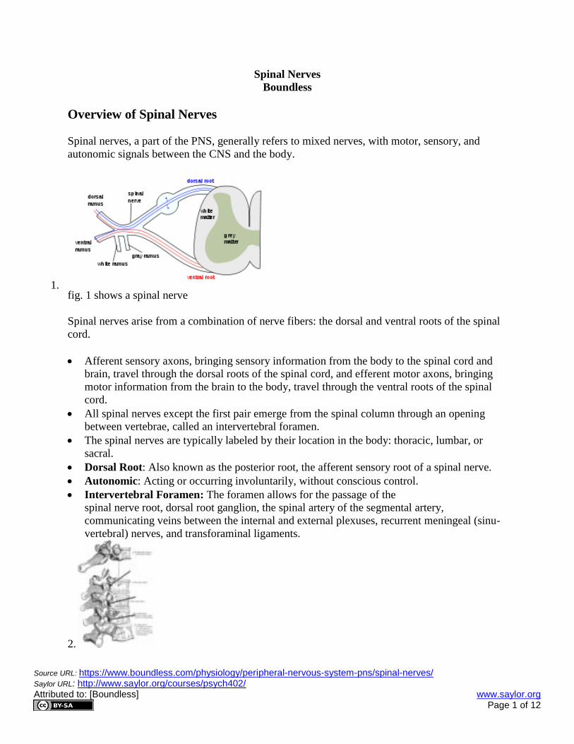
1. fig. 1 shows a spinal nerve
Spinal nerves arise from a combination of nerve fibers: the dorsal and ventral roots of the spinal
cord.
Afferent sensory axons, bringing sensory information from the body to the spinal cord and
brain, travel through the dorsal roots of the spinal cord, and efferent motor axons, bringing
motor information from the brain to the body, travel through the ventral roots of the spinal
cord.
All spinal nerves except the first pair emerge from the spinal column through an opening
between vertebrae, called an intervertebral foramen.
The spinal nerves are typically labeled by their location in the body: thoracic, lumbar, or
sacral.
Dorsal Root: Also known as the posterior root, the afferent sensory root of a spinal nerve.
Autonomic: Acting or occurring involuntarily, without conscious control.
Intervertebral Foramen: The foramen allows for the passage of the
spinal nerve root, dorsal root ganglion, the spinal artery of the segmental artery,
communicating veins between the internal and external plexuses, recurrent meningeal (sinu-
vertebral) nerves, and transforaminal ligaments.
2.
Page 2
Source URL: https://www.boundless.com/physiology/peripheral-nervous-system-pns/spinal-nerves/ Saylor URL: http://www.saylor.org/courses/psych402/ Attributed to: [Boundless] www.saylor.org Page 2 of 12
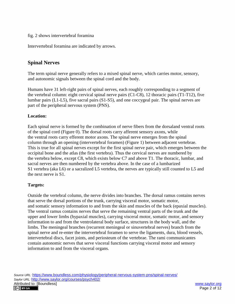
fig. 2 shows intervertebral foramina
Intervertebral foramina are indicated by arrows.
Spinal Nerves
The term spinal nerve generally refers to a mixed spinal nerve, which carries motor, sensory,
and autonomic signals between the spinal cord and the body.
Humans have 31 left-right pairs of spinal nerves, each roughly corresponding to a segment of
the vertebral column: eight cervical spinal nerve pairs (C1-C8), 12 thoracic pairs (T1-T12), five
lumbar pairs (L1-L5), five sacral pairs (S1-S5), and one coccygeal pair. The spinal nerves are
part of the peripheral nervous system (PNS).
Location:
Each spinal nerve is formed by the combination of nerve fibers from the dorsaland ventral roots
of the spinal cord (Figure 0). The dorsal roots carry afferent sensory axons, while
the ventral roots carry efferent motor axons. The spinal nerve emerges from the spinal
column through an opening (intervertebral foramen) (Figure 1) between adjacent vertebrae.
This is true for all spinal nerves except for the first spinal nerve pair, which emerges between the
occipital bone and the atlas (the first vertebra). Thus the cervical nerves are numbered by
the vertebra below, except C8, which exists below C7 and above T1. The thoracic, lumbar, and
sacral nerves are then numbered by the vertebra above. In the case of a lumbarized
S1 vertebra (aka L6) or a sacralized L5 vertebra, the nerves are typically still counted to L5 and
the next nerve is S1.
Targets:
Outside the vertebral column, the nerve divides into branches. The dorsal ramus contains nerves
that serve the dorsal portions of the trunk, carrying visceral motor, somatic motor,
and somatic sensory information to and from the skin and muscles of the back (epaxial muscles).
The ventral ramus contains nerves that serve the remaining ventral parts of the trunk and the
upper and lower limbs (hypaxial muscles), carrying visceral motor, somatic motor, and sensory
information to and from the ventrolateral body surface, structures in the body wall, and the
limbs. The meningeal branches (recurrent meningeal or sinuvertebral nerves) branch from the
spinal nerve and re-enter the intervertebral foramen to serve the ligaments, dura, blood vessels,
intervertebral discs, facet joints, and periosteum of the vertebrae. The rami communicantes
contain autonomic nerves that serve visceral functions carrying visceral motor and sensory
information to and from the visceral organs.
Page 3
Source URL: https://www.boundless.com/physiology/peripheral-nervous-system-pns/spinal-nerves/ Saylor URL: http://www.saylor.org/courses/psych402/ Attributed to: [Boundless] www.saylor.org Page 3 of 12
Branches:
The spinal nerves branch into the dorsal ramus, ventral ramus, the meningeal branches, and the
rami communicantes.
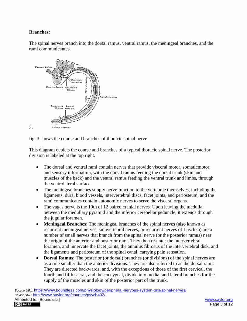
3.
fig. 3 shows the course and branches of thoracic spinal nerve
This diagram depicts the course and branches of a typical thoracic spinal nerve. The posterior
division is labeled at the top right.
The dorsal and ventral rami contain nerves that provide visceral motor, somaticmotor,
and sensory information, with the dorsal ramus feeding the dorsal trunk (skin and
muscles of the back) and the ventral ramus feeding the ventral trunk and limbs, through
the ventrolateral surface.
The meningeal branches supply nerve function to the vertebrae themselves, including the
ligaments, dura, blood vessels, intervertebral discs, facet joints, and periosteum, and the
rami communicates contain autonomic nerves to serve the visceral organs.
The vagas nerve is the 10th of 12 paired cranial nerves. Upon leaving the medulla
between the medullary pyramid and the inferior cerebellar peduncle, it extends through
the jugular foramen.
Meningeal Branches: The meningeal branches of the spinal nerves (also known as
recurrent meningeal nerves, sinuvertebral nerves, or recurrent nerves of Luschka) are a
number of small nerves that branch from the spinal nerve (or the posterior ramus) near
the origin of the anterior and posterior rami. They then re-enter the intervertebral
foramen, and innervate the facet joints, the annulus fibrosus of the intervertebral disk, and
the ligaments and periosteum of the spinal canal, carrying pain sensation.
Dorsal Ramus: The posterior (or dorsal) branches (or divisions) of the spinal nerves are
as a rule smaller than the anterior divisions. They are also referred to as the dorsal rami.
They are directed backwards, and, with the exceptions of those of the first cervical, the
fourth and fifth sacral, and the coccygeal, divide into medial and lateral branches for the
supply of the muscles and skin of the posterior part of the trunk.
Page 4
Source URL: https://www.boundless.com/physiology/peripheral-nervous-system-pns/spinal-nerves/ Saylor URL: http://www.saylor.org/courses/psych402/ Attributed to: [Boundless] www.saylor.org Page 4 of 12
Outside the vertebral column, the spinal nerves divide into branches. The dorsal ramus contains
nerves that serve the dorsal portions of the trunk carrying visceral motor, somatic motor, and
sensory information to and from the skin and muscles of the back. The ventral ramus contains
nerves that serve the remaining ventral parts of the trunk and the upper and lower limbs carrying
visceral motor, somatic motor, and sensory information to and from the ventrolateral body
surface, structures in the body wall, and the limbs. Themeningeal branches (recurrent meningeal
or sinuvertebral nerves) branch from the spinal nerve and re-enter the intervertebral foramen to
serve the ligaments, dura, blood vessels, intervertebral discs, facet joints, and periosteum of the
vertebrae. The rami communicants contain autonomic nerves that serve visceral functions
carrying visceral motor and sensory information to and from the visceral organs.
Some ventral rami merge with adjacent ventral rami to form a nerve plexus, a network of
interconnecting nerves. Nerves emerging from a plexus contain fibers from various spinal
nerves, which are now carried together to some target location. Major plexuses include the
cervical, brachial, lumbar, and sacral plexuses.
The vagas nerve is the 10th
of 12 paired cranial nerves. Upon leaving the medulla between the
medullary pyramid and the inferior cerebellar peduncle, it extends through the jugular foramen,
then passes into the carotid sheath between the internal carotid artery and the internal
jugular vein down below the head, to the neck, chest and abdomen, where it contributes to
the innervation of the viscera. Besides output to the various organs in the body, the
vagus nerve conveys sensory information about the state of the body's organs to the
central nervous system. 80-90% of the nerve fibers in the vagus nerve are afferent (sensory)
nerves communicating the state of the viscera to the brain.
Plexuses:
A nerve plexus is a network of intersecting nerve fibers consisting of nerves that serve the same
part of the body.
Page 5
Source URL: https://www.boundless.com/physiology/peripheral-nervous-system-pns/spinal-nerves/ Saylor URL: http://www.saylor.org/courses/psych402/ Attributed to: [Boundless] www.saylor.org Page 5 of 12
4.
fig. 4. shows a nerve plexus
The lumbar plexus is comprised of the ventral rami of the lumbar spinal nerves (L1-L5) and a
contribution from the thoracic nerve (T12). The posterior (green) and anterior (yellow) divisions
of the lumbar plexus are shown in the diagram.
Nerve plexuses throughout the body tend to be named after the area in which
theplexus occurs and the organs, limbs, and tissues it serves. Examples include the
cervical, brachial, lumbar, sacral, celiac, and coccygeal plexuses.
Auerbach’s plexus, which serves the gastrointestinal tract, is named after the first person
to describe this plexus, Leopold Auerbach, rather than the area of the body it serves.
The brachial plexus serves the chest, shoulders, arms and hands and is formed by
the ventral rami of C5-C8-T1 spinal nerves, and lower and upper halves of C4 and T2
spinal nerves.
Coccygeal Plexus: The coccygeal plexus is a plexus of nerves near the coccyx bone.
Nerve Plexus: A nerve plexus is a network of intersecting nerves.
Brachial Plexus: The brachial plexus is a network of nerve fibers, running from the
spine, formed by the ventral rami of the lower four cervical and first thoracic nerve roots
(C5-C8, T1). It proceeds through the neck, the axilla (armpit region), and into the arm. It
is a bunch of nerves passing through the cervico-axillary canal to reach axilla and
supplies the brachium, the antebrachium, and the hand.
Page 6
Source URL: https://www.boundless.com/physiology/peripheral-nervous-system-pns/spinal-nerves/ Saylor URL: http://www.saylor.org/courses/psych402/ Attributed to: [Boundless] www.saylor.org Page 6 of 12
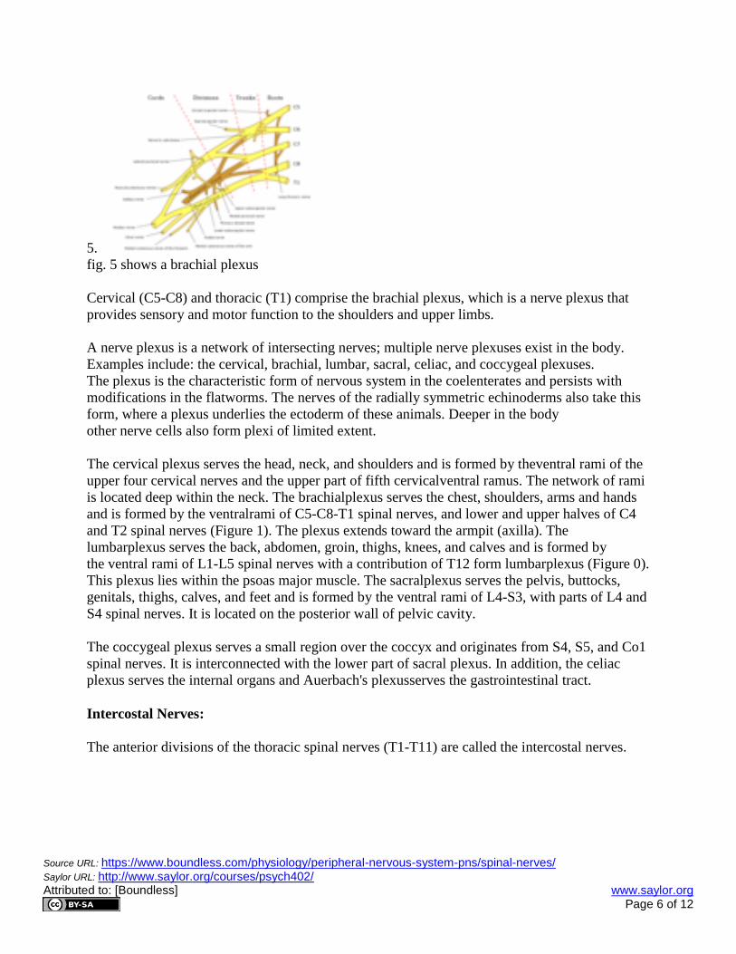
5.
fig. 5 shows a brachial plexus
Cervical (C5-C8) and thoracic (T1) comprise the brachial plexus, which is a nerve plexus that
provides sensory and motor function to the shoulders and upper limbs.
A nerve plexus is a network of intersecting nerves; multiple nerve plexuses exist in the body.
Examples include: the cervical, brachial, lumbar, sacral, celiac, and coccygeal plexuses.
The plexus is the characteristic form of nervous system in the coelenterates and persists with
modifications in the flatworms. The nerves of the radially symmetric echinoderms also take this
form, where a plexus underlies the ectoderm of these animals. Deeper in the body
other nerve cells also form plexi of limited extent.
The cervical plexus serves the head, neck, and shoulders and is formed by theventral rami of the
upper four cervical nerves and the upper part of fifth cervicalventral ramus. The network of rami
is located deep within the neck. The brachialplexus serves the chest, shoulders, arms and hands
and is formed by the ventralrami of C5-C8-T1 spinal nerves, and lower and upper halves of C4
and T2 spinal nerves (Figure 1). The plexus extends toward the armpit (axilla). The
lumbarplexus serves the back, abdomen, groin, thighs, knees, and calves and is formed by
the ventral rami of L1-L5 spinal nerves with a contribution of T12 form lumbarplexus (Figure 0).
This plexus lies within the psoas major muscle. The sacralplexus serves the pelvis, buttocks,
genitals, thighs, calves, and feet and is formed by the ventral rami of L4-S3, with parts of L4 and
S4 spinal nerves. It is located on the posterior wall of pelvic cavity.
The coccygeal plexus serves a small region over the coccyx and originates from S4, S5, and Co1
spinal nerves. It is interconnected with the lower part of sacral plexus. In addition, the celiac
plexus serves the internal organs and Auerbach's plexusserves the gastrointestinal tract.
Intercostal Nerves:
The anterior divisions of the thoracic spinal nerves (T1-T11) are called the intercostal nerves.
Page 7
Source URL: https://www.boundless.com/physiology/peripheral-nervous-system-pns/spinal-nerves/ Saylor URL: http://www.saylor.org/courses/psych402/ Attributed to: [Boundless] www.saylor.org Page 7 of 12
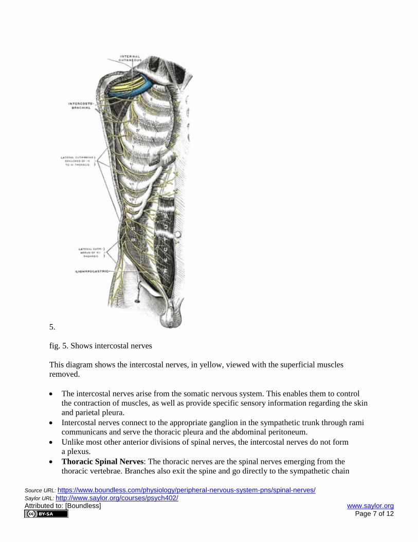
5.
fig. 5. Shows intercostal nerves
This diagram shows the intercostal nerves, in yellow, viewed with the superficial muscles
removed.
The intercostal nerves arise from the somatic nervous system. This enables them to control
the contraction of muscles, as well as provide specific sensory information regarding the skin
and parietal pleura.
Intercostal nerves connect to the appropriate ganglion in the sympathetic trunk through rami
communicans and serve the thoracic pleura and the abdominal peritoneum.
Unlike most other anterior divisions of spinal nerves, the intercostal nerves do not form
a plexus.
Thoracic Spinal Nerves: The thoracic nerves are the spinal nerves emerging from the
thoracic vertebrae. Branches also exit the spine and go directly to the sympathetic chain
Page 8
Source URL: https://www.boundless.com/physiology/peripheral-nervous-system-pns/spinal-nerves/ Saylor URL: http://www.saylor.org/courses/psych402/ Attributed to: [Boundless] www.saylor.org Page 8 of 12
ganglia of the autonomic nervous system where they are involved in the functions of organs
and glands in the head, neck, thorax, and abdomen.
Sympathetic Trunk: The sympathetic trunks (sympathetic chain, gangliated cord) are a
paired bundle of nerve fibers that run from the base of the skull to the coccyx.
Abdominal Peritoneum: The peritoneum is the serous membrane that forms the lining of
the abdominal cavity or the coelom—it covers most of the intra-abdominal (or coelomic)
organs—in amniotes and some invertebrates (annelids, for instance). It is composed of a
layer of mesothelium supported by a thin layer of connective tissue. The peritoneum both
supports the abdominal organs and serves as a conduit for their blood and lymph vessels and
nerves.
The intercostal nerves are part of the somatic nervous system, and arise from anterior divisions
(rami anteriores; ventral divisions) of the thoracic spinal nerves from T1 to T11. The intercostal
nerves are distributed chiefly to the thoracic pleura and abdominal peritoneum and differ from
the anterior divisions of the other spinal nerves in that each pursues an independent course
without plexus formation.
The first two nerves supply fibers to the upper limb in addition to their thoracic branches. The
anterior division of the first thoracic nerve divides into two branches: one, the larger, leaves the
thorax in front of the neck of the first rib, and enters the brachial plexus; the other smaller
branch, the first intercostal nerve, runs along the first intercostal space, and ends on the front of
the chest as the first anterior cutaneous branch of the thorax.
The next four are limited in their distribution to the parietes of the thorax; the lower five supply
the parietes of the thorax and abdomen. The seventh intercostalnerve terminates at the xyphoid
process, at the lower end of the sternum. The tenth intercostal nerve terminates at the umbilicus.
The twelfth (subcostal) thoracic is distributed to the abdominal wall and groin.
Unlike the nerves from the autonomic nervous system that innervate the visceral pleura of the
thoracic cavity, the intercostal nerves arise from the somatic nervous system. This enables them
to control the contraction of muscles, as well as provide specific sensory information regarding
the skin and parietal pleura. This explains why damage to the internal wall of the thoracic cavity
can be felt as a sharp painlocalized in the injured region. Damage to the visceral pleura is
experienced as an un-localized ache.
Dermatomes:
A dermatome is an area of skin that is supplied by a single spinal nerve.
Page 9
Source URL: https://www.boundless.com/physiology/peripheral-nervous-system-pns/spinal-nerves/ Saylor URL: http://www.saylor.org/courses/psych402/ Attributed to: [Boundless] www.saylor.org Page 9 of 12
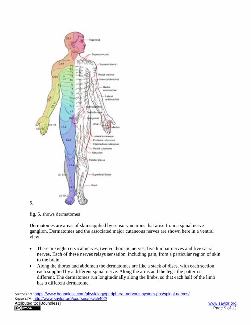
5.
fig. 5. shows dermatomes
Dermatomes are areas of skin supplied by sensory neurons that arise from a spinal nerve
ganglion. Dermatomes and the associated major cutaneous nerves are shown here in a ventral
view.
There are eight cervical nerves, twelve thoracic nerves, five lumbar nerves and five sacral
nerves. Each of these nerves relays sensation, including pain, from a particular region of skin
to the brain.
Along the thorax and abdomen the dermatomes are like a stack of discs, with each section
each supplied by a different spinal nerve. Along the arms and the legs, the pattern is
different. The dermatomes run longitudinally along the limbs, so that each half of the limb
has a different dermatome.
Page 10
Source URL: https://www.boundless.com/physiology/peripheral-nervous-system-pns/spinal-nerves/ Saylor URL: http://www.saylor.org/courses/psych402/ Attributed to: [Boundless] www.saylor.org Page 10 of 12
Dermatomes have clinical significance, especially in the diagnosis of certain diseases.
Symptoms that follow a dermatome, such as pain or a rash, may indicate a pathology that
involves the related nerve root. Examples include dysfunction of the spine or a
viral infection.
Chickenpox: A common childhood disease caused by the varicella-zoster virus (VZV).
Shingles: Also known as herpes zoster, shingles is an acute viral inflammation of the sensory
ganglia of spinal and cranial nerves associated with a vesicular eruption and neuralgic pains
and caused by reactivation of the poxvirus causing chicken pox.
6.
fig. 6. shows a shingle rash
The shingles rash appears across a dermatome. In this patient, one of the dermatomes in the arm
is affected, restricting the rash to the length of the back of the arm.
A dermatome is an area of skin that is supplied by a single spinal nerve (Figure 0). There are
eight cervical nerves, twelve thoracic nerves, five lumbar nerves and five sacral nerves. Each of
these nerves relays sensation, including pain, from a particular region of the skin to the brain.
Along the thorax and abdomen, the dermatomes are like a stack of discs, with each section
supplied by a different spinal nerve. Along the arms and the legs, the pattern is different. The
dermatomes run longitudinally along the limbs, so that each half of the limb has a
different dermatome. Although the general pattern is similar in all people, the precise areas
of innervation are as unique to an individual as fingerprints.
Dermatomes have clinical significance, especially in the diagnosis of certain diseases. Symptoms
that follow a dermatome, such as pain or a rash, may indicate a pathology that involves the
related nerve root. Examples include dysfunction of the spine or a viral infection. Viruses that
remain dormant in nerve ganglia, such as the Varicella zoster virus, which causes both
chickenpox and shingles, often cause either pain, rash or both in a pattern defined by
a dermatome (Figure 1). Shingles is one of the only diseases that causes a rash in a dermatomal
pattern, and as such, this is its defining symptom. The rash of shingles is almost always restricted
to a specific dermatome, such as on the chest, leg or arm, caused by the residual infection of
the nerve that supplies that area of skin with the varicella zoster virus. Shingles typically appears
years or decades after recovery from chickenpox.
Page 11
Source URL: https://www.boundless.com/physiology/peripheral-nervous-system-pns/spinal-nerves/ Saylor URL: http://www.saylor.org/courses/psych402/ Attributed to: [Boundless] www.saylor.org Page 11 of 12
Function and Physiology of the Spinal Nerves:
Spinal nerves connect the brain and spinal cord to the limbs and organs of the body.
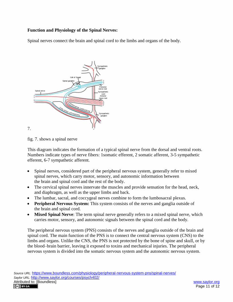
7.
fig. 7. shows a spinal nerve
This diagram indicates the formation of a typical spinal nerve from the dorsal and ventral roots.
Numbers indicate types of nerve fibers: 1somatic efferent, 2 somatic afferent, 3-5 sympathetic
efferent, 6-7 sympathetic afferent.
Spinal nerves, considered part of the peripheral nervous system, generally refer to mixed
spinal nerves, which carry motor, sensory, and autonomic information between
the brain and spinal cord and the rest of the body.
The cervical spinal nerves innervate the muscles and provide sensation for the head, neck,
and diaphragm, as well as the upper limbs and back.
The lumbar, sacral, and coccygeal nerves combine to form the lumbosacral plexus.
Peripheral Nervous System: This system consists of the nerves and ganglia outside of
the brain and spinal cord.
Mixed Spinal Nerve: The term spinal nerve generally refers to a mixed spinal nerve, which
carries motor, sensory, and autonomic signals between the spinal cord and the body.
The peripheral nervous system (PNS) consists of the nerves and ganglia outside of the brain and
spinal cord. The main function of the PNS is to connect the central nervous system (CNS) to the
limbs and organs. Unlike the CNS, the PNS is not protected by the bone of spine and skull, or by
the blood–brain barrier, leaving it exposed to toxins and mechanical injuries. The peripheral
nervous system is divided into the somatic nervous system and the autonomic nervous system.
Page 12
Source URL: https://www.boundless.com/physiology/peripheral-nervous-system-pns/spinal-nerves/ Saylor URL: http://www.saylor.org/courses/psych402/ Attributed to: [Boundless] www.saylor.org Page 12 of 12
The peripheral nervous system includes 12 cranial nerves and 31 pairs of spinal nerves, which
provide communication from the CNS to the rest of the body by nerve impulses that regulate the
functions of the human body. The term spinal nerve generally refers to a mixed spinal nerve,
which carries motor, sensory, and autonomic signals between the spinal cord and the body.
Humans have 31 left-right pairs of spinal nerves, each roughly corresponding to a segment of
thevertebral column: 8 cervical spinal nerve pairs (C1-C8), 12 thoracic pairs (T1-T12), 5 lumbar
pairs (L1-L5), 5 sacral pairs (S1-S5), and 1 coccygeal pair.
The first 4 cervical spinal nerves, C1 through C4, split and recombine to produce a variety of
nerves that subserve the neck and back of the head. Spinal nerve C1 is called the suboccipital
nerve, which provides motor innervation to muscles at the base of the skull. C2 and C3 form
many of the nerves of the neck, providing both sensory and motor control. These include the
greater occipital nerve which provides sensation to the back of the head, the lesser occipital
nerve which provides sensation to the area behind the ears, the greater auricular nerve, and the
lesser auricular nerve. The phrenic nerve arises from nerve roots C3, C4, and C5. It innervates
the diaphragm, enabling breathing. If the spinal cord is transected above C3, then spontaneous
breathing is not possible.
The last four cervical spinal nerves, C5 through C8, and the first thoracic spinal nerve, T1,
combine to form the brachial plexus, or plexus brachialis, a tangled array of nerves, splitting,
combining and recombining, to form the nerves that subserve the upper limb region and upper
back. Although the brachial plexus may appear tangled, it is highly organized and predictable,
with little variation between people.
The anterior divisions of the lumbar nerves, sacral nerves, and coccygeal nerve form the
lumbosacral plexus, the first lumbar nerve being frequently joined by a branch from the twelfth
thoracic. For descriptive purposes this plexus is usually divided into three parts: lumbar plexus,
sacral plexus, and pudendal plexus.