http://www.diva-portal.org This is the published version of a paper published in SpringerPlus. Citation for the original published paper (version of record): Husberg, B., Salehi, K., Peters, T., Gunnarsson, U., Michanek, M. et al. (2016) Congenital intestinal malrotation in adolescent and adult patients: a 12-year clinical and radiological survey. SpringerPlus, 5: 245 http://dx.doi.org/10.1186/s40064-016-1842-0 Access to the published version may require subscription. N.B. When citing this work, cite the original published paper. Permanent link to this version: http://urn.kb.se/resolve?urn=urn:nbn:se:umu:diva-119668
Transcript
http://www.diva-portal.org
This is the published version of a paper published in SpringerPlus.
Citation for the original published paper (version of record):
Husberg, B., Salehi, K., Peters, T., Gunnarsson, U., Michanek, M. et al. (2016)
Congenital intestinal malrotation in adolescent and adult patients: a 12-year clinical and
radiological survey.
SpringerPlus, 5: 245
http://dx.doi.org/10.1186/s40064-016-1842-0
Access to the published version may require subscription.
N.B. When citing this work, cite the original published paper.
Permanent link to this version:http://urn.kb.se/resolve?urn=urn:nbn:se:umu:diva-119668
Husberg et al. SpringerPlus (2016) 5:245 DOI 10.1186/s40064-016-1842-0
RESEARCH
Congenital intestinal malrotation in adolescent and adult patients: a 12-year clinical and radiological surveyBritt Husberg1,2,3,4, Karin Salehi5,6, Trevor Peters7, Ulf Gunnarsson8, Margareta Michanek3,4, Agneta Nordenskjöld5,6 and Karin Strigård8*
Abstract
Congenital intestinal malrotation is mainly detected in childhood and caused by incomplete rotation and fixation of the intestines providing the prerequisites for life-threatening volvulus of the midgut. The objective of this study was to evaluate a large cohort of adult patients with intestinal malrotation. Thirty-nine patients, 15–67 years, were diagnosed and admitted to a university setting with congenital intestinal malrotation 2002–2013. The patients were divided into three age groups for stratified evaluation. Medical charts were scrutinized, and clinical outcome of surgery was reviewed. Twelve patients presented as emergency cases, whereas 27 were admitted as elective cases. Diagnosis was established in 33 patients who underwent radiological investigation and in the remaining 6 during surgery. A Ladd’s operation was per-formed in 31 symptomatic patients; a conservative strategy was chosen in eight cases. Volvulus was more common in the younger age group. Twenty-six surgically treated patients were available for telephone interview, 1–12 years after surgery. All patients, except one, regarded their general condition improved to a high degree (n = 18) or with some reservation (n = 7). Twelve patients suffered remaining abdominal pain of a chronic and diffuse character. Due to recurrence of malro-tation six patients were reoperated. Symptomatic malrotation occurs in both children and the adult population. Improved awareness and an accurately performed CT scan can reveal the malformation and enable surgical treatment. A Ladd’s procedure relieved most patients from their severe complaints even when a history of several years of suffering existed.
BackgroundIn congenital intestinal malrotation an impaired embryo-logical development of the gut causes incomplete rota-tion and fixation of the intestines to the abdominal wall (Dott 1923). The fulfillment of the third embryonic rota-tion includes the traversing of the duodenum to the left side of the abdomen, forming the ligaments of Treitz, and the migration of the ileo-caecal junction to the lower right abdominal quadrant. The fixation of the full-length bowel is complete during the twelfth week (Penco et al. 2007). Congenital malformations such as diaphragmatic hernia, omphalocele or gastroschisis are associated with
a similar but secondary incomplete rotation and fixation of the intestines (Torres and Ziegler 1993).
The inadequate fixation of the bowel alongside remain-ing embryonic fibrous adhesions, the Ladd’s bands (Ladd 1932, 1936), may give rise to a variety of intestinal mal-function. In the worst case scenario, malrotation may develop into a midgut volvulus with torsion causing high risk of ischemia and necrosis of the parts of the intes-tine supplied by the superior mesenteric artery. This life-threatening condition is well known among pediat-ric surgeons and is always considered when physicians treat critically ill infants with abdominal symptoms and unknown diagnoses.
Malrotation has primarily been diagnosed in early childhood, with estimated onset of symptoms during the first year of life in 90 % of the cases (Vaos and Misiakos
Open Access
*Correspondence: [email protected] 8 Department of Surgical and Perioperative Sciences, Umeå University Hospital, 901 87 Umeå, SwedenFull list of author information is available at the end of the article
Page 2 of 7Husberg et al. SpringerPlus (2016) 5:245
2010; Pickhardt and Bhalla 2002; Stewart et al. 1976). There are recent reports of manifestation later in life, both as emergency conditions or more chronic gastrointes-tinal symptoms (Penco et al. 2007; Pickhardt and Bhalla 2002; Nehra and Goldstein 2011). The exact incidence of intestinal malrotation is thus still difficult to determine. It was earlier described to be approximately 0.2 % (Stewart et al. 1976; Donnellan and Kimura 1996; Clark and Old-ham 2002), but an incidence up to 1 % has been reported (Adams and Stanton 2014). Improved radiological facili-ties, including multi-detector CT-scans, provide new pos-sibilities to identify anatomical aberrations.
During a 12-year period, we have treated 39 consecu-tive cases of adult malrotation at the Karolinska Univer-sity Hospital, Huddinge. The aim of this study was to increase knowledge concerning this diagnosis by describ-ing symptoms, treatment and clinical outcome in our cohort of adolescent and adult patients with intestinal malrotation.
MethodsPatientsThirty-nine patients, 22 females and 17 males, aged between 15 and 67 years, were diagnosed with congeni-tal intestinal malrotation. The patients were prospectively investigated at the Karolinska University Hospital from 2002 to 2013. After identification of the first patient, it was decided to prospectively monitor patients treated for malrotation in order to analyze and publish data when a reasonable number of patients had been treated.
Medical chartsAll medical records were evaluated with regards to symp-toms, surgical procedures, previous disorders and out-comes. For analysis of differences according to age, the patients were divided into three groups (15–20, 21–50, 51–67 years).
Radiological diagnosticsTo establish the degree of malrotation, the radiologist identified the position of the duodenum and the proximal small bowel, the location of the caecum and the orienta-tion of the mesenteric vessels using intravenous, per oral as well as intrarectal contrast (triple-contrast). Twisting of the mesentery of the small bowel, the “whirlpool-sign”, typical for a volvulus was noted. This evaluation was also re-scrutinized and confirmed independently by one dedi-cated radiologist.
SurgerySymptomatic malrotation was treated by corrective sur-gery according to the technique originally described by Ladd. If a volvulus was present, the intestines were
de-rotated in a counter clockwise manner and all Ladd’s bands were carefully removed and dissected. If needed, the mesentery was broadened and the adhesions sur-rounding the mesenteric vessels dissected in order to avoid future recurrence of volvulus. When the dissec-tion was done, the small bowel was placed to the right and the colon to the left side of the abdominal cavity in a “non-rotational” position. Two different surgeons reg-istered data from medical charts on these surgical details independently.
Follow upThe patients were routinely assessed 6 weeks, 6 months and 12 months after surgery. After that, occasional con-tact occurred if further complaints presented. During 2012–2013 a research nurse performed telephone inter-views with a semi-structured concept concerning the patients’ past and present situation and possible remain-ing symptoms after surgery. The questions focused on remaining intense or chronic pain, postprandial nau-sea, vomiting and constipation. Patients were also asked whether they regarded their general physical condition as improved to a high degree, improved with some reserva-tion or without any notable improvement.
Ethical considerationsThe Regional Ethical Review Board approved this study 12-06-20. Dnr 2012/957-31/3.
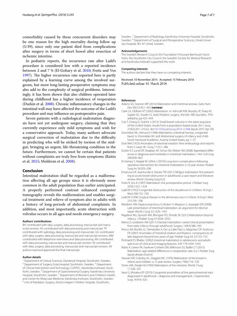
ResultsClinical dataTwelve patients presented as emergency cases, whereas the remaining 27 were admitted on an elective basis. The most common symptom was abdominal pain, followed by signs of intestinal obstruction (Table 1). Another predominant symptom was sensations of extreme full-ness and discomfort after meals, sometimes followed by nausea and vomiting, described by 29 patients (Table 2). Thirteen of these patients were previously assessed and diagnosed with gastro-oesophageal reflux. In six cases the diagnosis was achieved during surgical treatment focused on other conditions.
Concomitant malformations were observed in 15 patients (38 %), including seven patients with CNS dis-turbances and mental retardation. Other malformations noticed were bicorn uterus, vaginal atresia, double ure-ters, Tuberose sclerosis, Mb Hirschprung, pelvic kidney, Cornelia de Lange syndrome and scoliosis. Eight patients had a history of disease within the hepatobiliary and pan-creatic system with a history of pancreas divisum and in four cases pancreatitis. Six further patients had gastro-intestinal motility disturbances, verified by small bowel manometry and/or full thickness specimens.
Page 3 of 7Husberg et al. SpringerPlus (2016) 5:245
Table 1 Clinical data
a CDH n = 1, gastroschisis n = 1, omphalocele n = 1b “Imaging” denotes the radiologic procedure that lead to diagnosis. Two patients had no imaging due to emergency surgery (Age ≤20 years n = 1, age 21–50 years n = 1)c Out of 33 patients where CT-studies were available for reviewing
Page 4 of 7Husberg et al. SpringerPlus (2016) 5:245
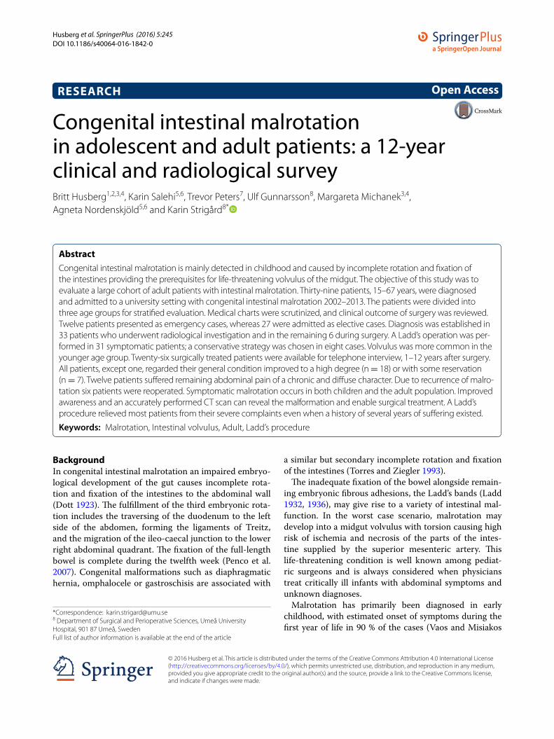
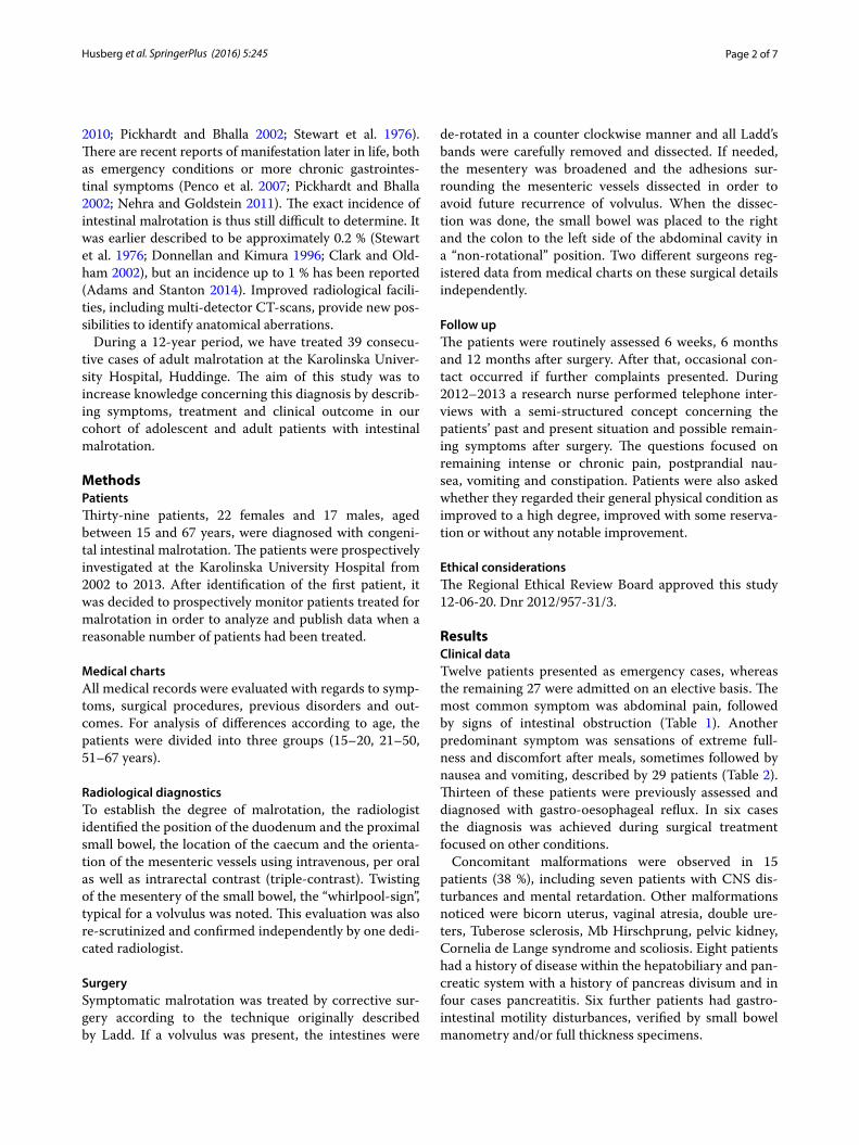
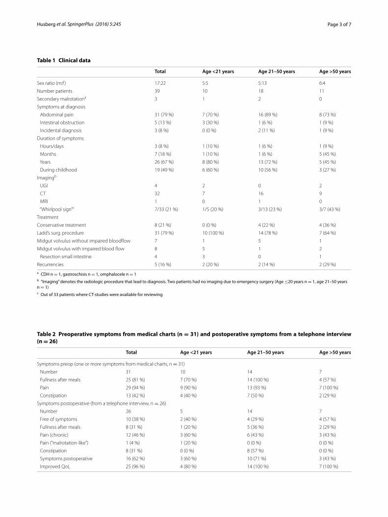
Radiological findingsInvestigation with multi-detector computer tomography was used in 32 cases and MRI in one. The three cardi-nal radiological diagnostic criteria were identified in 19 of the cases. In all but one of the patients, the small bowel was located to the right with a pathological ver-tical course of the duodenum that failed to traverse the vertebral spine. In the remaining patient, the duodenal course initially crossed the midline to the left, but turned back again forming a loop. In addition, the ascending colon had a short attachment to the parietal left side. In 22 cases the caecum had the expected abnormal posi-tion according to radiology, whereas the caecum in the remaining 11 patients was located on the right side. It was later revealed during surgery that in all these cases the ascending colon was mobile and not fixed to the pari-etal abdominal wall, except in one case where the right flexure of colon was shortly attached. Malposition of the superior mesenteric artery and vein was noted radiologi-cally in 26 patients, of whom 25 had inverted vessels and one presented vessels in a vertical position (Figs. 1, 2). A “whirlpool-sign” signifying a presence of rotation of the bowel could be detected in seven cases (Fig. 3).
SurgeryThirty-one patients were operated (Fig. 4). Sixteen patients had undergone previous abdominal surgery before the Ladd procedure, with chart notifications of intestinal malrotation in 11 of the cases. Emergency sur-gery was performed in 9 of 31 cases. In three patients the operation was performed semi-urgently because of progression of abdominal complaints. One patient had a complete mid-gut volvulus causing ischemia and necro-sis of the bowel, necessitating resection of the entire small bowel. Another seven patients exhibited signs of acute volvulus, compromising circulation in a segment of the small bowel (2 of them >50 years). Three of these required minor resections. There was a tendency towards an increased risk for volvulus in the younger patients (Table 1).
Seventeen symptomatic patients were operated on elec-tively after a radiological diagnosis. Appendectomy was Fig. 1 CT scan showing inverted vessels, front view
Fig. 2 CT scan in axial position showing the inverted vessels
Fig. 3 Whirl pool sign where when the mesenteric of the small intestine has been twisted
Page 5 of 7Husberg et al. SpringerPlus (2016) 5:245
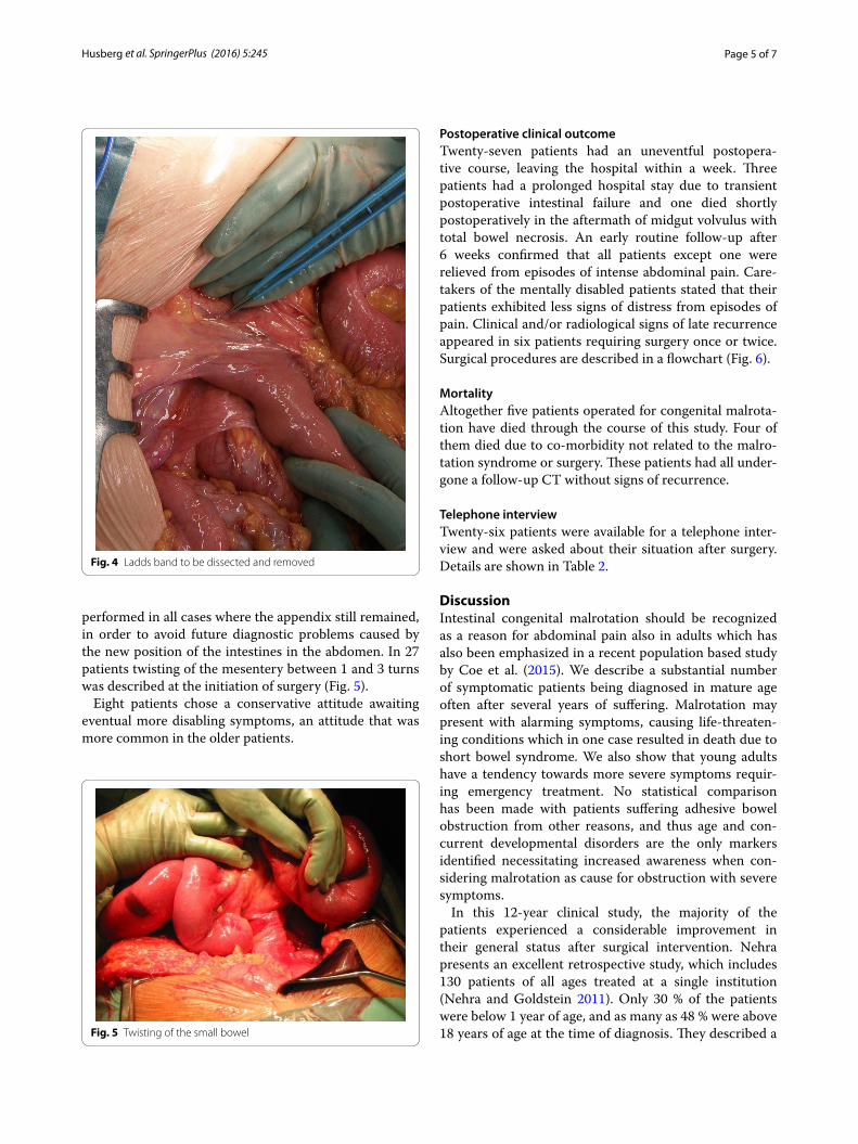
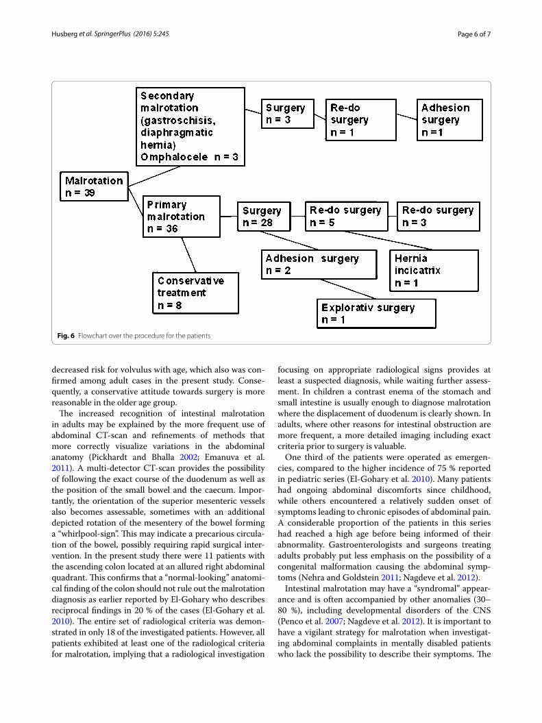
performed in all cases where the appendix still remained, in order to avoid future diagnostic problems caused by the new position of the intestines in the abdomen. In 27 patients twisting of the mesentery between 1 and 3 turns was described at the initiation of surgery (Fig. 5).
Eight patients chose a conservative attitude awaiting eventual more disabling symptoms, an attitude that was more common in the older patients.
Postoperative clinical outcomeTwenty-seven patients had an uneventful postopera-tive course, leaving the hospital within a week. Three patients had a prolonged hospital stay due to transient postoperative intestinal failure and one died shortly postoperatively in the aftermath of midgut volvulus with total bowel necrosis. An early routine follow-up after 6 weeks confirmed that all patients except one were relieved from episodes of intense abdominal pain. Care-takers of the mentally disabled patients stated that their patients exhibited less signs of distress from episodes of pain. Clinical and/or radiological signs of late recurrence appeared in six patients requiring surgery once or twice. Surgical procedures are described in a flowchart (Fig. 6).
MortalityAltogether five patients operated for congenital malrota-tion have died through the course of this study. Four of them died due to co-morbidity not related to the malro-tation syndrome or surgery. These patients had all under-gone a follow-up CT without signs of recurrence.
Telephone interviewTwenty-six patients were available for a telephone inter-view and were asked about their situation after surgery. Details are shown in Table 2.
DiscussionIntestinal congenital malrotation should be recognized as a reason for abdominal pain also in adults which has also been emphasized in a recent population based study by Coe et al. (2015). We describe a substantial number of symptomatic patients being diagnosed in mature age often after several years of suffering. Malrotation may present with alarming symptoms, causing life-threaten-ing conditions which in one case resulted in death due to short bowel syndrome. We also show that young adults have a tendency towards more severe symptoms requir-ing emergency treatment. No statistical comparison has been made with patients suffering adhesive bowel obstruction from other reasons, and thus age and con-current developmental disorders are the only markers identified necessitating increased awareness when con-sidering malrotation as cause for obstruction with severe symptoms.
In this 12-year clinical study, the majority of the patients experienced a considerable improvement in their general status after surgical intervention. Nehra presents an excellent retrospective study, which includes 130 patients of all ages treated at a single institution (Nehra and Goldstein 2011). Only 30 % of the patients were below 1 year of age, and as many as 48 % were above 18 years of age at the time of diagnosis. They described a
Fig. 4 Ladds band to be dissected and removed
Fig. 5 Twisting of the small bowel
Page 6 of 7Husberg et al. SpringerPlus (2016) 5:245
decreased risk for volvulus with age, which also was con-firmed among adult cases in the present study. Conse-quently, a conservative attitude towards surgery is more reasonable in the older age group.
The increased recognition of intestinal malrotation in adults may be explained by the more frequent use of abdominal CT-scan and refinements of methods that more correctly visualize variations in the abdominal anatomy (Pickhardt and Bhalla 2002; Emanuva et al. 2011). A multi-detector CT-scan provides the possibility of following the exact course of the duodenum as well as the position of the small bowel and the caecum. Impor-tantly, the orientation of the superior mesenteric vessels also becomes assessable, sometimes with an additional depicted rotation of the mesentery of the bowel forming a “whirlpool-sign”. This may indicate a precarious circula-tion of the bowel, possibly requiring rapid surgical inter-vention. In the present study there were 11 patients with the ascending colon located at an allured right abdominal quadrant. This confirms that a “normal-looking” anatomi-cal finding of the colon should not rule out the malrotation diagnosis as earlier reported by El-Gohary who describes reciprocal findings in 20 % of the cases (El-Gohary et al. 2010). The entire set of radiological criteria was demon-strated in only 18 of the investigated patients. However, all patients exhibited at least one of the radiological criteria for malrotation, implying that a radiological investigation
focusing on appropriate radiological signs provides at least a suspected diagnosis, while waiting further assess-ment. In children a contrast enema of the stomach and small intestine is usually enough to diagnose malrotation where the displacement of duodenum is clearly shown. In adults, where other reasons for intestinal obstruction are more frequent, a more detailed imaging including exact criteria prior to surgery is valuable.
One third of the patients were operated as emergen-cies, compared to the higher incidence of 75 % reported in pediatric series (El-Gohary et al. 2010). Many patients had ongoing abdominal discomforts since childhood, while others encountered a relatively sudden onset of symptoms leading to chronic episodes of abdominal pain. A considerable proportion of the patients in this series had reached a high age before being informed of their abnormality. Gastroenterologists and surgeons treating adults probably put less emphasis on the possibility of a congenital malformation causing the abdominal symp-toms (Nehra and Goldstein 2011; Nagdeve et al. 2012).
Intestinal malrotation may have a “syndromal” appear-ance and is often accompanied by other anomalies (30–80 %), including developmental disorders of the CNS (Penco et al. 2007; Nagdeve et al. 2012). It is important to have a vigilant strategy for malrotation when investigat-ing abdominal complaints in mentally disabled patients who lack the possibility to describe their symptoms. The
Fig. 6 Flowchart over the procedure for the patients
Page 7 of 7Husberg et al. SpringerPlus (2016) 5:245
comorbidity caused by these concurrent disorders may be one reason for the high mortality during follow up (5/39), since only one patient died from complications after surgery in terms of short bowel after resection of ischemic intestine.
In pediatric reports, the recurrence rate after Ladd’s procedure is considered low with a reported incidence between 2 and 7 % (El-Gohary et al. 2010; Freitz and Vos 1997). The higher recurrence rate reported here is partly explained by a learning curve among the involved sur-geons, but more long-lasting preoperative symptoms may also add to the complexity of surgical problems. Interest-ingly, it has been shown that also children operated later during childhood has a higher incidence of reoperation (Durkin et al. 2008). Chronic inflammatory changes in the intestinal wall may have affected the outcome of the Ladd’s procedure and may influence on postoperative pain.
Seven patients with a radiological malrotation diagno-sis have not yet undergone surgery, claiming that they currently experience only mild symptoms and wish for a conservative approach. Today, many authors advocate surgical correction of malrotation due to the difficulty in predicting who will be striked by torsion of the mid-gut, bringing an urgent, life-threatening condition in the future. Furthermore, we cannot be certain that patients without complaints are truly free from symptoms (Raitio et al. 2015; Moldrem et al. 2008).
ConclusionIntestinal malrotation shall be regarded as a malforma-tion affecting all age groups since it is obviously more common in the adult population than earlier anticipated. A properly performed contrast enhanced computer tomography reveals the malformation and enables surgi-cal treatment and relieve of symptom also in adults with a history of long-periods of abdominal complaints. In addition, and most importantly, acute obstruction with volvulus occurs in all ages and needs emergency surgery.
Authors’ contributionsBH contributed with idea, surgery, data processing, manuscript and manu-script revision. KS contributed with data processing and manuscript. TP contributed with radiology, data processing and manuscript. UG contributed with idea, surgery, data processing, manuscript and manuscript revision. MM contributed with telephone interviews and data processing. AN contributed with data processing, manuscript and manuscript revision. KS contributed with idea, surgery, data processing, manuscript and manuscript revision. All authors read and approved the final manuscript.
Author details1 Department of Clinical Sciences, Danderyd Hospital, Stockholm, Sweden. 2 Department of Surgery, Ersta Hospital, Stockholm, Sweden. 3 Department of Clinical Intervention and Technology, CLINTEC, Karolinska Institutet, Stock-holm, Sweden. 4 Department of Gastrointestinal Surgery, Karolinska University Hospital, Stockholm, Sweden. 5 Department of Women’s and Children’s Health, and Center for Molecular Medicine, Karolinska Institutet, Stockholm, Sweden. 6 Unit of Paediatric Surgery, Astrid Lindgren Children Hospital, Stockholm,
Sweden. 7 Department of Radiology, Karolinska University Hospital, Stockholm, Sweden. 8 Department of Surgical and Perioperative Sciences, Umeå Univer-sity Hospital, 901 87 Umeå, Sweden.
AcknowledgementsThe Swedish Research Council, the Foundation Frimurare Barnhuset Stock-holm, the Stockholm City Council, the Swedish Society for Medical Research and Karolinska Institutet supported this work.
Competing interestsThe authors declare that they have no competing interests.
Received: 10 November 2015 Accepted: 15 February 2016
ReferencesAdams SD, Stanton MP (2014) Malrotation and intestinal atresias. Early Hum
Coe T, Chang D, Sicklick J (2015) Small bowel volvulus in the adult populace of the United States: results from a population-based study. Am J Surg 210(2):201–210.e2. doi:10.1016/j.amjsurg.2014.12.048 (epub 2015 Apr 30)
Donnellan WL, Kimura K (1996) Malrotation, intestinal hernias, congenital band. In: Donnellan WL (ed) Abdominal surgery of infancy and child-hood. Harwood Academic, Austria-United States, pp 1–27
Dott NM (1923) Anomalies of intestinal rotation: their embryology and reports from 5 cases. Br J Surg 11:251–286
Durkin ET, Lund DP, Shaaban AF, Schurr MJ, Weber SM (2008) Agerelated differ-ences in diagnose and morbidity in intestinal malrotation. J Am Coll Surg 206:658–663
El-Gohary Y, Alagtal M, Gillick J (2010) Long-term complications following operative intervention for intestinal malrotation: a 10-year review. Pediatr Surg Int 26:203–206
Emanuva OF, Ayantunde A, Davies TW (2011) Midgut malrotation first present-ing as acute bowel obstruction in adulthood: a case report and literature review. World J Emerg Surg 6:22
Freitz R, Vos A (1997) Malrotation: the postoperative period. J Pediatr Surg 32(9):1322–1324
Ladd W (1932) Congenital obstruction of the duodenum in children. N Engl J Med 206:732–730
Ladd W (1936) Surgical disease in the alimentary tract in infants. N Engl J Med 215:705–708
Moldrem AW, Papaconstantinou H, Broker H, Megison S, Jeyarajah DR (2008) Late presentation of intestinal malrotation: an argument for elective repair. World J Surg 32:1426–1431
Nehra D, Goldstein AM (2011) Intestial malrotation: varied clinical presentation from early infancy through adulthood. Surgery 149(3):386–393
Penco JM, Murillo JC, Hernàndez A, De La Calle Pato U, Masjohan DF, Aceituno FR (2007) Anomalies of intestinal rotation and fixation: consequences of late diagnosis beyond two years of age. Pediatr Surg Int 23:723–732
Pickhardt PJ, Bhalla J (2002) Intestinal malrotation in adolescents and adults: spectrum of clinical and imaging features. AJR 179:1429–1435
Raitio A, Green PA, Fawkner-Corbett DW, Wilkinson DJ, Baillie CT (2015) Malrotation: age-related differences in reoperation rate. Eur J Pediatr Surg (epub ahead of print)
Stewart DR, Colodny AL, Daggett WC (1976) Malrotation of the bowel in infants and children: a 15 year review. Surgery 79(6):716–720
Torres AM, Ziegler M (1993) Malrotation of the intestine. World J Surg 17:326–331
Vaos G, Misiakos EP (2010) Congenital anomalities of the gastrointestinal tract diagnosed in adulthood—diagnose and management. J Gastrointest Surg 14:916–925