September 12, 2011 8:00-9:50 am STRUCTURE OF BIOLOGICAL MEMBRANES and BIOCHEMISTRY OF MEMBRANE TRANSPORT Lecturer: Dr. Eileen M. Lafer Contact Info: 415B, 567-3764, [email protected]Reading: Stryer Edition 6: Chapters 12 and 13, pp. 326-379 Please note that while the syllabus is printed in black and white, the notes were produced in color. Therefore, Dr. Lafer has deposited a pdf file containing these notes on blackboard so you can view them in color. Objectives: 1. Know the major components of biological membranes. 2. Know the generalized structure and function of biological membranes. 3. Understand how lipids are distributed in membranes. 4. Understand the differences between a micelle, lipid bilayer, and liposome. 5. Understand the chemical forces that stabilize lipid bilayers. 6. Understand how liposomes can be used as vehicles for drug delivery. 7. Understand the different modes of interaction of proteins with membranes. 8. Understand lipid and protein movement within membranes. 9. Know the properties associated with diffusion of molecules across membranes. 10. Understand the general properties of membrane translocation systems. 11. Understand the energetics of membrane transport. 12. Know the two major categories of membrane translocation systems: A. Channels B. Transporters 13. Know the difference between uniport, symport and antiport transport mechanisms. 14. Understand the key features and be able to recognize examples of membrane channels. 15. Understand the key features and be able to recognize examples of each class of transporter: A. Passive B. Active i. primary ii. secondary 16. Know the characteristics of ionophores, and how they can be used as antibiotics.
Transcript
September 12, 2011 8:00-9:50 am STRUCTURE OF BIOLOGICAL MEMBRANES and BIOCHEMISTRY OF MEMBRANE TRANSPORT Lecturer: Dr. Eileen M. Lafer Contact Info: 415B, 567-3764, [email protected] Reading: Stryer Edition 6: Chapters 12 and 13, pp. 326-379 Please note that while the syllabus is printed in black and white, the notes were produced in color. Therefore, Dr. Lafer has deposited a pdf file containing these notes on blackboard so you can view them in color. Objectives: 1. Know the major components of biological membranes. 2. Know the generalized structure and function of biological membranes. 3. Understand how lipids are distributed in membranes. 4. Understand the differences between a micelle, lipid bilayer, and liposome. 5. Understand the chemical forces that stabilize lipid bilayers.
6. Understand how liposomes can be used as vehicles for drug delivery.
7. Understand the different modes of interaction of proteins with membranes.
8. Understand lipid and protein movement within membranes. 9. Know the properties associated with diffusion of molecules across
membranes. 10. Understand the general properties of membrane translocation
systems. 11. Understand the energetics of membrane transport. 12. Know the two major categories of membrane translocation
systems: A. Channels
B. Transporters 13. Know the difference between uniport, symport and antiport
transport mechanisms. 14. Understand the key features and be able to recognize examples of
membrane channels. 15. Understand the key features and be able to recognize examples of
each class of transporter: A. Passive B. Active i. primary ii. secondary 16. Know the characteristics of ionophores, and how they can be used
as antibiotics.
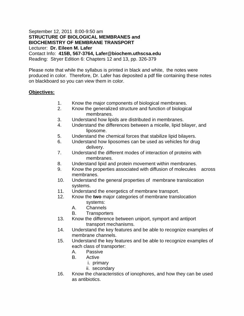
CHEMICAL SYNAPSE
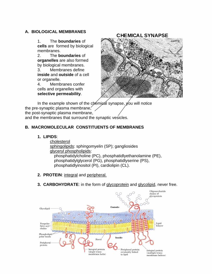
A. BIOLOGICAL MEMBRANES 1. The boundaries of
cells are formed by biological membranes.
2. The boundaries of organelles are also formed by biological membranes.
3. Membranes define inside and outside of a cell or organelle.
4. Membranes confer cells and organelles with selective permeability.
In the example shown of the chemical synapse, you will notice
the pre-synaptic plasma membrane, the post-synaptic plasma membrane, and the membranes that surround the synaptic vesicles.
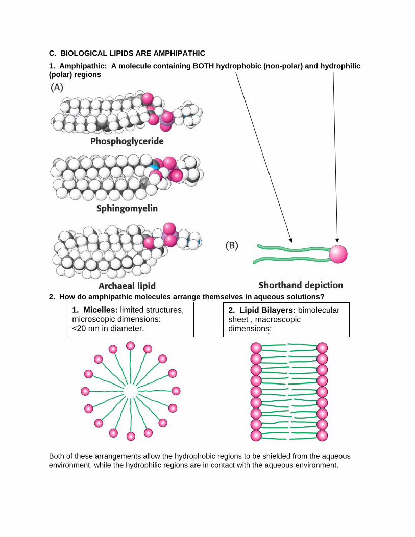
1. Micelles: limited structures, microscopic dimensions: <20 nm in diameter.
C. BIOLOGICAL LIPIDS ARE AMPHIPATHIC
1. Amphipathic: A molecule containing BOTH hydrophobic (non-polar) and hydrophilic (polar) regions
2. How do amphipathic molecules arrange themselves in aqueous solutions?
Both of these arrangements allow the hydrophobic regions to be shielded from the aqueous environment, while the hydrophilic regions are in contact with the aqueous environment.
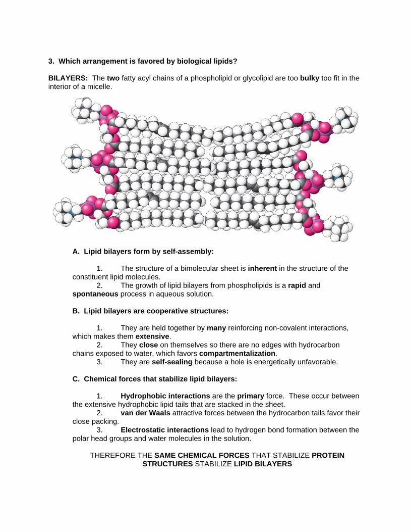
3. Which arrangement is favored by biological lipids? BILAYERS: The two fatty acyl chains of a phospholipid or glycolipid are too bulky too fit in the interior of a micelle.
A. Lipid bilayers form by self-assembly: 1. The structure of a bimolecular sheet is inherent in the structure of the constituent lipid molecules. 2. The growth of lipid bilayers from phospholipids is a rapid and spontaneous process in aqueous solution. B. Lipid bilayers are cooperative structures: 1. They are held together by many reinforcing non-covalent interactions, which makes them extensive. 2. They close on themselves so there are no edges with hydrocarbon chains exposed to water, which favors compartmentalization. 3. They are self-sealing because a hole is energetically unfavorable. C. Chemical forces that stabilize lipid bilayers: 1. Hydrophobic interactions are the primary force. These occur between the extensive hydrophobic lipid tails that are stacked in the sheet.
2. van der Waals attractive forces between the hydrocarbon tails favor their close packing. 3. Electrostatic interactions lead to hydrogen bond formation between the polar head groups and water molecules in the solution.
THEREFORE THE SAME CHEMICAL FORCES THAT STABILIZE PROTEIN STRUCTURES STABILIZE LIPID BILAYERS
A. Plasma membrane of an erythrocyte. B. Photoreceptor membrane of a retinal rod cell. C. Sarcoplasmic reticulum membrane of a muscle cell.
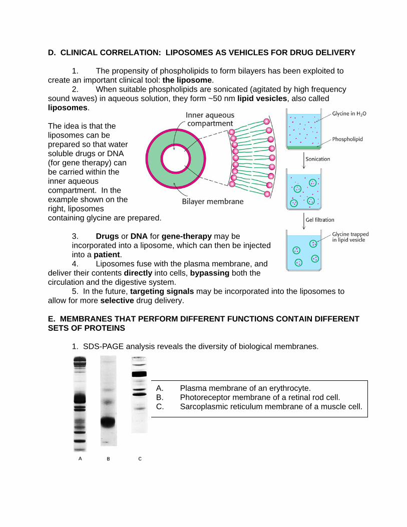
D. CLINICAL CORRELATION: LIPOSOMES AS VEHICLES FOR DRUG DELIVERY 1. The propensity of phospholipids to form bilayers has been exploited to create an important clinical tool: the liposome. 2. When suitable phospholipids are sonicated (agitated by high frequency sound waves) in aqueous solution, they form ~50 nm lipid vesicles, also called liposomes. The idea is that the liposomes can be prepared so that water soluble drugs or DNA (for gene therapy) can be carried within the inner aqueous compartment. In the example shown on the right, liposomes containing glycine are prepared.
3. Drugs or DNA for gene-therapy may be incorporated into a liposome, which can then be injected into a patient.
4. Liposomes fuse with the plasma membrane, and deliver their contents directly into cells, bypassing both the circulation and the digestive system. 5. In the future, targeting signals may be incorporated into the liposomes to allow for more selective drug delivery. E. MEMBRANES THAT PERFORM DIFFERENT FUNCTIONS CONTAIN DIFFERENT SETS OF PROTEINS 1. SDS-PAGE analysis reveals the diversity of biological membranes.
F. PROTEINS ASSOCIATE WITH THE LIPID BILAYER IN MANY WAYS
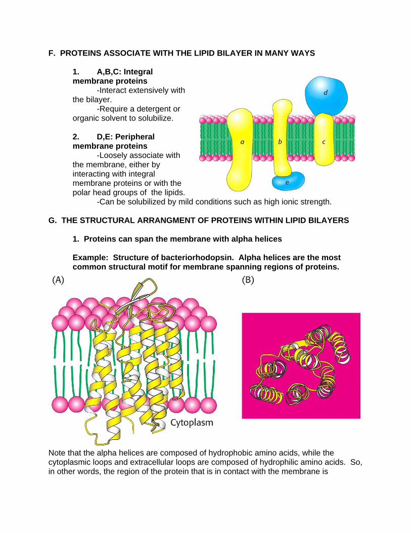
1. A,B,C: Integral membrane proteins -Interact extensively with the bilayer. -Require a detergent or organic solvent to solubilize. 2. D,E: Peripheral membrane proteins -Loosely associate with the membrane, either by interacting with integral membrane proteins or with the polar head groups of the lipids. -Can be solubilized by mild conditions such as high ionic strength.
G. THE STRUCTURAL ARRANGMENT OF PROTEINS WITHIN LIPID BILAYERS
1. Proteins can span the membrane with alpha helices
Example: Structure of bacteriorhodopsin. Alpha helices are the most common structural motif for membrane spanning regions of proteins.
Note that the alpha helices are composed of hydrophobic amino acids, while the cytoplasmic loops and extracellular loops are composed of hydrophilic amino acids. So, in other words, the region of the protein that is in contact with the membrane is
hydrophobic, while the region of the protein that is in contact with the cytosol or extracellular fluids is hydrophilic.
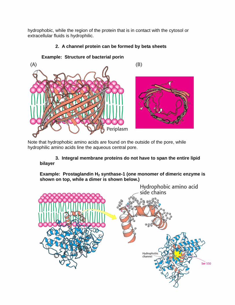
2. A channel protein can be formed by beta sheets Example: Structure of bacterial porin
Note that hydrophobic amino acids are found on the outside of the pore, while hydrophilic amino acids line the aqueous central pore.
3. Integral membrane proteins do not have to span the entire lipid bilayer Example: Prostaglandin H2 synthase-1 (one monomer of dimeric enzyme is shown on top, while a dimer is shown below.)
In this case, protein dimerization leads to the formation of a hydrophobic channel in the membrane.
A. Why is the localization of prostaglandin H2 synthase-1 in the membrane important? So that its substrate, arachidonic acid, which is a hydrophobic molecule generated by the hydrolysis of membrane lipids, does not have to leave the hydrophobic environment of the membrane to reach the active site of the enzyme.
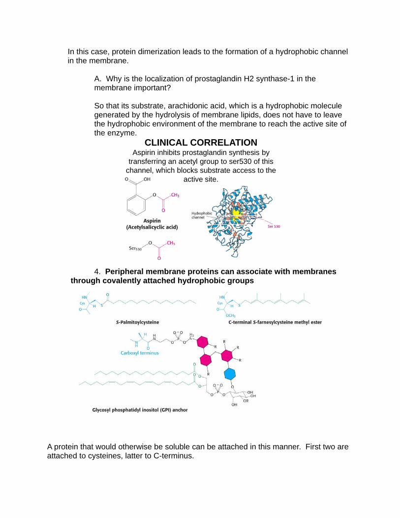
CLINICAL CORRELATIONAspirin inhibits prostaglandin synthesis by
transferring an acetyl group to ser530 of this channel, which blocks substrate access to the
active site.
4. Peripheral membrane proteins can associate with membranes through covalently attached hydrophobic groups
A protein that would otherwise be soluble can be attached in this manner. First two are attached to cysteines, latter to C-terminus.
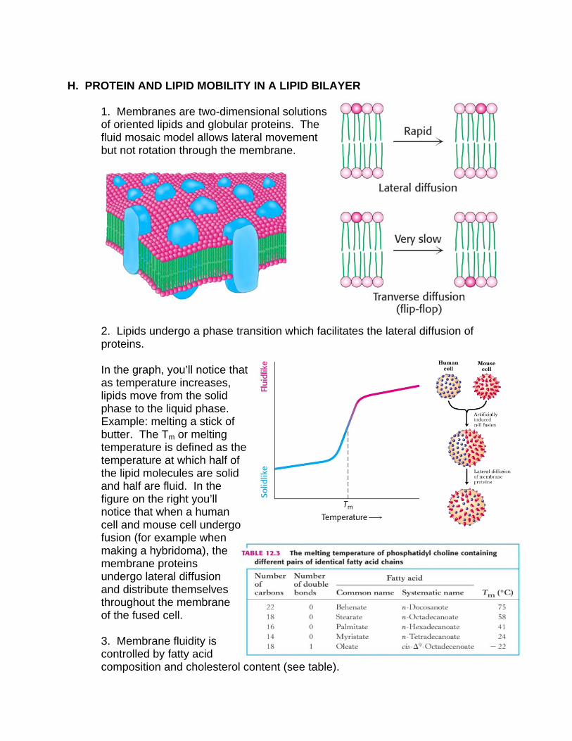
H. PROTEIN AND LIPID MOBILITY IN A LIPID BILAYER
1. Membranes are two-dimensional solutions of oriented lipids and globular proteins. The fluid mosaic model allows lateral movement but not rotation through the membrane.
2. Lipids undergo a phase transition which facilitates the lateral diffusion of proteins. In the graph, you’ll notice that as temperature increases, lipids move from the solid phase to the liquid phase. Example: melting a stick of butter. The Tm or melting temperature is defined as the temperature at which half of the lipid molecules are solid and half are fluid. In the figure on the right you’ll notice that when a human cell and mouse cell undergo fusion (for example when making a hybridoma), the membrane proteins undergo lateral diffusion and distribute themselves throughout the membrane of the fused cell. 3. Membrane fluidity is controlled by fatty acid composition and cholesterol content (see table).
OUT IN
10 mM 1 mM
5 mM 5 mM
1 mM 10 mM
no diffusion
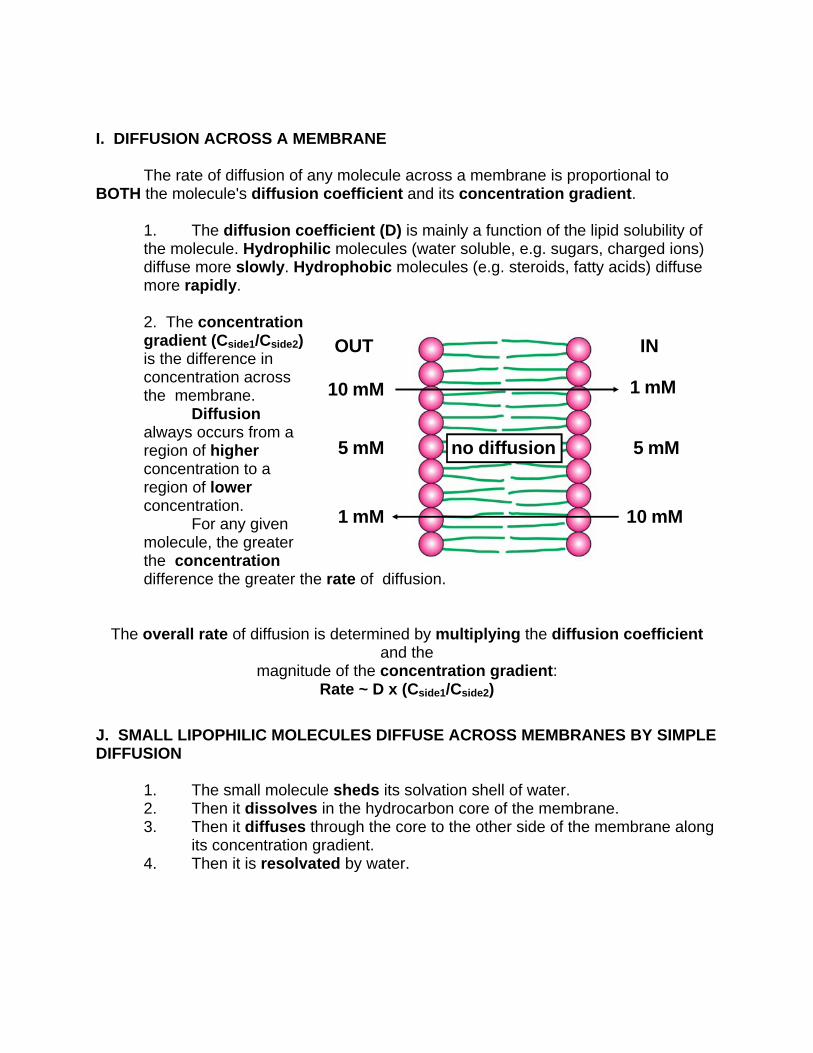
I. DIFFUSION ACROSS A MEMBRANE
The rate of diffusion of any molecule across a membrane is proportional to BOTH the molecule's diffusion coefficient and its concentration gradient.
1. The diffusion coefficient (D) is mainly a function of the lipid solubility of the molecule. Hydrophilic molecules (water soluble, e.g. sugars, charged ions) diffuse more slowly. Hydrophobic molecules (e.g. steroids, fatty acids) diffuse more rapidly. 2. The concentration gradient (Cside1/Cside2) is the difference in concentration across the membrane. Diffusion always occurs from a region of higher concentration to a region of lower concentration. For any given molecule, the greater the concentration difference the greater the rate of diffusion.
The overall rate of diffusion is determined by multiplying the diffusion coefficient and the
magnitude of the concentration gradient: Rate ~ D x (Cside1/Cside2)
J. SMALL LIPOPHILIC MOLECULES DIFFUSE ACROSS MEMBRANES BY SIMPLE DIFFUSION
1. The small molecule sheds its solvation shell of water. 2. Then it dissolves in the hydrocarbon core of the membrane. 3. Then it diffuses through the core to the other side of the membrane along its concentration gradient. 4. Then it is resolvated by water.
K. LARGE AND POLAR MOLECULES DO NOT READILY DIFFUSE ACROSS MEMBRANES BY SIMPLE DIFFUSION
1. Large and polar molecules are transported across membranes by proteinaceous membrane translocation systems
A. Passive Transport (also called facilitated diffusion): The transport goes in the same direction as the concentration gradient. This does not require an input of energy. Example: Acetylcholine Receptor: Passively transports sodium and potassium ions along their concentration gradients in response to neuronal signals. This is also an example of a ligand-gated ion channel. B. Active Transport: The transport goes in the opposite direction as the concentration gradient. This requires an input of energy.
Example: Sodium-Potassium Pump: Actively exchanges sodium and potassium against their concentration gradients utilizing the energy of ATP hydrolysis. Establishes the concentration gradients of sodium and potassium essential for synaptic transmission.
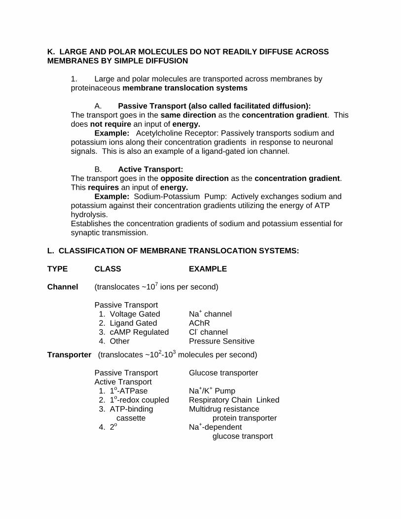
L. CLASSIFICATION OF MEMBRANE TRANSLOCATION SYSTEMS: TYPE CLASS EXAMPLE Channel (translocates ~107 ions per second) Passive Transport 1. Voltage Gated Na+ channel 2. Ligand Gated AChR 3. cAMP Regulated Cl- channel
4. Other Pressure Sensitive
Transporter (translocates ~102-103 molecules per second) Passive Transport Glucose transporter Active Transport 1. 1o-ATPase Na+/K+ Pump 2. 1o-redox coupled Respiratory Chain Linked 3. ATP-binding Multidrug resistance cassette protein transporter 4. 2o Na+-dependent glucose transport
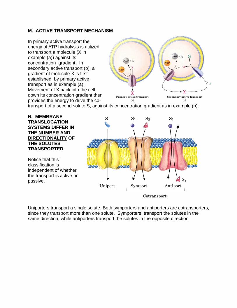
M. ACTIVE TRANSPORT MECHANISM In primary active transport the energy of ATP hydrolysis is utilized to transport a molecule (X in example (a)) against its concentration gradient. In secondary active transport (b), a gradient of molecule X is first established by primary active transport as in example (a). Movement of X back into the cell down its concentration gradient then provides the energy to drive the co-transport of a second solute S, against its concentration gradient as in example (b). N. MEMBRANE TRANSLOCATION SYSTEMS DIFFER IN THE NUMBER AND DIRECTIONALITY OF THE SOLUTES TRANSPORTED Notice that this classification is independent of whether the transport is active or passive. Uniporters transport a single solute. Both symporters and antiporters are cotransporters, since they transport more than one solute. Symporters transport the solutes in the same direction, while antiporters transport the solutes in the opposite direction
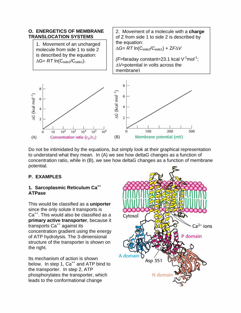
O. ENERGETICS OF MEMBRANE TRANSLOCATION SYSTEMS
Do not be intimidated by the equations, but simply look at their graphical representation to understand what they mean. In (A) we see how deltaG changes as a function of concentration ratio, while in (B), we see how deltaG changes as a function of membrane potential. P. EXAMPLES 1. Sarcoplasmic Reticulum Ca++ ATPase This would be classified as a uniporter since the only solute it transports is Ca++. This would also be classified as a primary active transporter, because it transports Ca++ against its concentration gradient using the energy of ATP hydrolysis. The 3-dimensional structure of the transporter is shown on the right. Its mechanism of action is shown below. In step 1, Ca++ and ATP bind to the transporter. In step 2, ATP phosphorylates the transporter, which leads to the conformational change
1. Movement of an uncharged molecule from side 1 to side 2 is described by the equation: G= RT ln(Cside2/Cside1).
2. Movement of a molecule with a charge of Z from side 1 to side 2 is described by the equation: G= RT ln(Cside2/Cside1) + ZFV (F=faraday constant=23.1 kcal V-1mol-1; V=potential in volts across the membrane)
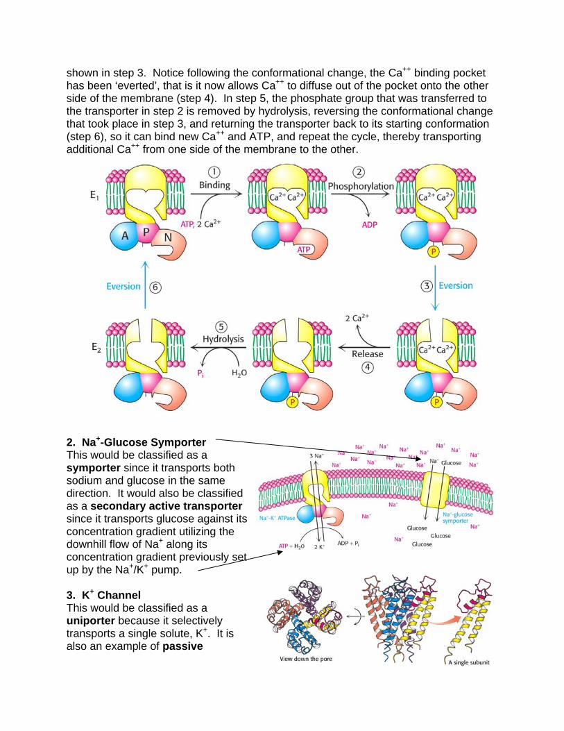
shown in step 3. Notice following the conformational change, the Ca++ binding pocket has been ‘everted’, that is it now allows Ca++ to diffuse out of the pocket onto the other side of the membrane (step 4). In step 5, the phosphate group that was transferred to the transporter in step 2 is removed by hydrolysis, reversing the conformational change that took place in step 3, and returning the transporter back to its starting conformation (step 6), so it can bind new Ca++ and ATP, and repeat the cycle, thereby transporting additional Ca++ from one side of the membrane to the other.
2. Na+-Glucose Symporter This would be classified as a symporter since it transports both sodium and glucose in the same direction. It would also be classified as a secondary active transporter since it transports glucose against its concentration gradient utilizing the downhill flow of Na+ along its concentration gradient previously set up by the Na+/K+ pump. 3. K+ Channel This would be classified as a uniporter because it selectively transports a single solute, K+. It is also an example of passive
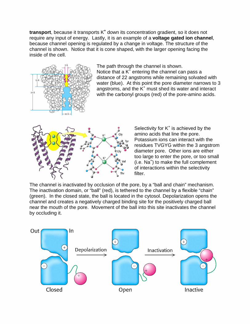
transport, because it transports K+ down its concentration gradient, so it does not require any input of energy. Lastly, it is an example of a voltage gated ion channel, because channel opening is regulated by a change in voltage. The structure of the channel is shown. Notice that it is cone shaped, with the larger opening facing the inside of the cell.
The path through the channel is shown. Notice that a K+ entering the channel can pass a distance of 22 angstroms while remaining solvated with water (blue). At this point the pore diameter narrows to 3 angstroms, and the K+ must shed its water and interact with the carbonyl groups (red) of the pore-amino acids.
Selectivity for K+ is achieved by the amino acids that line the pore. Potassium ions can interact with the residues TVGYG within the 3 angstrom diameter pore. Other ions are either too large to enter the pore, or too small (i.e. Na+) to make the full complement of interactions within the selectivity filter.
The channel is inactivated by occlusion of the pore, by a “ball and chain” mechanism. The inactivation domain, or “ball” (red), is tethered to the channel by a flexible “chain” (green). In the closed state, the ball is located in the cytosol. Depolarization opens the channel and creates a negatively charged binding site for the positively charged ball near the mouth of the pore. Movement of the ball into this site inactivates the channel by occluding it.
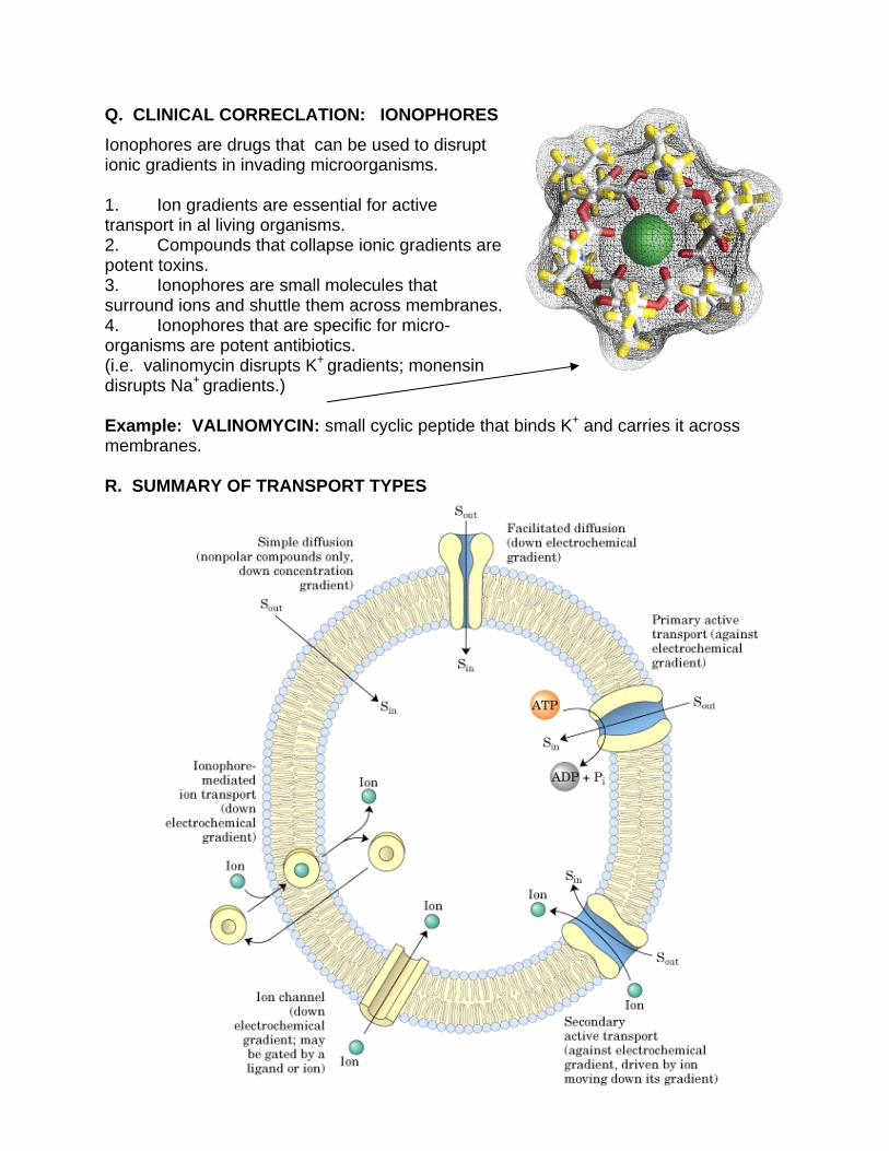
Q. CLINICAL CORRECLATION: IONOPHORES
Ionophores are drugs that can be used to disrupt ionic gradients in invading microorganisms. 1. Ion gradients are essential for active transport in al living organisms. 2. Compounds that collapse ionic gradients are potent toxins. 3. Ionophores are small molecules that surround ions and shuttle them across membranes. 4. Ionophores that are specific for micro-organisms are potent antibiotics. (i.e. valinomycin disrupts K+ gradients; monensin disrupts Na+ gradients.) Example: VALINOMYCIN: small cyclic peptide that binds K+ and carries it across membranes. R. SUMMARY OF TRANSPORT TYPES