ANTIMICROBIAL AGENTS AND CHEMOTHERAPY, Feb. 1993, p. 317-321 0066-4804/93/020317-05$02.00/0 Copyright ) 1993, American Society for Microbiology Susceptibilities of Mycoplasma bovis, Mycoplasma dispar, and Ureaplasma diversum Strains to Antimicrobial Agents In Vitro E. A. TER LAAK,* J. H. NOORDERGRAAF, AND M. H. VERSCHURE Department of Bacteriology, Central Veterinary Institute, P. O. Box 65, 8200 AB Lelystad, The Netherlands Received 10 August 1992/Accepted 1 December 1992 The purpose of this study was to determine the susceptibility of various strains of Mycoplasma bovis, Mycoplasma dispar, and Ureaplasma diversum, which are prevalent causes of pneumonia in calves, to 16 antimicrobial agents in vitro. The MICs of the antimicrobial agents were determined by a serial broth dilution method for 16 field strains and the type strain of M. bovis, for 19 field strains and the type strain of M. dispar, and for 17 field strains of U. diversum. Final MICs for M. bovis and M. dispar were read after 7 days and final MICs for U. diversum after 1 to 2 days. All strains tested were susceptible to tylosin, kitasamycin, and tiamulin but were resistant to nifuroquine and streptomycin. Most strains of U. diversum were intermediately susceptible to oxytetracycline but fuly susceptible to chlortetracycline; most strains of M. bovis and M. dispar, however, were resistant to both agents. Strains of M. dispar and U. diversum were susceptible to doxycycline and minocycline, but strains of M. bovis were only intermediately susceptible. Susceptibility or resistance to chloramphenicol, spiramycin, spectinomycin, lincomycin, or enrofloxacin depended on the species but was not equal for the three species. The type strains of M. bovis and M. dispar were more susceptible to various antimicrobial agents, including tetracyclines, than the field strains. This finding might indicate that M. bovis and M. dispar strains are becoming resistant to these agents. Antimicrobial agents that are effective in vitro against all three mycoplasma species can be considered for treating mycoplasma infections in pneumonic calves. Therefore, tylosin, kitasamycin, and tiamulin may be preferred over oxytetracycline and chlortetracycline. Several mycoplasma species, of the many that have been isolated from cattle, are pathogenic. Mycoplasma bovis is regarded as the most pathogenic, with the exception of Mycoplasma mycoides subsp. mycoides small-colony bio- type, the agent of contagious bovine pleuropneumonia. M. bovis causes mastitis in dairy cattle, respiratory tract infec- tions in calves, and arthritis in all age groups of cattle, and it is prevalent worldwide (5). In addition, Mycoplasma dispar and Ureaplasma diversum have frequently been isolated from the respiratory tracts of pneumonic calves, and their prevalence is assumed to be worldwide also (16). Their pathogenic significance in calf respiratory disease has been proven (7). U. diversum can also be isolated from the genital tracts of cows and bulls. M. bovis, M. dispar, and U. diversum are prevalent in the Netherlands (23, 25). Because bacteria and mycoplasmas are involved in calf pneumonia, calves are often treated with antimicrobial agents. The massive and timely use of macrolide antibiotics, singly or in combination with other drugs, contributed to the recovery of more than 90% of calves affected by pneumonia (12). Although antimicrobial agents cause calves to recover from the clinical signs of respiratory disease (12), they do not eliminate mycoplasmas from the herd. Although the suscep- tibility of M. bovis and U. diversum to antimicrobial agents has been studied in vitro (4, 8-11, 13, 14, 20, 22, 28), little is known about the susceptibility of M. dispar (9, 11). In the present study, we examined the susceptibility of numerous strains of these three bovine pathogens to 16 antimicrobial agents by using a serial broth dilution method. * Corresponding author. MATERMILS AND METHODS Mycoplasma strains. Type strain Donetta of M. bovis and type strain 462/2 of M. dispar were obtained from E. A. Freundt of the former FAO/WHO Collaborating Centre for Animal Mycoplasmas, Institute of Medical Microbiology, University of Aarhus, Arhus, Denmark. The type strain of U. diversum was not available to us during the study. From 1983 to 1988, field strains of M. bovis, M. dispar, and U. diversum were isolated and identified as described earlier (23). Strains of M. bovis were cultured in modified Edward media containing 0.4% tetrazolium chloride, strains of M. dispar were cultured in Friis NHS20 broth, and strains of U. diversum were cultured in Friis NHU pH 6.0 broth (23). Sixteen field strains of M. bovis were isolated from the lungs of pneumonic calves (B16, Fl, Q19, AN13b, AZ25, BD25, BG32, BY47b, DY20b, EA17, and EB14), from mastitic milk (AX20 and DL19), or from synovia collected from arthritic joints (DR40, EO11, and EU46). The strains were isolated from cattle from 14 farms. Nineteen field strains of M. dispar (E6, Q26, V24, Y15, AA9, AF7, AF18d, ASl5d, AS19, AZ11, BA20, BD5, BD8, BI35, BM10, BS18, BX43, BY47d, and DT10) were isolated from the lungs of pneumonic calves from 16 farms. Seventeen field strains of U. diversum were isolated from the lungs (B20, E5, G10, Y9, Y22, V25, AA9, AB13, AF18u, AN13u, ASl5u, AT5, BS19, BY48, and DT8) or noses (B15, Q22) of pneumonic calves from 15 farms. Strains B15, B20, E5, G10, AN13u, AT5, and BY48 had been identified as U. diversum serogroup A; strains Y9 and AA9 had been identified as serogroup B. The other Ureaplasma strains had not been serotyped. The farms from which the three mycoplasma species were isolated were located in various parts of the Netherlands. Primary cultures of M. bovis strains were purified three times by using a Pasteur pipette to suction an agar plug 317 Vol. 37, No. 2 Downloaded from https://journals.asm.org/journal/aac on 15 December 2021 by 191.8.239.54.
Transcript
ANTIMICROBIAL AGENTS AND CHEMOTHERAPY, Feb. 1993, p. 317-3210066-4804/93/020317-05$02.00/0Copyright ) 1993, American Society for Microbiology
Susceptibilities of Mycoplasma bovis, Mycoplasma dispar,and Ureaplasma diversum Strains to Antimicrobial
Agents In VitroE. A. TER LAAK,* J. H. NOORDERGRAAF, AND M. H. VERSCHURE
Department ofBacteriology, Central Veterinary Institute, P.O. Box 65, 8200AB Lelystad, The Netherlands
Received 10 August 1992/Accepted 1 December 1992
The purpose of this study was to determine the susceptibility of various strains of Mycoplasma bovis,Mycoplasma dispar, and Ureaplasma diversum, which are prevalent causes of pneumonia in calves, to 16antimicrobial agents in vitro. The MICs of the antimicrobial agents were determined by a serial broth dilutionmethod for 16 field strains and the type strain ofM. bovis, for 19 field strains and the type strain ofM. dispar,and for 17 field strains of U. diversum. Final MICs for M. bovis and M. dispar were read after 7 days and finalMICs for U. diversum after 1 to 2 days. All strains tested were susceptible to tylosin, kitasamycin, and tiamulinbut were resistant to nifuroquine and streptomycin. Most strains of U. diversum were intermediatelysusceptible to oxytetracycline but fuly susceptible to chlortetracycline; most strains ofM. bovis and M. dispar,however, were resistant to both agents. Strains ofM. dispar and U. diversum were susceptible to doxycyclineand minocycline, but strains of M. bovis were only intermediately susceptible. Susceptibility or resistance tochloramphenicol, spiramycin, spectinomycin, lincomycin, or enrofloxacin depended on the species but was notequal for the three species. The type strains of M. bovis and M. dispar were more susceptible to variousantimicrobial agents, including tetracyclines, than the field strains. This finding might indicate that M. bovisand M. dispar strains are becoming resistant to these agents. Antimicrobial agents that are effective in vitroagainst all three mycoplasma species can be considered for treating mycoplasma infections in pneumonic calves.Therefore, tylosin, kitasamycin, and tiamulin may be preferred over oxytetracycline and chlortetracycline.
Several mycoplasma species, of the many that have beenisolated from cattle, are pathogenic. Mycoplasma bovis isregarded as the most pathogenic, with the exception ofMycoplasma mycoides subsp. mycoides small-colony bio-type, the agent of contagious bovine pleuropneumonia. M.bovis causes mastitis in dairy cattle, respiratory tract infec-tions in calves, and arthritis in all age groups of cattle, and itis prevalent worldwide (5). In addition, Mycoplasma disparand Ureaplasma diversum have frequently been isolatedfrom the respiratory tracts of pneumonic calves, and theirprevalence is assumed to be worldwide also (16). Theirpathogenic significance in calf respiratory disease has beenproven (7). U. diversum can also be isolated from the genitaltracts of cows and bulls. M. bovis, M. dispar, and U.diversum are prevalent in the Netherlands (23, 25).Because bacteria and mycoplasmas are involved in calf
pneumonia, calves are often treated with antimicrobialagents. The massive and timely use of macrolide antibiotics,singly or in combination with other drugs, contributed to therecovery of more than 90% of calves affected by pneumonia(12). Although antimicrobial agents cause calves to recoverfrom the clinical signs of respiratory disease (12), they do noteliminate mycoplasmas from the herd. Although the suscep-tibility of M. bovis and U. diversum to antimicrobial agentshas been studied in vitro (4, 8-11, 13, 14, 20, 22, 28), little isknown about the susceptibility of M. dispar (9, 11).
In the present study, we examined the susceptibility ofnumerous strains of these three bovine pathogens to 16antimicrobial agents by using a serial broth dilution method.
* Corresponding author.
MATERMILS AND METHODS
Mycoplasma strains. Type strain Donetta of M. bovis andtype strain 462/2 of M. dispar were obtained from E. A.Freundt of the former FAO/WHO Collaborating Centre forAnimal Mycoplasmas, Institute of Medical Microbiology,University of Aarhus, Arhus, Denmark. The type strain ofU. diversum was not available to us during the study. From1983 to 1988, field strains of M. bovis, M. dispar, and U.diversum were isolated and identified as described earlier(23). Strains of M. bovis were cultured in modified Edwardmedia containing 0.4% tetrazolium chloride, strains of M.dispar were cultured in Friis NHS20 broth, and strains of U.diversum were cultured in Friis NHU pH 6.0 broth (23).
Sixteen field strains of M. bovis were isolated from thelungs of pneumonic calves (B16, Fl, Q19, AN13b, AZ25,BD25, BG32, BY47b, DY20b, EA17, and EB14), frommastitic milk (AX20 and DL19), or from synovia collectedfrom arthritic joints (DR40, EO11, and EU46). The strainswere isolated from cattle from 14 farms. Nineteen fieldstrains ofM. dispar (E6, Q26, V24, Y15, AA9, AF7, AF18d,ASl5d, AS19, AZ11, BA20, BD5, BD8, BI35, BM10, BS18,BX43, BY47d, and DT10) were isolated from the lungs ofpneumonic calves from 16 farms. Seventeen field strains ofU. diversum were isolated from the lungs (B20, E5, G10, Y9,Y22, V25, AA9, AB13, AF18u, AN13u, ASl5u, AT5, BS19,BY48, and DT8) or noses (B15, Q22) of pneumonic calvesfrom 15 farms. Strains B15, B20, E5, G10, AN13u, AT5, andBY48 had been identified as U. diversum serogroup A;strains Y9 and AA9 had been identified as serogroup B. Theother Ureaplasma strains had not been serotyped. The farmsfrom which the three mycoplasma species were isolatedwere located in various parts of the Netherlands.
Primary cultures of M. bovis strains were purified threetimes by using a Pasteur pipette to suction an agar plug
317
Vol. 37, No. 2
Dow
nloa
ded
from
http
s://j
ourn
als.
asm
.org
/jour
nal/a
ac o
n 15
Dec
embe
r 20
21 b
y 19
1.8.
239.
54.
ANTIMICROB. AGENTS CHEMOTHER.
bearing one colony (6). Cultures of M. dispar and U.diversum cannot be purified in this way because no viablecultures are obtained. Therefore, primary cultures of M.dispar and U. diversum were purified three times by con-ventional filtration cloning techniques, by using a 450-nm-pore-size membrane filter (26). After the first filtration step,the culture was serially diluted 10-fold in eight tubes withculture broth. Immediately thereafter, the contents of eachtube was transferred to eight wells (200 ,u per well) of amicrotiter plate that was sealed with adhesive tape andincubated at 37°C for 7 (U. diversum) or 14 (M. dispar) days.The contents of the well containing the highest dilution ofviable culture was serially diluted 10-fold in four tubes ofbroth medium. The tube containing the highest viable dilu-tion was used for the second and third filtration steps.
Antimicrobial agents. The following antimicrobial agentswere used: oxytetracycline, chloramphenicol, streptomycin,ampicillin, and benzylpenicillin potassium (Gist-Brocades,Delft, the Netherlands); chlortetracycline and minocycline(Cyanamid, Lederle, the Netherlands); doxycycline (Pfizer,Rotterdam, the Netherlands); spiramycin (Rhone-Merieux,Toulouse, France); tylosin (Elanco, Nieuwegein, the Neth-erlands); kitasamycin (Inffa, Houten, the Netherlands);spectinomycin and lincomycin (Upjohn, Ede, the Nether-lands); tiamulin (Coopers, Haarlem, the Netherlands); enro-floxacin (Bayer, Mijdrecht, the Netherlands); and nifuro-quine (quinaldofur) (Duphar, Amsterdam, the Netherlands).
Tylosin and tiamulin are used only in veterinary medicine.Tylosin is an antibiotic with a structure similar to that oferythromycin. Tiamulin is a semisynthetic antimicrobialagent that does not belong to a particular group. Kitasamy-cin is a macrolide antibiotic that is used in veterinarymedicine. In Japan it has been used successfully in humanmedicine (15). The three agents have a spectrum similar tothe spectrum of the macrolides. Nifuroquine is a quinolinederivative that is used in therapy of bovine mastitis.The antimicrobial agents were diluted in distilled water to
prepare stock solutions, except for the four tetracyclines,which were diluted in 10% methanol, and chloramphenicoland spiramycin, which were diluted in 0.5% N,N-dimethyl-formamide (no. 3034; E. Merck AG, Darmstadt, Germany).Concentrations were calculated as pure substances to pre-pare stock solutions. The activity of spiramycin was equiv-alent to 4,468 IU/mg, that is, 1.4 times the activity of theWorld Health Organization standard. The activity of peni-cillin was equivalent to 1,592 U/mg. Stock solutions weresterilized by filtration through a 200-nm-pore-size membranefilter and used immediately or stored at 4°C overnight.
Serial broth dilution method for determining MICs. Theserial broth dilution method was recommended by an ad hocworking group of the International Research Program onComparative Mycoplasmology (IRPCM), part of the Inter-national Organization for Mycoplasmology, as the mostuseful and reproducible assay (18). Each antimicrobial agentwas serially diluted twofold in culture broth in 10 wells of amicrotiter plate; each well contained 25 RI. A standardnumber of organisms grown in broth without bacterial inhib-itors was used. An amount of 175 RI, containing 1.7 x 10³ to1.7 x 104 color-changing units, was added to each well. Thesusceptibility or resistance of M. dispar strains to theantimicrobial agent was indicated by whether the culturewas able to metabolize glucose in the presence of one of theconcentrations of the antimicrobial agent. Strains of U.diversum were tested similarly, but urea instead of glucosewas the substrate to be metabolized. Metabolism of glucosewas demonstrated by a change of the phenol red indicator
from red to yellow; metabolism of urea was demonstrated bya change from yellow to red. The susceptibility or resistanceof M. bovis strains to the antimicrobial agent was indicatedby whether the culture was able to reduce the colorless2,3,5-triphenyltetrazolium chloride to the red formazan inthe presence of one of the concentrations of the antimicro-bial agent. Strains of M. bovis also produced a film layer ontop of the broth.On the day of inoculation, the required numbers of color-
changing units were prepared from stock cultures with aknown number of cells, which had been stored at -70°C.Organisms were counted again to verify the actual numbersof organisms in the system. The microtiter plates weresealed with adhesive tape and incubated aerobically at 37°Cin the dark to prevent spontaneous reduction of the tetra-zolium chloride. The MIC of the antimicrobial agent wasdetermined as the lowest concentration at which the mediumdid not change color or produce a film layer. The colorchanges were read several times for 7 days. Because the filmdeveloped more slowly than color change, it was read only atday 7. Initial MICs were recorded as soon as the inoculumcontrols (without the antimicrobial agent) changed color incomparison with the color of the culture medium controls.This was on day 1 to 2 for U. diversum, day 2 to 3 for M.bovis, and day 2 to 4 for M. dispar. Final MICs for M. bovisand M. dispar were read when color changes or film produc-tion had stopped for 1 to 2 days, that is, day 7. BecauseUreaplasma species grow rapidly, final MICs for U. diver-sum were read after only 24 to 48 h. These time points forreading MICs were in agreement with the recommendationsof the IRPCM working group, which recommended thatMICs for Mycoplasma species be read from 48 h to 7 daysand that MICs for Ureaplasma species be read after 24 h(18). All tests were performed in duplicate; when MICsdiffered by no more than a factor of two, the higher concen-tration of the two was recorded as the MIC.
Interpretation of MICs. When the MIC of the tetracyclinegroup was <1 ,ug/ml, the strain was considered susceptible;when the MIC was 2 or 4 ,ug/ml, it was considered interme-diately susceptible; and when the MIC was 28 ,ug/ml, thestrain was considered resistant. These values were c4, 8,and 216 ,ug/ml for chloramphenicol; <4, 8 or 16, and >32,ug/ml for streptomycin; <2, >4 to .16, and .32 ,ug/ml forampicillin; and .0.25, .0.5 to <4, and >8 ,g/ml forpenicillin (27). These MICs were based on guidelines fortesting the susceptibility of bacteria that affect humans.These criteria were used because criteria for animals are notgenerally available. Criteria for the other antimicrobialagents were not available.
RESULTS
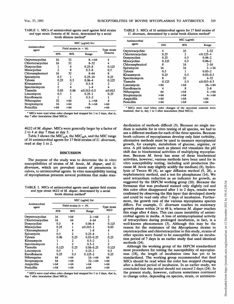
MICs of antimicrobial agents were determined for 50% ofthe strains tested (MIC50) and for 90% of the strains tested(MIC90). Table 1 shows the MIC50s, the MIC90s, and theMIC range of 15 antimicrobial agents for 16 field strains ofM. bovis, read at day 7; Table 1 also shows the MICs fortype strain Donetta of M. bovis. The MIC90 of doxycyclinewas larger at day 7 than at day 4 by a factor of 8, and theMIC90 of minocycline was larger by a factor of 16. MICgs ofthe other antimicrobial agents were larger than, by a factorof 2 to 4, or equal to the MIC90s read at day 4.
Table 2 shows the MIC50s, the MIC90s, and the MIC rangeof 16 antimicrobial agents for 19 field strains of M. dispar,read at day 7; Table 2 also shows the MICs for type strain
318 TER LAAK ET AL.
Dow
nloa
ded
from
http
s://j
ourn
als.
asm
.org
/jour
nal/a
ac o
n 15
Dec
embe
r 20
21 b
y 19
1.8.
239.
54.
SUSCEPTIBILITIES OF BOVINE MYCOPLASMAS TO ANTIBIOTICS 319
TABLE 1. MICs of antimicrobial agents used against field strainsand type strain Donetta of M. bovis, determined by a serial
broth dilution methoda
MIC (,ug/ml) for:Antimicrobial Field strains (n = 16) Type strain
agent Type_______strain______50% 90% Range Donetta
a MICs were read when color changes had stopped for 1 to 2 days, that is,day 7 after inoculation (final MICs).
462/2 of M. dispar. MICs were generally larger by a factor of2 to 4 at day 7 than at day 5.Table 3 shows the MIC50s, the MIC90s, and the MIC range
of 16 antimicrobial agents for 17 field strains of U. diversum,read at day 1 to 2.
DISCUSSION
The purpose of the study was to determine the in vitrosusceptibilities of strains of M. bovis, M. dispar, and U.diversum, which are prevalent causes of pneumonia incalves, to antimicrobial agents. In vitro susceptibility testingof mycoplasmas presents several problems that make stan-
TABLE 2. MICs of antimicrobial agents used against field strainsand type strain 462/2 of M. dispar, determined by a serial
broth dilution methoda
MIC (pg/ml) for:Antimicrobial Field strains (n = 19) Type strain
a MICs were read when color changes of the inoculum controls weremaximal, that is, day 1 to 2 after inoculation (final MICs).
dardization of methods difficult (3). Because no single me-dium is suitable for in vitro testing of all species, we had touse a different medium for each of the three species. Becausebroth cultures of mycoplasmas develop only faint turbidity,alternative methods must be used to measure mycoplasmalgrowth, for example, metabolism of glucose, arginine, orurea. A pH indicator such as phenol red visualizes the pHshift due to biochemical activities of multiplying mycoplas-mas. Because M. bovis has none of these biochemicalactivities, however, various methods have been used for invitro susceptibility testing, including acid production (be-cause M. bovis may slightly acidify the medium) (8), hydro-lysis of Tween 80 (4), an agar diffusion method (9, 28), anephelometric method, and a test for phosphatase (14). Weused tetrazolium reduction as an indicator for growth, assuggested by the IRPCM working group (18). Because theformazan that was produced stained only slightly red andthis color often disappeared after 1 to 2 days, results wereconfirmed by observing the film layer that developed slowlyand could be read only after 7 days of incubation. Further-more, the growth rate of the various mycoplasma speciesdiffers. For example, U. diversum reaches its stationarygrowth phase within 24 to 48 h, whereas M. dispar reachesthis stage after 4 days. This can cause instability of antimi-crobial agents in media. A loss of antimycoplasmal activityof tetracyclines during prolonged incubation, in fact, is awell-known phenomenon (3). Although this may be thereason for the resistance of the Mycoplasma strains tooxytetracycline and chlortetracycline in this study, strains ofother species were found to be susceptible after an incuba-tion period of 7 days in an earlier study that used identicalmethods (24).Although the working group of the IRPCM standardized
most procedures for testing the susceptibility of mycoplas-mas (18), the length of incubation time has yet to bestandardized. The working group recommended that finalMICs should be read when the color has stopped changingafter a defined period of incubation. In an earlier study, weconcluded that this period should not exceed 2 days (24). Inthe present study, however, cultures sometimes continuedto change color, depending on species, strain, or antimicro-
VOL. 37, 1993
Dow
nloa
ded
from
http
s://j
ourn
als.
asm
.org
/jour
nal/a
ac o
n 15
Dec
embe
r 20
21 b
y 19
1.8.
239.
54.
ANTIMICROB. AGENTS CHEMOTHER.
bial agent used. Therefore, final MICs for the Mycoplasmastrains were read when most strains had stopped changingcolor for 1 or 2 days. Another useful suggestion is to readfinal MICs after a period of time twice as long as thatrequired for the control to change color (17).
In several U. diversum tests, especially those with chlor-tetracycline, color continuously changed during the obser-vation period of 7 days. Probably, urea continued to bedegraded by the enzyme urease in the inhibited organisms.MICs could not be read for one particular strain of U.diversum after 2 days of incubation because an alkaline colorchange had developed in all inoculated wells. Bloomster andLynn (2) demonstrated that residual urease activity fromdead organisms considerably influenced the dynamics ofcolor changes in Ureaplasma urealyticum cultures. Thiscolor change could cause errors in evaluating the suscepti-bility of ureaplasmas to antimicrobial agents. Reading MICsfor U. urealyticum strains after incubation periods of differ-ent lengths is the main cause for the variation in the MICspublished for a particular antimicrobial agent (21). There-fore, and because Ureaplasma species grow rapidly, theworking group of the IRPCM recommended that final MICsof Ureaplasma cultures can generally be read after 24 h (18).We regarded the MICs for M. bovis that were read after 7
days of incubation as the final MICs, but other studies haveread final MICs after 2 to 3 (4), 3 (20), 4 (14), 5 (28), 6 (9), or7 (8) days. Only Hannan et al. (9) reported MICs for typestrain Donetta of M. bovis. Our results generally agree withthose reported earlier (4, 8, 14, 20).Although M. dispar grows more slowly than M. bovis,
final MICs were read after 7 days (Table 2). At day 7, colorchanges had stopped for only 1 day, in general. Only Hannanet al. (9) reported MICs for type strain 462/2 of M. dispar.We found a higher MIC for oxytetracycline (2 ug/nml) thanthat of Hannan et al. (0.25 p,g/ml), but methods also differed.MICs of tylosin and tiamulin were similar in both studies.Matsuoka et al. (11) reported MICs of tylosin for M. disparsimilar to those we found. No other reports on MICs for M.dispar are available.Andrews et al. (1) reported that M. dispar was more
frequently isolated when ampicillin in the media was substi-tuted for penicillin that was used in a concentration of 200IU/ml. The MICs of penicillin had not been determined,however. The highest concentration of penicillin that wetested was 64 p,g/ml, which is equivalent to 102 IU/ml. Wefound a final MIC of this value for three strains, so weconfirm that penicillin must not be incorporated in media forM. dispar.
Final MICs for U. diversum were read after only 1 to 2days of incubation, and strains were found to be resistant tovarious antimicrobial agents. Our results generally agreewith those reported earlier (10, 13, 20, 22). The resistance ofU. urealyticum to lincomycin was reported in 1968 (19). Weconfirmed the resistance of U. diversum to lincomycin.
It has been the experience in chemotherapy for decadesthat two groups of antimicrobial agents, i.e., tetracyclinesand macrolides, are of primary importance in treating animalmycoplasma infections. The older tetracyclines, such asoxytetracycline and chlortetracycline, are cheaper than thenewer tetracyclines, such as doxycycline and minocycline.The dosage of the newer tetracyclines, however, can belower, because they are better absorbed after oral adminis-tration (3).However, bovine mycoplasma strains are acquiring resis-
tance to tetracyclines (in particular, to oxytetracycline), asdemonstrated by our study. This finding is notable because
oxytetracycline is frequently used in calf husbandry in theNetherlands. M. bovis and M. dispar were less susceptibleto doxycycline and minocycline, however, than U. diversumwas.Mycoplasma strains are known to be susceptible to tiam-
ulin and tylosin, although strains are generally more suscep-tible to tiamulin than to tylosin. The MICs of these antimi-crobial agents were low for all strains studied; for one M.bovis strain, however, the MIC of tylosin was as high as 4,ug/ml. The U. diversum strains were generally less suscep-tible to tylosin than strains of M. bovis or M. dispar.Because in other countries some strains were less suscepti-ble to tiamulin (M. bovis) or tylosin (M. bovis and U.diversum), these strains may also be acquiring resistance totylosin (14, 20). Another indication that field strains of M.bovis are developing resistance is that the MICs of variousantimicrobial agents for type strain Donetta were lower thanthose for any field strain. The type strain of M. dispar wasmore susceptible to the tetracyclines than most field strains;this indicates that M. dispar strains are also acquiringresistance.Because the three mycoplasma species often occur in
pneumonic calves of one herd and even in the respiratorytract of one calf (23), antimicrobial agents that are effectivein vitro against all three species can be considered for use invivo. Therefore, tylosin, kitasamycin, and tiamulin may bepreferred over oxytetracycline and chlortetracycline.
ACKNOWLEDGMENTS
We thank L. B. Senterfit (Microbiology Department, MedicalCollege, Cornell University, New York, N.Y.) for sending thereport of the IRPCM working group and J. Haagsma and U. Vechtfor critically reading the manuscript.
REFERENCES1. Andrews, B. E., R. H. Leach, R. N. Gourlay, and C. J. Howard.
1973. Enhanced isolation of Mycoplasma dispar by substitutionof ampicillin for benzylpenicillin in growth media. Vet. Rec.93:603.
2. Bloomster, T. G., and R. J. Lynn. 1981. Effect of antibiotics onthe dynamics of color change in Ureaplasma urealyticum cul-tures. J. Clin. Microbiol. 13:598-600.
3. Brunner, H., and G. Laber. 1985. Chemotherapy of myco-plasma infections, p. 403-450. In S. Razin and M. F. Barile(ed.), The mycoplasmas, vol. 4. Academic Press, Inc., Orlando,Fla.
4. Devriese, L. A., and F. HaesebroucL 1991. Antibiotic suscepti-bility testing of Mycoplasma bovis using tween 80 hydrolysis asan indicator of growth. J. Vet. Med. Ser. B 38:781-783.
5. Ern0, H., and P. Perreau. 1985. Mycoplasmal infections incattle, p. 300-345. In I. Gylstorff (ed.), Infektionen durchMycoplasmatales. Ferdinand Enke Verlag, Stuttgart, Germany.
6. Freundt, E. A., H. Ern0, and R. M. Lemcke. 1979. Identificationof mycoplasmas, p. 377-434. In T. Bergan and J. R. Norris(ed.), Methods in microbiology, vol. 13. Academic Press Ltd.,London.
7. Gourlay, R. N., and C. J. Howard. 1979. Bovine mycoplasmas,p. 49-102. In J. G. Tully and R. F. Whitcomb (ed.), Themycoplasmas, vol. 2. Academic Press, Inc., New York.
8. Hamdy, A. H., and C. C. Miller. 1971. Antibiotics for bovinemycoplasmas. J. Dairy Sci. 54:1541-1544.
9. Hannan, P. C. T., P. J. O'Hanlon, and N. H. Rogers. 1989. Invitro evaluation of various quinolone antibacterial agentsagainst veterinary mycoplasmas and porcine respiratory bacte-rial pathogens. Res. Vet. Sci. 46:202-211.
10. Kishima, M., and K. Hashimoto. 1979. In vitro sensitivities toantimicrobial drugs of ureaplasmas isolated from the bovinerespiratory tract, genital tract and eye. Res. Vet. Sci. 27:218-222.
320 TER LAAK ET AL.
Dow
nloa
ded
from
http
s://j
ourn
als.
asm
.org
/jour
nal/a
ac o
n 15
Dec
embe
r 20
21 b
y 19
1.8.
239.
54.
SUSCEPTIBILITIES OF BOVINE MYCOPLASMAS TO ANTIBIOTICS 321
11. Matsuoka, T., 0. A. Muenster, E. E. Ose, and L. Tonkinson.1980. Orally administered tylosin for the control of pneumoniain neonatal calves. Vet. Rec. 107:149-151.
12. Pignatelli, P. 1978. Respiratory disease and the incidence ofpulmonary mycoplasmosis in intensively-reared calves in Italy,p. 284-294. In W. B. Martin (ed.), Respiratory diseases in cattle.Martinus Nijhoff, The Hague, The Netherlands.
13. Pilaszek, J., and M. Truszczynski. 1986. Ureaplasmas of cattle-sensitivity to antibiotics. Arch. Exp. Vet. Med. 40:88-93.
14. Poumarat, F., and J. L. Martel. 1989. Antimicrobial suscepti-bility in vitro of French Mycoplasma bovis strains. Ann. Rech.Vet. 20:145-152. (In French.)
15. Reynolds, J. E. F. (ed.). 1989. Martindale, the extra pharmaco-poeia, 29th ed., p. 252. The Pharmaceutical Press, London.
16. Ross, R. F. 1985. Mycoplasmas of cattle, p. 314-344. In H.Blobel and T. Schliesser (ed.), Handbuch der bakteriellenInfektionen bei Tieren, vol. 5. Gustav Fischer Verlag, Stuttgart,Germany.
17. Senterfit, L. B. 1983. Antibiotic sensitivity testing of mycoplas-mas, p. 397-401. In J. G. Tully and S. Razin (ed.), Methods inmycoplasmology, vol. 2. Academic Press, Inc., New York.
18. Senterfit, L. B., D. Taylor-Robinson, J. A. Robertson, C. Bebear,and G. Laber. 1986. Antibiotic sensitivity testing of mycoplas-mas. International Organization for Mycoplasmology, Edmon-ton, Alberta, Canada.
19. Shipley, A., S. J. Bowman, and J. J. O'Connor. 1968. T-strainmycoplasmas in non-specific urethritis. Med. J. Aust. 1:794-796.
20. Stipkovits, L., Z. Varga, G. Laber, and J. Bockmann. 1984. Acomparison of the effect of tiamulin hydrogen fumarate andtylosin tartrate on mycoplasmas of ruminants and some animal
ureaplasmas. Vet. Microbiol. 9:147-153.21. Taylor-Robinson, D., and P. M. Furr. 1986. Clinical antibiotic
resistance of Ureaplasma urealyticum. Pediatr. Infect. Dis. J.5:S335-S337.
22. Taylor-Robinson, D., M. H. Williams, and D. A. Haig. 1968. Theisolation and comparative biological and physical characteris-tics of T-mycoplasmas of cattle. J. Gen. Microbiol. 54:33-46.
23. Ter Laak, E. A., J. H. Noordergraaf, and R. P. J. W. Dieltjes.1992. Prevalence of mycoplasmas in the respiratory tracts ofpneumonic calves. J. Vet. Med. Ser. B 39:553-562.
24. Ter Laak, E. A., A. Pjpers, J. H. Noordergraaf, E. C. Schoevers,and J. H. M. Verheijden. 1991. Comparison of methods for invitro testing of susceptibility of porcine Mycoplasma species toantimicrobial agents. Antimicrob. Agents Chemother. 35:228-233.
25. Ter Laak, E. A., G. H. Wentink, and G. M. Zimmer. 1992.Increased prevalence of Mycoplasma bovis in the Netherlands.Vet. Quart. 14:100-104.
26. Tully, J. G. 1983. Cloning and filtration techniques for myco-plasmas, p. 173-177. In S. Razin and J. G. Tully (ed.), Methodsin mycoplasmology, vol. 1. Academic Press, Inc., New York.
27. Van Klingeren, B., and B. P. Mouton. 1990. The standardizationof susceptibility tests. Report of the working group on guide-lines for susceptibility testing. National Institute for PublicHealth and Environmental Hygiene, Bilthoven, The Nether-lands. (In Dutch.)
28. Wachowski, C., and H. Kirchhoff. 1986. Studies of sensitivity ofMycoplasma bovis field strains to various antibiotics and che-motherapeutics. Berl. Muench. Tierarztl. Wochenschr. 99:41-44. (In German.)