66
| Date post: | 04-Mar-2018 |
| Category: |
Documents |
| Upload: | phunghuong |
| View: | 213 times |
| Download: | 1 times |


I
“There is grandeur in this view of life, with its several powers, having been originally breathed into a few forms or into one; and that, whilst this planet has gone cycling on according to the fixed law of gravity, from so simple a beginning endless forms most
beautiful and most wonderful have been, and are being, evolved.”
Charles Darwin, 1859
Aos meus pais

II
I. ACKNOWLEDGMENTS No effort of this nature is feasible without assistance and supervision of others, directly or not related to the work itself. I feel I couldn´t have been more or better supported than I was, and that is reflected in the smile I wore along the journey herein reported.
It started with a lucky strike that made me arrive in Feijó´s lab at the IGC by the hand (a big one) of Alessandro, the myco-ruela-post-doc. For being the one that first explained me how to do science, for the endless happiness and inspiration, pure friendship and generosity, and for teaching me SO much, obrigado mané, meu irmão! Most of all, thanks for showing me how to aim the probe! Fungi will never be the same…cada um no seu quadrado!
Pollen tubes arrived in my life with the departure of Alessandro to Brazil. Monsieur Michard introduced me in a more than electrifying project, teaching me everything he could and guiding me throughout a fantastic work. These times were great…Merci pour tout mon ami Erwan!
For all the past and present Plant Dev phyto-urologists, and especially to my friends Catarina, Filipe, Eng. Doutor Moreno, Pedro, Kai, Barbie, Filipa and Herr Becker, who paciently gave me the opportunity to learn so much from them, thank you! “Assim se vê a força do DV”!
Dear JF, peer of the science Gods realm: thanks for receiving me in the lab, asking nothing in exchange except for rigorous and honest work. The pleasure of working in such environment, both physical and intellectual, goes much beyond words can convey. Thanks for all the support, for showing me ways to look at life and science, for giving me the opportunity to think and work freely, and for being a GREAT friend! You once advised me not to follow leaders. I totally agree, but as Dylan says: “You don't need a weather man to know which way the wind blows”… Obrigado Professor, até já! (can I have the same desk?)
A big thanks to my dear landlord José E. Sucena, the sexiest man alive. For the help he gave me improving in knowledge and scientific reasoning, and for being so incredibly intelligent and incisive when I most needed it.
My dear friend Kadu, the immunology master who kindly “visited the greenhouse”, advising me in all sort of experiments. Thanks for everything, and for bringing so much knowledge (poker included) and happiness to the institute and to myself.
To my life partners and circus troupe (ariops!) Sara, Aninhas, Zuca, Patraquim, Alex, Picão, Marialva, Isa, Cocas, Palha, Nhé, Jorge, Bebinca and a few more, thank you for being my friends. You are the most promising scientists and the most incredible chunk of biological splendor I know.
For all my IGC friends, and especially to Carol, Rui, Lu, Dani, Jorge Costa, you are the best!
All I accomplished was possible due to the support of my family. Thank you for absolutely everything, especially for the omnipresent love, outstanding food and for nurturing my genotype in the best way you could (the phenotype is clearly not good enough, but you tried…). A special thank you to my parents and brother, for guiding me through 24 years of profound happiness. For giving me so so much, and for the brilliant future ahead of us, thank you dear Sara.

III
II. GENERAL ABSTRACT
In the words of the german mycologist Anton de Bary, symbiosis is defined as the living
together of dissimilar organisms. The not unusual establishment of gradual physiological
and/or developmental interdependence among organisms underlies a selective force
attributable to symbiotic phenomena along extended co-evolutionary time. The commonly
cited example of flowering plants´major radiation events in the Cretaceous stands as an
impressive case of adaptive speciation linked to mutualistic co-evolution with insects. The
existence of a whole biological Kingdom of photosynthetic organisms is regarded as the
evolutionary derivation of a successful adaptation of the ancestral chloroplast as an
endosymbiotic entity. Understanding the basal features of symbiotic systems stands as a
fundamental topic in Biology, as it encompasses mechanisms that might have played roles
in the evolution of major biological themes, such as multicellularity and immunity. Plants
establish widespread, both phylogenetically and geographically speaking, mutualistic
symbiotic interactions with large groups of Fungi. Among other important differences,
according to whether the fungal hyphae penetrate or not the plant cell wall during proximate
cellular interactions these relationships are classified as endo- or ectomycorrhizas. These
comprise some of the most ubiquitous symbioses on Earth, with a large impact on
agriculture and in shaping ecological niches. Addressing inter-specific cell-cell
communication in these systems is crucial in order to properly understand its mechanistic
basis and evolutionary paths. In the first chapter we address the hypothesis of the existence
of a putative role played by ion dynamics in cell signaling between symbionts, as it has been
shown to be the case for well characterized plant-microbe interactions. We describe an
original process of ion flux modulation in ectomycorrhizal roots, elaborating on its relevance
for plant nutrition and ontogeny. In the second chapter, we theoretically address the
evolution of root endosymbioses, introducing useful evolutionary developmental biology
concepts, such as homology and modularity, aiming at further clarifying the emergence of
these systems. The transversal relevance of ionic behaviour in the establishment of biological
connections stands out as the main conclusion of the overall work. Playing a crucial role in
morphogenesis, modulation of ion fluxes and bioelectricity can provide an additional layer of
regulation and fine-tunning of biological processes, particularly in information flow and
processing during the establishment of symbiotic crosstalks.
IV
III. RESUMO GERAL
De acordo com o micologista alemão Anton de Bary, o termo “simbiose” pode ser definido
como “a vida em comum de diferentes organismos”. O vulgar estabelecimento de
interdependência fisiológica e/ou ontogénica entre organismos evidencia uma força selectiva
que pode ser atribuída a fenómenos simbióticos, operando durante o tempo co-evolutivo. O
exemplo comunmente citado dos grandes eventos de radiação das angiospérmicas durante
o Cretáceo destaca-se como um caso de especiação adaptativa estreitamente relacionado
com interacções mutualísticas com insectos. A própria existência de todo um Reino biológico
composto por organismos fotossintéticos é vista como tendo resutado da derivação evolutiva
da adaptação do cloroplasto ancestral ao ambiente endossimbiótico. A compreensão de
aspectos basais do funcionamento de sistemas simbióticos destaca-se como um tema
fundamental em Biologia, uma vez que relaciona mecanismos que poderão ter actuado ao
longo da evolução de grandes inovações biológicas, como a multicelularidade e a
imunidade. As plantas estabelecem relações mutualísticas com um variado número de
grupos de fungos, com enorme dispersão geográfica e filogenética. Entre outras diferenças
importantes que as distinguem, estas podem ser classificadas como endo- ou ectomicorrizas
consoante a capacidade exibida pelas hifas dos fungos penetrarem ou não a parede celular
vegetal. Correspondem a algumas das mais ubíquas simbioses na Terra, com grande
impacto na agricultura e na modificação de nichos ecológicos. O estudo da comunicação
interespecífica célula-célula nestes sistemas é fundamental, de forma a que possamos
melhor entender as suas bases mecanísticas e percursos evolutivos. Assim, no primeiro
capítulo debruçamo-nos sobre a hipótese da existência de um putativo papel desempenhado
pela dinâmica iónica celular em fenómenos de sinalização entre simbiontes, tal como
demonstrado em exemplos bem caracterizados de interacções planta-microorganismo.
Descrevemos um processo de modulação de fluxo iónico em raízes ectomicorrizadas,
elaborando em torno da sua relevância para a nutrição e desenvolvimento da planta. No
segundo capítulo apresenta-se uma abordagem teórica à evolução de endossimbioses
radiculares, introduzindo conceitos úteis provenientes da biologia evolutiva e do
desenvolvimento, tais como os de homologia e modularidade, com o objectivo de melhor
clarificar a emergência destes sistemas. A relevância transversal do comportamento iónico

V
no estabelecimento de conecções biológicas destaca-se como conclusão central do trabalho
aqui descrito. Apresentando um papel crucial em fenómenos de morfogénese, a modulação
de fluxos iónicos e bio-electricidade providenciam uma camada adicional de regulação e
afinação de processos biológicos, particularmente no fluxo e processamento de informação
durante a formação de interações simbióticas.

VI
TABLE OF CONTENTS ACKNOWLEDGMENTS….…………………………………………………………………………………….…..II
GENERAL ABSTRACT….….………………………………………………………………………………….….III
RESUMO GERAL……………………………………………………………………………………………….….IV
1. CHAPTER 1
ION CHOREOGRAPHIES IN ECTOMYCORRYZAL ROOTS…………………………………………..1
1.1 ABSTRACT…………………………………………………..…………………………………………………2
1.2 INTRODUCTION…………………………………………..…………………………………………………..3
1.3 RESULTS……………………………………………..………………………………………………………..4
1.3.1 ECM COLONIZATION EFFECTS ON PLANT GROWTH PARAMETERS…………………………………………..….4
1.3.2 H+ FLUX AND SURFACE PH SIGNATURES FOR ECM ROOTS….………………………………………………..6
1.3.3 CA2+ AND ANION FLUX PROFILES…………………………………………………………………………….7
1.3.4 TIME-COURSE VARIATION OF EXTERNAL CA2+ AND ANION CONCENTRATIONS…………………………………….7
1.3.5 PHARMACOLOGICAL ASSAYS ON H+, CA2+ AND ANION FLUXES………………………………………………….7
1.3.6 SPECTRAL ANALYSIS OF THE ION FLUX OSCILLATIONS………………………………………………....…...10
1.3.7 A DUAL EFFECT OF THE EXTERNAL PH AND CA2+ CONCENTRATION ON EXTRACELLULAR ION FLUXES…………...12
1.4 DISCUSSION………………………………………………………………………………………….……..13
1.4.1 THE CONTROL OF ROOT SURFACE PH IN ECM ROOTS BY EXTRACELLULAR H+ FLUXES IS LINKED TO PM H+-ATPASE
ACTIVITY………………………………………………………………………………………….…….….……13
1.4.2 CA2+ EFFLUX SUPPRESSION AND INCREASE UPON CA2+ UPTAKE IN ECM ROOTS……………….…….………..14
1.4.3 ACTIVATION OF ANION UPTAKE BY ECM FUNGUS………………………………………….……………….16
1.5 CONCLUDING REMARKS………………………………………………………………………….…….…17
1.6 METHODS………………………………………………………...................................................18
1.6.1 BIOLOGICAL MATERIAL, INOCULUM PRODUCTION AND IN VITRO SYNTHESIS OF ECTOMYCORRHIZAS…..………..18
1.6.2 MEASUREMENTS OF H+, CA2+ AND ANION FLUXES AND CURRENTS USING THE ION-SELECTIVE VIBRATING .PROBE
SYSTEM……………………………………………………………………………………………………………………………….19

VII
1.6.3 INHIBITION WITH VANADATE (VO43-), GADOLINIUM (GdCl3) AND 4,4′-DIISOTHIOCYANATOSTILBENE-2,2′-DISULFONIC
ACID (DIDS)………….……………………………………………………………………………………………………………..20
1.6.4 ION FLUX OSCILLATION ANALYSIS………………………………………………………………………………………....21
1.6.5 STATISTICAL ANALYSIS……….…………………………………………………………………………………………….21
1.7 REFERENCES………………………………………………………………………………………………..22
1.8 SUPPLEMENTARY DATA……………………………………………………………….………………….28
2. CHAPTER 2
ON THE EVOLUTION AND DEVELOPMENT OF ROOT SYMBIOTIC SYSTEMS….………………………..30
2.1 ABSTRACT…………………………………………………………………………..…………………..31
2.2 INTRODUCTION…………………………………………………………………………………………32
2.3 FOLLOWING THE ONTOGENY OF AM……………………………………………………..……....33
2.4 SETTING UP A RENDEZ-VOUS: DIFFUSIBLE SIGNALS IN PRESYMBIOSIS ……………………34
2.5 APPRESSORIA FORMATION…………………………………………………………………………..37
2.6 PLANTS DETERMINE THE SHAPE OF HYPHAE TO COME…….………………………….……..38
2.7 ESTABLISHING A “SYMBIOTIC SYNAPSE”………….………………………………………..…..39
2.8 SIGNALING IN ROOT ENDOSYMBIOSES: COMMON GENES, DIFFERENT OUTPUTS……….41
2.8.1 NODULATION AT A GLANCE…..……………………………………………………………………….41
2.9 LINKING AM TO NODULATION……………………………………………………………….…….43
2.10 HOW BACTERIA GOT EN ROUTE TO THE ROOT……..…………………………………………..45
2.11 CHOOSING BETWEEN STATES: CA2+ SETS THE FRONTIERS….………………………….....46
2.12 CONCLUDING REMARKS………………………………………………………………….………..48
2.13 REFERENCES…………………………………………….…………………………………………..50
3. CONCLUSIONS…………………………………………………………………………………………….56

1
CHAPTER I
1. ION CHOREOGRAPHIES IN ECTOMYCORRYZAL ROOTS
Work published under the title:
A pH signaling mechanism involved in the spatial distribution of calcium and anion fluxes in ectomycorrhizal roots
(2009) New Phytologist 181: 448–462
Ramos AC1, Lima PT1, Dias P1, Kasuya MCM2, Feijó JA1,3
Affiliations:
1Instituto Gulbenkian de Ciência, Centro de Biologia do Desenvolvimento, Oeiras, Portugal;
2Depto. Microbiologia, Universidade Federal de Viçosa, Viçosa-MG, Brazil;
3Depto. Biologia Vegetal, Faculdade de Ciências da Universidade de Lisboa,Portugal
Key Words: protons, anions, calcium, ectomycorrhizae, ion-selective vibrating probe, pH signaling,
Pisolithus microcarpus

2
1.1 ABSTRACT
Mycorrhization is a typical example of a host-microorganism symbiotic interaction where the
symbiont cell biology and the host immune response co-evolved several functional links. Here
we address the role played by ion fluxes across the root, concerning nutrient uptake,
osmoregulation, growth and signaling events. An ion-selective vibrating probe system was used
to assess the net fluxes of protons (H+), calcium (Ca2+) and anions (A-) along non-mycorrhizal
and ectomycorrhizal (ECM) roots of Eucalyptus globulus colonised with Pisolithus sp. Our data
showed that, from five root zones analysed, the main effect of fungal colonization was localised
to the elongation zone, where strong variations in ion dynamics and rhizophere acidification
capacity were observed. Additionally, ion fluxes exhibited periodic fluctuations. To verify
whether these fluctuations sustained oscillations, we applied continuous wavelet time spectrum
analysis and determined that H+ and A- fluxes from ECM roots show longer periods than non-
mycorrhizal roots. In contrast, Ca2+ oscillations are completely abolished following fungal
interaction. These results are interpreted in light of a working model in which nutrient uptake
and stimulation of growth are mediated by ECM fungi and may be pH-dependent. Furthermore,
the variations detected in ECM roots for H+ and A- fluxes suggest a main contribution from the
plant, while the results obtained for Ca2+ suggest a significant involvement of the fungus.

3
1.2 INTRODUCTION
Establishment of an effective ectomycorrhizal symbiosis encompasses a progression of
complex and overlapping developmental processes in both the colonizing mycelium and the
roots of host trees (Martin et al., 2007). During mycorrhizal symbiosis, host plants show
enhanced growth and increased soil nutrient uptake ability, which are believed to be promoted
by the fungal partner (Taylor & Peterson, 2005). The mechanisms by which this occurs are
poorly understood, although a number of anatomical and physiological factors are clearly
involved, namely an increase in the absorbing surface area promoted by the extraradical
mycelium (Marchner & Dell, 1994; Gobert & Plassard, 2002); the synthesis and exudation of
organic compounds (Ahonen-Jonnarth et al., 2000; van Scholl et al., 2006) and exoenzymes
(Pasqualini et al., 1992; Courty et al., 2006) to the soil in order to solubilize nutrients; and the
regulation of host root proteins involved in the nutrient transport across the plasma membrane
(PM) (Lei & Dexheimer, 1988; Javelle et al., 2003; Muller et al., 2007).
Changing ion fluxes across the root plasma membrane implies alterations of
transmembrane electrical potential, contributed by the electrogenic proton (H+) pumps, which
in turn controls ion transport systems (Tazawa, 2003). High H+- ATPase activities were found in
the PM of external hyphae and sheaths of ectomycorrhizal (ECM) fungi (Lei & Dexheimer,
1988). This enzyme was also found to be stimulated by external anion concentrations
(Churchill & Sze, 1984; Ullrich & Novacky, 1990) and inhibited by Ca2+ (Lino et al., 1998). In
this context, an induction in the uptake has been demonstrated for Pinus pinaster ECM roots
(Gobert & Plassard, 2002; Plassard et al., 2002; Boukcim & Plassard, 2003; Hawkins et al.,
2008). This supports the notion that H+ transport, PM H+-ATPase activity and root surface
acidification work together in order to promote uptake (Ullrich & Novacky, 1990; Glass et al.,
1992; Forde, 2000). Positive effects on ion uptake during mycorrhizal symbiosis have been
described for nitrogen, phosphate and some tracer elements such as copper and zinc
(Marchner & Dell, 1994), though previous results for calcium have been limited and difficult to
interpret (Bucking et al., 2002). For example, in the root cortex almost 100% of the cell wall
calcium content can be easily exchanged for an external 44Ca label (Peterson & Enstone, 1996;
Kuhn et al., 2000). Similarly, studies of nutrient mobilization in ECM symbiosis have been
performed by radioisotope coupling with laser microprobe mass analysis (LAMMA), energy-
dispersive X-ray spectroscopy (EDXS) and secondary ion mass spectroscopy (SIMS) (Peterson

4
& Enstone, 1996; Bucking & Heyser, 2000; Bucking et al., 2007). However, very few detailed
studies aiming to determine the regulation of ion dynamics in the ECM symbiosis have been
carried out.
There is a profound effect of pH in several biological processes, including nutrient
uptake, cell growth and plant–microbe interactions (Feijó et al., 1999; Felle, 2001; Michard et
al., 2008). Recently, we showed that extracellular H+ fluxes are involved in both presymbiotic
and symbiotic development of arbuscular mycorrhizal symbiosis (Ramos et al., 2008a,b). By
contrast, the possible impact of pH changes was not yet established for ECM associations.
Proton fluxes presumably generated by the PM H+-ATPase activity can modify the root surface
pH in ways that may trigger, for instance, modifications in the availability of free extracellular
Ca2+ or anion transport. As a first step to test the role of ion fluxes in ECM associations, we
performed a systematic analysis of the different root ion fluxes in the presence and absence of
fungal colonization. We measured these fluxes by means of ion-specific vibrating probes. Major
alterations were observed in the growing zone of the root, and are compatible with the notion
that pH modulates nutrient uptake. Furthermore, the major alterations detected in ECM roots
for H+ and A– seem to be associated with root-specific fluxes, while the results for Ca2+ suggest a
significant contribution of the fungus in the overall ion choreography.
1.3 RESULTS
1.3.1 ECM COLONIZATION EFFECTS ON PLANT GROWTH PARAMETERS
For ion flux analysis purposes (Fig. 1b), formation of ectomycorrhizas was performed under in
vitro conditions (Fig. 1a) in order to produce E. globulus with a high degree of colonization by
P. microcarpus isolate 90A (Fig. 1c,d). During the experiments, plants presented 78.3% of
ECM root colonization (Table 1; Fig. 1d). In addition, significant and positive effects of ECM
colonization were found both on plant height and on shoot and root fresh weights (P < 0.05;
Table 2). No changes in the number of root tips, at the time of the analysis, were detected. A
significant decrease in the length of root hairs was found in ECM roots (Table 1). Plant growth
was strongly correlated with ionic fluxes as significant Pearson’s correlation coefficients were
found between H+ fluxes and plant growth parameters (0.78; P < 0.008), root surface pH

5
(−0.82; P < 0.0001) and anion fluxes (−0.59; P < 0.002). Moreover, we also found significant
correlation coefficients between root surface pH and plant growth parameters (−0.72; P <
0.0102).
Significantly different by Student’s t-test (*P < 0.05; **P < 0.01; ***P < 0.001). For root tips, P = 0.051. nd, not determined.
PARAMETER ANALYZED CONTROL ECM
Fungal colonization (%) nd 78.3 Plant height (cm) 14.38 17.61* Shoot fresh weight (mg/plant) 33.94 45.33** Root fresh weight (mg/plant) 12.2 15.75* Root hair lenght (µm) 386.52 152.39** Root tips (nº) 12 17
Figure 1| (a) Ectomycorrhiza formation under in vitro germination conditions of Eucalyptus globulus seedlings and Pisolithus microcarpus before transplanting to hydroponic settings. The arrows show inoculum discs containing MNM medium and fungal mycelium. Bar, 9 mm. (b) Representation of a root apex during measurements with an ion-selective vibrating probe. Bar, 170 µm. (c) Representation of a lateral root (arrowhead) of E. globulus around P. microcarpus mycelium (arrow) under our experimental conditions. Bar, 450 µm. (d) Cross-section of E. globulus roots colonized by Pisolithus microcarpus. The arrow indicates the fungal colonization. Bar, 50 µm.
Table 1| Average values of fungal and plant growth parameters analyzed in nonmycorrhizal (control) or mycorrhizal roots of Eucalyptus globulus colonized by Pisolithus microcarpus (ECM), 10 d after transplanting to hydroponic conditions (n = 35)

6
1.3.2 H+ FLUX AND SURFACE PH SIGNATURES FOR ECM ROOTS
A differential pattern of H+ fluxes was observed along the zones of eucalyptus roots (Fig. 2a). In
both nonmycorrhizal and ECM roots, the apex, meristematic and elongation zones were
characterized as domains of significant H+ efflux. By contrast, root hair and mature zones were
characterized as domains of H+ influx (Fig. 2a). A sixfold stimulation on H+ effluxes was
observed at the elongation zone in the presence of colonizing P. microcarpus (P < 0.001). As
expected, surface pH values along the root system showed a pattern consistent with the flux
profile, and equally affected ECM colonization (Fig. 2b). The two domains described for H+
fluxes along the roots corresponded to patches of variable acidity, ranging from 5.56 in the
meristematic region to 5.68 in the apex. In ECM roots, significant acidification was observed in
the apex, meristematic and, most notably, elongation regions. The lowest pH value (< 5.4) was
observed in the elongation zone. In root hairs and mature zones, pH values were found to be
5.6 and 5.8, respectively. These results support an ECM-driven increase in overall H+ influx. All
regions showed significant differences in the surface pH after the establishment of ECM. The
global extracellular pH gradient increased by approx. 0.12 pH units in the control (Me vs Mat)
to 0.4 pH units after ECM (Elong vs Mat) (Fig. 2b).
Figure 2| Proton fluxes (a) and root surface pH (b) along nonmycorrhizal (control, open bars) and ectomycorrhizal (ECM) roots of Eucalyptus globulus colonized by Pisolithus microcarpus (ECM, closed bars). Apex, meristematic (Me), elongation (Elong), root hairs (RH) and mature (Mat) indicate the zones analyzed. Bars represent the mean values + SE of five independent experiments (*statistical difference at P < 0.01). Negative values correspond to ion influx and positive values to effluxes. For H+ fluxes and surface pH, by two-way ANOVA combined with Duncan’s test, the results showed that there was significant interaction between fungal treatment and root zones (P < 0.0001). For H+ fluxes, we found no statistically significant difference with fungal inoculation at the meristematic zone. For pH data interpretation, bars followed by the same capital letter, in the same root region, are not significantly different by Duncan’s test at P < 0.05. Bars followed by the same lower-case letter, in different root regions, are not significantly different at P < 0.05 (n = 5).

7
1.3.3 CA2+ AND ANION FLUX PROFILES
Interestingly, the patterns of the Ca2+ and anion fluxes in control and ECM roots revealed a
quite different scenario. In all zones analyzed, the inoculation of eucalyptus plants induced an
inhibition of the magnitude of Ca2+ fluxes (Fig. 3a). Furthermore, an inversion of flux direction
(efflux to influx) was observed in the elongation zone. On the other hand, a significant increase
of anion influx was observed primarily at the elongation zone (P < 0.001) and, to a lesser
extent, at the root hair zone (P < 0.01, Fig. 3b). The results also showed a significant inhibition
of the anion influx at the meristematic zone (P < 0.01), while no significant changes were
observed at the apex and mature zones (Fig. 3b).
1.3.4 TIME-COURSE VARIATION OF EXTERNAL CA2+ AND ANION CONCENTRATIONS
Analysis of the time-course variations in Ca2+ and anion concentrations in the medium with
nonmycorrhizal (control) and ECM roots after a 5 min exposure to the nutrient medium is
presented in Fig. 3(c) and (d). These results indicate that ECM roots were more efficient than
the control in taking up Ca2+ ions from the external medium (Fig. 3c). By contrast, control roots
seem to take up anions less efficiently than ECM (control change is nonsignificant) (Fig. 3d).
This correlates well with the root surface pH values, since ECM roots showed a superior
capacity to acidify the medium compared with the control (Fig. 2b).
1.3.5 PHARMACOLOGICAL ASSAYS ON H+, CA2+ AND ANION FLUXES
Highly significant changes in the ion fluxes were observed in the root system of E. globulus in
the presence of P. microcarpus ECM fungus, notably in the elongation zone (Figs 2, 3). We
further investigated the various fluxes in this region by detailed temporal analysis and
pharmacological inference of the putative entities involved in their generation. All fluxes showed
a clear oscillatory behavior in the elongation zone, irrespective of the conditions assayed.
Changes in the oscillatory components of the ion fluxes were also induced by fungal
colonization, mainly in the case of H+ and anion fluxes (Fig. 4; wavelet spectral analysis in Fig.
5). The addition of 100 µm orthovanadate, a P-type PM H+-ATPase inhibitor (Bowman, 1982;
Bowman et al., 1983), strongly inhibited all effluxes at the elongation zone (Fig. 4a). Ca2+ and
anion fluxes were differentially inhibited by 100 µm gadolinium and 50 µm DIDS, respectively
(Fig. 4b,c). Gadolinium (Gd3+) is a widely used inhibitor for Ca2+ channels (Yang & Sachs,

8
1989; Hedrich et al., 1990; Klusener et al., 1995; Caldwell et al., 1998; Antoine et al., 2000,
2001) and DIDS is a commonly used Cl− blocker (Schroeder et al., 1993; Zonia et al., 2001,
2002; Messerli et al., 2004). Vanadate treatment led to an almost complete blockage of H+
fluxes (Fig. 4a, Table 2), and the observed differences between nonmycorrhizal and ECM roots
were not significant (P > 0.05), suggesting that all effluxes detected were the result of the
% INHIBITION
TREATMENT Orthovanadate Gadolinium DIDS
Nonmycorrhizal 97.72 81.65* 100.00*
Ectomycorrhizal 82.6 75.41 73.83
For H+ fluxes, orthovanadate was applied to the final concentration of 100 μM. For Ca2+ and anion fluxes, 100 μM gadolinium and 50 μM DIDS, respectively, were applied. n = 5. *At the same column, the mean values are significantly different by Student’s t-test at P < 0.01.
Figure 3| Fluxes of calcium (a) and anions (b) along nonmycorrhizal (control, open bars) and ectomycorrhizal (ECM) roots of Eucalyptus globulus colonized by Pisolithus microcarpus (closed bars). Apex, meristematic (Me), elongation (Elong), root hairs (RH) and mature (Mat) refer to the root zones analyzed. Negative values correspond to ion influx and positive values to effluxes. Bars represent mean values + SE of five independent experiments. (c, d) Fluctuations on external Ca2+ (c) and anion (d) concentrations in nonmycorrhizal (control, circles) and ECM (squares) roots. For uptake analysis, roots were exposed for 5 min to Clark solution containing 0.2 mM Ca2+ (c) and 1.5 mM anions (d). Scale bars represent the mean values + standard error (n = 5). *Means significantly different by Student’s t-test at P < 0.001. For Ca2+ and anion fluxes, by two-way ANOVA combined with Duncan’s test, the results showed that there were significant interactions between fungal treatment and root zones (P < 0.0001). There were no significant effects of fungal inoculation for Ca2+ fluxes at the root hair zone, and for anion fluxes at the apex and mature zones. *, P < 0.01; **, P < 0.001.
Table 2| inhibition of H+, Ca2+ and anion fluxes (%) following the respective pharmacological assays

9
Figure 4| A representative graphical display of the standard output showing the oscillations in ion fluxes at the elongation zone of nonmycorrhizal (control) or mycorrhizal roots of Eucalyptus globulus colonized by Pisolithus microcarpus (ECM). (a) H+ flux oscillations in the absence and presence of 100 μM orthovanadate (VO4
3-). (b) Ca2+ flux oscillations in the absence and presence of 100 μM gadolinium (Gd3+). (c) Anion flux oscillations in the absence and presence of 50 μM 4,4′-diisothiocyanatostilbene-2,2′-disulfonic acid (DIDS). Negative values correspond to ion influx and the positive values to effluxes.
plasma membrane H+-ATPase activity. Considering the stronger values of H+ effluxes in ECM
roots and the presence of different H+-ATPase isoforms in the fungal hyphae, presumably with
different sensitivities to vanadate, this degree of inhibition came as a surprise. Taken literally,
one possible hypothesis is that the major proportions of these fluxes are actually generated by
the root epidermis. The Gd3+ inhibition of Ca2+ fluxes showed a more complex pattern than that
of vanadate on H+ fluxes (Fig. 4b; Table 2). As previously mentioned, ECM reverses the efflux to

10
influx in the elongation zone, a result which is difficult to interpret, as it implies a shift in the
balance of functional carriers for Ca2+, which are presumably derived from different equilibrium
conditions. In the context of ECM, Gd3+ inhibits close to 80% of the Ca2+ influx, a result
consistent with the hypothesis that the majority of Ca2+ is taken up via Gd3+--sensitive channels,
some of which could be the result of fungal Ca2+ channels. This conclusion is supported by the
observation that there is an almost total inhibition of Ca2+ channels in nonmycorrhizal roots
(Figs 4b, 5). However, it should be pointed out that inhibition of an efflux by Gd3+ is not a
straightforward interpretable result, and calls for further study. Anion influxes seem to be
proportionally inhibited by DIDS in the same way in both control and ECM roots. This supports
the notion that most anion fluxes are root-generated (Fig. 4c; Table 2). Furthermore, it was
observed that all inhibitors performed more effectively in control conditions, supporting the
hypothesis that in ECM roots there is a greater variety of ion transporters, some of which being
refractory to the broadband inhibitors used.
1.3.6 SPECTRAL ANALYSIS OF THE ION FLUX OSCILLATIONS
As illustrated in the traces presented in Fig. 4, most of the continuous time-course
measurements of fluxes showed components that were suggestive of sustained periodicity. To
the extent that the spectral properties of these temporal variations could enlighten aspects of
their regulation, we employed continuous wavelet time–frequency spectrum coupled to Fourier
analysis to further dissect these properties. In all cases analyzed, we found evidence for
underlying oscillations, sometimes with one single component, and in others with more than
one component (Fig. 5a,c,e). More interestingly, they all showed some degree of modification
upon colonization of Eucalyptus roots with the ECM fungus P. microcarpus (Fig. 5b,d,f).
Results shown in Fig. 5 reveal that, in the control H+ flux oscillations, there is one dominant
period of c. 3.1 min, which lengthens to 5.3 min in the presence of the ECM fungus (Fig.
5a,b). This broadening of the major components of the oscillations were confirmed by Fourier
analysis (P < 0.05; Fig. S1a,b). In addition, no significant oscillations were found in controls
without a biological sample (not shown). By contrast, Ca2+ flux oscillations seem to show an
opposing trend after ECM (Fig. 5c,d). Firstly, they seem to have two major components in the
control condition: a dominant one of c. 5.3 min (P < 0.01) and a second of c. 1.5 min.
However, both disappeared in the presence of the fungus (Fig. 5c,d), giving rise to a number of
small periods at the borderline of the S/N ratio of the system. The Fourier analysis confirmed

11
these results and showed that some of the high frequencies detected by continuous wavelet
time–frequency spectrum analysis were not statistically significant at P < 0.05 (Fig. S1c,d).
Finally, anion fluxes showed a third and different scenario. Control roots had at least one
significant oscillation period of c. 0.6 min, thus characterized as being very fast, together with
Figure 5| Continuous wavelet time spectrum analyses of the H+ (a, b), Ca2+ (c, d) and anion flux oscillations in the elongation zone of nonmycorrhizal (a, c, e) and mycorrhizal roots (ECM) of Eucalyptus globulus colonized by Pisolithus microcarpus (b, d, f), as presented in Fig. 4. The frequencies are represented in min–1 and the periods in min. Wavelet analysis was coupled to Fourier analysis in order to dissect the frequency components, and shows the oscillatory pattern of the ion fluxes. No significant periods of ion fluxes were found in the medium without any biological sample (see also Fig. S2).
12
others considered nonsignificant by Fourier analysis at P < 0.05 (Fig. 5e, S1e). In the presence
of the ECM fungus, however, there was a drastic change of behavior, giving rise to a longer
period of c. 3.0 min (Fig. 5e,f, S1f ). Fourier analysis revealed the same short period of 0.6
min in ECM roots as above the level of system noise, but with a much-reduced significance.
These results demonstrate that the ECM colonization changes the H+ and anion flux
oscillations, by increasing their periods by approx. double and sixfold, respectively, while for
Ca2+ flux the oscillations are completely disrupted in the presence of the fungus. In addition, all
ion flux oscillations were fully inhibited by the respective inhibitors such as orthovanadate,
gadolinium and DIDS (data not shown).
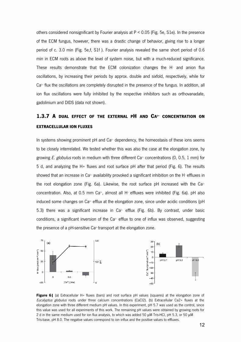
1.3.7 A DUAL EFFECT OF THE EXTERNAL PH AND CA2+ CONCENTRATION ON
EXTRACELLULAR ION FLUXES
In systems showing prominent pH and Ca2+ dependency, the homeostasis of these ions seems
to be closely interrelated. We tested whether this was also the case at the elongation zone, by
growing E. globulus roots in medium with three different Ca2+ concentrations (0, 0.5, 1 mm) for
5 d, and analyzing the H+ fluxes and root surface pH after that period (Fig. 6). The results
showed that an increase in Ca2+ availability provoked a significant inhibition on the H+ effluxes in
the root elongation zone (Fig. 6a). Likewise, the root surface pH increased with the Ca2+
concentration. Also, at 0.5 mm Ca2+, almost all H+ effluxes were inhibited (Fig. 6a). pH also
induced some changes on Ca2+ efflux at the elongation zone, since under acidic conditions (pH
5.3) there was a significant increase in Ca2+ efflux (Fig. 6b). By contrast, under basic
conditions, a significant inversion of the Ca2+ efflux to one of influx was observed, suggesting
the presence of a pH-sensitive Ca2+ transport at the elongation zone.
Figure 6| (a) Extracellular H+ fluxes (bars) and root surface pH values (squares) at the elongation zone of Eucalyptus globulus roots under three calcium concentrations (CaCl2). (b) Extracellular Ca2+ fluxes at the elongation zone with three different medium pH values. In this experiment, pH 5.7 was used as the control, since this value was used for all experiments of this work. The remaining pH values were obtained by growing roots for 2 d in the same medium used for ion flux analysis, to which was added 50 μM Tris-HCl, pH 5.3, or 50 μM Tris-base, pH 8.0. The negative values correspond to ion influx and the positive values to effluxes.

13
1.4 DISCUSSION
This study presents the novel observation that different Eucalyptus root zones experience a
differential modulation in their ion fluxes by the colonization of the ECM fungus P. microcarpus.
Our experimental approach was efficient to produce plants with a high degree of fungal
colonization at the stage of analysis. Thus, despite the inhibition of root hair growth, positive
effects of ECM fungus on plant growth were observed (Table 1). This is a new aspect of host-
pathogen interaction during ECM that reveals a potentially important aspect of coevolution
between the fungal cell biology and the plant immune system, and one that may open for new
paradigms of cell–cell communication through ion signaling pathways.
1.4.1 THE CONTROL OF ROOT SURFACE PH IN ECM ROOTS BY EXTRACELLULAR H+
FLUXES IS LINKED TO PM H+-ATPASE ACTIVITY
In comparison with control uninfected plants, the highest rates of H+ efflux and acidic surface
pH were located at the elongation zones of ECM roots (Fig. 2). These effluxes are dependent
on the PM H+-ATPase, as they were inhibited by 100 µm orthovanadate (inhibitor of P-type
plasma membrane H+-ATPase), a result which is conceptually sound since the elongation zone
is a specialized growing zone (Winch & Pritchard, 1999). In fact, it has been shown that this
zone shows notably higher immunolocalization and higher activity levels of PM H+-ATPase than
the apical and meristematic zones (Jahn et al., 1998; Palmgren, 2001; see details in Enriquez-
Arredondo et al., 2005). Using immunocytochemical approaches, Lei & Dexheimer (1988)
found strong PM H+-ATPase labeling in root cortical cells of Pinus sylvestris- Laccaria laccata, in
external hyphae sheaths and Hartig nets. This localization supports the concept of a coupling
mechanism between fungal and host H+ pumps in ECM roots (Fig. 2, Table 2). Indeed, it has
been demonstrated that for arbuscular mycorrhizal associations, some host PM H+-ATPase
isoforms show increased activity and gene expression after fungal colonization (Ferrol et al.,
2002; Ramos et al., 2005; details in Rosewarne et al., 2007). The H+ efflux mediated by the
PM H+-ATPase is important for the regulation of cytoplasmic pH (Felle, 2001; Palmgren, 2001;
Tazawa, 2003) and the activation of cell wall-loosening enzymes and proteins through

14
acidification of the apoplast (Hager, 2003). This effect is closely related to auxin-induced cell
growth as proposed by the ‘acid-growth theory’ by Rayle & Cleland (1992). This implies that
enhanced H+ efflux in ECM roots (Fig. 2a) results in an acidification of the apoplastic/ external
pH (Fig. 2b). Moloney et al. (1981) demonstrated that pH changes in the apoplast are crucial
for root growth, since acidic buffering conditions act as stimulators whilst neutral or basic pHs
act as inhibitors. Our results clearly show that when the H+ flux rate (Fig. 2a) and surface pH
values (Fig. 2b) are combined, highly significant Pearson’s correlation coefficients are obtained
(−0.82; P < 0.0001). Other candidates that contribute to the control of extracellular H+ flux in
ECM are the presence of anions in the growth medium. These are reported to act as
stimulators of the PM H+-ATPase (Churchill & Sze, 1984; Ullrich & Novacky, 1990; Glass et al.,
1992; Forde, 2000; Garnett et al., 2001). This concept is especially appealing taking into
account the observed oscillatory behavior (Figs 5, S1), where the ECM colonization induced
changes in the flux oscillations, leading to their increased periods. For Ca2+ flux oscillation, ECM
colonization abolished all significant periods observed in control roots (Figs 5b,c, S1).
Combined with the reversion from efflux to influx in the elongation zone, this result could be
interpreted as showing that the fungus contributes to the majority of the Ca2+ influx through
specific channels. These different activities would produce intricate temporal patterns
impossible to synchronize on an organized oscillatory pattern. This being the case, the
prediction would be that ectomycorrhizal plants should have an improved efficiency of Ca2+
uptake from the soil, a result partly confirmed in Fig. 3(c).
1.4.2 CA2+ EFFLUX SUPPRESSION AND INCREASE UPON CA2+ UPTAKE IN ECM
ROOTS
Calcium has a paradoxical effect on PM H+-ATPase, as it has been reported to be an inhibitor
via a Ca2+-dependent phosphorylation pathway (Lino et al., 1998; Tazawa, 2003) and an
activator in guard cells (Assmann et al., 1985). An inhibition of the PM Ca2+ influx channels in
both animal (Yang & Sachs, 1989) and plant cells (Allen & Sanders, 1994; Klusener et al.,
1995; Knight et al., 1996; Antoine et al., 2000, 2001) occurs by the addition of extracellular
Gd3+ in a micromolar range. Despite its use for detection of Ca2+ stretch-activated channels
(Caldwell et al., 1998), Gd3+ is likely to inhibit other cationic channels as well, because of its
relatively broad effect. Our pharmacological analysis suggested that the Ca2+ influx in the
elongation zone of ECM roots is the result of the activity of Gd3+-sensitive calcium channels

15
(Figs 3a, 4; Table 2). However, as the Ca2+ effluxes are largely governed by the chemical
potential gradient of Ca2+ generated by the PM Ca2+-ATPase, we hypothesized that the
suppression of effluxes in the control roots could represent an indirect dissipation of the Ca2+
gradient, as promoted by Gd3+ treatment (Fig. 4, Table 2). In addition, similar flux inhibition
profiles were obtained by Nemchinov et al. (2008) in Nicotiana benthamiana leaves. The
authors proposed a model in which Gd3+-sensitive Ca2+ influxes and Ca2+ pumps are involved in
the signal transduction pathways of the hypersensitive response mechanisms (Nemchinov et
al., 2008). As a passive Ca2+ efflux from the cell cytosol is thermodynamically improbable
(Shabala & Newman, 2000), an active mechanism must be involved. Two possible
mechanisms of Ca2+ efflux might occur, one through Ca2+ release from the cell wall and the
other by Ca2+ extrusion via the PM Ca2+-ATPase (Lecourieux et al., 2006; Nemchinov et al.,
2008). It remains to be determined which of these two mechanisms is responsible for this
event to occur. Alternatively, an increase in the activity of Ca2+ influx in ECM roots could reflect
an increased cytosolic concentration of this ion. Indeed, it has recently been demonstrated that
the exposure of E. globulus root hairs to hypaphorine (an indole alkaloid secreted by P.
microcarpus) led to an elevation of cytoplasmic Ca2+ concentration (Dauphin et al., 2007).
Thus, hypaphorine led to a reduction of the Ca2+ gradient across the plasma membrane, which
was correlated with the arrested growth of root hairs (Béguiristain & Lapeyrie, 1997; Dauphin
et al., 2007). These results seem similar to our own observations (Table 1), where root hair
length was reduced in ECM roots. Recently, Martin et al. (2008) published the genome of the
ECM fungus Laccaria bicolor, in which numerous and diverse Ca2+ channels are found to be
encoded (see details at http:// genome.jgi-psf.org/Lacbi1/Lacbi1.home.html). Accordingly, we
found ECM roots to have a higher uptake capacity of Ca2+ from the external medium (Fig. 3c).
In itself this would not necessarily lead to a major accumulation of Ca2+ in ECM of whole plants,
but clearly suggests a higher potential for ion uptake and storage in the cell wall (Peterson &
Enstone, 1996; Kuhn et al., 2000) promoted by the fungus. In ECM associations, such as
Suilus bovinus-Pinus sylvestris, an exposure to Ca2+ also led to an accumulation of this ion in
the interfacial apoplast in between symbionts and in the fungal sheath (Bucking et al., 2002).
Depending on the fungal species, Ca2+ can also accumulate as calcium oxalate in the fungal
hyphae (Malajczuk & Cromack, 1982). In the light of this, calcium dynamics in ECM
interactions needs to be more carefully investigated, not just using radioisotopes, but also by

16
means of an integration of techniques such as ion-selective vibrating probes, patch-clamp and
imaging analyses.
1.4.3 ACTIVATION OF ANION UPTAKE BY ECM FUNGUS
It is well known that an increase in the root surface concentration of H+ generates a proton-
motive force, which is necessary to drive the secondary transport of , SO42+, Cl−, Ca2+ and K+
(Portillo, 2000; Palmgren, 2001). Accordingly, we found that the changes in H+ efflux
attributable to ECM fungal infection in the elongation zone were strictly correlated to the root
surface pH values (−0.82; P < 0.0001), and, significantly, correlations of root surface
concentrations of H+ were found with both Ca2+ (−0.78, P < 0.001) and anion fluxes (0.66; P <
0.006). The correlation between Ca2+ and anions at the elongation zone (0.99, P < 0.001)
raised the possibility of an activation of anion influx by Ca2+, as demonstrated in other cells
(Hedrich et al., 1990). Since plant cells have adapted to low anion concentrations, anion
uptake is generally coupled to the electrochemical gradient generated by the PM H+-ATPase
activity (Evans et al., 1980; Zimmermann et al., 1994; Garnett et al., 2001). Consequently,
ECM roots possess strong anion influxes and H+ effluxes primarily at the elongation zone (Fig.
3b). Consistent with this, we observed high H+-ATPase activity in this root zone. It has been
reported that this enzyme is stimulated by anions in plant (Churchill et al., 1983; Churchill &
Sze, 1984; Zimmermann et al., 1994) and animal cell membranes (Vieira et al., 1995). The
induction of uptake in P. pinaster ECM roots, even at low external concentrations, was
previously shown by Gobert & Plassard (2002). The H+ efflux and consequent root surface
acidification are necessary for the uptake mechanism to operate (Ullrich & Novacky, 1990;
Glass et al., 1992; Forde, 2000), as this occurs via PM cotransporters (nH+/NO3-)
(Crawford,1995). This was already demonstrated for Eucalyptus nitens, where large H+ effluxes
were found in medium with NO3-. However, fluxes were quantitatively linked to H+ fluxes
(Garnett & Smethurst, 1999; Garnett et al., 2001, 2003). In addition, according to Garnett et
al. (2003), negative correlation coefficients can be obtained between and H+ fluxes. Nitrate is
thus a strong candidate to be a component of the anion fluxes we observed, but unfortunately
the technical limitations of the electrodes used do not warrant a straightforward conclusion in
this respect (see the Materials and Methods section and Fig. S2a,b), with chloride probably
playing also an important role. In normal conditions, the maintenance of the electrical
membrane potential depends on the H+ efflux and influxes of anions and potassium (Felle,

17
2001; Tazawa, 2003). In ECM symbiosis, fungi have a high capacity to uptake potassium in
their external hyphae (Rygiewicz & Bledsoe, 1984). One possible molecular basis for this was
recently discovered in the same type of hyphae, where Corratgé et al. (2007) cloned the
HcTrk1 transporter from Hebeloma cylindrosporum, and demonstrated it to encode for a
single-file pore channel that cotransports Na+/K+ into the hyphae. Pharmacological analyses
suggested the presence of anion channels at the elongation zone, since the influxes were
sensitive to DIDS. In guard cells, DIDS also inhibits anion uptake (Schroeder et al., 1993;
Schwartz et al., 1995) similar to what was observed in this study (Table 2). Further studies
should be focused on the proper discrimination of the specific anions involved on the observed
response at the elongation zone of ECM roots.
1.5 CONCLUDING REMARKS
Based on our results, we propose a model for pH signaling in ECM roots, which is directly
linked to nutrient uptake and plant growth (Table 1, Fig. 7). ECM fungi induce positive
modulation of the H+ efflux rates and rhizosphere acidification, mediated by PM H+-ATPase
activities from both host and fungal partners. In turn, this stimulation triggers a pH signal that
modulates Ca2+ transport and, indirectly, anion uptake (Hedrich et al., 1990). This hypothesis is
supported by our observation that external Ca2+ acts as a strong inhibitor of the H+ efflux and
root surface acidification in the elongation zone of eucalypt roots. By contrast, Ca2+ fluxes were
also affected by the medium’s pH, as has previously been reported in other plant cells (Foster,
1990). An increase in anion uptake and lower concentrations of external Ca2+ will thus occur,
which are reflected both in the promotion of plant growth and in PM H+-ATPase activity
(Zimmermann et al., 1994). The spectral analysis of the ion flux oscillations revealed itself to
be an efficient parameter to compare biophysical effects of the ECM fungus in the fast
oscillation components. This analysis can be used as an additional tool during the study of ion
dynamics using the ion-selective vibrating probe technique, on the assumption that shifts in the
main components of oscillations correspond to the activation/shift of a variety of molecular
transporters.

18
1.6 METHODS
1.6.1 BIOLOGICAL MATERIAL, INOCULUM PRODUCTION AND IN VITRO SYNTHESIS OF
ECTOMYCORRHIZAS
Three agar discs containing mycelium of the ECM gasteromycete Pisolithus microcarpus
isolate PT 90A were inoculated onto Petri dishes containing 20 mL of modified MNM (Marx,
1969) medium and incubated for 28 d at 28ºC. From the resulting colonies, 9 mm agar discs
were cut off from the edge of actively growing colonies. Eucalyptus globulus Labill. Seeds were
superficially sterilized with 5% sodium hypochlorite (v/v) for 15 min, rinsed with five changes of
sterile water, and plated on modified Clark solution at quarter-strength (Clark, 1975) to which
was added 2.9 µM thiamine-HCl and 1% sucrose in 0.5% (w/v) Phytagel (Sigma-Aldrich,
Gillingham, UK). The use of Phytagel produced a clear and colorless medium, which is
excellent for imaging and ion flux measurement with reduced electrical noise (Ramos et al.,
Figure 7| Proposed model for the pH signaling mechanism in ectomycorrhizal (ECM) roots and the differential modulation of anion (A−) and calcium (Ca2+) uptake

19
2008a). After 7 d, aseptically germinated seedlings were placed on the edge of 10-d-old ECM
fungal mycelium grown on the same medium used for seedlings. These were left for 15 d in a
controlled environment growth chamber, with 16 h of light (26ºC, 350 µmol m−2 s−1) and 8 h of
dark, for ectomycorrhiza formation. ECM plants were later transferred to hydroponic conditions
in the same solution and growth chamber settings for 10 d. Subsequently, ion fluxes
measurements were performed in secondary roots of intact plants. In addition, pieces of root
system were washed and samples were subsequently collected for microscopic evaluation of
mycorrhizal colonization, as described by Brundrett et al. (1996).
1.6.2 MEASUREMENTS OF H+, CA2+ AND ANION FLUXES AND CURRENTS USING THE ION-
SELECTIVE VIBRATING PROBE SYSTEM
A detailed description of the experimental setup of the ionselective vibrating probe technique
utilized in this study has been well described (Kochian et al., 1992; Feijó et al., 1999; Shipley
& Feijó, 1999; Zonia et al., 2002; Kunkel et al., 2006; Ramos et al., 2008a). In short, E.
globulus plants colonized or not by ECM fungus P. microcarpus isolate PT 90A under
hydroponic conditions, were placed in plastic Petri dishes (140x140 mm) filled with 30 mL of
modified Clark solution at quarter strength, except for Ca2+ measurements, where 100 µM Ca2+
was used. Visual Minteq analysis was performed according to Parker et al. (1995) using the
ion concentrations of the modified Clark solution applied in this study. We focused on
secondary roots, as they are biologically and physiologically more significant than primary roots
for nutrient supply to the plant. The volume occupied by secondary roots in the soil can reach
30–40% more than primary ones. Readings were taken in five defined root zones of
nonmycorrhizal (control) and mycelium-covered roots: apex (tip), meristematic (100–150 µm);
elongation (300–800 µm); root hairs (major presence of these structures); and finally mature
zone (posterior to root hair zone). Ion-specific vibrating microelectrodes were produced as
described by Feijó et al. (1999). Micropipettes were pulled from 1.5 mm borosilicate glass
capillaries and treated with dimethyl dichlorosilane (Sigma-Aldrich). After silanization, they were
backfilled with a 15–20 mm column of electrolyte (15 mM KCl and 40 mM KH2PO4, pH 6.0, for
H+; 100 mM KCl for anions; 100 mM CaCl2 for Ca2+) and then frontloaded with a 20–25 µm
column of the respective ion-selective liquid exchange cocktail (Fluka, Milwaukee, WI, USA). We

20
used Cl− electrodes to measure the anion fluxes given that this electrode has poor selectivity for
Cl− under our experimental conditions (Supporting information, Fig. S2a,b). Firstly the
measurement of chloride activity in the medium is slightly affected by the presence of other
ions (Fig. S2a), but these changes should be expressed below noise level within the microvolt
range usually measured on vibrating conditions for cellular fluxes. More importantly, the Cl−
electrode calibration with different anions showed that this electrode responds with a Nernstian
slope to chloride and nitrate, and while sub-Nernstian to sulfate and phosphate, also exhibits a
significant response within the concentrations used in this study (Fig. S2b). Last but not least,
the background concentrations in the medium of the individual anions span various orders of
magnitude, likewise affecting the signal-to-noise (S/N) ratio measurement of the fluxes in a way
that is inversely proportional to the concentration. Taken together, these considerations make it
almost impossible to discriminate the individual activities of every single anion, and therefore
we have opted to refer to these fluxes as reflecting the global ‘anionic’ concentration rather
than Cl− proper fluctuations. An Ag/AgCl wire electrode holder (World Precision Instruments,
Sarasota, FL, USA) was inserted into the back of the microelectrode and established electrical
contact with the bathing solution. The ground electrode was a dry reference (DRIREF-2, World
Precision Instruments) that was inserted into the sample bath. The microelectrodes were
calibrated at the beginning and end of each experiment using standard solutions covering the
experimental range of each ion, in order to obtain a calibration line. Both the slope and
intercept of the calibration line were used to calculate the respective ion concentration from the
mV values measured during the experiments.
1.6.3 INHIBITION WITH VANADATE (VO43-), GADOLINIUM (GdCl3) AND 4,4′-
DIISOTHIOCYANATOSTILBENE-2,2′-DISULFONIC ACID (DIDS)
Inhibitor treatments were performed in Eucalyptus roots after determination of each ion flux at
the elongation zone (n = 5). The data acquisition was stopped and the respective inhibitors
(Sigma-Aldrich) were added in the Petri dishes with the following concentrations: plasma
membrane H+-ATPase (100 µM orthovanadate), calcium channels (100 µM gadolinium) and
chloride channels (50 µM DIDS). Five to 10 min later, a background reference was taken and
ion fluxes were again recorded. Interference caused by the inhibitors was controlled for by

21
direct incubation with ionophore-loaded probes. No significant interference of the inhibitors was
found to occur for H+ and anions. For Ca2+, the interference was more pronounced with high
levels of gadolinium, which in the present study was used at lower concentrations (100 µM;
Fig. S3).
1.6.4 ION FLUX OSCILLATION ANALYSIS
Frequency analyses were performed using AutoSignal v1.7 (Systat Software, Inc.). For each set
of flux oscillations to be analyzed, a data trend removal was applied, consisting of a linear
least-squares fit subtraction to remove the very low frequency trend of the data. Two distinct
methods were then used to assess the frequency components of the oscillations: Fourier and
Wavelet analyses. For Fourier analysis, a fast Fourier transform Radix 2 algorithm was used,
ensuring that each data set was a continuous acquisition without breaks and with a constant
sampling rate. Peaks were detected by a local maxima detection algorithm and considered
relevant according to their significance levels (the higher the significance level, the less likely it
is that a detected spectral signal will arise from random noise). Significance levels are given in
the Results section. For wavelet analysis, a continuous wavelet time– frequency spectrum was
obtained with a noncomplex Morlet wavelet (wave number, 12). A peak-type critical limit was
used instead of the traditional confidence levels, as implemented in the software.
1.6.5 STATISTICAL ANALYSIS
All data was analyzed by one-way or two-way ANOVA in order to compare the mean values
(considering ‘fungal treatment’ and ‘root region’ as factors), which were validated by
convenient residual analyses and, when necessary, combined with Duncan’s test for multiple
comparisons. To compare the control and fungal treatment (Table 1), we applied Student’s t-
test for two independent samples and calculated confidence intervals for the mean difference,
in order to guarantee a global 95% confidence level. The results are expressed as means with
respective standard error, and the numbers of repetitions are given in each figure legend. All
statistical analyses were conducted using the R program and the level of significance was set
up at 5% (Ihaka & Gentleman, 1996).

22
1.7 REFERENCES
Ahonen-Jonnarth U, Van Hees PAW, Lundstrom US, Finlay RD. 2000. Organic acids produced by mycorrhizal
Pinus sylvestris exposed to elevated aluminium and heavy metal concentrations. New Phytologist 146: 557–567.
Allen GJ, Sanders D. 1994. Two voltage-gated calcium release channels coreside in the vacuolar membrane of
guard cells. Plant Cell 6: 685–694.
Antoine AF, Faure JE, Cordeiro S, Dumas C, Rougier M, Feijó JA. 2000. A calcium influx is triggered and
propagates as a wavefront in the zygote after in vitro fertilization of flowering plants. Proceedings of the National
Academy of Sciences, USA 97: 10643–10648.
Antoine AF, Faure JE, Dumas C, Feijó JA. 2001. Differential contributions of free cytosolic and extracellular fluxes
of calcium to gamete fusion and egg activation in flowering plants. Nature Cell Biology 3: 1120–1124.
Assmann SM, Simoncini L, Schroeder JI. 1985. Blue light activates electrogenic ion pumping in guard cell
protoplasts of Vicia faba. Nature 318: 285–287.
Béguiristain T, Lapeyrie F. 1997. Host plant stimulates hypaphorine accumulation in Pisolithus tinctorius hyphae
during ectomycorrhizal infection while excreted fungal hypaphorine controls root hair development. New
Phytologist 136: 525–532.
Boukcim H, Plassard C. 2003. Juvenile nitrogen uptake capacities and root architecture of two open-pollinated
families of Picea abies. Effects of nitrogen source and ectomycorrhizal symbiosis. Journal of Plant Physiology 10:
1211–1218.
Bowman BJ. 1982. Vanadate uptake in Neurospora crassa occurs via phosphate transport system II. Journal of
Bacteriology 153: 286–291.
Bowman BJ, Allen KE, Slayman CW. 1983. Vanadate-resistant mutants of Neurospora crassa are deficient in a
high-affinity phosphate transport system. Journal of Bacteriology 153: 292–296.
Brundrett M, Bougher NM, Dell B, Grove T, Malajczuck N. 1996. Working with mycorrhizas in forestry and
agriculture. Camberra, Australia: Pirie Printers.
Bucking H, Hans R, Heyser W. 2007. The apoplast of ectomycorrhizal roots – site of nutrient uptake and nutrient
exchange between the symbiotic partners. In: Sattelmacher B, Horst WJ, eds. The apoplast of higher plants:
compartment of storage, transport and reactions. Dordrecht, the Netherlands: Springer-Verlag, 97–108.
Bucking H, Heyser W. 2000. Subcellular compartmentation of elements in nonmycorrhizal and mycorrhizal roots
of Pinus sylvestris: an X-ray microanalytical study. II. The distribution of calcium, potassium and sodium. New
Phytologist 145: 321–331.

23
Bucking H, Kuhn AJ, Schröder WH, Heyser W. 2002. The fungal sheath of ectomycorrhizal pine roots: an
apoplastic barrier for the entry of calcium, magnesium, and potassium into the root cortex? Journal of
Experimental Botany 53: 1659–1669.
Caldwell RA, Clemo HF, Baumgarten CM. 1998. Using gadolinium to identify stretch-activated channels: technical
considerations. American Journal of Physiology – Cell Physiology 275: 619–621.
Churchill KA, Holaway B, Sze H. 1983. Separation of two types of electrogenic H+-pumping ATPases from oat
roots. Plant Physiology 73: 921–928.
Churchill KA, Sze H. 1984. Anion-sensitive, H+ pumping ATPase of oat roots: direct effects of Cl−, and a
disulfonic stilbene. Plant Physiology 76: 490–497.
Clark RB. 1975. Characterization of phosphatase of intact maize roots. Journal of Agricultural and Food Chemistry
23: 458–460.
Corratgé C, Zimmermann S, Lambilliotte R, Plassard C, Marmeisse R, Thibaud JB, Lacombe B, Sentenac H.
2007. Molecular and functional characterization of a Na+-K+ transporter from the Trk family in the
ectomycorrhizal fungus Hebeloma cylindrosporum. Journal of Biological Chemistry. 282: 26057–26066.
Courty PE, Pouysegur R, Buee M, Garbaye J. 2006. Laccase and phosphatase activities of the dominant
ectomycorrhizal types in a lowland oak forest. Soil Biology & Biochemistry 38: 1219–1222.
Crawford NM. 1995. Nitrate: nutrient and signal for plant growth. Plant Cell 7: 859–868.
Dauphin A, Gérard J, Lapeyrie F, Legué V. 2007. Fungal hypaphorine reduces growth and induces cytosolic
calcium increase in root hairs of Eucalyptus globulus. Protoplasma 231: 83–88.
Enriquez-Arredondo C, Sanchez-Nieto S, Rendon-Huerta E, Gonzalez-Halphen D, Gavilanes-Ruiz M, Diaz-Pontones
D. 2005. The plasma membrane H+-ATPase of maize embryos localizes in regions that are critical during the
onset of germination. Plant Science 169: 11–19.
Evans ML, Mulkey TJ, Vesper MJ. 1980. Auxin action on proton influx in corn roots and its correlation with growth.
Planta 148: 510–512.
Feijó JA, Sainhas J, Hackett GR, Kunkel JG, Hepler PK. 1999. Growing pollen tubes posses a constitutive alkaline
band in the clear zone and a growth-dependent acidic tip. Journal of Cell Biology 144: 483–496.
Felle HH. 2001. pH: signal and messenger in plant cells. Plant Biology 3: 577–591.
Ferrol N, Pozo MJ, Antelo M, Azcón-Aguilar C. 2002. Arbuscular mycorrhizal symbiosis regulates plasma
membrane H+-ATPase gene expression in tomato plants. Journal of Experimental Botany 53: 1683–1687.

24
Forde BG. 2000. Nitrate transporters in plants: structure, function and regulation. Biochimica et Biophysica Acta
1465: 219–235. Foster JF. 1990. Influence of pH and plant nutrient status on ion fluxes between tomato plants
and simulated acid mists. New Phytologist 116: 475–485.
Garnett TP, Shabala SN, Smethurst PJ, Newman IA. 2001. Kinetics of ammonium and nitrate uptake by eucalypt
roots and associated proton fluxes measured using ion selective microelectrodes. Functional Plant Biology 30:
1165–1176.
Garnett TP, Shabala SN, Smethurst PJ, Newman IA. 2003. Simultaneous measurement of ammonium, nitrate
and proton fluxes along the length of eucalypt roots. Plant and Soil 236: 55–62.
Garnett TP, Smethurst PJ. 1999. Ammonium and nitrate uptake by Eucalyptus nitens: effects of pH and
temperature. Plant and Soil 214: 133–140.
Glass AD, Shaff JE, Kochian LV. 1992. Studies of the uptake of nitrate in barley. IV. Electrophysiology. Plant
Physiology 99: 456–463.
Gobert A, Plassard C. 2002. Differential dependent patterns of uptake in Pinus pinaster, Rhizopogon roseolus and
their ectomycorrhizal association. New Phytologist 154: 509–516.
Hager A. 2003. Role of the plasma membrane H+-ATPase in auxin induced elongation growth. Historical and new
aspects. Journal of Plant Research 116: 483–505.
Hawkins BJ, Boukcim H, Plassard C. 2008. A comparison of ammonium, nitrate and proton net fluxes along
seedling roots of Douglas-fir and lodgepole pine grown and measured with different inorganic nitrogen sources.
Plant, Cell & Environment 31: 278–287.
Hedrich R, Busch H, Raschke K. 1990. Ca2+ and nucleotide dependent regulation of voltage dependent anion
channels in the plasma membrane of guard cells. EMBO Journal 9: 3889–3892.
Ihaka R, Gentleman R. 1996. R: a language for data analysis and graphics. Journal of Computational and
Graphical Statistics 5: 299–314.
Jahn T, Baluska F, Michalke W, Harper JF, Volkmann D. 1998. Plasma membrane H+-ATPase in the root apex:
evidence for strong expression in xylem parenchyma and asymmetric localization within cortical and epidermal
cells. Physiologia Plantarum 104: 311–316.
Javelle A, Andre B, Marini AM, Chalot M. 2003. High-affinity ammonium transporters and nitrogen sensing in
mycorrhizas. Trends in Microbiology 11: 53–55.
Klusener B, Boheim G, Liss H, Engelberth J, Weiler EW. 1995. Gadolinium-sensitive, voltage-dependent calcium
release channels in the endoplasmic reticulum of a higher plant mechanoreceptor organ. EMBO Journal 14:
2708–2714.

25
Knight H, Trewavas AJ, Knight MR. 1996. Cold calcium signaling in Arabidopsis involves two cellular pools and a
change in calcium signature after acclimation. Plant Cell 8: 489–50.
Kochian LV, Shaff JE, Ku " htreiber WM, Jaffe LF. 1992. Use of an extracellular, ion-selective, vibrating
microelectrodes system for the quantification of K+, H+ and Ca2+ fluxes in maize suspension cells. Planta 188:
601–610.
Kuhn AJ, Schröder WH, Bauch J. 2000. The kinetics of calcium and magnesium entry into mycorrhizal spruce
roots. Planta 210: 488–96.
Kunkel JG, Cordeiro S, Xu J, Shipley AM, Feijó JA. 2006. The use of noninvasive ion-selective microelectrode
techniques for the study of plant development. In: Volkov V, ed. Plant electrophysiology – theory and methods.
Berlin, Germany: Springer-Verlag, 109–137.
Lecourieux D, Ranjeva R, Pugin A. 2006. Calcium in plant defence signalling pathways. New Phytologist 171:
249–69.
Lei J, Dexheimer J. 1988. Ultrastructural localization of ATPase activity in the Pinus sylvestris/Laccaria laccata
ectomycorrhizal association. New Phytologist 108: 329–334.
Lino B, Baizabal-Aguirre VM, González de la Vara LE. 1998. The plasma membrane H+-ATPase from beet root is
inhibited by a calcium-dependent phosphorylation. Planta 204: 352–359.
Malajczuk N, Cromack K Jr. 1982. Accumulation of calcium oxalate in the mantle of ectomycorrhizal roots of
Pinus radiata and Eucalyptus marginata. New Phytologist 92: 527–531.
Marchner H, Dell B. 1994. Nutrient uptake in mycorhizal symbiosis. Plant and Soil 159: 89–102.
Martin F, Aerts A, Ahrén D, Brun A, Danchin EGJ, Duchaussoy F, Gibon J, Kohler A, Lindquist E, Pereda V et al.
2008. The genome of Laccaria bicolor provides insights into mycorrhizal symbiosis. Nature 452: 88–92.
Martin F, Kohler A, Duplessis S. 2007. Living in harmony in the wood underground: ectomycorrhizal genomics.
Current Opinion in Plant Biology 10: 204–210.
Marx DH. 1969. The influence of ectotrophic mycorrhizal fungi on the resistance of pine roots to pathogenic fungi
and soil bacteria. I. Antagonism of mycorrhizal fungi to root pathogenic fungi and soil bacteria. Phytopathology 59:
153–163.
Messerli MA, Smith PJS, Lewis RC, Robinson KR. 2004. Chloride fluxes in lily pollen tubes: a critical reevaluation.
Plant Journal 40: 799–812.
Michard E, Dias P, Feijó JA. 2008. Tobacco pollen tubes as cellular models for ion dynamics: improved spatial
and temporal resolution of extracellular flux and free cytosolic concentration of calcium and protons using
pHluorin and YC3.1 CaMeleon. Sexual Plant Reproduction 21: 169–181.

26
Moloney MM, Elliott MC, Cleland RE. 1981. Acid growth effects in maize roots: evidence for a link between auxin
economy and proton extrusion in the control of root growth. Planta 152: 285–291.
Muller T, Avolioa M, Olivia M, Benjdiab M, Rikirschb E, Kasarasa A, Fitza M, Chalotc M, Wipfa D. 2007. Nitrogen
transport in the ectomycorrhiza association: the Hebeloma cylindrosporum-Pinus pinaster model. Phytochemistry
68: 41–51.
Nemchinov LG, Shabala L, Shabala S. 2008. Calcium efflux as a component of the hypersensitive response of
Nicotiana benthamiana to Pseudomonas syringae. Plant and Cell Physiology 49: 40–46.
Palmgren MG. 2001. Plant plasma membrane H+-ATPases: powerhouses for nutrient uptake. Annual Review of
Plant Physiology and Plant Molecular Biology 52: 817–845.
Parker DR, Chaney RL, Norvell WA. 1995. Chemical equilibrium models: applications to plant nutrition research.
In: RH Loeppert, AP Schwab, S Goldberg, eds. Chemical equilibrium and reaction models. Madison, WI, USA: Soil
Science Society of America, Inc, 163–200, 253–269.
Pasqualini S, Panara F, Antonielli M. 1992. Acid-phosphatase-activity in Pinus-pinea-Tuber-albidum
ectomycorrhizal association. Canadian Journal of Botany 70: 1377–1383.
Peterson CA, Enstone DE. 1996. Functions of passage cells in the endodermis and exodermis of roots.
Physiologia Plantarum 97: 592–598.
Plassard C, Guérin-Laguette A, Véry AA, Casarin V, Thibaud JB. 2002. Local measurements of nitrate and
potassium fluxes along roots of maritime pine. Effects of ectomycorrhizal symbiosis. Plant, Cell & Environment 25:
75–84.
Portillo F. 2000. Regulation of plasma membrane H+-ATPase in fungi and plants. Biochimica et Biophysica Acta
1469: 31–42.
Ramos AC, Façanha AR, Feijó JA. 2008a. Proton (H+) flux signature for the presymbiotic development of the
arbuscular mycorrhizal fungi. New Phytologist 178: 177–188.
Ramos AC, Façanha AR, Feijó JA. 2008b. Ion dynamics during the polarized growth of arbuscular mycorrhizal
fungi: from presymbiosis to symbiosis. In: Varma A, ed. Mycorrhiza: structure function and biotechnology.
Heidelberg, Germany: Springer-Verlag, 241–261.
Ramos AC, Martins MA, Façanha AR. 2005. ATPase and pyrophosphatase activities in corn root microsomes
colonized with arbuscular mycorrhizal fungi. Brazilian Journal of Soil Science 29: 207–213.
Rayle DL, Cleland RE. 1992. The acid growth theory of auxin-induced cell elongation is alive and well. Plant
Physiology 99: 1271–1274.

27
Rosewarne GM, Smith FA, Schachtman DP, Smith SE. 2007. Localization of proton-ATPase genes expressed in
arbuscular mycorrhizal tomato plants. Mycorrhiza 17: 249–258.
Rygiewicz PT, Bledsoe CS. 1984. Mycorrhizal effects on potassium fluxes by northwest coniferous seedlings. Plant
Physiology 76: 918–923.
van Scholl L, Hoffland E, van Breemen N. 2006. Organic anion exudation by ectomycorrhizal fungi and Pinus
sylvestris in response to nutrient deficiencies. New Phytologist 170: 153–163.
Schroeder JI, Schmidt C, Sheaffer J. 1993. Identification of high-affinity slow anion channel blockers and evidence
for stomatal regulation by slow anion channels in guard cells. Plant Cell 5: 1831–1841.
Schwartz A, Ilan N, Schwarz M, Scheaffer J, Assmann SM, Schroeder JI. 1995. Anion-channel blockers inhibit S-
type anion channels and abscisic-acid responses in guard cells. Plant Physiology 109: 651–658.
Shabala S, Newman IA. 2000. Salinity effects on the activity of plasma membrane H+ and Ca2+ transporters in
bean leaf mesophyll: masking role of the cell wall. Annals of Botany 85: 681–686.
Shipley AM, Feijó JA. 1999. The use of the vibrating probe technique to study steady extracellular currents during
pollen germination and tube growth. In: Cresti M, Cai G, Moscatelli S, eds. Fertilization in higher plants: molecular
and cytological aspects. Heidelberg, Germany: Springer- Verlag, 235–252.
Taylor JH, Peterson CA. 2005. Ectomycorrhizal impacts on nutrient uptake pathways in woody roots. New Forests
30: 203–214. Tazawa M. 2003. Cell physiological aspects of the plasma membrane electrogenic H+ pump,
Journal of Plant Research 116: 419–442.
Ullrich CI, Novacky AJ. 1990. Extra- and intracellular pH and membrane potential changes induced by K+, CI−, ,
uptake and fusicoccin in root hairs of Limnobium storoniferum. Plant Physiology 94: 1561–1567.
Vieira L, Slotki I, Cabantchik ZI. 1995. Chloride conductive pathways which support electrogenic H+ pumping by
Leishmania major Promastigotes. Journal of Biological Chemistry 270: 5299–5304.
Ward JT, Lahner B, Yakubova E, Salt DE, Raghothama KG. 2008. The effect of iron on the primary root elongation
of Arabidopsis during phosphate deficiency. Plant Physiology 147: 1181–1191.
Winch S, Pritchard J. 1999. Acid-induced cell wall loosening is confined to the accelerating region of the root
growing zone. Journal of Experimental Botany 50: 1481–1487.
Yang X-C, Sachs F. 1989. Block of stretch-activated ion channels in Xenopus oocytes by gadolinium and calcium
ions. Science. 243: 1068–1071.
Zimmermann S, Thomine S, Guern J, Barbier-Brygoo H. 1994. An anion current at the plasma membrane of
tobacco protoplasts shows ATP-dependent voltage regulation and is modulated by auxin. Plant Journal 6: 707–
716.

28
Zonia L, Cordeiro S, Feijó JA. 2001. Ion dynamics and the control of hydrodynamics in the regulation of pollen
tube growth. Sexual Plant Reproduction 14: 111–116.
Zonia L, Cordeiro S, Tupy J, Feijó JA. 2002. Oscillatory chloride efflux at the pollen tube apical tip has a role in
growth and osmoregulation and is linked to inositol polyphosphate signaling pathways. Plant Cell 14: 2233–2249
1.8 SUPPLEMENTARY DATA
Figure S1| Fourier frequency spectrum analysis of H+ (A, B), Ca2+ (C, D) and anion flux oscillations in the elongation zone
of non-mycorrhizal (A, C and E) and mycorrhizal roots (ECM) of E. globulus colonized by P. microcarpus (B, D and F).
Frequencies are represented in min-1.

29
Figure S2| (A)- Interference on Cl− activity detection by the Cl− electrode (Control) by further anions (NO3-, SO4
2- and PO4
2-) present in ¼ strength Clark`s solution. The interference was more pronounced in lower Cl− concentrations, especially for NO3
- and SO42-. Yet, in real vibration measurement conditions, where differences are
usually measured in the microvolt range, this effect is minimal. * P-value < 0.1 and ** P -value < 0.01. (B)- Calibration of Cl− electrodes with different anions. Nitrate and Chloride show almost perfect Nersntian responses. Phosphate and Sulphate, while clearly sub-Nersntian, show significant responses on the interval measured.
Figure S3| Effect of different concentrations of Gadolinium (Gd3+) on the sensitivity of Ca2+ electrodes (n = 5). * P-
value < 0.01.
30
CHAPTER II
2. ON THE EVOLUTION AND DEVELOPMENT OF ROOT SYMBIOTIC SYSTEMS
Work published under the title:
Plant-microbe symbioses: new insights into common roots (2009) BioEssays 31 (11): 1233-1244
Pedro T. Lima1*, Vitor G. Faria1,2*, Pedro Patraquim1,2*, Alessandro C. Ramos1,4, José A. Feijó1,3, Élio Sucena1,2
*These authors contributed equally to this work
Affiliations
1 Instituto Gulbenkian de Ciência, Centro de Biologia do Desenvolvimento, Oeiras, Portugal
2 Universidade de Lisboa, Faculdade de Ciências, Depto. Biologia Animal, Lisboa, Campo Grande, 1700; Portugal
3 Universidade de Lisboa, Faculdade de Ciências, Depto. Biologia Vegetal, Lisboa, Campo Grande, 1700; Portugal
Key Words: Mycorrhiza, Nodulation, Symbiosis, Modularity, Glomeromycota, Evo-Devo

31
2.1 ABSTRACT
Arbuscular mycorrhiza (AM), a type of plant-fungal endosymbiosis, and nodulation, a bacterial-
plant endosymbiosis, are the most ubiquitous symbioses on earth. Recent findings have
established part of a shared genetic basis underlying these interactions. Here, we approach
root endosymbioses through the lens of the homology and modularity concepts aiming at
further clarifying the proximate and ultimate causes for the establishment of these biological
systems. We review the genetic structures that underlie interspecific signaling and its
concomitant shift in genetic programs for either partner. Also, through the comparative
analysis of AM and nodulation symbioses shared genetic modules, we identify the fundamental
nodes in these networks, suggesting the elemental steps that permitted symbiotic adaptation.
Here, we show that this approach, allied to recent technical advances in the study of genetic
systems architecture, can provide clear testable hypotheses for the advancement of our
understanding on the evolution and development of symbiotic systems.

32
2.2 INTRODUCTION
As early as 1905, Mereschkowsky stated that chloroplasts might have evolved from
free-living cyanobacteria, which were engulfed by a protozoan host cell (Mereschkowsky,
1905). The adaptation of the ancestral chloroplast as an endosymbiont was so successful that
entire new forms of life and a new kingdom arose from the evolution of this interaction
(Kutschera & Niklas, 2005). The establishment of symbioses between distantly-related species
recurred in this clade (Smith & Read, 2008), being the most ubiquitous result of such an event
the arbuscular mycorrhizal (AM) mutualistic association (Smith & Read, 2008). This interaction
is established between fungi belonging to the monophyletic phylum Glomeromycota (Schüssler
et al., 2001) and the roots of most vascular plants (Smith & Read, 2008), suggesting an
ancient evolutionary origin (Parniske, 2008). Arbuscular mycorrhiza-forming fungi (AMF) are
incapable of accomplishing their life cycle in the absence of a host plant (obligate biotrophy)
(Harrison, 2005; Reinhardt, 2007). The AMF dependence on this symbiotic relationship is
consistent with the expected progressive loss of functional redundancy over extended co-
evolutionary time (Moran, 2002). Further support comes from both fossil (Remy et al., 1994;
Taylor, 1995) and genetic (Simon et al., 1993) evidence of ancient arbuscular mycorrhizal-like
associations, which suggest that these predate the colonization of land by plants around 450
million years ago, and thus might have played a role in the subsequent radical change in the
biosphere. Despite this potentially pivotal role in the history of life on earth, the AM genetical
system has only recently started to be dissected. However, most of the genetical analysis of
AM establishment has focused on the plant symbiont alone (Smith & Read, 2008). This bias
can be attributed to the tractability of the plant system, both for its capability to develop
independently of the AMF, and for the development of plant genetics tools early in the history
of genetics as an experimental scientific discipline.
Several plant genome sequences are already available, including AM-forming plants
such as the model systems Oryza sativa (rice) (2005) and more recently Zea mays (maize),
two of the most important crops for human nutrition. Moreover, most data concerning AM
genetics has been generated in legumes, which are a relatively recent subgroup of the AM-
forming plants (Markmann et al., 2008; Parniske, 2008), constituting a further phylogenetic
bias on our current knowledge of AM biology. Nevertheless, studies in legumes have provided
some important insights such as that some nodulation mutants are unable to produce normal

33
AM phenotypes. We know of at least seven genes (common symbiosis genes) to be shared
between plant-rhizobia and AM endosymbioses (Kistner et al., 2005), one of which also plays a
role in the establishment of symbioses with actinobacteria (Gherbi et al., 2008; Markmann et
al., 2008). This is an invaluable cornerstone in our knowledge of the plant gene networks
underlying AM evolution, that must be complemented with an approach focusing on the
genetic bases underlying specificity in mycorrhizal endosymbioses. Recently, the first steps in
this direction have been taken with the description of mutants for the establishment of AM
symbiosis in non-legumes (Paszkowski et al., 2006; Reddy et al., 2007).
There are recent and very comprehensive reviews dealing with the collection of AM
genetic data (Parniske, 2004; Harrison, 2005; Requena et al., 2007; Parniske, 2008), so this
will not be the main topic of this paper. Here, we present an evodevo approach to root
endosymbioses, namely through the concepts of modularity and Gene Regulatory Networks
(GRNs), that can point the way to the most important features of these systems and prioritize
an experimental program designed at determining their basal biological principles and
mechanisms. We will develop the idea that the current knowledge of the plant genetics of
nodulation further supports the hypothesis that at least part of this genetic network originated
through co-option of AM symbiosis genes (Kistner & Parniske, 2002). We will also expand this
notion by reviewing recent literature on the evolution of root-nodule symbioses and integrating
in our analysis the concepts of homology and modularity (BOX 1).
2.3 FOLLOWING THE ONTOGENY OF AM
Plant mutualistic endosymbioses are characterized by the penetration of host cells by
microbial symbionts, followed by a stage during which the symbiont lives partially or wholly
within plant cells (Parniske, 2000). AM fungi receive plant-derived photosynthates from this
association, in turn transferring to the host cells phosphate and other minerals scavenged from
the soil (Harrison, 2005; Parniske, 2008). The development of AM fungi involves germination
of an asexual spore, hyphal polarized growth and profuse branching (Akiyama et al., 2005),
which ultimately differentiate into appressoria. Appressoria develop on the root surface and
functionally work as host cell penetration-helping structures (Figure 1) (Harrison, 2005;
Parniske, 2008). Arbuscular mycorrhiza-forming fungi (AMF) are obligate biotrophs, i.e. they
cannot complete their life-cycle without taking part in a symbiotic relationship with a plant
34
(Smith & Read, 2008). Despite this apparent limitation, these fungi have an initial asymbiotic
stage characterized by a coenocytic complex of hyphae, occurring after thegermination of a
single spore containing up to 2000 nuclei (Bécard, 1993). An interesting observation is that
they have the ability to, in the absence of host-derived signals, arrest and restart growth
(“flickering”) (Koske, 1981). AM fungal hyphae cross epidermal, sometimes hypodermal, and
outer cortex layers through a host derived intracellular assembly apparatus which determines
the future path of hyphal penetration (the prepenetration apparatus - PPA) (Genre et al., 2005;
Genre et al., 2008) (Figure 1e) and re-enter the apoplast crossing the inner cortex
intercellularly. Inner cortical cells are subsequently invaded and hyphae develop profuse
ramifications, giving rise to tree-shaped structures termed arbuscules within their cytoplasm,
surrounded by a plant-derived membrane (Parniske, 2008).
Mycorrhizal symbioses are multicellular complex systems in which a series of spatial
and temporal tightly regulated events involving inter-organismic signaling lead to division of
labour between the players. In particular, the obligate biotrophy of one of the players supports
the notion that AM symbiosis can be looked at as a single (and singular) developmental
program as typically applied to multicellular eukaryotes.
2.4 SETTING UP A RENDEZ-VOUS: DIFFUSIBLE SIGNALS IN PRESYMBIOSIS
A necessary step for the transition to the symbiotic stage is the pre-symbiotic recognition of the
phototrophic partner by the AMF. It is known that fungi recognize compatible host roots with a
certain level of specificity before undergoing hyphal branching (Requena et al., 2007).
Recently, it has been shown that hyphal branching in Gigaspora margarita is triggered by Lotus
japonicus constitutively-secreted strigolactones, molecules belonging to the sesquiterpene
lactones family (Akiyama et al., 2005). These branching factors are now known to be
recognized by phylogenetically distant fungal species (Besserer et al., 2006) and, in an
interesting evolutionary analogy, by some parasitic weeds among which are Orobanche and
Striga, from whom its name derives (Cook et al., 1966). Additionally, strigolactones have been
recently appointed as novel endogenous plant hormones, acting in diverse angiosperms,
including the non AM-forming Arabidopsis thaliana, as shoot branching inhibitor compounds
(Gomez-Roldan et al., 2008; Umehara et al., 2008). The original role played by strigolactones
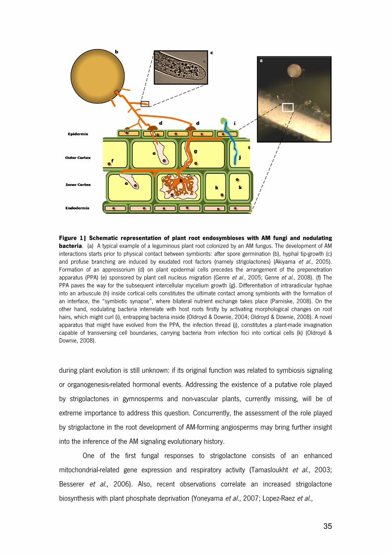
35
Figure 1| Schematic representation of plant root endosymbioses with AM fungi and nodulating bacteria. (a) A typical example of a leguminous plant root colonized by an AM fungus. The development of AM interactions starts prior to physical contact between symbionts: after spore germination (b), hyphal tip-growth (c) and profuse branching are induced by exudated root factors (namely strigolactones) (Akiyama et al., 2005). Formation of an appressorium (d) on plant epidermal cells precedes the arrangement of the prepenetration apparatus (PPA) (e) sponsored by plant cell nucleus migration (Genre et al., 2005; Genre et al., 2008). (f) The PPA paves the way for the subsequent intercellular mycelium growth (g). Differentiation of intraradicular hyphae into an arbuscule (h) inside cortical cells constitutes the ultimate contact among symbionts with the formation of an interface, the “symbiotic synapse”, where bilateral nutrient exchange takes place (Parniske, 2008). On the other hand, nodulating bacteria interrelate with host roots firstly by activating morphological changes on root hairs, which might curl (i), entrapping bacteria inside (Oldroyd & Downie, 2004; Oldroyd & Downie, 2008). A novel apparatus that might have evolved from the PPA, the infection thread (j), constitutes a plant-made invagination capable of transversing cell boundaries, carrying bacteria from infection foci into cortical cells (k) (Oldroyd & Downie, 2008).
during plant evolution is still unknown: if its original function was related to symbiosis signaling
or organogenesis-related hormonal events. Addressing the existence of a putative role played
by strigolactones in gymnosperms and non-vascular plants, currently missing, will be of
extreme importance to address this question. Concurrently, the assessment of the role played
by strigolactone in the root development of AM-forming angiosperms may bring further insight
into the inference of the AM signaling evolutionary history.
One of the first fungal responses to strigolactone consists of an enhanced
mitochondrial-related gene expression and respiratory activity (Tamasloukht et al., 2003;
Besserer et al., 2006). Also, recent observations correlate an increased strigolactone
biosynthesis with plant phosphate deprivation (Yoneyama et al., 2007; Lopez-Raez et al.,

36
BOX1: MODULARITY AND HOMOLOGY: TOOLS FOR UNDERSTANDING DEVELOPMENT
AND EVOLUTION
In a recent review on the developmental genetics of homologous structures, Wagner (2007)
elaborates on the conceptual challenge of ascribing a common ground, at the genetic level,
to characters known to be homologous. Such challenge stems from the realization that
homologous genes do not necessarily encode for homologous structures, the same holding
true for the reverse proposition. Epitomizing the elusiveness of the homology concept is the
extreme example of the ey/pax6 genes, in which homologous genes were “independently
recruited for superficially similar roles” (Abouheif et al., 1997) in divergent phyletic lines,
taking part in the development of the insect/vertebrate eyes, known to be independently
derived structures that also share a dissimilar developmental genetic network (Abouheif et
al., 1997; Wagner, 2007). To overcome this conceptual hurdle, and elaborating on the
concept of gene regulatory networks (GRNs), Wagner proposes that the genetic
correspondence of the homology observed between two characters is to be ascertained
through their “character-identity genetic networks”, or ChINs. These are to be differentiated
from the character-state genetic networks that encode for the often divergent
shapes/sizes/colours (states) of a homologous character and are downstream of and
activated by ChINs. This conceptualization predicts identical ChIN modules underlying
homologous structures and, reversely, non-homologous characters diverging in their ChINs
(Wagner, 2007). GRNs are modular, quasi-independent sets of genetic elements highly
correlated in phenotype and seemingly unaffected by variation in elements of other
modules (Davidson, 2001; Schlosser & Wagner, 2004). Genetic networks with a modular
architecture permit that a mutation affecting one module will have a small-scale effect in
other GRNs, permitting selective pressures to act on each individually. As ontogeny is
characterized by a succession of GRNs in time and space, the aforementioned dissociative
property implies that a change in the stereotypical order of events is not only easier in
mutational terms, because it is facilitated by the reduced number of genes controlling
module succession (inter-modular edges and respective nodes), but also carries with it less
probabilities of whole-system breakdown. As such, for their particular ability to generate
diversity, module connections are prime-targets for adaptive evolution (Davidson, 2001).

37
2008), in accordance with the crucial role of arbuscular mycorrhizal fungi in phosphate
acquisition and delivery.
Fungal hyphae in turn produce unidentified “Myc-factors”, which lead to the induction
of cytoplasmic calcium oscillations in host root epidermal cells (Kosuta et al., 2008) as well as
to the transcriptional induction of symbiosis-related genes in the host root (Kosuta et al.,
2003). On a recent work that dealt with root exudate-induced gene expression in
Glomeromycota, Tamasloukht and colleagues observed the up-regulation of 40 genes in
Gigaspora rosea (Tamasloukht et al., 2003). Twenty of these genes were found to have known
functional homologues in other organisms, and were categorized in four classes: mitochondrial
enzymes, DNA-synthesis, signal transduction and transcriptional regulation. Significantly, the
later class includes the majority of the genes described. This is consistent with the gene
expression profile shifts (activation of downstream gene regulatory networks - GRNs) that are
expected with the progression of ontogenetic modules. The detected gene regulatory apparatus
is expected to be involved in this transition from asymbiotic “flickering” to pre-symbiotic hyphal
branching; as such, these gene products connect ontogenetic modules, thus constituting
primary targets for adaptive evolution (Davidson, 2001). The description of this proposed GRN
component constitutes a primary research target for the understanding of adaptive evolution in
fungal development.
Indeed, information regarding this partner recognition module is scarce. One approach
to filling this gap would consist of ascribing functional roles to the 18 yet-unknown described
strigolactone responsive genes(Tamasloukht et al., 2003). This description should further
clarify the architecture of the putative ancestral module and/or reveal which specific additions
of new genetic elements to the network contributed to its novel symbiosis-establishment role.
Strigolactone recognition across Glomeromycota should be also assessed for further
evolutionary considerations, and also in plant-pathogenic fungi. If a conserved genetic
apparatus is confirmed, this module would become a focal point in the quest for basal
symbiotic mechanisms.
2.5 APPRESSORIA FORMATION
After plant recognition and subsequent hyphal branching of the AMF, physical contact
is established between the two symbionts (Figure 1d). Evidence shows that at this stage, AM
fungi recognize plant thigmotropic signals (Giovannetti, 1993) and undergo an abrupt change
in their ontogeny, developing a differentiated hyphal structure called appressorium (Harrison,

38
2005). Furthermore, this developmental module is shared with a number of known parasitic
fungi (Bechinger et al., 1999; Parniske, 2000), which is of relevance, as it may suggest the
appearance of this feature before AM symbiosis. On the other hand, appressoria formation
may have evolved more than once, representing convergent adaptations to the need of
crossing plant outer tissues in different fungal lineages. Further genetic data is necessary to
determine the evolutionary process resulting in this similarity.
Recent gene expression studies concerning appressoria formation in Glomus mosseae
show an increased expression of a H+-ATPase gene (Requena et al., 2003) and several
homologs of calcium-signaling related genes (Breuninger & Requena, 2004), indicating a
significant role of calcium signaling in fungal response to plant stimuli. Interestingly, 63% of the
clones identified in the latter expression study have no known homologues, “and may
represent putative novel genes specific for mycorrhizal symbiosis” (reviewed in Balestrini &
Lanfranco, 2006)). In an analogous manner to the 18 genes described by Tamasloukht and
colleagues (2003) for the pre-symbiotic phase, this result may reflect the current lack of
knowledge about AMF genetics and genomes.
2.6 PLANTS DETERMINE THE SHAPE OF HYPHAE TO COME
The development of appressoria, the ontogenic module preceding fungal entry into the
root, elicits dramatic rearrangements of epidermal organization(Genre & Bonfante, 2007). In
fact, recent ground-breaking studies (Genre et al., 2005; Genre et al., 2008) have shown that
Medicago truncatula and Daucus carota host cells set up a novel subcellular milieu, guiding
AMF hyphae towards the cortex. Following adhesion of the microorganism to the host, a
repositioning of the nucleus has been observed, which first moves towards the future fungal
entry point (Genre et al., 2005; Parniske, 2008). The cytoskeleton is reorganized and, together
with intense organelle movement across the cell, the formation of a transient tubular structure
enriched in endoplasmic reticulum cisternae takes place (Genre & Bonfante, 2007). The
resulting trans-cellular path, the prepenetration apparatus (PPA) (Genre et al., 2005),
constitutes an apoplastic column where the fungus is hosted after penetration (Genre &
Bonfante, 2007).
It has been proposed that plants co-opted cellular response mechanisms to
penetration of AMF from pathogenic fungi invasion-response strategies, or vice-versa, as a

39
number of elements appear to be common to both interactions (O'Connell & Panstruga, 2006;
Parniske, 2008; Genre et al., 2009). For instance, thigmo-stimulation per se can elicit nuclear
repositioning in epidermal cells (Gus-Mayer et al., 1998; Genre et al., 2009), while cytoplasmic
aggregation and nuclear movement are triggered after compatible pathogenic or mutualistic
fungi interaction (Genre et al., 2009). PPA formation, however, appears to be strictly correlated
with AMF adhesion (Genre et al., 2009), revealing a putative specific host developmental
module responsible for the intracellular accommodation of mutualistic fungi.
2.7 ESTABLISHING A “SYMBIOTIC SYNAPSE”
After passing through the outer cortex, supporting hyphae transverse inner cortical
cells, inside which arbuscules are formed (Harrison, 2005). Similar to the events triggered on
epidermal cells prior to fungal invasion, the proximity of hyphae elicit cell organization
rearrangements in the inner cortex, including vacuole disruption and the assembly of PPA-like
structures (Reinhardt, 2007; Genre et al., 2008), predicting which cells will be colonized.
Arbuscules are nutrient exchange structures. They are excluded from the plant cell cytoplasm,
as they are enveloped by a host cell-derived membrane (the periarbuscular membrane - PAM),
formed by the invagination of the host plasmalemma through delivery of membrane precursors
(Harrison, 2005; Parniske, 2008) (Figure 1h). It is at this stage of development that the
establishment of a functional “symbiotic synapse” (Figure 2) occurs, being formed by the
fungal plasma membrane, the PAM and the space in-between the two (Parniske, 2008). This
cortical cell-arbuscule synapse resembles, both in structure and function, the interface formed
in legume-rhizobia (Harrison, 2005) complexes and, although only structurally, the pathogenic
haustoria-plant interactions (Parniske, 2000). Interestingly, this synapse also shares similarities
with the interface formed by the only known non-AM-forming genus of Glomeromycota
(Geosiphon) and cyanobacteria (Box 2) (Schüssler, 2005). The fact that Geosiphon is derived
from the AM-forming ancestral to all the Glomeromycota adds further interest to the study of
the genetic basis behind the related diversity events, namely the hability to establish
mutualistic interactions with cyanobacteria.

40
BOX 2. Geosiphon pyriformis, a unique glomeromycotan Geosiphon pyriformis is a remarkable living system. Like all other known fungi of the phylum
Glomeromycota, its asexual, it forms coenocytic hyphae and obtains at least part of the nutrients
from an autotrophic symbiotic partner. What makes this species unique is that, unlike all the other
known Glomeromycota, it does not enter into AM symbioses, associating instead with
photoautotrophic and diazotrophic cyanobacteria that lie inside its own cells (Schüssler et al., 2001;
Schüssler, 2005). Geosiphon is able to obtain carbon from Nostoc punctiforme, a cyanobacteria that
can also be found as a free-living organism in freshwater. After being invaginated, cyanobacteria
become part of the symbiosome (the fungal intracellular compartment), along with a surrounding
fungal membrane, thus turning into an organelle-like structure within the Geosiphon’s cytoplasm.
The cyanobacteria grow in size and heterocysts are formed, where nitrogenase is protected from
oxygen, allowing for N2 fixation, which is provided along with carbohydrates to the fungus (Kluge,
1992). But nutrients flow both ways in the “symbiotic synapse”: Geosiphon provides phosphate to
its endosymbionts (Schüssler et al., 2006) (Figure 2). Although the Geosiphon-Nostoc (G-N) and AM
associations are fundamentally different in many aspects, the similarities between these symbiosis
are noteworthy: both involve a glomeromycotan fungus that provides phosphate to its phototrophic
symbiont, which in turn delivers carbohydrates to its partner (Schüssler et al., 2006). At the micro-
anatomical level, the resemblance is even greater: both symbioses involve a structurally similar
symbiotic synapse, where metabolite exchange takes place (Figure 2) (Schüssler, 2002; Schüssler
et al., 2006). As for the mapping of ancestrality partnerships onto the glomeromycotan phylogenetic
tree (Figure 3), the symbiosis with an autotrophic prokaryote is apparently a derived feature:
Geosiphon pyriformis is an exception with respect to the choice of symbiotic partner. The most
parsimonious explanation is that the ancestor to all Glomeromycota would be an intimate plant
mutualist fungus (Schüssler, 2002). Recent molecular data support this hypothesis, in agreement
with the whole fungal kingdom phylogenetic tree, thus suggesting that AM endosymbiosis represents
a derived feature, acquired solely by the glomeromycotan phyletic line (James et al., 2006).
Homology and modularity in AM/G-N symbioses
In light of the supracited phylogenetic, morphological and functional reasons, the AM and G-N
symbiotic synapses appear to be homologous. The recognition of this homology entails the
prediction of shared evolutionary mechanisms shaping symbiosis between fungi and
plants/cyanobacteria. Describing the shared components, topology and interactions of and between

41
BOX 2 (cont.) modules can elucidate the characteristics of the most-recent common ancestor of
the Glomeromycota, as much as delineate the basic characteristics of mycorrhizal symbiosis in
extant species. More specifically, we can generate hypotheses regarding the major genetic steps
allowing for the appearance of intracellular plant-fungal symbiosis. The genetics of symbiosis
synapse establishment and maintenance are, as with the rest of AMF genetics, relatively obscure.
The pervasive problem for evolutionary considerations on this phylum is that there is no study
comparing gene expression patterns in the two types of glomeromycotan mycobionts. Gene-
regulatory protein dynamics throughout fungal development should therefore constitute a major
topic of future research.
2.8 SIGNALING IN ROOT ENDOSYMBIOSES: COMMON GENES, DIFFERENT OUTPUTS
AM symbiosis is widespread among land plants and examples are found in
representatives of all major lineages. Nevertheless, these associations are promiscuous in that
no specific pairwise association between plant and AM fungi lineages has been described so
far (Figure 3). More recently in evolution, a group of plants restricted to four orders of the
Eurosid clade I of dicots, comprising legumes (Fabales) and a few non-legumes, engaged in
symbiotic relationships with dinitrogen reducing bacteria(Soltis et al., 1995) (Figure 3). These
species can consequently bypass the common nitrogen availability limitation in natural
habitats, hosting bacteria inside specialized root organs, termed nodules, within which bacteria
are provided with a carefully regulated oxygen and carbon supply, allowing for efficient nitrogen
reduction to ammonia(Oldroyd & Downie, 2008). Legume roots interrelate with Gram-negative
Rhizobium bacteria, whereas non-legume plants interact with the Gram-positive filamentous
Frankia actinobacteria (Pawlowski & Bisseling, 1996) (Figure 3).
2.8.1 Nodulation at a glance
Rhizobia produce lipochito-oligosaccharides in response to plant-derived flavonoids
signals which are recognized by NodD proteins. This recognition activates subsequent
transcriptional reprogramming leading to activation of nodulation (nod) genes. Some of the
resulting products of nod genes expression are called Nod Factors (NF) and represent the
signal that trigger the inception of the nodulation process (Riely et al., 2004; Oldroyd &
Downie, 2008). NF act as morphogens, causing resumption of cell division in terminally

42
Figure 2| Schematic view of the “symbiotic synapse” in (a) AM and (b) Geosiphon pyriformis/Nostoc punctiforme symbioses. Along the symbiotic synapse nutrients are bilaterally exchanged, most notably plant/cyanobacteria-derived photoassimilates and phosphate scavenged by fungi. Both fungal and plant plasma membrane H+-ATPases extrude protons, creating an electrochemical gradient across the membranes, fueling the secondary active transport of inorganic phosphate (Pi), Sucrose, Glucose (Glc), Fructose (Fru) and further nutrients via membrane transporters (Schüssler et al., 2006). Photosynthesized sucrose is hydrolyzed (probably by invertases (Schaarschmidt et al., 2006)) and sugars are transported to fungal cells through monosaccharide transporters (MST). Also, nitrogen flows are opposite in these symbioses: they are directed from fungus to photobiont in AM (Govindarajulu et al., 2005), while from photobiont to fungus in Geosiphon-Nostoc association.PT – phosphate transporter, PoliPase – poliphosphatase.
differentiated root cells (Yang et al., 1994) and resulting in enlargement of root tissue into a
nodule.
NF are thought to be perceived by the plant host probably by receptor-like kinases with
N-acetylglucosamine–binding lysin motifs. In fact, two LysM domain receptor-like kinases,
NFR1 and NFR5 from L. japonicus, have been implicated in Nod factor recognition (Madsen et
al., 2003; Radutoiu et al., 2003). The infection process starts when bacteria attach to root-hair

43
cells and induce their deformation, eventually getting entrapped within a curl. Concomitantly,
mitosis is triggered in cortical cells below the epidermal infection foci, giving origin to a nodule
meristem. Through a process mediated by actin cytoskeleton rearrangements(Yokota et al.,
2009), cylinder-like structures formed by invagination of the host plasma membrane and cell
wall deposition, termed infection threads, elongate from the middle of the curl and transverse
cells into the inner cortex (Gage et al., 1996; Oldroyd & Downie, 2008) (Figure 1i). The
mechanism through which plant cells establish infection threads is thought to have evolved
from the pre-penetration apparatus (PPA) ontogenetical programme (Parniske, 2008), an
interesting hypothesis still to be evaluated.
Once Rhizobia get inside specialized cells in the infection zone of the young nodule,
close to the nodule meristem, they enlarge and differentiate into nitrogen fixing forms, called
bacteroids (Gualtieri & Bisseling, 2000). These differentiated forms of bacteria residing inside
the nodule, surrounded by a peribacteroid membrane are best known as symbiosomes
(Oldroyd & Downie, 2004; Jones et al., 2007).
2.9 LINKING AM TO NODULATION
The restriction of root nodulation endosymbioses (RNE) to a monophyletic group of
four angiosperm orders, suggests that this phenomenon may constitute an evolutionary novelty
at the base of this clade (Gherbi et al., 2008; Markmann et al., 2008). Phenotypic observations
of legume mutants, more specifically the model plants Medicago truncatula and Lotus
japonicus (Harrison, 2005), shed light into a fundamental link between RNE and AM
symbioses. Mutant plants impaired in RNE symbiosis accomplishment are also unable to
associate with Glomeromycota. This suggests a bond between plant-phosphate scavenging
fungi and plant-nitrogen fixing bacteria symbioses genetic programmes (Kistner et al., 2005).
The link of plant–fungal and plant–bacterial endosymbioses in legumes, involving at least
seven genes (Kistner et al., 2005), best known as “common symbiosis genes” (Kistner &
Parniske, 2002), inspired the idea that during the evolution of bacterial endosymbiosis, some
genes were co-opted from the pre-existing AM genetic program (Parniske, 2004). This group of
genes includes a leucine-rich-repeat receptor kinase (Endre et al., 2002; Stracke et al., 2002)

44
Figure 3| Inter Kingdoms symbiotic species relationships. Mycorrhizal fungi belonging to the phylum Glomeromycota (+- 200 species) can establish endosymbiotic relationships with the vast majority of plants roots (around 80%). As depicted in the cladograms, there is no exclusiveness of relations between groups of fungi and plants, as it is possible to find the same fungal species associated with a large number of plant species. (*) In some phylogenetic groups of plants one can find disperse families with no described link with AM fungi (e.g. Brassicaceae, Caryophyllaceae, Amaranthaceae or Polygonaceae). These groups present a great surplus value for the comparative study of symbiotic characteristics. Additionally, numerous bacterial groups are involved in this complex interactive system, like plant-interacting nodulating bacteria (green circles), bacteria associated with Glomeromycota (orange circles) and autotrophic bacteria with Geosiphon (blue circles): (a) Frankia and Rhizobia are the most commonly found in association with families included in the Eurosids I clade; (b) Burkholderia bacteria are found connected with Rubiaceae (Euasterids I), a quite well described symbiosis. Bacterial interactions with AM fungi where found to occur, namely within the (c) Glomus, (d) Gigaspora (e) and Geosiphon genera, the latter being established by the only known non-AM-forming species of the Glomeromycota and a cyanobacteria (Geosiphon piriformis/ Nostoc punctiforme). The cladogram representing plants was prepared according to the APGII system (2003) and Glomeromycota phylogeny was consulted in Schwarzott et al., 2001). Nodulating bacteria and fungi-interacting bacteria species where consulted in Gualtieri & Bisseling, 2000 and Frey-Klett et al., 2007, respectively.
(Lotus japonicus SyMRK/M. truncatula DMI2/M. sativa NORK), two channels permeable to
cations (Charpentier et al., 2008), (LjCASTOR and LjPOLLUX/ MtDMI1), two putative
nucleoporines (LjNUP85 and LjNUP133) (Kanamori et al., 2006; Saito et al., 2007), a calcium
and calmodulin-dependent protein kinase (Levy et al., 2004; Mitra et al., 2004; Gleason et al.,
2006) (LjCCaMK/ MtDMI3) and LjCYCLOPS, encoding a protein that physically interacts in
vitro and colocalizes within the nucleus with CcaMK (Yano et al., 2008).

45
More particularly, the M. truncatula DMI (DOESN´T MAKE INFECTIONS) proteins play
pivotal roles in both early nodulation and mycorrhization signaling episodes and will serve as
proof-of-concept for our approach.
2.10 HOW BACTERIA GOT EN ROUTE TO THE ROOT
Plants express and make use of leucine-rich-repeat receptor-like kinases (LRR-RLKs)
for a variety of signal-transduction pathways. These receptors are structurally related to Toll
receptors identified in Drosophila melanogaster and Toll-like receptors present in mammal
cells, a family of important players in immunity (Leulier & Lemaitre, 2008). Leucine-rich
repeats are usually involved in protein-protein interactions, which might be implicated in
binding of signal molecules, whereas the intracellular kinase domain acts in protein
phosphorylation. The M. truncatula DMI2/SYMRK (symbiosis receptor-kinase) can be included
in this class of proteins. SYMRK takes an active part in the signaling pathway leading to the
accommodation of both rhizobia (Endre et al., 2002; Stracke et al., 2002) and, as recently
shown, of Frankia bacteria (Gherbi et al., 2008; Markmann et al., 2008). It acts near a point of
convergence of AM and legume-rhizobia signaling pathways and exists in at least three different
structural versions within angiosperms, particularly regarding its extracellular portion and exon
number (Markmann et al., 2008). This polymorphic extracellular domain is thought to
recognize exogenous molecules related to symbiosis. Recently published data regarding the
evolution of intracellular root symbioses with bacteria (Gherbi et al., 2008; Markmann et al.,
2008), reflects the particular role of SYMRK in the evolution of nodulation in a modern group of
legumes and close relatives, which present the version of the molecule with higher length (for
details see Figure 1 of Markmann et al., 2008)). De Mita and colleagues pointed out that
adaptive changes reflecting positive selective constraints can be traced in LRR and extracellular
regions of SYMRK genes from different Medicago species, but these do not correlate with shifts
in rhizobial specificity (De Mita et al., 2007). The functional adaptation of this specific receptor-
kinase, correlating with a novel architecture acquisition, was possibly due to an increase in the
number of exons in ancestral legumes (Markmann et al., 2008).
The critical events that arose from the appearance of a novel type of receptor in the
nodulating clade are yet unanswered, specifically those regarding the rearrangement of genetic

46
networks and developmental pattern shifts that allowed for the intracellular lodging of
dinitrogen reducing bacteria. We propose that the GRN containing SYMRK/DMI2 constitutes a
ChIN, an ancient network that lies at the origin of symbiosis formation. This ChIN feeds onto
alternative modules for nodulation or mycorrhization leading to different character states
present in extant species that establish these types of endosymbioses. Furthermore, a
comparative approach should be directed at the SYMRK and downstream genes of non-
nodulating species proximately related to legumes, shedding light over the genetic architecture
of the proposed character states.
As is now evident, the establishment of symbiotic interactions involves specific
developmental adaptations in both symbiotic partners and intricate coordination of their
developmental programs. As gene networks provide a set of control systems for the course of
development, they can provide not only an insight into the inner-workings of the ontogenic
program but also establish the topology of genetic relationships shaped by selection (Davidson,
2001). This GRN topology can prove invaluable in the hierarchization of the successive steps
that evolution has taken in the construction of their resulting biological features.
The similarities between Glomeromycota- and rhizobia- elicited symbiotic genetic
programmes in legumes have allowed for the proposition of a ChIN, revealing the adequacy of
the proposed comparative approach in the dissection of the genetic mechanisms underlying
mycorrhizal endosymbioses. But bearing in mind the specificities of each symbiotic system,
how can a basal common signaling structure transduce different incoming signals (e.g. NF and
still unknown mycorrhization factors) into different developmental symbiosis modules?
2.11 CHOOSING BETWEEN STATES: CA2+ SETS THE FRONTIERS
In fact, some advances have been recently made on the mechanisms underlying the
choices between nodulation and mycorrhization through the study of the downstream events
triggered by DIM2/SYMRK activation. At the top of the nodulation´s signal perception
hierarchy are Nod-factor receptors, LysM type receptor kinases implicated in the binding of N-
acetyl-glucosamine-containing molecules (Madsen et al., 2003; Radutoiu et al., 2003). Upon

47
Nod-factor perception, a transient increase in root hair´s cytosolic Ca2+ concentration is
observed (Cardenas et al., 1999; Felle et al., 2000), followed by repetitive nuclear-associated
oscillations (Ca2+ spiking) (Ehrhardt et al., 1996; Oldroyd & Downie, 2006). The aforementioned
M. truncatula DMI1 and DMI2/SYMRK are both required for calcium spiking (Endre et al.,
2002; Ane et al., 2004). Downstream of calcium oscillatory phenomena, the CCaMK DMI3
putatively acts as a Ca2+ signal decoder, subsequently activating both AM and nodulation
symbiosis-specific gene transcription (Levy et al., 2004; Oldroyd & Downie, 2006).
The system through which CCaMK integrates ion concentration oscillations (at a
frequency, amplitude or spike number level) remains to be clarified, though it has been shown
that, specifically for nodulation symbiosis, DMI3-activated gene expression is regulated by Ca2+
spike number (Miwa et al., 2006). The chimeric CCaMK has EF-hand Ca2+-binding sites and an
autoinhibitory domain (Harper & Harmon, 2005). If the autoinhibitory sequence is absent from
the DMI3 structure, nodule morphogenesis can be triggered in the cortex of mutant plants
devoid of bacterial elicitation (Gleason et al., 2006). This indicates that downstream of the
calcium dynamical trajectory, the activation of this protein is sufficient to trigger the nodule
ontogenetical programme. In a very recent paper, Kosuta and co-workers provide evidence for
the occurrence of calcium oscillatory rhythms also involved in AM symbiosis, with a different
signature from nodulation-associated spiking (Kosuta et al., 2008). Also, by means of extensive
mathematical analyses, these authors have shown that both [Ca2+] trajectories are chaotic in
nature. An intrinsic property of the common symbiosis pathway (ChIN) must be the plasticity to
integrate signals from bacteria and AM fungi into precise outcomes (non-linear Ca2+ signatures)
that can lead to differential DMI3 perception and consequent activation of symbiosis-type
specific genetic networks (character states). The specific dynamic patterns of Ca2+ and other
ions have long been related to nodulation (Cardenas et al., 2000) and systematically observed
in endo and ectomycorrhizal associations (Ramos, et al., 2008a; Ramos, et al., 2008b; Ramos
et al., 2009), thus allowing for the hypothesis that specific spatial and temporal coreographies
of free ion concentration may play a role in the integration of these signals.

48
2.12 CONCLUDING REMARKS
The study of organismal ontogeny is becoming a major tool to understand evolutionary
events (Gould, 1977; Raff, 1996; Carroll et al., 2005; Muller, 2007). This research program
stems from the realisation that among the rules of transformation maping genotype to
phenotype in multicellular organisms, development stands as the generator of form, and thus a
central target for adaptive evolution (de Beer, 1958; Lewontin, 1974).
Arbuscular mycorrhizae stand as a particularly successful and important symbiotic
relationship, considering its ubiquity and putative importance in the colonization of land by
plants. The knowledge of the fungal partner’s ontogeny is still in its infancy, despite novel
molecular genetics approaches (e.g. AM fugal genome sequencing projects, comparative
transcriptomics). Such advances are expected to contribute to the disclosure of the poorly
understood events leading to this intracellular tête-à-tête, providing a fertile, yet eminently
descriptive dataset that calls for an analytical approach. For example, the release of the
genome sequences of Glomus intraradices in the near-future and the possibility of a sequenced
dikaryan outgroup can contribute significantly to generate handles into the description and
comparison of the transcription profiles throughout fungal ontogeny. In addition, the gene
expression in AMF and its dynamics throughout development are becoming clearer. For
instance, there is a growing body of knowledge on the mechanisms through which diffusible
molecules are perceived and decoded(Akiyama et al., 2005; Besserer et al., 2006), triggering
gene expression reprogramming and ontogenetical shifts which are vital for life-cycle
completion.
In fact, this specific developmental step illustrates how the modular nature of ontogeny
can be helpful in guiding research on this symbiotic system. More specifically, strigolactone
has been shown to act acts as the trigger for a central developmental transition in the
presymbiotic phase. This particular transition between two developmental modules (groups of
events with inner-cohesion in space and time) is, as other edges between modules, a major
target for adaptive change, for its potential to generate diversity with seemingly low pleiotropic
effects. As such, the extent of strigolactone-based communication across the Glomeromycota
has to be quickly accessed. The comparative study of another developmental module, the
intracellular “symbiotic synapse” (arbuscule/plant cell, siphonal bladder/cyanobacteria and

49
simbiosome/ plant cell interface in nodulation) can also be crucial for hypothesis-generation
regarding the adaptive steps (putatively gene network rearrangements) that have allowed for
symbiosis appearance and, in this case, diversification. As this intracellular contact between
symbionts is also shared with plant-pathogenic fungi (Parniske, 2000), the knowledge of the
degree and detail of homology between these groups of fungi can generate hypotheses
regarding the stepwise genetic changes leading to the emergence of this mutualistic
evolutionary novelty.
With respect to the AM plant partner, the aforementioned research program is already
proving to be invaluable. A comparative approach has shown that some plant mutants were
impaired in both RNE and AM formation, suggesting a common GRN referred to as “common
symbiosis genes”. Recent studies detailing the plant gene expression involved in root
nodulation endosymbioses show that it parallels that of AM to a considerable extent (Kistner et
al., 2005). The phylogenetic mapping of this character points to the AM symbiosis as an
ancestral condition, and as such, the derived character of nodulation is predicted to have
arisen by a co-option event of some of the ancestral GRNs. This shared genetic network
upstream of two dissociable states (RNE/AM) points to both homologous and modular
properties of the underlying GRNs. We argue that these should be, as in the approach to AMF
ontogeny, guiding standpoints to understand the plant genetic adaptations that enabled the
establishment of AM mutualism.
The case-study of the DMI genes, identified in Medicago truncatula, serve as a proof-of-
concept for our systematization. In this case, a RNE/AM-shared signal-integration cascade was
shown to lay upstream of specialization GRNs. The homology and modularity concepts can
thus enable the further exploration of the bordering nodes of this shared character-identity
network, as for clarifying the genetic architecture behind each character state.
Although some transcription profiles are becoming available for different life-stages
(Breuninger & Requena, 2004), the link between signal integration and shifts in gene
expression is poorly understood. We propose an emphasis on the description of gene-
expression regulation, particularly the mapping of transcription factor/effector gene cis-
regulatory interactions. This emphasis relies on the fact that the progression of ontogenetic
modules in both plant and fungal partners must rely on shifts in gene expression. These are,
as stated earlier, particularly evolvable regions of genetic networks and thus prime candidates
for the evolutionary bases of plant-fungal mutualism.

50
We propose that the comparative method and the concept of homology can provide
the first level of systematization by mapping onto the phylogenies of plants and fungi the
characters that make up the establishment of mycorrhizal endosymbioses. Furthermore, we
consider that looking at comparative gene expression data through the eye of modularity and
gene network concepts can provide the foundations for a research program aimed at
advancing our understanding of the evolution and development of symbiotic systems.
2.13 REFERENCES
2003. An update of the angiosperm phylogeny group classification for the orders and families of flowering plants: Apg ii. Botanical Journal of the Linnean Society 141: 399-436.
2005. The map-based sequence of the rice genome. Nature 436: 793-800.
Abouheif E, Akam M, Dickinson WJ, Holland PW, Meyer A, Patel NH, Raff RA, Roth VL, Wray GA. 1997. Homology and developmental genes. Trends Genet 13: 432-433.
Akiyama K, Matsuzaki K, Hayashi H. 2005. Plant sesquiterpenes induce hyphal branching in arbuscular mycorrhizal fungi. Nature 435: 824-827.
Ane JM, Kiss GB, Riely BK, Penmetsa RV, Oldroyd GE, Ayax C, Levy J, Debelle F, Baek JM, Kalo P, Rosenberg C, Roe BA, Long SR, Denarie J, Cook DR. 2004. Medicago truncatula dmi1 required for bacterial and fungal symbioses in legumes. Science 303: 1364-1367.
Balestrini R, Lanfranco L. 2006. Fungal and plant gene expression in arbuscular mycorrhizal symbiosis. Mycorrhiza 16: 509-524.
Bécard GP, P.E. 1993. Status of nuclear division in arbuscular mycorrhizal fungi during in vitro development Protoplasma 174: 62-68.
Bechinger C, Giebel KF, Schnell M, Leiderer P, Deising HB, Bastmeyer M. 1999. Optical measurements of invasive forces exerted by appressoria of a plant pathogenic fungus. Science 285: 1896-1899.
Besserer A, Puech-Pages V, Kiefer P, Gomez-Roldan V, Jauneau A, Roy S, Portais JC, Roux C, Becard G, Sejalon-Delmas N. 2006. Strigolactones stimulate arbuscular mycorrhizal fungi by activating mitochondria. PLoS Biol 4: e226.
Breuninger M, Requena N. 2004. Recognition events in am symbiosis: Analysis of fungal gene expression at the early appressorium stage. Fungal Genet Biol 41: 794-804.
Cardenas L, Feijo JA, Kunkel JG, Sanchez F, Holdaway-Clarke T, Hepler PK, Quinto C. 1999. Rhizobium nod factors induce increases in intracellular free calcium and extracellular calcium influxes in bean root hairs. Plant J 19: 347-352.
Cardenas L, Holdaway-Clarke TL, Sanchez F, Quinto C, Feijo JA, Kunkel JG, Hepler PK. 2000. Ion changes in legume root hairs responding to nod factors. Plant Physiol 123: 443-452.
Carroll SB, Grenier JK, Weatherbee SD. 2005. From DNA to diversity: Molecular genetics and the evolution of animal design. Malden, Mass.: Blackwell Scientific.

51
Charpentier M, Bredemeier R, Wanner G, Takeda N, Schleiff E, Parniske M. 2008. Lotus japonicus castor and pollux are ion channels essential for perinuclear calcium spiking in legume root endosymbiosis. Plant Cell 20: 3467-3479.
Cook CE, Whichard LP, Turner B, Wall ME, Egley GH. 1966. Germination of witchweed (striga lutea lour.): Isolation and properties of a potent stimulant. Science 154: 1189-1190.
Davidson EH. 2001. Genomic regulatory systems: Development and evolution. San Diego: Academic Press
de Beer G. 1958. Embryos and ancestors. Oxford: Oxford University Press.
De Mita S, Santoni S, Ronfort J, Bataillon T. 2007. Adaptive evolution of the symbiotic gene nork is not correlated with shifts of rhizobial specificity in the genus medicago. BMC Evol Biol 7: 210.
Ehrhardt DW, Wais R, Long SR. 1996. Calcium spiking in plant root hairs responding to rhizobium nodulation signals. Cell 85: 673-681.
Endre G, Kereszt A, Kevei Z, Mihacea S, Kalo P, Kiss GB. 2002. A receptor kinase gene regulating symbiotic nodule development. Nature 417: 962-966.
Felle HH, Kondorosi E, Kondorosi A, Schultze M. 2000. How alfalfa root hairs discriminate between nod factors and oligochitin elicitors. Plant Physiol 124: 1373-1380.
Frey-Klett P, Garbaye J, Tarkka M. 2007. The mycorrhiza helper bacteria revisited. New Phytol 176: 22-36.
Gage DJ, Bobo T, Long SR. 1996. Use of green fluorescent protein to visualize the early events of symbiosis between rhizobium meliloti and alfalfa (medicago sativa). J Bacteriol 178: 7159-7166.
Genre A, Bonfante P. 2007. Check-in procedures for plant cell entry by biotrophic microbes. Mol Plant Microbe Interact 20: 1023-1030.
Genre A, Chabaud M, Faccio A, Barker DG, Bonfante P. 2008. Prepenetration apparatus assembly precedes and predicts the colonization patterns of arbuscular mycorrhizal fungi within the root cortex of both medicago truncatula and daucus carota. Plant Cell 20: 1407-1420.
Genre A, Chabaud M, Timmers T, Bonfante P, Barker DG. 2005. Arbuscular mycorrhizal fungi elicit a novel intracellular apparatus in medicago truncatula root epidermal cells before infection. Plant Cell 17: 3489-3499.
Genre A, Ortu G, Bertoldo C, Martino E, Bonfante P. 2009. Biotic and abiotic stimulation of root epidermal cells reveals common and specific responses to arbuscular mycorrhizal fungi. Plant Physiol 149: 1424-1434.
Gherbi H, Markmann K, Svistoonoff S, Estevan J, Autran D, Giczey G, Auguy F, Peret B, Laplaze L, Franche C, Parniske M, Bogusz D. 2008. Symrk defines a common genetic basis for plant root endosymbioses with arbuscular mycorrhiza fungi, rhizobia, and frankiabacteria. Proc Natl Acad Sci U S A 105: 4928-4932.
Giovannetti MA, L. Sbrana, C. Citernesi, A.S. 1993. Factors affecting appressorium development in the vesicular-arbuscular mycorrhizal fungus glomus mosseae. New Phytologist 123: 115-122.
Gleason C, Chaudhuri S, Yang T, Munoz A, Poovaiah BW, Oldroyd GE. 2006. Nodulation independent of rhizobia induced by a calcium-activated kinase lacking autoinhibition. Nature 441: 1149-1152.
Gomez-Roldan V, Fermas S, Brewer PB, Puech-Pages V, Dun EA, Pillot JP, Letisse F, Matusova R, Danoun S, Portais JC, Bouwmeester H, Becard G, Beveridge CA, Rameau C, Rochange SF. 2008. Strigolactone inhibition of shoot branching. Nature 455: 189-194.
Gould SJ. 1977. Ontogeny and phylogeny. Cambridge, Massachusetts
London, England: Harvard University Press.

52
Govindarajulu M, Pfeffer PE, Jin H, Abubaker J, Douds DD, Allen JW, Bucking H, Lammers PJ, Shachar-Hill Y. 2005. Nitrogen transfer in the arbuscular mycorrhizal symbiosis. Nature 435: 819-823.
Gualtieri G, Bisseling T. 2000. The evolution of nodulation. Plant Mol Biol 42: 181-194.
Gus-Mayer S, Naton B, Hahlbrock K, Schmelzer E. 1998. Local mechanical stimulation induces components of the pathogen defense response in parsley. Proc Natl Acad Sci U S A 95: 8398-8403.
Harper JF, Harmon A. 2005. Plants, symbiosis and parasites: A calcium signalling connection. Nat Rev Mol Cell Biol 6: 555-566.
Harrison MJ. 2005. Signaling in the arbuscular mycorrhizal symbiosis. Annu Rev Microbiol 59: 19-42.
James TY, Kauff F, Schoch CL, Matheny PB, Hofstetter V, Cox CJ, Celio G, Gueidan C, Fraker E, Miadlikowska J, Lumbsch HT, Rauhut A, Reeb V, Arnold AE, Amtoft A, Stajich JE, Hosaka K, Sung GH, Johnson D, O'Rourke B, Crockett M, Binder M, Curtis JM, Slot JC, Wang Z, Wilson AW, Schüssler A, Longcore JE, O'Donnell K, Mozley-Standridge S, Porter D, Letcher PM, Powell MJ, Taylor JW, White MM, Griffith GW, Davies DR, Humber RA, Morton JB, Sugiyama J, Rossman AY, Rogers JD, Pfister DH, Hewitt D, Hansen K, Hambleton S, Shoemaker RA, Kohlmeyer J, Volkmann-Kohlmeyer B, Spotts RA, Serdani M, Crous PW, Hughes KW, Matsuura K, Langer E, Langer G, Untereiner WA, Lucking R, Budel B, Geiser DM, Aptroot A, Diederich P, Schmitt I, Schultz M, Yahr R, Hibbett DS, Lutzoni F, McLaughlin DJ, Spatafora JW, Vilgalys R. 2006. Reconstructing the early evolution of fungi using a six-gene phylogeny. Nature 443: 818-822.
Jones KM, Kobayashi H, Davies BW, Taga ME, Walker GC. 2007. How rhizobial symbionts invade plants: The sinorhizobium-medicago model. Nat Rev Microbiol 5: 619-633.
Kanamori N, Madsen LH, Radutoiu S, Frantescu M, Quistgaard EM, Miwa H, Downie JA, James EK, Felle HH, Haaning LL, Jensen TH, Sato S, Nakamura Y, Tabata S, Sandal N, Stougaard J. 2006. A nucleoporin is required for induction of ca2+ spiking in legume nodule development and essential for rhizobial and fungal symbiosis. Proc Natl Acad Sci U S A 103: 359-364.
Kistner C, Parniske M. 2002. Evolution of signal transduction in intracellular symbiosis. Trends Plant Sci 7: 511-518.
Kistner C, Winzer T, Pitzschke A, Mulder L, Sato S, Kaneko T, Tabata S, Sandal N, Stougaard J, Webb KJ, Szczyglowski K, Parniske M. 2005. Seven lotus japonicus genes required for transcriptional reprogramming of the root during fungal and bacterial symbiosis. Plant Cell 17: 2217-2229.
Kluge MM, D. Mollenhauer, R. Kape, R. 1992. Geosiphon pyriforme, an endosymbiotic consortium of a fungus and a cyanobacterium (nostoc), fixes nitrogen. Botanica Acta 105: 343-344.
Koske RE. 1981. Gigaspora gigantea: Observations on spore germination of a va-mycorrhizal fungus. Mycologia 73: 288-300.
Kosuta S, Chabaud M, Lougnon G, Gough C, Denarie J, Barker DG, Becard G. 2003. A diffusible factor from arbuscular mycorrhizal fungi induces symbiosis-specific mtenod11 expression in roots of medicago truncatula. Plant Physiol 131: 952-962.
Kosuta S, Hazledine S, Sun J, Miwa H, Morris RJ, Downie JA, Oldroyd GE. 2008. Differential and chaotic calcium signatures in the symbiosis signaling pathway of legumes. Proc Natl Acad Sci U S A 105: 9823-9828.
Kutschera U, Niklas KJ. 2005. Endosymbiosis, cell evolution, and speciation. Theory Biosci 124: 1-24.
Leulier F, Lemaitre B. 2008. Toll-like receptors--taking an evolutionary approach. Nat Rev Genet 9: 165-178.
Levy J, Bres C, Geurts R, Chalhoub B, Kulikova O, Duc G, Journet EP, Ane JM, Lauber E, Bisseling T, Denarie J, Rosenberg C, Debelle F. 2004. A putative ca2+ and calmodulin-dependent protein kinase required for bacterial and fungal symbioses. Science 303: 1361-1364.

53
Lewontin RC. 1974. The genetic basis of evolutionary change. New York & London: Columbia University Press.
Lopez-Raez JA, Charnikhova T, Gomez-Roldan V, Matusova R, Kohlen W, De Vos R, Verstappen F, Puech-Pages V, Becard G, Mulder P, Bouwmeester H. 2008. Tomato strigolactones are derived from carotenoids and their biosynthesis is promoted by phosphate starvation. New Phytol 178: 863-874.
Madsen EB, Madsen LH, Radutoiu S, Olbryt M, Rakwalska M, Szczyglowski K, Sato S, Kaneko T, Tabata S, Sandal N, Stougaard J. 2003. A receptor kinase gene of the lysm type is involved in legume perception of rhizobial signals. Nature 425: 637-640.
Markmann K, Giczey G, Parniske M. 2008. Functional adaptation of a plant receptor-kinase paved the way for the evolution of intracellular root symbioses with bacteria. PLoS Biol 6: e68.
Mereschkowsky C. 1905. Über natur und ursprung der chromatophoren im pflanzenreiche. Biol Centralbl 25: 593-604.
Mitra RM, Gleason CA, Edwards A, Hadfield J, Downie JA, Oldroyd GE, Long SR. 2004. A ca2+/calmodulin-dependent protein kinase required for symbiotic nodule development: Gene identification by transcript-based cloning. Proc Natl Acad Sci U S A 101: 4701-4705.
Miwa H, Sun J, Oldroyd GE, Downie JA. 2006. Analysis of calcium spiking using a cameleon calcium sensor reveals that nodulation gene expression is regulated by calcium spike number and the developmental status of the cell. Plant J 48: 883-894.
Moran NA. 2002. Microbial minimalism: Genome reduction in bacterial pathogens. Cell 108: 583-586.
Muller GB. 2007. Evo-devo: Extending the evolutionary synthesis. Nat Rev Genet 8: 943-949.
O'Connell RJ, Panstruga R. 2006. Tete a tete inside a plant cell: Establishing compatibility between plants and biotrophic fungi and oomycetes. New Phytol 171: 699-718.
Oldroyd GE, Downie JA. 2004. Calcium, kinases and nodulation signalling in legumes. Nat Rev Mol Cell Biol 5: 566-576.
Oldroyd GE, Downie JA. 2006. Nuclear calcium changes at the core of symbiosis signalling. Curr Opin Plant Biol 9: 351-357.
Oldroyd GE, Downie JA. 2008. Coordinating nodule morphogenesis with rhizobial infection in legumes. Annu Rev Plant Biol 59: 519-546.
Parniske M. 2000. Intracellular accommodation of microbes by plants: A common developmental program for symbiosis and disease? Curr Opin Plant Biol 3: 320-328.
Parniske M. 2004. Molecular genetics of the arbuscular mycorrhizal symbiosis. Curr Opin Plant Biol 7: 414-421.
Parniske M. 2008. Arbuscular mycorrhiza: The mother of plant root endosymbioses. Nat Rev Microbiol 6: 763-775.
Paszkowski U, Jakovleva L, Boller T. 2006. Maize mutants affected at distinct stages of the arbuscular mycorrhizal symbiosis. Plant J 47: 165-173.
Pawlowski K, Bisseling T. 1996. Rhizobial and actinorhizal symbioses: What are the shared features? Plant Cell 8: 1899-1913.
Radutoiu S, Madsen LH, Madsen EB, Felle HH, Umehara Y, Gronlund M, Sato S, Nakamura Y, Tabata S, Sandal N, Stougaard J. 2003. Plant recognition of symbiotic bacteria requires two lysm receptor-like kinases. Nature 425: 585-592.
Raff RA. 1996. The shape of life: Genes, development, and the evolution of animal form. Chicago: University Of Chicago Press.

54
Ramos AC, Facanha AR, Feijo JA. 2008. Proton (h+) flux signature for the presymbiotic development of the arbuscular mycorrhizal fungi. New Phytologist 178: 177-188.
Ramos AC, Façanha AR, Feijó JA 2008. Ion dynamics during the polarized growth of arbuscular mycorrhizal fungi: From presymbiosis to symbiosis. In: A. H. Varma, B. ed. Mycorrhiza: Biology, genetics, novel endophytes and biotechnology: Springer-Verlag Germany, 241-261.
Ramos AC, Lima PT, Dias PN, Kasuya MC, Feijo JA. 2009. A ph signaling mechanism involved in the spatial distribution of calcium and anion fluxes in ectomycorrhizal roots. New Phytol 181: 448-462.
Reddy DMRS, Schorderet M, Feller U, Reinhardt D. 2007. A petunia mutant affected in intracellular accommodation and morphogenesis of arbuscular mycorrhizal fungi. Plant J 51: 739-750.
Reinhardt D. 2007. Programming good relations--development of the arbuscular mycorrhizal symbiosis. Curr Opin Plant Biol 10: 98-105.
Remy W, Taylor TN, Hass H, Kerp H. 1994. Four hundred-million-year-old vesicular arbuscular mycorrhizae. Proc Natl Acad Sci U S A 91: 11841-11843.
Requena N, Breuninger M, Franken P, Ocon A. 2003. Symbiotic status, phosphate, and sucrose regulate the expression of two plasma membrane h+-atpase genes from the mycorrhizal fungus glomus mosseae. Plant Physiol 132: 1540-1549.
Requena N, Serrano E, Ocon A, Breuninger M. 2007. Plant signals and fungal perception during arbuscular mycorrhiza establishment. Phytochemistry 68: 33-40.
Riely BK, Ane JM, Penmetsa RV, Cook DR. 2004. Genetic and genomic analysis in model legumes bring nod-factor signaling to center stage. Curr Opin Plant Biol 7: 408-413.
Saito K, Yoshikawa M, Yano K, Miwa H, Uchida H, Asamizu E, Sato S, Tabata S, Imaizumi-Anraku H, Umehara Y, Kouchi H, Murooka Y, Szczyglowski K, Downie JA, Parniske M, Hayashi M, Kawaguchi M. 2007. Nucleoporin85 is required for calcium spiking, fungal and bacterial symbioses, and seed production in lotus japonicus. Plant Cell 19: 610-624.
Schaarschmidt S, Roitsch T, Hause B. 2006. Arbuscular mycorrhiza induces gene expression of the apoplastic invertase lin6 in tomato (lycopersicon esculentum) roots. J Exp Bot 57: 4015-4023.
Schlosser G, Wagner GP 2004. The modularity concept in developmental and evolutionary biology. In: G. SchlosserG. P. Wagner eds. Modularity in development and evolution. Chicago, USA: The University of Chicago Press, 1-11.
Schüssler A. 2002. Molecular phylogeny, taxonomy, and evolution of geosiphon pyriformis and arbuscular mycorrhizal fungi. Plant and Soil 244.
Schüssler A, Martin H, Cohen D, Fitz M, Wipf D. 2006. Characterization of a carbohydrate transporter from symbiotic glomeromycotan fungi. Nature 444: 933-936.
Schüssler AK, M. 2001. Geosiphon pyriforme, an endocytosymbiosis between fungus and cyanobacteria, and its meaning as a model system for arbuscular mycorrhizal research. In: B. Hock ed. The mycota ix. Berlin Heidelberg New York: Springer Verlag, 151-161.
Schüssler AS, D. Walker, C. . 2001. A new fungal phylum, the glomeromycota: Phylogeny and evolution. Mycological Research 105: 1413-1421.
Schüssler AW, E. 2005. Geosiphon pyriformis - a glomeromycotan soil fungus forming endosymbiosis with cyanobacteria. In: S. S. Declerck, D-G. Fortin, J.A. ed. In vitro culture of mycorrhizas: Springer-Verlag Berlin Heidelberg, 271-289.
Schwarzott D, Walker C, Schüssler A. 2001. Glomus, the largest genus of the arbuscular mycorrhizal fungi (glomales), is nonmonophyletic. Mol Phylogenet Evol 21: 190-197.
55
Simon L, Bousquet J, Levesque RC, Lalonde M. 1993. Origin and diversification of endomycorrhizal fungi and coincidence with vascular land plants. Nature 363: 67-69.
Smith SE, Read DJ. 2008. Mycorrhizal symbiosis: Academic Press.
Soltis DE, Soltis PS, Morgan DR, Swensen SM, Mullin BC, Dowd JM, Martin PG. 1995. Chloroplast gene sequence data suggest a single origin of the predisposition for symbiotic nitrogen fixation in angiosperms. Proc Natl Acad Sci U S A 92: 2647-2651.
Stracke S, Kistner C, Yoshida S, Mulder L, Sato S, Kaneko T, Tabata S, Sandal N, Stougaard J, Szczyglowski K, Parniske M. 2002. A plant receptor-like kinase required for both bacterial and fungal symbiosis. Nature 417: 959-962.
Tamasloukht M, Sejalon-Delmas N, Kluever A, Jauneau A, Roux C, Becard G, Franken P. 2003. Root factors induce mitochondrial-related gene expression and fungal respiration during the developmental switch from asymbiosis to presymbiosis in the arbuscular mycorrhizal fungus gigaspora rosea. Plant Physiol 131: 1468-1478.
Taylor TNR, W. Hass, H. Kerp, H. 1995. Fossil arbuscular-mycorrhiza from the early devonian. Mycologia 87: 560-573.
Umehara M, Hanada A, Yoshida S, Akiyama K, Arite T, Takeda-Kamiya N, Magome H, Kamiya Y, Shirasu K, Yoneyama K, Kyozuka J, Yamaguchi S. 2008. Inhibition of shoot branching by new terpenoid plant hormones. Nature 455: 195-200.
Wagner GP. 2007. The developmental genetics of homology. Nat Rev Genet 8: 473-479.
Yang WC, de Blank C, Meskiene I, Hirt H, Bakker J, van Kammen A, Franssen H, Bisseling T. 1994. Rhizobium nod factors reactivate the cell cycle during infection and nodule primordium formation, but the cycle is only completed in primordium formation. Plant Cell 6: 1415-1426.
Yano K, Yoshida S, Muller J, Singh S, Banba M, Vickers K, Markmann K, White C, Schuller B, Sato S, Asamizu E, Tabata S, Murooka Y, Perry J, Wang TL, Kawaguchi M, Imaizumi-Anraku H, Hayashi M, Parniske M. 2008. Cyclops, a mediator of symbiotic intracellular accommodation. Proc Natl Acad Sci U S A 105: 20540-20545.
Yokota K, Fukai E, Madsen LH, Jurkiewicz A, Rueda P, Radutoiu S, Held M, Hossain MS, Szczyglowski K, Morieri G, Oldroyd GE, Downie JA, Nielsen MW, Rusek AM, Sato S, Tabata S, James EK, Oyaizu H, Sandal N, Stougaard J. 2009. Rearrangement of actin cytoskeleton mediates invasion of lotus japonicus roots by mesorhizobium loti. Plant Cell.
Yoneyama K, Takeuchi Y, Sekimoto H. 2007. Phosphorus deficiency in red clover promotes exudation of orobanchol, the signal for mycorrhizal symbionts and germination stimulant for root parasites. Planta 225: 1031-1038.

56
3. CONCLUSIONS
An integrated understanding of symbiotic events underlies the establishment of experimental
paradigms that allow for the dissection of its proximate and evolutionary causes. Looking at
these biological interactions from the point of view of not only co-evolution, but also of co-
development, might help to properly formulate hypothesis and further address them in a
systematic and coherent manner.
This work might contribute to the community in the sense that it provides 1) a physiological
study based on the modulation of ion dynamics in ectomycorrhizal roots, where the first
description of the molecular bases of cell-cell interactions are only now starting to appear,
despite its major ecological relevance; 2) A collection of hopefully fresh ideas that might help
the field addressing questions and formulating hypotheses regarding the evolution and
development of plant endosymbioses.
Future studies based on hypothesis-driven approaches, supported on the ever-growing
accumulation of data and knowledge, might reveal the fundamental steps that permitted the
living together of the constantly evolving endless forms, whose beauty will always intrigue us in
such a powerful way.
Do not feel absolutely certain of anything
Bertrand Russel, 1951


