1278 DR. D. B. LEES : INCIPIENT PULMONARY TUBERCULOSIS. process may be called sundering the organotropic and parasito- tropic effects. Whether this can be successfully accomplished with this class of compounds cannot be predicted. But if not the quest will be transferred to still other drugs When it is accomplished the victory will be won. By whom will the victory be won, and when ? Ours is the office of story-teller and not the vision of the prophet I In giving Huxley to science the Charing Cross School of Medicine conferred a great benefit upon the world. In imbuing him with the ideals of biological science it per- formed an especial service for America. For in 1876 Huxley journeyed to Baltimore to deliver the address at the formal opening of the Johns Hopkins University, at which time he outlined in essence the plan of medical education which, 20 years later, was adopted and put into practice at the Johns Hopkins Medical School. The example of this wise foundation, inspired by Huxley, has acted far and wide throughout the United States as a regenerating force upon medical education. The Bradshaw Lecture ON THE DIAGNOSIS AND TREATMENT OF INCIPIENT PULMONARY TUBERCULOSIS. Delivered at the Royal College of Physicians, London, on Nov. 5th, 1912, BY DAVID B. LEES, M.D. CANTAB., F.R.C.P. LOND., CONSULTING PHYSICIAN TO ST. MARY’S HOSPITAL AND TO THE HOSPITAL FOR SICK CHILDREN, GREAT ORMOND-STREET. SIR THOMAS BARLOW AND FELLOWS OF THE ROYAL COLLEGE OF PHYSICIANS,—William Wood Bradshaw, M.D., F.R.C.S.. M.R.C.P., in memory of whom this lectureship was established by his widow, was a Member of this College engaged in general practice. It seems appropriate, there- fore, that the Bradshaw lecturer should select a subject in which general practitioners are specially interested, and should endeavour to elucidate questions of diagnosis and of treatment in such a manner as shall be helpful to them in their daily work. No subject is more important to the general practitioner than the diagnosis and treatment of incipient pulmonary tuberculosis ; no morbid condition more frequently claims his attention ; in none is a correct diagnosis and a suitable treatment more necessary for the welfare of the patient and for the reputation of the practitioner. NEED OF CAREFUL CONSIDERATION OF MEASURES ADOPTED. There is at present a special need for a careful dis- cussion of this subject, for it is now proposed to spend a large sum of public money on an organised effort to secure the effective treatment of pulmonary tuberculosis, and if possible to stamp out the disease. The patient work of the last few years, bacteriological, clinical, and social, has thrown new light on the various problems involved in the subject of tuberculosis. Never before have we had so large an army of workers, both scientific and practical, engaged in the effort to combat it. In Great Britain, in Germany, in France, and in America, the atten- tion of the medical profession and of the public has been directed as never before to this most important matter, and there are journals entirely devoted to the consideration of tuberculosis and of everything connected with it. By these researches and discussions much valuable knowledge has been acquired, but it cannot be said that the proper treat- ment for pulmonary tuberculosis has been clearly and finally determined. Yet surely at this time, when a national effort is being organised, and when large contributions from the national resources are to be furnished in support of this effort, it is highly desirable that the most effective measures and (as far as possible) the least costly should be adopted, so that the greatest possible benefit may be secured, and the funds provided may be most economically administerecl, This unexpected but most welcome therapeutic opportunity is a call to the medical profession to consider carefully what measures they will recommend. It is ours to direct this campaign. Are we quite clear in our minds as to the: precise measures which ought to be employed ? ‘! The magnitude of the task before us is evident. According- to the reports of the Registrar-General for 1910 (the latest available) the deaths in England and Wales assigned to- tuberculous affections in the aggregate numbered 51,317, of which 36,334 (or 71 per cent.) were attributed to "phthisis." " The returns for Ireland for the same year give 10,016 deaths. from all forms of tuberculous disease, of which 7527 were due to pulmonary tuberculosis. The returns for Scotland for the- year 1910 are still incomplete, but the statement is made. that in eight of the principal towns, comprising 38 per cent. of the population of Scotland, the number of deaths from tuberculosis was 3458, of which 2175 were due to pulmonary tuberculosis. This would approximately correspond to 9100 and 5724 respectively for the whole population. Combining: these returns we have- Deaths in 1910- From all forms of From pulmonary tuberculosis. tuberculosis. England and Wales ......... 51,317 ............ 36,334=71 % Ireland .................. 10,016 ............ 7,527 = 75 % Scotland .................. 9,100 ............ 5,724 = 63 % Totals ......... 70,433 ............ 49,585 We may, therefore, say that about 50,000 deaths from pulmonary tuberculosis occur every year in the United Kingdom, and 20,000 more from other forms of tuberculosis.. RELATIVE INCIDENCE OF HUMAN AND BOVINE TYPES OF’ TUBERCULOSIS. The pathological, clinical, bacteriological, and experi- mental work of the last few years has taught us that the problem of tuberculosis may be resolved into two separate problems of unequal magnitude, which arise from the action of two distinct types of the bacillus tuberculosis-the " bovine" and the" human." In an address delivered before the International Conference on Tuberculosis at Rome in April, 1912, Professor G. Sims Woodhead reported the- results of the investigation of this subject by the British Royal Commission on Tuberculosis. 1 The sputum of 28 cases of pulmonary tuberculosis under treatment in hos- pital was examined : in 26 of these the human type of bacillus was found, in only 2 the bovine. In 14 cases of primary pulmonary tuberculosis examined post mortem the human type of bacillus was found in all, the bovine in none. In 3 cases of generalised tuberculosis, in all of which the lungs were affected, only the human type was found. In 14- cases of tuberculosis of bones and of joints the human type, was detected in all, the bovine in only one. Three cases of tuberculosis of the kidney, suprarenal, and testicle yieldecl the human type only. But in 5 cases of tuberculosis, of the bronchial glands the human type was found in 3 and the bovine in 2. Of 9 cases of tuberculosis of the cervical glands the human type was found in 6, the bovine in 3. In 29 cases of primary abdominal tuberculosis 13 proved to be of the human type, while no fewer than 14- were bovine, and in 2 instances both types were present. It is interesting to note that of the 14 purely bovine cases 10 were aged from 1-3 years, 3 from 4-5 years, and the remaining case was only 8 years old. The- result of the investigation is summed up thus : In 108 cases of tuberculosis in man 84 yielded the human type only,. 19 the bovine type only, and 5 both types-that is to, say, 89 human to 24 bovine, a ratio of nearly 4 to 1 in the specimens examined. The bovine type, however, was. found chiefly in tuberculosis of the abdominal organs, and of the cervical and bronchial glands, and even in these- almost exclusively in early childhood. It appears to have. very little influence in the production of pulmonary tuber- culosis, and as five-sevenths of the total mortality from tuberculosis is due to disease of the lungs, it is quite clear that the complete annihilation of the bovine type of the- bacillus would make very little difference to the mortality from phthisis. The problem of the bovine bacillus is therefore almost 1 THE LANCET, June 1st, 1912, p. 1451.
Transcript
1278 DR. D. B. LEES : INCIPIENT PULMONARY TUBERCULOSIS.
process may be called sundering the organotropic and parasito-tropic effects. Whether this can be successfully accomplishedwith this class of compounds cannot be predicted. But ifnot the quest will be transferred to still other drugsWhen it is accomplished the victory will be won. By whomwill the victory be won, and when ? Ours is the office ofstory-teller and not the vision of the prophet I
In giving Huxley to science the Charing Cross School ofMedicine conferred a great benefit upon the world. Inimbuing him with the ideals of biological science it per-formed an especial service for America. For in 1876Huxley journeyed to Baltimore to deliver the address at theformal opening of the Johns Hopkins University, at whichtime he outlined in essence the plan of medical educationwhich, 20 years later, was adopted and put into practice atthe Johns Hopkins Medical School. The example of thiswise foundation, inspired by Huxley, has acted far and widethroughout the United States as a regenerating force uponmedical education.
The Bradshaw LectureON
THE DIAGNOSIS AND TREATMENT OFINCIPIENT PULMONARY
TUBERCULOSIS.Delivered at the Royal College of Physicians, London, on
Nov. 5th, 1912,
BY DAVID B. LEES, M.D. CANTAB.,F.R.C.P. LOND.,
CONSULTING PHYSICIAN TO ST. MARY’S HOSPITAL AND TO THE HOSPITALFOR SICK CHILDREN, GREAT ORMOND-STREET.
SIR THOMAS BARLOW AND FELLOWS OF THE ROYALCOLLEGE OF PHYSICIANS,—William Wood Bradshaw, M.D.,F.R.C.S.. M.R.C.P., in memory of whom this lectureshipwas established by his widow, was a Member of this Collegeengaged in general practice. It seems appropriate, there-
fore, that the Bradshaw lecturer should select a subject inwhich general practitioners are specially interested, andshould endeavour to elucidate questions of diagnosis and oftreatment in such a manner as shall be helpful to them intheir daily work. No subject is more important to the
general practitioner than the diagnosis and treatment of
incipient pulmonary tuberculosis ; no morbid condition morefrequently claims his attention ; in none is a correct
diagnosis and a suitable treatment more necessary for thewelfare of the patient and for the reputation of thepractitioner.
NEED OF CAREFUL CONSIDERATION OF MEASURESADOPTED.
There is at present a special need for a careful dis-cussion of this subject, for it is now proposed to spenda large sum of public money on an organised effort tosecure the effective treatment of pulmonary tuberculosis,and if possible to stamp out the disease. The patientwork of the last few years, bacteriological, clinical,and social, has thrown new light on the various problemsinvolved in the subject of tuberculosis. Never before havewe had so large an army of workers, both scientific andpractical, engaged in the effort to combat it. In GreatBritain, in Germany, in France, and in America, the atten-tion of the medical profession and of the public has beendirected as never before to this most important matter, andthere are journals entirely devoted to the consideration oftuberculosis and of everything connected with it. By theseresearches and discussions much valuable knowledge hasbeen acquired, but it cannot be said that the proper treat-ment for pulmonary tuberculosis has been clearly and finallydetermined. Yet surely at this time, when a national effortis being organised, and when large contributions from thenational resources are to be furnished in support of thiseffort, it is highly desirable that the most effective measuresand (as far as possible) the least costly should be adopted,so that the greatest possible benefit may be secured, and the
funds provided may be most economically administerecl,This unexpected but most welcome therapeutic opportunityis a call to the medical profession to consider carefully whatmeasures they will recommend. It is ours to direct thiscampaign. Are we quite clear in our minds as to the:precise measures which ought to be employed ? ‘!The magnitude of the task before us is evident. According-
to the reports of the Registrar-General for 1910 (the latestavailable) the deaths in England and Wales assigned to-tuberculous affections in the aggregate numbered 51,317, ofwhich 36,334 (or 71 per cent.) were attributed to "phthisis."
"
The returns for Ireland for the same year give 10,016 deaths.from all forms of tuberculous disease, of which 7527 were dueto pulmonary tuberculosis. The returns for Scotland for the-
year 1910 are still incomplete, but the statement is made.that in eight of the principal towns, comprising 38 per cent.of the population of Scotland, the number of deaths fromtuberculosis was 3458, of which 2175 were due to pulmonarytuberculosis. This would approximately correspond to 9100and 5724 respectively for the whole population. Combining:these returns we have-
Deaths in 1910-
From all forms of From pulmonarytuberculosis. tuberculosis.
England and Wales ......... 51,317 ............ 36,334=71%Ireland .................. 10,016 ............ 7,527 = 75 %Scotland .................. 9,100 ............ 5,724 = 63 %
Totals ......... 70,433 ............ 49,585
We may, therefore, say that about 50,000 deaths from
pulmonary tuberculosis occur every year in the United
Kingdom, and 20,000 more from other forms of tuberculosis..
RELATIVE INCIDENCE OF HUMAN AND BOVINE TYPES OF’TUBERCULOSIS.
The pathological, clinical, bacteriological, and experi-mental work of the last few years has taught us that theproblem of tuberculosis may be resolved into two separateproblems of unequal magnitude, which arise from the actionof two distinct types of the bacillus tuberculosis-the" bovine" and the" human." In an address deliveredbefore the International Conference on Tuberculosis at Romein April, 1912, Professor G. Sims Woodhead reported the-results of the investigation of this subject by the BritishRoyal Commission on Tuberculosis. 1 The sputum of 28cases of pulmonary tuberculosis under treatment in hos-
pital was examined : in 26 of these the human type ofbacillus was found, in only 2 the bovine. In 14 cases of
primary pulmonary tuberculosis examined post mortem thehuman type of bacillus was found in all, the bovine in none.In 3 cases of generalised tuberculosis, in all of which thelungs were affected, only the human type was found. In 14-cases of tuberculosis of bones and of joints the human type,was detected in all, the bovine in only one. Three cases oftuberculosis of the kidney, suprarenal, and testicle yieldeclthe human type only. But in 5 cases of tuberculosis,of the bronchial glands the human type was found in 3and the bovine in 2. Of 9 cases of tuberculosis of thecervical glands the human type was found in 6, the bovinein 3. In 29 cases of primary abdominal tuberculosis 13proved to be of the human type, while no fewer than 14-were bovine, and in 2 instances both types were
present. It is interesting to note that of the 14 purelybovine cases 10 were aged from 1-3 years, 3 from 4-5years, and the remaining case was only 8 years old. The-result of the investigation is summed up thus : In 108cases of tuberculosis in man 84 yielded the human type only,.19 the bovine type only, and 5 both types-that is to,
say, 89 human to 24 bovine, a ratio of nearly 4 to 1 inthe specimens examined. The bovine type, however, was.found chiefly in tuberculosis of the abdominal organs, andof the cervical and bronchial glands, and even in these-almost exclusively in early childhood. It appears to have.very little influence in the production of pulmonary tuber-culosis, and as five-sevenths of the total mortality fromtuberculosis is due to disease of the lungs, it is quite clearthat the complete annihilation of the bovine type of the-bacillus would make very little difference to the mortalityfrom phthisis.The problem of the bovine bacillus is therefore almost
1 THE LANCET, June 1st, 1912, p. 1451.
1279DR. D. B. LEES : INCIPIENT PULMONARY TUBERCULOSIS.
insignificant in comparison with that of the human bacillus ;yet it is not to be neglected, for it is responsible for a con-siderable number of deaths in early life. Theoretically theproblem of the bovine bacillus is easy of solution. Hygienicmethods universally applied in all dairies and dairy-farms,- careful selection of cows with exclusion of tuberculous
.animals, proper arrangements for the conveyance and distri-bution of milk, and the practice of sterilisation of milk byheat ought to be capable of abolishing the bovine type
"
of tuberculosis in man.But how are we to attack the human type " of tuber-
,,culosis, which causes nearly 50,000 deaths annually in theUnited Kingdom from disease of the lungs, and probably;at least a half of the 20,000 deaths from tuberculosis of other- organs a This is a serious problem indeed, and one which
anight seem to be hopeless. The clue to its solution
.evidently lies in the predominant localisation in the
lungs. If it can be conquered here the battle wouldibe won. The crucial point of the struggle againsttuberculosis in general is this : Is it possible to abolishphthisis ? For if pulmonary tuberculosis could be anni-hilated there would be no spread of infection by means- of sputum, and there would be little or no infection of otherorgans in the patients themselves. This, then, is the
question which urgently needs consideration. For theanswer we must go far back-to the individual case ofincipient pulmonary tuberculosis and to the individualmedical practitioner who is called upon to diagnose and totreat it. The abolition of pulmonary tuberculosis is possible’on two conditions, and only on these conditions : first, that- every practitioner shall learn how to detect the disease at itsdirst appearance, long before any bacteriological evidence is.available; and secondly, that a method of treatment can be.employed by the practitioner, in the patient’s own home,which will be simple, harmless, completely effective, and yetdnexpensive. If these two conditions can be satisfied thesolution of the problem is in our hands. I propose,therefore, to devote this lecture to a discussion of thesetwo questions.
I. THE DIAGNOSIS OF INCIPIENT PULMONARYTUBERCULOSIS.
At present the great majority of general practitioners donot feel justified in giving a definite diagnosis of incipientpulmonary tuberculosis until the bacillus has been found inthe patient’s sputum, either by themselves or by some.acknowledged bacteriological expert. This is unfortunate,for the disease is often present for weeks or months, andmay have spread extensively in the lungs, before a positivebacteriological report can be given. Much time, therefore,has been lost before systematic treatment has been under-taken. The delay caused by waiting for a positive report isresponsible for a large amount of mortality.
Principiis obsta. Sero medicina paraturQuum mala per longas convaluere moras.
A negative bacteriological report may be entirely misleadingrand, indeed, calamitous in its results, for it may delude thepatient and the practitioner into a false sense of security,so that the disease goes on its way unchecked until itsmanifestations become too plain to be any longer ignored..Let the medical man constantly bear in mind that in the,detection of disease absence of proof is not the same thingas proof of absence. The fact that no bacilli can be detectedin the sputum is no proof whatever that they do not exist inthe lungs.
Value of Percussion.But if the practitioner, conscious of the danger of waiting
for a positive bacteriological report and of relying on anegative report, proceeds to make a careful physical exami-nation of the patient’s lungs he at once finds himself in adifficulty. For if he studies his text-books to ascertain pre-,cisely how this examination ought to be conducted, if heseeks advice from those who are acknowledged to be expertsin tuberculosis, if he reads the most recent monographs andarticles in the medical journals, he finds that they all, with (
’one consent, in considering the diagnostic evidence ofincipient pulmonary tuberculosis, lay stress on auscultation and say very little about percussion. Yet the auscultatorysigns may be very slight indeed when the percussion signs are well marked and entirely distinctive. Quite large dull i.areas may be present in the positions characteristic of a .tuberculous infection of the lungs, and yet there may be few
or no evidences of local catarrh ; there may be nothingabnormal observable by the most careful auscultation, excepta defect in the entry of air. We may learn much from the
stethoscope, but in early microbic infections of the lungswe may learn much more by the skilled use of our own
fingers. In the earliest stage of a pneumococcal invasionof the lungs, 24 or 36 hours after the initial rigor,it is usually possible by careful percussion to detecta small area of defective resonance, usually near thebase of one lung, with diminished air-entry as the onlyauscultatory phenomenon. This condition may exist forabout 24 hours, but as the consolidation of lung progressessharp inspiratory crepitus and bronchial breathing are
developed. It is not often that there is the opportunity fora post-mortem examination on a case of pneumonia in itsearliest stage, but I have found in such a case the suspectedarea of lung collapsed, almost airless, and very full ofblood.
Similarly in incipient pulmonary tuberculosis the earliest
signs are local areas of dullness with defective air-entry asthe only constant auscultatory phenomenon, though somecatarrhal sounds may possibly be present in addition. Butthere is a marked difference in the rapidity of developmentin the pneumococcal and in the tuberculous infection of thelungs. The pneumococcal invasion is extremely rapid andoften overwhelming; in six 6r seven days the tragedy is com-plete and the curtain falls. But the tuberculous invasionis as a rule very slow and insidious, as if the bacillus found
great difficulty in establishing itself. For weeks or monthsthe process may continue with very little general indication.Even cough may be entirely absent, no pyrexia may bedetected, and loss of weight, malaise, flushing, or occasionalnight-sweats may be the only symptoms. A worker in the
bacteriological laboratory at St. Mary’s Hospital was foundto have a varying opsonic index to tubercle, though he feltquite well. He came to me and requested me to examinehis lungs. I found minimal areas of dullness with defective
air-entry in the positions characteristic of an incipientpulmonary tuberculosis. He went to a sanatorium, wheretwo months later he had a pyrexial attack lasting a week,and a few tubercle bacilli were found in his (very scanty)sputum. He remained in the sanatorium four months and
gained 8 lb. in weight. I examined him again on his return,and found all the dull areas distinctly larger but quiescent.He has remained since then in good health and has doneexcellent and continuous work. It is, therefore, not sur-
prising that the practitioner too often fails to detect an
incipient pulmonary tuberculosis until the disease is faradvanced. In the earliest stage careful and accurate per-cussion is required for its detection, and the text-booksinsist almost wholly on auscultation I
Sites of Dull Arcas and Method of Percussion.It cannot be too much insisted on that if any practitioner
will acquire the habit of carefully and invariably examiningthe heart and lungs by accurate percussion before using hisstethoscope, he will enormously increase his ability to forma correct diagnosis, and will gain invaluable indications fortreatment. But at what part of the thorax may the prac-titioner hope to detect the earliest indications of an
incipient pulmonary tuberculosis ? He will naturally turn tothe "apex" of the lung, and on consulting some of hisauthorities will be instructed to percuss above the clavicle todetect a disease which such authorities imagine to commenceat the extreme summit of the lung and to advance steadilydownwards. The progress of the disease has even beendivided into stages" in accordance with the distancedownwards from the summit attained by the morbid process,the affection of the lower lobes being often ignored altogetherin such classification. Yet it was pointed out more than 20years ago, in a valuable paper by a distinguished Fellow ofthis College, Sir James Kingston Fowler, that post-mortemexamination of the precise site of the earliest tuberculouslesions of the lung shows that the tuberculous processdoes not begin in the summit of the lung, but at a spotabout an inch and a half below the summit, from whichthe morbid process may extend backwards and also down-wards. Sir James Kingston Fowler showed that a secondlocalisation in the outer part of the upper lobe at the samehorizontal lovel is also extremely common, and that a thirdearly focus is very frequently present at about an inch anda half below the summit of the lower lobe on each side.
1280 DR. D. B. LEES : INCIPIENT PULMONARY TUBERCULOSIS.
Can we detect these local areas of incipient pulmonarytuberculosis by accurate percussion 2 If we are to succeedin this attempt it is essential to adopt a correct method ofexamination. For the investigation of the front of thechest the patient must lie on his back on a comfortable couch,and must be completely at his ease and with his musclesrelaxed. It is impossible to obtain an accurate result frompercussion of the front of the chest while the patient isstanding or sitting erect ; the habit of physicians to exa-mine such patients in the erect position has been the causeof the delay in the recognition of the true picture of thelocalisation of an early pulmonary tuberculosis. If the
patient is in the recumbent position on a comfortable couchwhich supplies an adequate resistance, and at the same timeallows of complete muscular relaxation, it is quite easy bycareful percussion to detect dull areas in the first andsecond intercostal spaces which correspond to the state-ments made by Sir James Kingston Fowler, if it beremembered that a distance of an inch and a halfin the collapsed lung of the post-mortem room will
correspond to a distance of two inches or more in the
air-containing lung during life. Of course, it is im-
portant to adopt a proper method of percussion. Letthe practitioner practise light percussion on the terminalphalanx of a finger of his left hand firmly pressed on thespot to be percussed (the rest of that hand and forearm beingkept away from the patient’s chest-wall). After a littlecareful practice of this method he will have no difficulty indetermining in a case of incipient pulmonary tuberculosisdull areas in the inner and in the outer part of the firstintercostal space on each side. The second and the third
interspaces must then be examined in the same way, and theremaining parts of the anterior wall of the chest and also theaxillary regions. For the posterior aspect of the thorax thepatient should be sitting erect with his back to the
practitioner ; he should place his hands on the anterior
aspect of the opposite shoulders, should bend gently forwardsand relax his muscles. Careful percussion should then bepractised over the inner and the outer parts of the supra-scapular fossa on each side, and also over the posteriorend of the spine of the scapula and the surroundingregion. In a case of incipient pulmonary tuberculosisa dull area will be found in the inner part of the
suprascapular fossa, quite close to the first and seconddorsal vertebras (a region normally resonant), which corre-sponds anteriorly to the dull area in the inner part of thefirst intercostal space. Similarly, a dull area (smallerin size) will be found in the outer part of the supra-scapular fossa, which corresponds to a dull area inthe outer part of the first intercostal space. Thirdly,a very definite dull area, as large as or even largerthan the area first described, will be found in the upperpart of the lower lobe at the extremity of the spine of thescapula.
In examining the subclavicular dull areas more carefully,it will be found that they can usually be traced downwardsinto the second intercostal space, being in this space smallerbut nearer together than in the first space. In a severe casethe outer part of the second space may be involved, and(more rarely) the dullness may extend under the anteriorfold of the axilla into the axillary region. It should also beobserved that though at first the dull area in the inner paitof the first space extends quite up to the sternum, as thepatient improves resonance begins to appear at the sternaledge and may extend from one to two centimetres, so thatthe focus of morbid action is situated at about a fingerbreadthfrom the sternum. ,
There are, therefore, six dull areas to be detected in theupper part of the lungs in a case of incipient pulmonarytuberculosis, two in each upper lobe and one in each lowerlobe. Over these dull areas the only auscultatory pheno-menon in many cases is a defect in the air-entry. Even tle deepest possible inspiratory effort on the part of the patientproduces very little inspiratory sound at these localities,while in the lower part of the lungs the air-entry may bemuch more distinct. On careful auscultation one maysometimes detect a slight crepitant sound with inspirationor with expiration also ; it may or may not vanish after thepatient has coughed. Occasionally the inspiration will beslightly "wavy" in rhythm; occasionally the expirationwill be slightly prolonged. At this stage of the disease thereis rarely any increase in the conduction of voice-sounds.
The six dull areas above described may all be present andeven of considerable size, while the supraclavicular region,corresponding to the summit of the lung, may be still veryfairly resonant, though the clavicle itself may yield a dullnote in the positions corresponding to the dull areas.
below.These six areas at the four apices are the most important.
and the most easily detected part of the morbid signs of anincipient pulmonary tuberculosis. They do not represent the-complete picture-far from it, as I shall show presently.But they are sufficient for the diagnosis. They are, I’believe, invariably present in all cases of early pulmonary-tuberculosis, though in a small minority certain areas about.the angle of the scapula (of which I shall speak later) maybecome unusually definite, especially if any pleurisy develops,over them.
Diagnostic Value of Typical Dull Areas.To prove that these typical dull areas are not produced by
any other type of disease is obviously more difficult, andrequires a much wider range of careful clinical observation,Yet I believe it to be the fact. The condition which maysimulate them most nearly is the tendency to lobular
collapse of the apices which is not uncommon in feeble-children ; but this condition rarely, if ever, attains to the-
typical symmetrical distribution of the six dull areas of
pulmonary tuberculosis. An, influenzal or pneumococcalbroncho-pneumonia does not usually affect the apices in this.definite and symmetrical fashion, and the distribution of the-dull areas caused by pulmonary infarcts is very different.Those physicians who insist on the demonstration of the-
bacillus in the sputum a-a the only proof of the reality ofpulmonary tuberculosis will, of course, be sceptical as to.-the tuberculous nature of lesions found at so early a stage-of the disease that this convincing evidence cannot be pro-duced. Yet the exact similarity of the condition found inthese early cases, which promptly respond to treatment, tothat found in more developed cases in which the bacillus.can be demonstrated, seems to me to make it at least highlyprobable that they also are tuberculous. Much more extended’observation is, of course, necessary to attain to absolute-.
certainty that these six dull areas are not produced by anyother infection, but at all events it is quite clear that theyjustify a very strong suspicion of a tuberculous infection,.present or past.
It must be carefully noted that the discovery of these..six areas at the four apices does not necessarily prove thatthe tuberculous infection which has produced them is active-’at the time when they are discovered. For though theydiminish in size as the patient improves, they do not entirely-disappear. They may remain quiescent but detectable for-many years. While preparing this lecture I had the oppor-tunity of examining again the chest of a lady who had been,under my care 11 years ago for a slight pulmonary tuber-culosis. She made a complete recovery, did hard and con-tinuous work as a nurse, and for some years as a hospitalsister in a trying climate. She came to me again, not orbaccount of her lungs, but for examination of her heart. Yet.the characteristic dull areas at the pulmonary apices due to-the tuberculous infection 11 years before could be detected
quite easily, though there was absolutely no morbid sound to-be heard over them. It is probable that these old dull areas.caused by former tuberculous infection remain during the-rest of the patient’s life. They are no doubt in large part.due to local fibrosis, small pulmonary scars. How long theymay yet contain living bacilli, which may under favourablecircumstances again start a morbid process, it is impossible-to say. Certainly for some months after apparent recoverythese areas ought to be very carefully watched, for an increase-in the size of any of them may explain doubtful clinicalsymptoms, and may show the necessity for renewed treat-ment. But after a time they may certainly become completelyquiescent.
It is always wise, on the first discovery of the typicalareas at the four apices, to consider carefully whether there-are any indications of present activity, such as tenderness,pyrexia, cough, haemoptysis, or local crepitant sounds.Whenever any reasonable ground for suspicion of activedisease exists, a week or ten days of rest in bed with con-tinuous antiseptic inhalation should be instituted, as a pre-cautionary measure. The result will often show that this.treatment has been wise, for the active signs will subside:and the dull areas will become definitely smaller.
1281DR. D. B. LEES : INCIPIENT PULMONARY TUBERCULOSIS.
Progress of Case indicated by Periodical Measurement otDull Areas.
When the practitioner has detected the existence of thesix typical dull areas at the four apices in a case of suspectedpulmonary tuberculosis, it is most important that he shouldcarefully naeasrcre tlaeir diameter, and should keep a record oftheir size at the time of their first discovery, which can becompared with subsequent measurements made at intervalsof three or four weeks. I have a large number of such
records, and find them easy to make and of the greatestservice in prognosis and in estimating the progress towardsTecovery. The size can be determined with very considerable
:accuracy, though not, of course, with mathematical pre-cision. To measure them in inches or centimetres would
’give a false idea of the amount of exactness possible underthe circumstances, and would alarm the patient by the useof tape measures. But the measure by fingerbreadths ismade during the routine process of physical examination and’without the patient’s knowledge. The fingerbreadth is ameasure which is invariable for the same observer ; it is
(very literally) always at hand, and it admits of quitesufficient clinical accuracy. After a little practice it is
possible to estimate the size of a dull area to one-sixth or<even one-eighth of a fingerbreadth, approximately equivalentto one-third and one-quarter of a centimetre.As evidence of the truth of the statements just made I
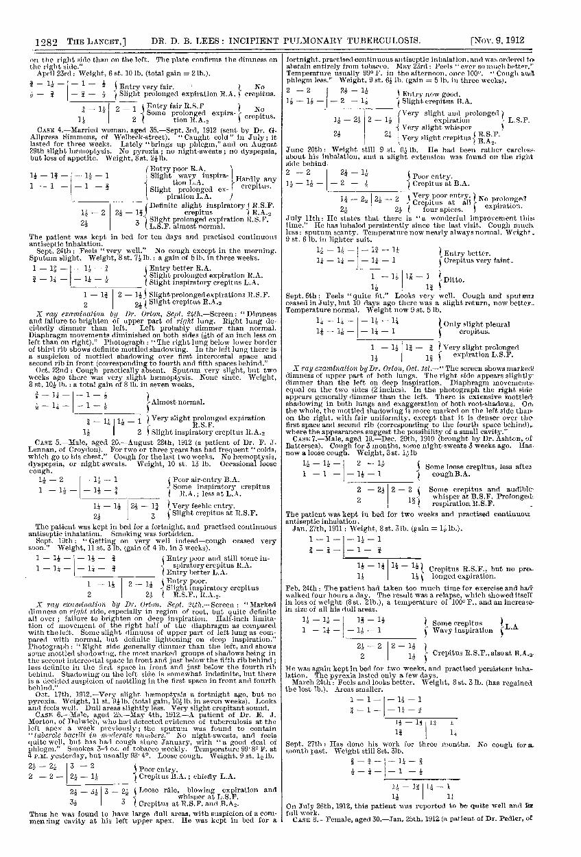
Bsubmit the following figures, obtained by myself in eightrecent (unpublished) cases. The first six are very earlyoases, in which an X ray examination was made, so that theresults of both methods of examination can be compared’The seventh case shows how a slight relapse, due to excessof exercise, gave evidence of increase in size of some of thedull areas, and how rest and continuance of the antisepticinhalations rapidly brought about subsidence of this ex-
tension. The eighth is a much more chronic case, whichhas made remarkable improvement under the use of con-tinuous antiseptic inhalation.
It must be explained that in each case the part of thediagram above the horizontal line presents the front view of’the chest and shows the size of the dull areas in the first:and second intercostal spaces, while the part below the’horizontal line presents the back view of the chest and’.shows the size of the dull areas in the inner and outer
parts of the supraseapular fossa, and at the extremity of’the spine of the scapula on each side. The figures are in- fingerbreadths and fractions of a fingerbreadth.
CASE 1.-Female, aged 24.-Jan. 26th, 1912 (sent by Dr. J. Ashton, of--Battersea). Only definite symptom occasional nipht-s2eeuts for nearly ayear; no cough or sputum. Her brother (Case 7 below) had been underany care for early pulmonary tuberculosis and had recovered. Weight,7 st. 12 lb. (it was formerly 8 st. 1 Ib.).
3 Distinct crepitus B.A."The patient was kept in bed for 10 days and she practised continuousantiseptic inhalation.
Feb. 16th: Weight, 8 st. 2½ lb. (gain = 41b. in three weeks). Feels.and looks better; still occasional night-sweats. Temperature to 99’4" F.for 3 days, now 98’8°.
- 1 - __ __ 2
2 2
March 5th: Weight, 8 st. 5 Ib. (total gain = 6½ lb.). Doing well. Night-sweats now "nothing like what they used to be." Temperature onlyonce to 99.4°.
3/4 - 1½ -
2
April 19th : Weight, 8 st. 8 lb. (total gain = 10 lb.). Night-sweatsslight. Temperature rarely reaches 99°. Dull areas a little smaller.X ray ea;aveination by Dr. Harrison Orton, April 23rd.-" The screen
:shows slight dimness on the 1’ight side; does not brighten so well as theleft on deep inspiration. Diaphragm movements approximately equal.and not matkediy abnormal. The photograph shows a definite area ofmottling in the first and second spaces in front (= the fourth and fifthbehind) on the right side ; there is also some mottling (but less definite)in the fifth right space in front. The left upper spaces show less definitemottling, but the left root-shadow is considerably more marked than.the right, and shows several very opaque areas." ;
June 7th: Weight, 8 et. 10 lb. in lighter dress (total gain = 13 lb.).
Feels and looks very well. Night-sweats almost disappear Tempera-ture entirely subnormal for last seven days.
)Very slight crepitus R.A.(Rather poor entry -
Rather poor entry { R.S.F.1 B R.A.2
July 26th : Weight still Sst. 10½ lb. "Not a single sweat since lastvisit."
Breathing quite normal in;} - - ;B: } front and at a11 posterior in
s 1 apices. posterior
CASE 2.-Female, aged 30.-March 6th, 1912 (sent by Dr. R. RMowll, of Surbiton). Has felt weak lately and tintit for work. Pain inleft side of chest came on suddenly at night iive weeks ago; for thefirst two days it hurt her to breathe. Since then feels a chronicsoreness in left axilla. No cough, dyspepsia, or night-sweats. Weight,8 st. Iglb. (8 st. 2 lb. last August).
121 ( L. front slightlya - 1 - tender. Slight -B.A.I s double crepitus
.Poor entry B.S.F.- 2 2 - 2 Very slight pro- j Also
distinct fric-expiration tion crepitus at2 2l t angle of scapula.crepitus B.A.2
She was kept in bed for ten days and practised continuous antisepticinhalation. March 27th : Left chest still rather sore. No cough.Weight, 8 st. 421 lb. (gain = 3 lb. in three weeks).
- Entry poor ( - - -- fair L.A. No crepitus.
1 - 1? Entry poor. Slight rub still-
Hardly any audible. still1 2 crepitus ) audible.
X ray examination by Dr. Ortnn, April 16th.-Il The screen showsdefinite dimness of upper part of righf lung as compared with the left.Diaphragm movements approximately equal on the two sides, but theirrange on both sides is well below the average. The photograph showsdefinite mottling on the right side, with several small opaque areaswhich suggest old fibrous nodules, especially in first space in front,corresponding to fourth space behind; in second space in front.corresponding to fifth space behind ; and in third space in front (mostmarked) corresponding to seventh rib behind. On the left side there isonly a little faint mottling lying over fourth rib behind and in firstintercostal space in front: otherwise indefinite."May 1st: No soreness during the last week. Still no cough. Weight,
8 st. 6 lb. (total gain = 4½ lb.).
:f
4 Nothing abnormal on auscul-tation except very slight fric-
1 tion at angle of left scapula.I June 5th : Feels well.
CASE 3.-Female, aged 28.-March 2nd, 1912 (sent by Dr. A. M.Hickley, of South Lambeth-road, under whose care she had been inAugust, 1911, for pharyngitis and post-nasal catarrh). At the end ofNovember, 1911, Dr. Hickley detected evidence of tuberculosis at theright apex, and prescribed treatment by continuous antiseptic inhala-tion ; but she was not kept in bed. Herweight on Dec. 1st was 6 st. 31b.;it rose gradually to 6 st. 9lb. On Feb. 22nd 1912, a few tubercle bacilli
were found in her sputum. Cough troublesome. Weight, 6 st. 81b.- 2 - Poor entry.- 1 - 1 crepitus.
2 Poor entry.No crepitus.2 Prolonged expiration R.S.F.The patient was kept in bed for a fortnight and practised continuousantiseptic inhalation.March 26th: Weight, 6 st. 9 Ib. (gain 1 in three weeks). Cough
not much better.
1 Much better entry.31 1 Some prolonged expiration R.A... ’ No crepitus.
X ray examination by Dr. Orton, April 2nd, 1912.-" The screen showsslight dimness at both apices, most marked at the right apex. Hesitat-ing movement of the right side of the diaphragm. The photographshows definite mottled shadowing more marked and more extensive
1282 DR. D. B. LEES: INCIPIENT PULMONARY TUBERCULOSIS.
on the right side than on the left. The plate confirms the dimness onthe right side."April 23rd : Weight, 6 st. 10 lb. (total gain = 21b.).
CASE 4.—Married woman, aged 35.-Sept. 3rd, 1912 (sent by Dr. G.Allpress Simmons, of Welbeck-street). " Caught cold " in July ; itlasted for three weeks. Lately "brings up phlegm," and on August29th slight hæmoptysis. No pyrexia ; no night-sweats ; no dyspepsia,but loss of appetite. Weight, 8st. 2½lb.
X ray examination by Dr. Orton, Sept. 24th.-Screen: "Dimnessand failure to brighten of upper part of right lung. Right lung de-cidedly dimmer than left. Left probably dimmer than normal.Diaphragm movements diminished on both sides of an inch less onleft than on right)." Photograph : " The right lung below lower borderof third rib shows definite mottled shadowing. In the left lung there isa suspicion of mottled shadowing over first intercostal space andsecond rib in front (corresponding to fourth and fifth spaces behind."Oct. 22nd : Cough practically absent. Sputum very slight, but two
weeks ago there was very slight haemoptysis. None since. Weight,8 st. 10 lb. : a total gain of 8 lb. in seven weeks.
CASE 5.-Male, aged 20.-August 28th, 1912 (a patient of Dr. F. J.Lennan, of Croydon). For two or three years has had frequent " colds,which go to his chest." Cough for the last two weeks. No haemoptysis,dyspepsia, or night-sweats. Weight, 10 st. 13 lb. Occasional loosecough.
2 2& t RS.F., R.A.2.X ray examiaation by Dr. Orton, Sept. 24th.—Screen : " marked
dimness on right side, especially in region of root, but quite definiteall over ; failure to brighten on deep inspiration. Half-inch limita-tion of movement of the right half of the diaphragm as comparedwith the left. Some slight dimness of upper part of left lung as com-pared with normal, but definite lightening on deep inspiration."Photograph: " Right side generally dimmer than the left, and showssome mottled shadowing, the most marked groups of shadows being inthe second intercostal space in front and just below the fifth rib behind ;less definite in the first space in front and just below the fourth ribbehind. Shadowing on the left side is somewhat indefinite, but thereis a decided suspicion of mottling in the first space in front and fourthbehind."
Oct. 17th, 1912.-Very slight h2emoptysis a fortnight ago, but nopyrexia. Weight, 11 st. 9 lb. (total gain, 10½lb. in seven weeks). Looksand feels well. Dull areas slightly less. Very slight crepitant sound.CASE 6.-Male, aged 25.-May 4th, 1912.-A patient of Dr. E. J.
Morton, of Dulwich, who had detected evidence of tuberculosis at theleft apex a week previously; the sputum was found to contain" tubercle bacilli in moderate numbers." No night-sweats, and feelsquite well, but has had cough since January, with " a goocl deal ofphlegm." Smokes 3-4 oz. of tobacco weekly. Temperature 99- 80 F. at4 P.M. yesterday, but usually 93’ 4°. Loose cough. Weight, 9 st. 1½lb.
3s I 3 Crepitus at R.S.F. and B.A2.Thus he was found to have large dull areas, with suspicion of a com-meming cavity at his left upper apex. He was kept in bed for a
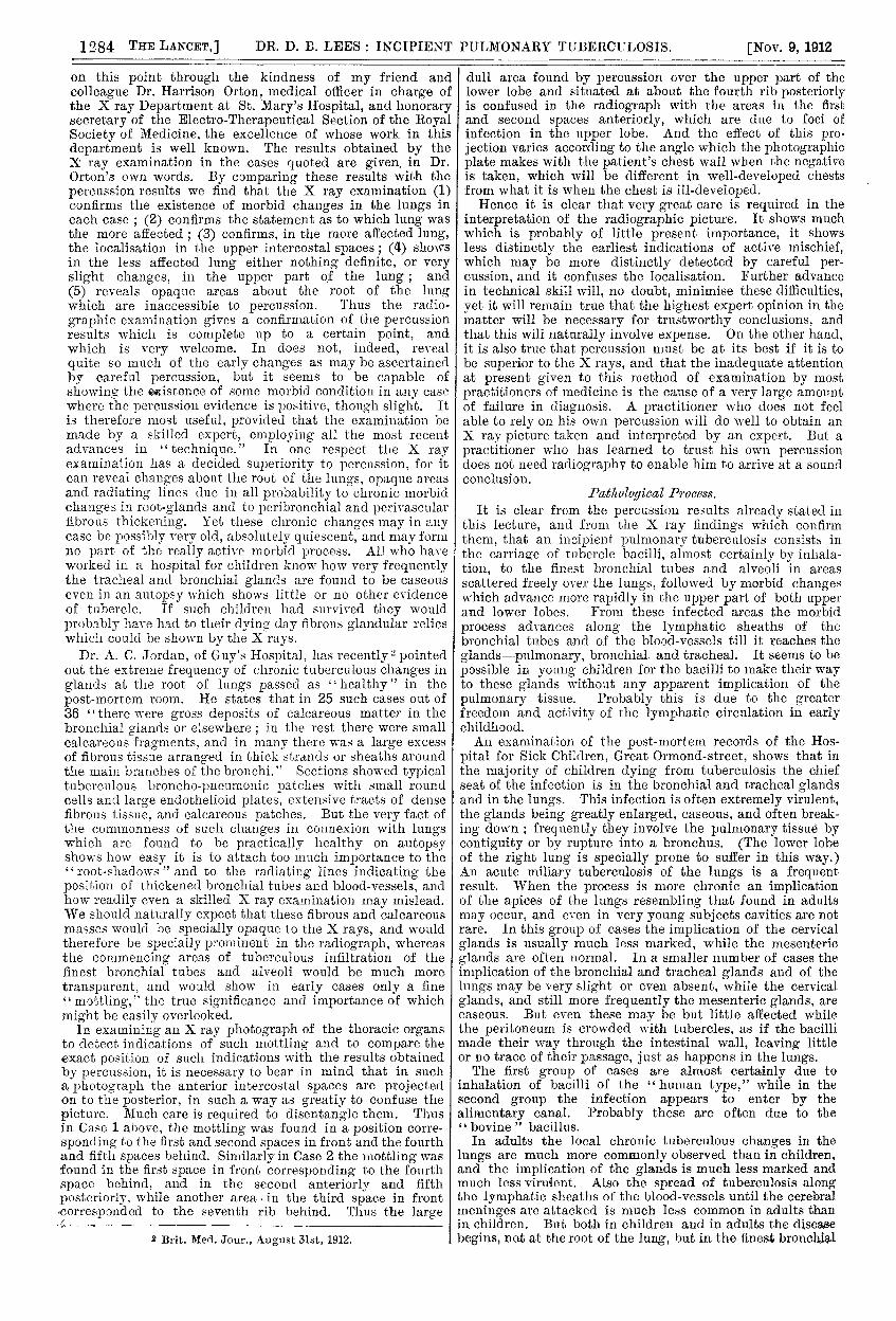
fortnight, practised continuous antiseptic inhalation, and was ordered toabstain entirely from tobacco. May 23rd : Feels ever so much better."Temperature usually 990 F. in the afternoon, once 100°. " Cough andphlegm less." Weight, 9 st. 6 lb. (gain = 5 lb. in three weeks).
B Entry now good.1 2 - 1 Slight crepitus B.A.
(Very slight and prolonged I1 - 2 2 - 1½ expiration L. S. P.
O! 0! Very slight whisper ( T? C 17 )2½ 2¼ Very slight crepitus B.A2-June 20th: Weight still 9 st. 6 lb. He had been rather careless.about his inhalation, and a slight extension was found on the rightside behind.2-2 - Is Poor entry.2 Poor entry.
— 1 — — 2 - I Crepitus at B.A.
Very poor entry. No prolongedCrepitus at all expiration.2 2 four apices. ) expiration.
July llth: He states that there is "a wonderful improvement thitime." He has inhaled persistently since the last visit. Cough muchless: sputum scanty. Temperature now nearly always normal. Weight,9 st. 6 lb. in lighter suit.
Ditto.Sept. 6th : Feels " quite lit." Looks very well. Cough and sput ceased in July, but 10 days ago there was a slight return, now better-Temperature normal. Weight now 9 st. 5 lb.
U Only slight pleural1 - - - 1 crepitus.
1 - 1 — Very slight prolongedl í expiration L.S.F.
X ray examination by Dr. Orton, Oct. lst.-Il The screen shows markeddimness of upper part of both lungs. The right side appears slightlydimmer than the left on deep inspiration. Diaphragm movements,equal on the two sides (2 inches). In the photograph the right sideappears generally dimmer than the left. There is extensive mottledshadowing in both lungs and exaggeration of both root-shadows. 0iijthe whole, the mottled shadowing is more marked on the left side thanon the right. with fair uniformity, except that it is denser over the-first space and second rib (corresponding to the fourth space behind),where the appearances suggest the possibility of a small cavity."CASE 7.-Male, aged 19.-Dec. 29th, 1910 (brought by Dr. Ashton, of
Battersea). Cough for 3 months, some night-sweats 3 weeks ago. Has.now a loose cough. Weight, 3 st. 1½ lb
1½ - 1& -B 2 Some loose crepitus, less after1 coughB.A.
2 - 2 2 - 2 ( Some crepitus and audible
2 J whisper at B.S.F. Prolongedrespiration R.S.F. _
The patient was kept in bed for two weeks and practised continuousantiseptic inhalation.Jan. 27th, 1911 : Weight, 8 st. 31b. (gain =
- 1 —
crepitus R.S.F., but no pra-1 I longed expiration.
Feb. 24th: The patient had taken too much time for exercise and had’walked four hours a day. The result was a relapse, which showed itselfin loss of weight (8 st. 21b.), a temperature of 100° F., and an increase-in size of all his clull areas.
He was again kept in bed for two weeks, and practised persistent inha-lation. The pyrexia lasted only a few days.March 24th: Feels and looks better. Weight, 8 st. 3 Ib. (has regained
the lost lb.). Areas smaller.
-
I Sept. 27th : Has done his work for three months. No cough for a.month past. Weight still 8st. 31b.
a g *
- - 1
1.lOn July 26th, 1912, this patient was reported to be quite well and infull work.CASE 8.-Female, aged 30.-Jan. 25th, 1912 (a patient of Dr. Pedler, of
1283DR. D. B. LEES : INCIPIENT PULMONARY TUBERCULOSIS.
Knightsbridge). More or less cough for several years. Some night-sweats and much weakness more than a year ago, but she had made agreat effort to remain at her work. Night-sweats three or four times aweek for the last three months. Weight, 8 st. 7 lb. (last August sheweighed 9st. 71b.). Frequent short cough.2 - 2 2 -
1{ Poorentry B.A.
1½ Moist crepitus and prolonged expiration R.A.( Slight crepitus L.A.
2 I 3 Faint crepitus H.S.F.Dr. Pedler reported that the bacillus is present in fair numbers." Thepatient was kept in bed for a fortnight, and practised continuous anti-septic inhalation.
Feb. 16th: Feels able to take longer breaths. Temperature 99’2"to99’8°F. in the evening (once it rose to 100°, once even to 104.2°.Weight, 8 st. 121b. (gain = 4½lb. in three weeks). Cough much better.
1½ - -
2j Entry better.Some prolonged expiration and crepitus R.A.;¼ less at L.A.
l’ - 2 Entry much better, but still poor at2 ’ lt.S.F., with some prolonged ex-2 2 { piration. Distinct crepitus B.A’2
March 15th: Feels " ever so much better--no comparison" ! Coughmuch better. Sputum " less than half of what it was." Temperaturehas been not above 98’6° since Feb. 16th. Weight, 9 st. 3 lb. (total gain
in seven weeks).- 1 - 1 - Double moist crepitus
1 - 1 Slight prolonged ex-’R.A.’’ piration )
1 - Ditto at ItS.F.2
Poor entry and slight crepitus( B.A.2.
April 25th: Weight, 9 st. 10 lb. (total gain = in 13 weeks).
- l Ausculatatory signs signs as before.1 - 1
2Thus far the patient had made remarkable progress. Unfortunately,her surroundings were now unsatisfactory, and she was the subject ofmuch mental anxiety. The result was a loss of weight, and moredefinite signs of cavity at her right apex. After some weeks sheobtained admission into a sanatorium, where she was allowed tocontinue her antiseptic inhalation.
These cases are sufficient to prove that it is easy tomeasure the size of the six typical dull areas at the four
apices in incipient pulmonary tuberculosis with sufficient
accuracy to give a very valuable guide to diagnosis and toprognosis. The practitioner will soon find the advantagewhich he will derive from a careful observation of them.
I I I I
/ ’ ’
Diagram showing the distribution of the dull areas inincipient pulmonary tuberculosis.
He will do well at first to confine his attention to thesesix areas, for the complete localisation of an incipientpulmonary tuberculosis is much more extensive and com-plicated. So far as I know, it has never been described indetail, and it is therefore necessary to point out the manyother positions on the chest wall where small dull areas
may be detected in an early case, in addition to the sixalready described. (See Figure.)
Other Dull A reccs.On the anterior wall of the chest a small dull area can
always be discovered by careful percussion in the thirdintercostal space on each side, above the nipple or somewhatmore internally ; and further out in the same space there is
on each side a dull area below the anterior fold of theaxilla. On the right side, internal to and below the nipple(in the region of the middle lobe of the right lung), there isa very definite dull area, with resonance between it and theborder of the right auricle (which normally extends a finger-breadth outwards into the fourth space, and in these casesof early pulmonary tuberculosis sometimes as much as a
fingerbreadth and a half). Nothing corresponding to thisarea can be detected on the left side, for in this positionthere is the dullness due to the heart. But on both sidesthere is a small dull area in the anterior part of the axillaat about the level of the fourth space or fifth rib. Posteriorly,there is always a dull area at the angle of the scapula oneach side, and another a little externally, in the posterioraxillary region. When the infection in this region extendsto the surface of the lung and sets up a local pleurisy, as isnot uncommon, these two areas unite into one, especiallyif any pleural effusion occurs. If the patient improves andthe effusion is absorbed the dullness diminishes, and thetwo separate areas can again be distinguished. But if the
pleural effusion increases it may produce a large area ofdullness, with defect of air-entry, vocal resonance, and vocalfremitus.
If the horizontal line through these two areas be prolongedtowards the spine a third area is met with on each side near,but not close to, the spine. These areas also are very con-stant. At a slightly higher level there is an interestingdifference between the two sides, for on the left side a dullarea is found at the posterior edge of the scapula, about twofingerbreadths above the angle, while on the right side theposition of the corresponding area is not at the scapularedge, but always nearer to the spinal column, in about thesixth space. This is a remarkable and invariable difference,which evidently depends on the different anatomical divisionof the lungs into three lobes on the right side and two onthe left. Furthermore, in the upper part of the back oneach side three small separate dull areas can often bedetected in the positions shown on the diagram, and belowthe angle of the scapula there are also several dull areasdetectable in positions which are arranged in a kind ofregular pattern, and are marked on the diagram.
This remarkable grouping of small dull areas may usuallybe detected by careful percussion in quite early cases ofpulmonary tuberculosis. Their relative positions are practi-cally invariable. From their scattered and symmetricaldistribution and from the slight differences between the twolungs which I have described, it seems clear that they mustcorrespond to the ultimate distribution of the bronchial tubesin their most direct course. The dullness and lessened air
entry are probably caused by multiple local centres ofinfection giving rise to local collapse, followed by localbroncho-pneumonia of a very chronic type, and finallyfibrosis.
Of these dull areas the largest are always the six atthe apices, as previously described. For the apical regionis the least expanded in ordinary respiration, and in a
tuberculous infection of the lung (especially if it atall implicates the pleura) there appears to be a reflexinhibition of respiration, just as the diaphragm movementsare checked by even a local peritonitis. In an early stage ofpulmonary tuberculosis the fluorescent screen will oftenshow a marked limitation of the inspiratory movement ofthe diaphragm, sometimes unequally on the two sides.There can, I think, be no doubt that a similar reflex inhibi-tion of the other inspiratory muscles is common in pul-monary tuberculosis. Every physician must have observedhow difficult it is to induce such patients to take a long deepbreath and thoroughly to expand the chest. This failure of
expansion in its turn makes the local conditions more
favourable for the development of the tubercle bacillus-another instance of a "vicious circle " in disease. Thus themorbid process develops more rapidly in the apical areas,and these areas are the most prone to soften down intoirregular cavities.
Results of X Ray Examination.It is important to ascertain whether the results obtained
by percussion in incipient pulmonary tuberculosis are or arenot confirmed by X ray examination of the chest.
Segaius irritant animum demissa per auremQuam quæ sunt oculis subjecta fidelibus.
"&ohgr;ra &ggr;&agr;&rgr; &tgr;&ngr;&khgr;&agr;&ngr;∈i dpOpt5roto-t ∈ó&ngr;&tgr;&agr; &agr;&pgr;i&sgr;&tgr;ó&tgr;∈&rgr;&agr; ò&phgr;&thgr;&agr;&lgr;µ&ohgr;&ngr;."I have been able to make some comparative observations
1284 DR. D. B. LEES : INCIPIENT PULMONARY TUBEROULOSIS.
on this point through the kindness of my friend andcolleague Dr. Harrison Orton, medical officer in charge ofthe X ray Department at St. Mary’s Hospital, and honorarysecretary of the Electro-Therapeutical Section of the RoyalSociety of Medicine, the excellence of whose work in thisdepartment is well known. The results obtained by theX ray examination in the cases quoted are given. in Dr.Orton’s own words. By comparing these results with thepercussion results we find that the X ray examination (1)confirms the existence of morbid changes in the lungs ineach case ; (2) confirms the statement as to which lung wasthe more affected ; (3) confirms, in the more affected lung,the localisation in the upper intercostal spaces ; (4) showsin the less affected lung either nothing definite, or veryslight changes, in the upper part of the lung; and
(5) reveals opaque areas about the root of the lungwhich are inaccessible to percussion. Thus the radio-
graphic examination gives a confirmation of the percussionresults which is complete up to a certain point, andwhich is very welcome. In does not, indeed, reveal
quite so much of the early changes as may be ascertainedby careful percussion, but it seems to be capable ofshowing the existence of some morbid condition in any casewhere the percussion evidence is positive, though slight. Itis therefore most useful, provided that the examination bemade by a skilled expert, employing all the most recentadvances in "technique." In one respect the X rayexamination has a decided superiority to percussion, for itcan reveal changes about the root of the lungs, opaque areasand radiating lines due in all probability to chronic morbidchanges in root-glands and to peribronchial and perivascularfibrous thickening. Yet these chronic changes may in anycase be possibly very old, absolutely quiescent, and may formno part of the really active morbid process. All who haveworked in a hospital for children know how very frequentlythe tracheal and bronchial glands are found to be caseouseven in an autopsy which shows little or no other evidenceof tubercle. If such children had survived they wouldprobably have had to their dying day fibrous glandular relicswhich could be shown by the X rays.
Dr. A. C. Jordan, of Guy’s Hospital, has recently pointed ;out the extreme frequency of chronic tuberculous changes inglands at the root of lungs passed as "healthy" in the
post-mortem room. He states that in 25 such cases out of36 "there were gross deposits of calcareous matter in the ibronchial glands or elsewhere ; in the rest there were small calcareous fragments, and in many there was a large excessof fibrous tissue arranged in thick strands or sheaths aroundthe main branches of the bronchi." Sections showed typical ituberculous broncho-pneumonic patches with small round cells and large endothelioid plates, extensive tracts of densefibrous tissue, and calcareous patches. But the very fact of the commonness of such changes in connexion with lungs iwhich are found to be practically healthy on autopsy shows how easy it is to attach too much importance to the "root-shadows "and to the radiating lines indicating theposition of thickened bronchial tubes and blood-vessels, and how readily even a skilled X ray examination may mislead. (
We should naturally expect that these fibrous and calcareous masses would be specially opaque to the X rays, and would therefore be specially prominent in the radiograph, whereas Ethe commencing areas of tuberculous infiltration of the ífinest bronchial tubes and alveoli would be much more i
transparent, and would show in early cases only a fine I
" mottling," the true significance and importance of which ímight be easily overlooked. (
In examining an X ray photograph of the thoracic organs t
to detect indications of such mottling and to compare the i
exact position of such indications with the results obtained c
by percussion, it is necessary to bear in mind that in sucha photograph the anterior intercostal spaces are projected ion to the posterior, in such a way as greatly to confuse the s
picture. Much care is required to disentangle them. Thus 2
in Case 1 above, the mottling was found in a position corre- ‘
sponding to the first and second spaces in front and the fourthand fifth spaces behind. Similarly in Case 2 the mottling was 1found in the first space in front corresponding to the fourth a
space behind, and in the second anteriorly and fifth r
posteriorly, while another area, in the third space in front t
<corresponded to the seventh rib behind. Thus the large r
2 Brit. Med. Jour., August 31st, 1912.
dull area found by percussion over the upper part of thelower lobe and situated at about the fourth rib posteriorlyis confused in the radiograph with the areas in the firstand second spaces anteriorly, which are due to foci ofinfection in the upper lobe. And the effect of this pro-jection varies according to the angle which the photographicplate makes with the patient’s chest wall when the negativeis taken, which will be different in well-developed chestsfrom what it is when the chest is ill-developed.Hence it is clear that very great care is required in the
interpretation of the radiographic picture. It shows muchwhich is probably of little present importance, it showsless distinctly the earliest indications of active mischief,which may be more distinctly detected by careful per-cussion, and it confuses the localisation. Further advancein technical skill will, no doubt, minimise these difficulties,yet it will remain true that the highest expert opinion in thematter will be necessary for trustworthy conclusions, andthat this will naturally involve expense. On the other hand,it is also true that percussion must be at its best if it is tobe superior to the X rays, and that the inadequate attentionat present given to this method of examination by mostpractitioners of medicine is the cause of a very large amountof failure in diagnosis. A practitioner who does not feelable to rely on his own percussion will do well to obtain anX ray picture taken and interpreted by an expert. But a
practitioner who has learned to trust his own percussiondoes not need radiography to enable him to arrive at a soundconclusion.
Pathological Process.It is clear from the percussion results already stated in
this lecture, and from the X ray findings which confirmthem, that an incipient pulmonary tuberculosis consists inthe carriage of tubercle bacilli, almost certainly by inhala-tion, to the finest bronchial tubes and alveoli in areas
scattered freely over the lungs, followed by morbid changeswhich advance more rapidly in the upper part of both upperand lower lobes. From these infected areas the morbidprocess advances along the lymphatic sheaths of the
bronchial tubes and of the blood-vessels till it reaches the
glands--pulmonary, bronchial. and tracheal. It seems to be
possible in young children for the bacilli to make their wayto these glands without any apparent implication of the
pulmonary tissue. Probably this is due to the greaterfreedom and activity of the lymphatic circulation in earlychildhood.An examination of the post-mortem records of the Hos-
pital for Sick Children, Great Ormond-street, shows that inthe majority of children dying from tuberculosis the chiefseat of the infection is in the bronchial and tracheal glandsand in the lungs. This infection is often extremely virulent,the glands being greatly enlarged, caseous, and often break-ing down ; frequently they involve the pulmonary tissue bycontiguity or by rupture into a bronchus. (The lower lobeof the right lung is specially prone to suffer in this way.)An acute miliary tuberculosis of the lungs is a frequentresult. When the process is more chronic an implicationof the apices of the lungs resembling that found in adultsmay occur, and even in very young subjects cavities are notrare. In this group of cases the implication of the cervicalglands is usually much less marked, while the mesentericglands are often normal. In a smaller number of cases theimplication of the bronchial and tracheal glands and of thelungs may be very slight or even absent, while the cervicalglands, and still more frequently the mesenteric glands, arecaseous. But even these may be but little affected whilethe peritoneum is crowded with tubercles, as if the bacillimade their way through the intestinal wall, leaving littleor no trace of their passage, just as happens in the lungs.The first group of cases are almost certainly due to
inhalation of bacilli of the human type," while in thesecond group the infection appears to enter by the
alimentary canal. Probably these are often due to the" bovine bacillus.In adults the local chronic tuberculous changes in the
lungs are much more commonly observed than in children,and the implication of the glands is much less marked andmuch less virulent. Also the spread of tuberculosis alongthe lymphatic sheaths of the blood-vessels until the cerebralmeninges are attacked is much less common in adults thanin children. But both in children and in adults the diseasebegins, not at the root of the lung, but in the finest bronchial
1285DR. D. B. LEES : INCIPIENT PULMONARY TUBERCULOSIS.’,
tubes and in the alveoli, in scattered areas which show achaiacteristic localisation.
two sum up this long discussion, I would claim that thefirst of my two questions may now be answered in the aflirma-tive. It is possible for any general practitioner of medicine,by the adoption of a correct method of examination and theexercise of a little care and patience, accurately to diagnosethe existence of an incipient pulmonary tuberculosis longbefore any bacteriological evidence can be obtained. It isnot necessary for him to wait for a positive bacteriologicalreport, and he will be foolish indeed if he allows a negativereport to shake a diagnosis which is founded on an accuratephysical examination. The possibility of a correct diagnosisis, in more senses than one, in his own hands.
II.—THE TREATMENT OF INCIPIENT PULMONARYTUBERCULOSIS.
.
In the minds of the majority of the medical professionthe question as to the proper treatment of pulmonary tuber-culosis seems to be answered by the one word " sanatorium,"to which some are now inclined to add (relying on the con-fident assertions of its advocates) the word "tuberculin."But with regard to the employment of tuberculin there isstill acute controversy as to whether it ought to be employedin small or in large doses, whether the grave symptoms whichmay follow its employment are due to it or to "mixedinfection," and whether it ought to be used only when thepatient is kept at rest and under skilled supervision, or maybe given to patients who are allowed to pursue their ordinaryoccupations.
It is quite certain that sanatoria have been extremelyuseful, and that by their help many patients have beenrestored to health. Yet it must also be sorrowfully confessedthat many patients who have lived in a sanatorium for sixmonths, at a very considerable cost, are still not completelycured, and that numerous instances of subsequent relapseoccur, so that in the minds of many practitioners there is agrowing scepticism as to the curative value of sanatoria.And when we consider the matter carefully we see that
though the treatment by absolute rest, abundance of freshair, and abundance of nutriment places the patient under themost favourable circumstances for fighting his battle with theinvading bacilli, it does not include any organised attack onthe bacilli theftisel?,es. It is the consciousness of this that hasled many to desire to make trial of tuberculin. But is thereno simpler and less dangerous method of direct attack ? Isit quite certain that ordinary antiseptic drugs are of no valuein the treatment of pulmonary tuberculosis ? On the con-
trary, it is quite certain that some cases have markedlyimproved under the use of large doses of creosote. Of latethe value of injections of a preparation of which iodine
appears to be the active agent has been prominentlyadvocatedin France. And a letter in the British Medical Jonrnal froma practitioner who had been using tuberculin and whose
supply was exhausted related that he had substituted injec-tions of carbolic acid and had obtained better results. Butcreosote by the mouth is apt to cause much disturbance ofthe organs of digestion, while hypodermic injections are apt <
to be painful, and require the most scrupulous precautions 1
against sepsis. i
Treatment by ContÍ’mwus Ighalation of Antiseptic J;-11([J01lr8.Why not administer these antiseptic drugs by a method of
inhalation ’? Too many physicians reply, "Because wecannot see how such a method can be of service." -Someeven go so far as to say, "Because it cannot possibly do anygood." " But is such scepticism justifiable? If a patientinhales a combination of antiseptic vapours persistently, dayand night (except when actually taking food), by theprinciple of diffusion of gases the antiseptics m2cst maketheir way into the pulmonary alveoli, if the inhaler be wornfor a sufficient length of time. This is surely indisputable.And if antiseptic drugs like creosote, carbolic acid, andiodine are dissolved in alcohol, ether, and chloroform, theremust be a gradual absorption of these anaesthetic agentsinto the blood, for this is an A.C.E. mixture, and we knowthat if used in greater strength it would prove the absorptionby producing general anaesthesia. The slow absorption ofthese agents in a very dilute condition must carry with theminto the blood the antiseptics dissolved in them. How these
may be transformed in the blood it is for physiologicalchemistry to determine, but it seems likely that some
antiseptic influences will thus be carried by the blood-streamto the tuberculous foci, which will then be attacked bothfrom without the lungs and from within.
Certain othei properties of this combination of drugsdeserve attention. First, with regard to the alcohol, we ehave learned from the remarkable observations of ProfessorCollingwood and Dr. W. H. Willcox how much the
stimulating and tonic effect of oxygen given by inhalation isincreased by passing the oxygen through absolute alcohol.Dr. Willcox has calculated the amount of alcohol absorbedand finds it to be very small, yet it exercises a mostremarkable influence, as has subsequently been proved byadministering oxygen bubbled through alcohol in cases ofcardiac failure. The effect of the continuous inhalationof an antiseptic mixture in which alcohol is the solvent musttherefore be a tonic and stimulating influence constantly atwork. Another constituent of the mixture, ether, will actsimilarly and perhaps more strongly. Lastly, chloroformhas a local sedative influence and strongly tends to diminishcough. I therefore recommend, and have constantlyemployed, the following formula for a solution to beused for continuous antiseptic inhalation.
Menthol and formalin appear to me too irritating, andunsuitable for continuous inhalation.Whether my argument from theoretical considerations
does or does not produce conviction in the minds of myhearers, the final appeal must be to the results of experience.If the results of experience can be shown to be satisfactory,theoretical objections will soon be swept away. For wemust always remember that however fascinating a scientificor apparently scientific basis for treatment may be, our mosttrusted remedies are the product of empirical observation.Quinine, opium, digitalis, mercury, and sodium salicylate are-triumphs of empiricism. Medicine is both a science and anart ; both must be cultivated with equal care and accuracy.For the art to ignore the science is stupid : for the science toneglect the art is suicidal. The medical science which has
any real claim to the name cannot be confined to the labora-
tory and its products ; it must be constantly in touch withan ever-increasing accuracy of clinical investigation. Theresults of treatment which claims to be scientific " must betested, not simply by the assertion of the patient that he is"better" (have we not all heard of the spes phthisica 1),but by diminution of pyrexia, of cough, and of sputum, byincrease in weight, and by careful observation and record ofthe physical signs. I therefore ask your careful considerationof the results recorded in the table of 70 cases treated withthe formula above stated (pp. 1286-87) which have beenalready published in the medical journals.3 The most recentof these cases ceased treatment a year ago. The table showsin each case : (1) the gain in weight during the first threeweeks ; (2) the total gain in weight; (3) the effect on thecough ; (4) the effect on the sputum ; (5) the effect on thephysical signs ; and (6) the result as to permanent capacityfor work. The present condition of each case in October,1912, with very few exceptions, has been ascertained byletters directed to the patient or to the medical man whosent him. The answers have been pleasant reading, for theyhave shown the most satisfactory results in nearly all cases,and they have been full of gratitude. Out of the 70 cases
Of the 10 incompletely cured, 9 have improved remarkably; 1 hasdeteriorated. Of the 7 deaths, 1 was due to apoplexy after recoveryfrom the lung disease, 1 was hopeless when first seen, 2 abandonedtreatment, 1 died from haemoptysis, 1 from abdominal tuberculosis,and 1 from tuberculous meningitis.
3 Cases 1-30 in British Medical Journal, Dec. 11th, 1909 ; Cases31-50 in THE LANCET, Nov. 19th, 1910; Cases 51-70 in BritishMedical Journal, April 6th, 1912.
1286 PROF. D. B. LEES : INCIPIENT PULMONARY TUBERCULOSIS.
TABLE GIVING SEVENTY CASES OF PULMONARY TUBERCULOSIS TREATED BY CONTINUOUS ANTISEPTIC INHALATION.
1287PROF. D. B. LEES : INCIPIENT PULMONARY TUBERCULOSIS.
Table giving Seventy Cases of Pulmonary Tiibercitlosis treated by Coiztinitoqts Antiseptic Inhalation-(continued).
* Had been treated with tuberculin injections weekly for six month.,T
1288 ,DR. D. B. LEES : INCIPIENT PULMONARY TUBERCULOSIS.
In 19 of the cases in this table a bacteriological examina-tion of the sputum was made. The results were as follows: —
I I -
I _..
’’ 111 one case (after removal of tuberculous cervical elands) notubercle bacilli were found in the sputum, but lJIll11Y strepto-cocci. The rapid gain in weight shown in this table is
worthy of notice. The gain in the first three weeks of treat-ment in 33 cases shows an average of 3’8 lb., the total gain I I
in weight in 59 cases an average of 12-4 Ib. In every one of Vthe 70 cases both lungs were found to be affected; but inalmost all one lung was more affected than the other, thoughthe, difference was usually slight. In only 2 cases the
right and left lungs were about equally affected. In 44 casesthe right lung was more affected than the left ; in 24 casesthe, left lung was more affected than the right. Thus inthis’, list of 70 cases the right lung was chiefly attackednearly twice as often as the left.
Resttlts obtained by Va1’iolls OÚSl’1’1’l’’l’S.
’The idea of administering antiseptic drugs by the air-passages in cases of phthisis suggested itself to physicians30 years ago. In 1877 the late Sir William Roberts claimedgood results from the use of antiseptic inhalation by meansof a " respirator-inhaler" " which covered the mouth only,and which he directed to be "worn for fifteen, thirty, orsixty minutes several times a day."
" Dr. Coghill also
.employed a similar apparatus, and directed that the patientshould inspire through the mouth and expire through the.nose, each such period of inhalation to last from 15 to 20minutes. To Dr. Burney Yeo we are indebted for the
invention, in 1882, of the simple inhaler of perforated zincwhich has proved of such great value. It is still being made-as Dr, Yeo recommended, but it has been found advisable(with Dr. Yeo’s consent) to increase the size a little for caseswith more prominent noses. This model is known as Yeo’s
inhaler (M), the original type being described as Yeo’sinhaler (F) ; they are made by Squire and Son, 413, Oxford-.street.When Dr. Hassall, in 1885, doubted the value of drugs s
given by inhalation, Dr. Wilson Fox (clarlt1n et venerabilenomen) replied :’r " The antiseptic effect has been doubted byHassall, but there can be -no question that inhalations prac-tised in this manner, with creosote, thymol, eucalyptus,iodoform, iodine, or terebene, tend to diminish cough andexpectoration, and that in some cases marked improvementin the’patient’s state occurs during their use, even in veryadvanced stages." This statement by Dr. Wilson Fox maybe commended to the careful consideration of physicians,’who are sceptical as to the value of such inhalations.
During the last’ 30 years the method of treatment thus’initiated has been adopted with success by a few physicians,among whom special mention must be made of Dr. Beverley- Robinson, of New York, who has followed the recommenda-tions of .Dr. Yeo, and made use of similar solutions forinhalation for many years. He has published numerous
papers’on the subject, in which he has given evidence of the’value of this method of treatment in cases of phthisis, andespecially in laryngeal tuberculosis.For several years past Dr. Muthu, of the Mendip Hills
.Sanatorium, has made much use of antiseptic inhalation inthe treatment of phthisis by means of solutions of mentholand formaldehyde employed for six or eight hours daily.He finds such inhalations very useful, and has publishedpapers in support of this method of treatment. Dr. Garry,of Cairo, has also published good results, and I am informed.that as long as 10 or 12 years ago the late Dr. Huggard, ofDavos, ordered antiseptic inhalation day and night con-
tinuously, with complete success, in a case known to my’informant.
4 Diseases of the Lungs, p. 884.
Methord C/’ Inhalation Treatment.For the last .seven years I have treated all my cases of
pulmonary tuberculosis (except some seen in consultation ata very advanced stage) by the method of continuous anti-septic inhalation, by means of a Yeo’s inhaler and the antiseptic solution of which the formula is’ stated above, Ina(lopting this plan of treatment it seemed to me that certainconditions were necessary if the greatest possible amount ofbenefit was to be obtained.
1. The inhalation must be r,crzttin2co2t.s, the inhaler beingworn all day and all night, except at meal-times.
2. At the beginning of the treatment a period qf rest inbed is essential. The Importance of this in the treatment oftuberculosis is well understood in sanatoria.. In addition,the compulsory rest directs the patient’s attention’to the
necessity for careful and thorough use of the method. , ’
3. Digestive disturbances, ’anorexia, and diarrhoea of
constipation must be treated by appropriate drugs.4. The patient must be supplic(l with a sufficiency of easily
digested fimcl. Malted milk dissolve(l in (scalded) milk givenat the end of each of the four meals has proved invaluablefor this purpose.
5. Careful disinfection of the mouth with equal parts ofSanitas fluid and water four times daily, after the milk, hasbeen always ordered. As soon as the patient has begun tomake definite progress carious teeth should be stopped orremoved. Antiseptic sprays may be used for unhealthy con-ditions of the fauces, and a spray of cocain, eucalyptus, andparolein to relieve any nasal obstruction.
‘
6. Smoking must be absolutely forbidden.These are the principles on which the 70 published cases
were all treated.I advise, therefore, that whenever a practitioner has
reason to suspect that a patient is infected with incipientpulmonary tuberculosis, and has by careful percussiondetected the presence of the six characteristic dull areas atthe four apices, he should carefully note the size of theseareas, the morbid sounds present, and the patient’s weight’;and at once order a period of rest in bed for 7, 10, or
14 days according to the extent of the dull areas and theauteness of the symptoms. Two Yeo’s inhalers (each costingonly 6d. or Is.) should be procured, so that they may beused on alternate days and may be properly cleansed byimmersion in boiling water and careful drying.The solution of which the formula has been already
mentioned should be supplied; and the patient instructed topour six to eight drops on the felt of the inhaler every hourluring the daytime, and once or twice during the night if helappens to wake. Nearly all patients soon become accus-omed to wearing the inhaler,at night. - The object shouldbe to keep the piece of felt constantly saturated with the;olution, but so that none of it wets the framework of thenhaler itself, The base of the inhaler should be carefullywiped before it is applied, and a little vaseline or lanolin;old cream smearecl over the nostrils and upper lip at first. By simple precautions like these, with the use of an
nhaler of proper size, any irritation of the skin of the nose)r lips may be avoided. The possibility of such irritations the only disadvantage of the method, and it may bejvoided if careful instructions are given at first. Th.nhaler must be worn persistently and removed only at meal-imes. After the first day or two the patient, while in bed,’.nay read, write, do hand-work, or play simple games.Dyspepsia and anorexia may be treated with sodiumbicarbonate, nux vomica, and a vegetable bitter given auarter of an hour before meals three times daily. ’.Thepatient should be encouraged to eat freely of’plain nutritiousood, and at the end of each of his four meals he should’rink slowly a half-pint of fresh milk which has been raised3 the boiling-point and allowed to cool, and to which, twoteaspoonfuls (gradually increased to a tablespoonful) ofhalted milk has afterwards been added. The bedroomendows should, of course, be kept open, but the patientmust be kept warm in bed and protected from draught. Afteris meals he should always rinse his mouth with equal parts ofanitas fluid and water. Smoking must be absolutelyforbidden.When the necessary period of rest in bed is at an end theaticnt may be allowed to sit np in his room, still wearing thelhaler persistently. Two or three days later he may be,Ilowed to lay it aside for an hour in the morning and take aliort walk out of doors. But when he returns he must at
1289MR, J. H. PARSONS: THE TREATMENT OF UNILATERAL CATARACT. ;
once replace his inhaler. At;’ the end of’ three weeks the
practitioner, should again weigh his patient and again care-fully,record- the size of his dull areas. Almost invariablya considerable gain in weight, often front 3 to 5 lb., will bediscovered, and. the size of the dull area will be found to bedefinitely smaller,. Cough will be mnch diminished -in manyearly cases it ceases altooctlrcr sputum will be less andmore easily expectorated ; and ihe temperature chart willbe mpre satisfactory. The patient generally feels so muchbetter that he is quite willing to continue the treatment.11’e should now be instructed to practise simple breathingexercises three times daily in order to obtain a better
expansion of the lungs. The inhalation treatment should be
continued persistently (except for an hour in the morning)during the next four weeks, at the end of which time the
weight should again be taken, and the size of the dull areascarefully ascertained and recorded. If the progress of thecase has been satisfactory during the first seven weeks it maynow be possible to reduce slightly the number of hours ofinhalation, but such reduction should be very gradual. The
danger at this period is that the patient. feeling himself
better, should grow careless. If lie does, there is a consider-able chance of a relapse, which will necessitate a returnto ’bed .for a fortnight with continuous inhalation. Expe-rience shows that the patients who are most conscientiousand,’.persevering in *. carrying, out the treatment improveinost rapidly and are least liable to relapse. A veryearly case may perhaps be able to return to ordinarylife after three or four months of treatment, but one in whichcavitation has occurred will need six months or longer. Afterapparent recovery the patient will do wisely to use his inhalerfor one or two ’hours daily, and also at night, for a furtherperiod of six months. Whenever he is conscious of having’caught cold" he should remain in bed for a day or twodays, and practise continuous inhalation all the time.This.treatment by antiseptic inhalation can be carried outin the patient’s own home if the hygienic and sanitary con-ditions are satisfactory. It is not usually necessary to sendthe patient to a sailatorium, so that there is a great saving inexpense, and the practitioner does not lose charge of him.But where the home conditions are unsatisfactorv it will bewise to send him to a sanatorium at which he will be per-mitted to carry out the treatment bv inhalation. No idea of
change of climate or foreign travel or sea voyage should beentertained until he has been through a course of inhalationtreatment, and even then ’he must take his inhaler with himand use it freely.
I now venture to claim that my second question, as wellas the first, may be answered in the affirmative. It is
possible to provide the practitioner with a simple, inexpen-sive ; and very effective form of treatment which can be
used in the patient’s own home. If every practitioner of medicine learned how to diagnoseaccurately at a very early stage the fact of incipient pul-monary tuberculosis, and, treated it at once according to themethod which I have described, it is quite certain that avery .large diminution in the mortality from pulmonarytuberculosis could be effected within a very few years, and itwould not be too much to hope that when the attention ofthe profession everywhere had been thoroughly aroused andthe intelligent cooperation of all medical men completelysecured, we might even see the dawn of a day when thedisease formerly knownas pulmonary phthisis had become athing of the past. May that day come soon !
UNIVERSITY OF LONDON : FACULTY OF MEDICINE.A meeting of the faculty was held at the University onNov. lst, the Dean, Dr. Sidney Martin, being in the ohair.
Dr. Reginald Cecil Wall was elected secretary to the faculty.The constitution for 1913 of the boarcis of studies withwhich the faculty is concerned was decided, as was also theconstitution of the board of the faculty for the remainder of1912. A report of the board of studies in hygiene and publichealth for’the provision in London for the post-graduateteaching of hygiene and public health was presented andafter’some discussion referred to the board of the faculty forreport. The election of Dean of the faculty for the period1912-14 then took place. The following nomination hadbeen received: Sir Alfred Pearce Gould, K.C.V.O., 31.8.,M.B., F.R.C.S., proposed by Profess.br Sir Rickinan J. Godlee,seconded by Dr. H. P. Hawkins, and supported by Mr. H. J.Waring. Sir Alfred Pearce Gould was unanimously elected.
THE TREATMENT OF UNILATERALCATARACT.1
BY J. HERBERT PARSONS, D.Sc. LOND., F.R.C.S. ENG.,SURGEON TO THE ROYAL LONDON (MOORFIELDS) OPHTHALMICHOSPITAL; OPHTHALMIC SURGEON TO UNIVERSITY COLLEGE
HOSPITAL.
BY "unilateral cataract" I mean cases of cataract in’which only one eye is affected, or in which the other eye, if°not absolutely normal, has sufficient vision to enable the’patient to carry out his ordinary duties. It is not always’easy to decide what is the best treatment of these cases ; I’’shall put before you the principles which guide me in my own’practice. I do not propose to deal with cases in whichother parts of the eye than the lens are seriously diseased,-but to confine my remarks to cases in which such’disease is so slight or quiescent as in itself to offer-no decided contra-indication to some form of cataractoperation.
°
-
The cases are best divided into two great groups accordingto the age of the patient, for, speaking generally, the indica-tion is to operate in cases of unilateral cataract in the young’and not to operate in the adult or aged. ’:
Let us first consider the main advantages which may be:expected to accrue to the patient from successful operation.’Perhaps the greatest, for reasons which will be apparent-from the sequel, is restoration of the full field of vision."The field of useful vision of the cataractous eye is practi-cally abolished. Moving objects on the side of the affectedeye are only seen by turning the head so that the nasalfield of the other eye is brought into play. This may be armatter of very great importance, especially to workingpeople who have to gain their living. They may have to)work in the midst of machinery in motion in close proximityto rotating wheels and bands. Any defect in the field ofvision may be a serious menace to life and limb. So, too,people who have to move about in the crowded thoroughfares of a great city are much handicapped, and may evenrun serious risks, if they are visually unconscious of’’’the.approach of vehicles on one side. Next as regards central vision. If the patient , had
originally little or no error of refraction he will withthe aid of a strong convex lens have his centralvision restored. This might a priori be regarded asa much greater asset than mere improvement of the fieldof vision. In reality it is not so. The operation results in ahigh degree of anisometropia or inequality in the refractiveconditions of the two eyes. Now it is well known that whenthe vision is corrected with suitable glasses under theseconditions, so that each eye separately has excellent vision,’the patient finds it impossible to wear the glasses and theyare soon discarded. This fact depends partly upon theidifference in size of the retinal images in the two eyes, adifference so great that the images cannot be fused; butprobably to a still greater degree upon the unequal prismaticeffects of the correcting lenses when they are looked throughobliquely, as occurs when the eyes roam from side to side;Moreover, the normal muscle balance of the eyes is interfered with. Hence it is not to be anticipated that after theoperation the patient will be able to wear a cataract
glass. The affected eye will, therefore, be practicallyuseless for central vision, and no particular gain accruesin this respect. It is true that if the cause of the cataract-isof a nature likely ultimately to produce a like effect in theother eye operation places the first affected eye in a condiltion to take up the visual function at the earliest moment;This must be considered an advantage, but generally one towhich too much weight must not be attached. It is true,too, that central vision is improved in the eye by operationeven if a cataract glass is not worn, but this, far from beingan advantage, is often a positive disadvantage. The. improvedvision acts as a disquieting feature, actually tending Ttoobscure the clarity of vision in the sound eye. Thus eventhe uncorrected anisometropia proves worrying and discon-certing, so that patients, far from being grateful for theresults of the operation, are much dissatisfied.
’
Next to be considered is the cosmetic effect. It .will
1 A paper fonnded on a clinical lecture delivered at the RoyalLondon (Moorfields) Ophthalmic Hospital on Oct. 12th.