R/F The Clinical Utility of Tomosynthesis in Lung Cancer Diagnosis Division of Thoracic Oncology, National Cancer Center Hospital East 1 National Cancer Center Hospital Masami Ito, MD Research Center for Cancer Prevention and Screening, National Cancer Center 2 Masami Ito 1 , Hironobu Ohmatsu 1 , Yoichi Naito 1 , Hirotsugu Kenmotsu 1 , Yuki Yamane 1 , Kiyotaka Yo 1 , Seiji Niho 1 , Koichi Goto 1 , Yuichiro Ohe 1 , Yutaka Nishiwaki 1 , Noriyuki Moriyama 2 1. Background The number of fatalities due to lung cancer is increasing. Therefore, the rapid detection of lung cancer through screening is extremely important. The chest radiography conventionally used for screening has the benefit of convenience and low exposure dose, but detection of cancer is difficult from the small, low-density shadow images. The introduction of helical CT for screening enhanced the sensitivity of shadow detection and increased the proportion of lung cancers detected early. However, CT with high shadow detection sensitivity can only be introduced into limited facilities and it suffers from problems with high exposure dose. We focused on a new imaging technology, tomosynthesis. This is a simple technique that offers high shadow detection sensitivity at a low exposure dose. This paper reports on our investigations into the clinical utility of tomosynthesis in the diagnosis of lung cancer. 2. Tomosynthesis The word “tomosynthesis” is a composite of “tomography” and “synthesis.” This method can reconstruct a coronal section at an arbitrary height from a single tomographic imaging operation. The tomography angle and tomography speed can be set, in addition to the radiography conditions, and radiography can be performed in the standing position (standing side-on permits tomography of lateral body sections). Tomosynthesis involves parallel, planar tomographic scanning with an R/F table system. Conventional tomography takes only a single section per imaging operation. A long time is required to take the series of images required for diagnosis. It produces images that are difficult to view, due to artifacts known as obstructive shadow. Conversely, tomosynthesis can reconstruct multiple sectional images from a single scan and offers image processing to reduce artifacts. Masami Ito, MD of the National Cancer Center Hospital East gave a presentation on the Shimadzu SONIALVISION safire R/F System with direct-conversion FPD at the 50th Annual Meeting of the Japan Lung Cancer Society (12/13 November 2009), entitled “The Clinical Utility of Tomosynthesis in Lung Cancer Diagnosis.” The contents of her presentation are introduced below. < News Flash >
Transcript
R/F
The Clinical Utility of Tomosynthesis in Lung Cancer Diagnosis
Division of Thoracic Oncology, National Cancer Center Hospital East1
National Cancer Center Hospital
Masami Ito, MD
Research Center for Cancer Prevention and Screening, National Cancer Center2
Masami Ito1, Hironobu Ohmatsu
1, Yoichi Naito
1, Hirotsugu Kenmotsu
1, Yuki Yamane
1,
Kiyotaka Yo1, Seiji Niho
1, Koichi Goto
1, Yuichiro Ohe
1, Yutaka Nishiwaki
1, Noriyuki Moriyama
2
1. Background
The number of fatalities due to lung cancer is
increasing. Therefore, the rapid detection of lung
cancer through screening is extremely important.
The chest radiography conventionally used for
screening has the benefit of convenience and low
exposure dose, but detection of cancer is difficult
from the small, low-density shadow images.
The introduction of helical CT for screening
enhanced the sensitivity of shadow detection and
increased the proportion of lung cancers detected
early. However, CT with high shadow detection
sensitivity can only be introduced into limited
facilities and it suffers from problems with high
exposure dose.
We focused on a new imaging technology,
tomosynthesis. This is a simple technique that
offers high shadow detection sensitivity at a low
exposure dose.
This paper reports on our investigations into the
clinical utility of tomosynthesis in the diagnosis of
lung cancer.
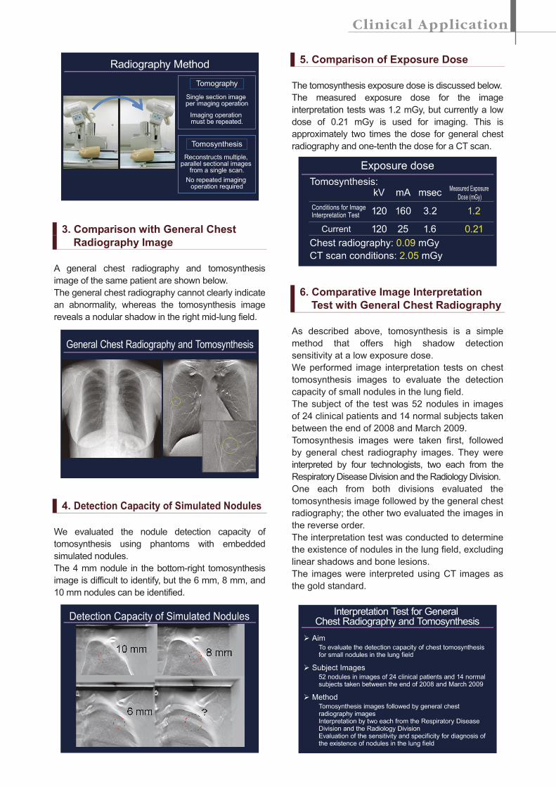
2. Tomosynthesis
The word “tomosynthesis” is a composite of
“tomography” and “synthesis.” This method can
reconstruct a coronal section at an arbitrary height
from a single tomographic imaging operation.
The tomography angle and tomography speed can
be set, in addition to the radiography conditions,