The Enzymes of Biological Membranes SECOND EDITION Volume 4 Bioenergetics of Electron and Proton Transport Edited by Anthony N. Martohosi State University of New York ...,·•• Syracuse, New York Plenum Press New York and London
Transcript
The Enzymes of Biological Membranes
SECOND EDITION
Volume 4
Bioenergetics of Electron and Proton Transport
Edited by
Anthony N. Martohosi State University of New York ...,·•• Syracuse, New York
Plenum Press · New York and London
Contents of Volume 4
46. The Enzymes and the Enzyme Complexes of the Mitochondrial Oxidative Phosphorylation System Youssef Hatefi, C. Ian Ragan, and Yves M. Galante
I . Introduction 1 I I . Complex I (NADH-Ubiquinone Oxidoreductase) 6
A. Composition of Complex 17 · B. Enzymic Properties of Complex 19 · C. Spectroscopic Properties of Complex I 10 · D. Resolution of Complex 111 · E. Structure of Complex 116 · F. Mechanism of Action of Complex I 18
I I I . Complex I I (Succinate-Ubiquinone Oxidoreductase) 20 A. Composition of Complex II 20 · B. Succinate Dehydrogenase 20 · C. Cytochrome b$w 23 · D. Activities of Complex II 25 · E. Ubiquinone-Binding Proteins 27
IV. Complex I I I (Ubiquinol-Cytochrome c Oxidoreductase) 28 A. Composition and Structure of Complex III 28 · B. Mechanism of Action of Complex III 32 · C. Inhibitors of Complex III 35
V. Complex IV (Ferrocytochrome c-Oxygen Oxidoreductase) 35 V I . Complex V (ATP Synthase) 36
A. Isolation of Complex V 36 · B. Composition of Complex V 37 · C. Structure of Complex V 39 · D. Role of the Subunits of Complex V 40 · E. Activities of Complex V 46
V I I . Mechanisms of ATP Hydrolysis and Synthesis 47 A. Mechanistic Considerations 47 · B. Structure of the ATPase Active Site 51 · C. The Nature of the "High-Energy" Intermediate 52
V I I I . Arrangement of Proteins in the Mitochondrial Inner Membrane 54 References 56
47. Proton Diffusion and the Bioenergies of Enzymes in Membranes Robert J. P. Williams
I . Introduction 71 A. Chemiosmosis and Local Domains 71
I I . Proton Diffusion 74 A. Models 74 · B. Summary of Inorganic Proton Channels 81
/χ
χ CONTENTS
I I I . The Injection of Protons to a Channel 81 IV. Biological Proton Channels: Introduction 83
A. Light-Activated Proton Migration 84 · B. Redox-Activated Proton Channels 86 · C. Gates 89 · D. The ATP-Synthase 93
V. The Kinases and ATP-Synthase: The F, Unit 100 A. Calcium-Binding Proteins—Energy Transfer 100 · B. The Energization of F0F, 101 · C. The ATP-ADP Reaction 102
V I . Proton Transfer from Generator to ATP-Synthase 104 References 107
48. Relationships between Structure and Function in Cytochrome Oxidase
Märten Wikström, Matti Saraste, and Timo Penttilä
I . Introduction and Scope I l l I I . Composition of the Enzyme 112
A. The Prosthetic Groups 112 · B. The Apoprotein 114 I I I . Quaternary Structure 114 IV. The Mitochondrial^ Coded Subunits 117 V. Cytoplasmic Subunits 119
V I . Location and Structure of the Prosthetic Groups 123 A. Copper of Subunit II 123 · B. Heme of Subunit II 126 · C. Heme and Copper of Subunit I 128
V I I . The Binding of Cytochrome c 133 VI I I . The Mechanism of Reduction of Dioxygen 134
IX. Electron Transfer, Proton Translocation, and Subunit I I I 136 A. Electron Transfer 137 · B. Proton Translocation and the Role of Subunit III 137 · C. Structure and Possible Function of Subunit III 139 · D. On Possible Mechanisms of Proton Translocation 140
X. Concluding Remarks 142 References 142
49. Η+-ATPase as an Energy-Converting Enzyme
Toshiro Hamamoto and Yasuo Kagawa
I . Introduction 149 I I . General Properties 150
A. Distribution 151 · B. Isolation 153 · C. Structure 154 D. Reconstitution from Subunits 156
I I I . Reconstitution into Lipid Bilayers 158 A. Phospholipids 158 · B. Proteoliposomes 158
IV. Ligand-Binding Activity 159 A. Nucleotide Binding 159 · B. Inhibitors 160
CONTENTS χ/'
V. Energy-Transducing Activity 161 A. ATP Synthesis 162 · B. ATP Formed on Nonenergized F, 163 · C. Exchange Reactions 163 · D. H + Translocation 164
V I . Mechanisms 167 References 170
50. The Proton-Translocating Membrane ATPase (FXFQ) in Streptococcus faecalis (faecium)
Adolph Abrams
I . Introduction and Historical Perspective 177 I I . The Fi ATPase Sector 178
A. Solubilization 178 · B. Purification and Characterization of the F, ATPase 179 · C. Binding Interactions between F, and F 0 180 • D. Attachment Factors 181 · E. Endogenous Nucleotides 182
I I I . The F,F 0 ATPase Complex 183 A. Inhibition by ΝΛ'-Dicyclohexylcarbodiimide (DCCD) 183 · B. The DCCD-Resistant Mutant of S. faecalis (SFdcc8) 184 · C. Isolation and Subunits of F,F0 185
IV. Physiological Role of the FjFo ATPase in 5. faecalis (faecium) 186 V. Concluding Remarks 190
Addendum 191 References 191
57. Cytochrome b of the Respiratory Chain
Henry R. Mahler and Philip S. Perlman
I . Introduction 195 I I . Structure 196
A. Isolation and Properties of Cytochrome b of Complex III 196 · B. Major Unanswered Questions Concerning the Cytochrome b Molecule 203
I I I . Function 205 A. Complex III [bcx Complex; Ubiquinol:Cytochrome c (Oxido)reductase, EC 1.10.2.2] 205 · B. Cytochrome b in Complex II 217
IV. Genetics and Biogenesis 218 A. Organization of Genes for Cytochrome ^ 218 · B. Expression and Regulation of Genes for Cytochrome b 219 · C. Mutations Leading to Resistance to Inhibitors of Complex III 224 · D. Biogenesis of Cytochrome b and Complex III 225 References 227
χ/'/' CONTENTS
52. Cytochrome b5 and Cytochrome b5 Reductase from a Chemical and X-Ray Diffraction Viewpoint F. Scott Mathews and Edmund W. Czerwinski
I . Introduction 235 I I . Cytochrome b5 237
A. Heme-Binding Fragment 237 · B. Structure 246 · C. Intact Cytochrome b5 265 · D. Nonpolar Polypeptide Fragment 268
I I I . NADH Cytochrome b5 Reductase 274 A. Soluble Catalytic Fragment 274 · B. Intact Enzyme 284
IV. Interactions among Components 285 A. Cytochrome b$ and b5 Reductase in Membrane Vesicles 285 · B. Reconstitution of the Fatty Acid Desaturase System 287 · C. NADPH-Cytochrome P-450 Reductase 289 · D. Structural Aspects of b5 Interactions 290
V. Evolutionary Relationships 291 A. Mitochondrial Cytochrome b$ 29\ * B. The Cytochrome b5 Fold 292
V I . Summary 294 References 295
53. Iron-Sulfur Clusters in Mitochondrial Enzymes
Thomas P. Singer and Rona R. Ramsay
I . Structure and Properties of Known Fe-S Clusters 301 I I . Detection and Analysis of Fe-S Clusters 304
A. Chemical Analysis 304 · B. Absorbance Spectrum 304 · C. EPR Spectrum 304 · D. Cluster Extrusion 305 • E. Mössbauer Spectroscopy 307 · F. Resonance Raman Spectroscopy 307 • G. Magnetic Circular Dichroism 308 · H. EXAFS Studies 309
I I I . Newer Knowledge of the Properties and Function of Fe-S Clusters in Mitochondrial Enzymes 309 A. Aconitase 309 · B. The Iron-Sulfur Protein of the Cytochrome b-C\ Complex 316 · C. ETF-Q Oxidoreductase 318 · D. Succinate Dehydrogenase 319 · E. NADH Dehydrogenase 323 References 326
54. The Structure of Mitochondrial Ubiquinol.Cytochrome c Reductase
Hanns Weiss, Stephen J. Perkins, and Kevin Leonard
I . Introduction 333 I I . Isolation and Cleavage 334
I I I . The Hydrophilic, Amphiphilic, and Hydrophobic Subunits 336 IV. The Cytochrome-c-Binding Subunit of Cytochrome Reductase . . 336
CONTENTS χ/'/'/'
V. Three-Dimensional Structures Determined by Electron Microscopy of Membrane Crystals 336
V I . Low-Resolution Structures Determined by Neutron Scattering in Detergent Solution 341
VI I . Topography of the Subunits within the Structure and Orientation of the Structure in the Membrane 343
. 344
55. The Mechanism of the Ubiquinol.Cytochrome c Oxidoreductases of Mitochondria and of Rhodopseudomonas sphaeroides
Antony R. Crofts
I . Introduction 347 I I . Redox-Linked Proton-Pumping Mechanisms 349
A. Protolytic Reactions 350 · B. Proton Wells and Proton Channels 351 I I I . Topological and Structural Aspects 352
A. The Mitochondrial Complex 352 · B. The Chromatophore Complex 354
IV. Kinetic and Thermodynamic Properties 355 A. Overview 355 · B. Mechanisms 356 · C. The Kinetics of Oxidation of the FeS Center 359 · D. The Mechanism of the Quinol Oxidase Site 360 · E. Role of the Quinone Pool 362 · F. Kinetics of Reduction of the High-Potential Chain in the Presence of Antimycin 364 · G. The Mechanism of Inhibition by UHDBT, UHNQ, and Myxothiazol 365 · H. The Mechanism of Quinone Reduction by the Complex 366 · I . The Quinone Reductase Site 368 · J. The Mechanism of Inhibition by Antimycin 370
V. Mechanism of the Complex as a Proton Pump 371 VI . Summary 373
References 374
56. Functions of the Subunits and Regulation of Chloroplast Coupling Factor 1 Richard E. McCarty and James V. Moroney
I . Introduction 383 I I . Isolation and Purification of C?! 385
A. Comparison of Isolation Procedures 385 · B. Criteria for Purity 387 · C. Removal of Rubisco Contamination 389 · D. Small-Scale Preparations 390
I I I . Structure and Physical Properties of CF! 390 A. Molecular Weight of CF, 391 · B. Subunit Stoichiometry 392 · C. Physical Properties of CF, 394
x/v CONTENTS
IV. Functions of the Subunits of CF, 395 A. The ε-Subunit 395 · B. The δ-Subunit 397 · C. The 7-Subunit 398 · D. The a- and ß-Subunits 400
V. Active-Inactive Transitions: Regulation 402 A. Thiol Activation 403 · B. Protease Activation 405 · C. Heat Activation 406 · D. Alcohol and Detergent Activations 407 · E. Is There a Chloroplast ATPase Inhibitor? 407
V I . Summary and Conclusions 408 References 408
57. Biosynthesis of the Yeast Mitochondrial Η+-ATPase Complex
Sangkot Marzuki and Anthony W. Linnane
I . Introduction 415 I I . Subunit Composition and Structure of the Yeast Mitochondrial
Η +-ATPase 415 I I I . Mitochondrially Synthesized Subunits 417
A. Mitochondrial Mutants with Lesions in the Structural Genes of the Mitochondrially Synthesized H + -ATPase Subunits 417 · B. Biosynthesis of Subunit 9 419 · C. Biosynthesis of Subunit 6 420 · D. Biosynthesis of Subunit 8 422 · E. The Site of Synthesis of the Mitochondrially Coded Η +-ATPase Subunits 423
IV. Cytoplasmically Synthesized Subunits 424 A. Structural Genes of the Cytoplasmically Synthesized Η +-ATPase Subunits 424 · B. F|-Subunits Are Synthesized in the Extramitochondrial Cytoplasm as Larger Precursors 424 · C. Transport of the FrSubunit Precursors across the Mitochondrial Membranes and Their Processing into Mature Subunits 425
V. Assembly of the Mitochondrial Η +-ATPase 426 A. In the Absence of Mitochondrial Protein Synthesis, Cytoplasmically Synthesized Η + -ATPase Subunits Are Assembled into a Membrane-Associated Complex 426 · B. Defect in the Assembly of the Mitochondrially Synthesized Subunits of Η +-ATPase in mir Mutants of Yeast 427 References 428
58. Synthesis and Intracellular Transport of Mitochondrial Proteins
Matthew A. Harmey and Walter Neupert
I . Introduction 431 I I . Synthesis of Nuclear Coded Proteins 434
I I I . Transport of Mitochondrial Precursor Proteins from Cytosol into Mitochondria 442
IV. Mitochondrial Recognition of Precursors 444 V. Insertion of Precursors into and Transport across Membranes . . . 447
CONTENTS xv
V I . Mitochondrial Proteases and the Processing of Precursors 450 V I I . General and Specific Transport Features of Individual Proteins
Destined for Different Compartments 452 V I I I . Proteins Coded for by the Mitochondrial Genome 456
IX. Conclusions 458 A. Precursors 458 · B. Recognition 458 · C. Translocation 458 · D. Proteolytic Processing 459 References 459
59. Plasma Membrane Redox Enzymes F. L . Crane, H. Low, and M. G. Clark
I . Introduction 465 I I . Intrinsic Enzymes 466
A. Endodehydrogenases 466 · B. Transdehydrogenases 478 · C. Ectodehydrogenases 493 · D. Dehydrogenases of Endocytic and Exocytic Vesicles 496
I I I . Cytochromes and Other Redox Carriers 497 IV. Extrinsic Dehydrogenases 500
References 501
60. The ADΡI ATP Carrier in Mitochondrial Membranes
Martin Klingenberg
I . Introduction ' 511 I I . Metabolic Localization of ADP/ATP Transport 512
I I I . The Mitochondrial Adenine Nucleotide Pool 512 IV. Kinetics 514 V. Energy Control of Exchange 518
V I . The Nucleotide Transport in the Reconstituted System 521 V I I . Inhibitors of ADP/ATP Transport 524
V I I I . Definition of Carrier Sites 525 IX. The Reorientation Mechanism of Ligand Interaction 530 X. Conformational Changes of the Membrane on Binding of ADP 533
XL The Influence of Amino Acid Reagents 535 X I I . The ADP/ATP Carrier in Submitochondrial (Sonic) Particles . . . 536
X I I I . The Isolation of the ADP/ATP Carrier 538 XIV. Physical Characteristics of the Isolated Carrier 539 XV. Chemical Characteristics 540
X V I . Conformational Change 543 X V I I . Transition of the Isolated Protein between the c-State
and the m-State 545
XV/ CONTENTS
X V I I I . The Carrier Mechanism 545 References 547
61. Bacteriorhodopsin arid Rhodopsin: Structure and Function Yuri A. Ovchinnikov and Nazhmutdin G. Abdulaev
I . Introduction 555 I I . Functional Characteristics of Bacteriorhodopsin and Rhodopsin 556
A. Bacteriorhodopsin 556 · B. Rhodopsin 557 I I I . Amino Acid Sequence of Bacteriorhodopsin 557 IV. Location of Bacteriorhodopsin in the Purple Membrane 559 V. Retinal-Binding Site 562
V I . Amino Acid Sequence of Bovine Rhodopsin 564 VI I . Retinal-Binding Site of Bovine Rhodopsin 567
VII I . Location of the Rhodopsin Polypeptide Chain in Membranes . . . 567 References 574
Index 579
58
Synthesis and Intracellular Transport of Mitochondrial Proteins Matthew A. Harmey and Walter Neupert
I. INTRODUCTION
The biogenesis of the mitochondrion represents the result of a coordinated synergism between two distinct and spatially separate genetic systems. Not only are these two systems separate but they also have distinct modes of transcription and translation (Barrell etal.t 1979; Borst, 1981; Borst and Grivell, 1978, 1981). Genetic and inhibitor studies on the development of mitochondria established that the majority of mitochondrial proteins are coded for by the nuclear DNA and are the products of translation on the cytoplasmic ribosomes (Lamb et aL, 1968; Schatz and Mason, 1974; Neupert and Schatz, 1981; Heinrich, 1982). As the protein constituents of mitochondria are distinct from the bulk of cellular proteins, some mechanism must exist for the sorting of proteins destined for the mitochondria.
Transport of proteins into mitochondria and assembly of mitochondrial membranes is one example of the general phenomenon of intracellular protein sorting and protein insertion into and translocation across cellular membranes. Similar reactions must occur for the assembly of other cellular membranes and compartments such as the plasma membrane and the endomembrane system including the endoplasmic reticulum, Golgi apparatus, transport vesicles, and endocytic vesicles, for peroxisomes and glyox-ysomes, and analogous to mitochondria, for chloroplasts. Furthermore, the transport of proteins out of the cell, i.e., secretion of proteins, requires basically the same reactions.
Matthew A. Harmey · Department of Botany, University College Dublin, Dublin, Ireland. Walter Neupert · Institute of Biochemistry, University of Göttingen, Göttingen, West Germany.
431
432 MATTHEW A. HARMEY and WALTER NEUPERT
The import of protein into organelles and organelle membranes is directly related to the problem of how membranes maintain their identity and their continuity in space and time. Since almost all proteins of the cell are made on cytoplasmic ribosomes but end up in a number of different compartments, specific mechanisms must exist to direct them into these compartments. The identity of a compartment is determined by the membrane surrounding this compartment. Thus, membranes must have devices to recognize not only newly made components for themselves but also for the compartment enclosed. Formation of cellular compartments or organelles, therefore, must entail as a first step recognition of new components or precursor proteins. This process must be highly specific since the compartment is believed to be absolutely unique. This recognition is, thus, the primary step in the formation of cellular membranes. Identity and continuity of membranes is determined by recognizing structures on their membranes. This explains at the same time why a membrane may undergo modulation but cannot arise de novo, since a membrane requires for its formation the continued presence of recognizing structures or "receptors" to maintain identity. Therefore, in a way, membranes are self-replicating structures. As in the replication of DNA, their formation entails an initial recognition. In the case of DNA replication, this is the base pairing step; in membrane replication, it is the binding of a newly made protein precursor to its receptor. The second step then is fixation; in the case of DNA replication, this is the formation of the diester bond between two nucleotides; in the case of membrane replication, this is the insertion into and or translocation across the membrane.
The details of these two basic steps are far from being understood. The second step, especially, appears to be particularly complex. We do not know how a polypeptide enters into and leaves a membrane. In the case of mitochondria, some proteins must traverse one membrane (the outer membrane) to reach another membrane (the inner membrane). Mitochondria have four clearly recognizable compartments (Ernster and Kuyienstierna, 1970) each with a distinctive protein complement related to the particular activities of each compartment, viz., outer membrane, intermembrane space, inner membrane, and matrix. Moreover, it is clear that the mechanisms involved in recognition of and transfer across membranes are not common to all the different sorting reactions. Two general mechanisms have been postulated, viz., cotranslational and posttranslational. In the case of secretory proteins which must pass into the lumen of the endoplasmic reticulum prior to secretion, a cotranslational mechanism has been shown to operate in all cases studied so far. The initiation of protein synthesis takes place on free polysomes. The emerging signal peptide of the nascent polypeptide chain is recognized by a signal recognition particle (SRP; Walter and Blobel, 1982) which arrests elongation until the complex of polysomes and SRP is bound to the microsomal membrane via the docking protein (Meyer and Dobberstein, 1980; Meyer et al.t 1982; Walter and Blobel, 1982). The complex may be further stabilized by interactions with the ribophorins (Kreibich et al.t 1978, Kreibich, 1982). The elongation recommences with the polypeptide being inserted into and across the membrane. A signal or leader peptidase on the luminal face of the ER cleaves the signal peptide to yield the mature polypeptide. This may already occur before the transmembrane journey is complete (Blobel et al.y 1979, Kreil, 1981). Proteins which do not contain a cleaved signal sequence have also been found. One example of a secreted protein is that of ovalbumin.
MITOCHONDRIAL PROTEINS 433
A number of proteins ending up in the plasma membrane which follow a very similar pathway are not initially synthesized with a cleavable sequence. One example is ovalbumin. A number of proteins ending up in the plasma membrane which follow a very similar pathway are not initially synthesized with a cleavable sequence (Kreil, 1981). This mechanism appears to obtain in the transfer of proteins into mitochondria, which will be discussed in detail. The weight of evidence indicates that proteins of these organelles are synthesized as precursors on free polysomes and are run off into the cytosol where they can be detected (Hallermayer et al., 1977; Harmey et al., 1977) and are subsequently imported into the mitochondria. It also appears that a posttran-slational mechanism works in the transport of proteins into chloroplasts, peroxisomes, and glyoxysomes and of some proteins into the endoplasmic reticulum.
Furthermore, insertion and translocation of at least a few proteins into or across the plasma membrane of bacteria has been found to occur by a posttranslational mechanism. In particular, the insertion of the major coat protein of the bacteriophage Μ13 has been studied in great detail (Wickner, 1980). It has been proposed that, in this case, no specific recognizing structure in the recipient membrane is required. It remains to be shown whether this mechanism is of a general importance or restricted to this phage protein. It seems, however, already clear from genetic studies that secretion of periplasmic proteins in bacteria requires protein components associated with the membranes (Inouye and Beck with, 1977; Emr and Silhavy, 1982). It has become apparent in recent years that there is not a clear demarcation between what is regarded as cotranslational and posttranslational transport. These two types may represent extremes of a system with graded intermediate conditions. For instance, it is not clear whether secretory proteins are translocated across the membranes in a linear fashion as the chain elongates or whether stretches of the nascent chain fold on the ribosomal side of the membrane and these folded "domains" are translocated across in a discontinuous fashion (Randall, 1983).
The transfer of proteins from cytosol to mitochondria is generally regarded as a one-way process, so that the ingress pathway is not available for exit. The mitochondria appear to be impermeable to added mature mitochondrial proteins (Neupert and Schatz, 1981). I f the internalized proteins behave in a similar manner, then the cleavage and ensuing conformational changes provide a mechanism for the containment of mature mitochondrial proteins and provide a logic for proteolytic cleavage. There have been a number of reports that mitochondria take up mature aspartate amino transferase (Marra et al., 1977) and more recently, the same group reported a similar uptake of malate dehydrogenase (Passarella et al., 1980). This process was considered as a model system for protein translocation. The described phenomenon cannot be clearly identified with precursor uptake; this is apparent from large number of differences between the characteristics of precursor uptake in vivo and the described systems (Sonderegger et al., 1980; Sakakibara et al, 1980; Aziz et al, 1981).
In approaching the problem of how mitochondrial proteins are transported into mitochondria, a number of questions may be posed, some of which can be answered while others can only be partly answered.
• How do precursor proteins travel through the cytosol • How are precursor proteins recognized by mitochondria? • How are proteins translocated across the mitochondrial membranes?
434 MATTHEW Α. HARMEY and WALTER NEUPERT
• How do proteins reach their specific compartment and how are they integrated into their functional locations?
• How are protein subunits assembled into multimeric complexes? We shall attempt to review the available answers to these questions.
//. SYNTHESIS OF NUCLEAR CODED PROTEINS
Of the total mitochondrial proteins, almost all are synthesized on cytoribosomes. This can be clearly demonstrated by the effect of cycloheximide on the incorporation of labeled amino acids into mitochondrial proteins. As this is a most effective inhibitor of cytoplasmic protein synthesis, it causes a striking decrease in the synthesis of mitochondrial proteins. It does not, however, prevent the import of proteins per se into the mitochondria and this is one of the observations that led to the proposal that import of mitochondrial proteins was a posttranslational phenomenon (Hallermayer and Neupert, 1977).
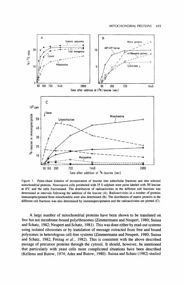
The biosynthesis of mitochondrial proteins in vivo has been followed principally by kinetic studies involving dual labeling and pulse- and chase-labeling studies carried out at low temperture (5-8°C). These studies have been particularly useful in following the synthesis of mitochondrial proteins in organisms such as Neurospora and yeast, but have also been applied to vertebrate cells. When coupled with the use of specific antibodies, kinetic studies have allowed investigators to locate and follow the movement of individual proteins from their sites of synthesis to their final location. Hal-lermayer and Neupert (1977) using Neurospora cells, and Schatz (1979) using yeast cells, demonstrated that newly synthesized mitochondrial proteins could first be detected in the cytosol of the cell and were subsequently imported into the mitochondria. A distinct time lag could be demonstrated between the two phenomena. Newly synthesized material could be recognized in the case of dual label studies such as those shown in Figure 1. Different lag times were found with different proteins suggesting that the various extramitochondrial precursor proteins have different extramitochondrial concentrations, or pool sizes. The transfer from the cytosol could be readily observed either by the application of a chase of cold amino acid or after additon of cycloheximide to stop translation.
The pool of precursor proteins, at least of those investigated, in the cytosol is very small; in fact, at temperatures of 20-30°C in both yeast and Neurospora cells, the size of the pool and the dwell time of the precursors is so small as to make it difficult to detect the precursors (Morita et al., 1982; Schatz and Butow, 1983; Hal-lermayer et al., 1977; Schmidt et al., 1983a,b). However, as will be discussed later, incorporation at low temperatures or in the presence of uncouplers has made the detection of many precursors possible.
In vitro synthesis of mitochondrial proteins has been shown by translation of extracted mRNA in either the rabbit reticulocyte lysate (Hunt and Jackson, 1974) or the wheat germ system (Roberts and Paterson, 1973). These translation systems have been used extensively in the synthesis of mitochondrial proteins as attested to in the data presented in Table 1. The list given is one to which new proteins are continually being added as the proteins and their precursor forms are identified.
MITOCHONDRIAL PROTEINS 435
20-
10
Cytosolic polysomes
· ·
..">-_-:—ν-—• .· ^ Total homogenale
• ' ^ z Cytosol _ - -o
' \ Mitochondria
/ /
90 360 720 1U0 2860 Time after addition of [ 3H] leucine (sec
90 360
10 3 cpm
•Β. 5 0 -
I Chase Mitochondria
L
90 180 360 720 1 U 0 Time after addition of 3 H-leucine [sec]
2880
Figure I . Pulse-chase kinetics of incorporation of leucine into subcellular fractions and into selected mitochondrial proteins. Neurospora cells prelabeled with 35 S sulphate were pulse labeled with 3H leucine at 8°C and the cells fractionated. The distribution of radioactivities in the different cell fractions was determined at intervals following the addition of the leucine (A). Radioactivities in a number of proteins immunoprecipitated from mitochondria were also determined (B). The distribution of matrix proteins in the different cell fractions was also determined by immunoprecipitation and the radioactivities are plotted (C).
A large number of mitochondrial proteins have been shown to be translated on free but not membrane-bound polyribosomes (Zimmermann and Neupert, 1980; Suissa and Schatz, 1982; Neupert and Schatz, 1981). This was done either by read-out systems using isolated ribosomes or by translation of message extracted from free and bound polysomes in heterologous cell-free systems (Zimmermann and Neupert, 1980; Suissa and Schatz, 1982; Freitag et al., 1982). This is consistent with the above described passage of precursor proteins through the cytosol. It should, however, be mentioned that particularly with yeast cells more complicated situations have been described (Kellems and Butow, 1974; Ades and Butow, 1980). Suissa and Schatz (1982) studied
Table 1. Cytoplasmic Precursors of Mitochondrial Proteins
Apparent Apparent molecular Post Energy or molecular weight in K: translational membrane
Mitochondrial weight in K: mature import potential compartment Protein Tissue precursor protein demonstrated dependence References
Outer membrane Porin Neurospora crassa
30 30 + None Freitag et al. (1982)
Porin Yeast 29 29 + None Mihara et al. (1982) 35K protein Rat liver 35 35 + None Shore tr al. (1981) Monoamine oxidase Rat liver 59 59 ND" ND Sagara and Ito (1982)
Intermembrane Cytochrome c Yeast 39.5 33.5 + + Maccechini et al. (1979a.b) space peroxidase
Cytochrome b2 Yeast 67 57 + + Gasser et al. (1982a,b) Adenylate kinase Chick liver 28 28 ND None Watanabe and Kubo (1982) Sulfite oxidase Rat liver 59 55 ND ND Mihara et al. (1982)
Inner membrane Cytochrome c Neurospora crassa
12 12 + None Korb and Neupert (1978)
Rat liver 12 12 + None Matsuura et al. (1981) ADP/ATP carrier Neurospora
crassa 32 32 + + Zimmermann et al. (1979a,b)
F0F| ATPase Neurospora 12 8 + + Michel et al. (1979) subunit 9 crassa
bc\ complex Neurospora 51 50 + + Teintze et al. (1982) subunit I crassa
Cytochrome c\ Yeast 44.5 44 + + Cote et al. (1979) Subunit II Neurospora
crassa 47.5 45 + + Teintze et al. (1982)
Yeast 40.5 40 + + Cote et al. (1979)
Matrix
Cytochrome C)
Subunit V
Subunit VI
Subunit VII
Subunit VIII
Cytochrome oxidase Subunit IV
Subunit V
Subunit VI Subunit VII
F, ATPase Subunit α Subunit β Subunit 7 Subunit β
Fi Inhibitor Citrate synthase
Neurospora crassa
Yeast Neurospora
crassa Yeast Neurospora
crassa Yeast Neurospora
crassa Yeast Neurospora
crassa Yeast
Rat liver Yeast Rat liver Yeast Yeast Yeast
Yeast Yeast Yeast Neurospora
crassa Yeast Neurospora
crassa
38
37 28
27 14
25 12
14
11.6
11 19.5 17 15.5 15 14 5
64 56 40
12 47
31
31 25
25 14
17 11.5
14 11.2
I I
16.5 14 12.5 12.5 12.5 5
58 54 34
10 45
+ + Teintze et al. (1982)
+ + Nelson and Schatz (1979) + + Teintze et al. (1982)
+ + Cote et al. (1979) ND ND Teintze et al. (1982)
+ ND Cote et al. (1979) + + Teintze et al. (1982)
ND ND Cote et al. (1979) ND ND Teintze et al. (1982)
ND ND Cote et al. (1979)
ND ND Heinrich (1982) ND ND Mihara and Blobel (1980) ND ND Heinrich (1982) ND ND Mihara and Blobel (1980) ND ND Mihara and Blobel (1980) ND ND Mihara and Blobel (1980)
+ + Maccechini et al. (1979) g 4- + Nelson and Schatz (1979) Ο + + Suissa and Schatz (1982) Q + + Zwizinski et al. (1983) g
+ + Yoshida et al. (1983) £ ND ND Harmey and Neupert (1979)
Ο
(Continued)
Table 1. (Continued)
Apparent Apparent molecular Post Energy or molecular weight in K: translational membrane
Mitochondrial weight in K: mature import potential compartment Protein Tissue precursor protein demonstrated dependence References
Malate Rat liver 38 36 ND ND Mihara et al. (1982) dehydrogenase Aziz et αϊ. (1981)
δ-Amino Rat liver 75 66 ND ND Yamamoto et al. (1982) levulinate 75 65 ND ND Ades and Harpe (1981) synthase Chick liver 76 68 ND ND Srivastava et al. (1982)
Glutamate Rat liver 60 54 ND ND Mihara et al. (1982) dehydrogenase
Superoxide Yeast 26 24 + + Autor (1982) dismutase
Ornithine carbamyl Rat liver 39.4 36 + + Mori et al. (1980) transferase 43 39 + + Conboy and Rosenberg (1981)
Morita et αϊ. (1982) Carbamyl phosphate Rat liver 165 160 + + Mori et al. (1979)
synthase Raymond and Shore (1979) Shore et al. (1979)
Serine pyruvate Rat liver 42 40 ND ND Oda et al. (1981) ami τ* υ transferase
RNA polymerase Rat liver 47 45 ND ND Lustig et al. (1982a,b) Aspartate amino Chick heart 47.5 44.5 ND ND Sonderegger et al. (1980)
transferase Rat liver 47 45 + ND Sakakibara et al. (1980) Ornithine amino 49 46 + ND Mueckler et al. (1982)
transferase Adrenodoxin Bovine
adrenal cortex
20 12 + + Nabi and Omura (1980)
—I —I X m 5
m -<
I PO
2: m e
' ND = not determined.
MITOCHONDRIAL PROTEINS 439
the synthesis of some 12 mitochondrial proteins with respect to the location of the messenger RNA for these proteins. They showed that polysomes bound to mitochondria contain messenger RNA for mitochondrial proteins, which is in accord with the findings of Kellems and Butow (1974). However, the bulk of the translatable RNA for these proteins was found to be associated with free cytosolic ribosomes. The occurrence of the mitochondrial bound polyribosomes has been explained by the fact that, in the presence of cycloheximide, the cytosolic precursor pools are so reduced that the mitochondrial capacity for import is far from saturated, a situation which could lead to incomplete nascent chains still attached to polysomes being bound to the mitochondria (Schatz and Butow, 1983).
The majority of the proteins listed in Table 1 have been shown to be synthesized in a form that is different to the mature mitochondrial protein (Figure 2). The commonest feature is that the initial translation product is larger by anything up to 10K daltons than the mature protein. These additional pieces are all amino terminal additions as far as have been determined (Viebrock et al., 1982; Kaput et al., 1982); none have yet been discovered on the carboxy terminus. A number of exceptions to this behavior have been found. Cytochrome c is synthesized as apocytochrome c which essentially is not any different to the holoprotein in terms of amino acid composition (Korb and Neupert, 1978). A further exception is the ADP/ATP carrier protein. The mature protein is an integral membrane protein of the inner membrane and the precursor is made on cytoribosomes in a form that has the same apparent molecular weight as the mature protein (Zimmermann et al., 1979a,b). Both of these proteins were shown not to have an amino terminal extension by synthesis in a heterologous system in the presence of 3 5S labeled formylmethionyl tRNA (Figure 3). I f the protein retains a 3 5S labeled amino terminus on being imported into the mitochondria then the precursor retained its initiating methionine, and can be adjudged to have no cleavable amino terminal extension piece. This has been shown to be the case for cytochrome c and
Figure 2. Transfer of the Neurospora a b C d Ζ f g precursor to subunit 9 of the Fi ATPase into Neurospora mitochondria (a-c) and into yeast mitochondria (d-g). Precursor was synthesized by translation of polyA RNA in a rabbit reticulocyte lys-ate. Following synthesis, a postribo- (pSu9) somal supernatant was prepared and isolated mitochondria incubated in this supernatant at 25°C for 30 min after (rnSll9) which subunit 9 was immunoprecipi- ^ ^ ^ ^ ^ ^ ^ ^ ^ ^ ^ ^ ^ tated from the mitochondria. Immunoprecipitates were analyzed by SDS-gel electrophoresis and autoradiography, (a) Precursor from reticulocyte lysate supernatent. (b) Transfer of precursor into Neurospora mitochondria, (c) Mature subunit 9 immunoprecipitated from 35 S labeled mitochondria. Transfer into yeast mitochondria, (d) Control transfer, (e)" After transfer* mitochondria were suspended in isotonic medium and treated with proteinase Κ for 60 min at 0°C. (f) Transfer in the presence of 2 μΜ valinomycin. (g) Transfer in the presence of 10 μΜ oligomycin, 4 μΜ antimycin A, and 1 mM KCN. pSu9, precursor protein; mSu9, mature protein.
440 MATTHEW A. HARMEY and WALTER NEUPERT
Synthesis _m vitro Isolated proteins
< ex.
^ - I -J Ρ
.2 ο ο ο ο ο ο Ο Ω. Ω. Ω. Ο Ο Ω.
Χ < < < Χ Χ <
ΜΓχ10"3 ': ' ' . O r i g i n
43 —
30 —
20 —
Figure 3. In vitro synthesis of apocytochrome c. Apocytochrome c was immunoprecipitated from rabbit reticulocyte (RL) and wheat germ (WG) translation systems programmed with Neurospora polyA RNA in the presence of 35S methionine or 3SS formylmethionyl-tRNA. The immunoprecipitates were subjected to SDS-gel electrophoresis and the dried gels autoradiographed. Holocytochrome c was immunoprecipitated from labeled mitochondria. Coomassie stained isolated proteins are included.
ADP/ATP carrier (Zimmermann et al., 1979a,b). This does not rule out the possibility of an unstable carboxy terminal extension, but there is no evidence for such an extension nor any precedent on any of the proteins studied to date. The outer membrane protein porin from Neurospora (Freitag et al., 1982) and from yeast (Mihara et al., 1982) has been found on translation to give a product which has the same molecular weight as the mature protein. In addition, Schatz has used labeling with 35S-fmet-tRNA to show that this protein in yeast does not have an additional amino terminal sequence (Gasser and Schatz, 1983). Adenylate kinase and monoamine oxidase from rat liver have been shown to behave in a like manner (Watanabe and Kubo, 1982; Sagara and Ito, 1982). The matrix protein isopropyl malate synthase has also been shown to be made without an amino terminal extension (Hampsey et al., 1983). No correlation
MITOCHONDRIAL PROTEINS 441
between the presence and absence of an additional sequence and its final location can thus be made. Perhaps one exception is that, until now, no proteolytic processing has been found to be involved in the assembly of outer membrane proteins.
Structural characteristics of precursor proteins have proved quite difficult to determine, principally because it is quite difficult to procure sufficient quantities of most precursors to carry out such studies. Nonetheless, it has been possible in a few instances to obtain reasonable amounts of precursor. The apo form of cytochrome c prepared by the removal of the heme moiety is not distinguishable from the native precursor (Korb and Neupert, 1978). A water soluble porin has been prepared which will bind to mitochondria and insert into the outer membrane in a manner identical to the native precursor (Freitag and Neupert, unpublished). Transport studies which will be discussed below have also provided a means of accumulating precursor proteins in the cytosol in yeast cells (Reid and Schatz, 1982). Notwithstanding the problems of acquiring sufficient amounts of the precursors, a number of general features have been ascribed to these precursors. A l l appear to be quite soluble in a hydrophilic medium, i.e., the cytosol, although the mature proteins are in many cases extremely hydrophobic intrinsic membrane proteins (Klingenberg, 1976). A l l appear to have different conformations from their mature counterparts, e.g., the antibody to apocytochrome c does not react with holocytochrome c (Korb and Neupert, 1978). Kraus et al. (1981) have also described differences in reaction to antibodies in the case of ornithine transcarbamylase. The precursor to the ADP/ATP carrier does not bind carboxyatractyloside, a characteristic inhibitor of the functional carrier (Schleyer et al., 1982). In those precursors having amino terminal extensions, one can predict an influence on the conformation depending on the size and nature of the extension piece. In the small number of extensions which have been sequenced so far, they appear to be basically hydrophilic and may assist in the maintenance of the precursors in a soluble form in their aqueous milieu (Viebrock et al., 1982; Adolphus et al., 1982; Kaput et al., 1982). A further feature of many of the precursors is a marked tendency to form aggregates (Zimmermann and Neupert, 1980; Schmidt et al., 1983a,b; Mori et al., 1979; Lewin et al., 1980). This may be a means of retaining solubility by the masking of hydrophobic regions of the proteins by hydrophobic interactions between precursor subunits.
One characteristic of the precursors is that they are selectively recognized by the mitochondria. Korb and Neupert (1978) demonstrated that, in the presence of a large excess of holocytochrome c, the apo form was selectively taken up by the mitochondria. The interpretation of this is that the configuration of the precursor is sufficiently different to allow the mitochondrial recognition system to differentiate between the precursor and mature forms and that, in effect, these two forms do not compete for uptake by the mitochondria. Differential stability of precursors to proteolytic enzymes has also been described (Harmey and Neupert, 1979; Jaussi et al., 1981).
The genes coding for mitochondrial precursor proteins have recently been isolated in a number of laboratories. Viebrock etal. (1982) described the cloning and sequencing of the messenger RNA for subunit 9 of the ATPase ("proteolipid" or "DCCD-binding protein") and so the entire amino acid sequence could be deduced. The amino terminal extension of 66 amino acids has a high percentage of polar and basic amino acids while the mature protein of 81 amino acids is rich in hydrophobic components. Kaput
442 MATTHEW Α. HARMEY and WALTER NEUPERT
et al. (1982) have cloned and sequenced the gene for cytochrome c peroxidase and consequently have been able to propose a model for the shape and insertion of this protein in the mitochondrial membrane prior to proteolytic processing. The amino terminal extension of 68 amino acids contains a stretch of 23 nonpolar residues, including ten consecutive alanines, and is basic in character. These 23 amino acids have been suggested to span the inner membrane as an α-helix, with the more polar stretch of the first 18 amino acids protruding into the matrix. There appears to be no similarity between the presequence of the proteolipid and the cytochrome c peroxidase. It has not been possible to detect common sequences, nor any size uniformity between the presequences of the various precursor proteins. There is currently a burgeoning interest in the cloning approach to the study of precursor proteins as it allows an amino sequence determination readily and it is hoped that it may be exploited to provide increased amounts of precursors to allow the construction of simplified reconstitution systems to study precursor transport and also to allow conformational considerations with a view to understanding the molecular details of precursor movement into and across membranes.
///. TRANSPORT OF MITOCHONDRIAL PRECURSOR PROTEINS FROM CYTOSOL INTO MITOCHONDRIA
The transport process is really a composite of a number of integrated processes. In order to dissect the steps involved in transport in vitro, assays of import were devised (Harmey et al., 1977; Maccechini et al., 1979a; Zimmermann and Neupert, 1980). These assays were based on the provision of a pool of precursor proteins either by read-out or by translation of isolated mRNA. Incubation of mitochondria in postri-bosomal supernatants of such systems led to uptake of precursors into the mitochondria. While the precursors were external to the mitochondria, they were sensitive to added proteases; however, when internalized, the proteins became inaccessible to such proteases. Termination of the proteolysis allowed immunoprecipitation and localization of the imported proteins (Figure 2). These assays allowed a comparison of newly converted proteins with the external precursor forms (Neupert and Schatz, 1981).
That the proteins have reached their final destination and have been integrated into their functional location is difficult to demonstrate in an unequivocal manner and can only be deduced from alterations in the characteristics of the precursors en route. Apocytochrome c is antigenically different to its holo counterparts (Korb and Neupert, 1978). The transport of the apo molecule into the mitochondria results in the covalent attachment of heme and results in a change in conformation such that the antibody to the apo enzyme no longer recognizes the imported protein. The ADP/ATP carrier on transfer to the mitochondria acquires the ability to bind carboxyatractyloside, a specific inhibitor of the carrier, and to reorient its subtrate binding sites in the same manner as the functional carrier molecule (Zimmerman and Neupert, 1980; Schleyer and Neupert, unpublished). Furthermore, the mature protein binds both ATP and carbox-yatracytloside, while the precursor does not (Zimmermann and Neupert, 1980). In the case of subunit 9 of the F! part of the F Q F J ATPase, the imported subunit can be
MITOCHONDRIAL PROTEINS 443
immunoprecipitated from the mitochondria subsequent to import, by antibody to the F! ATPase, while the precursor is not recognized by this antibody (Schmidt et al., 1983a,b). Lewin and Norman (1983) have observed that subunits of F, ATPase imported into isolated yeast mitochondria become assembled into F complexes. Gasser et al. (1982a,b) and Daum et al., (1982a) studied uptake of a number of proteins into yeast mitochondria and demonstrated by fractionation of the mitochondria that the imported proteins were now located in the same compartment as their mature counterparts. They also were able to show by nondestructive criteria that preproteins requiring proteolytic processing had reached the matrix. These cited instances suggest that the imported proteins reach and at least in some instances are integrated into their functional locations.
It was found that the import of most proteins into mitochondria was inhibited in vivo and in vitro by treatment with uncouplers of oxidative phosphorylation (Haller-mayer and Neupert, 1976; Harmey etaL, 1977; Nelson and Schatz, 1979; Zimmermann etal., 1981; Reid and Schatz, 1982; Schleyer etaL, 1982; Mori etaL, 1980; Kolansky et al., 1982; Jaussi et al., 1981; Daum et aL, 1982b). There are, however, proteins whose import is not energy dependent; these exceptions wil l be considered later. Table 1 lists many proteins whose import has been shown to be energy dependent (see also Figure 2). The one common feature that is shown by the precursors whose transport requires coupling of oxidation and phosphorylation is that at some time they are either inserted into or actually cross the inner mitochondrial membrane (Gasser et al., 1982a&b). Which form of energy is required for this transport of proteins? The application of uncouplers leads to a depletion of ATP due to the continuing action and stimulation of the oligomycin-sensitive ATPase (Stigall et aL, 1979) and also to discharge of the membrane potential. It became important to diagnose which of these events, viz., the dissipation of the membrane potential or the depletion of ATP is responsible for the inhibition of the transport process. A series of experiments reported by Schleyer et al. (1982) demonstrated that in Neurospora it is the electrochemical potential across the inner membrane that is required for the import of proteins into or across this membrane. They studied the transport of the ADP/ATP carrier under conditions where (1) the ATP was high and the membrane potential depleted, and (2) ATP was low and membrane potential high. In the first instance, in vitro transfer of proteins into mitochondria was performed in the presence of CCCP and oligomycin. The oligomycin inhibits the ATPase while the CCCP dissipates the membrane potential. Under these conditions, the ATP level in the mitochondria was high due to the fact that in uncoupled mitochondria the ADP/ATP carrier system tends to equilibrate the ATP inside and outside the mitochondria and the incubation medium had a high level of ATP. The transport of all proteins studied except porin and cytochrome c was inhibited (Schleyer et aL, 1982; Teintze et aL, 1982). On the other hand, in the second instance when the mitochondria were first treated with oligomycin and carboxyatractyloside to deplete their ATP pool but to leave the membrane potential intact, transport of the proteins proceeded uninhibited. Similar results have been obtained for yeast cells by Schatz and his group (Gasser et aL, 1982a,b) and for rat liver by Conboy and Rosenberg (1981).
In a further series of experiments (Zwizinski et aL, 1983), mitochondria were incubated in the presence of antimycin A and oligomycin and incubated in a reticulocyte
444 MATTHEW Α. HARMEY and WALTER NEUPERT
lysate supernatant. This treatment prevents the generation of a membrane potential and so inhibits the transport of the precursors into the mitochondria. It does not, however, prevent the binding of the precursors to the outer surface of the mitochondria where they remain sensitive to added proteases. The membrane potential could be restored by the addition of ascorbate and TMPD which funnels electrons through cytochrome c to cytochrome oxidase (Wikstrom and Krab, 1982). The reestablishment of a membrane potential restored the transport of the precursor proteins. This experimental procedure also differentiates between binding and transport. On washing, the mitochondria retained the bound precursors and, on restoration of the membrane potential, the bound precursors were directly imported from their bound location. These results not only differentiate between binding and transport but they further show that the membrane potential is necessary only for the transfer across or insertion into the inner membrane.
Schatz (1979) and Nelson and Schatz (1979) have observed that the transfer of a number of mitochondrial precursor proteins does occur in rho- mutants of yeast. These cells lack a functional oligomycin-sensitive ATPase and also some essential components of the respiratory chain and, thus, cannot produce a membrane potential either by respiration or ATP hydrolysis (Nelson and Schatz, 1979). They do, however, have an ATP/ADP carrier which can internalize ATP in an electrogenic manner (Klingenberg and Rottenberg, 1977) and so may generate a membrane potential. More recent reports by Gasser et al. (1982a) based on the use of CCCP, oligomycin, and valinomycin have indicated that the transport of precursor proteins into yeast mitochondria may be driven by either a pH gradient, electrical potential, or both. In this context, it is interesting to note that Schleyer et al. (1982) found that addition of nigericin, which dissipates the proton gradient by exchanging K + for H + did not inhibit the uptake of the ADP/ATP carrier precursor. This finding would argue against a proton gradient being involved in the transport phenomenon. However, much more detailed studies are necessary to understand the exact role of the membrane potential in the transport process.
Outer membrane proteins typified by mitochondrial porin have been shown in Neurospora and in yeast to be posttranslational ly incorporated into a protease-in sensitive location in an energy-independent manner (Freitag et aL, 1982; Mihara et aL, 1982). Apocytochrome c has also been shown to be transported into mitochondria without dependence on a membrane potential as reflected by responses to added uncouplers or ATP levels. Only proteins whose transport is governed by the inner membrane appear to require a membrane potential for their uptake. Different proteins, therefore, appear to use different pathways to reach their final functional destination subsequent to recognition by the mitochondria.
IV. MITOCHONDRIAL RECOGNITION OF PRECURSORS
In all cases of transport, one common feature applies, viz., the precursors are initially bound tightly (Hennig et aL, 1983; Zwizinski et al., 1983) to the outer membrane surface. The bound precursors while resistant to washing can be readily
MITOCHONDRIAL PROTEINS 445
removed by added proteases or can be exchanged for added precursor. Pretreatment of mitochondria with trypsin destroys their ability to import precursors and to specifically bind them on their surface (Gasser et aL, 1982a; Zwizinski, unpublished). These findings clearly indicate the presence of recognition proteins on the outer mitochondrial surface which mediate the binding of the precursors.
The mitochondria only bind the precursor protein form and the binding has been clearly shown to be selective. When mitochondria were incubated in the presence of labeled apocytochrome c and a large excess of unlabeled holocytochrome c, the apo form was selectively bound and the binding was not inhibited by the presence of a large excess of holocytochrome c (Korb and Neupert, 1978). Well-defined binding studies have been few because of the problem of unavailability of reasonable quantities of purified precursor proteins. Two proteins exist, however, whose precursors can be produced in reasonable quantities, viz., cytochrome c and porin.
Hennig et al. (1983) have studied the binding of apocytochrome c to mitochondria in the presence of the protoheme analogue deuterohemin. Under these conditions, the conversion of the apo to holo form was inhibited and import of the apocytochrome c did not take place. They found that binding takes place in a saturable manner. Using Scatchard plots, high-affinity binding sites with a frequency of 60-90 pmoles/mg mitochondrial protein were demonstrated and a Ka of 2.2 x 10⁷ M " 1 was obtained. A low-affinity component was also detected; however, only the high-affinity sites were sensitive to trypsin (Koehler et aL, 1983). Apocytochrome c did not compete with other mitochondrial preproteins for the binding sites. The only proteins which were found to displace bound apocytochrome c from Neurospora mitochondria were apo-cytochromes from other species (Figure 4). The apocytochrome c from Paracoccus was exceptional in that it had no effect on the binding behavior of the homologous apocytochrome c. This may be due to the fact that Paracoccus is a prokaryote and that its apocytochrome c lacks the highly conserved sequence at amino acid positions 60-80 present in all eukaryotic cytochromes c and which was implicated in the binding of apocytochrome c (Matsuura et aL, 1981; Hennig and Neupert, unpublished). The other apocytochromes inhibited the binding process to a varying degree; the extent of inhibition may be related to their phylogenetic affinity to Neurospora cytochrome c, a behavior which emphasizes the specificity of the binding reaction. The possibility that a nonspecific charge interaction is involved in the apocytochrome c binding can be discounted in view of the fact that neither holocytochrome c nor amino terminal fragments with similar charge to the whole apomolecule could displace the bound apo molecules. Poly lysine and apocytochrome c denatured by repeated freezing and thawing were also ineffective in displacing the bound apocytochrome (Hennig et al., 1983). It appears that apocytochrome is recognized by some surface component of the outer mitochondrial membrane with a high degree of specificity. The exact nature of the surface component is presently under investigation.
Freitag and Neupert (unpublished) have recently isolated porin from Neurospora mitochondria and transformed it into a water soluble form which is very similar in behavior to the precursor protein synthesized in vitro. This water soluble form of porin binds to mitochondria at low temperature in a rapid and saturable manner, while the conversion to the mature form proceeds slowly at the low temperature. The water-
Figure 4. Competition between Neurospora apocytochrome c and apocytochrome c from various species for binding to Neurospora mitochondria. Neurospora mitochondria were incubated in the presence of deuterohemin (10 nmoles mg mitochondrial protein) for 5 min at 25°C. Apocytochrome was synthesized in a cell-free homogenate in the presence of 3H leucine for 10 min, after which time further protein synthesis was blocked by the addition of cycloheximide. A postribosomal supernatant was prepared and the deute-rohemin-treated mitochondria were incubated in this supernatant for 15 min at 25°C. Following incubation, the mitochondria were reisolated and washed in a sucrose-MOPS buffer. The mitochondria containing the bound apocytochrome c were then incubated in an unlabeled postribosomal supernatant containing varying amounts of apocytochrome from different sources. After equilibration of free and bound apocytochrome c, the mitochondria were reisolated, twice washed, and lysed with 1% Triton X-100. Apocytochrome was immunoprecipitated from the lysates and analyzed by SDS-gel electrophoresis. Radioactivity was determined in the sliced gels by scintillation counting (From Koehler et al., 1983).
soluble porin, therefore, may be used as a second system to study mitochondrial precursor recognition. One interesting feature emerged from the binding studies with porin and that was that increasing amounts of water-soluble porin when bound to mitochondria inhibited the transport of a fraction of the bulk precursor proteins into the mitochondria. This would infer that porin and some other precursor proteins share the same recognition sites.
A further feature of the specificity of the mitochondrial receptors is that precursor proteins from one species are recognized by mitochondria from another phylogeneti-cally far-removed species. Not only are such proteins specifically recognized but they are also processed to the correct mature form. For instance, it has been observed that rat liver and yeast mitochondria import several Neurospora precursor proteins such as the ADP/ATP carrier, porin, or the Fi ATPase subunit 9 (Zimmermann and Neupert,
MITOCHONDRIAL PROTEINS 447
1980, Schmidt et al., 1983a; Freitag et aL, 1982; Hennig et aL, 1983). Furthermore, mitochondria from rat kidney were found to import the precursor of rat liver ornithine transcarbamylase, an enzyme only present in liver mitochondria (Mori et aL, 1980).
Subunit 9 of the F] ATPase is an especially interesting protein in that, in yeast, this protein is coded by the mitochondrial genome and is not translated as a larger precursor (Macino and Tzagoloff, 1979), whereas, in Neurospora crassa, it is coded by a nuclear gene and translated as a larger molecular weight precursor (Michel et aL, 1979; Schmidt et aL, 1983b). The yeast mitochondria can selectively bind this precursor and cleave off the prepiece in the same manner as Neurospora. It would appear, therefore, that the receptor proteins are highly conserved in evolution. With respect to the number of different receptor proteins on the mitochondria, it would appear that more than one type exists, as apocytochrome c does not compete for the binding sites of other precursor proteins (Zimmermann et al., 1981). However, it is not clear how many recognition proteins are involved in the binding of precursors. It is clear, on the other hand, that each mitochondrial protein cannot have its own specific receptor. A detailed analysis has to wait until more precursor proteins are available on such scale that the ligand receptor interaction can be studied in the same manner as in the case, e.g., of hormone receptors of the plasma membrane.
V. INSERTION OF PRECURSORS INTO AND TRANSPORT ACROSS MEMBRANES
Subsequent to binding to the outer membrane, the precursors must either insert into a membrane as in the case of porin or else be translocated across a single membrane as apocytochrome c or be translocated across the outer membrane and inserted into the inner membrane, e.g., ADP/ATP carrier protein. For some proteins, transport across both the outer and the inner membrane is necessary. How can the transition from receptor-bound precursor to translocated precursor be analyzed? As outlined above, the mitochondrial proteins can be divided into two general groups in respect of transport: (1) those requiring no membrane potential, and (2) those which require a membrane potential. Apocytochrome c which belongs to the first of these groups is bound to mitochondria but not translocated in the presence of deuterohemin. I f an excess of protohemin is added to the mitochondria, the inhibition of heme addition is reversed and the mitochondria import the bound apocytochrome c directly from its bound location on the mitochondrial outer surface. In studies on the import of the outer membrane porin by Neurospora mitochondria, Freitag et al. (1982) have shown that binding of the precursor protein can take place at 0°C but the insertion of the proteins into the outer membrane is slow at this temperature. The bound protein can be distinguished from the inserted protein on the basis of its sensitivity to added protease. The precursor form of porin is tightly bound to the mitochondria and is not readily washed off. On transferring mitochondria with precursor porin bound to the outer membrane to a higher temperature, the porin is immediately inserted into the outer membrane (Freitag et al., 1982). In this instance, the insertion of the bound molecules takes place without any apparent detachment of the bound molecules. Mihara
448 MATTHEW Α. HARMEY and WALTER NEUPERT
et al. (1982) have also found that yeast porin synthesized in rabbit reticulocyte lysate was bound and inserted into yeast mitochondria. However, attempts to bind it to or insert it into other cell membranes failed. Recently, Gasser and Schatz (1983) showed that yeast porin inserted into isolated outer mitochondrial membrane.
It is possible that the recognition of binding sites may play a role in positioning the precursors in such a way that the next step, membrane insertion or transmembrane transport, can occur. Zwizinski et al. (1983) showed that ADP/ATP carrier binds to mitochondria in the presence of antimycin A and oligomycin. Subsequent restoration of a membrane potential resulted in the translocation and insertion of the bound carrier directly into a protease-resistant carboxyactractyloside binding location.
The majority of the mitochondrial precursor proteins carry an additional polypeptide sequence and must, therefore, be processed proteolytically before they can be integrated into their final location. In view of the fact that all reports to date indicate that the initial proteolytic processing takes place in the matrix (Boehni et al.t 1980; Schatz and Butow, 1983), then it follows that all larger molecular weight precursors must be exposed in total or in part to the matrix protease at some stage during processing. The import of all of the precursor proteins destined for the inner membrane and beyond has been found to require a membrane potential. A question immediately springs to mind, viz., which of the two functions, transport across the membrane or proteolytic processing requires the membrane potential or do both processes require a membrane potential? Two lines of experimental evidence can be offered to provide the answer. Zwizinski et al. (1983) inhibited the processing of the Β subunit of Fi ATPase by Neurospora mitochondria in a reticulocyte lysate postribosomal supernatant by EDTA and ophenanthroline (Figure 5). The unprocessed protein was transported into the mitochondria, clearly showing that import and processing are independent phenomena. The second line of evidence comes from the results of the Schatz group. They have shown that the simultaneous import and processing of the ß-subunit of
Processing of Pre-Fj β - S u b u n i t Import of Pre-F, β - S u b u n i t Imported in the Presence of o-Phenanthroline
in the Presence of o-Phenanthroline Mn2+(2mM) -
ό Ά < * < Β Protease -
Figure 5. Import of precursor to ß-subunit of the F| ATPase in the presence of o-phenanthroline and EDTA. Isolated mitochondria were incubated with 35 S labeled reticulocyte lysate supernatant. Transfer was performed in the presence of EDTA 7.5 mM and 100 μΜ o-phenanthroline (A). Mitochondria were allowed import pre β in the presence of o-phenanthroline and EDTA and were incubated in the presence and absence of MnCl (B). Protease sensitivity was assessed by treatment of the mitochondria at 0°C with proteinase Κ (Zwizinski et al., 1983).
MITOCHONDRIAL PROTEINS 449
ATPase required a membrane potential. Proteolytic processing of the precursor could, on the other hand, be carried out by hypotonic extract of the mitochondria (Gasser et al., 1982a). Kolansky et al. (1982) have found similar behavior in the case of the translocation of the precursor of ornithine transcarbamylase across the inner membrane of rat liver mitochondria. The presence of uncoupler was without effect on processing by hypotonic extracts. Furthermore, Reid and Schatz (1982) found uncleaved precursors in yeast mitochondria, the further processing of which was independent of added CCCP. It may, therefore, be concluded that it is the transport across the inner membrane that requires the membrane potential.
In yeast, the import of cytochrome b2, an intermembrane space enzyme, has been shown to require a membrane potential. Similarly, import of cytochrome c l whose functional location is on the outer side of the inner membrane is energy dependent. The processing of the precursors of both of these proteins requires exposure to the matrix. Therefore, it is necessary for them to cross the inner membrane, hence, the requirement of a membrane potential (Gasser et al., 1982a,b).
The details of how precursor proteins cross the mitochondrial membranes are not known. In fact, the same can equally be said about proteins crossing cellular membranes in general (Wickner, 1980; Randall, 1983). In her studies on protein translocation in E. coli., Randall produced evidence more in favor of the idea of domains of protein crossing the membranes stepwise than of the progressive linear insertion of the emergent nascent chain. A similar type of situation could be envisaged for the translocation of mitochondrial proteins where, at least in vitro, whole polypeptides can cross the membranes. Some precursor proteins have a quite large molecular weight such as precarbamyl phosphate synthase (165 K) and a folded diameter at least equal to that of the membrane bilayer. The problem is how does such a molecule with a predominantly hydrophilic exterior pass through both mitochondrial membranes. Are there translocator proteins which combine with these precursors and carry them across the membrane? The presence of surface receptor proteins is generally agreed but the question of translocators cannot yet be answered. One could envisage some combination of the hydrophilic precursor being necessary to modulate their hydrophilic character and thus facilitate their passage through the lipid bilayer. Little attention has been paid to the role of the membrane lipids in the process of translocation. Tanner and his group (Komar et al., 1979) have shown that the energy status of the cells has a marked effect on the sensitivity of the membranes to added detergents. When the membranes are energized, they are susceptible to damage by detergents such as Triton X-100. On the other hand, if the cells are treated with uncouplers to deplete their membrane potential, they become insensitive to the detergents. This would suggest that the membrane potential makes the lipid bilayer more labile or penetrable and may go some way towards explaining the need for a membrane potential in protein translocation. Curiously, the magnitude of the potential does not appear to be related to uptake or the threshold potential required may be rather low. Energized membranes have a definite polarity. In mitochondria and in bacteria, the membranes are negatively charged on the inside, yet the direction of protein translocation is opposite in both. It would not appear that translocation is a simple electrophoretic phenomenon. For some of the precursors investigated (Viebrock et al., 1982; Kaput et al., 1982), the pre-
450 MATTHEW A. HARMEY and WALTER NEUPERT
sequences were predominantly basic and would therefore hardly be attracted to the positively-charged face of the membrane which faces out. However, one could envisage the binding of the precursor to the receptor leading to the positioning of the precursor in such a way that positively-charged domains could now respond to the negatively-charged inside of the membrane. Not all of the precursors have been shown to be more basic than the mature proteins. Wada and his group have described the precursor to aspartate amino transferase as being more acidic (Kamisaki et al., 1982). It remains to be shown whether the presequences play a direct role in translocation across membranes or whether their role is to alter the conformation of the proteins so that recognition, or receptor attractive, domains are exposed which are hidden or masked in the mature proteins. The whole question of how the actual translocation takes place is one of the challenging mysteries of cell biology today.
A further unresolved problem is how polypeptides destined for the inner membrane or the matrix space traverse the outer and inner membranes. Are there two individual steps which can be experimentally separated or does translocation occur in a single step across both membranes? In the case of the second alternative, the two membranes would have to come into intimate contact or even fuse at certain points. The answers to these questions are really not known. Contact sites of outer and inner membranes have repeatedly been reported on the basis of electron microscopic studies. Kellems et al. (1975) have described the preferential association of cytoplasmic polyribosomes at sites where outer and inner membranes were in close proximity. This could be taken as an indication that precursor proteins cross both outer and inner membranes at specific sites in a single step.
VI. MITOCHONDRIAL PROTEASES AND THE PROCESSING OF PRECURSORS
The processing of imported precursors involves, in most cases, a proteolytic cleavage of a larger molecular weight precursor. In a number of instances, proteolysis may involve two proteolytic steps (Neupert and Schatz, 1981). In the case of cytochrome Ci and cytochrome b2 of yeast, two separate proteases are involved; other phenomena such as heme addition may attend this process (Gasser et al., 1982b).
In all cases studied so far, the initial proteolytic event appears to be carried out by a metal-dependent enzyme located in the mitochondrial matrix first described by Boehni et αϊ. (1980). They found that a hypotonic extract of rat liver and of yeast mitochondria could cleave the precursors of the β- and 7-subunits of the F{ ATPase to the correct mature size. Fractionation of the mitochondria indicated that the enzyme was located in the matrix. They also found that a similar enzyme was present in the matrix of rat liver mitochondria. The proteolytic activity was inhibited by chelating agents such as o-phenanthroline and EDTA, but unaffected by inhibitors such as TLCK/ TPCK and PMSF. Gasser et αϊ. (1982a,b) showed that hypotonic extracts of yeast mitochondria could process the precursors of cytochrome cx and cytochrome b2. In this instance, however, the matrix enzyme cleaved the precursors to the intermediate form but not to the mature form. The final proteolytic step was heme dependent. The
MITOCHONDRIAL PROTEINS 451
assay of these proteases presents problems in that it is extremely difficult to set up standard assays using precursor proteins as substrates.
A partially purified enzyme preparation from rat liver has been described by Miura et al. (1982) which cleaved the precursor of ornithine transcarbamylase to a size intermediate between that of precursor and mature size. It remains to be shown that this activity is actually involved in the in vivo processing since the significance of the intermediate form is not known. McAda and Douglas (1982) suggested that the me-talloprotease from yeast is a dimeric protein with a molecular weight of 105,000 since, in partially purified preparations, a 59000 M r polypeptide was prominent. However, from the data by others (Boehni et al., 1983; Miura et al., 1982), the question of the enzyme being monomelic or dimeric remains open. McAda and Douglas (1982) found that low levels of the detergents Triton X-100 and deoxycholate inactivated the enzyme, but this inactivation could be reversed by the addition of phospholipids. Recently Schmidt et al. (1983b) have partially purified an enzyme from whole Neurospora cells which can process a number of precursors. The precursor to cytochrome c} was cleaved to the intermediate form and the precursor of subunit 9 of the F! ATPase through an intermediate to the mature protein. Attempts to separate this activity into two separate entities have not yet been successful.
The only firm consensus on the nature of the protease responsible for the proteolytic cleavage of the mitochondrial precursor protein is that it is a soluble matrix located enzyme which has a requirement for divalent metals such as Zn 2 + , M n 2 + , or C o 2 + and can be inhibited by chelating agents. Reports on the reversibility of inhibition are contradictory.
A second protease has been postulated for the final processing of precursors of proteins such as cytochrome b2, cytochrome c peroxidase, and cytochrome c{ (Gasser et al., 1982b). Cytochrome c peroxidase is an intermembrane space protein as is cytochrome b2 (Daum et al., 1982a,b)and their processing by a matrix enzyme appears to involve a detour of all or part of the molecule across the inner mitochondrial membrane. This is consistent with the need for a membrane potential in the processing of these proteins by whole mitochondria as discussed above. In the case of cytochrome cu a second heme-dependent cleavage has been postulated to take place at the outer face of the inner membrane (Ohashi et al., 1982). A similar location has been suggested for the second proteolytic step in cytochrome b2 (Gasser et al., 1982b). This would locate the second hypothetical protease outside the inner membrane. However, no active extracts showing the proteolytic activity have yet been described so it remains a hypothetical entity.
One interesting feature of the matrix protease is its apparently widespread distribution and its specificity for mitochondrial precursor proteins. It can apparently recognize mitochondrial proteins from other cellular proteins. However, in most cases, it remains to be demonstrated that the proteolytic cleavage affected by this enzyme extract takes place at the correct molecular site. This latter point is particularly relevant in view of the number of instances where addition of partially purified enzyme to precursor containing translation supernatants results in processing to intermediate forms. It has been shown, however, that the processing of subunit 9 of the F! ATPase in Neurospora results in the generation of the correct amino terminus (Schmidt et al.,
452 MATTHEW Α. HARMEY and WALTER NEUPERT
1983a,b). It has also been shown that, in yeast, the protease generates the correct amino terminus of subunit 5 of cytochrome c oxidase (Cerletti et al., 1983).
The protein precursors have varied itineraries depending on their final location (see Figures 6-9). In general, proteins of the outer membrane appear to be made not as larger precursors but as proteins of the same molecular weight as the mature proteins; a typical example is porin (Freitag et aL, 1982; see Figure 6). Other outer membrane proteins of unidentified function have been analyzed in yeast and have been found to be made without extensions (Riezman et aL, 1983). Neither the binding nor the insertion show an energy or membrane potential requirement. The two processes show different temperature requirements; insertion may depend to a greater extent on membrane fluidity than does binding. Since insertion of porin into the outer membrane was inhibited by treatment with trypsin, receptor proteins on the outer membrane surface appear to be involved. It is not possible to make valid generalizations on the outer membrane proteins as Shore et al. (1981) have reported a slightly larger precursor for an outer membrane protein from rat liver. However, proteolytic cleavage of this protein remains to be demonstrated.
Intermembrane proteins have been variably reported as having polypeptide ex-
Figure 6. Hypothetical import pathway of an outer membrane protein, porin. (a) The water-soluble precursor is released from the polysomes to give a cytosolic pool. The precursor is here shown as monomers but aggregation may occur, the extent to which this happens in vivo remains to be clarified, (b) Precursor molecules are recognized by a surface receptor which results in the precursors being firmly bound to the protease accessible exterior surface of the mitochondria, (c) The bound precursors are inserted into the outer membrane and now become inaccessible to external proteases and undergo conformational changes which result in changes in the solubility characteristics and in the formation of oligomeric complexes, probably a trimer in the case of porin.
V//. GENERAL AND SPECIFIC TRANSPORT FEATURES OF INDIVIDUAL PROTEINS DESTINED FOR DIFFERENT COMPARTMENTS
5' 3'
MITOCHONDRIAL PROTEINS 453
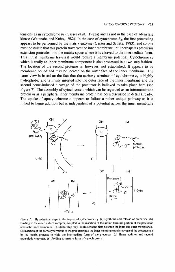
tensions as in cytochrome b2 (Gasser et al., 1982a) and as not in the case of adenylate kinase (Watanabe and Kubo, 1982). In the case of cytochrome b2, the first processing appears to be performed by the matrix enzyme (Gasser and Schatz, 1983), and so one must postulate that this protein traverses the inner membrane until perhaps its precursor extension protrudes into the matrix space where it is cleaved to the intermediate form. This initial membrane traversal would require a membrane potential. Cytochrome cx
which is really an inner membrane component is also processed in a two-step fashion. The location of the second protease is, however, not established. It appears to be membrane bound and may be located on the outer face of the inner membrane. The latter view is based on the fact that the carboxy terminus of cytochrome Ci is highly hydrophobic and is firmly inserted into the outer face of the inner membrane and the second heme-induced cleavage of the precursor is believed to take place here (see Figure 7). The assembly of cytochrome c which can be regarded as an intermembrane protein or as a peripheral inner membrane protein has been discussed in detail already. The uptake of apocytochrome c appears to follow a rather unique pathway as it is linked to heme addition but is independent of a potential across the inner membrane
Figure 7. Hypothetical steps in the import of cytochrome c\. (a) Synthesis and release of precursor, (b) Binding to the outer surface receptor, coupled to the insertion of the amino terminal portion of the precursor across the inner membrane. This latter step may involve contact sites between the inner and outer membranes, (c) Insertion of the carboxy terminus of the precursor into the inner membrane and cleavage of the presequence by the matrix protease to yield the intermediate form of the precursor, (d) Heme addition and second proteolytic cleavage, (e) Folding to mature form of cytochrome c.
m-Cytq
454 MATTHEW Α. HARMEY and WALTER NEUPERT
Free Ribosomes
HS' α) Apocytochrome c b)
OM IM OM
IM
IM
Figure β. Hypothetical import pathway of cytochrome c. (A) Synthesis and release of apocytochrome c into the cytosol. (B) Recognition by a specific apocytochrome c receptor. (C) Heme addition to the receptor-bound apocytochrome c by cytochrome c heme lyase (CHL). (D) Internalization and refolding of the cytochrome c. (E) Binding of the completed and internalized cytochrome to the outer surface of the inner membrane (CCO, cytochrome c oxidase; CCR, cytochrome c reductase).
(Figure 8). It is also characterized by having an apparently separate or unique receptor protein (Hennig and Neupert, 1981). It may be speculated that the apocytochrome c is positioned by the receptor in such a way that addition of the heme group can take place. The enzyme catalyzing the formation of the thioether bonds between the vinyl groups of the heme and the thiols of cysteines 14 and 17 of the apoprotein may be located in the intermembrane space. It can be further speculated that the free energy of refolding of the polypeptide chain occurring after heme linkage provides the energy to completely pull the molecule through the outer membrane.
ADP/ATP carrier, on the other hand, inserts into the inner membrane (Zimmermann and Neupert, 1980) and traverses it. In common with other proteins studied, it binds to the outer membrane as a protease-sensitive precursor which does not have the ability to bind carboxyatractyloside nor ATP, as the mature protein does. The evidence presented by Zimmermann and Neupert (1980) and Schleyer et al. (1982) indicate that there is a change of conformation from a relatively hydrophilic form of the protein to a highly hydrophobic one. This transformation requires the presence of an energized inner membrane. It has recently been shown that the imported precursor binds carboxyatractyloside in a manner identical to the mature protein (Schleyer, unpublished). The possible pathways of two processed precursors, ß subunit and subunit 9 of the ¥<F\ ATPase are outlined in Figure 9A. Obviously, most of the steps proposed are quite hypothetical and may have to be amended in the future.
MITOCHONDRIAL PROTEINS 455
The majority of matrix proteins described so far are synthesized as larger molecular weight precursors and their transport behavior accords with the belief that passage across their inner membrane requires a membrane potential (Figure 9b). The fact that in the matrix both peripheral membrane and soluble matrix proteins require both proteolytic processing and a membrane potential has led in some cases to the belief that the two processes were obligately linked. Import of precursors into the matrix
pre-F,ß
Functional Dimer
Figure 9. Hypothetical import pathways of (a) subunits of the Fi ATPase. Receptors on the outer membrane channel; the proteins across the inner membrane. The ß-subunit precursor has been located in the matrix while the precursor to subunit 9 remains membrane associated (Zwizinski and Neupert, unpublished). The presequence of su9 must protrude into the matrix to allow the protease access to the cleavage site. Following cleavage the mature protein is incorporated into the Fi ATPase complex. Su9 undergoes a change of conformation and acquires its characteristic hydrophobic properties and is firmly embedded in the inner membrane. Post cleavage, both proteins are precipitable by antibody to the F| ATPase complex, (b) Citrate synthase, a soluble matrix protein. Binding and transfer is similar to above. Following transfer, the precursor is cleaved and the cleaved precursors can now dimerize to yield the mature dimeric protein.
456 MATTHEW A. HARMEY and WALTER NEUPERT
occurs in the absence of proteolytic cleavage. Thus, proteolytic processing is apparently not an essential step for translocation. In agreement with this view, the precursor to isopropylmalate synthase, a matrix enzyme in yeast, does not differ in molecular weight from the mature protein and was reported to be taken up by yeast mitochondria in a membrane potential-dependent inininer without a change in its apparent molecular weight (Hampsey et aL, 1983).
VIII. PROTEINS CODED FOR BY THE MITOCHONDRIAL GENOME
The mitochondrial genome has been extensively studied in a few organisms. The whole genome has been sequenced in man and mouse while in species such as yeast it has been only partly sequenced (Attardi, 1981). At the other end of the scale, the genome of plant mitochondria which appears to be relatively large (Leaver and Grey, 1982) has had little sequence analysis applied to it. The mitochondrial genetic system shows a number of unique common features. The code usage is at variance with that in the nucleocytoplasmic system in that the same codons do not code for the same amino acids in both systems, e.g., mammalian mitochondria read AGA and AGG as stop signals rather than as codons for the amino acid arginine and translate AUA as methionine rather than as isoleucine (Grivell, 1983). The differences between the two systems make it unlikely that messages from the nucleocytoplasmic system would be imported into the mitochondria for translation.
Despite a wide range of variation in the sizes of mitochondrial DNA, as measured by contour length and restriction analyses, from a range of species, the products of translation appear limited to much the same repertoire in most cases. In vitro studies on the mitochondrial translation system have yielded various estimates on the number of mitochondrial products. Bhat et al. (1981) quote the number of translation products from mitochondria from some mouse cell lines as 18, ranging in size from 3.8-67.OK. The former appear somewhat small for a functional polypeptide. The proteins synthesized by all mitochondria studied so far are cytochrome oxidase subunits I , I I , and I I I , subunit V I of the ATPase, and the apocytochrome b. Subunit 9 of ATPase is coded by the mitochondrial DNA in yeast but by nuclear DNA in Neurospora and in higher organisms. Recently, a 10K mitochondrial translation product has been found in yeast which appears to be associated with the ATPase; however, no function could be assigned to this component (Macreadie et al., 1983). There is also in yeast and Neurospora mitochondrial ribosomes one protein which appears to be essential for proper ribosomal function (varl protein in yeast) which is a mitochondrial translation product. There is experimental evidence (Leaver and Gray, 1982) that plant mitochondria do synthesize a greater number of proteins than do their fungal or mammalian counterparts. One of the additional translation products in plants has been identified as the α-subunit of the F t ATPase. Apart from the identified translation products there are variable numbers of unassigned reading frames depending on the source of the mitochondria (Borst and Grivell, 1981). The translation system of mitochondria resembles that of prokaryotes with respect to antibiotic sensitivity, as typified by its sensitivity to chloramphenicol and tetracyclines. The exact nature of the mitochondrial
MITOCHONDRIAL PROTEINS 457
translation products from many species remains to be classified. Studies on the synthesis of cytochrome oxidase subunits has shown that in some cases they may be synthesized as large precursors such as subunit I in Neurospora (Werner and Bertrand, 1979) and subunit I I in yeast (Poyton and McKemmie, 1979; Pratje etal., 1983). Little is known about the exact location of the translation, i.e., free or bound polysomes. Mitoribo-somes have been reported by many to be associated with the inner face of the inner membrane; however, no clear evidence in favor of either a post- or cotranslational mechanism of precursors has been presented so far, nor is there any definitive data on the nature of the protease involved in the processing of mitochondrially synthesized precursors.
An interesting feature has been observed in a comparison of a subunit of the ATPase in yeast and Neurospora. In yeast, subunit 9 (proteolipid) is synthesized without any detectable polypeptide extension and is coded for by the mitochondrial DNA. In Neurospora, the same protein is coded by the nuclear genome and is synthesized as a larger molecular weight precursor on cytoplasmic ribosomes as discussed above. Interestingly, a gene with striking homology to those of either yeast or Neurospora subunit 9 has been detected in the mitochondrial genome of Neurospora crassa. However, expression of this gene has not been observed although it carries the characteristics of a duplication rather than a transposition (van den Boogart et al., 1982).
A general feature of all functionally identified mitochondrial gene products is that they are all constituents of multisubunit complexes and occur combined with nuclear gene products. Therefore, a problem of mitochondrial biogenesis is that of understanding how the assembly of the translation products of the two systems, i.e., nucleocy-toplasmic and mitochondrial takes place and how it is regulated. In yeast, there is evidence of a degree of coupling between the activities of the cytoplasmic translation system and that of the mitochondria. Lustig et al. (1982a,b) followed the levels of mRNA for the cytoplasmically synthesized subunits of cytochrome c oxidase under conditions of catabolite repression and derepression of mitochondria. The levels of mRNA for the subunits were low under conditions of repression and gradually increased with derepression. They also studied the level of transcripts for the nuclear coded mitochondrial RNA polymerase and found that the changes in messenger activity for the subunits of the oxidase were mirrored by those for the RNA polymerase. This result would suggest that the mitochondrial transcription increases to accommodate the influx of precursors from the cytoplasm. It may of course also be argued that this increase in the polymerase was merely due to a general increase in the synthesis of cytoplasmically synthesized mitochondrial protein precursors and, as such, does not represent a coordination of the two systems. In Neurospora cells, no tight coupling of the two translation systems has been found. Weiss and Kolb (1979) grew cells in the presence of chloramphenicol; these cells were deficient in cytochrome b, nevertheless, they continued to synthesize normal amounts of cytochrome c,. This was taken up and processed by the mitochondria and inserted into a cytochrome ^-deficient complex. Rucker and Neupert (1976) showed that inhibition of cytoplasmic translation led to continued production of subunits I—III of cytochrome oxidase at least for the duration of a doubling period of the Neurospora cells. However, increased breakdown was observed of the mitochondrially synthesized subunits which could not be assembled
458 MATTHEW Α. HARMEY and WALTER NEUPERT
into functional enzyme complexes. Poyton and McKemmie (1979) suggested from their studies on the biosynthesis of cytochrome oxidase that the synthesis of subunits I , I I , and I I I by mitochondria was stimulated by a precursor to subunits I V - V I I . The data on which this suggestion was made is open to question as they also suggested that the precursor was a polyprotein which is not in accordance with recent findings by other groups (Schmelzer and Heinrich, 1980; Lewin et aL, 1980; Mihara and Blobel, 1980).
/X. CONCLUSIONS
A. Precursors
Mitochondria import precursor proteins from the cytosol in a posttranslational manner. Precursors are mostly translated with amino terminal extensions on free ribosomes. A number of exceptions to this rule exist, e.g., ADP/ATP carrier, adenylate kinase, apocytochrome c, porin, and isopropylmalate synthase. Many precursors have a tendency to aggregate. It is not yet known whether there is an equilibrium between monomelic and oligomeric forms or whether there is selective recognition or uptake of one form vs. the other.
ß . Recognition
Specific receptor sites exist on the outer mitochondrial surface. Competition experiments indicate the existence of more than one type. Treatment with proteases indicate that recognition is mediated by proteins. Requirements for binding and translocation across the membrane are different. Precursors can be bound at low temperature in the absence of insertion or translocation. On elevating the temperature to 25°C, the proteins are inserted or translocated without intervening detachment from the binding sites. Binding of precursor proteins is independent of the energy status of the mitochondria. Mitoplasts have been described as importing precursors; it is unclear, however, whether uptake is occurring via remnants of outer membrane or via specific receptors on the mitoplast surface as distinct from those on the outer membrane.
C. Translocation
Translocation across or insertion into the inner membrane requires a membrane potential. The nature of the process of transfer or translocation across the inner membrane remains a mystery. The role of the membrane potential is equally obscure, the magnitude of the potential does not seem to be directly related to the extent of import. Interactions of outer and inner membranes may play a role in the transfer of proteins into inner membrane and beyond but this is essentially a speculation. It appears that there are different pathways of translocation for different precursors depending on both the final location and solubility characteristics of the mature protein.
MITOCHONDRIAL PROTEINS 459
D. Proteolytic Processing
A matrix located metalloenzyme is responsible for proteolysis of the precursor protein to their mature form. The process is inhibited by chelating agents and is independent of membrane potential or ATP. The processing is indirectly influenced by these factors as they control translocation of the substrates to the site of action. In a number of cases, a second, as yet uncharacterized, protease is required to fully process the precursors.
The current practice of cloning and sequencing the genes for individual precursors will hopefully lead to the availability of sufficient amounts of individual precursors to allow investigation of binding and transport of individual proteins under defined conditions.
As stated at the outset we would attempt to provide answers to a number of questions posed. It is perhaps appropriate also to finish this chapter by outlining a number of open questions which remain to be answered.
How do precursors of insoluble membrane proteins move through the predominantly aqueous milieu of the cytosol? What is the nature of the receptors which mediate the recognition of mitochondrial precursor proteins? How many types of recognition proteins exist and how are they arranged in the membranes? Are there specific receptors on the inner membrane? How do polypeptides cross the membranes, i.e., as compact folded entities or do extended polypeptide chains move through the membrane? Are other membrane components besides receptors (translocators) required for translocating polypeptides across membranes? What exactly is the role of the membrane potential in polypeptide chain translocation? What is the role of the additional sequence; do they play a role in recognition or are the recognition domains associated with the mature position of the protein?
Are cytosolic pools of precursors involved in the regulation of synthesis of mitochondrial proteins?
REFERENCES
Ades, I . Z., and Butow, R. Α., 1980, The products of mitochondria bound cytoplasmic polysomes in yeast, J. Biol. Chem. 225:9918-9924.
Ades, I . Z., and Harpe, Κ. G., 1981, Biogenesis of mitochondrial proteins. Identification of the mature and precursor forms of the subunit of d-aminolevulinate synthase from embryonic chick liver, J. Biol. Chem. 256:9329-9333.
Adolphus, P. G., Van Loon, M. , de Groot, R., Van Eyk, E., and Grivell, L. Α., 1982, Isolation and characterization of nuclear genes coding for subunits of the yeast ubiquinol-cyt c reductase complex, Gene 20:323-337.
Attardi, G., 1981, Organisation and expression of the mammalian mitochondrial genome, Trends Biochem. 6:86-89.
Autor, A. P., 1982, Biosynthesis of mitochondrial manganese superoxide dismutase in Saccharomyces cerevisiae. Precursor form of mitochondrial superoxide dismutase made in the cytoplasm, J. Biol. Chem. 257:2713-2718.
Aziz, L. E., Chien, S. M. , Patel, Η. V., and Freeman, Κ. B., 1981, A putative precursor of rat liver mitochondrial malate dehydrogenase, FEBS Lett. 133:127-129.
Barrell, B. G., Barbier, A. T., and Drouin, J., 1979, A different genetic code in human mitochondria, Nature 282:189.
460 MATTHEW Α. HARMEY and WALTER NEUPERT
Bhat, Ν. K., Niranjan, B. G., and Avadhani, N. G., 1981, The complexity of mitochondrial translation products in mammalian cells, Biochem. Biophys. Res. Commun. 103:621-628.
Blobel, G., Walter, P., Chang C. N. , Goldman, Β. M., Erickson, A. H., Lingappa, V. R., 1979, Translocation of Proteins across Membranes, the Signal Hypothesis and Beyond, Symp. of Soc. for Exp. Biol. XXXIII Secretory Mechanisms (C. R. Hopkins and C. J. Duncan, eds.), Cambridge University Press, Cambridge, pp. 9-36.
Boehni, P., Grasser, S., Leaver, C , and Schatz, G., 1980, A matrix-localized mitochondrial protease processing cytoplasmically-made precursors to mitochondrial proteins, in: The Organisation and Expression of the Mitochondrial Genome (A. Kroon and C. Saccone, eds.), Elsevier, Amsterdam, pp. 423-433.
Boehni, P., Daum, G., and Schatz, G., 1983, Import of proteins into mitochondria. Partial purification of a matrix located protease involved in cleavage of mitochondrial precursor polypeptides, J. Biol. Chem. 258:4937-4943.
Boogart, van den, P., Sanallo, J., and Agsteribbe, E., 1982, Similar genes for a mitochondrial ATPase subunit in the nuclear and mitochondrial genomes of Neurospora crassa, Nature 298:1870-1872.
Borst, P., 1981, Biosynthesis of mitochondria and chloroplasts, in: Molecular Biology of the Cell (S. Alberts, K. Porter, M. C. Raff, K. Roberts, and J. D. Watson, eds.), Garland, New York.
Borst, P., and Grivell, L. Α., 1978, The mitochondrial genome of yeast, Cell 15:705-723. Borst, P., and Grivell, L. Α., 1981, Small is beautiful: Portrait of a mitochondrial genome, Nature
290:443^144. Cerletti, N. , Boehni, P. C , and Suda, K., 1983, Import of proteins into mitochondria. Isolated yeast
mitochondria and a solubilised matrix protease correctly process cytochrome c oxidase subunit V precursor at the N-terminus, J. Biol. Chem. 258:4944-4949.
Conboy, J. G., and Rosenberg, L. E., 1981, Post-translational uptake and processing of in vitro synthesized ornithine transcarbamylase precursor by isolated rat liver mitochondria, Proc. Natl. Acad. Sei. USA 78:3073-3077.
Cote, C., Solioz, Μ., and Schatz, G., 1979, Biogenesis of the cytochrome be complex of yeast mitochondria. A precursor form of the cytoplasmically made subunit V, J. Biol. Chem. 254:1437-1439.
Daum, G., Bohni, P., and Schatz, G., 1982a, Import of proteins into mitochondria: Cyt b and cyt c peroxidase are located in the intermembrane space of yeast mitochondria, J. Biol. Chem. 257:13028-13033.
Daum, G., Gasser, S. Μ., and Schatz, G., 1982b, Import of proteins into mitochondria, energy-dependent two-step processing of the intermembrane space enzyme cyt b by isolated yeast mitochondria, J. Biol. Chem. 257:13075-13080.
Emr, S. D., and Silhavy, T. J., 1982, Molecular components of the signal sequence that function in the initiation of protein export, J. Cell Biol. 95:689-696.
Ernster, L., and Kuylenstierna, B., 1970, Outer membrane of mitochondria, in: Membranes of Mitochondria and Chloroplasts (E. Racker, ed.), von Nostrand, New York, pp. 172-212.
Freitag, Η., Janes, Μ., and Neupert, W., 1982, Biosynthesis of mitochondrial porin and insertion into the outer mitochondrial membrane of Neurospora crassa, Eur. J. Biochem. 126:197-202.
Gasser, S. M. , and Schatz, G., 1983, Import of protein into mitochondria, in vitro studies on the biogenesis of the outer membrane, J. Biol. Chem. 258:3427-3430.
Gasser, S. M . , Daum, G., and Schatz, G., 1982a, Import of proteins into mitochondria, energy-dependent uptake of precursors by isolated mitochondria, J. Biol. Chem. 257:13034-13041.
Gasser, S. M . , Ohashi, Α., Daum, G., Boehni, P. C , Gibson, J., Reid, G. Α., Yonetani, T., and Schatz, G., 1982b, Imported mitochondrial proteins cytochrome b2 and cytochrome cl are processed in two steps, Proc. Natl. Acad. Sei. USA 79:261-211.
Grivell, L. Α., 1983, Mitochondrial DNA, Sei. Am. 248:60-73. Hallermayer, G. and Neupert, W., 1976, Studies on the synthesis of mitochondrial proteins in the cytoplasm
and on their transport into mitochondria, in: Genetics and Biogenesis of Chloroplasts and Mitochondria (T. Bucher, W. Neupert, W. Sebald, and S. Werner, eds.) North-Holland, Amsterdam, pp. 807-812.
Hallermayer, G., Zimmermann, R., and Neupert, W., 1977, Kinetic studies on the transport of cytoplasmically synthesized proteins into mitochondria in intact cells of Neurospora crassa, Eur. J. Biochem. 81:523-532.
Hampsey, M. D., Levin, A. S., and Kohlnow, G. B., 1983, Submitochondrial localisation, cell free
MITOCHONDRIAL PROTEINS 461
synthesis and mitochondrial import of two isopropylmalate synthase of yeast, Proc. Natl. Acad. Sei. USA 80:1270-1274.
Harmey, Μ. Α., and Neupert, W., 1979, Biosynthesis of mitochondrial citrate synthase in Neurospora crassa, FEBS Lett. 108:385-389.
Harmey, Μ. Α., Hallermayer, G., Korb, Η., and Neupert, W., 1977, Transport of cytoplasmically synthesized proteins into the mitochondria in a cell free system, Eur. J. Biochem. 81:533-544.
Heinrich, P. C., 1982, Proteolytic processing of polypeptides during the biosynthesis of subcellular structures, Rev. Physiol. Biochem. Pharmacol. 93:115-187.
Hennig, B., and Neupert, W., 1981, Assembly of cytochrome c. Apocytochrome c is bound to specific sites on mitochondria before its conversion to holocytochrome c, Eur. J. Biochem. 121:203-212.
Hennig, B., Koehler, H., and Neupert, W., 1983, Receptor sites involved in posttranslational transport of apocytochrome c into mitochondria, Cell Biol., in press.
Hunt, T., and Jackson, R. J., 1974, The rabbit reticulocyte as a system for studying mRNA, in: Modern Trends in Human Leukaemia (R. Netz, ed.), Lehmans Verlag, Munich, pp. 300-307.
Inouye, M. , and Beckwith, J., 1977, Synthesis and processing of an alkaline phosphatase precursor in vitro, Proc. Natl. Acad. Sei. USA 74:1440-1444.
Jaussi, R., Sonderegger, P., Fluckinger, J., and Christen, P., 1981, Biosynthesis and topogenesis of aspartate aminotransferase isoenzymes in chicken embryo fibroblasts, J. Biol. Chem. 257:13334-13340.
Kamisaki, Y., Sakakibara, R., Horio, Y., and Wada, H., 1982, Low isoelectric point of precursor of mitochondrial glutamic oxalacetic transaminase isoenzyme synthesized in vitro, Biochem. Int. 4:289-296.
Kaput, J., Goltz, S., and Blobel, G., 1982, Nucleotide sequence of the yeast nuclear gene for cytochrome c peroxidase precursor, functional implications of the presequence for protein transport into mitochondria, J. Biol. Chem. 257:15054-15058.
Kellems, R. E. and Butow, R. Α., 1974, Cytoplasmic type 80S ribosomes associates with yeast mitochondria III . Changes in the amount of bound ribosomes in response to changes in metabolic state, J. Biol. Chem. 249:3304-3310.
Kellems, R. E., Allison, V. F., and Butow, R. Α., 1975, Cytoplasmic type 80S ribosomes associated with yeast mitochondria, IV, Attachment of ribosomes to the outer membrane of isolated mitochondria, J. Cell Biol. 65:1-14.
Klingenberg, Μ., 1976, The ADP-ATP carrier in mitochondrial membranes, in: Enzymes of Biological Membranes Vol. 4 (A. Martonosi, ed.), Plenum Press, New York pp. 383^38.
Klingenberg, Μ., and Rottenberg, Η., 1977, Relation between the gradient of the ATP/ADP ratio and the membrane potential across the mitochondrial membrane, Eur. J. Biochem. 73:125-130.
Koehler, H., Hennig, B. and Neupert, W., 1983, Mitochondria 1983 (R. T. Schweyen, K. Wolf, and F. Kaudewitz, eds.) Walter de Gruyter, Berlin and New York.
Kolansky, D. M. , Conboy, J. G., Fenlon, W. Α., and Rosenberg, L. , 1982, Energy-dependent translocation of the precursor of ornithine transcarbamylase by isolated rat liver mitochondria, J. Biol. Chem. 257:8467-8471.
Komar, E., Weber, H., and Tanner, W., 1979, Greatly decreased susceptibility of nonmetabolising cells towards detergents, Proc. Natl. Acad. Sei. USA 76:1814-1818.
Korb, Η., and Neupert, W., 1978, Biogenesis of cytochrome c in Neurospora crassa. Synthesis of apocytochrome c, transfer, Eur. J. Biochem. 91:609-620.
Kraus, J. P., Conboy, J. G., and Rosenberg, L. E., 1981, Preornithine transcarbamylase, properties of the cytoplasmic precursor of a mitochondrial matrix enzyme, J. Biol. Chem. 256:10739-10742.
Kreibich, P. C , 1982, Proteolytic processing of polypeptides during the biosynthesis of subcellular structures, Rev. Physiol. Biochem. Pharmacol. 93:116-187.
Kreibich, G., Czako-Graham, M. , Grebenau, R., Mok, W., Rodriguez-Boulan, E., and Sabatini, D. D., 1978, Characterization of the ribosomal binding site in rat liver rough microsomes: Ribophorins I and II , two integral membrane proteins related to ribosome binding, J. Supramol. Struct. 8:279- 302.
Kreil, G., 1981, Transfer of proteins across membranes, Annu. Rev. Biochem. 50:317-348. Lamb, A. J., Clark-Walker, G. D., and Linnane, A. W., 1968, The biogenesis of mitochondria, the
differentiation of mitochondrial and cytoplasmic protein synthesizing systems in vitro by antibiotics, Biochim. Biophys. Acta 161:415^35.
462 MATTHEW Α. HARMEY and WALTER NEUPERT
Leaver, C. J., and Gray, M. W., 1982, Mitochondrial genome, organisation and expression in higher plants, Annu. Rev. Plant Physiol. 33:373^*02.
Lewin, A. S., and Norman, D., 1983, Assembly of F ATPase in isolated mitochondria, J. Biol. Chem. 258:6750-6755.
Lewin, A. S., Gregor, I . , Mason, T. L. , Nelson, N. , and Schatz, G., 1980, Cytoplasmically made subunits of yeast mitochondrial F-ATPase and cytochrome c oxidase are synthesized as individual precursors, not as polyproteins, Proc. Natl. Acad. Sei. USA 77:3998^002.
Lustig, Α., Levens, D., and Rabinowitz, M . , 1982a, The biogenesis and regulation of mitochondrial RNA polymerase, J. Biol. Chem. 257:5800-5808.
Lustig, Α., Padmanaban, G., and Rabinowitz, M . , 1982b, Regulation of the nuclear coded peptides of yeast cytochrome c oxidase, Biochemistry 21:309-316.
Maccechini, M. , Rudin, Y., Blobel, G., and Schatz, G., 1979a, Import of proteins into mitochondria: Precursor forms of the extramitochondrially made F ATPase subunits in yeast, Proc. Natl. Acad. Sei. USA 76:343-347.
Maccechini, M.-L., Rudin, Y., and Schatz, G., 1979b, Transport of proteins across the mitochondrial outer membrane. A precursor form of the cytoplasmically made intermembrane enzyme cytochrome c peroxidase, J. Biol. Chem. 254:7468-7471.
Macino, G., and Tzagoloff, Α., 1979, Assembly of the mitochondrial membrane system, J. Biol. Chem. 254:4617^1623.
Macreadie, I . G., Novitski, G. E., Maxwell, R. J., John, V., Ooi, B. G., McMullen, G. C , Lukins, H. B., Linnane, A. W., and Nagley, P., 1983, Biogenesis of mitochondria: The mitochondrial gene (aapl) coding for mitochondrial ATPase subunit 8 in Saccharomyces cerevisiae, Nucleic Acid Res. 11:4435-4451.
Marra, E., Doonan, S., Saccone, C , and Quagliariello, E., 1977, Selective permeability of rat liver mitochondria to purified aspartate amino transferase in vitro, Biochem. J. 164:685-691.
Matsuura, S., Arpin, M . , Hannum, C , Margoliash, E., Sabatini, D. D., and Morimoto, T., 1981, In vitro synthesis and posttranslational uptake of cytochrome c into isolated mitochondria: Role of a specific addressing signal in the apocytochrome, Proc. Natl. Acad. Sei. USA 78:4368-4372.
McAda, P., and Douglas, M. G., 1982, A neutral metalloendoprotease involved in the processing of an Fj-ATPase subunit precursor in mitochondria, J. Biol. Chem. 257:3177-3182.
Meyer, D. I . , and Dobberstein, B., 1980, Identification and characterization of a membrane component essential for the translocation of nascent protein across the membrane of the endoplasmic reticulum, J. Cell Biol. 87:503-508.
Meyer, D. I . , Krause, Ε., and Dobberstein, B., 1982, Secretory protein translocation across membranes: The role of the "docking protein," Nature 297:647-650.
Michel, R., Wächter, Ε., and Sebald, W., 1979, Synthesis of a larger precursor for the proteolipid subunit of the mitochondrial ATPase complex of Neurospora crassa in a cell-free wheat germ system, FEBS Lett. 101:373-376.
Mihara, K., and Blobel, G., 1980, The four cytoplasmically made subunits of yeast mitochondrial cytochrome c oxidase are synthesized individually and not as a polyprotein, Proc. Natl. Acad. Sei. USA 77:4160-4164.
Mihara, K., Blobel, G. and Sato, R., 1982, in vitro synthesis and integration into mitochondria of porin, a major protein of the outer mitochondrial membrane of Saccharomyces cerevisiae, Proc. Nat. Acad. Sei. USA. 79:7102-7106.
Mihara, K., Omura, T., Harano, T., Brenner, S., Fleischer, S., Rajagopalan, Κ. V., and Blobel, G., 1982, Rat liver L-glutamate dehydrogenase, D-fr-hydroxybutyrate dehydrogenase, and sulfite oxidase are each synthesized as larger precursors by cytoplasmic free polysomes, J. Biol. Chem. 257:3355-3358.
Miura, S., Mori, M. , Amaya, Y., and Tatibana, M. , 1982, A mitochondrial protease that cleaves the precursor of ornithine carbamyltransferase. Purification and properties, Eur. J. Biochem. 122:641-647.
Mori, M., Miura, S., Tatibana, M . , and Cohen, P. P., 1979, Cell-free synthesis and processing of a putative precursor for mitochondrial carbamyl phosphate synthetase I of rat liver, Proc. Natl. Acad. Sei. USA 76:5071-5075.
Mori, M. , Miura, S., Tatibana, M. , and Cohen, P. P., 1980, Processing of a putative precursor of rat liver ornithine transcarbamylase by a mitochondrial matrix enzyme, J. Biochem. 88:1829-1836.
MITOCHONDRIAL PROTEINS 463
Morita, T., Miura, S., Mori, Μ., and Tatibana, Μ., 1982, Transport of the precursor for rat-liver ornithine carbamy transferase into mitochondria in vitro, Eur. J. Biochem. 122:501-509.
Mueckler, M. M. , Himeno, M. , and Pitot, H. C , 1982, In vitro synthesis and processing of a precursor to ornithine amino transferase, J. Biol. Chem. 257:7178-7180.
Nabi, N. , and Omura, T., 1980, In vitro synthesis of adrenodoxin and adrenodoxin reductase: Existence of a putative large precursor form of adrenodoxin, Biochem. Biophys. Res. Commun. 97:680-686.
Nelson, N. , and Schatz, G., 1979, Energy-dependent processing of cytoplasmically made precursors to mitochondrial proteins, Proc. Natl. Acad. Sei. USA 76:4365-4369.
Neupert, W., and Schatz, G., 1981, How proteins are transported into mitochondria, Trends Biochem. Sei. 6 : 1 ^ .
Oda, Τ., Ichiyoma, Α., Miura, S., Mori, M. , and Tatibana, M . , 1981, In vitro synthesis of a putative precursor of serine: Pyruvate aminotransferase of rat liver mitochondria, Biochem. Biophys. Res Commun. 102:568-573.
Ohashi, Α., Gibson, J., Gregor, I . , and Schatz, G., 1982, Import of proteins into mitochondria. The precursor of cytochrome c\ is processed in two steps, one of them heme-dependent, J. Biol. Chem. 257:13042-13047.
Passarella, S., Marra, E., Doonan,. S., and Quagliarello, E., 1980, Selective permeability of rat liver mitochondria to purified malate dehydrogenase isoenzymes in vitro, Biochem. J. 192:649-658.
Poyton, R. O., and McKemmie, E., 1979, Posttranslational processing and transport of the polyprotein precursor to subunits IV to VII of yeast cytochrome c oxidase, J. Biol. Chem. 254:6772-6780.
Pratje, Ε., Mannhaupt, G., Michaelis, G., and Beyreuther, Κ., 1983, A nuclear mutation prevents processing of a mitochondrially encoded membrane protein in Saccharomyces cerevisiae, EMBO J. 7:1049-1054.
Randall, L., 1983, Translocation of domains of nascent periplasmic proteins across the cytoplasmic membrane is independent of elongation, Cell 33:231-240.
Raymond, Y., and Shore, G. C., 1979, The precursor for carbamyl phosphate synthetase is transported to mitochondria via a cytosolic route, J. Biol. Chem. 254:9335-9338.
Reid, G., and Schatz, G., 1982, Import of proteins into mitochondria, J. Biol. Chem. 257:13056-13067. Riezman, H., Hay, R., Witte, C., Nelson, N., and Schatz, G., 1983, Yeast mitochondrial outer membrane
specifically binds cytoplasmically synthesized precursors of mitochondrial proteins, EMBO J 2:1113-1118. Roberts, Β. E., and Paterson, Β. M. , 1973, Efficient translation of tobacco mosaic virus RNA and rabbit
globin 9S RNA in a cell free system from commercial wheat germ, Proc. Natl. Acad. Sei. USA 70:2330-2334.
Rucker, V. Α., and Neupert, W., 1976, Coordination of mitochondrial and cytoplasmic protein synthesis in Neurospora crassa, in: Genetics and Biogenesis of Chloroplasts and Mitochondria (T. Bucher, W. Neupert, W. Sebald, and S. Werner, eds.), North-Holland, Amsterdam pp. 231-238.
Sagara, Y., and Ito, Α., 1982, In vitro synthesis of monoamine oxidase of rat liver outer mitochondrial membrane, Biochem. Biophys. Res. Commun. 109:1102-1107.
Sakakibara, R., Huynh, Q. K., Nishida, Y., Watanabe, T., and Wado, H., 1980, In vitro synthesis of glutamic oxaloacetic transaminase isozymes of rat liver, Biochem. Biophys. Res. Commun. 95:1781-1788.
Schatz, G., 1979, How mitochondria import proteins from the cytoplasm, FEBS Lett. 103:201-211. Schatz, G., and Butow, R. Α., 1983, How are proteins imported into mitochondria, Cell 32:316-318. Schatz, G., and Mason, T. L., 1974, The biosynthesis of mitochondrial proteins, Annu. Rev. Biochem.
43:51-87. Schleyer, Μ., Schmidt, Β., and Neupert, W., 1982, Requirement of a membrane potential for the post
translational transfer of proteins into mitochondria, Eur. J. Biochem. 125:109-116. Schmelzer, Ε., and Heinrich, P. C , 1980, Synthesis of a larger precursor for the subunit IV of rat liver
cytochrome c oxidase in a cell-free wheat germ system, J. Biol. Chem. 255:7503-7506. Schmidt, B., Hennig, Β., Simmermann, R., and Neupert, W., 1983a, Biosynthetic pathway of mitochondrial
ATPase subunit 9 in Neurospora crassa, J. Cell Biol. 96:248-255. Schmidt, B., Hennig, B., Kohler, H., and Neupert, W., 1983b, Transport of precursor to Neurospora
ATPase subunit 9 into yeast mitochondri, J. Biol. Chem., in press. Shore, G. C , Carignan, P., and Raymond, Y., 1979, In vitro synthesis of a putative precursor to the
mitochondrial enzyme, carbamyl phosphate synthetase, J. Biol. Chem. 254:3141-3144.
464 MATTHEW Α. HARMEY and WALTER NEUPERT
Shore, G. C., Power, F., Bendayon, M . , and Carignan, P., 1981, Biogenesis of a 35K dalton protein associated with outer mitochondrial membrane in rat liver, J. Biol. Chem. 256:8761-8766.
Sonderegger, P., Jaussi, R., and Christen, P., 1980, Cell-free synthesis of a putative precursor of mitochondrial aspartate aminotransferase with higher molecular weight, Biochem. Biophys. Res. Commun. 94:1256-1260.
Srivastava, G., Borthwick, I . Α., Brooker, J. D., May, Β. K., and Elliot, W. H., 1982, Purification of rat liver mitochondrial d-aminolaevulinate synthase, Biochem. Biophys. Res. Commun. 109:305-312.
Stigall, D. L. , Galante, Υ. M . , and Hatefi, Y., 1979, Preparation and properties of complex V, Meth. Enzymol. 55:308-315.
Suissa, M. , and Schatz, G., 1982, Import of proteins into mitochondria: Translatable mRNAs for imported proteins are present in free as well as mitochondria bound cytoplasmic polysomes, J. Biol. Chem. 257:13048-13055.
Teintze, M. , Slaughter, M. , Weiss, H., and Neupert, W., 1982, Biogenesis of mitochondrial ubiquinol: cytochrome c reductase (cytochrome be complex). Precursor proteins and their transfer into mitochondria, J. Biol. Chem. 257:10364-10371.
Viebrock, Α., Perz, Α., and Sebald, W., 1982, The imported preprotein of the proteolipid subunit of the mitochondrial ATP synthase from Neurospora crassa. Molecular cloning and sequencing of the mRNA, EMBO J. 1:565-571.
Walter, P., and Blobel, G., 1982, Signal recognition particle contains a 7S RNA essential for protein translocation across the endoplasmic reticulum, Nature 299:691-698.
Watanabe, K., and Kubo, S., 1982, Mitochondrial adenylate kinase from chicken liver, Eur. J. Biochem. 123:587-592.
Weiss, H., and Kolb, H. J., 1979, Isolation of mitochondrial succinate: ubiquinone reductase, cytochrome c reductase and cytochrome c oxidase from Neurospora crassa using non-ionic detergent, Eur. J. Biochem. 99:139-150.
Werner, S., and Bertrand, Η., 1979, Conversion of a mitochondrial precursor polypeptide into subunit 1 of cytochrome oxidase in the Ml3 mutants of Neurospora crassa, Eur. J. Biochem. 99:463-470.
Wickner, W., 1980, Assembly of proteins into membranes, Science 210:861-862. Wikstrom, Μ., and Krab, K., 1982, Proton pumping cytochrome c oxidase, Biochim. Biophys. Acta
549:177-222. Yamamoto, M. , Hayashi, N. , and Kikuchi, G., 1982, Evidence for the transcriptional inhibition by heme
of the synthesis of d-aminolevulinate synthase in rat liver, Biochem. Biophys. Res. Commun. 105:985-990. Yoshida, Y., Hasimoto, T., and Tagawa, K., 1983, Cell free synthesis of mitochondrial ATPase inhibitor
precursor and its transport into yeast mitochondria, J. Biochem. 94:283-290. Zimmermann, R., and Neupert, W., 1980, Transport of proteins into mitochondria. Posttranslational transfer
of ADP/ATP carrier into mitochondria in vitro, Eur. J. Biochem. 109:217-229. Zimmermann, R., Paluch, U., and Neupert, W., 1979a, Cell-free synthesis of cytochrome c, FEBS Lett.
108:141-146. Zimmermann, R., Paluch, U., Sprinzl, M . , and Neupert, W., 1979b, Cell-free synthesis of the mitochondrial
ADP/ATP carrier protein of Neurospora crassa, Eur. J. Biochem. 99:247-252. Zimmermann, R., Hennig, B., and Neupert, W., 1981, Different transport pathways of individual precursor
proteins in mitochondria, Eur. J. Biochem. 116:455-460. Zwizinski, C , Schleyer, Μ., and Neupert, W., 1983, Transfer of proteins into mitochondria. Precursor to
the ADP/ATP carrier binds to receptor sites on isolated mitochondria, J. Biol. Chem. 258:4071-4074.