Stomatologija, Baltic Dental and Maxillofacial Journal, 2015, Vol. 17, No. 2 41 SCIENTIFIC ARTICLES Stomatologija, Baltic Dental and Maxillofacial Journal, 17:41-7, 2015 SUMMARY The aim of the study was to assess the relationship between vertical skeletal pattern in terms of mandibular rotation and osseous structural changes of the TMJ in pre- surgical or- thognatic patients. TMJ skeletal morphology was evaluated in cone beam computer tomog- raphy images of 117 consecutive patients with Class II and Class III dentofacial deformities according to the research diagnostic criteria of the osseous components of the TMJ related to the maxillary-mandibular plane (MM) angle. The distribution of the number and percentage of joints with structural changes in Class II was markedly different in groups divided according to the MM angle. Statistically significant increase was found in the percentage of TMJ’s with osseous changes separately for each side, i.e., right (p=0.001), left (p=0.04) and both together (p=0.0001), in the Class II patient group, an increased MM angle indicated backward rotation of the mandible. In Class III patients, there were no statistically significant differences in the number of joints with TMJ structural changes. The presence of mentioned changes was asym- metrical between the left and right joints in both the Class II and Class III patient groups. In conclusion, structural changes in the osseous parts of the TMJ are more common in patients with Class II skeletal dentofacial deformities with backward rotation of the mandible than in Class III pre-surgery orthognathic patients. Key words: TMJ, dentofacial deformities, orthognathic surgery, CBCT. The relationship between mandibular rotation and osseous structure of the TMJ in pre-surgery orthognathic patients: A cone beam CT study Zane Krisjane, Ilga Urtane, Katrina Gardovska, Iveta Jankovska, Gaida Krumina 1 Department of Orthodontics, Institute of Stomatology, Rigas Stradins University, Riga, Latvia 2 Faculty of Dentistry, Rigas Stradins University, Riga, Latvia 3 Institute of Radiology, Riga Stradins University, Riga, Latvia Zane Krisjane 1 – Dr. Med. Ilga Urtane 2 – D.D.S., Dr. Med., professor Katrina Gardovska 1 – PhD student in orthodonics Iveta Jankovska 1 – Dr. Med. Gaida Krumina 3 – M.D., PhD, professor Address correspondence to Dr. Zane Krisjane, 20 Dzirciema street, Riga, LV 1007, Latvia. E-mail address: [email protected]INTRODUCTION An important factor that should be considered when planning treatment for dentofacial deformities is existing relapses caused by destruction of the osse- ous tissue of the TMJ which is frequently associated with inflammatory or degenerative changes that can result in mandibular morphological and functional changes (1). Although the TMJ is anatomically characterised as load-receiving, its structure can change through excessive or unbalanced functional loading to pro- duce TMJ dysfunctional remodelling, which in the majority of cases passes asymptomatically and affects the function of the joint and occlusal stability, i.e., the volume of the osseous tissue changes, and the length of the mandibular condylar head and mandibular ramus decreases. Reduced mandibular growth in children and adolescents in addition to progressive mandibular backward rotation in adults can develop in such cases (2). Various controversial beliefs exist regarding changes of the TMJ skeletal structure depending on the type of deformation and its manifestation de- termined using two-dimensional (2D) radiological investigation methods (3-9). In maxillofacial radiology, cone beam computed tomography (CBCT) provides a three-dimensional (3D) image and provides a more qualitative evalu- ation of the TMJ osseous tissue structure than con- ventional CT; it also offers the advantages of low radiation and the ability to be used in dental practice (10, 11). Evaluation of the morphological features and quality of TMJ articulating surfaces in 3D CBCT
SCIENTIFIC ARTICLES Stomatologija, Baltic Dental and Maxillofacial Journal, 17:41-7, 2015
SUMMARY
The aim of the study was to assess the relationship between vertical skeletal pattern in terms of mandibular rotation and osseous structural changes of the TMJ in pre- surgical or-thognatic patients. TMJ skeletal morphology was evaluated in cone beam computer tomog-raphy images of 117 consecutive patients with Class II and Class III dentofacial deformities according to the research diagnostic criteria of the osseous components of the TMJ related to the maxillary-mandibular plane (MM) angle. The distribution of the number and percentage of joints with structural changes in Class II was markedly different in groups divided according to the MM angle. Statistically significant increase was found in the percentage of TMJ’s with osseous changes separately for each side, i.e., right (p=0.001), left (p=0.04) and both together (p=0.0001), in the Class II patient group, an increased MM angle indicated backward rotation of the mandible. In Class III patients, there were no statistically significant differences in the number of joints with TMJ structural changes. The presence of mentioned changes was asym-metrical between the left and right joints in both the Class II and Class III patient groups. In conclusion, structural changes in the osseous parts of the TMJ are more common in patients with Class II skeletal dentofacial deformities with backward rotation of the mandible than in Class III pre-surgery orthognathic patients.
The relationship between mandibular rotation and osseous structure of the TMJ in pre-surgery orthognathic
patients: A cone beam CT studyZane Krisjane, Ilga Urtane, Katrina Gardovska, Iveta Jankovska, Gaida Krumina
1Department of Orthodontics, Institute of Stomatology, Rigas Stradins University, Riga, Latvia
2Faculty of Dentistry, Rigas Stradins University, Riga, Latvia3Institute of Radiology, Riga Stradins University, Riga, Latvia
Zane Krisjane1 – Dr. Med.Ilga Urtane2 – D.D.S., Dr. Med., professorKatrina Gardovska1 – PhD student in orthodonicsIveta Jankovska1 – Dr. Med.Gaida Krumina3 – M.D., PhD, professor
Address correspondence to Dr. Zane Krisjane, 20 Dzirciema street, Riga, LV 1007, Latvia.E-mail address: [email protected]
INTRODUCTION
An important factor that should be considered when planning treatment for dentofacial deformities is existing relapses caused by destruction of the osse-ous tissue of the TMJ which is frequently associated with inflammatory or degenerative changes that can result in mandibular morphological and functional changes (1).
Although the TMJ is anatomically characterised as load-receiving, its structure can change through excessive or unbalanced functional loading to pro-duce TMJ dysfunctional remodelling, which in the
majority of cases passes asymptomatically and affects the function of the joint and occlusal stability, i.e., the volume of the osseous tissue changes, and the length of the mandibular condylar head and mandibular ramus decreases. Reduced mandibular growth in children and adolescents in addition to progressive mandibular backward rotation in adults can develop in such cases (2).
Various controversial beliefs exist regarding changes of the TMJ skeletal structure depending on the type of deformation and its manifestation de-termined using two-dimensional (2D) radiological investigation methods (3-9).
In maxillofacial radiology, cone beam computed tomography (CBCT) provides a three-dimensional (3D) image and provides a more qualitative evalu-ation of the TMJ osseous tissue structure than con-ventional CT; it also offers the advantages of low radiation and the ability to be used in dental practice (10, 11).
Evaluation of the morphological features and quality of TMJ articulating surfaces in 3D CBCT
on the magnitude of the MM angle, the patients of Class II and Class III were divided into study groups (Table 1). A neutral vertical relationship of the jaws was characterized by an MM angle of 22°-32° or average angle of 27°±5°; an angle smaller than 22° indicated forward rotation, whereas an angle larger than 32° indicated backward rotation of the mandible (13).
CBCT imagingIn all of the included patients, diagnosis and
treatment planning were performed using cone beam computed tomography (CBCT) equipment CBCT (iCAT New Generation, Imaging Sciences Interna-tional, Inc. Hatfield, PA, USA) before the orthodontic treatment was started.
During the examination, each patient was in a sitting position, with the head in a natural position to ensure maximum intercuspation. A standardised protocol was used for the equipment (voltage, 120 KV; current, 38 mA; field of view (FOV), 17 cm; resolution, 0.4 voxels; approximate dose of radiation, 36 μSv). For the cephalometric analyses of CBCT data, the Dolphin programme, version 11.0, was used (Dolphin imaging, CA, USA). Analysis of all CBCT images was performed by one of the authors (ZK). The acquired examination data was processed and analysed by applying the software supplied with the iCAT Vision equipment. The presence of structural changes in the osseous structures was assessed in the coronal and sagittal planes (Figures 1, 2) according
Z. Krisjane et al. SCIENTIFIC ARTICLES
reconstruction images allows precise judgements regarding osseous structural incompatibilities (12) Evaluation of these using 3D CBCT is thus helpful for investigating in greater depth the morphology of the TMJ and for recognizing the potential degenera-tive processes in the TMJ leading to increased risk of occlusal stability.
The aim of the current study was to determine the relationship between the vertical rotation of the mandible and the osseous structural changes of the TMJ in pre-surgery orthognathic patients with skel-etal Class II and Class III dentofacial deformities using CBCT images.
The hypothesis of our study was that structural changes of the osseous components of the TMJ are related to a backward rotation of the mandible.
MATERIAL AND METHODS
The study included 117 pre-surgery orthognathic patients with dentofacial deformities and without complaints related to the TMJ: 56 skeletal Angle Class II patients (42 patients in Class II subdivision 1 and 14 patients in Class II subdivision 2) and 61 skel-etal Angle Class III patients. The average age of the patients was 20.58±4.27 years, and the study groups included 55 or 47% males and 62 or 53% females, respectively. The exclusion criteria for the study were as follows: congenital dentofacial syndromes (including labial and/or palatal cleft), clinically vis-ible skeletal facial asymmetry, rheumatoid or other types of arthritis, trauma in the maxillofacial area in the patient history, complaints regarding temporo-mandibular disorders, pain in the maxillofacial area, pronounced noise in the temporomandibular joint and previous orthodontic treatment with functional devices and/or fixed appliance.
The study was approved by the permission of the Ethics Committee of Riga Stradins University, with the principles laid down in the Declaration of Helsinki.
The degree of severity of the dentofacial de-formity was determined from data obtained from cephalometric analyses of CBCT images in the sagittal plane based on the ANB angle and Witts appraisal values.
Distribution of groups according to the MM angleThe vertical relationships of the jaws were ana-
lysed based on the cephalometric data. To classify the study groups, we used the maxillary plane (ANS- PNS) and mandibular plane (Go-Me) angle (MM) to determine the rotation of one or both jaws that devi-ates from the intersection of the other planes. Based
Fig. 1. Coronary plane Fig. 2. Sagittal plane
Table 1. Distribution of patients by MM angle in study groups
age analysis of osseous components established by Dworkin (1992) and Ahmad (2009). This analysis
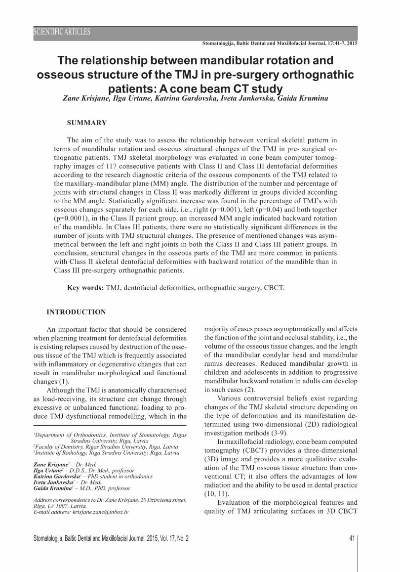
included a description of the structural qual-ity and quantity of the articular condyle and articular fossa/eminence complex in the TMJ with regards to condylar hypoplasia, condylar hyperplasia, articular surface flattening (Fig-ure 3), subcortical sclerosis, subcortical cysts, surface erosion, osteophytes, generalised sclerosis, loose joint body, deviation in form (Figure 4) and ankylosis.
Statistical analysis The aim of the statistical data analysis
was to evaluate the distribution of the osse-ous structural changes in the study groups. Data regarding the presence of descriptive signs was entered into the database, which later was converted into the database for-mat of the statistical software SPSS (Inc., USA). All calculations were performed us-ing this software. After at least a two-week interval, 71 randomly selected patient scans (i.e., 60% of all included patients) were re-evaluated. Dahlberg`s approach was used for the calculation of measurement error (Dahlberg, 1940), and an error less than 1 was regarded as tolerable. Mean values and standard deviations were calculated. Distri-bution frequency/prevalence was assessed. Pearson’s Chi-squared test and Fisher’s exact test were used to evaluate the statisti-cal significance of differences in prevalence among the groups.
The difference of means between groups was assessed using t-tests for paired data and for non-paired data. For comparison of means among more than two groups, an analysis of variance (one-way ANOVA) with Bonferroni correction (to control for overall II type er-ror) was used.
Fig. 3. Articular surface flattening and slight erosive changes of mandibular condyle
to the research diagnostic criteria for temporoman-dibular disorders (RDC/TMD) Axis I for the CT im-
Table 2. Presence of osseous structural changes of TMJ in Class II subdivisions on the left side
Presence of changes
Class II subdivision 1 Class II subdivision 2 TotalNumber of the joints
Fig. 4. Deformation of mandibular condyle, presenting subchondral sclerosis and osteophyte formation
Z. Krisjane et al. SCIENTIFIC ARTICLES
To reject the zero hypothesis and accept the al-ternative hypothesis, a level of statistical significance of p≤0.05 was used in all cases.
RESULTS
Analysis of the prevalent osseous struc-tural changes of TMJ revealed a statistically significant difference (p=0.014) between Class II and Class III patients. One or several signs were determined in 38.4% of the Class II group and 28.7% of the Class III group.
Class II subdivision 1 and Class II sub-division 2 patients presented no statistically significant differences in the prevalence of joints affected by structural changes between Class II subdivisions (Tables 2 and 3). The numbers of the joints in which no changes were found and those in which at least one was found was almost equal.
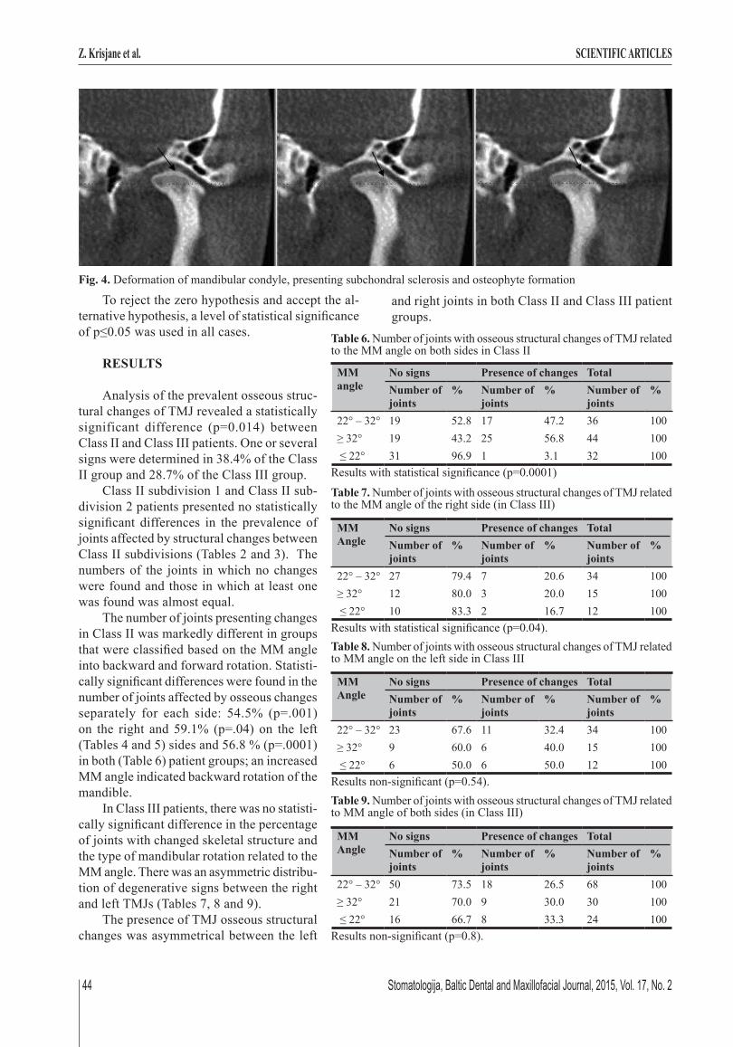
The number of joints presenting changes in Class II was markedly different in groups that were classified based on the MM angle into backward and forward rotation. Statisti-cally significant differences were found in the number of joints affected by osseous changes separately for each side: 54.5% (p=.001) on the right and 59.1% (p=.04) on the left (Tables 4 and 5) sides and 56.8 % (p=.0001) in both (Table 6) patient groups; an increased MM angle indicated backward rotation of the mandible.
In Class III patients, there was no statisti-cally significant difference in the percentage of joints with changed skeletal structure and the type of mandibular rotation related to the MM angle. There was an asymmetric distribu-tion of degenerative signs between the right and left TMJs (Tables 7, 8 and 9).
The presence of TMJ osseous structural changes was asymmetrical between the left
and right joints in both Class II and Class III patient groups.
Table 7. Number of joints with osseous structural changes of TMJ related to the MM angle of the right side (in Class III)
MM Angle
No signs Presence of changes TotalNumber of joints
A detailed analysis of the prevalence of each type of changes in the condyle and fossa eminence in the Class II patient group indicated that condylar hypoplasia, flattening, subsclerosis, osteophytes, deviation in form and the fossa flattening were pres-ent. Subcortical cyst, generalized sclerosis, loose joint body and ankylosis were not found. A higher percentage of previously mentioned signs were ob-served in the right and left sides of Class II patients with mandibular backward rotation than in the groups with neutral position or forward rotation. Subcortical sclerosis and deviation in form more frequent in the group with neutral mandibular position. The most common sign of osseous changes in patients of the Class III group was flattening of the condyle. There was no difference in the prevalence of TMJ structural
incompatibilities related to mandibular rotation. The percentage of joints showing destruction signs was not significantly different among MM angle groups in the Class II and Class III patient groups. The prevalence of osseous changes between the right and left sides was asymmetrical (Tables 10-13).
DISCUSSION
This study of CBCT images evaluated man-dibular condyle and articular/fossa structure disorders according to the RDC/TMD related to mandibular rotation in patients with skeletal Class II and Class III dentofacial deformities and found a statistically significant relationship between the radiographic features of TMJ osseous structure changes and man-
dibular backward rotation in Class II patients but not Class III patients.
Our results suggest differing prevalence of TMJ structural changes in different study groups; signs were more frequent in Class II patients than in Class III patients which indicates the role of skeletal discrepancy of the jaws in the development of TMJ osseous destruction. The study groups were classified according to the jaw skeletal discrepancy as determined by the MM rotation angle, which represents the vertical jaw rotation, as the literature data refers to the relationship among vertical planes rotation by a palatal and/or mandibular plane of one or both jaws in pa-tients with dentofacial deformities (16). The number of joints affected by TMJ structural changes was identified in the groups classi-fied according to the MM angle and was the highest in Class II patients with an increased MM angle. TMJ structural changes were ob-served asymmetrically between the left and rights joints, although these patients had no clinically visible jaw asymmetry. A previous study observed that TMJ problems had wide inter-individual variation even in patients with clinically similar malocclusions (17).
The most frequently occurring TMJ skeletal structural change in the Class II and Class III study groups with mandibular back-ward rotation was flattening of the articular surface of the condyle and fossa eminence. In the literature, flattening of the articular sur-face of the TMJ was described to result from remodelling (3), which can be radiologically defined as minor changes in the shape of the bone (4). Such flattening may present in both symptomatic and asymptomatic joints as a
Table 10. Osseous structural changes of TMJ related to MM angle of the right side in Class II
sign of indeterminate osteoarthritis (14), and it can be considered as a functional adaptation (18). The other commonly observed changes of TMJ structure in Class II and Class III patients with mandibular backward ro-tation were erosion and osteophytes of the condyle. The presence of erosion in joint surfaces characterizes the initial stage of degenerative changes in osteoarthritis and indicates the likely instability of the TMJ osseous structure (14) accompanied by the risk of an altered area of the joint surfaces, which can cause occlusal changes (19). The presence of osteophytes in the joint together with other signs is an important criteria for radiological diagnosis of osteoarthritis; in contrast to erosions, osteophytes occur in the late stage of degen-erative changes during adaptation (20).
Based on the RCD/TMD criteria used in the study, the temporomandibular joints were radiologi-cally assessed as a whole with regard to structural changes of osseous tissue quality. Only part of the mentioned diagnostic system was used in the study, and results describe the prevalence of signs in osse-ous structures but do not provide information about severity.
The literature contains studies that assess the disease based not only on the presence of signs but also on the severity as assessed by the intensity of erosions and osteophytes (21, 22); such analyses allow further consid-erations regarding the course of degenerative joint destruction.
All of Dworkin`s (1) criteria for assess-ment of changes in the TMJ can be catego-rized in the following groups: 1) the result of chronic adaption or active on-going changes; 2) changes that have an impact on function of the joint and quality of life, or asymptomatic; 3) changes that are reversible or irreversible. The literature indicates that bone loss in the mandibular condyle may result from the dysfunctional remodelling by orthognathic surgery, systemic and local arthritis, post-traumatic remodelling and hormonal imbal-ance (2, 23). The clinical manifestations of condylar destruction adversely affect the mechanics and function of the joint, and oc-clusion is characterised by progressive man-dibular backward rotation in adults (2) and is the reason for mandibular growth deviations in childhood and adolescence (24).
An opinion exists, that considerations regarding on-going processes in the TMJ can be based on established changes before orthodontic and orthognathic surgical treat-ment. However, in the literature, there is
insufficient data on the relationship between TMJ structural changes and mandibular vertical rotation, which may be an important factor in the prediction of the stability of orthodontic and orthognathic sur-gery treatment results. It is difficult to compare our results with existing data in the literature because of different radiological methods and the diversity of the diagnostic criteria used in the studies.
A conventional cephalometric study of mandibu-lar advancement surgery in Class II patients demon-strated that high-angle patients were associated with both a higher frequency and a greater magnitude of horizontal relapse (25). Clinical examination of the TMJ before and after orthognathic surgery showed that counter-clockwise rotation of the mandible is related to a slight increase in muscular symptoms after bilateral sagittal split osteotomy. All symptoms tended to decline over time, which suggests that the amount of advancement and mandibular rotation should not be considered as risk factors for the de-velopment of TMD in patients without pre-existing conditions (26).
Table 12. Osseous structural changes of TMJ related to MM angle of the right side in Class III
Received: 14 03 2014Accepted for publishing: 26 06 2015
1. Dworkin SF, LeResche L. Research diagnostic criteria for temporomandibular disorders: review, criteria, examina-tions and specifications, critique. J Craniomandib Disord 1992;6: 301-55.
2. Arnett GW, Milam SB, Gottesman L. Progressive mandibular retrusion idiopathic condylar resorption. Part I. Am J Orthod Dentofac Orthop 1996;101:8-15.
3. Kurita H, Ohtsuka A, Kobayashi H, Kurashina K. Flattening of the articular eminence correlates with progressive internal derangement of the temporomandibular joint. Dentomaxil-lofac Radiol 2000;29:277-79.
4. Honda K, Larheim TA, Sano T, Hashimoto K, Shinoda K, Westensson PL. Thickening of the glenoid fossa in osteo-arthritis of the temporomandibular joint. An autopsy study. Dentomaxillofac Radiol 2001;30:10-3.
5. Katsavrias EG, Halazonetis DJ. Condyle and fossa shape in Class II and Class III skeletal patterns: a morphometric tomography study. Am J Orthod Dentofac Orthoped 2005; 128:337-46.
6. Katsavrias EG. Morphology of the temporomandibular joint in subjects with Class II division 2 malocclusions. Am J Orthod Dentofac Orthoped 2006;129:470-8.
7. Hussain AM, Packota G, Major PW, Flores-Mir C. Role of different imaging modalities in assessment of temporoman-dibular joint erosions and osteophytes: a systematic review. Dentomaxillofac Radiol 2008;37:63-71.
8. Vitral RWF, Telles CS, Fraga MR, Fortes de Oliveira RSM, Tanaka OM. Computed tomography evaluation of temporo-mandibular joint alterations in patients with Class II division 1 subdivision malocclusions: condyle-fossa relationship. Am J Orthod Dentofac Orthop 2004;126:48-52.
9. Vitral RWF, da Silva Campos MJ, Rodrigues AF, Fraga MR. Temporomandibular joint and normal occlusion: is there any-thing singular about it? A computed tomography evaluation. Am J Orthod Dentofac Orthop 2011;140:18-24.
10. Loubele M, Bogaerts R, van Dijck E, Pauwels R, Vanheusden S, Suetens P, et al. Comparison between effective radiation dose of CBCT and MSCT scanners for dentomaxillofacial applications. Eur J Radiol 2009;71:461-68.
11. Davies J, Johnson B, Drage NA. Effective dose from cone beam CT investigation of the jaws. Dentomaxillofac Radiol 2012;41:30-6.
12. Petterson A. Imaging of the temporomandibular joint. In: Manfredini D. Current concepts on temporomandibular disorders. London: Quintessence Publishing; 2010.
13. Dung DJ, Smith RJ. Cephalometric and clinical diag-nozes of open bite tendency. Am J Orthod Dentofac Orthop 1988;94:484-90.
14. Ahmad M, Hollender L, Anderson Q, Kartha K, Ohrbach R, Truelove EL, et al. Research diagnostic criteria for tem-poromandibular disorders (RDC/TMD): development of image analysis criteria and examiner reliability for image analysis. Oral Surg Oral Med Oral Pathol Oral Radiol Endod 2009;107:844-59.
15. Dahlberg G. Statistical methods for medical and biological students. London: George Allen and Unwin; 1940.
16. Proffit WR, White RP, Sarver DM. Contemporary treatment of dentofacial deformity. Mosby: 2003, chapter 5, p. 153-7.
17. Krisjane Z, Urtane I, Krumina G, Neimane L, Rogovska I. The prevalence if TMJ osteorathritis in asymptomatic patients with dentofacial deformities: a cone-beam CT study. Int J Oral Maxillofac Surg 2012;41:690-95.
18. Brooks SL, Westesson PL, Eriksson L, Hansson LG, Bar-sotti JR, Arbor A. Prevalence of osseous changes in the temporomandibular joint of asymptomatic persons without internal derangement. Oral Surg Oral Med Oral Pathol 1992;73:122-26.
19. Hussain AM, Packota G, Major PW, Flores-Mir C. Role of different imaging modalities in assessment of temporoman-dibular joint erosions and osteophytes: a systematic review. Dentomaxillofac Radiol 2008;37:63-71.
20. van der Kraan PM, van den Berg WB. Osteophytes: relevance and biology. Osteoarthritis Cartilage 2007;15:237-44.
21. Alexiou KE, Stamatakis HC, Tsiklakis K. Evaluation of the severity of temporomandibular joint osteoarthritic changes related to age using cone beam computed tomography. Den-tomaxillofac Radiol 2009;38:141-7.
22. Cevidanes LHS, Hajati AK, Paniagua B, Lim PF, Walker DG, Palconet G, et al. Quantification of condylar resorption in temporomandibular joint osteoarthritis. Oral Surg Oral Med Oral Pathol Oral Radiol Endod 2010;110:110-17.
23. Gunson MJ, Arnett W, Milam SB. Pathophysiology and phar-macologic control of osseous mandibular condylar resorption. J Oral Maxillofac Surg 2012;70:1918-34.
24. Pirttiniemi P, Peltomaki T, Muller L, Luder HU. Abnormal mandibular growth and the condylar cartilage. Eur J Orthod 2009;31:1-11.
25. Mobarak KA, Espeland L, Krogstad O, Lyberg T. Mandibular advancement surgery in high-angle and low-angle Class II patients: Different long-term skeletal responses. Am J Orthod Dentofac Orthop 2001;119:368-81.
26. Frey DR, Hatch JP, Van Sickels JE, Dolce C, Rugh JD. Effects of surgical mandibular advancement and rotation on signs and symptoms of temporomandibular disorder: A 2 year follow-up study. Am J Orthod Dentofac Orthop 2008;133:490.e1-49.e8.
REFERENCES
CONCLUSION
The most common changes of osseous structures in the TMJ are related to skeletal Class II pre-orthognathic surgery patients with mandibular backward rotation.
Long-term observation allows the understand-ing of whether mandibular rotation is the cause or consequence of TMJ destruction in orthodontic and orthognathic surgery, and aids in determining instabil-ity in patients with dentofacial deformities.