150
The Use of High-Resolution Ultrasound and Ultrasound-Guided Needle Core Biopsy in the Diagnosis of Major Salivary Gland Lesions. David Crispin Howlett PhD June 2015
The Use of High-Resolution
Ultrasound and Ultrasound-Guided
Needle Core Biopsy in the Diagnosis
of Major Salivary Gland Lesions.
David Crispin Howlett
PhD June 2015
2
Abstract
Over recent years there have been significant changes in both the diagnosis and
management of palpable swellings of the parotid and submandibular major
salivary glands. There is a requirement and an expectation from both clinicians
and patients that a pre-operative diagnosis is available and that it is accurate and
obtained in a manner which is safe and timely, this is of particular importance with
the advent of minimally invasive surgical techniques. There is also increasing
recognition that the principal current salivary gland biopsy technique, fine needle
aspiration cytology (FNAC), is associated with significant drawbacks and is not
performing reliably across a network of institutions.
This critical appraisal concentrates on two key areas of my research and utilises
eleven published works and numerous selected references to examine and detail
the contribution my research has made to changes in modern salivary gland
clinical practice. Firstly, the appraisal demonstrates the diagnostic utility of high
resolution ultrasound in symptomatic salivary gland evaluation. The included
publications and references extend over a twelve year period and when combined
with other relevant literature have helped confirm ultrasound as the initial imaging
modality of choice in this clinical scenario, with ultrasound now widely used in
North America, as well as the United Kingdom and Europe.
The second, and more extensive component of the appraisal, concentrates on the
demonstration of the successful implementation of ultrasound guided core biopsy
(USCB) as a primary biopsy tool of choice in the salivary glands. Works
publications and references are included, spanning the initial and innovative
3
description of this technique applied to a series of parotid patients in 1999, through
to the largest current published series in 2015, where good patient tolerability and
high diagnostic accuracy of USCB are confirmed.
This critical appraisal demonstrates the significant and sustained contribution of
my research with a clear and defined impact on clinical practice. Changes in
salivary gland investigation and diagnosis are confirmed at both national and
international level, with evidence of increasing acceptance and utilisation of both
diagnostic ultrasound and USCB as the primary investigative tools of choice for
salivary gland swellings.
4
Title Page
Abstract 2
Contents 4
Acknowledgements 7
Author’s declaration 8
Aims of the Critical Appraisal 9
1. Background and Overview 11
1.1 Clinical presentation 11
1.2 Pathology involving the major salivary glands 12
1.3 The need for change in the management of salivary gland
neoplasms
14
1.4 The importance of a pre-operative diagnosis in the management
of a major salivary gland lesion
16
2. Imaging a Palpable Swelling of the Major Salivary Glands 18
2.1 High resolution ultrasound 18
2.2 Magnetic resonance imaging (MRI) 20
2.3 Computed tomography (CT) 21
2.4 Other imaging modalities 22
3. Biopsy Diagnosis of Major Salivary Gland Lesions 23
3.1 Fine needle aspiration cytology (FNAC) 24
3.2 Ultrasound guided core biopsy (USCB) 25
3.3 The technique of ultrasound guided core biopsy (USCB) 28
5
4. Discussion of Selected Published Works Included in 36
this Critical Appraisal
a) Diagnostic Ultrasound in Major Salivary Gland Assessment 40
4.1 Salivary gland: Oncologic imaging 40
4.2 A study to evaluate the accuracy of ultrasound in the diagnosis
of parotid lumps and to review the sonographic features of
parotid lesions – results in 200 patients
41
4.3 Sonoelasteography techniques in the evaluation and diagnosis
of parotid neoplasms
42
b) The Use of Ultrasound-Guided Core Biopsy (USCB) in the 43
Histological Diagnosis of a Major Salivary Gland Lesion
4.4 Ultrasound-guided cutting needle biopsy of the parotid gland 43
4.5 Ultrasound-guided biopsy in the evaluation of focal lesions and
diffuse swelling of the parotid gland
44
4.6 Diagnosing a parotid lump: Fine needle aspiration cytology or
core biopsy?
45
4.7 Sonographically guided core biopsy of a parotid mass 46
4.8 The use of fine needle core biopsy under ultrasound guidance in
the diagnosis of a parotid mass
47
4.9 Evaluation of biopsy methods in the diagnosis of submandibular
space pathology
49
4.10 Diagnostic investigation of parotid neoplasms – a 16 year
experience of freehand fine needle aspiration cytology and
ultrasound guided core biopsy
50
6
4.11 Establishing an accurate diagnosis of a parotid lump: An
evaluation of the current biopsy methods – fine needle aspiration
cytology, ultrasound-guided core biopsy and intra-operative
frozen section
52
5. Conclusion 54
6. References 56
7. Appendices: Published Works included as primary sources within the 64
critical appraisal and supporting correspondence
7
Acknowledgements
I would like first to express my gratitude to Professor Kevin Davies from Brighton
and Sussex Medical School (BSMS) who agreed to act as my Supervisor and
without his patience, kindness and support this project would simply not have been
possible. Dr Anne Mandy at the University of Brighton Doctoral College has
provided insight and timely feedback, guiding me throughout, and I would also like
to thank Professor Helen Smith at BSMS who, all that time ago, originally reviewed
my CV and suggested my research could lend itself to a thesis.
I am indebted to Nicholas Taylor, Medical Photographer at Eastbourne Hospital,
who has helped me in so many ways and over so many years, in the successful
production of publications and presentations – he has been invaluable in the
production of this document also. My secretary, Kirstie Vine, has contributed
significantly to manuscript preparation.
I would like to acknowledge the junior doctors and also Consultant colleagues from
numerous specialties, with whom I have collaborated and worked with closely over
many years.
Finally, my thanks and gratitude to my family to whom this manuscript is
dedicated; my parents, Ken and Margaret, my dear wife Lara and the children
Thomas, Ella, Robert and Miles, remembering my late wife Joanna and dear little
Christopher.
8
Declaration
I declare that the research contained in this critical appraisal, unless otherwise
formally indicated within the text, is the original work of myself, the author. The
appraisal has not been previously submitted to this or any other university for a
degree, and does not incorporate any material already submitted for a degree. All
included published works for which I am the sole author were entirely my own
work: those for which I am the first author or final (senior) author are substantially
my own work and solely my own work with reference to material discussed within
the critical appraisal document.
Signed:
Dated:
9
Aims of the Critical Appraisal
The recommended management of major salivary gland lesions has been in a
state of flux over the past thirty to forty years with persisting debate as to the
optimum management strategy for parotid lesions in particular. Over this time
period there have been, however, significant changes and improvements in how
salivary gland lesions are managed. This critical appraisal documents the
contribution my research has made over a sixteen year period to these alterations
in practice and includes a series of eleven supporting published works and also
selected references.
The appraisal examines two main areas within pre-operative major salivary gland
assessment and diagnosis. The first component documents my research and
educational contribution to the published literature covering imaging the salivary
glands, with particular reference to the utilisation of high resolution diagnostic
ultrasound. Ultrasound is now considered the initial imaging modality of choice in
salivary gland assessment in most institutions and the selected works / references
in this critical aapraisal have been chosen to reflect my on-going and sustained
contribution to this change in practice.
The second and more substantial component of the appraisal revolves around the
use of ultrasound guided core needle biopsy (USCB) in confirming the histological
nature of a salivary gland lesion. Here a more direct link can be made to a practice
change, following the publication from 1999 describing the first successful use of
this biopsy technique in a series of parotid patients (works publication 4.4) and
then a series of papers following an enlarging patient cohort, with now the largest
10
published series of parotid neoplastic USCB in the world literature (works
publication 4.10). USCB does address many of the diagnostic issues that surround
FNAC and USCB is now the biopsy technique of choice in many centres.
11
Background and Overview
1.1 Clinical Presentation
The parotid and submandibular glands comprise the major salivary glands and the
investigation and diagnosis of pathology involving these structures is the subject of
this critical appraisal. The minor salivary glands, including the sublingual glands,
occupy a more diverse range of locations, usually more deeply in the neck and
pharyngeal regions and tend to present and to be investigated in a different
manner.
Typically a parotid or submandibular tumour presents as a mass, or swelling, in
the parotid / pre-auricular or submandibular regions. The majority of parotid
tumours are within the superficial lobe and, as with submandibular lesions, they
are usually palpable clinically. Parotid deep lobe lesions are generally impalpable
and present in a different way, with pain or evidence of local effects such as nerve
paresis or dysphagia.
Benign salivary gland tumours tend to be relatively slow growing, painless and
non-fixed to adjacent structures. Malignant tumours are more inclined to be hard to
palpation, there may be fixity to adjacent structures and skin ulceration, rapid or
changing speed of growth, cervical lymphadenopathy and facial nerve paralysis
(parotids) are signs all strongly suggestive of malignancy. The malignant grade of
the tumour may affect presentation, it is recognised that certain well-differentiated
primary parotid malignancies can present in a relatively ‘benign’ manner. Pain is a
poor discriminator of benign from malignant disease and overall it is important to
recognize that the clinical differentiation of a benign from a malignant tumour and
indeed of neoplastic from non-neoplastic disease is unreliable [1].
12
1.2 Pathology Involving the Major Salivary Glands
The head and neck and the major salivary glands in particular are complex
structures histologically, containing not only salivary gland tissue, but also fat,
lymph nodes and peripheral nerves, all of which can give rise to benign and
malignant tumours. The salivary glands are a potential site for metastatic
carcinoma, particularly from primary malignant skin neoplasms. Consequently, a
broad range of pathologies are recognised in the salivary glands, inflammatory
and benign non-neoplastic lesions are common. Neoplasms are relatively
uncommon and the majority of these are benign, although benign:malignant
tumour ratios do vary between the different salivary glands.
Primary salivary gland neoplasms have an estimated incidence of 70 – 75 benign
and 8 – 14 malignant tumours annually per million population in the United
Kingdom [2]. Salivary gland malignancies comprise approximately 5% of cancers
of the head and neck, and yet they are the most diverse with at least 24 different
types recognised within the 2005 World Health Organisation (WHO) categorisation
[3]. Seventy percent of salivary carcinomas arise in the parotid gland [3]. As a rule
of thumb 80% of all salivary gland tumours arise in the parotid gland also (the
majority are superficial lobe in origin) and of these 80% are benign. The smaller
the salivary gland the greater the chance of a tumour being malignant (sublingual
> submandibular > parotid). Compared to the parotid the benign to malignant ratio
falls in the submandibular glands with a submandibular gland malignancy rate of
about 50%.
13
It is beyond the scope of this critical appraisal to discuss in detail major salivary
gland pathology, although some discussion is helpful and relevant as it informs
some of the diagnostic difficulties that arise.
Salivary gland tumours can show significant morphological diversity and also
cross-over of cytological / histological features between different tumour types and
even sometimes within an individual tumour. Hybrid tumours, dedifferentiation and
malignant degeneration within a benign tumour all serve to complicate histological
analysis and accurate diagnosis [3]. Additionally, as some of the histological
tumour subtypes are rare, this can further add to diagnostic difficulty, particularly in
smaller pathology departments with less exposure to these tumour types.
Histological grading of malignant salivary gland tumours is important for treatment
and prognostication and some (well-differentiated) malignant tumours will require
assessment of their capsule and interaction with surrounding glandular
parenchyma for accurate diagnosis – these factors become important when
considering fine needle biopsy techniques and are discussed later.
Non-neoplastic salivary gland lesions are relatively frequent, they may include
acute / chronic sialadenitis, granulomatous disorders (tuberculosis, sarcoidosis)
and Sjögren’s syndrome, calculi, benign lymph nodes and benign simple cystic
lesions.
A classification of salivary gland neoplasms is included in the 2005 WHO
publication [3]. A copy of this summary classification for epithelial tumours is
included in the works publication 4.1.
14
Briefly, salivary gland tumours are divided up into five main categories:
i) Malignant epithelial tumours
Including the more common primary malignant tumours: acinic cell carcinoma,
mucoepidermoid carcinoma, adenoid cystic carcinoma and adenocarcinoma.
ii) Benign epithelial tumours
Including the most common benign tumour – pleomorphic adenoma, also Warthin
tumour, basal cell adenoma and other less common adenomas.
iii) Soft tissue tumours
Including haemangioma, this group would also incorporate other tumour types,
such as lipoma and neurofibroma.
iv) Haematolymphoid tumours
Including Hodgkin’s lymphoma, diffuse large B-cell lymphoma and the marginal
zone B-cell lymphomas (these latter tumours can develop within the salivary gland
itself outside of lymph nodes, there is a known association with Sjögren’s
syndrome).
v) Secondary malignancy
Commonly from head and neck squamous cell carcinoma or malignant skin
tumours.
1.3 The need for change in the management of major salivary gland
neoplasms
The main driver for management change has been the recognition that an
accurate and also a pre-operative diagnosis is essential in most patients prior to
consideration for major salivary gland surgery. As already described the majority
of parotid and submandibular gland lesions present with a clinically palpable mass.
15
Historically these patients were referred to a surgeon, who might have a head and
neck interest, but might equally be a generalist, and following clinical examination
lesions were often surgically excised as both a diagnostic and therapeutic
manoeuvre. Inevitably this meant that for a significant proportion of patients
lesions were either inadequately excised then requiring further surgery, or lesions
were excised where surgery would have been better avoided. In recognition of the
challenges and complications of surgery in this region there was increasing
subspecialisation in head and neck surgery. Many surgeons adopted the practice
of initial open biopsy of salivary gland lesions in theatre to decide on further
surgery whilst the patient was under general anaesthesia. It became clear,
however, that incisional open biopsy, although it could provide a histological
diagnosis, had many associated problems – these included the need for general
anaesthetic, possible facial nerve damage, bleeding, sialocele and fistula
formation and also increased rates of tumour spillage/seeding [5]. In the early
1980s these concerns led to the demise of open biopsy in most centres and about
this time there were the first reports of utilisation of fine needle aspiration cytology
(FNAC) in obtaining pre-operative head and neck diagnosis. This technique is
covered in more detail later, but it is worth noting at this stage the widespread
difficulties that have been encountered by FNAC in salivary gland diagnosis,
particularly when performed non-guided (“blind”) in the clinic setting as was
originally the case in most centres.
During this period of diagnostic surgical excision, open biopsy and “blind” FNAC
there was, in addition, a relative under-utilisation of the available imaging
techniques (importantly computed tomography (CT), ultrasound and magnetic
resonance (MR) imaging) that could assist with lesion characterisation and
16
demarcation. With the introduction of newer imaging techniques and
improvements in existing imaging technology there have also been significant
changes in surgical treatment, in particular the relatively recent developments in
parotid-sparing surgical techniques such as extracapsular dissection. These are
beneficial to patients in that they reduce surgical trauma to the parotid gland in
particular and potential complications, they are however dependent on obtaining
an accurate pre-operative diagnosis [6,7].
1.4 The importance of pre-operative diagnosis in the management of a
major salivary gland lesion
With increasing patient and clinician awareness of the importance of an accurate
diagnosis prior to salivary gland surgical intervention, a process of “triple”
assessment has developed in many centres, mirroring the system initially
developed for breast lesions. This concept involves initial clinical evaluation,
imaging and finally cytological/histological diagnosis as needed. It is around the
latter two components of the three stage diagnostic process that this critical
appraisal revolves – namely the appropriate selection and optimisation of
diagnostic imaging modalities and then utilisation of a biopsy technique that is
safe, well tolerated, reliable and capable of high levels of diagnostic adequacy and
accuracy.
An accurate pre-operative diagnosis is considered essential for several reasons:
• Allows appropriate operative timing and selection.
17
• Facilitates informed and pre-operative patient consent, particularly important
with larger or malignant parotid lesions where facial nerve integrity may be
threatened or sacrificed.
• Gives the opportunity for both full tumour staging prior to surgery in malignant
lesions and also for the use of adjuvant and neo-adjuvant pre-operative
treatments for some malignancies.
• Allows the avoidance of surgery in some tumours (eg. Warthin’s tumour) in the
elderly or the unfit.
• Identifies pathology where surgery is best avoided (eg.
tuberculosis);demonstrates pseudo-tumourous lesions (ie. a ‘palpable’ lump,
but no lesion present on imaging); diagnoses benign and non-neoplastic
palpable lesions, eg. Intraparotid retention cyst, calculus, reactive lymph node,
where surgery would not usually be indicated.
18
2. Imaging a Palpable Swelling of the Major Salivary Glands
A variety of imaging modalities have been used for major salivary gland
assessment, often in combination and all have relative strengths and weaknesses.
A core component of this critical appraisal examines the contribution of selected
published works (4.1 – 4.3) and also references [13,14,15,16,22,23,24,25,26] to
the acceptance and utilisation of high-resolution ultrasound in major salivary gland
assessment and diagnosis.
2.1 High-Resolution Ultrasound
Ultrasound has been historically relatively under-utilised as an imaging modality in
the head and neck region, particularly in North America, although this situation has
changed over the past 10 - 15 years with increasing appreciation of the
advantages of ultrasound [8,9]. The selected works publications and references
have played an important role in this change in perception and alteration in clinical
practice. The works publications also reflect changes and improvements in
ultrasound technology over time and have been supplemented by other author
publications [10,11,12].
Ultrasound employs a transducer which generates a sound wave which penetrates
soft tissues and is reflected back to the transducer. The type of tissue will dictate
whether sound is absorbed, refracted and dispersed or reflected back and the
amount of sound reflection/absorption is detected at the transducer and creates an
image. There is a pay-off with transducers between penetration and resolution – a
lower frequency beam is needed to penetrate soft tissue further, e.g. in the
abdomen, which then reduces resolution. As the major salivary glands are
superficial the need for depth penetration is reduced allowing higher resolution
19
images and increased detail appreciation. Ultrasound is safe, non-ionising,
portable and relatively inexpensive. High levels of accuracy in lesion
characterisation have been demonstrated (works publication 4.2) and ultrasound
can also be used to guide needle biopsy. Ultrasound however cannot penetrate
bone and is not useful in either the assessment of bone or of structures with bone
overlying them. Ultrasound is also highly operator dependent and importantly there
is poor visualisation of the parotid deep lobe sonographically due to bony
mandibular obscuration. Although capable of a high level of accuracy, ultrasound
alone (as with all imaging modalities) cannot predict the nature of all salivary gland
pathology. Ultrasound also cannot reliably demonstrate the intra-parotid path of
the facial nerve – this again is a problem for all imaging modalities, the nerve is
best seen with magnetic resonance imaging, although not routinely. The main
intra-parotid vessels are however clearly demonstrated sonographically and the
plane of nerve passage inferred passing superficial to the vessels, thereby
allowing delineation of superficial from deep parotid lobe and thereby lesion
compartmentalisation. Ultrasound as a modality will be discussed in more detail in
the first section of works publications.
Ultrasound has several potential roles in salivary gland assessment:
• To ensure that the clinically palpable lesion actually represents an intra-
glandular and a real lesion and that this requires biopsy e.g. palpable
calculus or reactive intraglandular lymph node can be diagnosed confidently
with ultrasound alone and biopsy avoided.
• Ultrasound is used pre-biopsy to both characterise and compartmentalise
parotid tumours. If the lesion is confined to the superficial lobe, and the
20
lesion is confirmed benign on biopsy, then if excision is appropriate parotid
sparing surgery/superficial parotidectomy can be performed.
• Diagnostic ultrasound will often demonstrate additional clinically impalpable
and significant lesions (works publication 4.2).
• The use of ultrasound allows precise lesion sampling at time of biopsy e.g. if
the lesion is complex and solid/cystic then cystic areas can be avoided and
diagnostic yield increased with more solid components biopsied.
• Ultrasound will guide the need for further imaging, usually MRI, in large,
atypical or probable malignant lesions.
2.2 Magnetic Resonance Imaging (MRI)
This technique uses high magnetic field strengths to create an image and has
excellent spatial resolution and tissue contrast definition capabilities. MRI is
accurate in salivary gland lesion assessment, with similar reported results to
ultrasound [1,8] in lesion characterisation and MRI also does not involve ionising
radiation. MRI is discussed further within both selected works publications and
also references. As a technique MRI is relatively expensive, not always readily
available and is associated with significant failure rate due to patient
claustrophobia. MRI usually involves the administration of intravenous contrast
when utilised in the salivary glands which has the associated risks of
nephrotoxicity (in renal impairment) and allergy.
The parotid deep lobe and deeper pharyngeal and parapharyngeal spaces are
clearly demonstrated using MRI which is a diagnostic advantage and MRI is the
most effective technique at delineating the facial nerve directly and for
21
demonstrating malignant neural infiltration (adenoid cystic carcinoma) [1,9,13,14].
MR is not useful in assessing calculi or calcifications.
2.3 Computed Tomography (CT)
This has been a historically popular technique for salivary gland imaging
particularly in North America and the Far East [15]. Its use has declined however
in primary lesion assessment with increasing acceptance of ultrasound and MRI.
This critical appraisal demonstrates the important contribution of the selected
works publications and references to this change in role for CT. CT has excellent
contrast resolution and remains the technique of choice for the detection of
calcification and bone erosion, however it is associated with high levels of ionising
radiation and usually requires the use of intravenous iodinated contrast for salivary
gland imaging with the associated risks of nephrotoxicity and allergy/anaphylaxis.
CT remains the technique of choice for lung staging in cases of salivary gland
malignancy and is used in acutely ill patients with suspected
parotid/submandibular abscess formation as it can acquire a set of data rapidly to
include the deep lobe and parapharyngeal spaces.
22
2.4 Other Imaging Modalities
Plain film and sialography (conventional contrast, CT or MR sialography) have no
role in salivary gland tumour assessment, but are still used for the assessment of
possible duct stricture or stone (usually after a negative ultrasound study).
PET (positron emission tomography) has a secondary role in salivary gland
malignancy and it is increasingly combined with CT as a staging technique.
Nuclear medicine studies have a secondary role in evaluating salivary gland
function (eg Sjögren’s syndrome).
23
3. Biopsy Diagnosis of Major Salivary Gland Lesions
As discussed, an accurate diagnosis of a salivary gland swelling is essential to
allow appropriate and timely patient management and for neoplasm
characterisation lesion cytology/histology is usually necessary. How this biopsy
material is best obtained has been, and to some extent remains, controversial.
This topic is the focus of the second part of the critical appraisal which describes
how selected works publications (4.4 – 4.11) and references [19,26,27,28,
29,30,32,33,34,35] have initiated and then contributed to a significant change in
biopsy practice, namely, the introduction of ultrasound-guided core biopsy (USCB)
and its successful application in the major salivary glands. This section
concentrates on FNAC and USCB, but mention will be made of two other less
common forms of biopsy. Intra-operative frozen section analysis is a form of open
biopsy that has been previously utilised and has recently been re-evaluated [17]
and is discussed in more detail in the works publications. It does have potential
benefits, it is recognised however that due to the wide variety, complex and often
overlapping appearances of salivary gland tumours, that this technique is not
generally applicable or appropriate for complex decision making in theatre. It also
does not allow informed pre-operative patient consent and may put unacceptable
strains on histopathology departments. Punch biopsy is a further biopsy technique
that is sometimes used for submandibular space lesions and is discussed in paper
4.9 in the works publications. This leaves the two principal pre-operative biopsy
techniques currently being utilised, both using a small bore needle – namely fine
needle aspiration cytology (FNAC) and core biopsy, this usually performed with
ultrasound guidance – ultrasound guided core biopsy (USCB).
24
3.1 Fine Needle Aspiration Cytology (FNAC)
This technique developed following the demise of open biopsy in the 1980s. It
involves the use of a small bore needle without local anaesthesia, usually 20G or
smaller, with sampling of the lesion in question and the sample smeared on slides
and sent to for cytological analysis. Traditionally FNAC was undertaken by
clinicians, usually in clinic and performed “blind” i.e. non-guided. FNAC as a
technique is fast, cheap and safe. There have been numerous studies looking at
the diagnostic performance of FNAC, many reporting high sensitivity and
specificity. However, it has become increasingly clear (and the subject of a recent
large meta-analysis [18]) that there is a wide variety in the diagnostic performance
of FNAC across centres, with high reported non-diagnostic, false negative and
false positive rates [19] and also low diagnostic accuracy in FNAC cases reported
as non-neoplastic and benign [18].
The performance of FNAC is affected and can be improved by a number of factors
[18]: -
• Reduced number of trained operators undertaking the procedure, rather than a
wide spectrum of operators of variable experience.
• Cytologist/cytology technician either performing FNAC with the option of
repeat sampling or available at the time of FNAC to ensure an adequate
sample has been obtained.
• The provision of ancillary cytology facilities, such as flow cytometry or in-situ
hybridisation techniques, to improve diagnostic performance.
• The use of ultrasound-guidance to improve sampling accuracy.
25
However, not all these options are generally available, in particular, in the UK,
there are shortages of trained cytologists and cytology technicians and ancillary
equipment is expensive and access is restricted. Even where optimised
circumstances do exist to improve the sampling performance of FNAC, there are
inherent problems for a technique which provides a cellular aspirate in the salivary
glands, as already alluded to. Salivary gland tumours have a wide variation in
architectural and cytomorphonuclear features and many of these features overlap
making cytological analysis difficult. Squamous or mucinous metaplasia, for
example, is common in a range of both benign and malignant tumours, and
lymphoid hyperplasia is also not reliably diagnosed on FNAC. FNAC cannot
provide information on tumour grade, tumour margins or interaction of tumour with
adjacent tissue. The diagnostic limitations of FNAC are discussed in more detail
later in the critical appraisal, in particular works paper 4.11. Heterogeneity in
provision of optimisation resources in part explains the reported variability of
performance of FNAC, however, the limitations imposed by a cellular aspirate are
more fundamental. The perceived and reported limitations of FNAC have lead
interested clinicians to explore other biopsy techniques to improve diagnostic yield.
3.2 Ultrasound Guided Core Biopsy (USCB)
USCB involves the use of a spring-loaded biopsy device that discharges a small
bore needle and obtains a core of tissue contained in a central biopsy tray in a
single automated movement. Ultrasound is a “real-time” modality and when
performing biopsy the probe, needle and patient can all be actively positioned to
facilitate safe needle access. Originally USCB was described in the abdomen/liver
and breast and demonstrated superior performance over FNAC in these areas
26
where USCB is now the technique of choice. Core biopsy uses a larger needle
than FNAC, usually 18G in salivary gland work – an 18G needle is 1.2 mm thick
and crucially USCB obtains a core of tissue that can be sent for
immunohistochemical analysis, rather than relying on a needle aspirate of cells.
This additional histopathological facility allows both typing and grading of tumours
and also improved characterisation of lymphoid proliferation, namely the
differentiation of reactive nodal hyperplasia from low-grade lymphoma [20, 21]. A
core of tissue allows assessment of tissue architecture, avoiding the broad
category diagnoses associated with FNAC such as “malignant” or “favour
neoplasm” and giving a precise and definitive diagnosis. An intact core of tissue
will also allow diagnosis of salivary involvement by systemic disease processes.
Core biopsy offers increased potential to assess lesion capsular integrity and
infiltration of surrounding tissues, although this is an area that remains difficult to
evaluate in some cases (discussed in works publications).
Core biopsy does have potential weaknesses when compared to FNAC which are
mentioned here and discussed in more detail later in the works publications.
• Minimally more invasive than FNAC with a larger bore needle and requiring
local anaesthesia and a small skin incision.
• There is a potentially increased risk of tumour seeding with the larger bore
needles used in USCB when compared to FNAC [20,21].
• Not as widely available as FNAC, currently fewer trained operators
• Theoretical increased risk of vascular/nerve injury and bleeding using a larger
needle than FNAC – this is more of an issue in the parotids.
27
• As there is a built-in delay around histological reporting of USCB specimens,
USCB does not lend itself to proposed “one-stop” clinic scenarios which
revolve around FNAC as a diagnostic tool.
• Potentially more expensive than FNAC as the procedure is a little more
complex and time consuming.
28
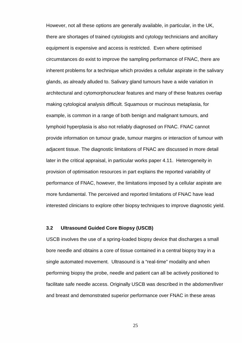
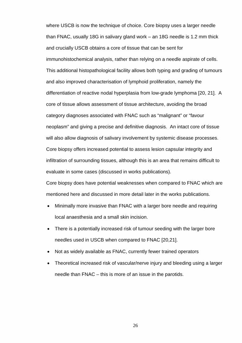
3.3 The technique of Ultrasound-Guided Core Biopsy (USCB)
Figure 1 – An image demonstrating a biopsy gun currently used for USCB and an
18 gauge needle alongside. This biopsy device has a variable throw facility of
either 15mm or 22mm.
Figure 2
29
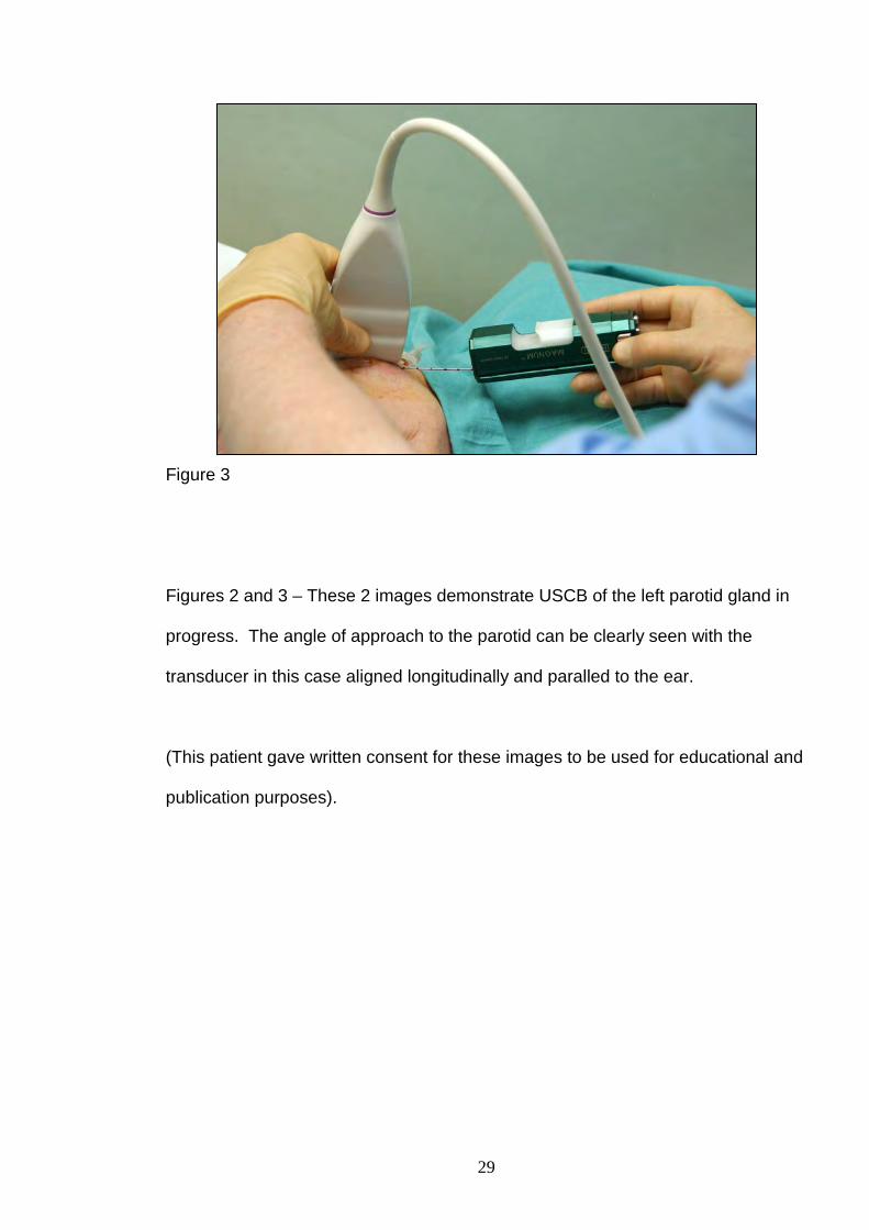
Figure 3
Figure 3
Figures 2 and 3 – These 2 images demonstrate USCB of the left parotid gland in
progress. The angle of approach to the parotid can be clearly seen with the
transducer in this case aligned longitudinally and paralled to the ear.
(This patient gave written consent for these images to be used for educational and
publication purposes).
30
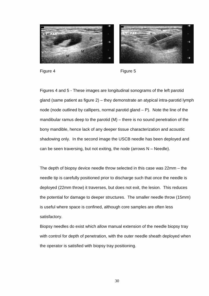
Figure 4 Figure 5
Figures 4 and 5 - These images are longitudinal sonograms of the left parotid
gland (same patient as figure 2) – they demonstrate an atypical intra-parotid lymph
node (node outlined by callipers, normal parotid gland – P). Note the line of the
mandibular ramus deep to the parotid (M) – there is no sound penetration of the
bony mandible, hence lack of any deeper tissue characterization and acoustic
shadowing only. In the second image the USCB needle has been deployed and
can be seen traversing, but not exiting, the node (arrows N – Needle).
The depth of biopsy device needle throw selected in this case was 22mm – the
needle tip is carefully positioned prior to discharge such that once the needle is
deployed (22mm throw) it traverses, but does not exit, the lesion. This reduces
the potential for damage to deeper structures. The smaller needle throw (15mm)
is useful where space is confined, although core samples are often less
satisfactory.
Biopsy needles do exist which allow manual extension of the needle biopsy tray
with control for depth of penetration, with the outer needle sheath deployed when
the operator is satisfied with biopsy tray positioning.
31
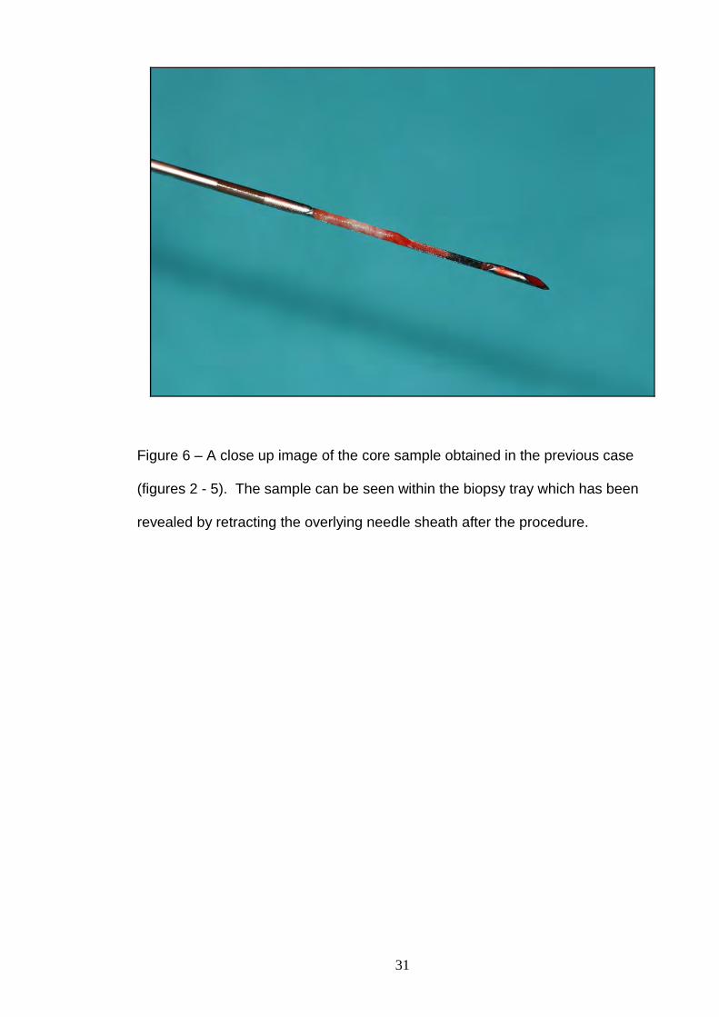
Figure 6 – A close up image of the core sample obtained in the previous case
(figures 2 - 5). The sample can be seen within the biopsy tray which has been
revealed by retracting the overlying needle sheath after the procedure.
32
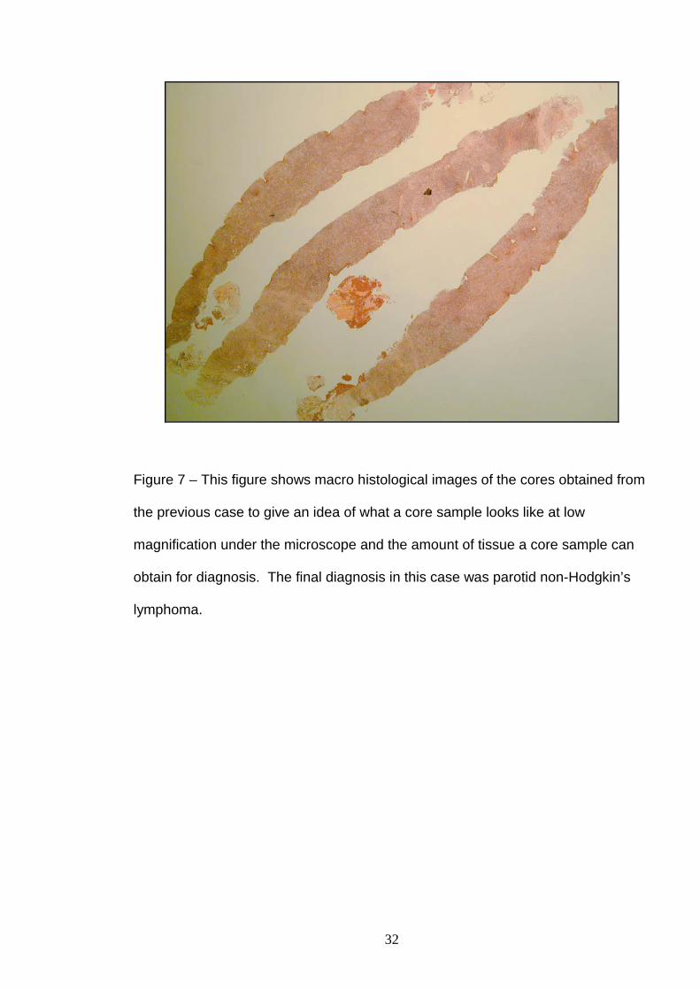
Figure 7 – This figure shows macro histological images of the cores obtained from
the previous case to give an idea of what a core sample looks like at low
magnification under the microscope and the amount of tissue a core sample can
obtain for diagnosis. The final diagnosis in this case was parotid non-Hodgkin’s
lymphoma.
33
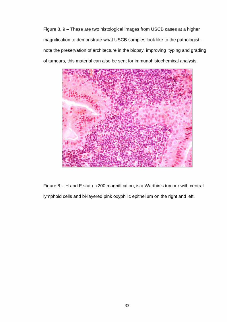
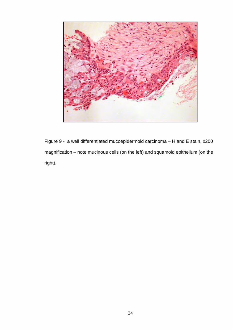
Figure 8, 9 – These are two histological images from USCB cases at a higher
magnification to demonstrate what USCB samples look like to the pathologist –
note the preservation of architecture in the biopsy, improving typing and grading
of tumours, this material can also be sent for immunohistochemical analysis.
Figure 8 - H and E stain x200 magnification, is a Warthin’s tumour with central
lymphoid cells and bi-layered pink oxyphilic epithelium on the right and left.
34
Figure 9 - a well differentiated mucoepidermoid carcinoma – H and E stain, x200
magnification – note mucinous cells (on the left) and squamoid epithelium (on the
right).
35
Newer biopsy needles are becoming available which offer several advantages
over the current available biopsy devices. Newer generation needles:-
• Employ a side cutting, not end cutting, mechanism which reduces potential
distal structure needle trauma.
• Increased throw variations are available – eg. 13, 23, 33mm
• Due to changes in internal needle design a needle of a certain gauge can
deliver a larger needle sample – eg. a 18G size needle can deliver an
equivalent 16G core, which is helpful for histology and delivers more tissue for
diagnosis, with a smaller biopsy hole and reduced trauma.
• Newer needles are disposable after a single use, improving infection control
aspects (current needles are likewise single use, but the gun is used
repeatedly, ideally requiring cleaning between patients).
• The main downside of the newer needles types is that of cost – typically about
£120 per needle compared to approximately £15 for a current needle for
example (current biopsy device about £800 as a single purchase), and this is a
rate-limiting step for their more widespread introduction.
36
4. Discussion of Selected Published Works Included in this
Critical Appraisal
The selected works publications, eleven in total, are divided into two sections
where the salient findings are discussed, together with an analysis of their
contribution to the published literature and current clinical practice –
a) The initial three publications (4.1 - 4.3) examine the use of ultrasound in
salivary gland assessment and diagnosis.
b) The remaining eight papers (4.4 - 4.11) study the performance, current
and future potential roles of USCB in major salivary gland diagnosis.
37
a) Diagnostic Ultrasound in Major Salivary Gland Assessment
______________________________________________________
4.1 Mandalia U Y, Porte F N, Howlett D C
Salivary Gland: Oncologic Imaging.
Ultrasound Clinics of North America 2014; 9; 99-113
4.2 Sriskandan N, Hannah A, Howlett D C
A Study to Evaluate the Accuracy of Ultrasound in the Diagnosis of Parotid
Lumps and to Review the Sonographic Features of Parotid Lesions –
Results in 220 Patients.
Clinical Radiology 2010; 65:366-372
4.3 Westerland O, Howlett D C
Sonoelastography Techniques in the Evaluation and Diagnosis of Parotid
Neoplasms
European Radiology 2012; 22 (9) 66-969
38
b) The Use of Ultrasound Guided Core Biopsy (USCB) in the
Histological Diagnosis of a Major Salivary Gland Lesion
4.4 Buckland J R, Manjaly G, Violaris N, Howlett D C
Ultrasound Guided Cutting Needle Biopsy of the Parotid Gland
Journal of Laryngology and Otology 1999; 113; 988-992
4.5 Keese KW, Manjaly G, Violaris N, Howlett D C
Ultrasound Guided Biopsy in the Evaluation of Focal Lesions and Diffuse
Swelling of the Parotid Gland.
British Journal of Oral and Maxillofacial Surgery 2002; 40; 364-368
4.6 Howlett D C
Diagnosing a Parotid Lump: Fine Needle Aspiration Cytology or Core
Biopsy?
British Journal of Radiology 2006; 79: 295-297
4.7 Howlett D C, Menezes LJ, Lewis K, Moody AB, Violaris N, Williams M D
Sonographically Guided Core Biopsy of a Parotid Mass
American Journal of Roentgenology 2007; 188-223-227
4.8 Breeze J, Andi A, Williams M D, Howlett D C
The Use of Fine Needle Core Biopsy Under Ultrasound Guidance in the
Diagnosis of a Parotid Mass
British Journal of Oral and Maxillofacial Surgery 2009; 47: 78-79
39
4.9 Olubaniyi B O, Chow V, Mandalia U, Haldar S, Gok G, Michl P, Ramesar K,
Sellon E, Williams M, Howlett D C
Evaluation of Biopsy Methods in the Diagnosis of Submandibular Space
Pathology.
International Journal of Oral and Maxillofacial Surgery 2014; 43:281-285
4.10 Haldar S, Mandalia U, Skelton E, Chow V, Tighe D, Ramesar K,
Williams M D, Howlett D C
Diagnostic Investigation of Parotid Neoplasms – a 16 Year Experience of
Freehand Fine Needle Aspiration Cytology and Ultrasound Guided Core
Biopsy.
International Journal of Oral and Maxillofacial Surgery – 2015; 44(2):151-
157
4.11 Howlett D C, Skelton E, Moody AB
Establishing an Accurate Diagnosis of a Parotid Lump: An Evaluation of
Current Biopsy Methods – Fine Needle Aspiration Cytology, Ultrasound
Guided Core Biopsy and Intra-operative Frozen Section.
British Journal of Oral and Maxillofacial Surgery – accepted for publication
August 2014
40
a) Diagnostic Ultrasound in Major Salivary Gland Assessment
4.1. Salivary Gland: Oncologic Imaging
This paper was published in 2014 in the North American journal, Ultrasound
Clinics of North America, a journal dedicated to updates and reviews of current
ultrasound best practice. This review article contains a detailed and
comprehensive examination of the ultrasound features of salivary gland neoplastic
disease utilising the most up-to-date sonographic technology. This selected works
publication is supplemented by a series of published ultrasound opinion papers
and pictorial imaging reviews from over a twelve year period as detailed in the
References section [22,23,24,25]. These articles have informed radiological
practice supplementing articles from other authors and have encouraged the wider
use and acceptance of ultrasound, which now represents the imaging modality of
choice in the initial assessment and diagnosis of major salivary gland lesions in
most institutions.
The publication of this 2014 paper (4.1) in a North American journal was important
– traditionally ultrasound has been relatively under-utilised in North America,
where there has been a tendency to rely upon MRI / CT. This publication was
extremely encouraging, suggesting an increased understanding and uptake of
ultrasound by the North American audience.
Paper 4.1 and also the selected references provide an overview and illustration of
the ultrasound findings in salivary gland disease over a prolonged period,
incorporating improvements in ultrasound technology and they give a clear
demonstration of the importance of ultrasound in the diagnostic pathway.
41
4.2 A Study to Evaluate the Accuracy of Ultrasound in the Diagnosis of
Parotid Lumps and to Review the Sonographic Features of Parotid Lesions –
Results in 220 Patients.
This was another important paper published in 2010 in the leading UK based
radiology journal, Clinical Radiology, which aimed to assess the accuracy of initial
sonographic diagnosis and also to review the sonographic features of parotid
lesions when compared to final histology. The paper included 220 patients with
data collected over an 11 year period. This published series remains the largest of
its kind in the World literature.
A variety of ultrasound machines were employed over the 11 year study period,
which affected, at least in part, the diagnostic capabilities of ultrasound as the
technology improved. This paper established a diagnostic accuracy of ultrasound
in the determination of malignancy of 93%, results not dissimilar to previous
smaller published series and comparable to MRI. The paper also highlighted the
ability of ultrasound to differentiate benign from malignant disease in most patients
and to detect additional lesions impalpable clinically. However, false negative and
false positive findings of ultrasound were described and a cross-over of
sonographic findings was demonstrated between some benign neoplasms and
also between certain benign and malignant neoplasms. The article confirmed
ultrasound as a valuable adjunct to clinical examination within the triple
assessment pathway, able to characterise and compartmentalise lesions
accurately in most patients. However to maximise diagnostic yield it was
recommended that ultrasound should be combined with needle biopsy as
appropriate.
42
The paper won the Clinical Radiology Ellis-Barnett prize in 2010 for “best paper
published on Ultrasound” that year in the journal.
4.3 Sonoelastography Techniques in the Evaluation and Diagnosis of
Parotid Neoplasms.
This was an invited expert review and opinion article, the invitation from the Editor
in Chief of European Radiology, the leading European radiology journal
publication. This paper provides a critique of two proffered publications to be
published in the journal, investigating two novel ultrasound applications in parotid
neoplasm assessment. The two papers looked at variations of the technique of
sonoelastography: essentially this involves manual compression of tissue applied
by an ultrasound probe and the degree of tissue deformation/stiffness is detected
and characterised. It was hoped that the technique measuring tissue compliance
or stiffness would improve the diagnosis of parotid neoplasia, namely malignant
lesions are “stiffer”. Neither technique however was found to offer significant new
diagnostic information to existing ultrasound techniques, although a potential
advantage over conventional ultrasound was postulated, with a recommendation
to undertake larger follow up prospective studies.
This paper is included within the thesis as it demonstrates a published contribution
not only to current ultrasound best practice, but also to potential future
developments in the technique.
43
b) The Use of Ultrasound Guided Core Biopsy (USCB) in the
Histological Diagnosis of a Major Salivary Gland Lesion
4.4 Ultrasound-Guided Cutting – Needle Biopsy of the Parotid Gland
This publication is likely to represent the single most important and potentially
influential paper contained within this critical appraisal. This article was published
in the leading UK clinical ear, nose and throat journal – The Journal of
Laryngology and Otology and describes the novel and successful application of
USCB to a series of patients with parotid lesions.
In clinical practice most referrals for salivary lump investigation originate from
either Ear, Nose or Throat or Oral/Maxillofacial surgeons. At the time of the paper
clinician “blind” FNAC was the norm in my institution, with high non-diagnostic
rates and a high number of patients undergoing surgical excision for diagnosis. It
was clear that changes were necessary to provide an accurate pre-operative
diagnosis for patients thereby facilitating appropriate operative management. On
review of the existing literature on core biopsy in the abdomen and breast it
seemed rational and reasonable that this technique could potentially translate itself
safely and effectively to the major salivary glands.
In paper 4.4, 16 patients were included, all had parotid lesions and 13/16 had a
previous non-diagnostic blind FNAC. USCB was undertaken with an older type of
spring-loaded biopsy gun with a fixed 20mm throw, 18G needles were used in all
cases. The range of pathologies encountered is included in the paper.
Key findings from this study:
44
• USCB using local anaesthesia was well tolerated by all patients, with no
significant complications.
• There was high diagnostic yield with all samples adequate for histological
analysis and with 100% diagnostic accuracy in the seven patients who had
subsequent surgery, nine patients therefore avoided surgical excision biopsy
on the basis of their USCB biopsy result.
• Ultrasound detected additional significant clinically occult disease in 31% of
cases.
This paper was important – it was the first publication in the world literature
describing the use of USCB applied to the parotid glands in a series of patients,
previous reports were sporadic, isolated cases. The paper confirmed USCB could
be performed safely in the parotid glands and that it offered potential significant
diagnostic benefits when compared to FNAC and this work was presented at
national surgical and radiological meetings. Over the next few years a series of
correspondence type articles discussing and highlighting the potential benefits of
USCB were also published and these are included in the References section [26 -
30]. A further paper confirmed that ultrasound alone was sufficient as a pre-
operative imaging modality for superficial lobe benign parotid tumours [31].
4.5 Ultrasound Guided Biopsy in the Evaluation of Focal Lesions and
Diffuse Swelling of the Parotid Gland
This paper was published in 2002 in the UK based British Journal of Oral and
Maxillofacial Surgeons. The paper looked at core biopsy performance and
45
accuracy in a cohort of 54 patients with parotid swelling (including the original
group of 16 patients). USCB was 100% accurate in the differentiation of benign
from malignant disease in the 28 patients who had subsequent surgery and
histological correlation. These findings were extremely encouraging, there were
also no significant biopsy related complications and there was no evidence of
tumour seeding at biopsy sites in the clinical follow up period. The paper
acknowledged that tumour seeding can occur up to 20 years following parotid
instrumentation and the follow-up period for these patients was short. Interestingly
clinician performed blind FNAC had effectively ceased in the institution at this
stage after the initial publication (4.4) had confirmed the safety and efficacy of
USCB.
4.6 Diagnosing a Parotid Lump: Fine Needle Aspiration Cytology or Core
Biopsy.
This paper was a review/opinion article published in the British Journal of
Radiology. From a literature review and personal experience of both USCB and
FNAC it was becoming clear that USCB did potentially offer significant advantages
over FNAC in salivary gland diagnosis, advantages that were also becoming
apparent in biopsy series of cervical lymphadenopathy [32,33,34].
This British Journal of Radiology paper was the first publication in the peer-
reviewed literature where the argument for potential use and selection of USCB
over FNAC was made. The paper also effectively argued against NICE (National
Institute of Clinical Excellence) guidance published at the time which advocated a
“one stop” neck lump service based on the use of FNAC, proposing patients to be
46
seen, examined, imaged, biopsied and diagnosed in one prolonged clinic
attendance. A further related publication at this time commented on the potential
disadvantages of the “one stop” model, advocating instead increased utilisation of
diagnostic ultrasound and USCB in neck lump assessment, especially in units with
poor results from FNAC [35].
4.7 Sonographically Guided Core Biopsy of a Parotid Mass
For USCB as a technique this was another important and landmark publication,
published in the North American radiology journal, the American Journal of
Roentgenology. The paper contained continuation and new patient data from the
original parotid USCB series, with a total of 135 patients included (data from 1998-
2004). This was not only the first publication of dedicated USCB parotid data in a
radiology journal, but first also in the North American literature and this was the
largest published series of parotid USCB patients in the literature at that time. In
this paper USCB showed itself to be highly accurate in the diagnosis of a range of
both neoplastic and non-neoplastic parotid gland pathology. All biopsies were
considered satisfactory for histological analysis. In 74 of the 76 patients who
underwent surgery there was biopsy correlation with final surgical histological
findings. In the 2 discrepant patients there was malignancy diagnosed in each
case at USCB, only the sub-type of malignancy differed between USCB and
surgical histology, patient management was unaffected. Fifty-nine patients were
diagnosed and managed on USCB results alone, thereby avoiding surgery.
Interestingly in this paper there were 17 cases with a USCB diagnosis of
Hodgkin’s disease or non-Hodgkin’s lymphoma. Lymphoma, as previously
47
discussed, is not usually possible to diagnose/characterise on FNAC results and
such cases would normally have needed to progress to surgical excision for
definitive diagnosis and instigation of treatment. In this paper it was shown that
11/17 cases were diagnosed and treated on the bases of USCB results alone, only
6 cases requiring surgical excision. These findings in lymphoma patients
demonstrate not only the good sample yield obtained at USCB, but importantly
also increased understanding, familiarity and acceptance of USCB samples and
results by histopathologists and treating clinicians. USCB was also well tolerated
by patients in the series, it was performed quickly as an out-patient procedure,
with no significant complications and with no reported tumour seeding in the
biopsy series to date.
4.8 The Use of Fine Needle Core Biopsy Under Ultrasound Guidance in
the Diagnosis of a Parotid Mass
This was a follow-up study published in the British Journal of Oral and Maxillofacial
Surgeons as a technical procedural note. This was a valuable paper as it
confirmed the optimised procedural and technical aspects of biopsy – a modified
and upgraded automated biopsy device was now available which was capable of
varying the throw of the biopsy needle – a 15 or 22mm throw could be selected,
(see earlier). This paper included updated biopsy data in 200 patients, an
additional 65 patients to the previous North American journal publication (4.7).
In most cases an 18G needle was utilised with an average of two needle passes
per patient. USCB again performed extremely well – in 198/200 patients samples
were considered suitable for histological analysis, with a diagnostic accuracy of
48
99.1% and1 non-correlation – (a false negative) between USCB and final surgical
histology.
These results are of interest as they were the first inadequate and also false
negative samples in the case series.
The two non-diagnostic cases both had subsequent surgery and were parotid
tumours, one a Warthin’s tumour and one an adenocarcinoma – these lesions had
been largely cystic on ultrasound explaining the difficulty in obtaining a diagnostic
solid sample. The false negative case was in a 28 year old male with a 4 month
history of a slowly enlarging, although hard, right parotid mass – ultrasound
suggested likely malignancy and USCB had been undertaken and histology
suggested likely benign basal cell adenoma. This patient had also had a blind
FNAC in clinic pre-USCB – this had reported a pleomorphic adenoma. It is
interesting that the clinician still sent the patient for USCB despite having
performed FNAC and without knowledge of the FNAC results suggesting a lack of
trust in FNAC. Final surgical histology from superficial parotidectomy confirmed a
well differentiated basal cell adenocarcinoma, this was only diagnosable once the
pathologist could observe the entire resected specimen and could visualise focal
capsular infiltration consistent with malignancy. The lesion was completely
excised but the patient did proceed to completion parotidectomy with post-
operative radiotherapy. This false negative case was educational and highlighted
a potential pitfall for USCB – already well recognised with FNAC in the literature –
a well differentiated salivary gland malignancy can be difficult to diagnose on
needle sampling alone as capsular loss of integrity can be important for diagnosis
and may not be apparent on a needle biopsy. In this case there was already a high
49
clinical/sonographic index of suspicion in the other two limbs of triple assessment.
No significant complications had been observed in the series to date – one patient
had experienced a small haematoma post-biopsy, no patients had demonstrated
tumour seeding (tumour seeding is discussed in more detail in works publication
4.11). The option for surgeons to excise the needle biopsy tract at the time of
surgery is available to reduce the potential for tumour seeding and occurs in some
centres, although this has not been considered necessary in our institution and
there is no evidence to suggest this approach is routinely required.
4.9 Evaluation of Biopsy Methods in the Diagnosis of Submandibular
Space Pathology
This paper was published in 2014 in the International Journal of Oral and
Maxillofacial Surgery and records the performance of three biopsy techniques in
diagnosing pathology of the submandibular space/gland from 1999-2011.
Submandibular gland tumours are far less common than parotid tumours,
comprising 10-15% of all salivary gland tumours. This study included biopsy data
in 44 patients, the relatively small number of patients reflecting the rarity of
tumours in this region. Biopsy techniques included clinician performed blind
FNAC, USCB and also punch biopsy, a technique that is sometimes used in the
submandibular region and is performed in theatre by the surgeon under local
anaesthesia. The total of 81 biopsy specimens in the study represents the largest
such series in the published literature, with a paucity of previously published work
on this subject. FNAC in this series was diagnostic in only 2/15 cases, USCB in
20/24 and punch biopsy in 5/7. USCB was accurate in diagnosing submandibular
50
space pathology, although a higher proportion of non-diagnostic (2/24) or
equivocal (2/24) cases was observed then in the parotids, the reasons for this are
unclear. An accurate pre-operative diagnosis is important in the submandibular
glands also, this allows informed patient consent and appropriate operative
intervention in malignancy. However the submandibular gland is relatively
straightforward to excise with no major intraglandular vessels or nerves and if a
lesion is present in the gland operative excision can be undertaken as a diagnostic
procedure if other biopsy techniques have failed.
4.10 Diagnostic Investigation of Parotid Neoplasms – a 16 Year Experience
of Freehand Fine Needle Aspiration Cytology and Ultrasound Guided Core
Needle Biopsy.
This publication is the tenth works publication, it was published in the International
Journal of Oral and Maxillofacial Surgery in early 2015. This paper reviews all
parotid neoplasm biopsy data on the updated cohort of patients in Eastbourne
performed over a 16 year period. Three hundred and ninety-eight patients are
included, all with parotid tumours and including 313 USCB specimens, the largest
parotid USCB series in the current world literature. This paper does differ in two
aspects from previous publications in the thesis – only parotid neoplasms are
included, non-neoplastic biopsies are excluded and FNAC data and primary
diagnostic surgical excision (direct surgical removal with no pre-operative biopsy
diagnosis) data is included along with data from USCB. This paper does not
attempt to make a direct comparison between USCB and FNAC.
51
FNAC in all cases had been performed blind by clinicians and was not being
undertaken under the optimised circumstances already alluded to that can
increase the diagnostic yield. However, this situation probably does reflect actual
clinical practice in many institutions and the performance of blind FNAC did merit
recording and comment. USCB recorded a 4.2% non-diagnostic rate, sensitivity of
93% and specificity of 100% in the surgical group. There was a further false
negative USCB result – a different pathology to the previous case (paper 4.8) but
a not dissimilar explanation. This case was rare, a well-differentiated parotid
malignancy (myoepithelial carcinoma) and USCB reported a likely basal cell
adenoma or benign myoepithelioma. Histological examination of the integrity of the
capsule of the entire resected specimen was necessary for a final malignant
diagnosis.
One hundred and thirty two patients with a parotid neoplasm had their final
diagnosis confirmed with USCB and avoided surgery – the majority of these
patients had Warthin’s tumours, the remainder were elderly, unfit, had metastatic
disease or simply declined resection. It can be seen within the paper that the use
of surgical excision biopsy (and FNAC) as the primary diagnostic tool had reduced
significantly over time, reflecting increased acceptance and utilisation of USCB.
The results of FNAC are recorded in the paper, the most striking figure is that of a
56% non-diagnostic rate. Several meta-analyses have been recently published,
already alluded to in this thesis, looking at the published performance of FNAC
[18] but also of USCB [20] with a supplementary paper updating USCB data
published in 2014 [21] in addition to the initial 2011 publication [20]. The 2014 [21]
paper includes information from 12 published series (paper 4.10 is not included), a
smaller number than available FNAC publications, but still considered by the
52
authors to be representative and applicable to USCB across a wider network of
institutions. USCB was shown to have no significant heterogeneity in performance
across centres when compared to FNAC, also a reduced non-diagnostic and false
negative rate and improved diagnostic accuracy. The paper suggested that USCB
should be considered as the diagnostic alternative to FNAC particularly in
institutions where FNAC performed poorly on audited results.
4.11 Establishing an Accurate Diagnosis of a Parotid Lump: An Evaluation
of the Current Biopsy Methods – Fine Needle Aspiration Cytology,
Ultrasound – Guided Core Biopsy and Intra-Operative Frozen Section.
This is the eleventh and final selected works publication within the critical
appraisal, accepted for publication in August 2014 in the British Journal of Oral
and Maxillofacial Surgery. This is a review/opinion article which examines the
current literature surrounding the performance of the currently described parotid
biopsy techniques. FNAC and USCB, but also includes discussion around intra-
operative frozen section following several recent publications supporting this
technique. The paper refers in detail to the content of recent published meta-
analyses looking at performance of FNAC, USCB and frozen section [17,18,21].
As already discussed FNAC may perform poorly even when a cellular aspirate is
obtained and underperformance can also occur even when a cytologist can offer
repeat sampling [36]. A large series of parotid frozen section cases was published
in 2013 [37] with 1339 cases and describing sensitivity of 98.5% and specificity of
99% and the authors proposed this technique as an intra-operative decision
making tool. Fakhry et al [36] likewise suggested frozen section as a confirmatory
53
tool in cases of equivocal or non-diagnostic FNAC. Interestingly in the 2013
publication [37] the authors mention that FNAC is rarely performed in their
institution (presumably due to problems with the technique) and neither publication
makes any mention of USCB – which is surprising.
In reality, it is hard to recommend the use of intra-operative frozen section as a
primary diagnostic tool for most patients. An accurate and pre-operative diagnosis
represents the ideal for most clinicians and their patients for reasons already
discussed. Intra-operative frozen section shares the concerns associated with
traditional open biopsy and is also not a procedure most pathologists find
acceptable due to the pressures of reaching a potentially difficult diagnosis whilst
the patient is anaesthetised. Frozen section does have definite advantages for a
minority of patients as it does allow more accurate diagnosis of capsular or local
invasion. A role can certainly be postulated for frozen section / excision of parotid
deep lobe lesions not amenable to percutaneous biopsy, or where FNAC/USCB
are felt to be non-representative for a superficial parotid lesion. Frozen section is
not the answer to underperforming FNAC, the solution to this, in most cases,
should be USCB (if available). Paper 4.11 concludes by suggesting the USCB is
likely to supplant FNAC as the primary diagnostic tool of choice in the major
salivary glands, however, an increase of USCB trained operators is likely to be
necessary to enable this.
54
5. Conclusion
This critical appraisal demonstrates how a selection of my published works over
16 years, from 1998-2014, have helped inform and improve the diagnosis of major
salivary gland swellings. The works publications and selected references
pertaining to ultrasound as a diagnostic technique have contributed significantly to
the published literature, supporting ultrasound as the initial imaging modality of
choice in the evaluation of symptomatic parotid and submandibular glands. The
second and larger component of the critical appraisal concentrates on the use of
USCB in making both an accurate and pre-operative histological diagnosis of
major salivary gland lesions, including the first description of the technique
successfully applied to a series of parotid patients, through to safe and accurate
results in the largest current published cohort of parotid neoplasia.
Recent published meta-analyses have confirmed the limitations of FNAC even
under optimised circumstances and have supported the use of USCB as a biopsy
technique of choice. There is evidence of increasing utilisation of USCB
worldwide with published data supporting high levels of diagnostic accuracy and
patient tolerability of this technique.The improvements in accuracy of USCB when
compared to FNAC will result in accelerated diagnosis and facilitate appropriate
and timely patient treatment – these benefits are likely to outweigh any additional
financial costs of USCB when compared to FNAC.
It is likely, from personal experience and informed by the published literature, that
over time USCB will largely replace FNAC as the diagnostic biopsy technique of
choice for major salivary gland lesions in many institutions, replicating what has
already occurred in breast practice. For this to occur there will be resource issues

55
with appropriate staffing and training needed to provide more widespread USCB
facilities, it is entirely possible in the future that ultrasound and USCB may be
provided by suitably qualified clinicians, other than radiologists, in appropriate
circumstances.
56
6. References
[1] Silvers AR, Som PM
Salivary Glands
Radiologic Clinics of North America 1998; 36 (5); 941-966
[2] Bradley PJ, McGurk M,
Incidence of Salivary Gland Neoplasms in a Defined UK Population.
British Journal of Oral and Maxillofacial Surgery 2013; 51: 399-403
[3] Eveson JW, Auclair PL, Gnepp DR et al
Tumours of the Salivary Glands: Introduction.
In: Barnes EL, Eveson JW, Reichart P, Sidransky D (Eds) World Health
Organisation classification of Tumours, Pathology and Genetics. Head and
Neck Tumours. Lyon: IARC Press; 2005: p221-222
[4] Spiro RH
Salivary Neoplasms: Overview of a 35 year Experience with 2807 Patients.
Head and Neck Surgery 1986; 8: 177-184
[5] Nelson BL, Thompson LDR.
Incisional or Core Biopsy of Salivary Gland Tumours: How Far Should We
Go?
Diagnostic Histopathology 2012; 18(9): 358-365
57
[6] Dell ‘Aversana Orabona G, Bonavolonta P Iacconetta G.
Surgical Management of Benign Tumours of the Parotid Gland:
Extracapsular Dissection Versus Superficial Parotidectomy – Our
Experience in 232 Cases.
Journal of Oral and Maxillofacial Surgery 2013; 71 (2): 410-413
[7] Zbaren P, Vander Poorten V, Witt RL
Pleomorphic Adenoma of the Parotid Gland; Formal Parotidectomy or
Limited Surgery?
American Journal of Surgery 2013; 205 (1): 109-118
[8] Orioff LA, Hwang HS, Jecker P
The Role of Ultrasound in the Diagnosis and Management of Salivary
Gland Disease.
Operative Technology Otolaryngology and Head and Neck
Surgery 2009; 20 (2): 136-144
[9] Lee YY, Wong KT, King AD
Imaging of Salivary Gland Tumours.
European Journal of Radiology 2008; 66 (3): 419-436
58
[10] Kim J, Kim EK, Park CS.
Characteristic Sonographic Findings of Warthin’s
Tumour of the Parotid Gland.
Journal of Clinical Ultrasound 2004; 32; 78-81
[11] Bialek EJ, Jakubowski W, Karpinska G
Role of Ultrasonography in Diagnosis and Differentiation of Pleomorphic
Adenomas: Work in Progress
Archives of Otolaryngology and Head and Neck Surgery 2003;
129: 929-933
[12] Bialek EJ, Jakubowski W, Zajkowski P
Ultrasound of the Major Salivary Glands: Anatomy and Spatial
Relationships, Pathological Correlation and Pitfalls.
Radiographics 2006; 26: 745-763
[13] Kesse KW, Violaris N, Howlett DC
An Unusual Cause of Facial Pain: Malignant Change in Calcified
Pleomorphic Adenoma in the Deep Lobe of the Parotid Gland
Ear, Nose and Throat Journal 2003; 82; 623-625
[14] Breeze J, Ramesar K, Williams MD, Howlett DC
Pleomorphic Adenoma Arising From Accessory Parotid Tissue Presenting
as Dysphonia.
Journal of the Royal Army Medical Corps 2007; 154 (1): 57-59
59
[15] Thomas R, Burke C, Howlett DC
CT “Invisible” Lesion of the Major Salivary Glands
Clinical Radiology 2009; 64 (11): 1137
[16] Lewis K, Vandervelde C, Grace R, Ramesar K, Williams M, Howlett DC
Salivary Gland Mucosa – Associated Lymphoid Tissue Lymphoma in 2
Patients with Sjogren’s Syndrome: Clinical and Sonographic Features with
Pathological Correlation.
Journal of Clinical Ultrasound 2007; 36 (2): 97-101
[17] Schmidt RL, Hunt JP, Hall BJ, Wilson AR, Layfield LJ
A Systematic Review and Meta-Analysis of the Diagnostic Accuracy of
Frozen Section for Parotid Gland Lesions.
American Journal of Clinical Pathology 2011; 136; 729-738
[18] Schmidt RL, Hall BJ, Wilson AR, Layfield LJ
A Systematic Review and Meta-Analysis of the Diagnostic Accuracy of Fine
Needle Aspiration Cytology for Parotid Gland Lesions.
American Journal of Clinical Pathology 2011; 136; 45-59
60
[19] Howlett DC, Harper B, Quante M, Berresford A, Morley M, Grant J,
Ramesar K, Barnes S
Diagnostic Adequacy and Accuracy of Fine Needle Aspiration Cytology in
Neck Lump Assessment: Results From Regional Cancer Network Over a
One Year Period.
Journal of Laryngology and Otology 2007; 121: 571-579
[20] Schmidt RL, Hall BJ, Layfield LJ
A Systematic Review and Meta-Analysis of the Diagnostic Accuracy of
Ultrasound-Guided Core Needle Biopsy for Salivary Gland Lesions.
American Journal of Clinical Pathology 2011; 136: 516-526.
[21] Witt BL, Schmidt RL
Ultrasound Guided Core Needle Biopsy of Salivary Gland Lesions; A
Systematic Review and Meta – Analysis.
The Laryngoscope 2014; 124: 694-700
[22] Howlett DC, Kesse KW, Hughes DV, Sallomi DF
The Role of Imaging in the Evaluation of Parotid Disease
Clinical Radiology 2002; 57; 692-701
[23] Howlett DC
High Resolution Ultrasound Assessment of the Parotid Gland
British Journal of Radiology 2003; 76:271—277
61
[24] Howlett DC, Alyas F, Wong KT, Lewis K, Williams M, Moody AB, Ahuja AT
Sonographic Assessment of the Submandibular Space
Clinical Radiology 2004; 59: 1070 – 1078
[25] Alyas F, Lewis K, Williams M, Moody AB, Wong KT, Ahuja AT, Howlett DC
Diseases of the Submandibular Gland as Demonstrated Using High
Resolution Ultrasound.
British Journal of Radiology 2005; 78: 362-369
[26] Sellon E, Moody AB, Howlett DC
Ultrasound Guided Core Biopsy is the Diagnostic Tool of Choice in Salivary
Gland Swellings
British Medical Journal 2012; 345; e7782
[27] Howlett DC, Moody AB, Williams M
Fine Needle Aspiration in the Management of a Parotid Mass
The Surgeon 2006;4 (3); 185
[28] Mandalia UY, Moody AB, Howlett DC
Parotid Cancer Treatment with Surgery Followed by Radiotherapy
Annals of the Royal College of Surgeons of England 2011; 93: 561-565
[29] Bajwa J, Christodolou D, Howlett DC
Biopsy of Major Salivary Gland Masses
American Journal of Roentgenology 2012, 198: 319
62
[30] Pierre S, Moody AB, Howlett DC
The Diagnostic Value of Fine Needle Aspiration in Parotid Lumps
Annals of the Royal College of Surgeons of England 2014; 93 (3): 253
[31] Brennan PA, Herd MK, Howlett DC, Gibson D, Oeppen RS
Is Ultrasound Alone Sufficient for Imaging Superficial Lobe Benign Parotid
Tumours Before Surgery?
British Journal of Oral and Maxillofacial Surgery. 2012; 50 (4): 333-337
[32] Howlett DC, Menezes L, Bell DJ, Ahmed I, Witcher T, Bhatti N, Ramesar K,
Williams MD
Ultrasound Guided Core Biopsy for the Diagnosis of Lumps in the Neck:
Results in 82 Patients.
British Journal of Oral and Maxillofacial Surgery 2006; 44: 34-37
[33] Saha S, Woodhouse NR, Gok G, Ramesar K, Moody AB, Howlett DC
Ultrasound Guided Core Biopsy, Fine Needle Aspiration Cytology and
Surgical Excision Biopsy in the Diagnosis of Metastatic Squamous Cell
Carcinoma in the Head and Neck
European Journal of Radiology; 2011: 80; 792-795
63
[34] Burke C, Thomas R, Inglis C, Baldwin A, Ramesar K, Grace R, Howlett DC
Ultrasound Guided Core Biopsy in the Diagnosis of Lymphoma of the Head
and Neck – A 9 Year Experience.
British Journal of Radiology 2011; 84: 727-732
[35] Witcher T, Williams MD, Howlett D C
“One-Stop” Clinics in the Investigation and Diagnosis of Head and Neck
Lumps.
British Journal of Oral and Maxillofacial Surgery 2007; 45: 19-22
[36] Fakhry N, Santini A, Lagier N,
Fine Needle Aspiration Cytology and Frozen Section in the Diagnosis of
Malignant Parotid Tumours.
International Journal of Oral and Maxillofacial Surgery 2014; 43:802 – 805
[37] Olsen KD, Moore EJ, Lewis JE.
Frozen Section Pathology for Decision Making in Parotid Surgery
JAMA Otolaryngology – Head and Neck Surgery 2013; 139(12): 1275-1278
64
7. Appendices: Published Works and Supporting
Correspondence
a) Diagnostic Ultrasound in Major Salivary Gland Assessment
Papers 4.1 – 4.3
b) The Use of Ultrasound Guided Core Biopsy (USCB) in the Histological
Diagnosis of a Major Salivary Gland Lesion
Papers 4.4 – 4.11
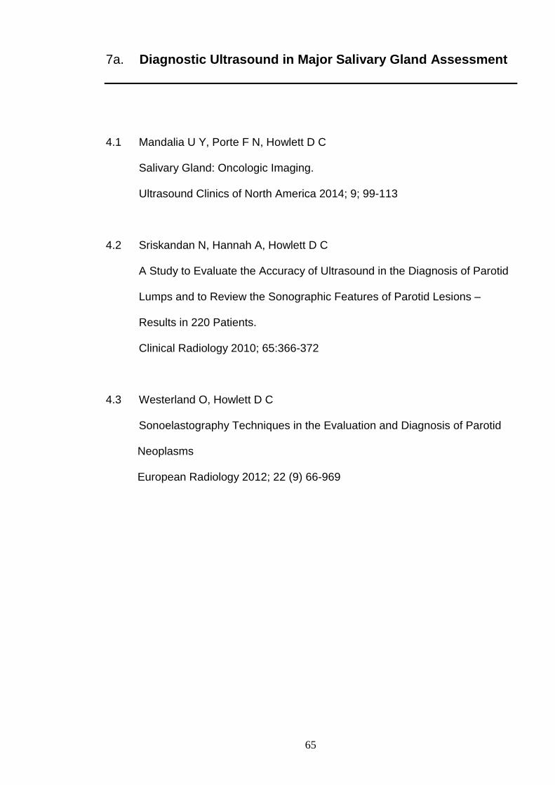
65
7a. Diagnostic Ultrasound in Major Salivary Gland Assessment
4.1 Mandalia U Y, Porte F N, Howlett D C
Salivary Gland: Oncologic Imaging.
Ultrasound Clinics of North America 2014; 9; 99-113
4.2 Sriskandan N, Hannah A, Howlett D C
A Study to Evaluate the Accuracy of Ultrasound in the Diagnosis of Parotid
Lumps and to Review the Sonographic Features of Parotid Lesions –
Results in 220 Patients.
Clinical Radiology 2010; 65:366-372
4.3 Westerland O, Howlett D C
Sonoelastography Techniques in the Evaluation and Diagnosis of Parotid
Neoplasms
European Radiology 2012; 22 (9) 66-969
66
7b. The Use of Ultrasound Guided Core Biopsy (USCB) in the
Histological Diagnosis of a Major Salivary Gland Lesion
4.4 Buckland J R, Manjaly G, Violaris N, Howlett D C
Ultrasound Guided Cutting Needle Biopsy of the Parotid Gland
Journal of Laryngology and Otology 1999; 113; 988-992
4.5 Keese KW, Manjaly G, Violaris N, Howlett D C
Ultrasound Guided Biopsy in the Evaluation of Focal Lesions and Diffuse
Swelling of the Parotid Gland.
British Journal of Oral and Maxillofacial Surgery 2002; 40; 364-368
4.6 Howlett D C
Diagnosing a Parotid Lump: Fine Needle Aspiration Cytology or Core
Biopsy?
British Journal of Radiology 2006; 79: 295-297
4.7 Howlett D C, Menezes LJ, Lewis K, Moody AB, Violaris N, Williams M D
Sonographically Guided Core Biopsy of a Parotid Mass
American Journal of Roentgenology 2007; 188-223-227
67
4.8 Breeze J, Andi A, Williams M D, Howlett D C
The Use of Fine Needle Core Biopsy Under Ultrasound Guidance in the
Diagnosis of a Parotid Mass
British Journal of Oral and Maxillofacial Surgery 2009; 47: 78-79
4.9 Olubaniyi B O, Chow V, Mandalia U, Haldar S, Gok G, Michl P,
Ramesar K, Sellon E,Williams M, Howlett D C
Evaluation of Biopsy Methods in the Diagnosis of Submandibular Space
Pathology
International Journal of Oral and Maxillofacial Surgery 2014; 43:281-285
4.10 Haldar S, Mandalia U, Skelton E, Chow V, Tighe D, Ramesar K,
Williams M D, Howlett D C
Diagnostic Investigation of Parotid Neoplasms – a 16 Year
Experience of Freehand Fine Needle Aspiration Cytology and Ultrasound
Guided Core Biopsy
International Journal of Oral and Maxillofacial Surgery 2015; 44(2): 151-157
4.11 Howlett D C, Skelton E, Moody AB
Establishing an Accurate Diagnosis of a Parotid Lump: An Evaluation of
the Current Biopsy Methods – Fine Needle Aspiration Cytology,
Ultrasound Guided Core Biopsy and Intra-operative Frozen Section.
British Journal of Oral and Maxillofacial Surgery – accepted for publication
August 2014
68
4.1 Mandalia U Y, Porte F N, Howlett D C
Salivary Gland: Oncologic Imaging. Ultrasound Clinics of North America
2014; 9:99-113
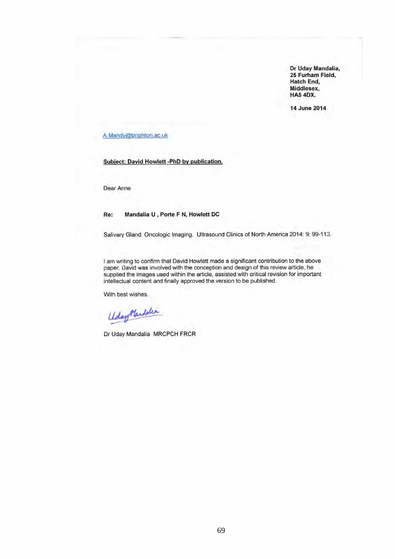
69
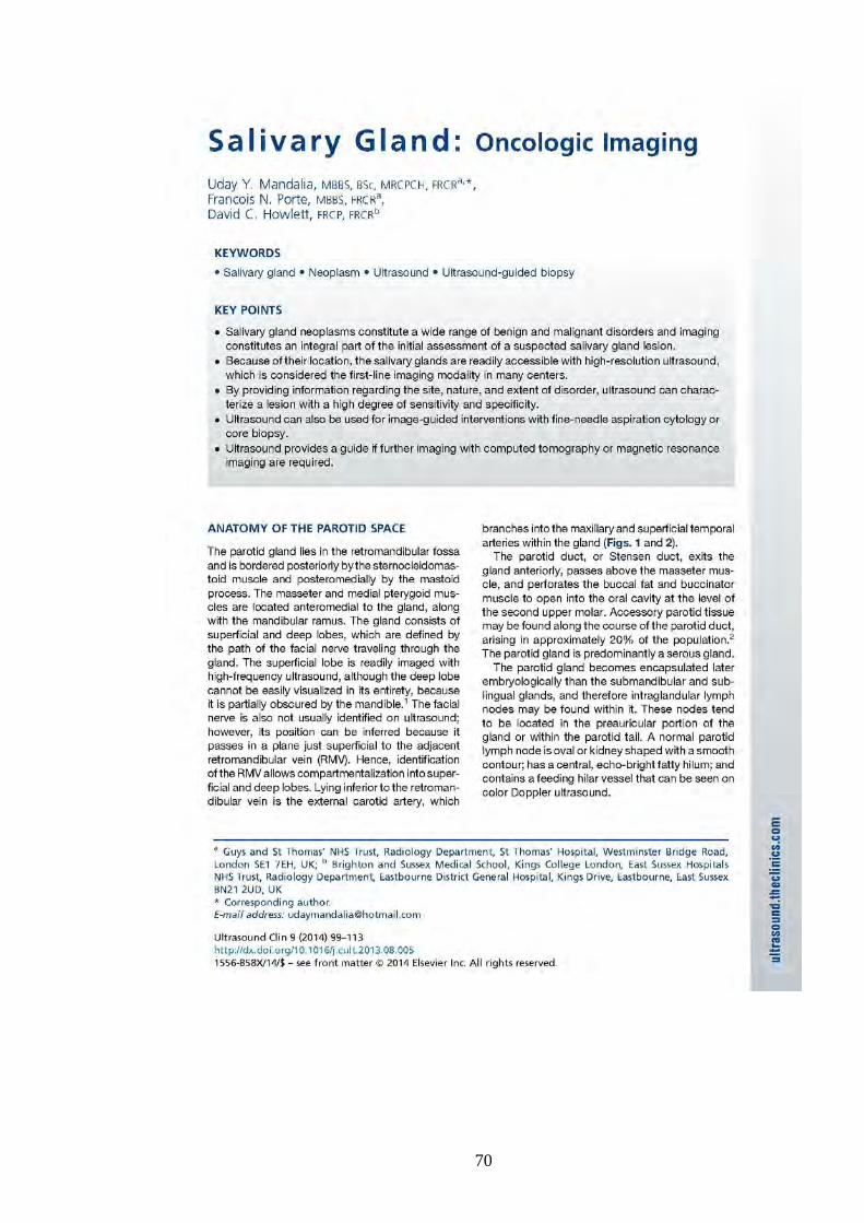
70
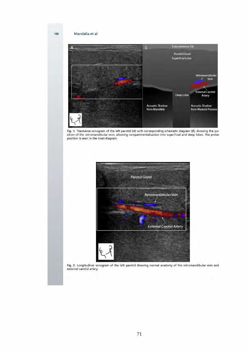
71
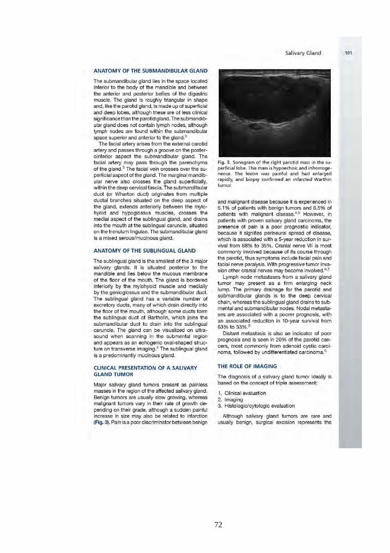
72
73
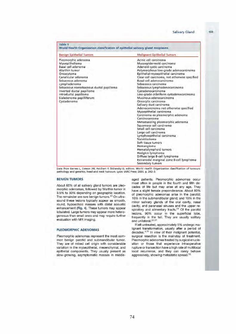
74
75
76
77
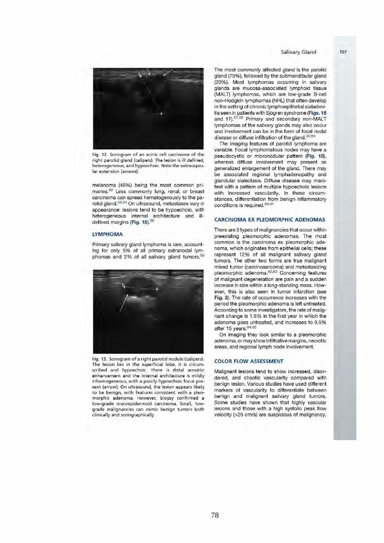
78
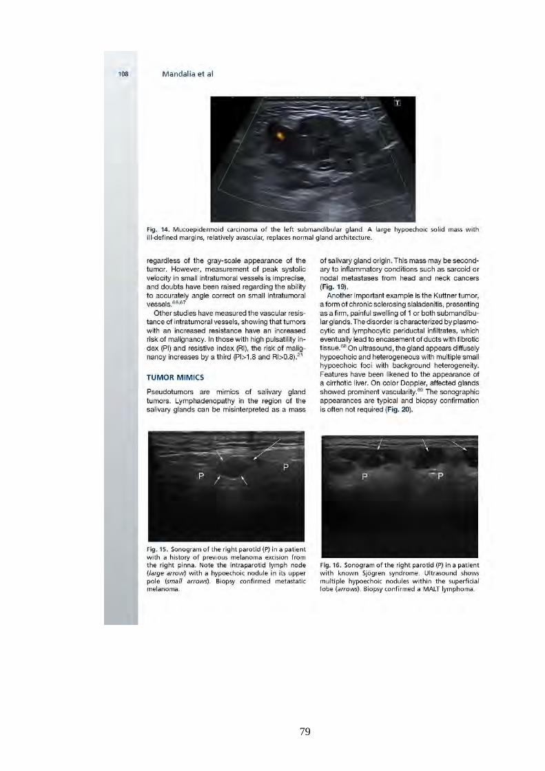
79
80
81
82
83
84
85
4.2 Sriskandan N, Hannah A, Howlett D C
A Study to Evaluate the Accuracy of Ultrasound in the Diagnosis of Parotid
Lumps and to Review the Sonographic Features of Parotid Lesions: Results
in 220 Patients.
Clinical Radiology 2010; 65:366-372
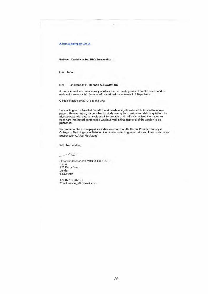
86
87
88
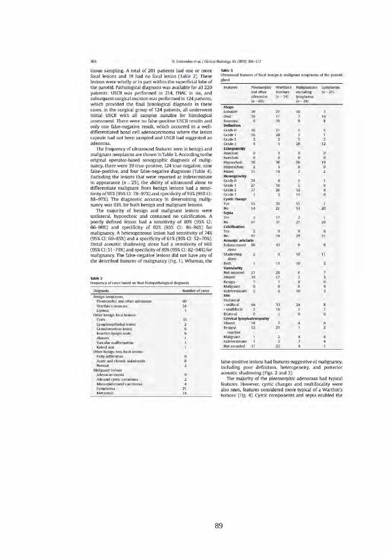
89
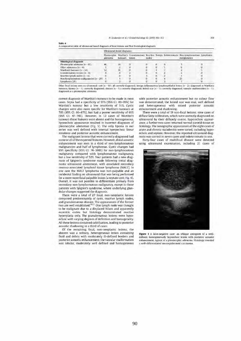
90
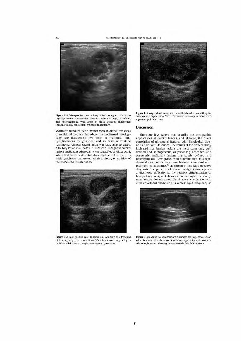
91
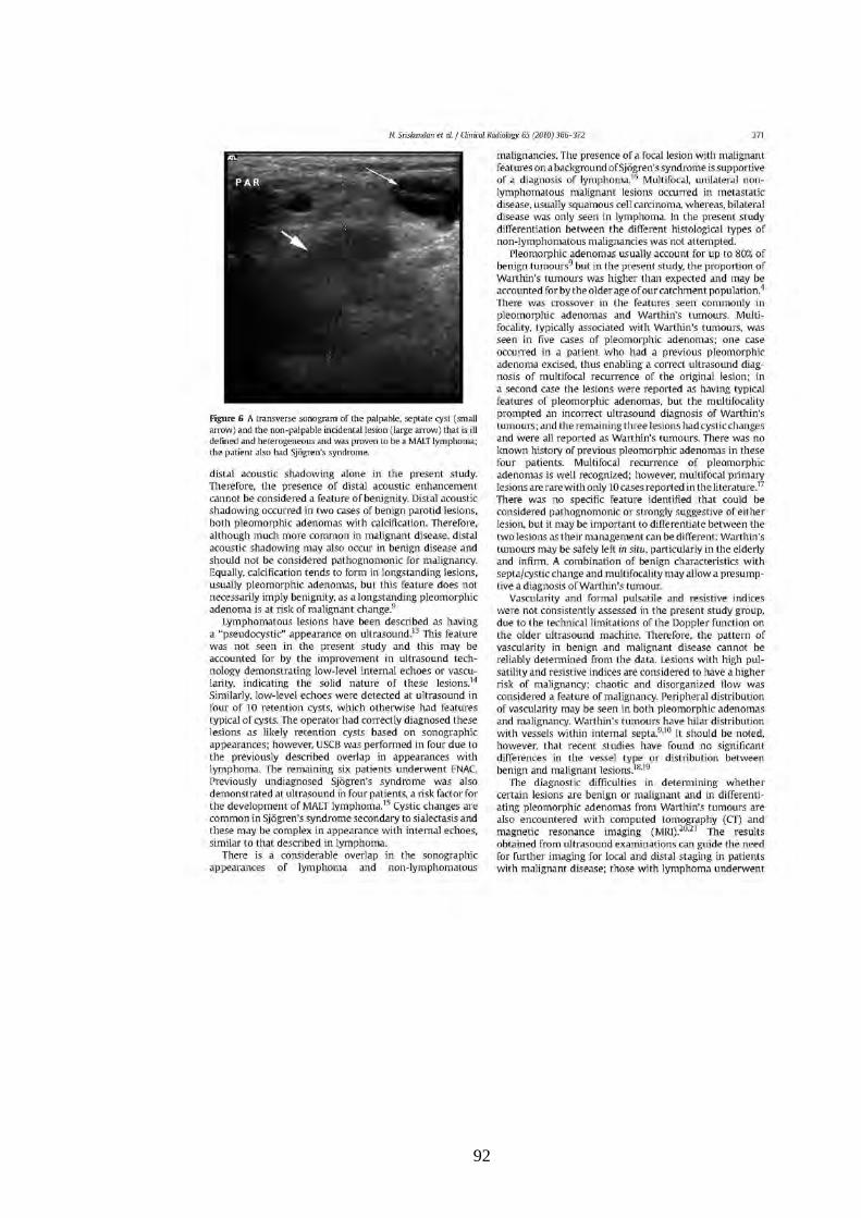
92
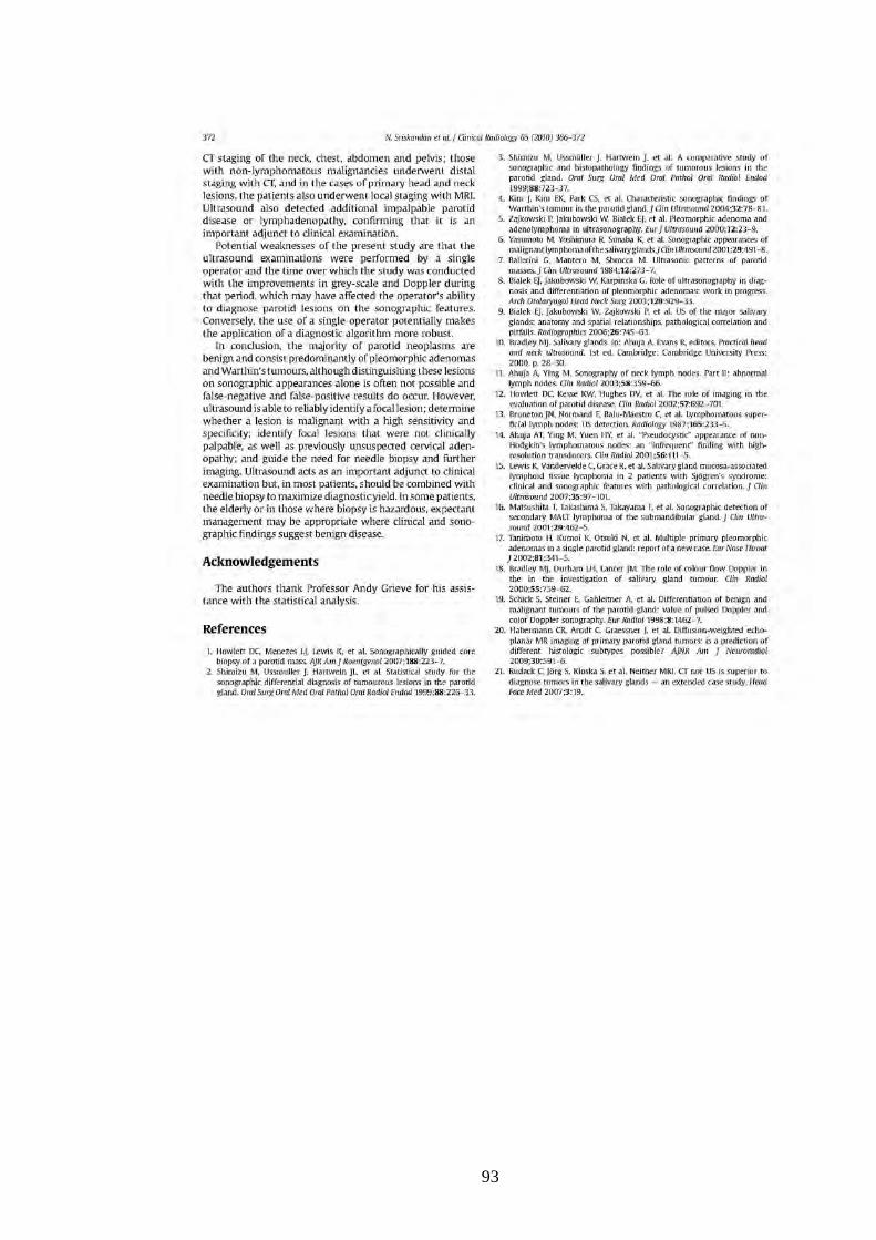
93
94
4.3 Westerland O, Howlett D C
Sonoelastography Techniques in the Evaluation and Diagnosis of Parotid
Neoplasms
European Radiology 2012; 22 (9) 66-969
95
96
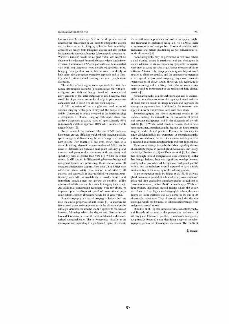
97
98
99
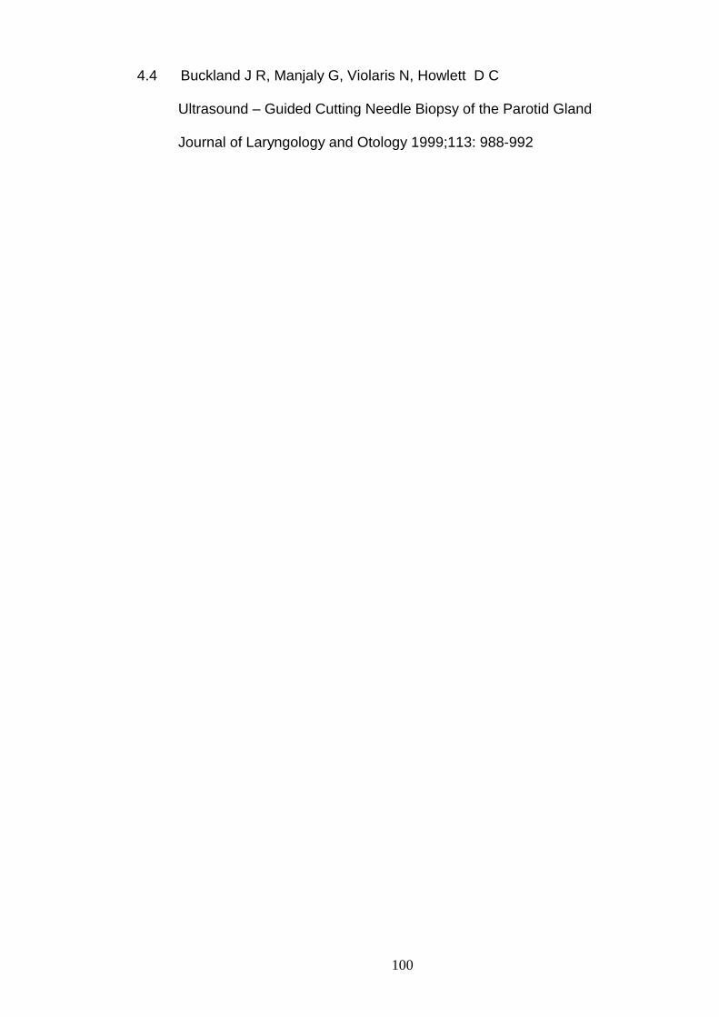
100
4.4 Buckland J R, Manjaly G, Violaris N, Howlett D C
Ultrasound – Guided Cutting Needle Biopsy of the Parotid Gland
Journal of Laryngology and Otology 1999;113: 988-992
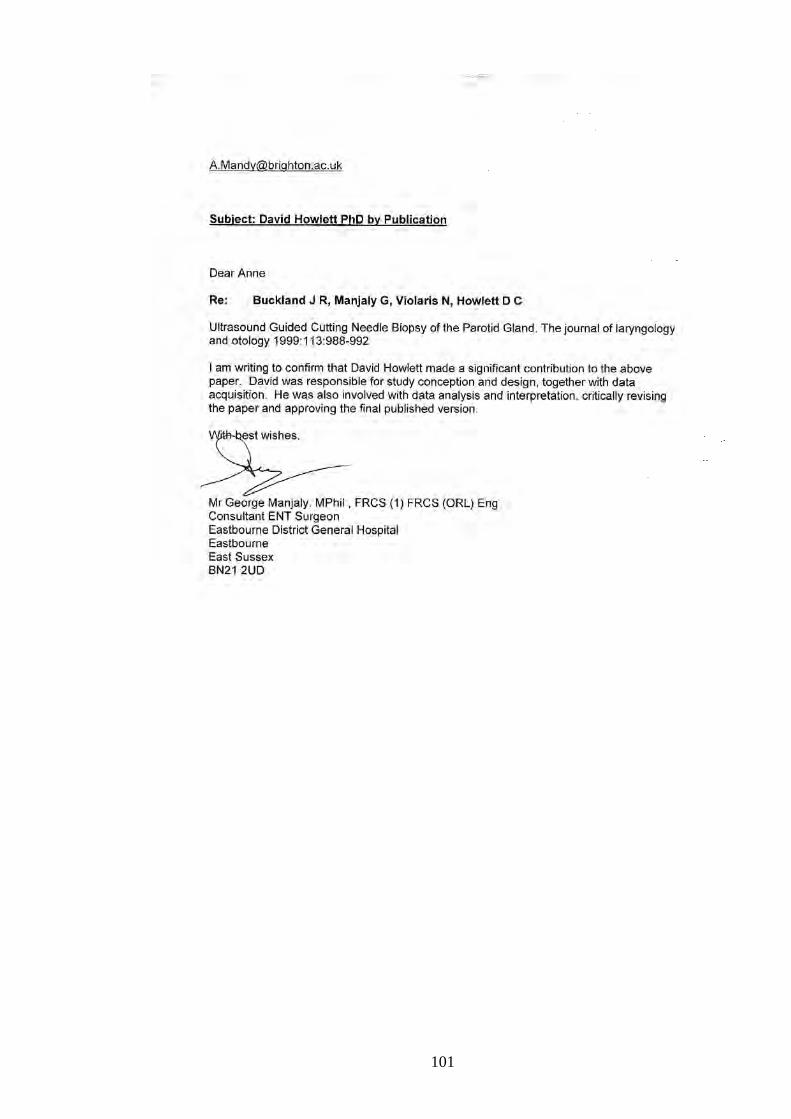
101
102

103
104
105

106
107
4.5 Kesse K W, Manjaly G, Violaris N, Howlett D C
Ultrasound Guided Biopsy in the Evaluation of Focal Lesions and Diffuse
Swelling of the Parotid Gland
British Journal of Oral and Maxillofacial Surgery 2002; 40: 364-368
108
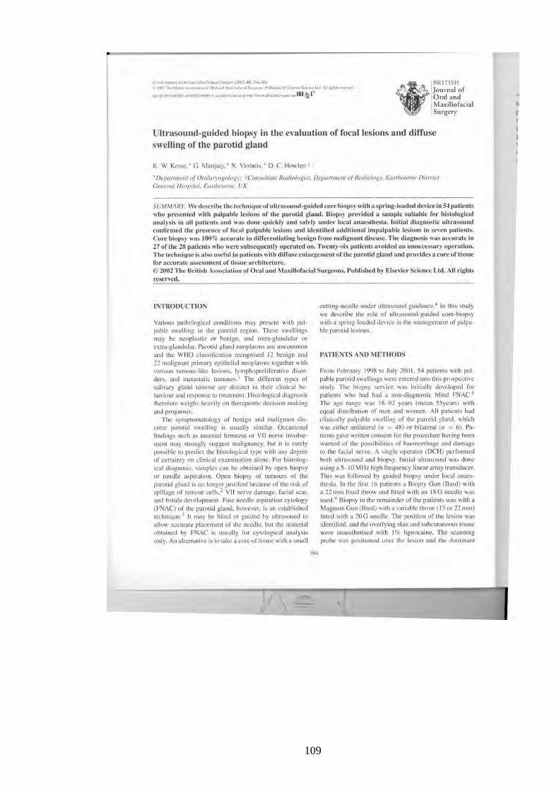
109
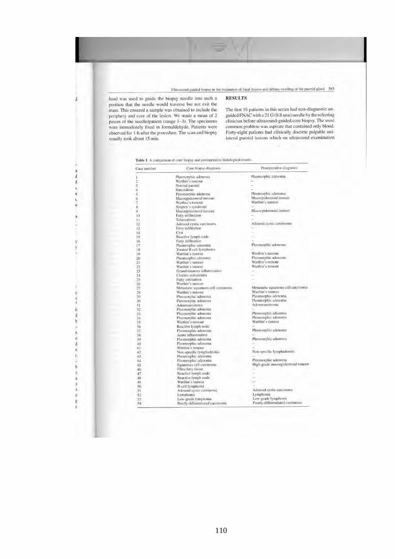
110
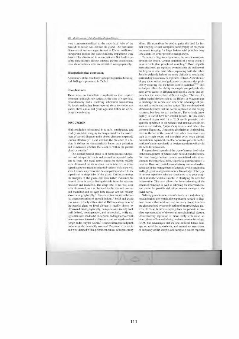
111
112
113
114
4.6 Howlett D C
Diagnosing a Parotid Lump: Fine Needle Aspiration Cytology or Core
Biopsy?
British Journal of Radiology 2006; 79: 295-297
115
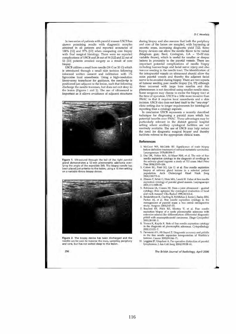
116
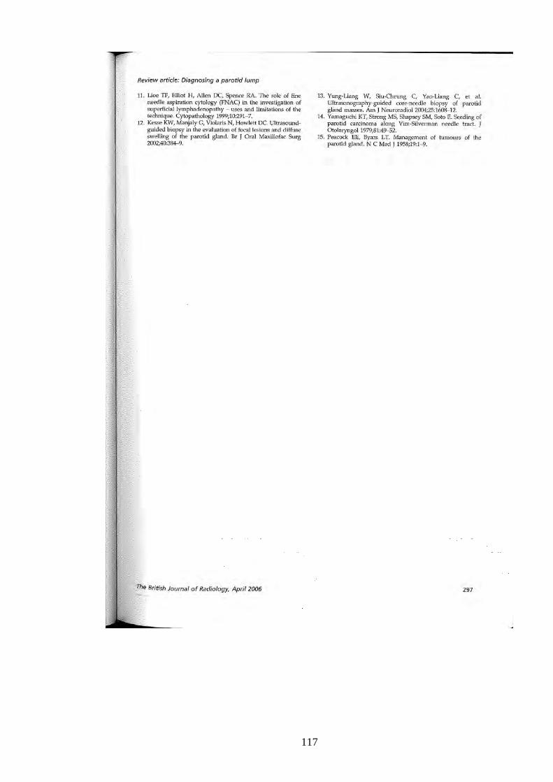
117
118
4.7 Howlett D C, Menezes L J, Lewis K, Moody A B, Violaris N, Williams M D
Sonographically Guided Core Biopsy of a Parotid Mass
American Journal of Roentgenology 2007; 168: 223-227
119
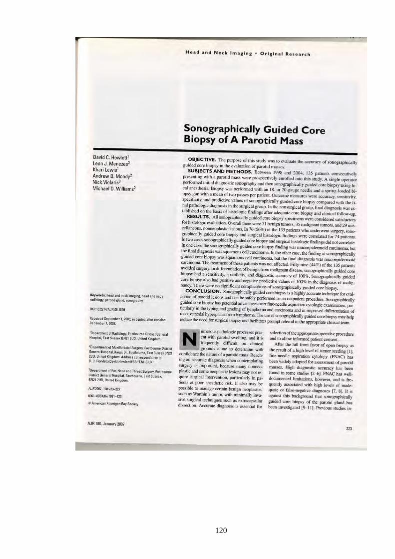
120
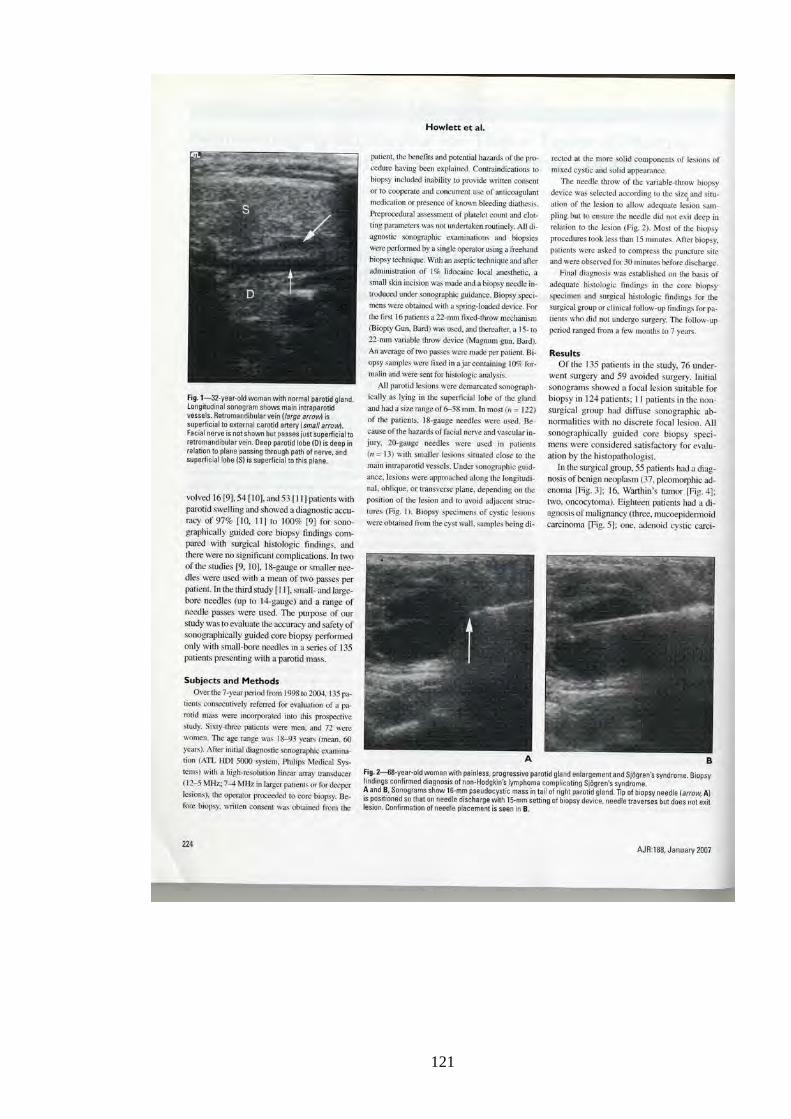
121
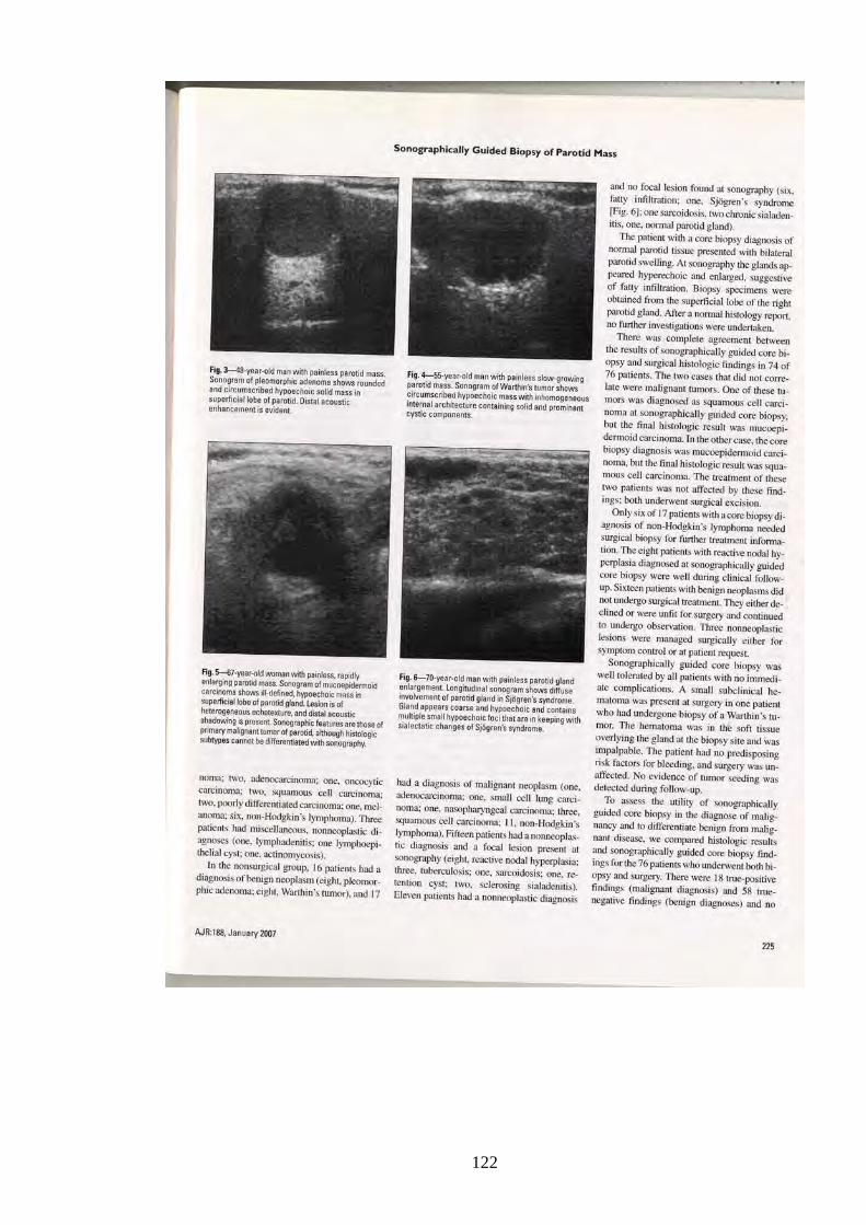
122
123
124
125
4.8 Breeze J, Andi A, Williams M D, Howlett D C
The Use of Fine Needle Core Biopsy Under Ultrasound Guidance in the
Diagnosis of a Parotid Mass
British Journal of Oral and Maxillofacial Surgery 2009; 47: 78-79
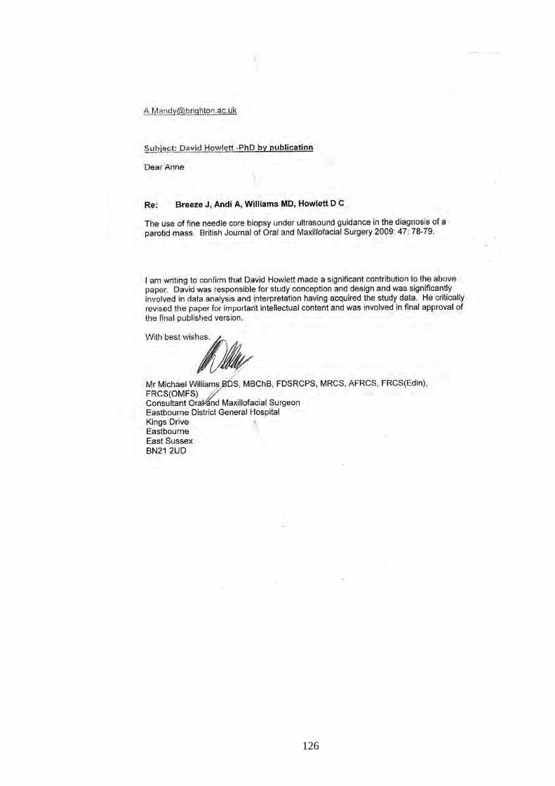
126
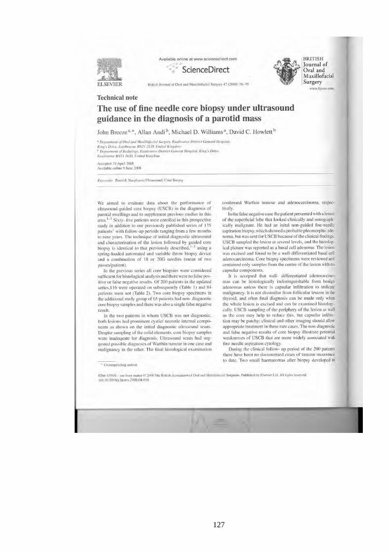
127
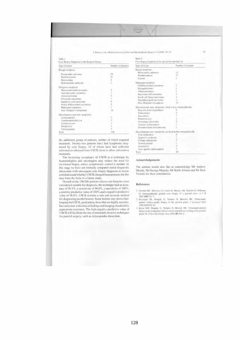
128
129
4.9 Olubaniyi B O, Chow V, Mandalia U, Haldar S, Gok G, Michl P, Ramesar K,
Sellon E, Williams M, Howlett D C
Evaluation of Biopsy Methods in the Diagnosis of Submandibular Space
Pathology
International Journal of Oral and Maxillofacial Surgery 2014; 43: 281-285
130
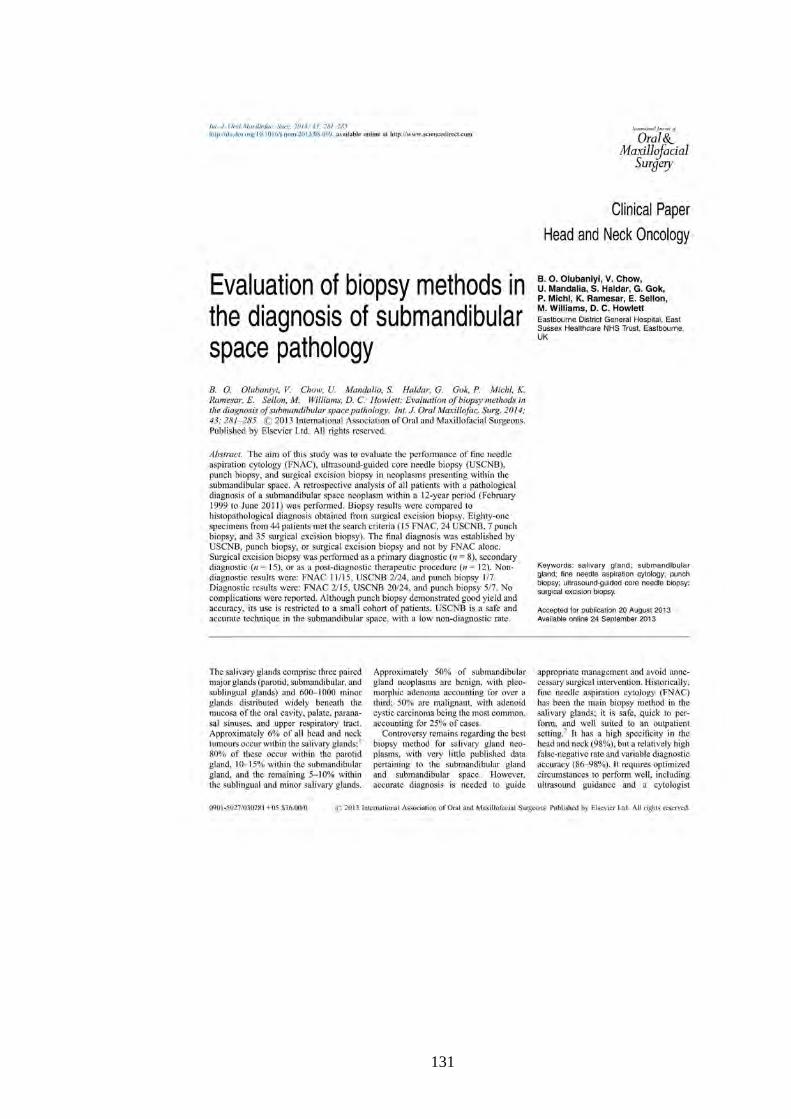
131
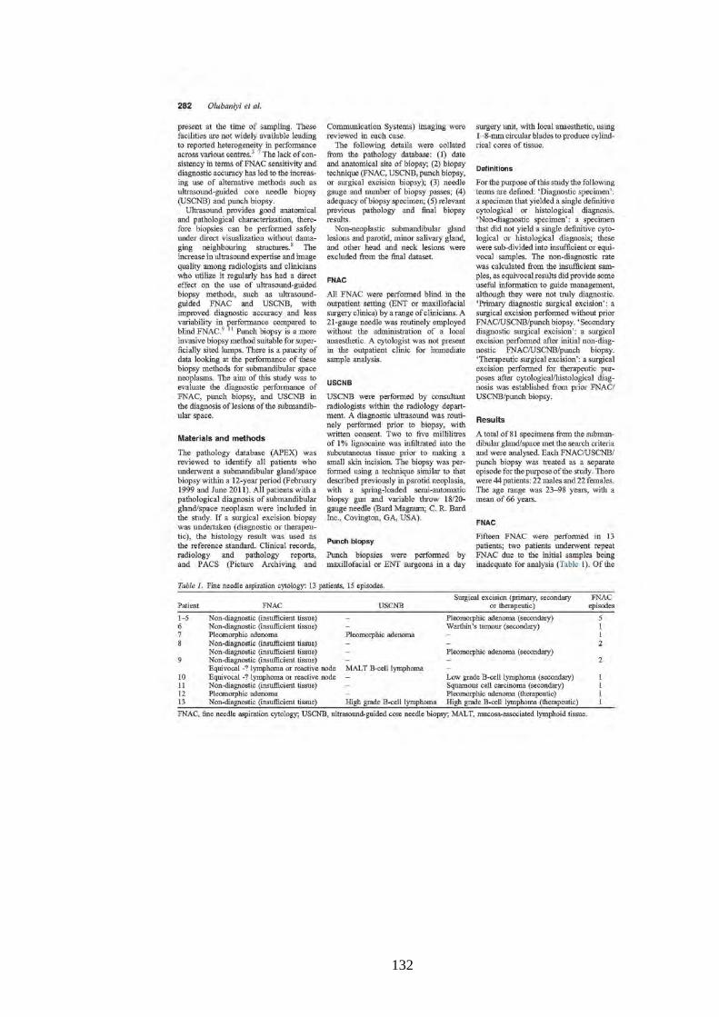
132
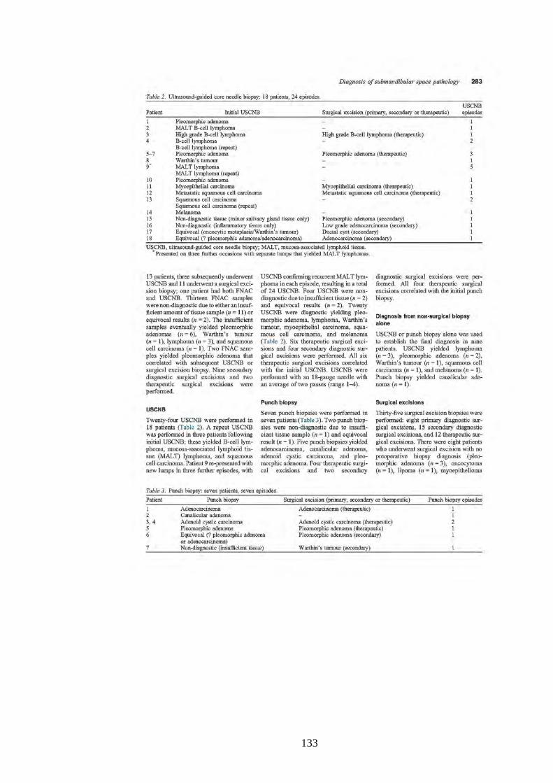
133
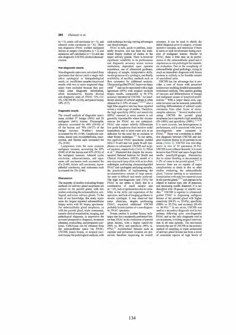
134
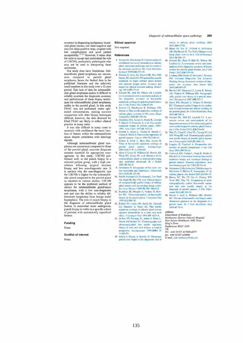
135
136
4.10 Haldar S, Mandalia U, Skelton E, Chow V, Tighe D, Ramesar K,
Williams M D, Howlett D C
Diagnostic Investigation of Parotid Neoplasms – a 16 Year Experience of
Freehand Fine Needle Aspiration Cytology and Ultrasound Guided Core
Needle Biopsy
International Journal of Oral and Maxillofacial Surgery 2015; 44(2): 151-157
137
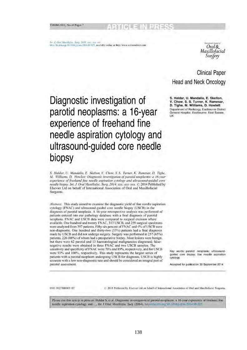
138
139
140
141
142
143
144
145
4.11 Howlett D C, Skelton E, Moody AB
Establishing an Accurate Diagnosis or a Parotid Lump: An Evaluation of the
Current Biopsy Methods – Fine Needle Aspiration Cytology, Ultrasound
Guided Core Biopsy and Intra-operative Frozen Section.
British Journal of Oral and Maxillofacial Surgery – accepted for publication
August 2014.
146
147
148
149
150


