� Ultrasound is a detailed anatomic assessment of the muscles and surrounding organs ofthe pelvic floor.
� Anatomic variability and pathology, such as prolapse, fecal incontinence, urinary inconti-nence, vaginal wall cysts, synthetic implanted material, and pelvic pain, can be easily as-sessed with endoluminal vaginal ultrasound.
� Knowledge of pelvic floor anatomy is essential for effective ultrasound imagingtechniques.
INTRODUCTION
The pelvic floor is a complex system, and adequate assessment of pelvic floor disor-ders is greatly supplemented by pelvic floor imaging. Rather than focusing on the clin-ical examination of pelvic floor surface structures, imaging modalities, such assonography, allow for immediate, real-time confirmation of anatomic findings. Pelvicfloor ultrasound offers a low cost, minimally invasive method of assessing pelvic flooranatomy and function. For example, clinical assessment of the anatomy of the levatorani by palpation requires significant skill and teaching.1–3 Clinical diagnosis by imaginghas been shown to be more reproducible than palpation and provides a more objec-tive method of teaching.3
PELVIC FLOOR ANATOMY
The female pelvic floor and the levator ani complex are composed of muscle fibers anda fascial network, which spans the area underneath the pelvis. An intact, well-innervated pelvic floor is necessary to maintain pelvic organ support, facilitate
Disclosures: royalties from UpToDate (L.H. Quiroz).Department of Obstetrics and Gynecology, University of Oklahoma Health Sciences Center, 920Stanton L. Young, WP2430, Oklahoma City, OK 73104, USA* Corresponding author.E-mail address: [email protected]
Obstet Gynecol Clin N Am 43 (2016) 141–153http://dx.doi.org/10.1016/j.ogc.2015.10.007 obgyn.theclinics.com0889-8545/16/$ – see front matter � 2016 Elsevier Inc. All rights reserved.
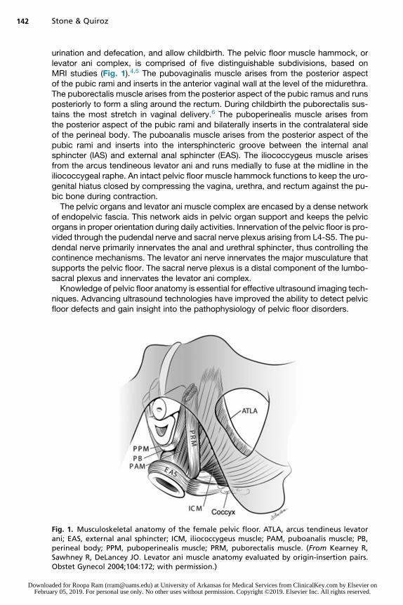
urination and defecation, and allow childbirth. The pelvic floor muscle hammock, orlevator ani complex, is comprised of five distinguishable subdivisions, based onMRI studies (Fig. 1).4,5 The pubovaginalis muscle arises from the posterior aspectof the pubic rami and inserts in the anterior vaginal wall at the level of the midurethra.The puborectalis muscle arises from the posterior aspect of the pubic ramus and runsposteriorly to form a sling around the rectum. During childbirth the puborectalis sus-tains the most stretch in vaginal delivery.6 The puboperinealis muscle arises fromthe posterior aspect of the pubic rami and bilaterally inserts in the contralateral sideof the perineal body. The puboanalis muscle arises from the posterior aspect of thepubic rami and inserts into the intersphincteric groove between the internal analsphincter (IAS) and external anal sphincter (EAS). The iliococcygeus muscle arisesfrom the arcus tendineous levator ani and runs medially to fuse at the midline in theiliococcygeal raphe. An intact pelvic floor muscle hammock functions to keep the uro-genital hiatus closed by compressing the vagina, urethra, and rectum against the pu-bic bone during contraction.The pelvic organs and levator ani muscle complex are encased by a dense network
of endopelvic fascia. This network aids in pelvic organ support and keeps the pelvicorgans in proper orientation during daily activities. Innervation of the pelvic floor is pro-vided through the pudendal nerve and sacral nerve plexus arising from L4-S5. The pu-dendal nerve primarily innervates the anal and urethral sphincter, thus controlling thecontinence mechanisms. The levator ani nerve innervates the major musculature thatsupports the pelvic floor. The sacral nerve plexus is a distal component of the lumbo-sacral plexus and innervates the levator ani complex.Knowledge of pelvic floor anatomy is essential for effective ultrasound imaging tech-
niques. Advancing ultrasound technologies have improved the ability to detect pelvicfloor defects and gain insight into the pathophysiology of pelvic floor disorders.
Fig. 1. Musculoskeletal anatomy of the female pelvic floor. ATLA, arcus tendineus levatorani; EAS, external anal sphincter; ICM, iliococcygeus muscle; PAM, puboanalis muscle; PB,perineal body; PPM, puboperinealis muscle; PRM, puborectalis muscle. (From Kearney R,Sawhney R, DeLancey JO. Levator ani muscle anatomy evaluated by origin-insertion pairs.Obstet Gynecol 2004;104:172; with permission.)
Requirements for two-dimensional (2D) perineal ultrasound include a B-mode-capable 2D ultrasound system, and a 3.5- to 6-MHz transducer.7 To perform the ex-amination, the patient is placed in dorsal lithotomy position after the patient voids. Theprobe may be covered with a nonpowdered glove or probe cover. Ultrasound gel isapplied to the probe and the probe is placed firmly on the perineum. Once a midsag-ittal view is obtained, the following structures should be identified from ventral to dor-sal: symphysis pubis, urethra and bladder neck, vaginal canal, uterus and cervix,anorectal canal, and the central portion of the puborectalis muscle. Parting the labiamay improve image quality. By rotating the probe 90�, one can obtain a coronalview and by placing a dorsal inclination on the probe, the anal canal and sphinctercomplex are seen and assessed. Once adequate images are obtained, the probe isremoved from the perineum and cleaned.2D images can be integrated into three-dimensional (3D) volume data either by a
free-hand acquisition of images or using a probe equipped with a motor to allow formotorized automatic acquisition of images. To perform the examination the patientis in the same position as described for 2D ultrasound. Ultrasound gel is applied tothe 3D-capable probe and placed firmly on the perineum in a midsagittal orientation.The probe is then held in place while the images are obtained. Postprocessing of theimages then occurs and is evaluated with the appropriate software.8 Four-dimensionalimaging refers to the real-time acquisition of data to produce and save cineloops of theimages obtained. To perform four-dimensional imaging, one must record images dur-ing a prompted maneuver, such as a squeeze or a Valsalva.
Endoluminal Ultrasound
Although a fair amount of information is obtained with an abdominal 2D probe whenplaced on the perineum (in the technique detailed previously), additional informa-tion is obtained by endoluminal ultrasound (endovaginal and endoanal ultrasound)with such equipment as the BK Medical Pro Focus Ultra View and Flex Focus (Pea-body, MA). These systems provide high performance with efficiency and speed,high resolution, and a sensitive color Doppler with great spatial resolution andsensitivity.Endovaginal ultrasound is performed with the patient in the dorsal lithotomy posi-
tion, with the patient having a comfortable amount of urine in the bladder. Multipletypes of probes are used including an electronic biplane 5- to 12-MHz probe, a highmultifrequency 9- to 16-MHz probe, a 360� rotational mechanical probe, or with aradial electronic probe,9,10 such as the one by BK Medical. To perform the examina-tion, the transducer is inserted into the vagina in a neutral position, avoiding excessivepressure on the surrounding structures. The biplane electronic probe provides 2D im-aging of the anterior and posterior compartments. Typically it is performed at rest, onValsalva, and during pelvic floor muscle contraction. 3D images are obtained by con-necting this transducer to an external 180� rotational mover. Other methods of obtain-ing 3D images are using a radial electronic probe or a rotational mechanical probe toobtain a 3D 360� view image of the pelvic floor. The 3D volume can be used on thescanner, but the better functionality is to use the free software, which can be installedin any personal computer. This allows for the volume to be exported to a CD, DVD,USB, or external drive and be viewed and analyzed at any time. The available functionsof the software allow for manipulation of the 3D cube in the x, y, or z planes, andobtaining linear, angle, area, and volume measurements.
Endoanal ultrasound is the gold standard for evaluating anal sphincter pathology.11 Itis performed with the patient either in dorsal lithotomy, left lateral, or prone position. Itcan be performed either with a high multifrequency 360� rotational probe or a radialelectronic probe. Irrespective of patient positioning, the transducer should be posi-tioned so that the anterior aspect of the anal canal is superior on the screen at the12-o’clock position. The distal end of the probe should be at the level of the puborec-talis muscle or 6 cm into the anal canal. The mechanical rotational probe, once acti-vated, automatically obtains 3D images.9 3D image acquisition with the radialelectronic probe involves manual withdrawal.12
CLINICAL UTILITY
This discussion is focused on the clinical use of ultrasound imaging of the pelvic floormostly to endoluminal ultrasound. Although pelvic floor imaging can be initiated with atransperineal ultrasound, having the advantage of dynamic imaging with minimal tis-sue distortion, the resulting images have a lower resolution.
Anterior Compartment
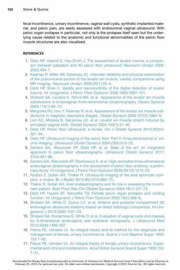
The anterior compartment of the pelvis is often assessed with transperineal and endo-vaginal ultrasound. Because there is an inherent displacement of tissue with endova-ginal ultrasound, transperineal ultrasound is preferred for visualization of bladder neckdescent, urethral hypermobility, cystocele, and cystourethrocele.13 3D endovaginal ul-trasound is used to provide a detailed anatomic depiction of anterior compartmentstructures including the trigone, compressor urethra, and the urogenital sphincter.14
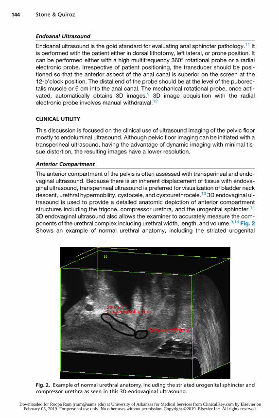
3D endovaginal ultrasound also allows the examiner to accurately measure the com-ponents of the urethral complex including urethral width, length, and volume.9,14 Fig. 2Shows an example of normal urethral anatomy, including the striated urogenital
Fig. 2. Example of normal urethral anatomy, including the striated urogenital sphincter andcompressor urethra as seen in this 3D endovaginal ultrasound.
sphincter and compressor urethra. It is an excellent tool for visualizing anterior vaginalcysts and masses including Skene gland cysts, urethral diverticula, and Gartner ductcysts.15
The female urethra is a complex organ that plays a central role in urinary inconti-nence. Normal anatomy, innervation, position, and proper relation to the surroundingpelvic floor structures ensure normal function of the urethra.16–18 The signs and symp-toms of pelvic floor dysfunction often overlap with signs and symptoms of urethraldiverticula, ectopic ureters, urethral tumors, and periurethral cystic lesions. Ectopicureters and ureteroceles are usually diagnosed in childhood and rarely present inadults. Nevertheless, these conditions should be considered in patients with urinarytract infections or urinary incontinence because surgery can correct these disorders.19
Endovaginal ultrasound is a useful tool in the diagnosis of ectopic and dystopic ure-ters.20 Urethral diverticula are also occasionally found in women complaining of uri-nary incontinence and/or pain and dyspareunia. Ultrasound can help differentiate adiverticulum from other periurethral cystic lesions, such as ectopic ureters, calcifica-tions, and injected material, and is useful in surgical planning because it can offerinvaluable information, such as shape, size, and location in relation to the urethraand bladder.21 Endovaginal ultrasound is very useful in the diagnosis and monitoringof urethral tumors.22 An example is a urethral leiomyoma, which is a rare benignsmooth muscle tumor that may grow in pregnancy and cause dysuria. On ultrasoundthese tumors appear well defined, homogenous, with increased vascularity.23 Urethralcarcinoma is a rare cancer that accounts for less than 0.02% of all malignancies inwomen and typically appears as an anterior urethral tumor.23 Endovaginal ultrasoundof the bladder can provide important information regarding the bladder neck, and thediagnosis of foreign bodies and bladder diverticula. Bladder wall thickness can also bemeasured by endovaginal ultrasound and has been found to positively correlate withdetrusor instability.24
Central Compartment
Assessment of the central compartment is usually performed with transperineal ultra-sound. Endovaginal techniques have a limited role in assessing the central compart-ment because the probe impedes descent of the uterus or vault. Additionally uterine orvaginal vault prolapse is typically diagnosed on clinical examination.
Posterior Compartment
For the posterior compartment, endovaginal ultrasound with a biplane transducer pro-vides important information including ensuring integrity of the rectovaginal septum,and measuring the anorectal angle. During Valsalva, several other important aspectsare appreciated including descent of an enterocele, rectocele, and movement of thepuborectalis and anorectal angle to evaluate for pelvic floor dyssynergy, and visuali-zation of intussusception.9 Transvaginal 3D ultrasound also is a useful tool in visual-izing rectovaginal fistulae.25 Assessment of the anal canal is typically performedwith endoanal ultrasound (discussed later).11,26
The anatomic causes associated with defecatory dysfunction are best visualized ondynamic transperineal or translabial and endovaginal scans.27 Many clinicians use theterm “rectocele” to refer to any prolapse of the posterior vaginal wall; a true rectoceleis defined as herniation of the anterior rectal wall into the vagina.28 Although examina-tion is usually adequate to diagnose prolapse of the posterior vaginal wall, ultrasoundis a useful tool in diagnosing a true rectocele. Typically this is done with a transperinealultrasound because the endoluminal ultrasound probe may prevent the prolapse fromoccurring.
An enterocele is a hernia of the most inferior point of the abdominal cavity into thevagina or pouch of Douglas. On ultrasound, it is visualized as the downward move-ment of abdominal contents into the vagina, ventral to the rectal ampulla and anal ca-nal. A sigmoidocele can usually be seen by differentiating hyperechoic stoolmovement from the surrounding tissue. Being able to differentiate a sigmoidocelefrom an enterocele is important in planning a surgical procedure. By definition, anintussusception occurs when the rectal wall telescopes into the rectal lumen andmay involve the rectal mucosa or full thickness of the rectal wall. It is defined as intra-rectal, intra-anal, or external if it forms a complete rectal prolapse. Pelvic floor dyssy-nergy is typically described as a lack of normal relaxation of the puborectalis muscleduring defecation. This is a difficult condition to verify through clinical examination.However, during Valsalva, it is documented by ultrasound because the anorectal anglebecomes narrower, the levator hiatus is shortened, and the puborectalis musclethickens.27
Lateral Compartments
Until recently, the concept of pelvic floor trauma focused mainly on perineal, vaginal,and anal sphincter injuries. But over the past several years, with advancements of 3Dultrasound technology, the concept of levator ani injury has become an importantcomponent of pelvic floor trauma. The pelvic floor is composed of symmetricallypaired levator ani muscles, which together form a sheet of muscles attached to the in-ternal surface of the true pelvis. The levator ani is divided into three muscle groups,named for their attachments: (1) pubovisceralis, (2) puborectalis, and (3) iliococcy-geus. The pubovisceralis is further divided into the puboperinealis, pubovaginalis,and puboanalis muscles.29 Together, these muscles support the urogenital organsand the anorectum.Studies have shown that levator ani injury is common. Levator avulsion is the
disconnection of the levator ani from its insertion on the pubic ramus or pelvic sidewall.Levator tears can occur at any part of the muscle between the two insertion points.Avulsion and tears are a common complication of overstretching of themuscles duringchildbirth and occur in 10% to 36% of women in their first vaginal delivery.28,30–32 Notonly can childbirth injure the muscles of the pelvic floor, but it can also disrupt theinnervation of the muscles.33 Electromyography abnormalities in the pelvic floor asso-ciated with defecation disorders, stress urinary incontinence, and prolapse are morefrequently seen in women who are multiparous, who have had prolonged secondstages of labor, forceps delivery, and high birth weight.34–37
Levator ani injury has been shown to be negatively correlated with pelvic floorstrength, and positively correlated with fecal incontinence and stage of pelvic organprolapse. Steensma and colleagues38 found that weak pelvic floor muscles occurredmore often in women who had levator ani avulsion, with injuries being present in53.8% of women with weak pelvic floor strength compared with being present in16.1% of women with normal strength. Women with levator ani injury have been foundto have a greater incidence of fecal incontinence.39 Particularly the puborectalis mus-cle plays an important role in maintaining continence, as shown in a case-controlstudy that found that fecal incontinence was more common in women with puborec-talis abnormalities compared with control subjects.40 It is well established that womenwith levator ani injury have a higher risk of developing pelvic organ prolapse.41 Levatoravulsion seems to play a significant role particularly in the central and anterior com-partments because the risk of having prolapse in these compartments doubles withlevator ani avulsion.42 The relationship with stress incontinence and levator ani injuryis unclear because some studies shown no correlation,43 whereas others have shown
a negative correlation.44 However, women with previous stress incontinence are twiceas likely to have a levator ani injury during childbirth.45
Levator avulsion can be accurately seen through 3D endovaginal ultrasound imag-ing, especially during a pelvic floor muscle contraction. Because levator damage fromchildbirth is not always an “all or none” phenomenon, a scoring systemwas developedto quantify levator ani injury with 3D endovaginal ultrasound. This scoring system hasbeen shown to have high interrater reliability.46,47
Implanted Vaginal Material
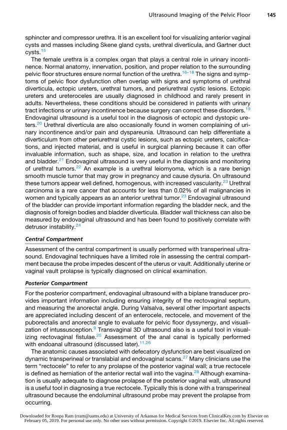
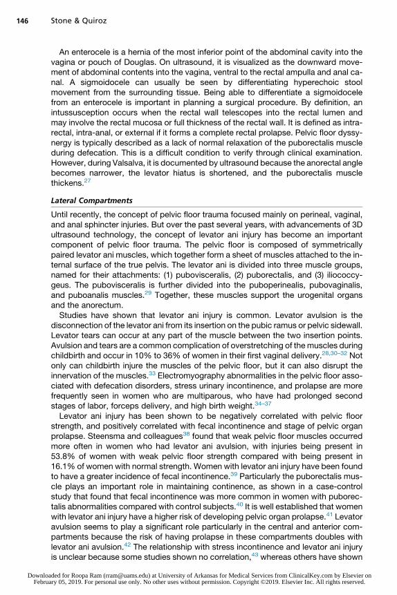
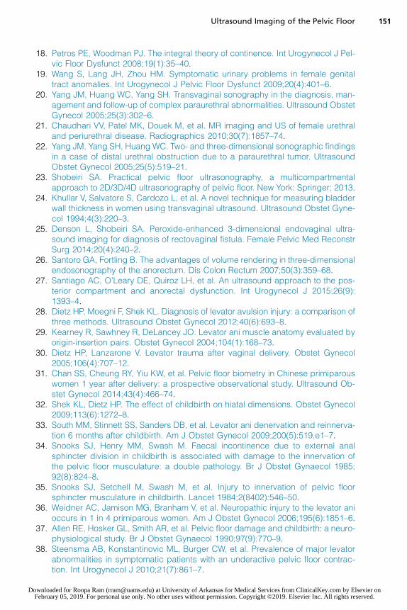
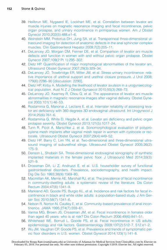
Over the last two decades, pelvic floor surgeries involving permanent vagina implantsused to aid in pelvic organ prolapse and stress incontinence have risen in popularity.Polypropylene mesh is one of the most commonly used meshes in vaginal surgery andis highly echogenic on endovaginal ultrasound. Imaging can determine the size,shape, position, distortion, and mobility of the implants. Position of the mesh is typi-cally either along the posterior vaginal wall sometimes extending to the perinealbody, along the anterior wall, or near the midurethra in the case of midurethral slings.Often the implanted mesh is found to be shrunken, contracted, or folded.48 Fig. 3shows an example of anterior and posterior vaginal wall mesh materials. In the caseof midurethral slings, ultrasound can help distinguish transobturator versus retropubicslings, and help locate and map the location of the synthetic material on axial imagingof the anterior compartment relative to the bladder neck.49,50 Fig. 4 shows an exampleof a transobturator sling visualized on ultrasound. This is helpful in planning a proce-dure to remove permanent synthetic materials in the absence of an operative report, oras adjunctive imaging in cases where the synthetic materials appear displaced. Peri-urethral bulking agents can also easily be seen on endovaginal ultrasound.50
Endoanal Imaging
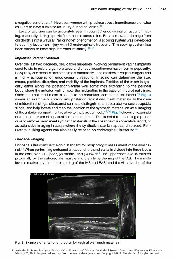
Endoanal ultrasound is the gold standard for morphologic assessment of the anal ca-nal.11 When performing endoanal ultrasound, the anal canal is divided into three levelsin the axial plan: (1) upper, (2) middle, and (3) lower.9 The uppermost level is markedproximally by the puborectalis muscle and distally by the ring of the IAS. The middlelevel is marked by the complete ring of the IAS and EAS, and the visualization of the
Fig. 3. Example of anterior and posterior vaginal wall mesh materials.
Fig. 4. Example of a transobturator sling visualized on ultrasound.
Stone & Quiroz148
Downloa Februa
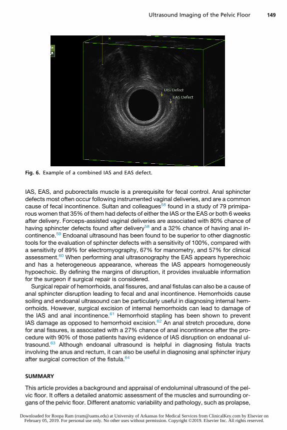
transverse perinei muscles. The lower level is marked by the subcutaneous part of theEAS. Fig. 5 shows an example of normal IAS and EAS anatomy.Endoanal ultrasound is often the diagnostic test of choice in patients presenting with
fecal incontinence and a history of a traumatic childbirth. Fig. 6 shows an example of acombined IAS and EAS defect. Fecal incontinence is defined as the involuntary loss offeces, whereas anal incontinence is the involuntary loss of flatus or feces. It is anembarrassing problem that has a devastating impact on a woman’s life. Because ofthe embarrassing nature of the problem, women often do not complain to their physi-cian, making the true incidence of anal incontinence likely underestimated. Reportedprevalence varies widely, ranging from 1% to 24% of the US adult population, and isprojected to increase 59% by 2050.51–57 An intact, innervated, and well-functioning
IAS, EAS, and puborectalis muscle is a prerequisite for fecal control. Anal sphincterdefects most often occur following instrumented vaginal deliveries, and are a commoncause of fecal incontinence. Sultan and colleagues58 found in a study of 79 primipa-rous women that 35% of them had defects of either the IAS or the EAS or both 6 weeksafter delivery. Forceps-assisted vaginal deliveries are associated with 80% chance ofhaving sphincter defects found after delivery58 and a 32% chance of having anal in-continence.59 Endoanal ultrasound has been found to be superior to other diagnostictools for the evaluation of sphincter defects with a sensitivity of 100%, compared witha sensitivity of 89% for electromyography, 67% for manometry, and 57% for clinicalassessment.60 When performing anal ultrasonography the EAS appears hyperechoicand has a heterogeneous appearance, whereas the IAS appears homogeneouslyhypoechoic. By defining the margins of disruption, it provides invaluable informationfor the surgeon if surgical repair is considered.Surgical repair of hemorrhoids, anal fissures, and anal fistulas can also be a cause of
anal sphincter disruption leading to fecal and anal incontinence. Hemorrhoids causesoiling and endoanal ultrasound can be particularly useful in diagnosing internal hem-orrhoids. However, surgical excision of internal hemorrhoids can lead to damage ofthe IAS and anal incontinence.61 Hemorrhoid stapling has been shown to preventIAS damage as opposed to hemorrhoid excision.62 An anal stretch procedure, donefor anal fissures, is associated with a 27% chance of anal incontinence after the pro-cedure with 90% of those patients having evidence of IAS disruption on endoanal ul-trasound.63 Although endoanal ultrasound is helpful in diagnosing fistula tractsinvolving the anus and rectum, it can also be useful in diagnosing anal sphincter injuryafter surgical correction of the fistula.64
SUMMARY
This article provides a background and appraisal of endoluminal ultrasound of the pel-vic floor. It offers a detailed anatomic assessment of the muscles and surrounding or-gans of the pelvic floor. Different anatomic variability and pathology, such as prolapse,
fecal incontinence, urinary incontinence, vaginal wall cysts, synthetic implanted mate-rial, and pelvic pain, are easily assessed with endoluminal vaginal ultrasound. Withpelvic organ prolapse in particular, not only is the prolapse itself seen but the under-lying cause related to the anatomic and functional abnormalities of the pelvic floormuscle structures are also visualized.
REFERENCES
1. Dietz HP, Hyland G, Hay-Smith J. The assessment of levator trauma: a compari-son between palpation and 4D pelvic floor ultrasound. Neurourol Urodyn 2006;25(5):424–7.
2. Kearney R, Miller JM, Delancey JO. Interrater reliability and physical examinationof the pubovisceral portion of the levator ani muscle, validity comparisons usingMR imaging. Neurourol Urodyn 2006;25(1):50–4.
3. Dietz HP, Shek C. Validity and reproducibility of the digital detection of levatortrauma. Int Urogynecol J Pelvic Floor Dysfunct 2008;19(8):1097–101.
4. Shobeiri SA, Leclaire E, Nihira MA, et al. Appearance of the levator ani musclesubdivisions in endovaginal three-dimensional ultrasonography. Obstet Gynecol2009;114(1):66–72.
5. Margulies RU, Hsu Y, Kearney R, et al. Appearance of the levator ani muscle sub-divisions in magnetic resonance images. Obstet Gynecol 2006;107(5):1064–9.
6. Lien KC, Mooney B, DeLancey JO, et al. Levator ani muscle stretch induced bysimulated vaginal birth. Obstet Gynecol 2004;103(1):31–40.
7. Dietz HP. Pelvic floor ultrasound: a review. Am J Obstet Gynecol 2010;202(4):321–34.
8. Dietz HP. Ultrasound imaging of the pelvic floor. Part II: three-dimensional or vol-ume imaging. Ultrasound Obstet Gynecol 2004;23(6):615–25.
9. Santoro GA, Wieczorek AP, Dietz HP, et al. State of the art: an integratedapproach to pelvic floor ultrasonography. Ultrasound Obstet Gynecol 2011;37(4):381–96.
10. Santoro GA, Wieczorek AP, Stankiewicz A, et al. High-resolution three-dimensionalendovaginal ultrasonography in the assessment of pelvic floor anatomy: a prelim-inary study. Int Urogynecol J Pelvic Floor Dysfunct 2009;20(10):1213–22.
11. Abdool Z, Sultan AH, Thakar R. Ultrasound imaging of the anal sphincter com-plex: a review. Br J Radiol 2012;85(1015):865–75.
12. Thakar R, Sultan AH. Anal endosonography and its role in assessing the inconti-nent patient. Best Pract Res Clin Obstet Gynaecol 2004;18(1):157–73.
13. Dietz HP, Haylen BT, Vancaillie TG. Female pelvic organ prolapse and voidingfunction. Int Urogynecol J Pelvic Floor Dysfunct 2002;13(5):284–8.
14. Shobeiri SA, White D, Quiroz LH, et al. Anterior and posterior compartment 3Dendovaginal ultrasound anatomy based on direct histologic comparison. Int Uro-gynecol J 2012;23(8):1047–53.
15. Shobeiri SA, Rostaminia G, White D, et al. Evaluation of vaginal cysts and massesby 3-dimensional endovaginal and endoanal sonography. J Ultrasound Med2013;32(8):1499–507.
16. Petros PE, Ulmsten UI. An integral theory and its method for the diagnosis andmanagement of female urinary incontinence. Scand J Urol Nephrol Suppl 1993;153:1–93.
17. Petros PE, Ulmsten UI. An integral theory of female urinary incontinence. Exper-imental and clinical considerations. Acta Obstet Gynecol Scand Suppl 1990;153:7–31.
18. Petros PE, Woodman PJ. The integral theory of continence. Int Urogynecol J Pel-vic Floor Dysfunct 2008;19(1):35–40.
19. Wang S, Lang JH, Zhou HM. Symptomatic urinary problems in female genitaltract anomalies. Int Urogynecol J Pelvic Floor Dysfunct 2009;20(4):401–6.
20. Yang JM, Huang WC, Yang SH. Transvaginal sonography in the diagnosis, man-agement and follow-up of complex paraurethral abnormalities. Ultrasound ObstetGynecol 2005;25(3):302–6.
21. Chaudhari VV, Patel MK, Douek M, et al. MR imaging and US of female urethraland periurethral disease. Radiographics 2010;30(7):1857–74.
22. Yang JM, Yang SH, Huang WC. Two- and three-dimensional sonographic findingsin a case of distal urethral obstruction due to a paraurethral tumor. UltrasoundObstet Gynecol 2005;25(5):519–21.
23. Shobeiri SA. Practical pelvic floor ultrasonography, a multicompartmentalapproach to 2D/3D/4D ultrasonography of pelvic floor. New York: Springer; 2013.
24. Khullar V, Salvatore S, Cardozo L, et al. A novel technique for measuring bladderwall thickness in women using transvaginal ultrasound. Ultrasound Obstet Gyne-col 1994;4(3):220–3.
25. Denson L, Shobeiri SA. Peroxide-enhanced 3-dimensional endovaginal ultra-sound imaging for diagnosis of rectovaginal fistula. Female Pelvic Med ReconstrSurg 2014;20(4):240–2.
26. Santoro GA, Fortling B. The advantages of volume rendering in three-dimensionalendosonography of the anorectum. Dis Colon Rectum 2007;50(3):359–68.
27. Santiago AC, O’Leary DE, Quiroz LH, et al. An ultrasound approach to the pos-terior compartment and anorectal dysfunction. Int Urogynecol J 2015;26(9):1393–4.
28. Dietz HP, Moegni F, Shek KL. Diagnosis of levator avulsion injury: a comparison ofthree methods. Ultrasound Obstet Gynecol 2012;40(6):693–8.
30. Dietz HP, Lanzarone V. Levator trauma after vaginal delivery. Obstet Gynecol2005;106(4):707–12.
31. Chan SS, Cheung RY, Yiu KW, et al. Pelvic floor biometry in Chinese primiparouswomen 1 year after delivery: a prospective observational study. Ultrasound Ob-stet Gynecol 2014;43(4):466–74.
32. Shek KL, Dietz HP. The effect of childbirth on hiatal dimensions. Obstet Gynecol2009;113(6):1272–8.
33. South MM, Stinnett SS, Sanders DB, et al. Levator ani denervation and reinnerva-tion 6 months after childbirth. Am J Obstet Gynecol 2009;200(5):519.e1–7.
34. Snooks SJ, Henry MM, Swash M. Faecal incontinence due to external analsphincter division in childbirth is associated with damage to the innervation ofthe pelvic floor musculature: a double pathology. Br J Obstet Gynaecol 1985;92(8):824–8.
35. Snooks SJ, Setchell M, Swash M, et al. Injury to innervation of pelvic floorsphincter musculature in childbirth. Lancet 1984;2(8402):546–50.
36. Weidner AC, Jamison MG, Branham V, et al. Neuropathic injury to the levator anioccurs in 1 in 4 primiparous women. Am J Obstet Gynecol 2006;195(6):1851–6.
37. Allen RE, Hosker GL, Smith AR, et al. Pelvic floor damage and childbirth: a neuro-physiological study. Br J Obstet Gynaecol 1990;97(9):770–9.
38. Steensma AB, Konstantinovic ML, Burger CW, et al. Prevalence of major levatorabnormalities in symptomatic patients with an underactive pelvic floor contrac-tion. Int Urogynecol J 2010;21(7):861–7.
39. Heilbrun ME, Nygaard IE, Lockhart ME, et al. Correlation between levator animuscle injuries on magnetic resonance imaging and fecal incontinence, pelvicorgan prolapse, and urinary incontinence in primiparous women. Am J ObstetGynecol 2010;202(5):488.e1–6.
40. Weinstein MM, Pretorius DH, Jung SA, et al. Transperineal three-dimensional ul-trasound imaging for detection of anatomic defects in the anal sphincter complexmuscles. Clin Gastroenterol Hepatol 2009;7(2):205–11.
41. DeLancey JO, Morgan DM, Fenner DE, et al. Comparison of levator ani muscledefects and function in women with and without pelvic organ prolapse. ObstetGynecol 2007;109(2 Pt 1):295–302.
42. Dietz HP. Quantification of major morphological abnormalities of the levator ani.Ultrasound Obstet Gynecol 2007;29(3):329–34.
43. DeLancey JO, Trowbridge ER, Miller JM, et al. Stress urinary incontinence: rela-tive importance of urethral support and urethral closure pressure. J Urol 2008;179(6):2286–90 [discussion: 2290].
44. Dietz HP, Kirby A. Modelling the likelihood of levator avulsion in a urogynaecolog-ical population. Aust N Z J Obstet Gynaecol 2010;50(3):268–72.
45. DeLancey JO, Kearney R, Chou Q, et al. The appearance of levator ani muscleabnormalities in magnetic resonance images after vaginal delivery. Obstet Gyne-col 2003;101(1):46–53.
46. Rostaminia G, Manonai J, Leclaire E, et al. Interrater reliability of assessing leva-tor ani deficiency with 360 degrees 3D endovaginal ultrasound. Int Urogynecol J2014;25(6):761–6.
47. Rostaminia G, White D, Hegde A, et al. Levator ani deficiency and pelvic organprolapse severity. Obstet Gynecol 2013;121(5):1017–24.
48. Tunn R, Picot A, Marschke J, et al. Sonomorphological evaluation of polypro-pylene mesh implants after vaginal mesh repair in women with cystocele or rec-tocele. Ultrasound Obstet Gynecol 2007;29(4):449–52.
49. Dietz HP, Barry C, Lim YN, et al. Two-dimensional and three-dimensional ultra-sound imaging of suburethral slings. Ultrasound Obstet Gynecol 2005;26(2):175–9.
50. Denson L, Shobeiri SA. Three-dimensional endovaginal sonography of syntheticimplanted materials in the female pelvic floor. J Ultrasound Med 2014;33(3):521–9.
51. Drossman DA, Li Z, Andruzzi E, et al. U.S. householder survey of functionalgastrointestinal disorders. Prevalence, sociodemography, and health impact.Dig Dis Sci 1993;38(9):1569–80.
52. Macmillan AK, Merrie AE, Marshall RJ, et al. The prevalence of fecal incontinencein community-dwelling adults: a systematic review of the literature. Dis ColonRectum 2004;47(8):1341–9.
53. Markland AD, Goode PS, Burgio KL, et al. Incidence and risk factors for fecal in-continence in black and white older adults: a population-based study. J Am Ger-iatr Soc 2010;58(7):1341–6.
54. Nelson R, Norton N, Cautley E, et al. Community-based prevalence of anal incon-tinence. JAMA 1995;274(7):559–61.
55. Varma MG, Brown JS, Creasman JM, et al. Fecal incontinence in females olderthan aged 40 years: who is at risk? Dis Colon Rectum 2006;49(6):841–51.
56. Whitehead WE, Borrud L, Goode PS, et al. Fecal incontinence in US adults:epidemiology and risk factors. Gastroenterology 2009;137(2):512–7, 512.e1–2.
57. Wu JM, Vaughan CP, Goode PS, et al. Prevalence and trends of symptomatic pel-vic floor disorders in U.S. women. Obstet Gynecol 2014;123(1):141–8.
58. Sultan AH, Kamm MA, Hudson CN, et al. Anal-sphincter disruption during vaginaldelivery. N Engl J Med 1993;329(26):1905–11.
59. Sultan AH, Johanson RB, Carter JE. Occult anal sphincter trauma following ran-domized forceps and vacuum delivery. Int J Gynaecol Obstet 1998;61(2):113–9.
60. Sultan AH, Nicholls RJ, Kamm MA, et al. Anal endosonography and correlationwith in vitro and in vivo anatomy. Br J Surg 1993;80(4):508–11.
61. Speakman CT, Burnett SJ, Kamm MA, et al. Sphincter injury after anal dilatationdemonstrated by anal endosonography. Br J Surg 1991;78(12):1429–30.
62. Altomare DF, Rinaldi M, Sallustio PL, et al. Long-term effects of stapled haemor-rhoidectomy on internal anal function and sensitivity. Br J Surg 2001;88(11):1487–91.
63. Khubchandani IT, Reed JF. Sequelae of internal sphincterotomy for chronicfissure in ano. Br J Surg 1989;76(5):431–4.
64. Kennedy HL, Zegarra JP. Fistulotomy without external sphincter division for highanal fistulae. Br J Surg 1990;77(8):898–901.