rspb.royalsocietypublishing.org Research Cite this article: Dheilly NM et al. 2015 Who is the puppet master? Replication of a parasitic wasp-associated virus correlates with host behaviour manipulation. Proc. R. Soc. B 282: 20142773. http://dx.doi.org/10.1098/rspb.2014.2773 Received: 12 November 2014 Accepted: 12 January 2015 Subject Areas: behaviour, ecology, microbiology Keywords: parasitoid wasp, virus, holobiont, symbiont, behavioural manipulation, host– parasite interaction Author for correspondence: Nolwenn M. Dheilly e-mail: [email protected]† Present address: School of Marine and Atmospheric Sciences, Stony Brook University, Stony Brook, NY 11794-5000, USA. ‡ These authors contributed equally to this work. Electronic supplementary material is available at http://dx.doi.org/10.1098/rspb.2014.2773 or via http://rspb.royalsocietypublishing.org. Who is the puppet master? Replication of a parasitic wasp-associated virus correlates with host behaviour manipulation Nolwenn M. Dheilly 1,2,† , Fanny Maure 2,3 , Marc Ravallec 4 , Richard Galinier 1 , Jose ´e Doyon 3 , David Duval 1 , Lucas Leger 2 , Anne-Nathalie Volkoff 4 , Dorothe ´e Misse ´ 2 , Sabine Nidelet 5 , Vincent Demolombe 5 , Jacques Brodeur 3,‡ , Benjamin Gourbal 1,‡ , Fre ´de ´ric Thomas 2,‡ and Guillaume Mitta 1,‡ 1 UMR 5244, Ecologie et Evolution des Interactions (2EI), CNRS, Universite ´ de Perpignan, Perpignan 66860, France 2 MIVEGEC (UMR CNRS/IRD/UM1/UM2 5290), 911 Avenue Agropolis, BP 64501, Montpellier Cedex 5 34394, France 3 De ´partement de Sciences Biologiques, Institut de Recherche en Biologie Ve ´ge ´tale, Universite ´ de Montre ´al, 4101 rue Sherbrooke est, Montre ´al, Que ´bec, Canada H1X 2B2 4 INRA (UMR 1333), ‘Insect-Microorganisms Diversity, Genomes and Interactions’, Universite ´ de Montpellier 2, Place Euge `ne Bataillon, CC101, Montpellier Cedex 34095, France 5 Montpellier Genomics and Bioinformatics Facility, MGX-Montpellier GenomiX, Montpellier 34396, France Many parasites modify their host behaviour to improve their own transmission and survival, but the proximate mechanisms remain poorly understood. An original model consists of the parasitoid Dinocampus coccinellae and its coccinel- lid host, Coleomegilla maculata; during the behaviour manipulation, the parasitoid is not in contact with its host anymore. We report herein the discov- ery and characterization of a new RNA virus of the parasitoid (D. coccinellae paralysis virus, DcPV). Using a combination of RT-qPCR and transmission electron microscopy, we demonstrate that DcPV is stored in the oviduct of parasitoid females, replicates in parasitoid larvae and is transmitted to the host during larval development. Next, DcPV replication in the host’s nervous tissue induces a severe neuropathy and antiviral immune response that corre- late with the paralytic symptoms characterizing the behaviour manipulation. Remarkably, virus clearance correlates with recovery of normal coccinellid behaviour. These results provide evidence that changes in ladybeetle behaviour most likely result from DcPV replication in the cerebral ganglia rather than by manipulation by the parasitoid. This offers stimulating prospects for research on parasitic manipulation by suggesting for the first time that behaviour manipulation could be symbiont-mediated. 1. Introduction Parasites have the capacity to alter the biology of their hosts in many ways to improve their own fitness [1,2], host behaviour manipulation being one of the most striking outcomes. Behaviour manipulations may favour completion of the parasite’s life cycle by (i) rendering intermediate hosts more susceptible to predation by definitive hosts, (ii) inducing the parasitized host to move to habitats suitable for the parasite and/or its progeny, (iii) increasing the appetite of vectors in cases of vector-borne transmission, and (iv) providing protection to the developing parasite against biotic or abiotic factors, a condition called bodyguard manipulation [1,3]. Understanding how manipulation of host behaviour works remains a chal- lenge. Until recently, the study of proximate mechanisms has mostly focused on neuromodulatory systems [4] and experimental evidence of parasite genes indu- cing a direct change in host behaviour is very limited [5,6]. Bodyguards have & 2015 The Author(s) Published by the Royal Society. All rights reserved. on May 17, 2018 http://rspb.royalsocietypublishing.org/ Downloaded from
Transcript
on May 17, 2018http://rspb.royalsocietypublishing.org/Downloaded from
rspb.royalsocietypublishing.org
ResearchCite this article: Dheilly NM et al. 2015 Who
Stony Brook, NY 11794-5000, USA.‡These authors contributed equally to this
work.
Electronic supplementary material is available
at http://dx.doi.org/10.1098/rspb.2014.2773 or
via http://rspb.royalsocietypublishing.org.
& 2015 The Author(s) Published by the Royal Society. All rights reserved.
Who is the puppet master? Replication ofa parasitic wasp-associated viruscorrelates with host behaviourmanipulation
Nolwenn M. Dheilly1,2,†, Fanny Maure2,3, Marc Ravallec4, Richard Galinier1,Josee Doyon3, David Duval1, Lucas Leger2, Anne-Nathalie Volkoff4,Dorothee Misse2, Sabine Nidelet5, Vincent Demolombe5, Jacques Brodeur3,‡,Benjamin Gourbal1,‡, Frederic Thomas2,‡ and Guillaume Mitta1,‡
1UMR 5244, Ecologie et Evolution des Interactions (2EI), CNRS, Universite de Perpignan, Perpignan 66860,France2MIVEGEC (UMR CNRS/IRD/UM1/UM2 5290), 911 Avenue Agropolis, BP 64501, Montpellier Cedex 5 34394,France3Departement de Sciences Biologiques, Institut de Recherche en Biologie Vegetale, Universite de Montreal,4101 rue Sherbrooke est, Montreal, Quebec, Canada H1X 2B24INRA (UMR 1333), ‘Insect-Microorganisms Diversity, Genomes and Interactions’, Universite de Montpellier 2,Place Eugene Bataillon, CC101, Montpellier Cedex 34095, France5Montpellier Genomics and Bioinformatics Facility, MGX-Montpellier GenomiX, Montpellier 34396, France
Many parasites modify their host behaviour to improve their own transmission
and survival, but the proximate mechanisms remain poorly understood. An
original model consists of the parasitoid Dinocampus coccinellae and its coccinel-
lid host, Coleomegilla maculata; during the behaviour manipulation, the
parasitoid is not in contact with its host anymore. We report herein the discov-
ery and characterization of a new RNA virus of the parasitoid (D. coccinellaeparalysis virus, DcPV). Using a combination of RT-qPCR and transmission
electron microscopy, we demonstrate that DcPV is stored in the oviduct of
parasitoid females, replicates in parasitoid larvae and is transmitted to the
host during larval development. Next, DcPV replication in the host’s nervous
tissue induces a severe neuropathy and antiviral immune response that corre-
late with the paralytic symptoms characterizing the behaviour manipulation.
Remarkably, virus clearance correlates with recovery of normal coccinellid
behaviour. These results provide evidence that changes in ladybeetle behaviour
most likely result from DcPV replication in the cerebral ganglia rather than by
manipulation by the parasitoid. This offers stimulating prospects for research
on parasitic manipulation by suggesting for the first time that behaviour
manipulation could be symbiont-mediated.
1. IntroductionParasites have the capacity to alter the biology of their hosts in many ways to
improve their own fitness [1,2], host behaviour manipulation being one of the
most striking outcomes. Behaviour manipulations may favour completion of
the parasite’s life cycle by (i) rendering intermediate hosts more susceptible
to predation by definitive hosts, (ii) inducing the parasitized host to move to
habitats suitable for the parasite and/or its progeny, (iii) increasing the appetite
of vectors in cases of vector-borne transmission, and (iv) providing protection
to the developing parasite against biotic or abiotic factors, a condition called
bodyguard manipulation [1,3].
Understanding how manipulation of host behaviour works remains a chal-
lenge. Until recently, the study of proximate mechanisms has mostly focused on
neuromodulatory systems [4] and experimental evidence of parasite genes indu-
cing a direct change in host behaviour is very limited [5,6]. Bodyguards have
He, healthy Be, before egression Ae, after egression Res, resistant R, recovering
D5 D13
normal behaviour recoveringstatic + tremors
D20 D21 D25 D35
egression pupation adult emergence
Figure 1. Life cycle of the parasitoid exploiting its host (drawing by Franz Vanoosthuyse). Boxes indicate when the samples were collected for analyses: healthyladybeetle (He), before parasitoid larval egression (Be), after parasitoid larval egression (Ae), resistant ladybeetle (Res) and following host recovery (R).
820
L 1A
VP21 VP4 VP1 VP3 Hel VpG Pro RdRp
1B 1C 1D (2A) (3A) 3B 3C
3007
3D
9840 10168
Poly(A)
3¢UTR
(2B) 2CVpG
5¢UTR
Figure 2. Schematic diagram of the predicted DcPV genome structure (electronic supplementary material, Note S2). Numbers on the top indicate nucleotide pos-itions, numbers on the bottom indicate amino acid positions, and the long shaded box represents the single ORF. A leader sequence was found upstream of the viralcapsid proteins (VP). Predicted proteins are indicated using the L434 nomenclature system. Boxes indicate the position of recognizable protein domains of structuralproteins (VP 1 to 4; open boxes) and non-structural proteins (dark boxes).
rspb.royalsocietypublishing.orgProc.R.Soc.B
282:20142773
2
on May 17, 2018http://rspb.royalsocietypublishing.org/Downloaded from
only been reported in hosts of parasitic wasps (parasitoids) that
pupate outside of their hosts [7,8], and the mechanisms
involved have never been explored. We investigated this ques-
tion using the Dinocampus coccinellae—Coleomegilla maculataassociation as a model system [8].
The female D. coccinellae lays its eggs in the lady-
beetle and the parasitoid larvae develop inside the body of
the coccinellid host. After about 20 days, a single prepupa
egresses and spins a cocoon between the ladybeetle’s
legs. At this time, the ladybeetle’s behaviour is modified: it
remains static and displays tremors. Throughout parasitoid
pupation, the host remains alive and positioned on top of
the parasitoid cocoon, serving as a bodyguard to protect
the parasitoid cocoon from predation [8]. After a week, the
adult parasitoid emerges from the cocoon. Some ladybeetles
recover from the paralysis, resume feeding and can even
reproduce [9,10].
Endoparasitoid larvae grow inside their hosts and rely on
a variety of weapons, including polydnaviruses and venom
proteins that are typically injected with parasitoid eggs and
disturb the host’s immune defence, or development [11,12].
Dinocampus coccinellae is a Braconidae from the Helconoid sub-
family in which no polydnavirus had been found. However,
we identified a virus, named D. coccinellae paralysis virus
(DcPV), in the head of parasitized ladybeetles. Given (i) the
delay between oviposition by D. coccinellae and the onset of
bodyguard behaviour in C. maculata, (ii) that the parasitoid
is no longer in physical contact with its host during the behav-
iour manipulation, and (iii) the frequent and diverse roles
played by viruses in host–parasitoid relationships, we
hypothesized that DcPV could be associated with D. coccinel-lae and infective for the nervous tissue of the coccinellid host,
thus participating in the behaviour manipulation.
2. Material and methodsThe detailed material and methods are available in the electronic
supplementary material.
(a) SamplingAdult C. maculata were exposed to female D. coccinellae from
Quebec. Following parasitism, D. coccinellae larvae (L) and
C. maculata heads (H) and abdomens (Ab) were sampled before
parasitoid egression: 5 days after parasitism (Be D5), 13 days after
parasitism (Be D13), 20 days after parasitism (Be D20), immediately
after emergence (Ae) and following ladybeetle recovery from para-
sitism (R) (figure 1). The abdomens of resistant ladybeetles (Res) in
which parasitoid eggs had been encapsulated were collected 25 days
after parasitism. Note that the behaviour of these individuals was
not affected. The heads and abdomens of unparasitized C. maculata(He) and adult D. coccinellae (Adult) were collected as controls.
In addition, larva of D. coccinellae from Poland, Japan and
The Netherlands were collected separately.
(b) RNA sequencingFor conditions He, Be D20, Ae and R, a pool of RNA was gener-
ated for L, H and Ab and used to perform mRNA sequencing
using an Illumina Genome Analyzer (electronic supplementary
material, table S1). Data were used for de novo transcriptome
assembly using Velvet and Oases (v. 0.2.05; http://www.ebi.
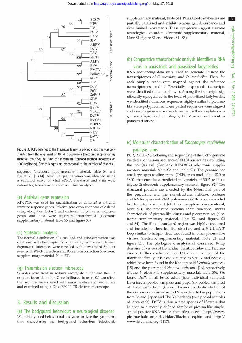
Figure 3. DcPV belongs to the Iflaviridae family. A phylogenetic tree was con-structed from the alignment of 30 RdRp sequences (electronic supplementarymaterial, table S3) by using the maximum-likelihood method (bootstrap on1000 replicates). Branch lengths are proportional to the number of changes.
rspb.royalsocietypublishing.orgProc.R.Soc.B
282:20142773
3
on May 17, 2018http://rspb.royalsocietypublishing.org/Downloaded from
sequence (electronic supplementary material, table S4 and
figure S6) [13,14]. Absolute quantification was obtained using
a standard curve of viral cDNA standards and data were
natural-log-transformed before statistical analyses.
(e) Antiviral gene expressionRT-qPCR was used for quantification of C. maculata antiviral
immune response genes. Relative gene expression was calculated
using elongation factor 2 and carbonic anhydrase as reference
genes and data were square-root-transformed (electronic
supplementary material, table S5 and figure S8).
( f ) Statistical analysesThe normal distribution of virus load and gene expression was
confirmed with the Shapiro Wilk normality test for each dataset.
Significant differences were revealed with a two-sided Student
t-test with Welch correction and Bonferroni correction (electronic
supplementary material, Note S3).
(g) Transmission electron microscopySamples were fixed in sodium cacodylate buffer and then in
osmium tetroxide buffer. Once infiltrated in resin, 0.1 mm ultra-
thin sections were stained with uranyl acetate and lead citrate
and examined using a Zeiss EM 10 CR electron microscope.
3. Results and discussion(a) The bodyguard behaviour: a neurological disorderWe initially used behavioural assays to analyse the symptoms
that characterize the bodyguard behaviour (electronic
supplementary material, Note S1). Parasitized ladybeetles are
partially paralysed and exhibit tremors, gait disturbance and
slow limited movements. These symptoms suggest a severe
Figure 4. Abundance and replication of DcPV in D. coccinellae. Quantity of negative- (a) and positive- (b) strand copies of DcPV in 500 mg of RNA from parasitoideggs (E) and larvae (L) collected 5, 13 and 20 days following oviposition (E Be D5, E/L Be D13, L Be D20), immediately after larval egression from the host (L Ae)and in adult parasitoids. Results are mean+ s.e.m. of biological replicates. Asterisks (*, ** and ***) indicate results are significantly different for a two-sidedStudent’s t-test (electronic supplementary material, Note S3), with q , 0.05, q , 0.01 and q , 0.001, respectively. (c) TEM image of the oviduct of D. coccinellae.Beneath the cuticular intima, a series of microvilli line the lumen. Viral particles are observed within unilamellar vesicles. (d ) TEM image of a vesicle packed withviruses showing a typical crystal structure. Cu, cuticular intima lining the oviductal lumen; mv, microvilli; L, lumen; arrow heads, viral particles. Scale bars, 500 nm.
350
(a) (b)
no. c
DN
A c
opie
s
no. c
DN
A c
opie
s
***
***
*** ***
***
**
*
*
*
300250200150100500
Res He
abdomens heads
Ae
Ae
HeR R
6 × 10⁵
5 × 10⁵
4 × 10⁵
3 × 10⁵
2 × 10⁵
1 × 10⁵
0
Be
D5
Be
D5
Be
D13
Be
D20
Be
D13
Be
D20
Res He
abdomens heads
Ae
Ae
HeR R
Be
D5
Be
D5
Be
D13
Be
D20
Be
D13
Be
D20
Figure 5. Abundance and replication of DcPV in healthy and parasitized C. maculata. Quantity of negative- (a) and positive- (b) strand copies of DcPV in 500 mg of RNAfrom abdomens and heads of ladybeetles collected healthy (He), 5, 13 and 20 days post-oviposition (Be D5, Be D13 and Be D20), immediately after larval egression (Ae),during recovery of a normal behaviour (R) and in resistant ladybeetles (Res). Results are mean+ s.e.m. of biological replicates. Asterisks (*, ** and ***) indicate thatresults are significantly different for a two-sided Student’s t-test (electronic supplementary material, Note S3) with q , 0.05, q , 0.01 and q , 0.001, respectively.
rspb.royalsocietypublishing.orgProc.R.Soc.B
282:20142773
4
on May 17, 2018http://rspb.royalsocietypublishing.org/Downloaded from
(d) Dinocampus coccinellae paralysis virus is associatedto the parasitoid wasp Dinocampus coccinellae
In early development (e.g. eggs collected five days after ovipos-
ition, Be D5), DcPV genomes were below detectable level,
suggesting that very few virus particles are present at this
stage (figure 4). Thirteen days post-oviposition (Be D13), we col-
lected either eggs or larva. Interestingly, virus was absent in
eggs but abundant following egg hatching, resulting in a
high variance at this time point. During larval development,
virus load increased significantly (figure 4a,b, Be D20). In the
adult stage, viral genome abundance was significantly higher
than in larva, whereas the replication intermediates were
significantly lower (figure 4). It resulted in a ratio of positive-
strand to negative-strand genomes of about 100 : 1 in larva
and 3000 : 1 in adult wasps, suggesting a high viral replication
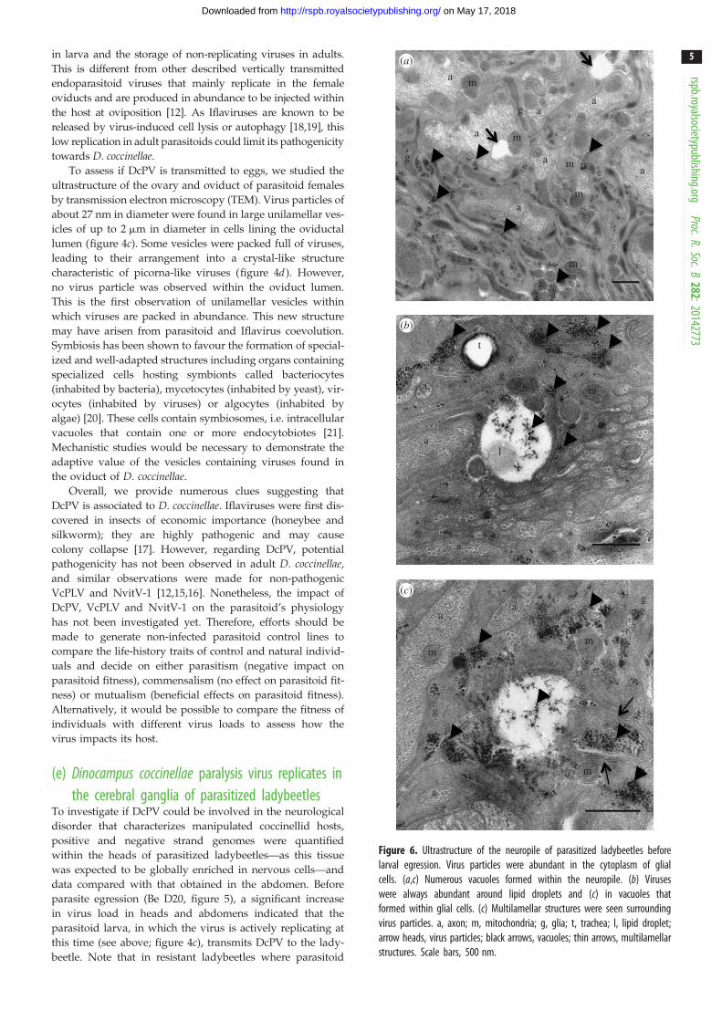
Figure 6. Ultrastructure of the neuropile of parasitized ladybeetles beforelarval egression. Virus particles were abundant in the cytoplasm of glialcells. (a,c) Numerous vacuoles formed within the neuropile. (b) Viruseswere always abundant around lipid droplets and (c) in vacuoles thatformed within glial cells. (c) Multilamellar structures were seen surroundingvirus particles. a, axon; m, mitochondria; g, glia; t, trachea; l, lipid droplet;arrow heads, virus particles; black arrows, vacuoles; thin arrows, multilamellarstructures. Scale bars, 500 nm.
rspb.royalsocietypublishing.orgProc.R.Soc.B
282:20142773
5
on May 17, 2018http://rspb.royalsocietypublishing.org/Downloaded from
in larva and the storage of non-replicating viruses in adults.
This is different from other described vertically transmitted
endoparasitoid viruses that mainly replicate in the female
oviducts and are produced in abundance to be injected within
the host at oviposition [12]. As Iflaviruses are known to be
released by virus-induced cell lysis or autophagy [18,19], this
low replication in adult parasitoids could limit its pathogenicity
towards D. coccinellae.
To assess if DcPV is transmitted to eggs, we studied the
ultrastructure of the ovary and oviduct of parasitoid females
by transmission electron microscopy (TEM). Virus particles of
about 27 nm in diameter were found in large unilamellar ves-
icles of up to 2 mm in diameter in cells lining the oviductal
lumen (figure 4c). Some vesicles were packed full of viruses,
leading to their arrangement into a crystal-like structure
characteristic of picorna-like viruses (figure 4d ). However,
no virus particle was observed within the oviduct lumen.
This is the first observation of unilamellar vesicles within
which viruses are packed in abundance. This new structure
may have arisen from parasitoid and Iflavirus coevolution.
Symbiosis has been shown to favour the formation of special-
ized and well-adapted structures including organs containing
specialized cells hosting symbionts called bacteriocytes
(inhabited by bacteria), mycetocytes (inhabited by yeast), vir-
ocytes (inhabited by viruses) or algocytes (inhabited by
algae) [20]. These cells contain symbiosomes, i.e. intracellular
vacuoles that contain one or more endocytobiotes [21].
Mechanistic studies would be necessary to demonstrate the
adaptive value of the vesicles containing viruses found in
the oviduct of D. coccinellae.
Overall, we provide numerous clues suggesting that
DcPV is associated to D. coccinellae. Iflaviruses were first dis-
covered in insects of economic importance (honeybee and
silkworm); they are highly pathogenic and may cause
colony collapse [17]. However, regarding DcPV, potential
pathogenicity has not been observed in adult D. coccinellae,
and similar observations were made for non-pathogenic
VcPLV and NvitV-1 [12,15,16]. Nonetheless, the impact of
DcPV, VcPLV and NvitV-1 on the parasitoid’s physiology
has not been investigated yet. Therefore, efforts should be
made to generate non-infected parasitoid control lines to
compare the life-history traits of control and natural individ-
uals and decide on either parasitism (negative impact on
parasitoid fitness), commensalism (no effect on parasitoid fit-
ness) or mutualism (beneficial effects on parasitoid fitness).
Alternatively, it would be possible to compare the fitness of
individuals with different virus loads to assess how the
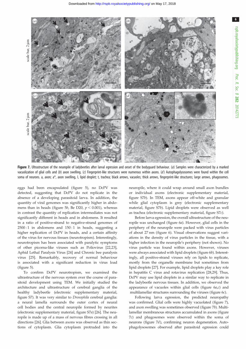
Figure 7. Ultrastructure of the neuropile of ladybeetles after larval egression and onset of the bodyguard behaviour. (a) Samples were characterized by a markedvacuolization of glial cells and (b) axon swelling. (c) Fingerprint-like structures were numerous within axons. (d ) Autophagolysosomes were found within the cellsoma of neurons. a, axon; a*, axon swelling, l, lipid droplet; t, trachea; black arrows, vacuoles; thick arrows, fingerprint-like structures; large arrows, phagosomes.
rspb.royalsocietypublishing.orgProc.R.Soc.B
282:20142773
6
on May 17, 2018http://rspb.royalsocietypublishing.org/Downloaded from
eggs had been encapsulated (figure 5), no DcPV was
detected, suggesting that DcPV do not replicate in the
absence of a developing parasitoid larva. In addition, the
quantity of viral genomes was significantly higher in abdo-
mens than in heads (figure 5b, Be D20, p , 0.001), whereas
in contrast the quantity of replication intermediates was not
significantly different in heads and in abdomens. It resulted
in a ratio of positive-strand to negative-strand genomes of
2500 : 1 in abdomens and 150 : 1 in heads, suggesting a
higher replication of DcPV in heads, and a certain affinity
of the virus for nervous tissues (neurotropism). Interestingly,
neurotropism has been associated with paralytic symptoms
of other picorna-like viruses such as Poliovirus [22,23],
Aphid Lethal Paralysis Virus [24] and Chronic bee paralysis
virus [25]. Remarkably, recovery of normal behaviour
is associated with a significant reduction in virus load
(figure 5).
To confirm DcPV neurotropism, we examined the
ultrastructure of the nervous system over the course of para-
sitoid development using TEM. We initially studied the
architecture and ultrastructure of cerebral ganglia of the
Figure 8. Ultrastructure of the neuropile of parasitized ladybeetles that recovered from bodyguard manipulation. (a,b) Glia expanded between the cortex andneuropile, and (c,d ) surrounded axons and axon bundles. a, axon; n, nucleus; m, mitochondria; thick arrows, expanding glial cells.
rspb.royalsocietypublishing.orgProc.R.Soc.B
282:20142773
7
on May 17, 2018http://rspb.royalsocietypublishing.org/Downloaded from
also be associated with antiviral immune defence [30–32].
Indeed, autophagy is a key feature of innate antiviral immunity,
although mechanisms are poorly understood [30–32].
Finally, vacuolization was limited in ladybeetles that recov-
ered from behaviour manipulation and survived parasitism,
and we found few phagosomes and phagolysosomes. How-
ever, highly electron-dense glial cells expanded between the
cortex and neuropile (figure 8a,b). These glial cells were
observed surrounding axons and axon bundles (figure 8c,d ).
Images were characteristic of a strong glial regenerative
response, i.e. the expansion of electron-dense glial cells
around axons and axon bundles [33]. In Drosophila and cock-
roach, stabbing injury induces proliferation of glial cells and
phagocytosis of cellular debris, which restore glial numbers,
axonal enwrapment and normal nervous system function
[33–37]; glia promote axonal regrowth and protect against
axonal degeneration and neuronal death. Therefore, the
spreading of glia in the neuropile of ladybeetles and engulf-
ment of axons could explain how normal brain functions are
restored and how the ladybeetle recovers from paralysis.
DcPV replication in glia may directly or indirectly alter the
behaviour of the ladybeetle by glial cell lysis. Indeed, insect glia
play a role in behavioural regulation via neurotransmitter clear-
ance. Thus, glial cell lysis could ultimately affect vision,
locomotion, sexual behaviour and host survival [38]. Also,
neurons greatly depend on glia, so when glia integrity is threa-
tened by DcPV replication, it would indirectly affect neuronal
functioning. Alternatively, the virus may have been transmitted
to neurons, which would explain the induction of autophagosis
involved in virus clearing (xenophagosis) [30–32]. It has been
demonstrated that xenophagic degradation allows virus clear-
ance and limits virus replication [30,37,39]. Therefore, DcPV
spread to neurons could result in a transient alteration of the
central nervous system that is restored once the viruses have
been eliminated. The impaired locomotion and overall lack of
ladybeetle motricity are the most remarkable symptoms of the
bodyguard behaviour. Based on our observations of the cer-
ebral ganglia, we assume that the DcPV induced neuropathy
spread to the entire nervous system, including the abdominal
and thoracic ganglia. The latter is involved in the control of loco-
motion by innervating the legs and wings. Some aspects of the
bodyguard behaviour, including the tremors and reduced
reflex, may also be owing to other indirect side effects of the
neuropathy, such as changes in ion concentration in haemo-
lymph owing to malphighian tubule malfunction, endocrine
disruption or an impairment of the processing of sensory
information collected by antennae and eyes.
( f ) The role of the antiviral immune responsePresence of virus particles in the ladybeetle is expected
to induce an antiviral response. We followed transcript
levels of key genes involved in antiviral autophagy (Toll 7
and PI3K) and in antiviral RNA interference (Dicer 2,
Ago 2, R2D2 and C3PO) throughout the infectious process
(figure 9; electronic supplementary material, table S5 and
figure S8) [32,39,40]. In abdomens, no significant variation
of gene expression was observed except for a downregulation
of Dicer 2 after egression and an upregulation of C3PO, a
homologue of R2D2 [41], in recovering individuals. However,
Figure 9. Gene expression profiles of selected genes involved in antiviral immune response as obtained by RT-qPCR analysis: gene expression profiles in abdomensand heads of ladybeetles collected healthy (He), 5, 13 and 20 days post-oviposition (Be D5, Be D13 and Be D20), immediately after larval egression (Ae), duringrecovery of a normal behaviour (R) and in resistant ladybeetles (Res). (a) Dicer 2; (b) Ago2, Argonaute 2; (c) C3PO; (d ) R2D2 are involved in antiviral RNA inter-ference. (e) Toll 7 and ( f ) PI3K, phosphatidylinositol 3 kinase are involved in antiviral autophagosis. Results are means+ s.d. of biological replicates for eachexperimental condition. Asterisks (*, ** and ***) indicate results are significantly different to He for a two-sided student’s t-test (electronic supplementary material,Note S3) with q , 0.05, q , 0.01 and q , 0.001, respectively.
rspb.royalsocietypublishing.orgProc.R.Soc.B
282:20142773
8
on May 17, 2018http://rspb.royalsocietypublishing.org/Downloaded from
Ago 2, R2D2, Toll7 and PI3K were found significantly upre-
gulated in ladybeetles resistant to parasitism (figure 9). Of
significance, Dicer 2, Ago2, Toll 7 and PI3K expression was
significantly downregulated in parasitized ladybeetle heads
at D5 and D13 post-oviposition (figure 9). Their expression
in heads was re-established from D20 onward, whereas
R2D2 expression was significantly downregulated from D20
onward. Thus, the transient downregulation of multiple
genes involved in the antiviral response in the ladybeetle ner-
vous tissue could allow DcPV neurotropism in the first stages
of the infectious process. Then, the re-establishment of an
antiviral immune response in ladybeetle nervous tissue corre-
lates with the appearance of phagolysosomes and with the
onset of the bodyguard behaviour and result in the virus
elimination. Thus, healthy ladybeetles are equipped to
recognize and eliminate the virus. The antiviral immuno-
suppression (that may be induced by the developing
D. coccinellae larva or by DcPV itself ) could trigger the
observed transient pathogenic infection and accumulation
of DcPV in the nervous tissue. Then, elimination of the con-
sequently high virus load could trigger the neuropathy
responsible for the paralysis. A link between antiviral
immune responses in nervous tissues and behavioural
changes has previously been observed in other host–virus
interactions. Immune response and associated widespread
inflammatory response are known to be involved in behav-
ioural changes induced by the Borna disease virus [42] or
in patients with rabies [43]. Here, the antiviral immune
response could result in the accumulation of phago-
somes that induce paralytic symptoms characterizing the
bodyguard behaviour.
4. Concluding remarksUntil now, studies of the mechanisms responsible for
host behaviour manipulation have mostly focused on neuro-
modulatory systems [1,4,44,45]. On rare occasions, a single
parasite gene explains the extended phenotype [5]. Here,
we revealed the involvement of a third protagonist, a symbio-
tic virus of the wasp—called DcPV—that is transmitted to
the host during parasite larval development and is neurotro-
pic. The presence of unilamellar vesicles containing large
amounts of DcPV particles within the oviduct cells of
D. coccinellae suggests that DcPV could be transmitted to
the wasp eggs. Our results suggest that changes in ladybeetle
behaviour most likely result from DcPV replication in the cer-
ebral ganglia rather than by a direct manipulation by the
parasitic wasp. We propose a theoretical scenario within
which DcPV is employed as a biological weapon by
D. coccinellae to manipulate the behaviour of C. maculata(life cycles in figure 10).
Further experiments are now necessary to characterize
the nature of the symbiosis between D. coccinellae and
DcPV. It remains unknown if symbiotic individuals display
a better fitness than asymbiotic individuals and if the
DcPV life cycle deposited with parasitoid egg at oviposition
oviposition replication in
parasitoid larva
latent inparasitoid oviduct
transmission toladybeetle
replication inladybeetle nervous
system
immuno-suppression
immuneresponse
adult
pupation
egg hatchingand larval
development
D. coccinnellae life cycle
elimination by ladybeetle immune response
nerve cells' restorationnormal behaviour
recovery
neuropathy: phagocytosis and neuronswelling
paralysis and tremors
Figure 10. Life cycles of the parasitoid D. coccinellae and its endosymbiotic virus (D. coccinellae paralysis virus, DcPV) together with responses to parasitism andinfection of the ladybeetle host C. maculata (drawing by Franz Vanoosthuyse). The DcPV is stored in abundance in the oviduct of D. coccinellae female. Followingoviposition and egg hatching, DcPV replicates in the parasitoid larva and is transmitted to C. maculata. The antiviral immune system of the ladybeetle is thensuppressed, which allows DcPV to replicate in glial cells in the host’s nervous system. The re-establishment of the antiviral immune response correlates with asevere neuropathy in the ladybeetle and the onset of the bodyguard behaviour. The synchronized egression of the larva allows it to take advantage of the paralyzedladybeetle: it steals between the ladybeetle legs and spins its cocoon under its protection. The DcPV is being eliminated from the ladybeetle, which progressivelyrecovers through nerve cell restoration. By then, the parasitoid resumes pupation and emerges with a new load of DcPV in its oviduct.
rspb.royalsocietypublishing.orgProc.R.Soc.B
282:20142773
9
on May 17, 2018http://rspb.royalsocietypublishing.org/Downloaded from
symbiosis is obligate for the host. In addition, and in order
to validate the proposed scenario, the effect of DcPV and
D. coccinellae on C. maculata physiology should be tested
independently via a combination of RNA interference
and injection of purified viruses. These complementary
analyses will also provide information regarding the surpris-
ingly precise simultaneous timing of larvae egression, re-
establishment of antiviral immune response, accumulation
of phagosomes and the induction of bodyguard behaviour
by determining which mechanisms is responsible for the
induction of the others.
The role of associated microorganisms, including eukar-
yotes, bacteria and viruses, in all components of organisms’
biology has been recently revealed [46]. This study contrib-
utes to the realization that host–parasite interactions should
be considered as holobiont–holobiont interactions [47] by
revealing that parasite-associated microorganisms could par-
ticipate in all components of host exploitation strategies,
including behaviour manipulation.
Data accessibility. DcPV genome sequence: EMBL GenBank accessionKF843822. Raw fastq files: NCBI’s sequence read archive referencePRJNA227418 for C. maculata samples and PRJNA227420 for D. coc-cinellae samples. de novo assembled transcriptomes: http://2ei.univ-perp.fr/telechargement/transcriptomes/ALL_Cocc_95.zip for para-sitized C. maculata and http://2ei.univ-perp.fr/telechargement/transcripto mes/ALL_Larve_95.zip for D. coccinellae .
Acknowledgements. We thank C. Cazevieille (CRIC, Montpellier, France)for help in TEM and L. Devine for English revision.Funding statement. N.M.D. and F.M. were supported by the AgenceNationale de la Recherche (ANR) Blanc, SVSE7, project Bodyguardto F.T. and by FQRNT to J.B.
References
1. Hughes DP, Brodeur J, Thomas F. 2012 Hostmanipulation by parasites, p. 224. Oxford, UK:Oxford University Press.
2. Moore J. 2002 Parasites and the behavior ofanimals, p. 315. Oxford, UK: Oxford University Press.
3. Poulin R. 2010 Parasite manipulation of hostbehavior: an update and frequently asked questions.
4. Perrot-Minnot M-J, Cezilly F. 2013 Investigatingcandidate neuromodulatory systems underlyingparasitic manipulation: concepts, limitations andprospects. J. Exp. Biol. 216, 134 – 141. (doi:10.1242/jeb.074146)
5. Hoover K, Grove M, Gardner M, Hughes DP, McNeilJ, Slavicek J. 2011 A gene for an extendedphenotype. Science 333, 1401. (doi:10.1126/science.1209199)
on May 17, 2018http://rspb.royalsocietypublishing.org/Downloaded from
gene induces enhanced locomotory activity in alepidopteran host. Proc. Natl Acad. Sci. USA 102,2584 – 2589. (doi:10.1073/pnas.0409457102)
7. Maure F, Daoust SP, Brodeur J, Mitta G, Thomas F.2013 Diversity and evolution of bodyguardmanipulation. J. Exp. Biol. 216, 36 – 42. (doi:10.1242/jeb.073130)
8. Maure F, Brodeur J, Ponlet N, Doyon J, Firlej A,Elguero E, Thomas F. 2011 The cost of abodyguard. Biol. Lett. 7, 843 – 846. (doi:10.1098/rsbl.2011.0415)
9. Triltsch H. 1996 On the parasitisation of the ladybirdCoccinella septempunctata. J. Appl. Entomol. 120,375 – 378. (doi:10.1111/j.1439-0418.1996.tb01622.x)
10. Maure F, Doyon J, Thomas F, Brodeur J. 2014 Hostbehavior manipulation as an evolutionary routetoward attenuation of parasitoid virulence. J. Evol.Biol. 27, 2871 – 2875. (doi:10.1111/jeb.12530)
11. Whitfield JB, Asgari S. 2003 Virus or not?Phylogenetics of polydnaviruses and their waspcarriers. J. Insect Physiol. 49, 397 – 405. (doi:10.1016/S0022-1910(03)00057-X)
12. Beckage NE, Drezen J-M. 2012 Parasitoid viruses:symbionts and pathogens, p. 312. New York, NY:Academic Press.
13. Komurian-Pradel F, Perret M, Deiman B, Sodoyer M,Lotteau V, Paranhos-Baccala G, Andre P. 2004 Strandspecific quantitative real-time PCR to study replicationof hepatitis C virus genome. J. Virol. Method 116,103 – 106. (doi:10.1016/j.jviromet.2003.10.004)
14. Plaskon NE, Adelman ZN, Myles KM. 2009 Accuratestrand-specific quantification of viral RNA. PLoS ONE4, e7468. (doi:10.1371/journal.pone.0007468)
15. Reineke A, Asgari S. 2005 Presence of a novelsmall RNA-containing virus in a laboratory cultureof the endoparasitic wasp Venturia canescens(Hymenoptera: Ichneumonidae). J. InsectPhysiol. 51, 127 – 135. (doi:10.1016/j.jinsphys.2004.05.005)
16. Oliveira DCSG, Hunter WB, Ng J, Desjardins CA,Dang PM, Werren JH. 2010 Data mining cDNAsreveals three new single stranded RNA viruses inNasonia (Hymenoptera: Pteromalidae). Insect Mol.Biol. 19, 99 – 107. (doi:10.1111/j.1365-2583.2009.00934.x)
17. van Oers MM. 2010 Genomics and biology ofIflaviruses. In Insect virology (eds S Asgari,K Johnson), pp. 231 – 250. Norfolk, Virginia: CaisterAcademic Press.
18. Buck KW, Maramorosch K, Murphy FA, Shatkin AJ.1996 Comparison of the replication of positive-stranded RNA viruses of plants and animals. Adv.Vir. Res. 47, 159 – 251. (doi:10.1016/S0065-3527(08)60736-8)
19. Richards AL, Jackson WT. 2013 Behind closedmembranes: the secret lives of Picornaviruses? PLoSPathog. 9, e1003262. (doi:10.1371/journal.ppat.1003262)
20. Seckbach J, Nardon P, Charles H. 2002Morphological aspects of symbiosis. In Symbiosis,pp. 13 – 44. Dordrecht, The Netherlands: Springer.
21. Ahn G, Choi E, Jeon K. 1990 A symbiosome-membrane-specific protein in symbiont-bearingAmoeba proteus as studied with a monoclonalantibody. Endocytobiosis Cell Res 7, 45 – 50.
22. Bodian D. 1949 Histopathologic basis of clinicalfindings in poliomyelitis. Am. J. Med. 6, 563 – 578.(doi:10.1016/0002-9343(49)90130-8)
23. Bodian D, Howe HA. 1941 Neurotropism and thegenesis of cerebral lesions in poliomyelitis: anexperimental study. Bull. Johns Hopkins Hosp. 68,58 – 76.
24. Williamson C, Rybicki EP, Kasdorf GGF, VonWechmar MB. 1988 Characterization of a newpicorna-like virus isolated from aphids. J. Gen. Virol.69, 787 – 795. (doi:10.1099/0022-1317-69-4-787)
25. Blanchard P, RibiAre M, Celle O, Lallemand P, SchurrF, Olivier V, Iscache AL, Faucon JP. 2007 Evaluationof a real-time two-step RT-PCR assay forquantitation of Chronic bee paralysis virus (CBPV)genome in experimentally-infected bee tissues andin life stages of a symptomatic colony. J. Virol.Methods 141, 7 – 13. (doi:10.1016/j.jviromet.2006.11.021)
26. Cardona A, Saalfeld S, Preibisch S, Schmid B, ChengA, Pulokas J, Tomancak P, Hartenstein V. 2010 Anintegrated micro- and macroarchitectural analysis ofthe Drosophila brain by computer-assisted serialsection electron microscopy. PLoS Biol. 8, e1000502.(doi:10.1371/journal.pbio.1000502)
27. Martın-Acebes MA, Vazquez-Calvo A, Caridi F, SaizJ-C, Sobrino F. 2012 Lipid involvement in viralinfections: present and future perspectives for thedesign of antiviral strategies. In Lipid metabolism,pp. 291 – 322. Rijeka, Croatia: InTech.
28. Cheung W et al. 2010 Rotaviruses associate with cellularlipid droplet components to replicate in viroplasms, andcompounds disrupting or blocking lipid droplets inhibitviroplasm formation and viral replication. J. Virol. 84,6782 – 6798. (doi:10.1128/JVI.01757-09)
29. Ogawa K, Hishiki T, Shimizu Y, Funami K, SugiyamaK, Miyanari Y, Shimotohno K. 2009 Hepatitis C virusutilizes lipid droplet for production of infectiousvirus. Proc. Jpn Acad. Ser. B Phys. Biol. Sci. 85,217 – 228. (doi:10.2183/pjab.85.217)
30. Orvedahl A, Levine B. 2008 Autophagy and viralneurovirulence. Cell Microbiol. 10, 1747 – 1756.(doi:10.1111/j.1462-5822.2008.01175.x)
31. Richetta C, Faure M. 2013 Autophagy in antiviralinnate immunity. Cell Microbiol. 15, 368 – 376.(doi:10.1111/cmi.12043)
32. Xu J, Cherry S. 2014 Viruses and antiviral immunityin Drosophila. Dev. Comp. Immunol. 42, 67 – 84.(doi:10.1016/j.dci.2013.05.002)
33. Kato K, Forero MG, Fenton JC, Hidalgo A. 2011 Theglial regenerative response to central nervous
system injury is enabled by pros-notch and pros-NFkB feedback. PLoS Biol. 9, e1001133. (doi:10.1371/journal.pbio.1001133)
34. Kato K, Awasaki T, Ito K. 2009 Neuronalprogrammed cell death induces glial cell division inthe adult Drosophila brain. Development 136,51 – 59. (doi:10.1242/dev.023366)
35. Smith P, Leech C, Treherne J. 1984 Glial repair in aninsect central nervous system: effects of selectiveglial disruption. J. Neurosci. 4, 2698 – 2711.
36. Smith PJ, Howes EA, Treherne JE. 1987 Mechanismsof glial regeneration in an insect central nervoussystem. J. Exp. Biol. 132, 59 – 78.
37. Treherne J, Harrison J, Treherne J, Lane N. 1984Glial repair in an insect central nervous system:effects of surgical lesioning. J. Neurosci. 4,2689 – 2697.
38. Edwards TN, Meinertzhagen IA. 2010 The functionalorganisation of glia in the adult brain of Drosophilaand other insects. Prog. Neurobiol. 90, 471 – 497.(doi:10.1016/j.pneurobio.2010.01.001)
39. Ding S-W, Voinnet O. 2007 Antiviral immunitydirected by small RNAs. Cell 130, 413 – 426. (doi:10.1016/j.cell.2007.07.039)
40. Sabin LR, Zheng Q, Thekkat P, Yang J, Hannon GJ,Gregory BD, Tudor M, Cherry S. 2013 Dicer-2processes diverse viral RNA species. PLoS ONE 8,e55458. (doi:10.1371/journal.pone.0055458)
41. Tomoyasu Y, Miller SC, Tomita S, Schoppmeier M,Grossmann D, Bucher G. 2008 Exploring systemicRNA interference in insects: a genome-wide surveyfor RNAi genes in Tribolium. Genome Biol. 9, R10.(doi:10.1186/gb-2008-9-1-r10)
42. Carbone KM, Duchala CS, Griffin JW, Kincaid AL,Narayan O. 1987 Pathogenesis of Borna diseasein rats: evidence that intra-axonal spread is themajor route for virus dissemination and thedeterminant for disease incubation. J. Virol. 61,3431 – 3440.
43. Hemachudha T, Laothamatas J, Rupprecht CE. 2002Human rabies: a disease of complexneuropathogenetic mechanisms and diagnosticchallenges. Lancet Neurol. 1, 101 – 109. (doi:10.1016/S1474-4422(02)00041-8)