CELL INJURY Lecture 3 Objectives: ● Understand the causes of and pathologic changes occurring in fatty change (steatosis), accumulations of exogenous and endogenous pigments (carbon, silica, iron, melanin, bilirubin and lipofuscin). ● Understand the causes of and differences between dystrophic and metastatic calcifications. 1 Color Index: Girl’s Slides Important Male’s Notes Female’s Notes Extra information { ْ مَ لْ عَ ي ْ مَ ا لَ مَ انَ نسِْٕ الَ مَّ لَ ع}
Transcript
CELL INJURYLecture 3
Objectives:
● Understand the causes of and pathologic changes occurring in fatty change (steatosis), accumulations of exogenous and endogenous pigments (carbon, silica, iron, melanin, bilirubin and lipofuscin).
● Understand the causes of and differences between dystrophic and metastatic calcifications.
1
Color Index:Girl’s SlidesImportantMale’s NotesFemale’s NotesExtra information
لم } ع ان ما لم ي س ن م ال { عل
2
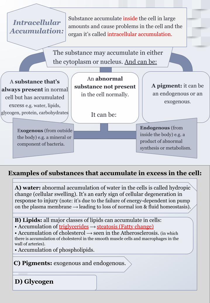
Intracellular Accumulation:
Substance accumulate inside the cell in large amounts and cause problems in the cell and the organ it’s called intracellular accumulation.
The substance may accumulate in either the cytoplasm or nucleus. And can be:
A substance that’s always present in normal
cell but has accumulated excess e.g. water, lipids,
glycogen, protein, carbohydrates
An abnormal substance not present
in the cell normally.
It can be:
Exogenous (from outside the body) e.g. a mineral or component of bacteria.
Endogenous (from inside the body) e.g. a product of abnormal synthesis or metabolism.
A pigment: it can be an endogenous or an
exogenous.
Examples of substance that accumulate in excess in the cell:
A) water: abnormal accumulation of water in the cells is called hydropic change (cellular swelling). It’s an early sign of cellular degeneration in response to injury (note: it’s due to the failure of energy-dependent ion pump on the plasma membrane ⟶ leading to loss of normal ion & fluid homeostasis).
B) Lipids: all major classes of lipids can accumulate in cells:• Accumulation of triglycerides ⟶ steatosis (Fatty change)• Accumulation of cholesterol ⟶ seen in the Atherosclerosis. (in which there is accumulation of cholesterol in the smooth muscle cells and macrophages in the wall of arteries).• Accumulation of phospholipids.
D) Glycogen
C) Pigments: exogenous and endogenous.
Examples of substances that accumulate in excess in the cell:
Light microscope:
in mild cases liver looks normal, in severe liver looks yellow and greasy.Gross:
Pregnancy Severe anemia
3
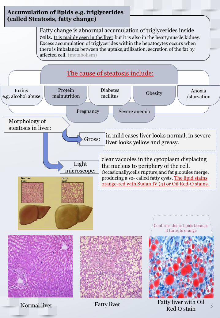
Accumulation of lipids e.g. triglycerides (called Steatosis, fatty change)
Fatty change is abnormal accumulation of triglycerides inside cells. It is mainly seen in the liver,but it is also in the heart,muscle,kidney.Excess accumulation of triglycerides within the hepatocytes occurs when there is imbalance between the uptake,utilization, secretion of the fat by affected cell. (metabolism)
The cause of steatosis include:
toxins e.g. alcohol abuse
Diabetes mellitus
Protein malnutrition Obesity Anoxia
/starvation
Morphology of steatosis in liver:
clear vacuoles in the cytoplasm displacing the nucleus to periphery of the cell.Occasionally,cells rupture,and fat globules merge, producing a so- called fatty cysts. The lipid stains orange-red with Sudan IV (4) or Oil Red-O stains.
Confirms this is lipids because it turns to orange
Normal liver Fatty liver Fatty liver with Oil Red O stain
Exogenous pigments: they are not synthesized within the body itself, it’s from outside the body.
Glucose is the main source of fuel for cells. Excess glucose is stored in the liver or muscles in the form of Glycogen.
4
Accumulation of Glycogen
Excessive intracellular deposits of Glycogen can be seen in patient with abnormally glucose or glycogen metabolism.
Glycogen is stored in the cell cytoplasm.
Glycogen stains pink/violet with periodic acid schiff(PAS).
Glycogen appears as clear vacuoles with the cell cytoplasm
Diabetes mellitus: it is disorder of glucose metabolism. In this disease glycogen is found in the proximal convoluted tubules of kidney,liver,the beta cells of islets of langerhans,heart muscle cell etc.
Glycogen storage disorders: it is group of genetic diseases in which there is abnormal glycogen metabolism and there can be abnormal accumulation of glycogen in the liver,muscle and other tissues.
Glycogen accumulation is seen in:
Accumulation of pigments:
Pigments are colored substance it can be:
Endogenous: synthesized within the body itself. some endogenous pigments are normal constituent of cells (e.g. melanin) and others are not.
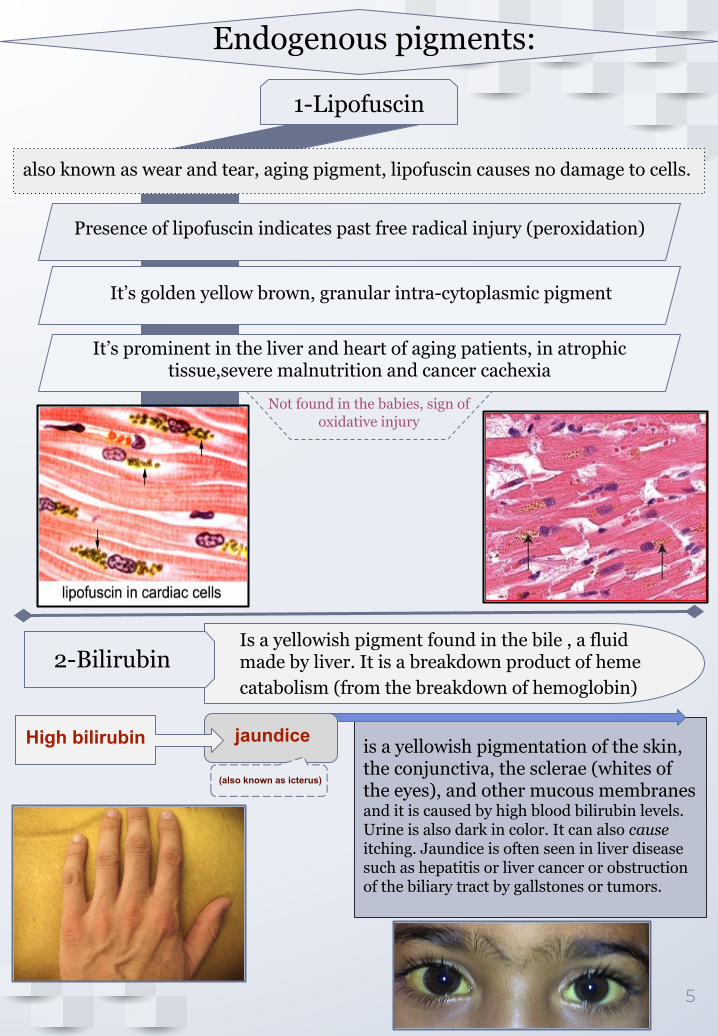
is a yellowish pigmentation of the skin, the conjunctiva, the sclerae (whites of the eyes), and other mucous membranes and it is caused by high blood bilirubin levels. Urine is also dark in color. It can also cause itching. Jaundice is often seen in liver disease such as hepatitis or liver cancer or obstruction of the biliary tract by gallstones or tumors.
5
Endogenous pigments:
1-Lipofuscin
also known as wear and tear, aging pigment, lipofuscin causes no damage to cells.
Presence of lipofuscin indicates past free radical injury (peroxidation)
It’s golden yellow brown, granular intra-cytoplasmic pigment
It’s prominent in the liver and heart of aging patients, in atrophic tissue,severe malnutrition and cancer cachexia
Not found in the babies, sign of oxidative injury
(also known as icterus)
Is a yellowish pigment found in the bile , a fluid made by liver. It is a breakdown product of heme catabolism (from the breakdown of hemoglobin)
2-Bilirubin
jaundice High bilirubin
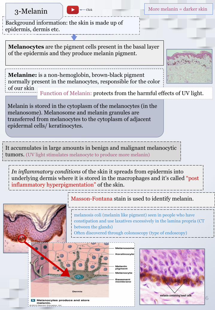
Melanocytes are the pigment cells present in the basal layer of the epidermis and they produce melanin pigment.
Background information: the skin is made up of epidermis, dermis etc.
6
More melanin = darker skin
Melanine: is a non-hemoglobin, brown-black pigment normally present in the melanocytes, responsible for the color of our skin
3-Melanin
Melanin is stored in the cytoplasm of the melanocytes (in the melanosome). Melanosome and melanin granules are transferred from melanocytes to the cytoplasm of adjacent epidermal cells/ keratinocytes.
Function of Melanin: protects from the harmful effects of UV light.
Masson-Fontana stain is used to identify melanin.
melanosis coli (melanin like pigment) seen in people who have constipation and use laxatives excessively in the lamina propria (CT between the glands)Often discovered through colonoscopy (type of endoscopy)
It accumulates in large amounts in benign and malignant melanocytic tumors. (UV light stimulates melanocyte to produce more melanin)
In inflammatory conditions of the skin it spreads from epidermis into underlying dermis where it is stored in the macrophages and it’s called “post inflammatory hyperpigmentation” of the skin.
Sickle shaped RBC (shrivel-shaped RBC) might go into necrosis
1st degree hemochromatosis (haemochromatosis) is genetic
**Deposits in macrophages all over blood
systemic hemosiderosis**: there is systemic overload of iron.
7
localized hemosiderosis e.g. common bruise: there is lysis of rbcs, release of hemoglobin and the iron in it is converted to hemosiderin
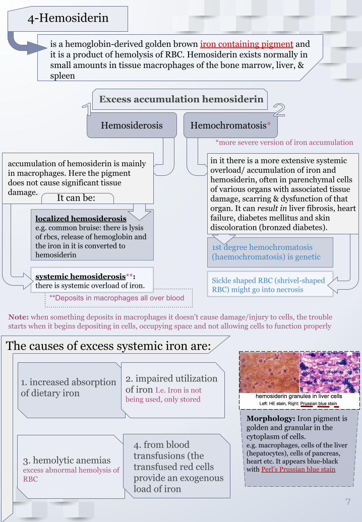
4-Hemosiderin
is a hemoglobin-derived golden brown iron containing pigment and it is a product of hemolysis of RBC. Hemosiderin exists normally in small amounts in tissue macrophages of the bone marrow, liver, & spleen
Excess accumulation hemosiderin
Hemosiderosis Hemochromatosis*
*more severe version of iron accumulation
in it there is a more extensive systemic overload/ accumulation of iron and hemosiderin, often in parenchymal cells of various organs with associated tissue damage, scarring & dysfunction of that organ. It can result in liver fibrosis, heart failure, diabetes mellitus and skin discoloration (bronzed diabetes).
accumulation of hemosiderin is mainly in macrophages. Here the pigment does not cause significant tissue damage.
It can be:
The causes of excess systemic iron are:
1. increased absorption of dietary iron
2. impaired utilization of iron I.e. Iron is not being used, only stored
3. hemolytic anemias excess abnormal hemolysis of RBC
4. from blood transfusions (the transfused red cells provide an exogenous load of iron
Note: when something deposits in macrophages it doesn't cause damage/injury to cells, the trouble starts when it begins depositing in cells, occupying space and not allowing cells to function properly
Morphology: Iron pigment is golden and granular in the cytoplasm of cells.e.g. macrophages, cells of the liver (hepatocytes), cells of pancreas, heart etc. It appears blue-black with Perl’s Prussian blue stain
Coal workers’ pneumoconiosis: in the coal mining industry, there is too much carbon dust in the lung of coal miners, it gets deposited in lung cells and it leads to a lung disease known as coal workers’ pneumoconiosis.
8
Exogenous pigments:
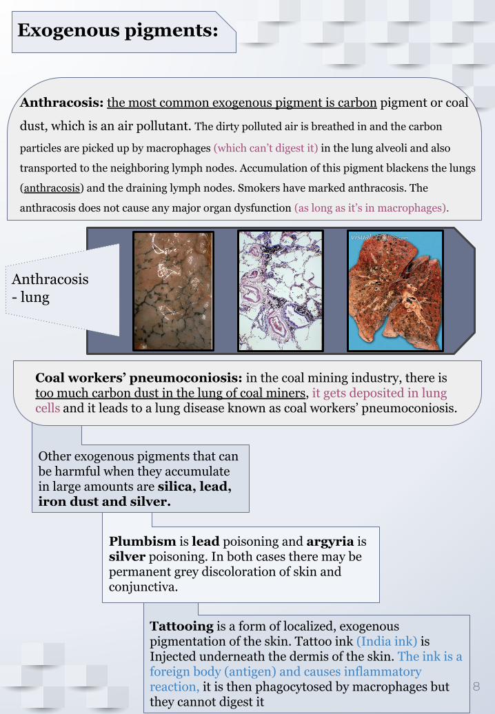
Anthracosis: the most common exogenous pigment is carbon pigment or coal
dust, which is an air pollutant. The dirty polluted air is breathed in and the carbon
particles are picked up by macrophages (which can’t digest it) in the lung alveoli and also
transported to the neighboring lymph nodes. Accumulation of this pigment blackens the lungs
(anthracosis) and the draining lymph nodes. Smokers have marked anthracosis. The
anthracosis does not cause any major organ dysfunction (as long as it’s in macrophages).
Anthracosis - lung
Other exogenous pigments that can be harmful when they accumulate in large amounts are silica, lead, iron dust and silver.
Plumbism is lead poisoning and argyria is silver poisoning. In both cases there may be permanent grey discoloration of skin and conjunctiva.
Tattooing is a form of localized, exogenous pigmentation of the skin. Tattoo ink (India ink) is Injected underneath the dermis of the skin. The ink is a foreign body (antigen) and causes inflammatory reaction, it is then phagocytosed by macrophages but they cannot digest it
*When it accumulate outside cells, it starts crushing them.
9
(additional)
Amyloidosis is a disorder of protein misfolding, which results in the extracellular deposition and accumulation of a fibrillary protein called amyloid*.
EXTRACELLULAR ACCUMULATION: Amyloidosis
Amyloid is composed of non-branching fibrils of β-pleated sheets.
It is deposited in various organs (kidney, liver, blood vessels, heart, tongue, intestine, lymph etc.) leading to damage of that organ.
Amyloidosis is associated with a number of inherited and inflammatory disorders.
There are 2 main clinical forms of amyloidosis:
Primary: associated with plasma cell abnormalities e.g. multiple myeloma; has “AL” type of amyloid.
Secondary: is secondary to chronic inflammatory or autoimmune diseases e.g. tuberculosis, rheumatoid arthritis etc.; has “AA” type of amyloid associated protein.
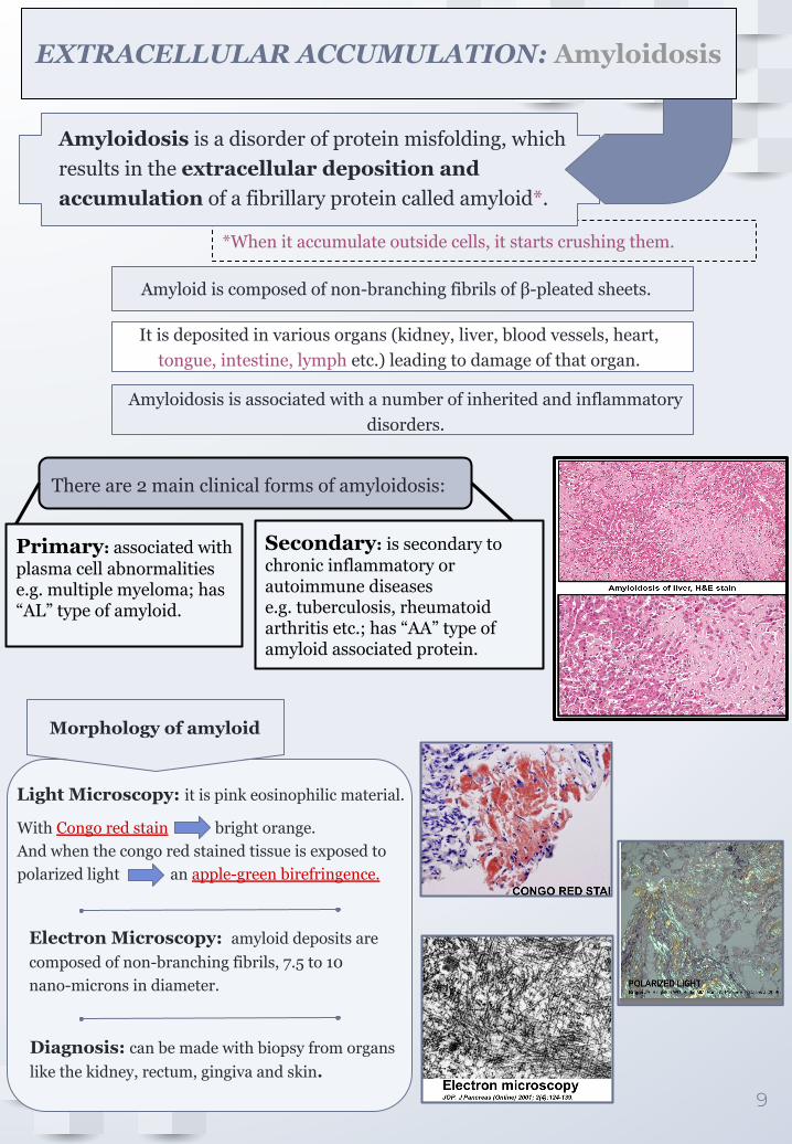
Morphology of amyloid
Light Microscopy: it is pink eosinophilic material.
With Congo red stain bright orange. And when the congo red stained tissue is exposed to polarized light an apple-green birefringence.
Electron Microscopy: amyloid deposits are composed of non-branching fibrils, 7.5 to 10 nano-microns in diameter.
Diagnosis: can be made with biopsy from organs like the kidney, rectum, gingiva and skin.
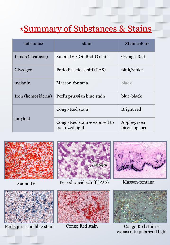
substance stain Stain colour
Lipids (steatosis) Sudan IV / Oil Red-O stain Orange-Red
Glycogen Periodic acid schiff (PAS) pink/violet
melanin Masson-fontana black
Iron (hemosiderin) Perl’s prussian blue stain blue-black
amyloid
Congo Red stain Bright red
Congo Red stain + exposed to polarized light
Apple-green birefringence
⭑Summary of Substances & Stains
Sudan IV Periodic acid schiff (PAS) Masson-fontana
Perl’s prussian blue stain Congo Red stain Congo Red stain + exposed to polarized light
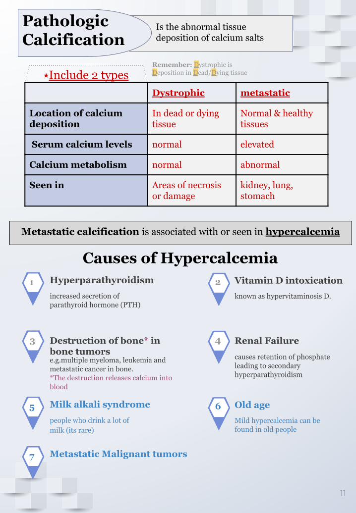
Pathologic Calcification
⭑Include 2 types Dystrophic metastatic
Location of calcium deposition
In dead or dying tissue
Normal & healthy tissues
Serum calcium levels normal elevated
Calcium metabolism normal abnormal
Seen in Areas of necrosis or damage
kidney, lung, stomach
Metastatic calcification is associated with or seen in hypercalcemia
1 2
3 4
increased secretion of parathyroid hormone (PTH)
Hyperparathyroidism
causes retention of phosphate leading to secondary hyperparathyroidism
Renal Failure
known as hypervitaminosis D.
Vitamin D intoxication
e.g.multiple myeloma, leukemia and metastatic cancer in bone.*The destruction releases calcium into blood
Destruction of bone* in bone tumors
Causes of Hypercalcemia
11
Is the abnormal tissue deposition of calcium salts
5 6people who drink a lot of milk (its rare)
Milk alkali syndrome
Mild hypercalcemia can be found in old people
Old age
7 Metastatic Malignant tumors
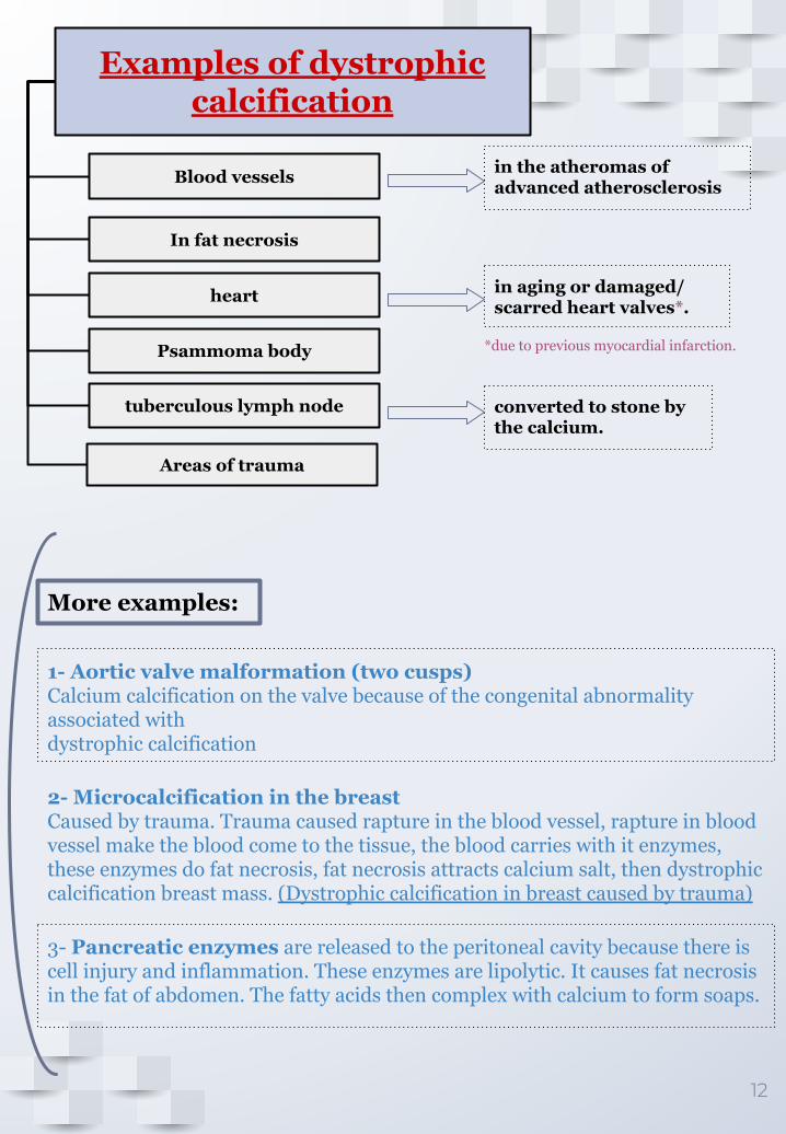
Remember: Dystrophic is Deposition in Dead/Dying tissue
Areas of trauma
Examples of dystrophic calcification
Blood vessels
In fat necrosis
heart
Psammoma body
tuberculous lymph node
in the atheromas of advanced atherosclerosis
in aging or damaged/scarred heart valves*.
converted to stone by the calcium.
*due to previous myocardial infarction.
12
3- Pancreatic enzymes are released to the peritoneal cavity because there is cell injury and inflammation. These enzymes are lipolytic. It causes fat necrosis in the fat of abdomen. The fatty acids then complex with calcium to form soaps.
2- Microcalcification in the breastCaused by trauma. Trauma caused rapture in the blood vessel, rapture in blood vessel make the blood come to the tissue, the blood carries with it enzymes, these enzymes do fat necrosis, fat necrosis attracts calcium salt, then dystrophic calcification breast mass. (Dystrophic calcification in breast caused by trauma)
1- Aortic valve malformation (two cusps) Calcium calcification on the valve because of the congenital abnormality associated withdystrophic calcification
More examples:
13
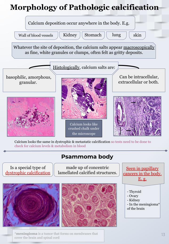
Morphology of Pathologic calcification
Calcium looks the same in dystrophic & metastatic calcification so tests need to be done to check for calcium levels & metabolism in blood
Calcium looks like crushed chalk under
the microscope
Calcium deposition occur anywhere in the body. E.g.
Wall of blood vessels Kidney Stomach lung skin
Whatever the site of deposition, the calcium salts appear macroscopically as fine, white granules or clumps, often felt as gritty deposits.
Histologically, calcium salts are:
basophilic, amorphous, granular.
Can be intracellular, extracellular or both.
Psammoma body
*meningioma is a tumor that forms on membranes that cover the brain and spinal cord
Is a special type of dystrophic calcification
made up of concentric lamellated calcified structures.
Seen in papillary cancers in the body.
E. g.
- Thyroid- Ovary- Kidney- In the meningioma* of the brain
14
MCQs
14
1- Slides 10 2- Slide 123- Slide 74- Slide 11
Q1- Accumulation of …… could be an early sign of cellular degeneration
A) Water B) Lipids C) Glycogen D) Pigments
Q2- Accumulation of carbon pigment causes:
A) atherosclerosis B) Melanosome granule C) Anthracosis D) Yellowish pigment
Q3- In secondary amyloidosis, Amyloid has “……” type of Amyloid associated proteins
A) AS B) AP C) AA D) AL
Q4- Stain that identifies Steatosis:
A) Masson-fontana B) Congo Red stain C) Sudan IV stain with Yellow-Black color
D) Oil Red-O stain with Orange-Red color
Q5: Psammoma bodies can be found in Papillary cancer of :
A) Heart B) Kidney C) Liver D) Both B & C
Q6: Which pigment accumulation can be seen as golden yellow brown?
A) Lipofuscin B) Bilirubin C) Melanin D) Hemosiderin
1-A 4-D
2-C 5-B
3-C 6-A
SAQs
Q1: Name some of the accumulating substances and the stain used to identify them.
Q2: Name examples of Dystrophic calcification.
Q3: The causes of excess systemic iron are?
Q4: What are the differences between Dystrophic and metastatic calcification?