APMIS 106: 959-969, 1998 Printed in Denmark . All rights reserved Copyright 8 APMIS 1998 l!Mi.lU8 ISSN 090346641 Exmession of keratin 13.14 and 19 in oral sauamous cell carcinomas from Sudanese SnuffdiDDers: Lack of association with human DaDillomavirus infection SALAH 0. IBRAHIM,',2,3BJ0RN BERTELSEN,4MAY BRITT KALVENES,4ALI M. IDRIS,5 ENDRE N. VASSTRAND? RUNE NILSEN3 and ANNE C. JOHANNESSEN' Departments of 'Odontology-Oral Pathology and Forensic Odontology, *Biochemistry and Molecular Biology, 3Centre for International Health, 4Department of Pathology, The Gade Institute, University of Bergen, Norway, 5The Toombak Research Centre and Oral Cancer Campaign, Khartoum, Sudan Ibrahim, S. O., Bertelsen, B., Kalvenes, M. B., Idris, A. M., Vasstrand, E. N., Nilsen, R. & Johannes- sen, A. C. Expression of keratin 13, 14 and 19 in oral squamous cell carcinomas from Sudanese snuff dippers: Lack of association with human papillomavirus infection. APMIS 106: 959-969, 1998. In stratified squamous epithelia, altered expression of keratins (Ks) is one possible marker of malig- nant potential. In the epithelium of the uterine cervix, presence of human papillomaviruses (HPVs) is increasingly regarded as a marker of risk for cervical cancer. However, a similar role in oral cancer and precancer remains controversial. To address these questions, formalin-fixed, paraffin-embedded oral carcinomas from Sudanese snuff dippers (n= 14) and oral carcinomas from Sudanese (n= 14), Swedish (n= 19) and Norwegian (n=41) non-snuff dippers were examined by immunohistochemistry for expression of K types 13, 14 and 19 using monoclonal antibodies. HPV infection was searched for in all the carcinomas by in situ hybridization (ISH) using the cocktail HPV OmniProbe and the ViraType probe. Carcinomas from Sudanese (snuff dippershon-snuff dippers) were also examined for HPV infection by polymerase chain reaction (PCR) using the general HPV primers GP5+/GP6+. For the oral carcinomas from snuff dippers, moderate to intense expression of K13 (71%; 10/14), K14 (86%; 12/14) and K19 (93%; 13/14) was found. For the oral carcinomas from non-snuff dippers, weak to moderate expression of K13 (64%; 47/74), K14 (43%; 32/74) and K19 (45%; 33/74) was found. HPV DNA was not detected in any of the carcinomas from three countries when examined by ISH. The Sudanese (from snuff dippershon-snuff dippers) oral carcinomas were also negative for HPV DNA with the PCR. The present study shows that (i) there is a high level of expression of K13, K14 and K19 in oral carcinomas from snuff dippers compared to those from non-snuff dippers, (ii) this high level of expression may arise from dysregulation of keratinocyte proliferation and maturation caused by damaging effects of snuff, (iii) the HPV genome is not found in Sudanese (snuff dippers/ non-snuff dippers), Swedish or Norwegian oral carcinomas, and (iv) this may suggest that these viruses do not play a prominent role in the aetiology of oral carcinomas from these countries. Key words: Keratins; human papillomavirus; toombak; Sudan; oral cancer. Salah 0. Ibrahim, Department of Odontology-Oral Pathology and Forensic Odontology, Haukeland Hospital, 5021 Bergen, Norway. Basic epidemiological information is lacking on different forms of smokeless tobacco used in Africa (19). In the Sudan, the habit of oral use of snuff is old, and the product, known as toom- Received October 9, 1997. Accepted April 2, 1998. bak in the local language, is widely used (17). Epidemiological and biochemical studies from the Sudan have documented that toombak use is an important risk factor for the development of oral cancer, because of the inordinately high levels of tobacco-specific nitrosamines (TSNAs) (16-18). In Sweden, oral use of snuff is increas- 959
Transcript
APMIS 106: 959-969, 1998 Printed in Denmark . All rights reserved
Copyright 8 A P M I S 1998
l!Mi.lU8 ISSN 090346641
Exmession of keratin 13.14 and 19 in oral sauamous cell carcinomas from Sudanese SnuffdiDDers:
Lack of association with human DaDillomavirus infection
SALAH 0. IBRAHIM,',2,3 BJ0RN BERTELSEN,4 MAY BRITT KALVENES,4 ALI M. IDRIS,5 ENDRE N. VASSTRAND? RUNE NILSEN3 and ANNE C. JOHANNESSEN'
Departments of 'Odontology-Oral Pathology and Forensic Odontology, *Biochemistry and Molecular Biology, 3Centre for International Health, 4Department of Pathology, The Gade Institute, University of
Bergen, Norway, 5The Toombak Research Centre and Oral Cancer Campaign, Khartoum, Sudan
Ibrahim, S. O., Bertelsen, B., Kalvenes, M. B., Idris, A. M., Vasstrand, E. N., Nilsen, R. & Johannes- sen, A. C. Expression of keratin 13, 14 and 19 in oral squamous cell carcinomas from Sudanese snuff dippers: Lack of association with human papillomavirus infection. APMIS 106: 959-969, 1998.
In stratified squamous epithelia, altered expression of keratins (Ks) is one possible marker of malig- nant potential. In the epithelium of the uterine cervix, presence of human papillomaviruses (HPVs) is increasingly regarded as a marker of risk for cervical cancer. However, a similar role in oral cancer and precancer remains controversial. To address these questions, formalin-fixed, paraffin-embedded oral carcinomas from Sudanese snuff dippers (n= 14) and oral carcinomas from Sudanese (n= 14), Swedish (n= 19) and Norwegian (n=41) non-snuff dippers were examined by immunohistochemistry for expression of K types 13, 14 and 19 using monoclonal antibodies. HPV infection was searched for in all the carcinomas by in situ hybridization (ISH) using the cocktail HPV OmniProbe and the ViraType probe. Carcinomas from Sudanese (snuff dippershon-snuff dippers) were also examined for HPV infection by polymerase chain reaction (PCR) using the general HPV primers GP5+/GP6+. For the oral carcinomas from snuff dippers, moderate to intense expression of K13 (71%; 10/14), K14 (86%; 12/14) and K19 (93%; 13/14) was found. For the oral carcinomas from non-snuff dippers, weak to moderate expression of K13 (64%; 47/74), K14 (43%; 32/74) and K19 (45%; 33/74) was found. HPV DNA was not detected in any of the carcinomas from three countries when examined by ISH. The Sudanese (from snuff dippershon-snuff dippers) oral carcinomas were also negative for HPV DNA with the PCR. The present study shows that (i) there is a high level of expression of K13, K14 and K19 in oral carcinomas from snuff dippers compared to those from non-snuff dippers, (ii) this high level of expression may arise from dysregulation of keratinocyte proliferation and maturation caused by damaging effects of snuff, (iii) the HPV genome is not found in Sudanese (snuff dippers/ non-snuff dippers), Swedish or Norwegian oral carcinomas, and (iv) this may suggest that these viruses do not play a prominent role in the aetiology of oral carcinomas from these countries.
Key words: Keratins; human papillomavirus; toombak; Sudan; oral cancer.
Salah 0. Ibrahim, Department of Odontology-Oral Pathology and Forensic Odontology, Haukeland Hospital, 5021 Bergen, Norway.
Basic epidemiological information is lacking on different forms of smokeless tobacco used in Africa (19). In the Sudan, the habit of oral use of snuff is old, and the product, known as toom-
Received October 9, 1997. Accepted April 2, 1998.
bak in the local language, is widely used (17). Epidemiological and biochemical studies from the Sudan have documented that toombak use is an important risk factor for the development of oral cancer, because of the inordinately high levels of tobacco-specific nitrosamines (TSNAs) (16-18). In Sweden, oral use of snuff is increas-
959
IBRAHIM et a/.
ing with a possible association with develop- ment of oral epithelial changes (lo), while in Norway, snuff use is relatively uncommon (20). In Africa, incidence rates of oral cancer are gen- erally low, but the relative frequency is particu- larly high in the Sudan (17%) (16). In Sweden, the incidence rates of oral cancer are 2.1% (27), and in Norway they are 1.9% for males and 2.5% for females (11). Previously, we found a low level of p53 expression in oral carcinomas from Sudanese snuff dippers, in contrast to the high level found in oral carcinomas from non- snuff dippers from the Sudan, Sweden and Nor- way (14).
Keratins (Ks) are a group of insoluble pro- teins that make up a family of 20 polypeptides in humans, and are subject to differential ex- pression (22, 23). They constitute two types: acidic, or type 1, which includes K9 to K20; and basic, or type 2, which includes K1 to K8 (5, 8, 36). Of the acidic types, K13 is known as a differentiation marker for non-keratinizing epi- thelium, while K14 is constitutionally expressed by all stratified squamous epithelium (5, 8, 22, 23, 36). K19 is present as a major component in simple epithelia, and may be present as a minor component in stratified squamous epithelium (5, 8, 22, 23, 36). In the oral mucosa, distri- bution of these Ks has been found to reflect the differentiation pattern of particular intraoral sites affected by pathological processes ( 5 , 8,22, 23, 26, 36). Expression of these Ks as differen- tiation markers in oral cancer and precancerous lesions from Western populations has been studied (5 , 8, 22, 23, 26, 36). To our knowledge, no studies have examined the expression of any type of Ks in oral mucosal lesions from African populations.
In Western countries, many studies have iden- tified human papillomavirus (HPV) antigens and viral DNA in potentially malignant and malignant oral lesions (6, 9, 37, 44, 46, 48-50). Out of the 77 or more different HPV types, more than 12 have been found in these lesions, including HPV 1, 2, 4, 6, 7, 11, 13, 16, 18, 30, 32 and 57 (37). Nevertheless, the prevalence de- scribed ranges from &8O% (38). Studies on HPV infection from African populations are limited (28-31, 47), suggesting that HPV is possibly not an aetiological factor for develop- ment of oral cancer (42, 43). HPVs have been shown to disturb the keratinocyte differen-
960
tiation in the basal cell layer of the oral epithel- ium, with the suggestion that K19 staining may help an understanding of the interaction that may possibly occur between virus and host cell (4). Additionally, expression of K19 at high levels has been reported in virally and chemic- ally transformed human epidermal keratino- cytes (1, 25).
The aim of the present study was to examine the expression of K13, K14 and K19 in oral car- cinomas from snuff dippers and non-snuff dip- pers, and to associate the K expression with the occurrence of HPV infection and use of snuff.
mens of oral carcinomas were obtained from the De- partments of: Oral Pathology, Faculty of Dentistry, University of Khartoum, Sudan; Oral Pathology, Faculty of Odontology, Goteborg, Sweden; and Path- ology, the Gade Institute, Haukeland University Hos- pital, Norway. The study subjects had previously been grouped into snuff dippers and non-snuff dip- pers (14). Briefly, Sudanese patients were interviewed about snuff use, duration of usage, frequency of daily consumption, position of quid placement in the mouth, and use of other forms of tobacco products. Of these patients, 12 males (86%) were found to dip snuff with a mean dipping period of 32 years, while 2 females (14%) were found to dip snuff with a mean dipping period of 10 years. The biopsy specimens from Sudanese snuff dippers were derived from the oral mucosa of the lower lip where the snuff quid is usually placed, while the biopsies from the non-snuff dippers from the Sudan, Sweden and Norway were derived from the lower lip, floor of the mouth, ventral surface of the tongue, gingiva (free/attached), buccal mucosa and soft palate. All the biopsy specimens had been fixed in 4% formaldehyde for 20-36 h, and then processed through alcohol and toluene to paraffin. Descriptions, of the study subjects and site distri- butions of the biopsy specimens selected from the three countries are given in Table 1. Data on history of cigarette smoking and/or alcohol consumption were not available for the non-snuff-dipper group se- lected from Sweden and Norway. For the Sudanese material (a predominantly Muslim population), ciga- rette smoking is uncommon (17). All the non-snuff dipper groups were selected from hospital records from the three countries during the period 1990- 1995.
As controls for K and HPV investigations, 10 formalin-fixed specimens of normal oral mucosa (buccal/tongue/gingiva) from non-snuff dippers were
KERATINS, HPV, ORAL CANCER AND SNUFF DIPPING
TABLE 1. Oral squamous cell carcinoma specimens: Distribution according to country of origin, anatomical site and snuff dipping
' Including only mucosal surface of the lower lip. * Including gingiva, ventral surface of the tongue, floor of the mouth, buccal mucosa and soft palate.
included from the Department of Pathology, the Gade Institute, University of Bergen, Norway. Form- alin-fixed specimens of genital condylomas (n = lo), carcinomas of the uterine cervix (n=5) and oral pap- illomas (n= 10) from non-snuff dippers from Norway were included as controls for the HPV investigation. All the control biopsy specimens were selected from the hospital records from Norway during the period 1990-1996, and all were processed as described above.
Immunohistochemistry The protocol used for the investigation of Ks is
described elsewhere (15). Briefly, 5 pm-thick sections were deparaffinized in three changes of xylene for 5 min each, followed by three rinses in absolute and 80% ethanol for 2 min, respectively, and then rinsed in PBS (pH 7.2) for 10 min. Before incubation with the primary monoclonal antibodies (MAbs), sections were incubated with 0.1% (mglml) protease in PBS for 10 min at 37"C, and thereafter rinsed in PBS for 5 min. Endogenous peroxidase activity was blocked by using 1% hydrogen peroxide in methanol for 30 min. The sections were then submerged in a plastic jar containing sodium-citrate buffer (0.01 M Na- citrate, pH 6.0), heated in a microwave oven for 5 rnin at a higher power setting (700W) and for another 5 min at a lower power setting (425W) to boiling point. The sections were further incubated for 15 rnin in the hot sodium-citrate acid buffer, rinsed in dis- tilled water, and placed in PBS for 5 min. Sections were pre-incubated for 30 rnin in normal rabbit serum (NRS; X902; DAKO) diluted in PBS/S% BSA (1 : 10) followed by overnight (1 8-20 h) incubation with the primary MAbs at room temperature (20- 22°C). The MAbs used, their sources and dilutions, are summarized in Table 2. Sections were rinsed in PBS for 5 rnin and incubated for 60 rnin with the biotinylated rabbit anti-mouse IgG (E 354; DAKO) at a dilution of 1:200 in PBS/S% BSA. The sections were then washed in PBS for 10 min, further incu- bated with an avidin biotin complex (ABC; K 355; DAKO) for 30 min, and washed in PBS for 5 min. They were developed in peroxidase substrate 3- amino-9-ethylcarbazole (AEC, Vector, Burlingame,
CA) solution for 30 min, washed in distilled water for 5 min, lightly counterstained with haematoxylin (20 s) dissolved in water, and mounted with a water- soluble mounting medium (Immunomount, Shan- don, Pittsburgh, PA). Negative controls included sub- stitution of the primary antibody with PBS and/or normal rabbit serum, normal mouse serum of the same isotype, and application of the ABC substrate alone (to determine the quenching of endogenous peroxidase activity). For Ks, immunoreactivity was recorded semiquantitatively by estimating the percen- tage of total positive cells in each tissue section (in- cluding the tumour tissue and oral epithelium ad- jacent to the non-malignant, premalignant and ma- lignant areas when present in the specimen) by using a three-grade scoring system: 0 (no positive cells); 1 (550% positive cells); and 2 (>50% positive cells). Intensity of staining was also determined for each percentage of total positive cells in each tissue section and recorded as (-) negative; (+) weak; (+ +) moder- ate; (+ + +) intense. Localization of staining intensity observed and percentage of immunoreactivity were recorded for the basal and suprabasal cell layers as well as for the infiltrating tumour islands.
In situ hybridization (ISH) A commercially available kit containing probes
TABLE 2. The primary monoclonal antibodies (MAb) used. their dilutions and sources
MouseMab Dilution* Source (IsotvDe)
Ks K13 1:lOO Sigma Immuno (IgGI) Chem@, USA K14 1 :200 Sigma Immuno (IgM) Chem@, USA K19 1:lOO Dako*, Denmark ( W 1 , kappa)
* Dilution was done in PBS containing 5% BSA.
96 1
IBRAHIM er a/.
42, 43, 44, 45, 51, 52, and 56 (Digene Tissue Hy- bridization Kit and HPV OmniProbeTM; Digene Diagnostics, Inc., USA) was used (3). Sections screened as positive for HPV were further subtyped using the ViraType@ in situ HPV Probe Set (Digene Diagnostics, Inc., USA) speciflc for HPV types 6/11, 16/18 and 31/33/35. A biotinylated human genomic DNA was used as positive control, while a probe specific for the plasmid vector pBR322 that produces no hybridization signal was used as nega- tive control. The test procedures recommended by the manufacturer were followed: the specimens were hybridized for 18-24 h with the OmniProbeTM and subtyped for 2 h with the ViraType@ probe, at 37°C. The slides were counterstained in nuclear fast red (Digene Diagnostics Inc., USA), dehydrated, and mounted. Cases with distinct blue-black nu- clear staining were counted as HPV DNA positive.
perslnon-snuff dippers) were re-examined for HPV infection by PCR. The genital condylomas (n= lo), carcinomas of the uterine cervix (n=5) and oral pap- illomas (n=10) used in the ISH were added as posi- tive controls. The normal oral mucosa (n=10), was included as negative control. Before sectioning, the paraffin blocks were washed with absolute alcohol, and two (5 pm-thick) sections were cut, but not used. Thereafter, 2-3 (10 pm-thick) sections were cut and DNA was extracted following the procedure de- scribed by Shibata (39). For total cellular DNA ex- traction, sections were deparaffinized in a series of xylene and ethanol, and dried for 30 min at 37°C. Extraction buffer containing 100 mM Tris and 1 mM EDTA, pH 8.0, was added together with proteinase K (Sigma, St. Louis, MO) at a concentration of 400 pglml. Samples were then incubated overnight at 37°C and boiled for 7 min. Supernatant was trans- ferred into new microcentrifuge tubes, and centri- fuged to leave DNA in the supernatant. The recently modified version of the general HPV inner primers GPYGP6, the GP5 +/GP6+ primer pair (KeboLab, Stockholm, Sweden) which amplifies HPV 6, 11, 13, 16, 18, 30, 31, 32, 33, 35, 39, 40, 43, 45, 51, 52, 54, 55, 56, 58, 59 and 60 (7), was used in a 50 p1 reaction mixture prepared as described earlier (3). The PCR procedure included a 5 min denaturation step at 94°C followed by 40 cycles of amplification in a DNA ther- mal cycler (Perkin-Elmer Cetus Instruments, USA). Each cycle consisted of 1 rnin denaturation at 94"C, 2 rnin annealing at 43"C, and 1.5 rnin elongation at 7TC, followed by an extra extension step of 5 min at 72°C (3). Determination of the PCR product was done by electrophoresis and staining with 0.5 pglml ethidium bromide in a 3% agarose gel. The positive cases were then registered directly where a product of 150 base pairs was considered positive for HPV
962
Statistical analysis Chi-square and Fisher's exact tests were performed
to examine for associations between expression of K, snuff dipping andlor non-dipping and HPV infection in the oral carcinomas from the Sudan, Sweden and Norway.
RESULTS
Keratins Normal control oral mucosa. The normal con-
trol oral mucosa (gingiva/tongue/buccal) showed staining for K13 in a few cells of the suprabasal cell layer, while K14 was found in the basal cell layer with a few positive cells in the suprabasal cell layers. K19 was negative.
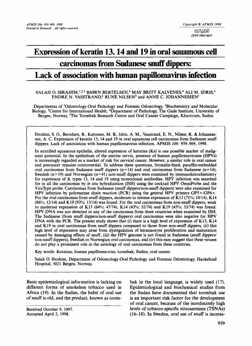
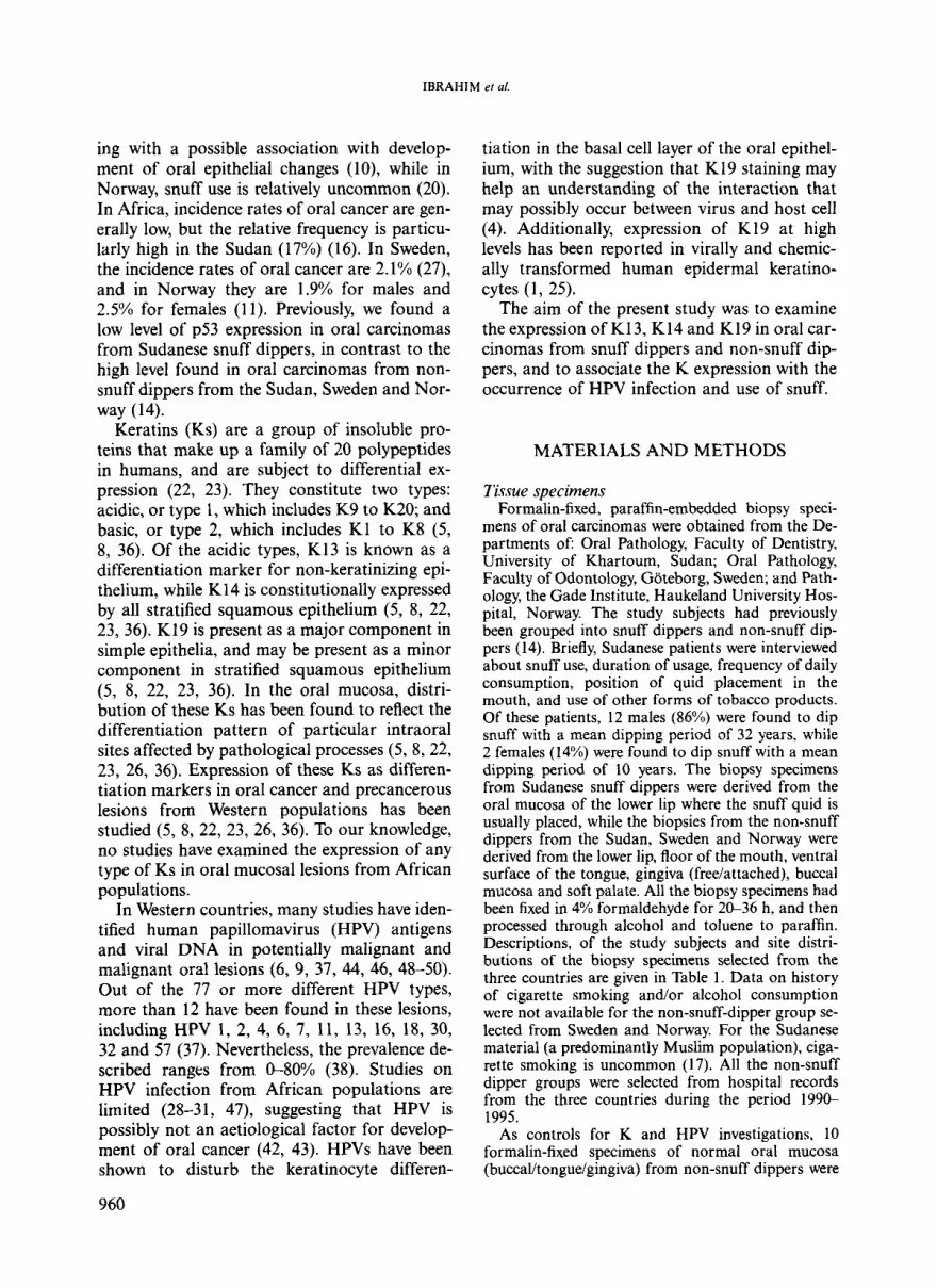
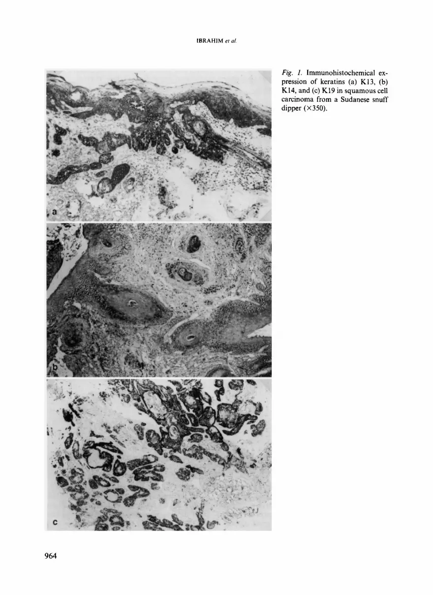
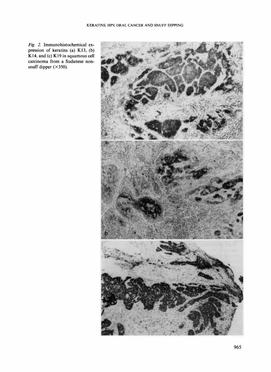
Oral carcinomas. Distribution and expression of K13, K14 and K19 found in oral carcinomas from snuff dippers and non-snuff dippers are given in Tables 3a & b. Most of the carcinomas from snuff dippers showed a wide distribution of these Ks in large areas of the specimen (Tables 3a & b; Fig. lA, B & C ) , and in the cases where basal cells of either the lining epi- thelium or that located adjacent to the infiltrat- ing tumour islands were found sporadically positive for these Ks, the positivity was uni- formly seen in the suprabasal cell layers and the infiltrating tumour islands. Oral carcinomas from non-snuff dippers showed a closely similar pattern of expression of K13, K14 and K19 (Tables 3a & b) as found in smaller areas of the specimens (Figs. 2A, B & C ) , and in the cases where basal cells of these carcinomas expressed these Ks, the expression varied in relation to that found in the suprabasal cell layers as well as the infiltrating tumour islands. Generally, the pattern of expression of K13, K14 and K19 in oral carcinomas from snuff dippers was more heterogeneous compared to that found in oral carcinomas from non-snuff dippers from the three countries.
There was a statistically significant difference in expression of K14 and K19 in oral carci- nomas from snuff dippers compared to non- snuff dippers from the Sudan as well as from Sweden/Norway (Table 3a), while for K13 ex- pression, no statistically significant differences were found. For the lip carcinomas alone, a stat- istically significant difference in expression of
KERATINS, HPV, ORAL CANCER AND SNUFF DIPPING
TABLE 3a. Expression of K13, K14 and K19, and distribution of total positive cells and staining intensity in oral squamous cell carcinomas from snuff dippers and non-snuff dippers from all three countries
Lesion *Grade of the number of positive cells and distribution of the staining intensity
Immunoreactivity
Negative Positive Basal Suprabasal Tumour n (%) n (%) layer layers islands
* Grade of the positive cells: 0 (negative); 1 (550% positive cells); 2 (>50% positive cells). Intensity of staining: (-) Negative; (+) Weak; (++) Moderate; (+++) Intense. 'p=0.006; 2p=0.02; for K14 and 3p=0.001; 'p= 0.0001; for K19 in oral carcinomas from Sudanese snuff dippers vs those from Sudanese non-snuff dippers and vs those from SwedishNorwegian non-snuff dippers, respectively.
TABLE 3b. Expression of K13, K14 and K19, and distribution of total positive cells in lip squamous cell carci- nomas from snuff dippers and those from non-snuff dippers from all three countries
Lesion Immunoreactivity *Grade of the number of positive cells and distribution of the staining intensity
Negative Positive Basal Suprabasal Tumour n (%) n (%) layer layers islands
2,++ K14 2 (17) 10 (83) 2,++ 2 ,++ K19 1 (8) 11 (92)' 2,++ 2 , + + + 2,+ + Non-snuff dippers Sudan, Sweden and Norway (n=36) K13 15 (42) 21 (58) 2, + 1,++ 1,++ K14 16 (44) 20 (56) 1,++ 2, + 1,++ K19 15 (42) 21 (58)' 2, + 1,++ 1,++ * Grade of the positive cells: 0 (negative); 1 (550% positive cells); 2 (>50% positive cells). Intensity of staining: (-) Negative; (+) Weak; (+ +) Moderate; (+ + +) Intense. p=0.04 for K19 in lip carcinomas from Sudanese snuff dippers vs those from non-snuff dippers from all three countries.
K19 was only found between lip carcinomas from Sudanese snuff dippers and those from non-snuff dippers from all the three countries (Table 3b). Owing to the low number of intra- oral carcinomas from Sudanese snuff dippers (n=2), no separate statistical analysis was per- formed.
ISH and PCR for HPV infection All carcinomas were negative for HPV DNA
by ISH. All the Sudanese oral carcinomas (from snuff dipperdnon-snuff dippers) were negative for HPV DNA with the PCR technique. 8/10 genital condylomas, 3/5 carcinomas of the uter- ine cervix and 5/10 oral papillomas from non-
963
IBRAHIM et al.
Fig. 1. Immunohistochemical ex- pression of keratins (a) K13, (b) K14, and (c) K19 in squamous cell carcinoma from a Sudanese snuff dipper (X 350).
964
KERATINS, HPV, ORAL CANCER AND SNUFF DIPPING
Fig. 2. Immunohistochemical ex- pression of keratins (a) K13, (b) K14, and (c) K19 in squamous cell carcinoma from a Sudanese non- snuff dipper (X 350).
965
IBRAHIM et al,
snuff dippers from Norway were positive for HPV DNA with the ISH, and showed a strong bond for both GP5+/GP6+ primers with the PCR. All the control normal oral mucosa was negative for HPV DNA with the ISH and the PCR.
DISCUSSION
In the present study, we found moderate to in- tense expression of K13, K14 and K19 in oral carcinomas from Sudanese snuff dippers com- pared to weak and moderate expression in oral carcinomas from non-snuff dippers. These find- ings validate the use of these Ks as markers for following the development, differentiation, de- differentiation and malignant transformation in oral epithelial lesions from Sudanese snuff dip- pers. Expression of Ks in carcinomas from snuff dippers perhaps arises from dysregulation of keratinocyte proliferation and maturation, probably induced by the physical action caused by continuous all day application of snuff on the oral mucosa, or by the chemical action of the TSNAs thought to be involved in the devel- opment of these cancers (13, 18). The present study was performed on formalin-fixed, paraf- fin-embedded tissues investigated by immuno- histochemistry with no technical differences or factors present between the Sudanese, Swedish and Norwegian materials used. It therefore sup- ports other studies which have suggested the use of formalin-fixed, paraffin-embedded tissues and showed expression of K (2, 32, 40, 41).
High prevalence of oral leukoplakia in any community may be indicative of a strong predis- position to oral cancer (33, 35). Snuff dipping and cigarette smoking were found to correlate strongly with the presence of oral leukoplakia that showed an altered pattern of K expression (33, 3 9 , and expression of K4 was found to in- crease in association with cigarette smoking in these lesions (34). In the Sudan, the snuff quid is usually placed on the mucosal surface of the lower lip (non-keratinized) (17). Our findings showed that K13, K14 and K19 were commonly expressed in carcinomas from Sudanese snuff dippers compared to those from non-snuff dip- pers from the three countries. This may indicate that TSNAs stimulate keratinocytes to undergo squamous dedifferentiation, and thus these Ks
966
may represent potential markers for early diag- nosis, prognosis or evaluation of effects of treat- ment in potentially malignant and malignant oral lesions from Sudanese snuff dippers. Inter- estingly, distribution of expression of K19 in carcinomas has been found to show remarkable heterogeneity even within the individual tu- mours, with the suggestion of its possible use in typing and subtyping of tumours (22, 24).
Results of HPV investigations in oral lesions using dot blot hybridization, ISH and PCR techniques are inconsistent (6, 9, 37, 44, 46, 48- 50), making it difficult to interpret the preva- lence of HPV in the development of these lesions. Although ISH and PCR are the most powerful techniques for investigating HPV DNA, we could not detect HPV DNA in any of the Sudanese carcinomas. These results, which are in line with other reports from African populations (42, 43), suggest that these viruses are of limited importance in oral squamous cell carcinogenesis of oral carcinomas in the Sud- anese population. HPVs might disturb the kera- tinocyte differentiation in the basal cell layer of oral tissues in relation to expression of K19 (4), and expression of this keratin at high levels has been described in virally and chemically trans- formed human epidermal keratinocytes (1, 25). It has also been indicated that HPV-16 E6 and E7 oncogenes are mutagenic in human oral keratinocytes (2 1). In the present study, express- ion of K19 was accompanied by absence of HPV infection.
The low level of p53 expression found in the same carcinomas from Sudanese snuff dippers (14) contradicts the high level of Ks expression found in the present study. It has been suggested that p53 degradation by the HPV E6 protein (45) or formation of complexes of the p53 pro- tein and other cytoplasmic proteins ( I 2) may ex- plain the low level of p53 expression in human neoplasms. The low level of p53 expression found in the carcinomas from Sudanese snuff dippers may be due to formation of cytoplasmic complexes of the p53 protein and the Ks ex- pressed as suggested earlier (12).
In conclusion, the present study suggests: (i) the high prevalence of expression of K13, K14 and K19 found in carcinomas from snuff dip- pers may arise from dysregulation of keratino- cyte proliferation and maturation caused by the damaging effects of snuff, (ii) the presence of
KERATINS, HPV, ORAL CANCER AND SNUFF DIPPING
high levels of TSNAs in the type of snuff used in the Sudan as well as the high incidence of expression of Ks demands further studies to elucidate if the relationship is of aetiological sig- nificance regarding the interaction between the TSNAs and host cells, and (iii) HPVs may not play a prominent role in the aetiology of oral carcinomas from the Sudan.
This study was supported by the Colgates Forsknings- fond, Colgate-Palmolive, Norway NS. We thank Pro- fessor B. Magnusson for providing the Swedish ma- terial from non-snuff dippers. We are grateful to G. Albrektsen for advice on statistical analysis and G. Qijordsbakken and G. Fjell for their skilled technical assistance. We thank Tor Christensen for taking the photographs.
REFERENCES
1. Banks-Schlege, S. & Rhim, J. S.: Keratin express- ion of both chemically and virally transformed human epidermal keratinocytes during the pro- cess of neoplastic conversion. Carcinogenesis 7:
2. Battifora, H. & Kopinski, M.: The influence of protease digestion and duration of fixation on the immunostaining of keratins. A comparison of formalin and ethanol fixation. J. Histochem. Cytochem. 34: 1095-1 100, 1986.
3. Bertelsen, B.. Kalvenes, M.-B. & Harveit, F.: Hu- man papillomavirus infection in progressive and non-progressive cervical intraepithelial neo- plasia. APMIS 104: 900-906, 1996.
4. Chang, F., Syrjanen, S., Nuutinen, J.. Karja, J. & Syrjunen, K.: Detection of human papillomavirus DNA in oral squamous cell carcinomas by in situ hybridization and polymerase chain reaction. Arch. Dermatol. Res. 282: 493497, 1990.
5 . Clausen, H.. Moe, D. , Buscard, D. & Dabelsteen, E.: Keratin proteins in human oral mucosa. J. Oral Pathol. 15: 3642, 1986b.
6. Dekmezian, R. P.. Batsakis, J. G. & Goepfert, H.: In situ hybridization of papillomavirus DNA in head and neck squamous carcinomas. Arch. Oto- laryngol. Head Neck Surg. 113: 8 19-82 1, 1987.
7. de Roda Husinan, A.-M., Walboomers, J. M. M. , van den Brule, A. J. C., Meijer, C. J. L. M. & Snijders, P. J. E: The use of general primers GP5 and GP6 elongated at their 3’ ends with adjacent highly conserved sequences improves human papillomavirus detection by PCR. J. Gen. Virol.
8. Eichner, R., Bonitz, P. & Sun, T.-T: Classifi- cation of epidermal keratins according to their immunoreactivity, isoelectric point and mode of expression. J. Cell Biol. 98: 1388-1396, 1984.
153-157, 1986.
76: 1057-1062, 1995.
9. Greer, R. 0. Jr., Douglas, J. M . Jr., Breese, P. & Crosby, L. K.: Evaluation of oral and laryngeal specimens for human papillomavirus (HPV) DNA by dot blot hybridization. J. Oral Pathol. Med. 19: 35-38, 1990.
10. Hirsch, J.-M. & Johansson, S. L.: Effect of long- term application of snuff on the oral mucosa - an experimental study in the rat. J. Oral. Pathol. 12: 187-198, 1983.
11. Hakulinen, T., Andersen, A. A., Malker. B., Pu- kala, E., Schou, G. & Tulinius, H. : Trends in can- cer incidence in the Nordic countries. Acta path. microbiol. immunol. scand. A. 94 (Suppl. 288):
2. Harlow. E., Williamson. N. M. , Ralston, R. , Helf- man, D. M. & Adams, T. E.: Molecular cloning and in vitro expression of a cDNA clone for hu- man cellular tumour antigen p53. Moll. Cell Biol. 5: 1601-1610, 1985.
3. Hecht, S. S. & Hoffmann, D.: The relevance of tobacco-specific nitrosamines to human cancer. Cancer Surv. 8: 273-294, 1989.
4. Ibrahim, S. O., Johannessen, A. C. Idris, A . M. , Hirsch. J.-M., Vasstrand, E. N., Magnusson, B. & Nilsen, R.: Immunohistochemical detection of p53 in non-malignant and malignant oral lesions associated with snuff dipping in the Sudan and Sweden. Int. J. Cancer 68: 749-753, 1996.
5. Ibrahim, S. O., Johannessen, A. C., Vasstrand, E. N., Lillehaug, J. R. & Nilsen, R.: Immunohisto- chemical detection of p53 in archival formalin- fixed tissues of lip and intraoral squamous cell carcinomas from Norway. APMIS 105: 757-764, 1997.
16. Idris, A. M. , Ahmed, H. M. & Malik, M. 0. A.: Toombak dipping and cancer of the oral cavity in the Sudan; a case control study. Int. J. Cancer 63: 477-480, 1995.
17. Idris, A. M. , Prokopczyk, B. & Hoffmann, D.: Toombak: a major risk factor for cancer of the oral cavity in Sudan. Prev. Med. 23: 832-839, 1994.
18. Idris, A . M. , Nair, J., Ohshima, H.. Friesen, M. , Brouet, I., Faustman, E. M . & Bartsch, H.: Un- usually high levels of carcinogenic tobacco-speci- fic nitrosamines in Sudan snuff (toombak). Car- cinogenesis 12: l 115-1 118, 1991.
19. IARC Monographs on the Evaluation of the Carcinogenic Risk of Chemicals to Humans. Vol. 37. Tobacco habits other than smoking; betel quid and areca nut chewing; and some related nitrosamines. Lyon: International Agency for Research on Cancer, 1985.
20. Kraft, P. & Svendsen, T . : Rerykevaner og bruk av snus i Norge 1973-95. Tidsskr. Nor. Lregeforen. 5: 629-634, 1996.
21. Liu, X. , Hun, S., Baluda, M . A. & Park, N . H.: HPV-16 oncogenes E6 and E7 are mutagenic in normal oral keratinocytes. Oncogene 14: 2347- 2353, 1997.
1-151, 1986.
967
IBRAHIM et al.
22. Moll, R., Franke, W. W., Schiller, D. L., Greiger, B. & Krepler, R.: The catalogue of human cyto- keratins: Patterns of expression in normal epi- thelia, tumors and cultured cells. Cell 31: 1-24, 1982.
23. Moll, R., Schiller, D. L. & Franke, W. W.: Identi- fication of protein IT of the intestinal cytoskel- eton as a novel type 1 cytokeratin with unusual properties and expression patterns. J. Cell. Biol.
24. Moll, R., Krepler, R. & Franke, W. W.: Complex cytokeratin polypeptide patterns observed in cer- tain human carcinomas. Differentiation 23: 256- 269, 1983.
25. Morris, A., Steinberg, M. L. & Defendi, V.: Kera- tin gene expression in simian virus 40-transform- ed human keratinocytes. Proc. Natl. Acad. Sci. USA: 82: 8498-8502, 1985.
26, Morgan, P. R., Leigh, I. M., Purkis, P. E., Gardner, I. D., Van Muijen, G. N. P. & Lane, E. B.: Site variation in keratin expression in human oral epithelia - An immunocytochemical study of individual keratins. Epithelia I: 3143, 1987.
27. Ostman, J., Anneroth, G.. Gustafsson, H. & Tav- elin, B.: Malignant oral tumours in Sweden 1960-1984 - an epidemiological study. Eur. J. Cancer B. Oral Oncol. 3ZB: 106-1 12, 1995.
28. Padayachee, A., Sanders, C. M. & Maitland, N. J.: A polymerase chain reaction (PCR) investiga- tion of oral verrucae which contain HPV types 2 and 57 by in situ hybridization. J. Oral Pathol. Med. 24: 329-334, 1995.
29. Padayachee, A.: Human papillomavirus (HPV) types 2 and 57 in oral verrucae demonstrated by in situ hybridization. J. Oral Pathol. Med. 23: 413417, 1994.
30. Padayachee, A. & van- Wyk, C. W.: Human pap- illomavirus (HPV) DNA in focal epithelial hy- perplasia by in situ hybridization. J. Oral Pathol. Med. 20: 210-214, 1991
3 1. Padayachee, A. & van- Wyk, C. W. : Human pap- illomavirus (HPV) in oral squamous cell papil- lomas. J. Oral Pathol. 16: 353-355, 1987.
32. Pinkus, G. S., Etheridge, C. L. & O’Connor, E. M.: Are keratin proteins a better tumor marker than epithelial membrane antigen? A compara- tive immunohistochemical study of various par- affin-embedded neoplasms using monoclonal and polyclonal antibodies. Am. J. Clin. Pathol.
33. Pindborg, J. J.: Oral cancer and precancer, John Wright and Sons Ltd., Bristol, 1980.
34. Pritlove-Carson, S, , Charlesworth, S., Morgan, P. R. & Palmer, R. M.: Cytokeratin phenotypes at the dento-gingival junction in relative health and inflammation, in smokers and non-smokers. Oral Dis. 3: 19-24, 1997.
35. Reibel, J., Kenrad, B. & Schwartz, 0.: Architec- tural organisation of oral epithelium as visual-
111: 567-580, 1990.
85: 269-277, 1986.
968
ised by keratin staining pattern in tobacco-as- sociated leukoplakias. J. Oral Pathol. Med. 20: 265-270, 1991.
36. Sun, T. -T., Eichner, R., Schemer, A., Cooper, D., Nelson, W. G. & Weiss, A.: Classification, ex- pression, and possible mechanisms of evolution of mammalian epithelial keratins: a unifying model. In: Levine, A. J., Van de Woude, G. F., Topp, W. C. & Watson, J. D. (Eds.): Cancer cells. The transformed phenotype. Cold Spring Harb- or Laboratory, New York 169: 1984.
37. Snijders, P. J. F., Van Den Brule, A. J. C., Meij- er, C. J. L. M. & Walboomers, J. M. M.: Papil- lomaviruses and cancer of the upper digestive and respiratory tracts. Cum. Topics Microbiol. Immunol. 186: 177-198, 1994.
38. Sugerman, P. B. & Shillitoe, E. J.: The high risk human papillomaviruses and oral cancer: evi- dence for and against a causal relationship. Oral Dis. 3: 130-147, 1997.
39. Shibata, K.: The polymerase chain reactioil and the molecular genetic analysis of tissue biopsies. In: Diagnostic Molecular Pathology, a Practical Approach, Vol. 11. Herrrington, C. S. & McGee, J. O’D. (Eds.). Oxford University Press, Oxford, New York, Tokyo, pp. 85-1 11, 1992.
40. Takahashi, H., Shikata, N., Senzaki, H., Shin- taku, M. & Tsubura, A.: Immunohistochemical staining patterns of keratins in normal oeso- phageal epithelium and carcinoma of the oeso- phagus. Histopathology 26: 45-50, 1995.
41. Tsubura, A., Okada, H., Sasaki, M., Dairkee, S. H. & Morii, S.: Immunohistochemical demon- stration of keratins 8 and 14 in benign tumours of the skin appendage. Virchows Arch. A Pathol. Anat. Histopathol. 418: 503-507, 1991.
42. Van-Rensburg, E. J., Van-Heerden, W. F., Ven- ter, E. H. & Raubenheimer, E. J.: Detection of human papillomavirus DNA with in situ hybridi- zation in oral squamous carcinoma in a rural black population. S. Afr. Med. J. 85: 894-896, 1995.
43. Van-Rensburg, E. J., Engelbrecht, S. , Van-Heerd- en, W. F., Raubenheimer, E. J. & Schoub, B. D.: Human papillomavirus DNA in oral squamous cell carcinomas from an African population sample. Anticancer Res. 16: 969-973, 1996.
44. Villiers, E. M. de.: Heterogeneity of the human papillomavirus group. J. Virol. 63: 4898-4903, 1989.
45. Vousden, K. H.: Interactions between papil- lomavirus proteins and tumour suppressor gene products. Adv. Cancer Res. 64: 1-24, 1994.
46. Watts, S. L., Brewer, E. E. & Fry, T. L.: Human papillomavirus DNA types in squamous cell car- cinomas of the head and neck. Oral Surg. Oral Med. Oral Pathol. 71: 701-707, 1991.
47. Williamson, A. L., Jaskiesicz, K. & Gunning, A.: The detection of human papillomavirus in oeso-
Schantz, S. P . & Adler-Storthz, K. : Analysis of human papillomavirus DNA in oral squamous cell carcinomas. J. Oral Pathol. Med. 22: 101- 108, 1993.
49. Young, S. K. & Min, K. W.: In situ DNA hybridi-
zation analysis of oral papillomas, leukoplakias, and carcinomas for human papillomavirus. Oral
48. Woods, K. V., Shillitoe, E. J., Spitz, M. R., Surg. Oral Med. Oral Pathol. 71: 726-729, 1991. 50. Zeuss, M . S., Miller, C. S. & White, D. K.: In situ
hybridization analysis of human papillomavirus DNA in oral mucosal lesions. Oral Surg. Oral Med. Oral Pathol. 71: 714-720, 1991.