Cellular Immune Responses to Human Papillomavirus (HPV) type 16 at the Cervix of Women with HPV-Associated Squamous Intraepithelial Neoplasia By Michelle Milner, BSc (Hons) This Dissertation is submitted in fulfillment of the requirements for the degree ofMSc (Medicine) in the Division of Medical Virology, Institute of Infectious Diseases and Molecular Medicine' January 2005 i
Transcript
Cellular Immune Responses to Human
Papillomavirus (HPV) type 16 at the Cervix of
Women with HPV-Associated Squamous
Intraepithelial Neoplasia
By
Michelle Milner, BSc (Hons)
This Dissertation is submitted in fulfillment of the requirements
for the degree ofMSc (Medicine) in the Division of Medical Virology,
Institute of Infectious Diseases and Molecular Medicine'
January 2005
i
The copyright of this thesis vests in the author. No quotation from it or information derived from it is to be published without full acknowledgement of the source. The thesis is to be used for private study or non-commercial research purposes only.
Published by the University of Cape Town (UCT) in terms of the non-exclusive license granted to UCT by the author.
Declaration
I, Michelle Milner, hereby declare that the work on which this thesis is based is
original (except where acknowledgements indicate otherwise) and that neither the
whole work, nor any part thereof, has been, is being or is to be submitted for another
degree in this or any other university.
I empower the university to reproduce for the purpose of research either the whole or
any portion of the contents in any manner whatsoever.
Name: (V\1 'U'\.t,{J..t �· �
ii
Index to Tables Index to Figures Abbreviations List Acknowledgements Abstract
CHAPrERONE
Contents Page
vii viii x xi xii
1.1 The Prevalence of Cervical Cancer in the Female Population 1 1.2 Progression from CIN to Cancer of the Cervix 1 1.3 The Biological Structure of the Cervix 3 1.4 Role of HPV in Cervical Cancer 5 1.5 HPV: Properties and Characteristics 6 1.5.1 Ll 8 1.5.2 E6 and E7 9 1.5.3 Diversity of HPV Genotypes 10 1.5.4 HPV: Infection Mechanism and Viral Lifestyle 11 1.6 Immune Responses to HPV Infection 14 1.6.1 Antibodies: the mediators of Humoral Immunity 14 1.6.2 Cell Mediated Immunity at the Female Reproductive Tract 15 1.6.2.1 Antigen Presentation at the Cervix and the Cells Facilitating the Process
16 1.6.2.2 T cell Responses to HPV Infection 18 1.6.2.3 Cytotoxic T cell responses to HPV antigens 19 1.6.2.4 Proliferation of T cells to HPV antigens 20 1.6.2.5 Cervical Cancer Patients Have Impaired Immune Responses 22 1.6.2.6 CD4 Responses in HPV Immunity: the Thl versus Th2 Paradigm 22 1.6.2.7 Impact of HIV Infection on Progression of HPV associated CIN 23 1.6.2.8 Cytokines Play an Integral Role in the Progress of HPV Infection 24 1.6.2.9 Conclusion of T cell Mediated Immunity to HPV Infection 27 1.7 Objectives of Project 28 1.7.1 Development of Techniques for Investigating Cervical T cell Responses
29 1.7.2 Determination of cervical versus peripheral blood T cell intracellular
cytokine responses to HPV16 speciftc antigens 29 1.7.3 Determination of the cytotoxic ability of HPV specific T cells 30
CHAPrERTwo 2.1 Introduction 31 2.2 Materials and Methods 33 2.2.1 Collection of cervical specimens using a Digene Cervical cytobrush
sampler 33 2.2.2 Isolation of cervical T cells from cytobrush specimens 33 2.2.3 CD3+ Screen to accurately determine the quantity of T cells in specimens
34 2.2.4 Determination of Red Blood Cell (RBC) contamination of cervical
specimens 35 2.2.4.1 Sensitivity of CD235 staining for use on cervical specimens 36
iii
2.2.5 Trypan Counting method using haemcytometer 37 2.2.6 7 AAD Staining to measure cell viability of cervical cells by FA CS analysis
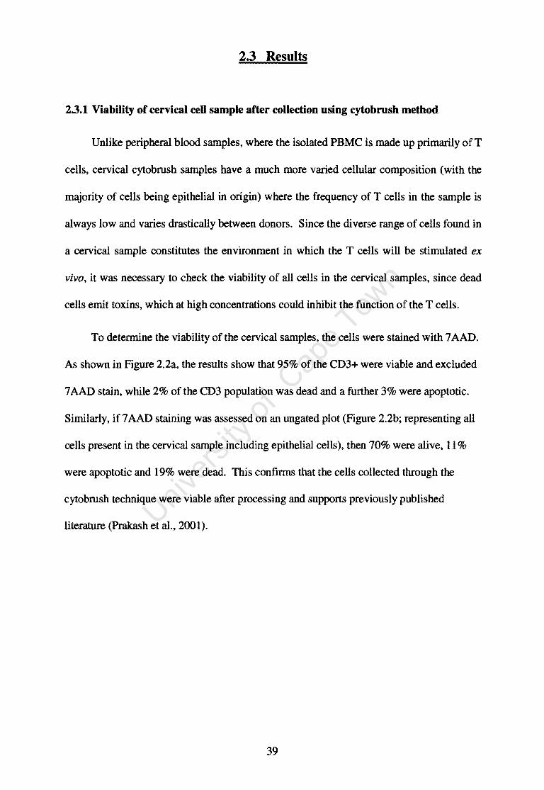
37 2.3 Results 39 2.3.1 ViabHlty of cervical cell sample after collection using cytobrush method
39 2.3.2 Quantity of CD3+ cells obtained using cytobrush technique 2.3.2.1 Reliability of Various Counting Methods 2.3.2.2 Validity of cervical cellular sample size for further use in statistical analyses 2.4 Conclusion
CHAFfER THREE
40 40
44 46
3.1 Introduction 48 3.2 Materials and Methods 51 3.2.1 Study Population 51 3.2.2 Procedures for Processing of Donor Samples 51 3.2.2.1 Extraction of Serum from Clotted Peripheral Blood Sample 52 3.2.2.2 Isolation of PBMC from Anti-Coagulated Peripheral Blood Sample 52 3.2.2.3 Collection and Processing of Cervical Cells from Cervi-Brush Sample 53 3.2.2.4 Stimulation of cervical and peripheral blood T cells with HPV-16 Ll and
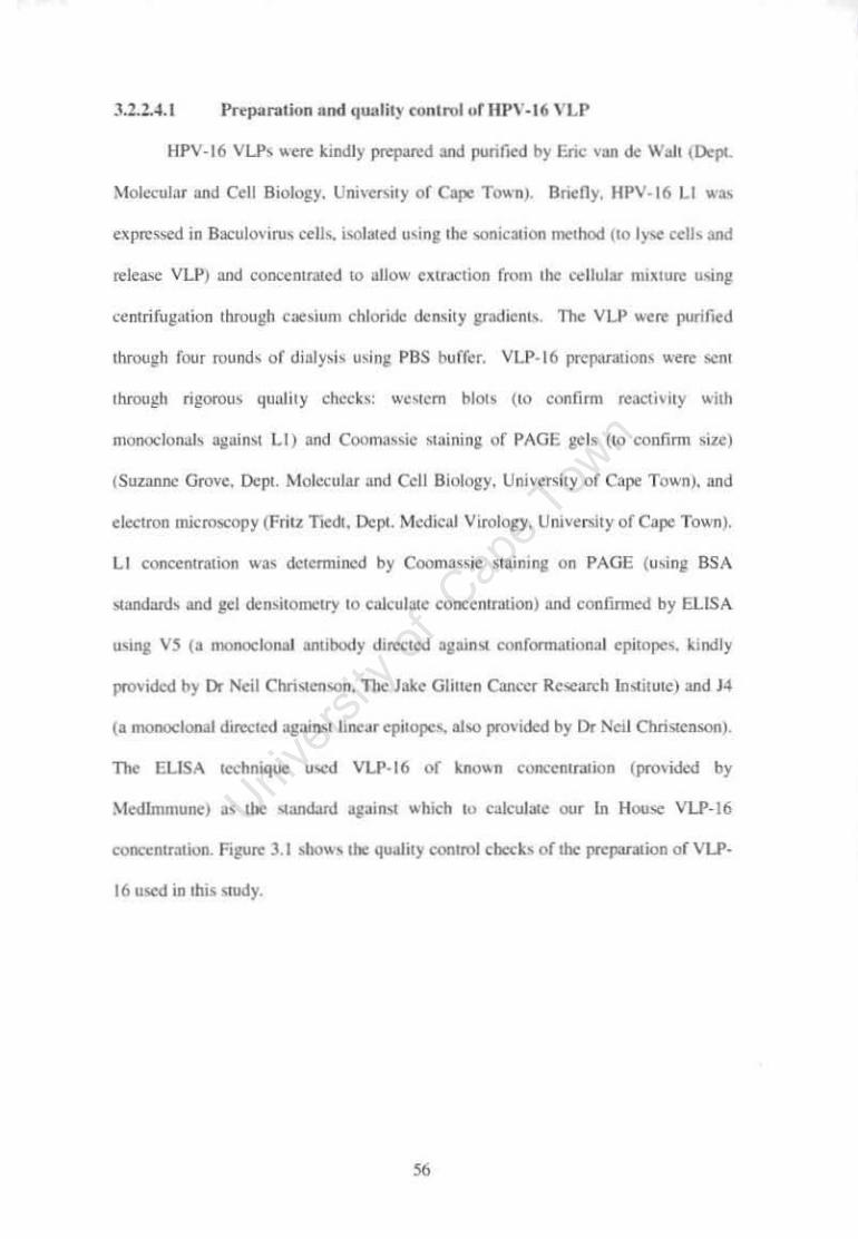
E7 antigens 55 3.2.2.4.1 Preparation and quality control of HPV -16 VLP 56 3.2.2.4.2 Preparation and Purification ofHPV-16 E7 57 3.2.2.4.3 Stimulation of cervical and PBMC-derived T cells with Ll and E7
58 3.2.2.5 Staining of stimulated cell populations 59 3.2.2.6 Digene Cylobrush for evaluating HPV DNA infection, HPV typing and
relative viral load 60 3.2.3 Testing for active cervical HPV infection, HPV typing and Viral Load
Determination 60 3.2.3.1 Digene Hybrid Capture@ II HPV Test 61 3.2.3.2 HPV Consensus PCR and Genotyping utilising Reverse Line Blots 62 3.2.4 Enzyme Linked Immunosorbent Assays (ELISA) to Assess HPV -16
specific antibody (lgG) reactivity to HPV VLPs 66 3.2.5 CBA Bead Kit to test for Inflammation at the Cervix 68 3.2.6 Statistical Analysis 70 3.3 Results 71 3.3.1 DesCription of women with varying grades of CIN attending the Groote
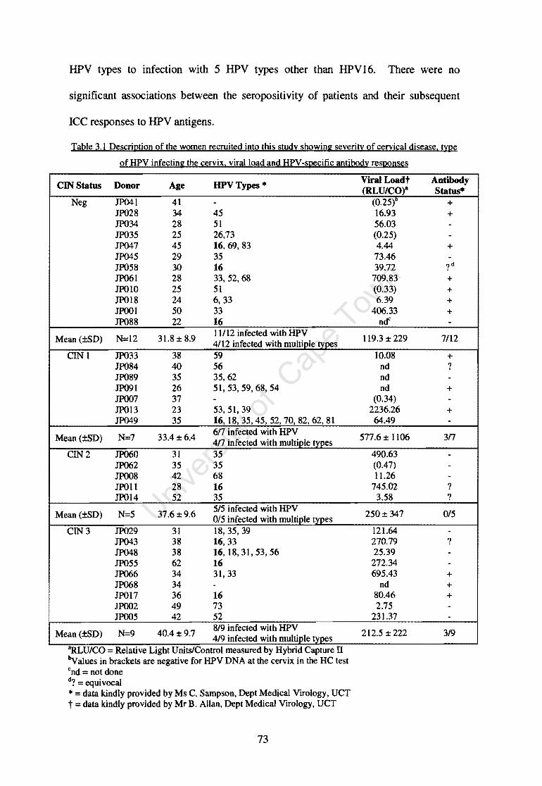
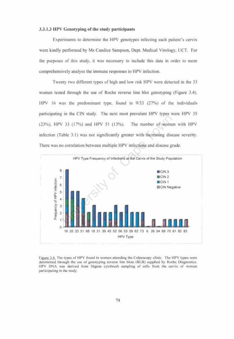
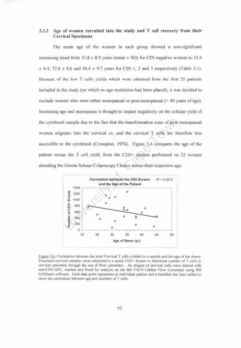
Schuur Hospital Outpatients Colposcopy clinic 71 3.3.1.1 CIN status of the study participants 71 3.3.1.1 Antibody Seropositivity of the study participants 72 3.3.1.3 HPV Genotyping of the study participants 74 3.3.1.4 Correlation of cervical disease severity with HPV Viral Load 75 3.3.2 Age of women recruited into the study and T cell recovery from their
Cervical Specimens 77
iv
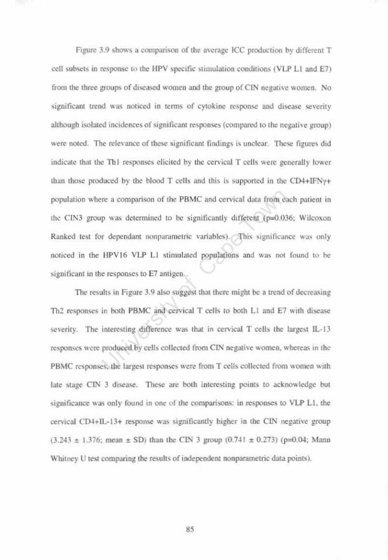
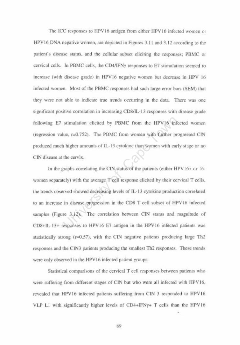
3.3.2 Intracellular Cytokine (IFN-yand IL-13) Production Following Stimulation of Cervical and Peripheral T cells using HPV16 Specific Antigens 78
3.3.2.1 Individual Patients ICC Responses 78 3.3.2.2 Comparison of ICC Responses from all study participants according to
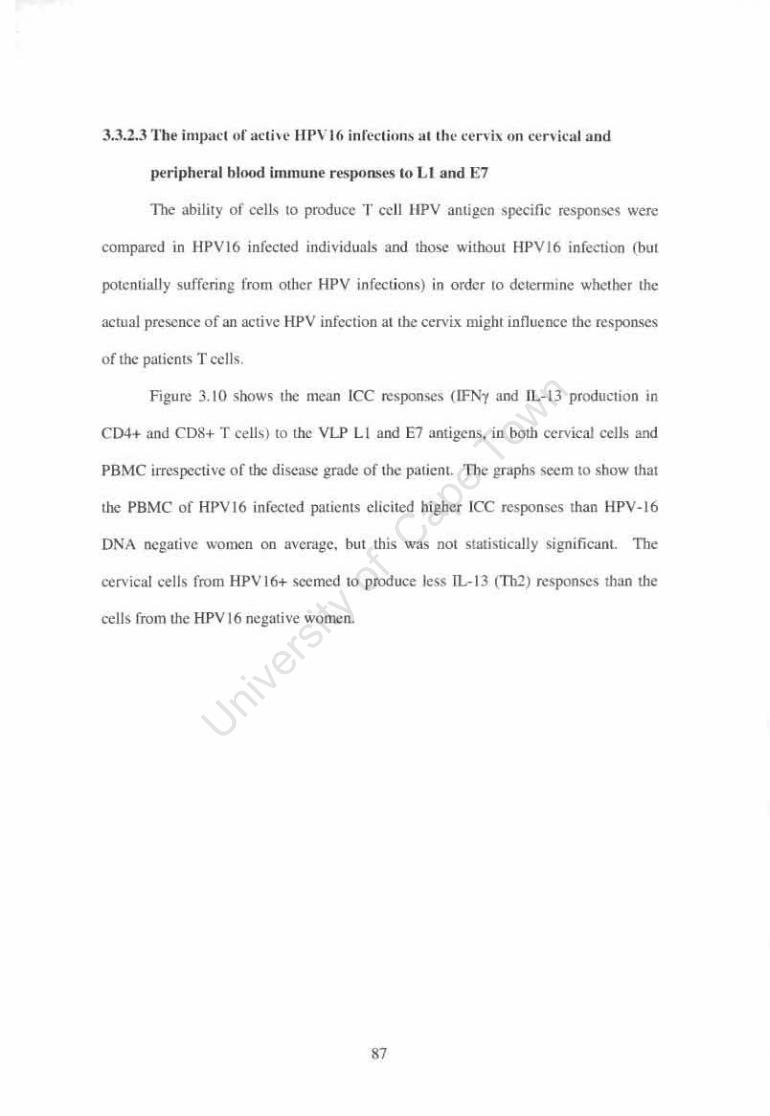
disease grade 83 3.3.2.3 The impact of active HPV16 infections at the cervix on cervical and
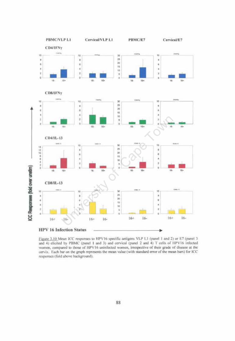
peripheral blood immune responses to Ll and E7 87 3.3.4 Correlation between HPV Viral Load and cytokine response at the cervix
and systemically 93 3.3.5 Thl versus Th2 responses in the blood versus at the cervix to HPV
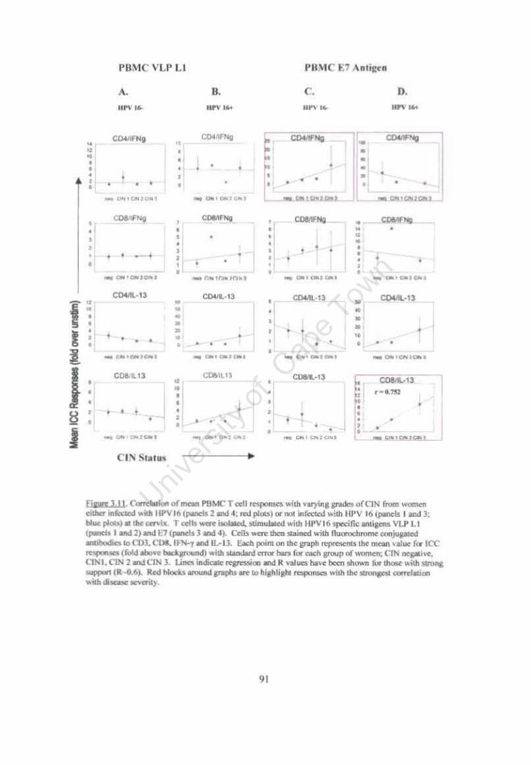
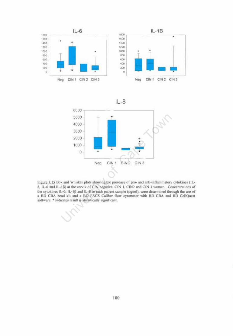
antigens Ll and E7 95 3.3.7 The cervical cytokine microenvironment of HPV infected women 98 3.4 Discussion 101 3.4.1 Does age correlate with a decrease in T cells in the cervical epithelium?
102 3.4.2 Lack of HPV16 Prevalence in Study Population 103 3.4.3 Type of cytokine microenvironment at the cervix 104 3.4.4 Individual ICC Responses of four interesting patients 106 3.4.5 Women with CIN 1 consistently showed the strongest responses to HPV
antigens 107 3.4.6 Trends in T helper responses between patients with varying grades of
cervical disease 108 3.4.7 Impact of HPV·16 infection on local and systemic T cell responses 110 3.4.8 Effect of HPV viral load on T cell responses at the cervix 112 3.4.9 Summary of the cellular immune environment in response to HPV
infection at the cervix of women with varying grades of CIN 113
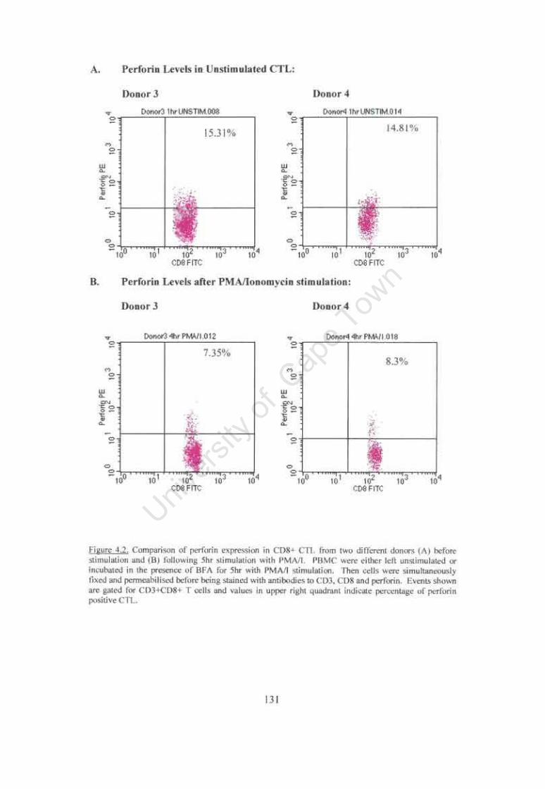
CnAYI'ER FOUR 4.1 Introduction 115 4.2 Materials and Methods 120 4.2.1 Isolation of PBMC 120 4.2.2 Analysis of Perforln as a marker of cytotoxic activity 121 4.2.2.1 Stimulation of PBMC to determine constitutive and post·stimulation
levels of perforin 121 4.2.2.2 Intracellular Staining for Perforin 121 4.2.3 Staining of PBMC for CDI07a expression 122 4.2.3.1 Intracellular staining for CDI07a 122 4.2.3.2 Kinetics of cell surface CDI07a versus intracellular Perf orin expression
following stimulation 123 4.2.3.3 Comparison of PHA, PMA/Ionomycln and SED-stimulation for induction
of CDI07a expression 124 4.2.4 HPV·16 Ll and E7 specific cytotoxicity (CDI07a) Assay on PBMC from
women with HPV ·associated CIN 126 4.2.4.1 Colposcopy Clinic Study Population 126 4.2.4.2 HPV Typing using Roche Reverse Line Blots 127 4.2.4.3 CDI07a Cytotoxicity Assay following stimulation with HPV .. 16 Ll and E7
127
v
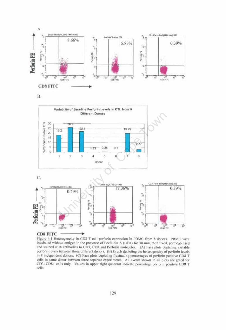
4.2.5 Statistical analysis 127 4.2 Results 128 4.2.1 Heterogeneity in the level of intracellular Perforin expression in CTLs
from ditTerent donors 128 4.2.2 Impact of T cell activation and degranulation on intracellular Perf orin
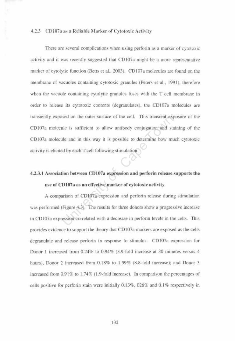
expression 130 4.2.3 C0107a as a Reliable Marker of Cytotoxic Activity 132 4.2.3.1 Association between C0107a expression and perforin release supports the
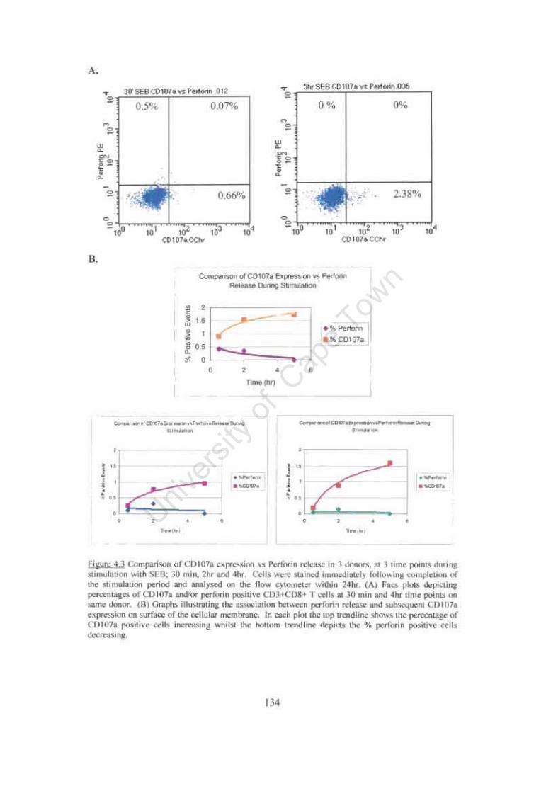
use of C0107a as an etTective marker of cytotoxic activity 132 4.2.3.2 Inter·assay reproducibility of C0107a as a marker of degranulation 135 4.2.4 Comparison of PHA, SED and PMAlIonomycin as agents to induce
maximal C0107a expression on activated T cells 136 4.2.5 C0107a expression following stimulation with HPV·16 L1 and E7 Antigen
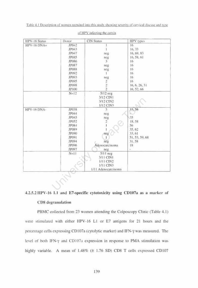
by PDMC from patients with cervical disease 138 4.2.5.1 Description of women with HPV ·associated cervical disease enrolled in
this study 138 4.2.5.2 Hpv·16 L1 and E7.specific cytotoxicity using C0107a as a marker of
C08 degranulation 139
4.3 Discussion 148
CHAPTER FIVE 5.1 Overall Objectives of this Project 155 5.2 Development of Methods to Process Cervical Samples 156 5.3 Analysis of HPV .specific cervical and peripheral blood T cell Responses
by intracellular cytokine staining and flow cytometry 157 5.4 Markers of HPV·16 specific Cytolytic Activity by peripheral blood T
cells: C0107a versus perf orin 161 5.5 Future Considerations for the analysis of HPV·specific cervical T cell
responses 163
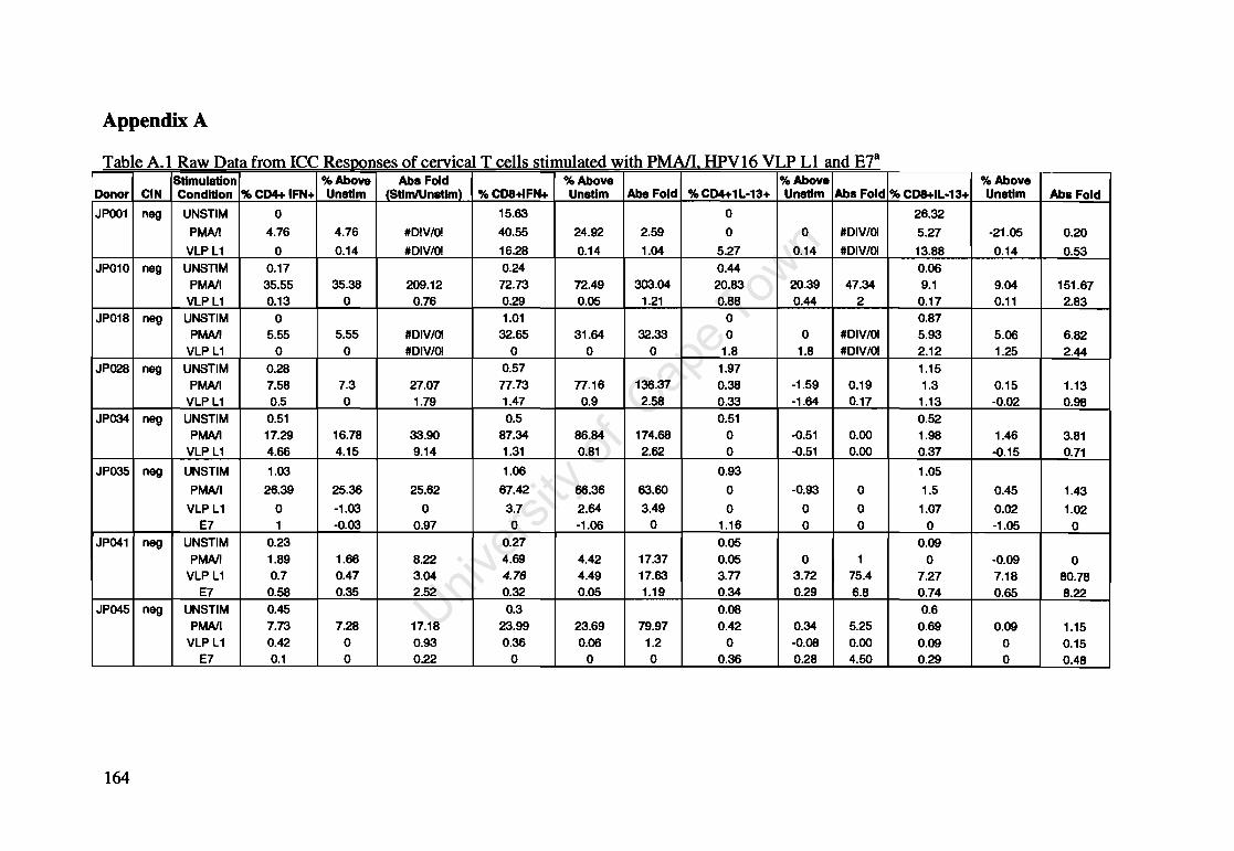
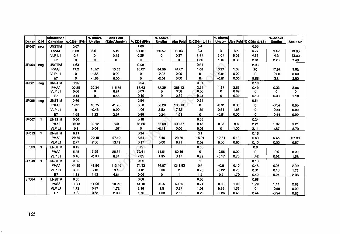
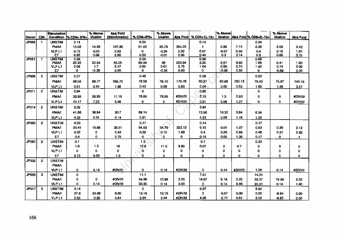
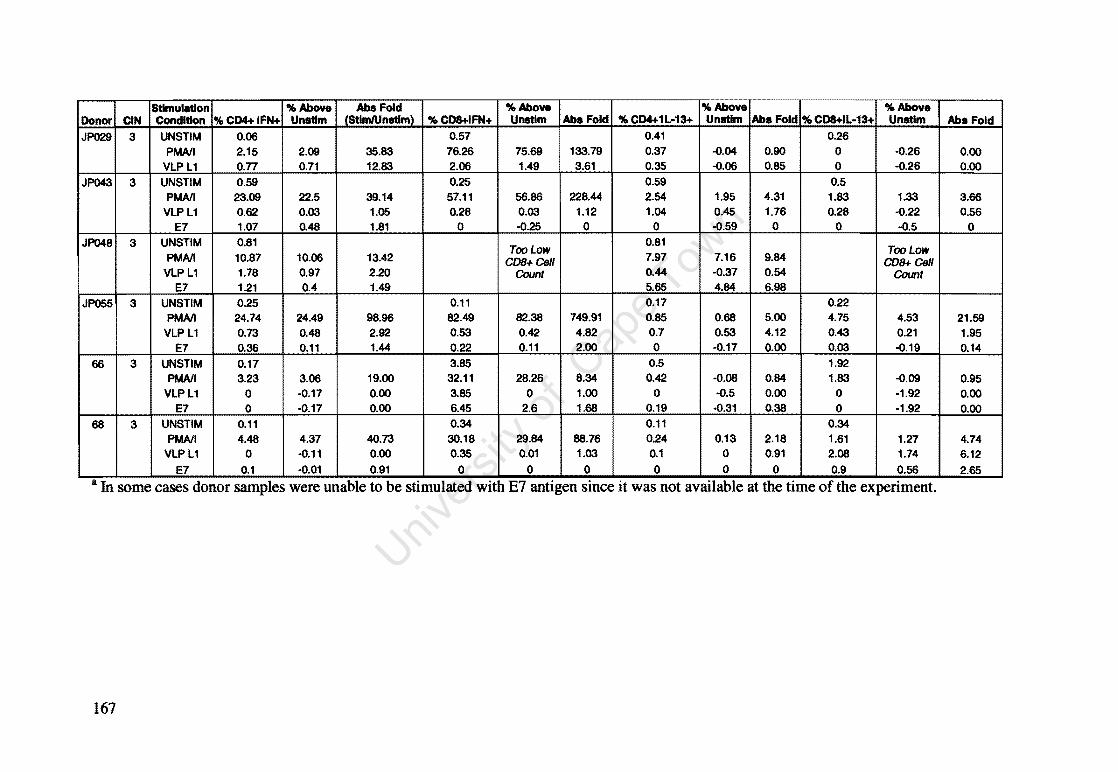
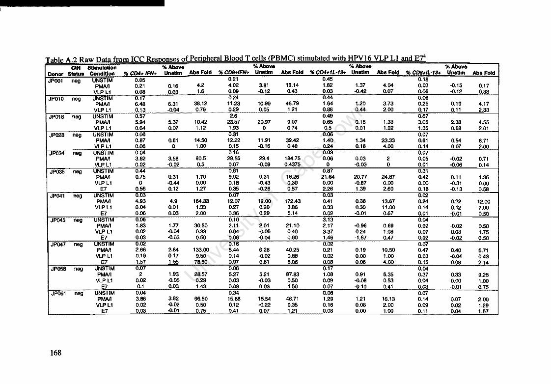
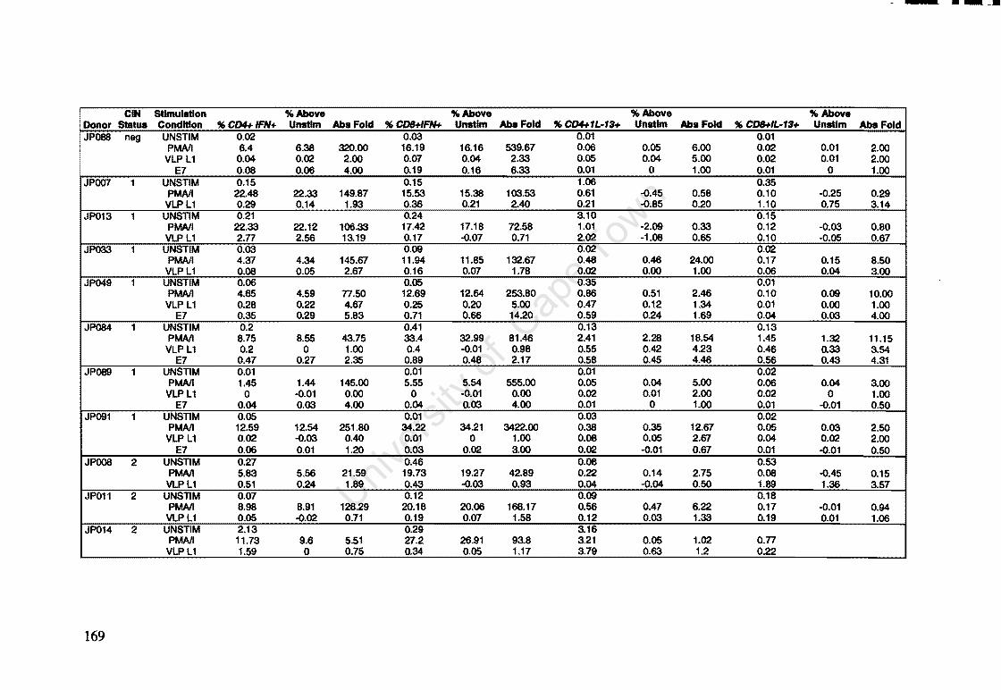
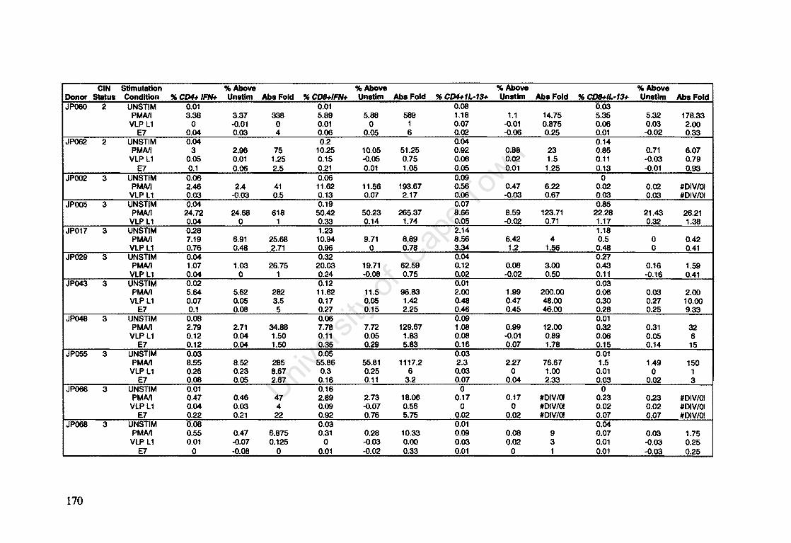
Appendix A: Raw Data of Intracellular Cytokines determined by Flow Cytometry 164
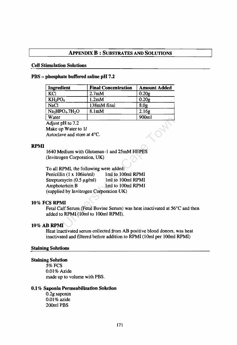
Appendix D: Substrates and Solutions 171 References 174
vi
Index to Tables
Table 1.1
Table 2.1
Table 3.1
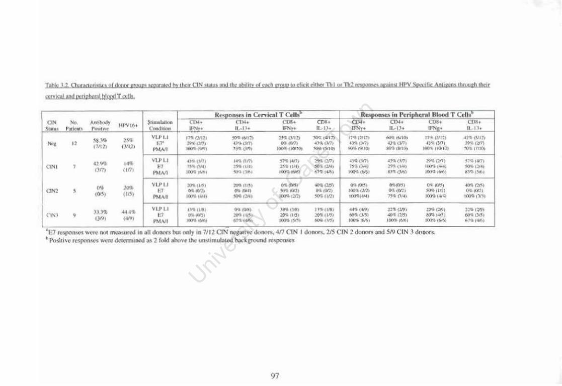
Table 3.2
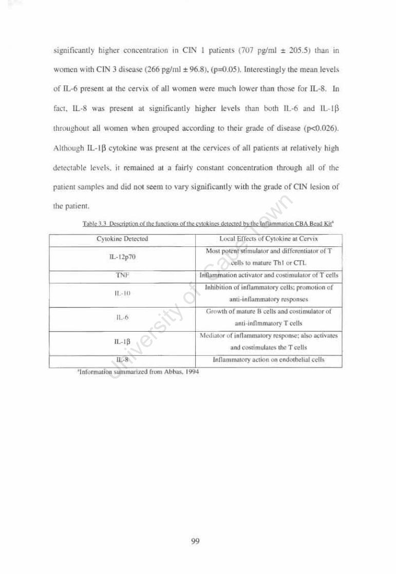
Table 3.3
Table 4.1
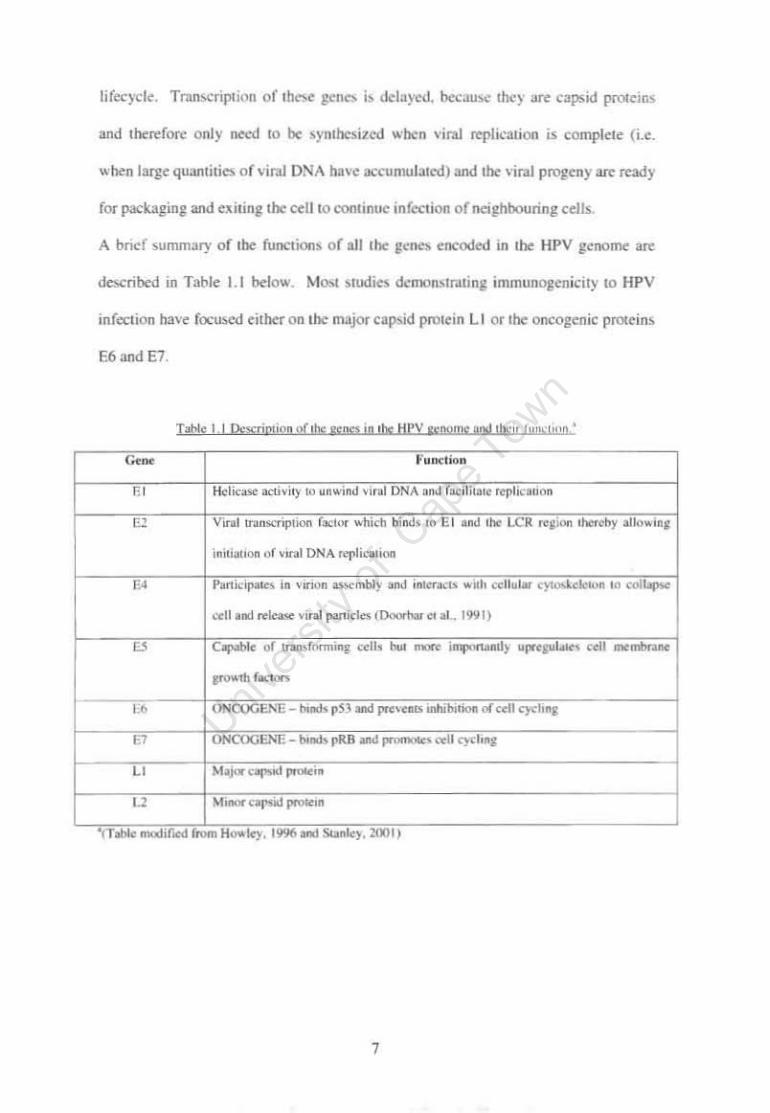
Description of the genes in the HPV genome and their function Page 7
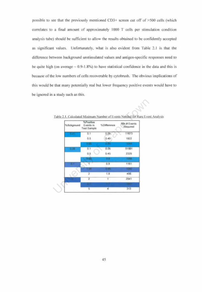
Calculated Minimum Number of Events Needed for Rare Event Analysis
4S
Description of the women recruited into this study showing severity of cervical disease, type of HPV infecting the cervix, viral load and HPV -specific antibody responses
73
Characteristics of donor groups separated by their CIN status and the ability of each group to elicit either Thl or Th2 responses against HPV Specific Antigens through their cervical and peripheral blood T cells
Description of the functions of the cytokines detected by the Innammation CBA Bead Kit
Description of women recruited into this study showing
97
severity of cervical disease and type of HPV infecting the cervix 139
vii
Index to Figures Figure 1.1. Structure of the Female Reproductive System Page 4
Figure 1.2. Picture of the squamo-col umnar junction at the cervix of an 18 year old girl 4
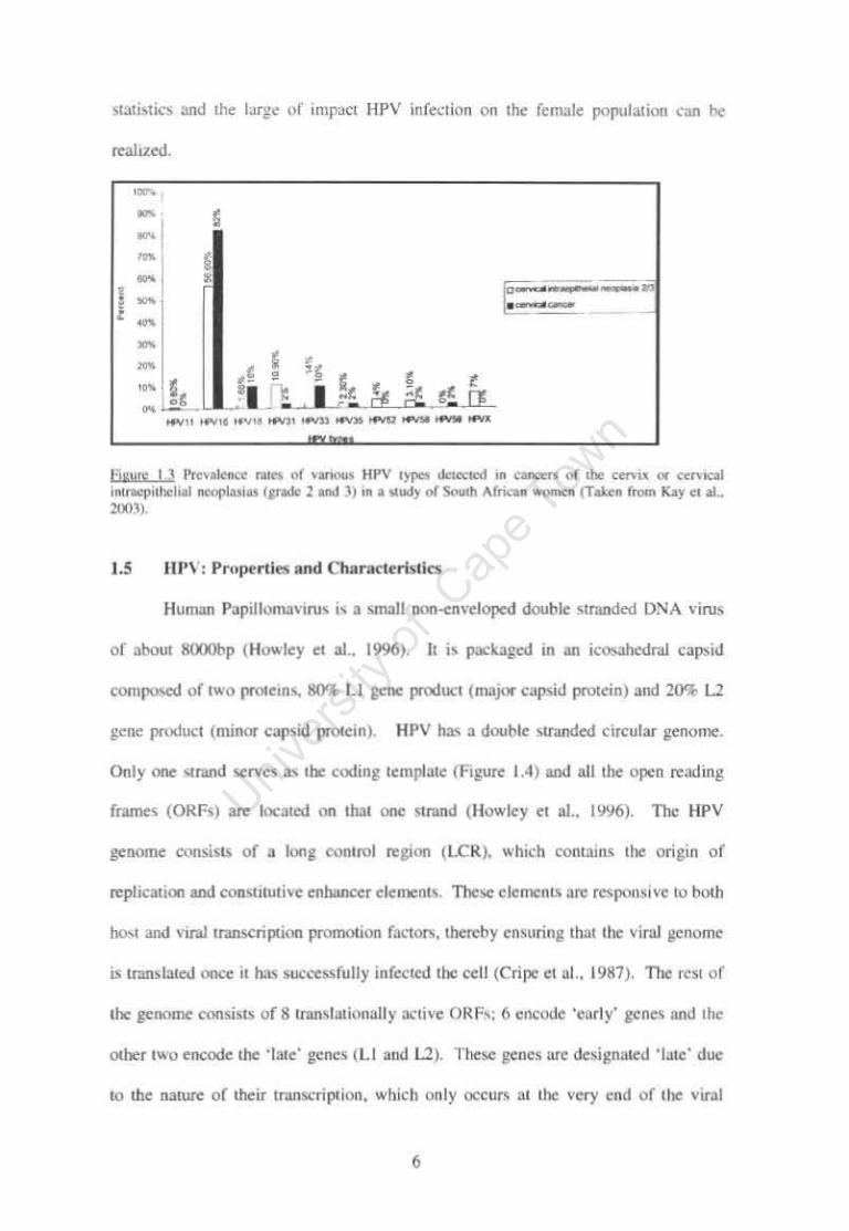
Figure 1.3 Prevalence rates of various HPV types in a study of South African women 6
Figure 1.4 Genomic map of Human Papilloma virus (HPV) 8
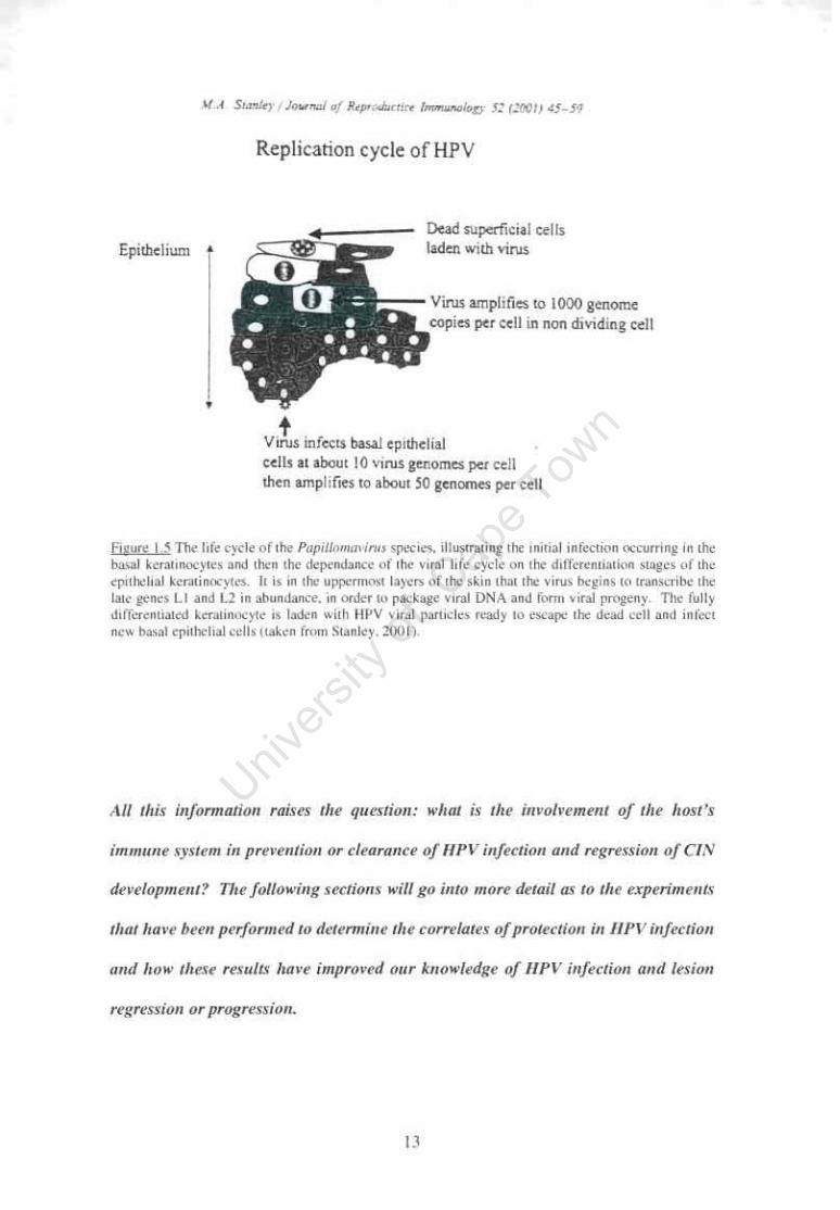
Figure 1.5 The life cycle of the Papilloma virus species. 13
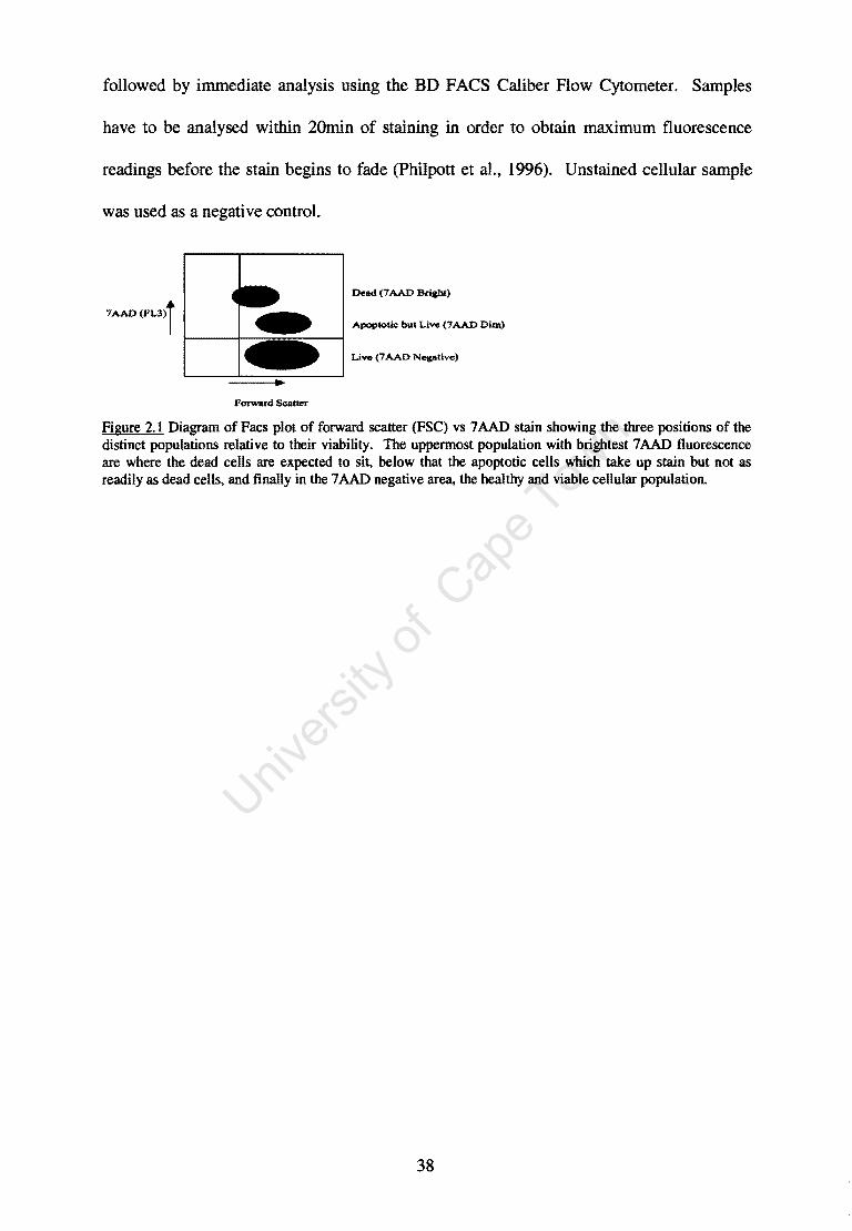
Figure 2.1 Diagram of Facs plot of forward scatter (FSC) vs 7 AAD stain showing the three positions of the distinct popUlations relative to their viability. 38
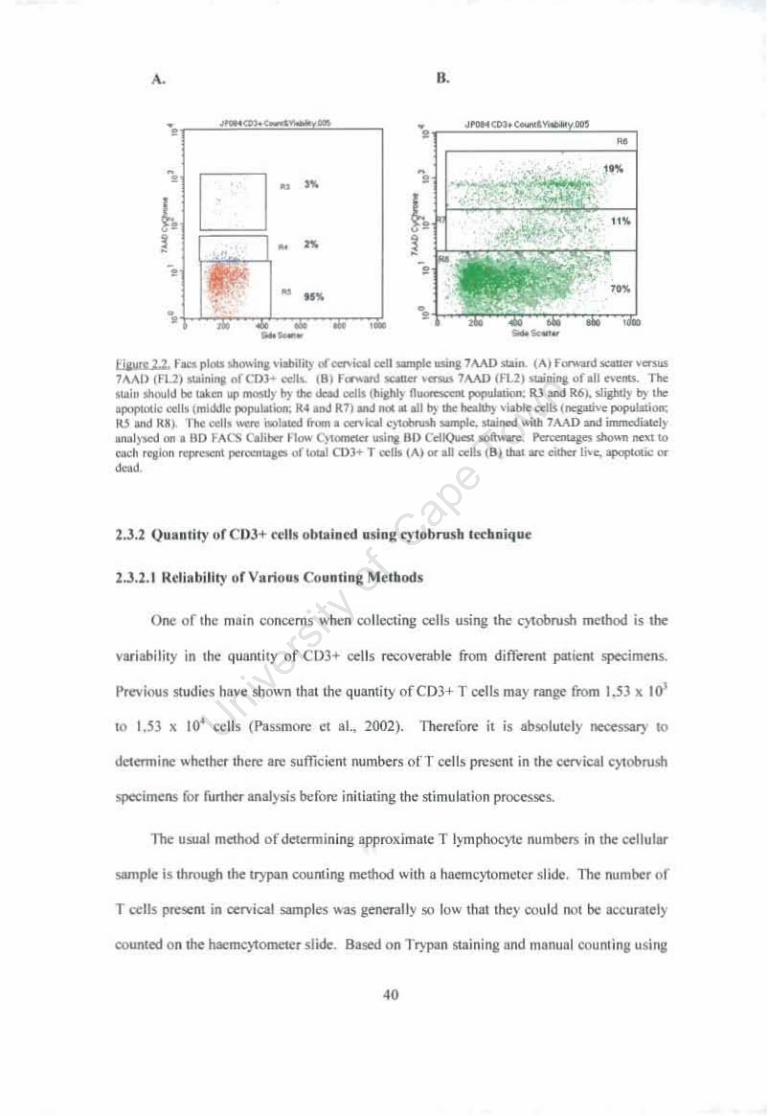
Figure 2.2 Facs plots showing viability of cervical cell sample using 7 AAD stain. 40
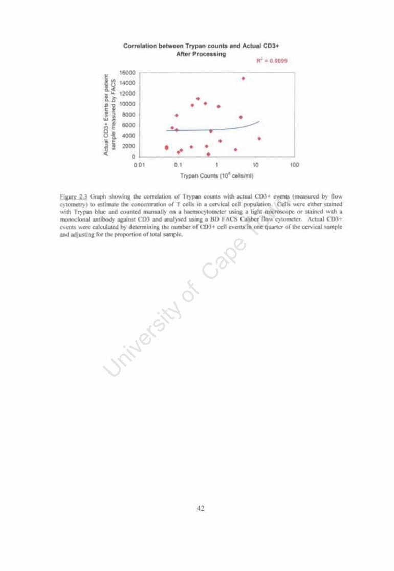
Figure 2.3 Graph showing the correlation ofTrypan counts with actual CD3+ events (measured by flow cytometry) to estimate the concentration of T cells in a cervical cell population. 42
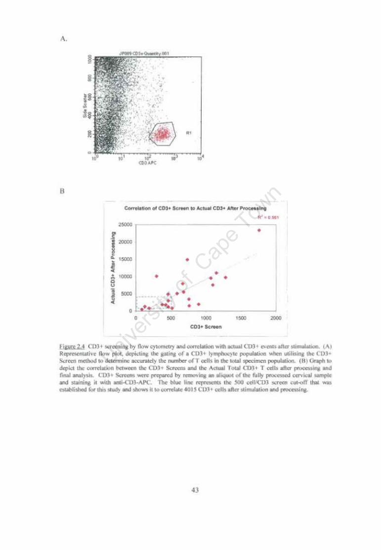
Figure 2.4 CD3+ screening by flow cytometry and correlation with actual CD3+ events after stimulation. 43
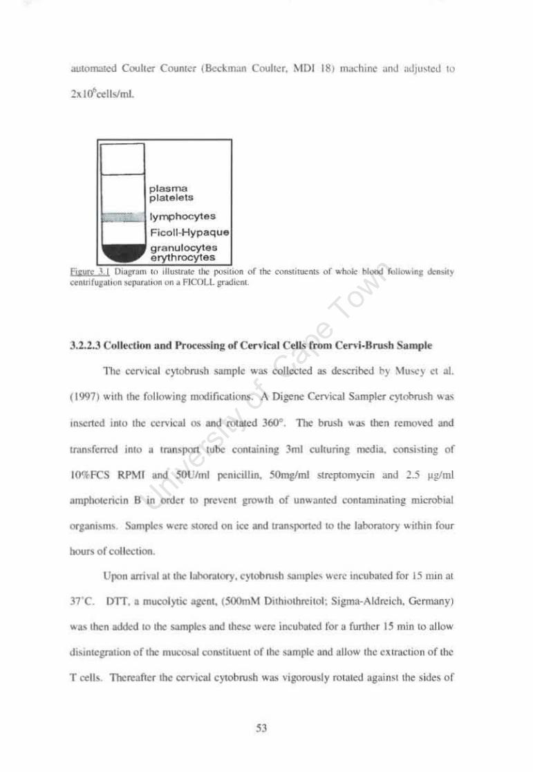
Figure 3.1 Diagram to illustrate FICOLL gradient. 53
Figure 3.2 An example of the characterization ofVLP-16 preparation used in this study 57
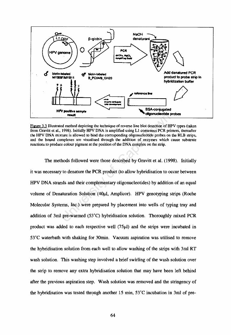
Figure 3.3 Illustrated technique of reverse line blot detection of HPV types. 64
Figure 3.4 The types of HPV found in women attending the Groote Schuur Colposcopy clinic. 74
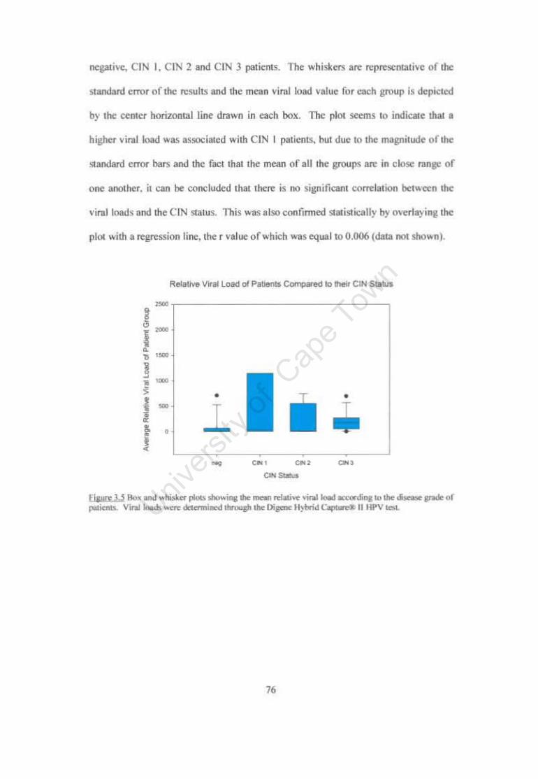
Figure 3.5 Box and whisker plots showing the mean relative viral load according to the disease grade of patients. 76
Figure 3.6 Correlation between Cervical T cells yielded in a sample and age of the donor. 77
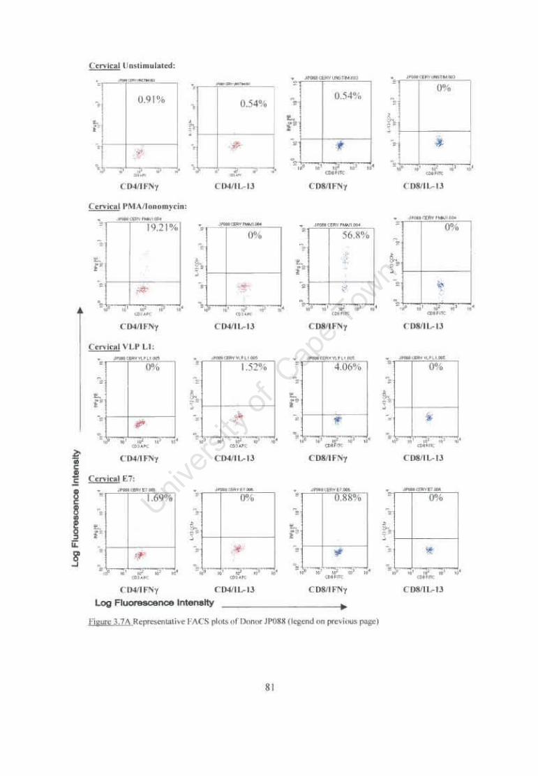
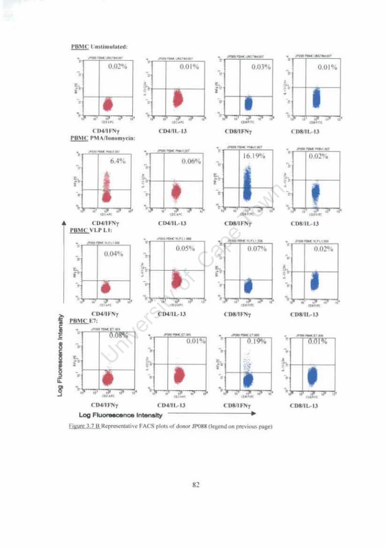
Figure 3.7 Representative FACS plots of ICC staining of cervical and PBMC T cells following stimulation with HPV 16 VLP L1 and E7 antigens. 81
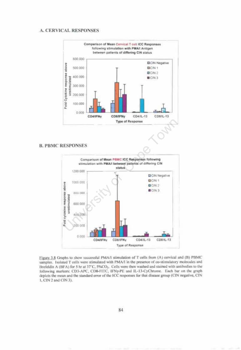
Figure 3.8 Graphs to show successful PMAII stimulation of T cells. 84
Figure 3.9 Bar graphs to show the mean ICC responses to each stimulation condition in the PBMC or cervical T cell populations from all women (irrespective of HPV infection status) compared with disease severity (negative, CIN1, CIN2 and CIN3). 86
Figure 3.10 Mean ICC responses to HPV16 specific antigens VLP L1 or E7 elicited by PBMC and cervical T cells of HPV16 infected women, compared to those ofHPV16 uninfected women, irrespective of their grade of disease at the cervix. 88
Figure 3.11 Correlation of mean PBMC T cell responses with varying grades of CIN from women either infected with HPV16 or not infected with HPV 16 at the cervix. 91
Figure 3.12 Correlation of mean cervical T cell responses with varying grades of CIN from women either infected with HPV16 or not infected with HPV 16 at the cervix. 92
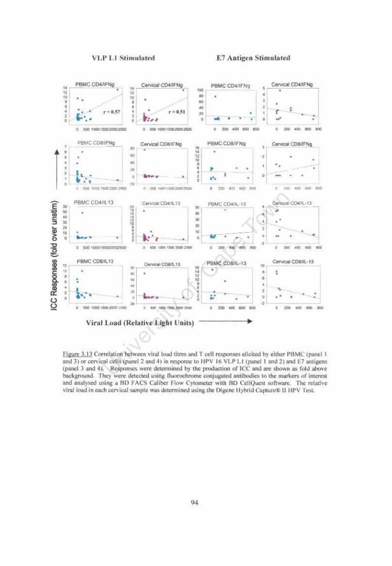
Figure 3.13 Correlation between viral load titres and T cell responses elicited by either PBMC or cervical cells in response to HPV 16 VLP L1 and E7 antigen. 94
viii
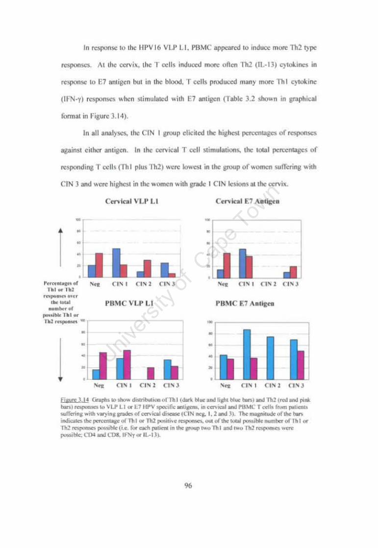
Figure 3.14 Graphs to show distribution ofThI and Th2 responses to VLP Ll or E7 HPV specific antigens, in cervical and PBMC T cells from patients suffering with varying grades of cervical disease (CIN neg. 1,2 and 3). %
Figure 3.15 Box and Whisker plots showing the presence of pro- and anti-inflammatory cytokines (IL-S, IL-6 and IL-IP) at the cervix of CIN negative, CIN I, CIN2 and CIN 3 women. 100
Figure 4.1 Heterogeneity in CDS T cell perforin expression in PBMC from S donors. 129
Figure 4.2. Comparison of perforin expression in CDS+ CfL from two different donors before stimulation and following 5hr stimulation with PMAII. 131
Figure 4.3 Comparison ofCDI07a expression vs Perf orin release in 3 donors. at 3 time points during stimulation with SEB. 134
Figure 4.4. Interassay variability when using CDI07a as a marker of cytotoxic activity of CDS+ T cells. 135
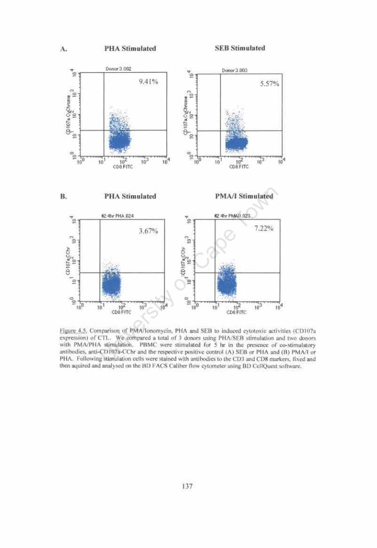
Figure 4.5. Comparison of PMAlIonomycin, PHA and SEB induction of cytotoxic activity (CD 107 a expression) in CfL 137
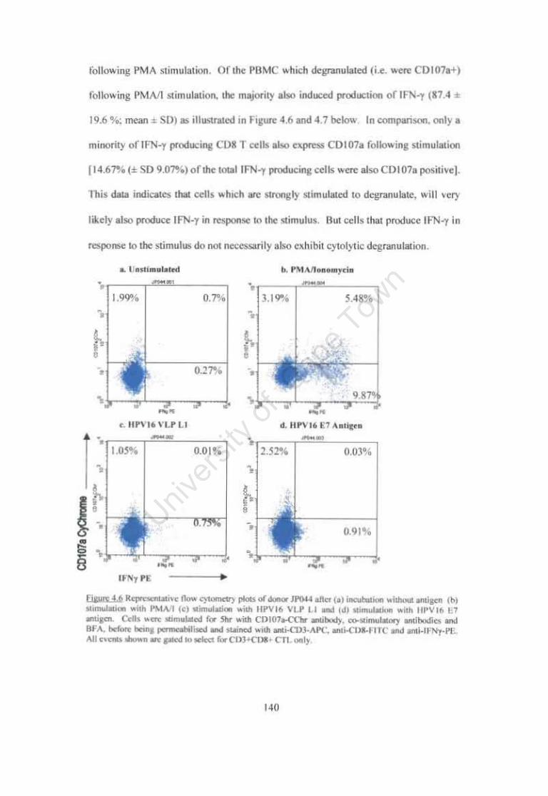
Figure 4.6. Representative flow cytometry plots of donor JP044 after (a) incubation without antigen (b) stimulation with PMAII (c) stimulation with HPVI6 VLP Ll and (d) stimulation with HPV16 E7 antigen. 140
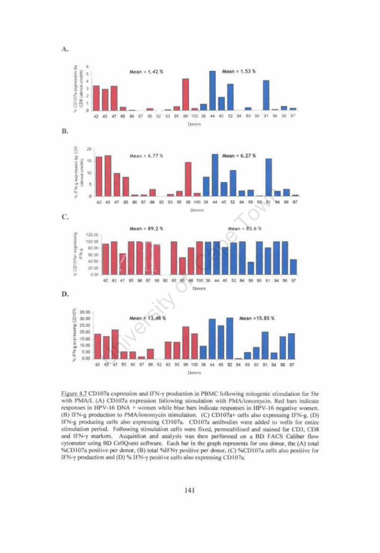
Figure 4.7. CDl07a expression and IFN-y production in PBMC following mitogenic stimulation for 5hr with PMAII. 141
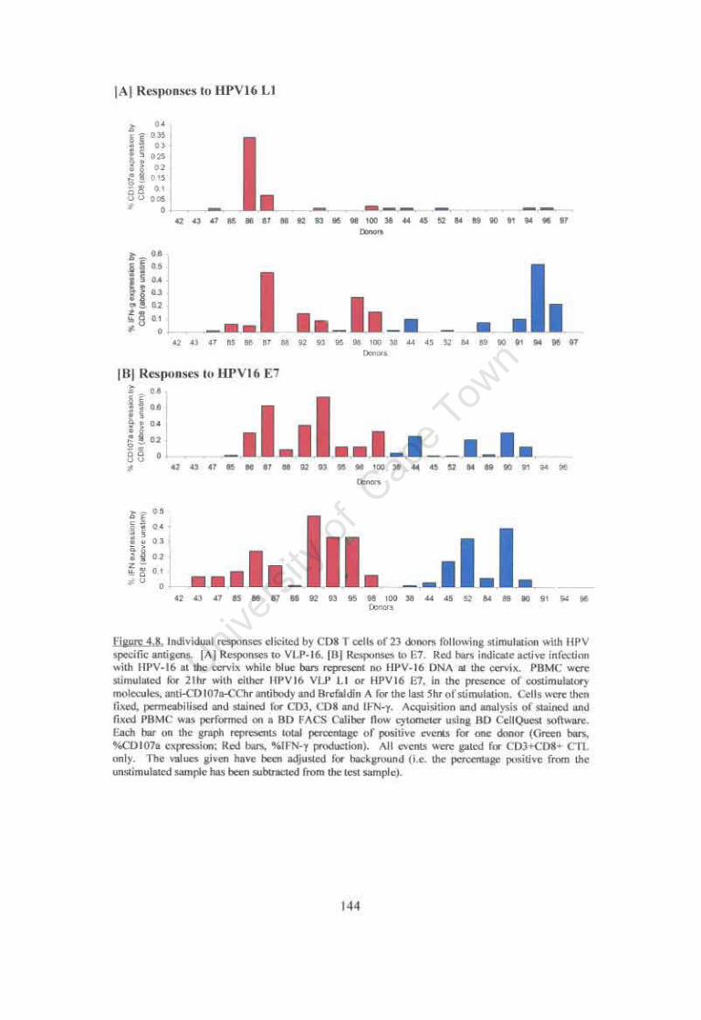
Figure 4.S. Individual responses elicited by CDS T cells of 23 donors following stimulation with HPV specific antigens. 144
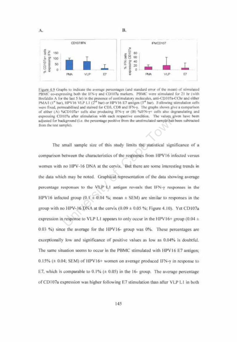
Figure 4.9. Graphs to indicate the average percentages of stimulated PBMC co-expressing both the IFN-y and CDI07a markers. 145
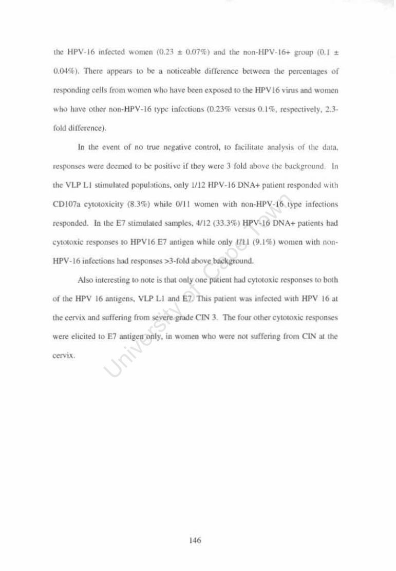
Figure 4.10. Comparison between the cytokine (IFN-y) and cytotoxic (CDI07a) responses elicited by the CDS T cells ofHPV16 infected (HPVI6+) versus HPV16 negative donors. following stimulation with HPV16 specific antigens. 147
ix
mM Ilg mg III ml min hr kDa RT
7AAD APC APC BCG BFA BSA CANSA CBA CChr CIN CMI CO CTL DC dH20 DMSO DTT ELISA FACS FCS FITC FSC ICC IFNy IgG IgA IL-13 HC HIV HPV LAMP LC LCR MHCWI NCI NK OD
7 Amino Actinomycin D Allophycocyanin Antigen Presenting Cells M. Bovis Bacillus Calmette Guerin Brefaldin A Bovine Serum Albumin Cansa Association of South Africa Cytometric Bead Array CyChrome Cervical Intraepithelial Neoplasia Cell Mediated Immunity cut off Cytotoxic T Lymphocytes Dendritic Cells Distilled water Dimethylsulphoxide Dithiothreitol Enzyme-Linked Immunosorbent Assay Fluorescence Activated Cell Sorter Fetal Calf Serum Fluorescein Forward Scatter Intracellular Cytokine Interferon - gamma Immunoglobulin G Immunoglobulin A Interleukin - 13 Hybrid Capture Human Immunodeficiency Virus Human Papillomavirus Lysosomal Associated Membrane Glycoprotein Langerhans Cells Long Control Region Major Histocompatability Class I or II National Cancer Institute Natural Killer cells Optical Density
x
ORF PAGE Pap PBMC PBS PE PHA PMA pRB PV RBC RLB RLU SA-HRP SD SEB SEM SSC TB TCR Th1l2/0 1NFa TZ VLP WHO WPBTS
Open Reading Frame Polyacrylamide gel electrophoresis Papanicolaou Peripheral Blood Mononuclear Cells Phosphate Buffered Saline Phycoerythrin Phytohaemagglutinin Phorbyl Myristate Ester protein Retino Blastoma Papillomavirus Red Blood Cells Reverse Line Blot Relative Ught Units streptavidin conjugated horse radish peroxidase Standard Deviation Staphyllococcus Enterotoxin B Standard Error of the Mean Side Scatter Tuberculosis T Cell Receptor T Helper cells type 1 t 2 or 0 Tumour Necrosis Factor-alpha Transformation Zone Virus Uke Particle World Health Organisation Western Province Blood Transfusion Service
xi
Acknowledgements
I, wish to acknowledge and sincerely thank the following people for their much appreciated help during the course of my project:
My Supervisor Dr. Jo-Ann Passmore, for her guidance, support and absolute commitment to helping me realize my goal.
My Co-supervisor Prof. Anna-Lise Williamson for making herself available when I needed her assistance.
Prof Lynette Denny and Sister Janine Jones for facilitating the recruitment of study participants and collecting the necessary specimens used in this project.
Ms Candice Sampson for kindly allowing me to use the data from an experiment which she performed (Serum antibody status and HPV genotyping of cervical samples).
Mr Bruce Allan also for kindly allowing me to use the data from the Hybrid Capture experiment which he performed for the benefit of this project.
The following people for their assistance either with kind donations of reagents or for providing much needed enthusiasm and support in the lab! Tracy Muller IngaBecker Eric van der Walt Di Marais Suzanne Grove Neil Christenson John Schiller Fritz Tiedt Gerald Chege
Finally, my family and friends who were there to support me when I most needed it, and Michael for giving me the added strength and vision which helped me to reach my goal-
Thank you.
This work was supported in part by grants from the Poliomyelitis Research Foundation (PRF) and the Medical Research Council (MRC) of South Africa.
xii
ABSTRACT
Cervical cancer is the most common cause of cancer-related death in black South
African women. Human papillomavirus (HPV) has been found to be a necessary
causative agent of cervical cancer and has been reported to be associated with 84% of
cervical intraepithelial neoplasia (CIN). HPV type 16 (HPV-16) is the most prevalent
HPV type associated CIN and cervical cancer with -56% of women with cervical
disease being infected with HPV 16. Yet studies have shown that 47-85% of CIN
regressed, suggesting that perhaps an effective immune response could result in HPV
clearance and lesion regression. Since HPV infection does not disseminate and there
is no systemic phase of infection, it is hypothesized that local cervical immune
responses are important in lesion regression and clearance of HPV infection. There
are, however, very few studies of mucosal immune responses to HPV infection. The
aim of this study was to determine the type of mucosal immune response elicited by
the CD4 and CD8 T cell subsets to HPV infection at the cervix of women diagnosed
with varying grades of CIN and to compare these to systemic responses. One hundred
women with varying grades of histologically confirmed CIN attending the Groote
Schuur Hospital Colposcopy Outpatient Clinic were enrolled into the study. Cervical
T cells were isolated from the endocervixltransfonnation zone of these women using a
Digene cytobrush. Of these 100 women, only 33 were found to have suitable cervical
cytobrush specimens for analysis of mucosal T cell responses based on CD3+ T cell
numbers and absence of red blood cell contamination.
Peripheral and cervical T cells were stimulated with the major capsid protein
Ll of HPV-16 which self assembles into virus like particles (VLP) and the major
xiii
HPV -16 oncogenic protein E7 and the subsequent production of intracellular
cytokines (ICC) was detected through flow cytometry.
Women with CIN 1 consistently had the strongest CD4 IFN-y (but not
necessarily CDS T cell) responses at the cervix to HPV-16 antigens compared to
women with no cervical neoplasia or those with more severe disease (CIN 213). This
was observed particularly if one focused on women with active HPV-16 infection but
also if one looked at the group as a whole (irrespective of the type of HPV causing
infection). There was a significant trend towards decreasing Th2 responses (IL-13
production) with increasing disease severity, in cervical CD4 and CDS T cells to both
HPV antigens (Ll and E7) if one looked at the group as a whole. Conversely, this
trend was reversed with increasing Th2 responses with increasing disease severity in
the women with active HPV-16 infection. When PBMC responses from women with
HPV-16 DNA at the cervix were compared with those that were infected with other
HPV types, the HPV-16 DNA+ women generally produced a Thl dominant response
(more IFN-y and less IL-13) which changed to a Th2 dominant response with
increasing disease severity (particularly for E7 antigen). In contrast, the HPV-16
negative women (infected with other HPV types) showed a complete reversal of this
profile with increasing IFN-y responses and decreasing IL-13 responses with
increasing disease grade. The only cervical immune response that correlated with
disease grade in this study was that both CD4 and CDS T cell IL-13 production
decreased with increasing disease severity but this was observed in both women
infected with HPV -16 and those infected with other HPV types. Although evidence
ofHPV-16 specificity is lacking, the results do imply that Th2 dominant responses are
associated with a "healthier" disease state and IL-13 responses (possibly driving a
protective antibody-mediated response) diminish with increasing disease grade. The
xiv
magnitude of Thl responses elicited by cervical T cells was generally lower than
those produced by T cells from peripheral blood. This study shows that the cytobrush
method of obtaining cervical lymphocytes combined with intracellular cytokine
analysis and flow cytometry is a non-invasive and potentially useful approach to
studying immune responses in the genital tract and also suggests various
modifications to improve the low numbers of suitable cervical specimens for study.
xv
CHAPTER ONE:
LITERATURE REVIEW
1.1 The Prevalence of Cervical Cancer in the Female Population
Cancer of the cervix is a worldwide problem. It is estimated that in 2002 a
total of 493,243 women were diagnosed with cervical cancer (Ferlay et al., 2002).
International cancer registries have estimated that it is the second most prevalent
cancer disease among women (after breast cancer), making up 16.8% of the total
number of cancer cases reported (International Association of Cancer Registries,
2002). Here in South Africa, the statistics are just as alarming. The Cancer
Association of South Africa announced in 2002 that cervical cancer has now become
the leading cause of cancer related deaths in black Mrican women (CANSA, 2002).
This means that South African women are now at higher risk of suffering from
cervical cancer than they are of breast cancer. It was also pointed out that of all the
women between the ages of 15 and 29 yr who were diagnosed with cancer, 13.4%
were suffering with cervical cancer, making it the most common cancer in that age
group. Therefore this is an area of growing concern, especially for South Mricans,
and there is much research being undertaken to understand the disease itself and
various other factors, which may either predispose women to, or protect them from,
cancer progression.
1.2 Progression from CIN to Cancer of the Cervix
Many clinics in South Africa offer screening to detect early onset of cervical
cell abnormalities. The initial screening is usually performed by papanicolaou (pap)
1
smear, which involves smearing a scraping of the cervical cells onto a glass slide for
analysis of cellular abnormality under a microscope (World Health Organisation,
1988). This is referred to as a cytological diagnosis. If unusually high cellular
abnormality is detected, the patient is referred for colposcopic or histological
diagnosis, since these are both more accurate than pap smears. A colposcopy
diagnosis involves washing the cervix with acetic acid and thereafter using a
colposcoscope to visualize any white (HPV infected) lesions (WHO, 1988). A
histological diagnosis makes use of the microscope to analyse the patients degree of
cellular abnormality, but a biopsy specimen (2-4mm of cervical epithelium) is taken
and therefore allows determination under the microscrope of the degree to which the
epithelium is dysplastic (WHO, 1988). The lesions are graded under the microscrope
according to the extent of the abnormal cells; if 1/3 of the epithelium is cytologically
abnormal, the lesion will be graded as a cervical intraepithelial neoplasia grade 1
(CIN 1) (NCI Workshop, 1988; Shah and Howley, 1996). As the lesions get
progressively worse (and therefore more of the cervical epithelium is classed as
abnormal cell growth) the lesions will be graded CIN 2 and CIN 3, depending on the
severity of the lesion.
A woman diagnosed with CIN is either immediately treated to prevent
progression to cervical cancer or depending on the severity of the lesion, asked to
return for a follow up visit at a later stage in order to check the lesion progression.
This is due to the fact that not all cervical lesions will progress to cervical cancer. In
fact studies have shown that 47-58% of grade 2 and 3, and up to 85% of grade 1
cervical lesions regress naturally (Chan et al., 2003; Iatrakis et al., 2004). Therefore
although a woman is diagnosed with CIN this does not mean that the lesion will
necessarily become cancerous.
2
This raises the question then, what other factors playa role in the progression
or regression of CIN? Articles published in the interest of increasing public
awareness regarding cervical cancer have cited the following as risk factors for
progressive worsening of the cervical lesion: sexually transmitted diseases (including
Human Papillomavirus [HPV] , Human Immunodeficency Virus [HN] , Chlamydia
and various Herpesviruses), high sexual activity with multiple partners, high parity,
long term use of hormonal contraceptives, socioeconomic and ethnic differences
(cervical cancer incidence is higher in less developed countries - although this is
probably due to the improved and more accessible screening facilities in well
developed countries which allow for earlier detection and treatment of cervical
lesions), smoking and an inherited genetic predisposition to cancer (Ho et al., 1998;
Well-Connected, 2002; Bosch and de Sanjose, 2(03).
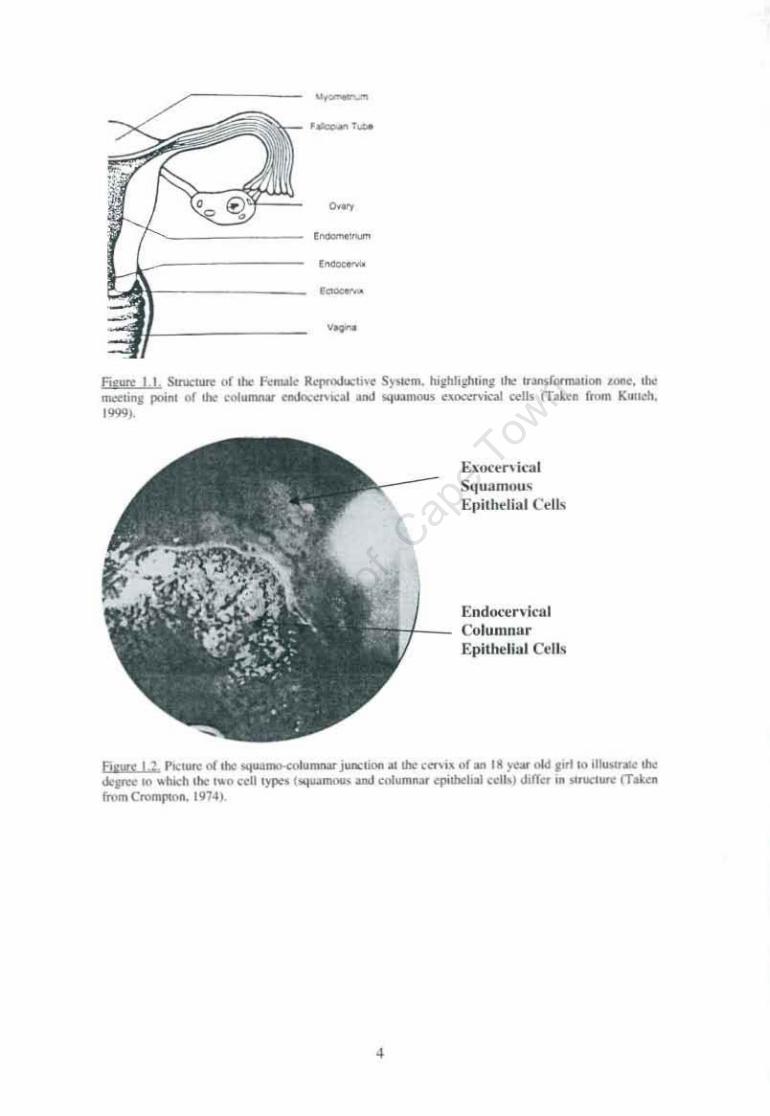
1.3 The Biological Structure of the Cervix
Apart from the above-mentioned factors, the cervix itself is predisposed to
abnormal cytological growth in the way that it is biologically structured. The cervix
is the meeting point of the columnar endocervical cells from the uterus and the
squamous ectocervical cells of the upper vaginal tract (Figure 1.1 and 1.2) (Crompton,
1976; Cartier, 1984). The area where these two very different cell types have to meet
in the cervix is labelled the 'Transformation Zone' (TZ), and this is where most of the
cervical lesions occur (Shah and Howley, 1996). At the TZ there is a high rate of cell
turnover and this is conducive to cellular proliferation and tumour formation.
3
'-'-
Flf"" 1.1 . StNcun (>(!he F.:no..lk R ............ "H SY'I"m. Iu.hliihti"l ,ho ",_fonnl'w.. ..:1M. ,ho mcain, poonl '" !he t'I'lIU/Mar fIdoc~f>l("al and III:jtWOOIn ~-"""""'iocal celli n-.~a from " .. !clI.
''''' E:uICt'n k al SIIIlanlnUS Epithelial C",lIs
Endol.'enical Culumnllr Epith~ I i:,1 Ct'JI.~
6,," 1.2. P1c:'1Ife o( ,~ "'I""1II<KOIu ...... J ..... ,""' :at !he c<."" '" or .. t 8 )'UI old .or! '0 il~ tho: .kpN lO ... hidt ,ho h"o c~1I ' yp<'II l;q1WlOOU< and cilium"", I.'pIdltlJllt ("clb ) Ihlfct ' D ~'11<1"'" (Tau. fromC""""""," t974 ).
1.:& Role or HI' .... ill C.,nical Can cer
The greatesl risk faclor for progression 10 c~rvical cancer is Human
Papillomavirus (HPV) infeelion. It has bce-n reponed Ilml al leasl 94% of o;eryical
c:meers and grealer than 84% of CIN were associaled ",ilh HPV D~A (K!ly el aI ..
2003; Chan el a1 .. 2(03). HPV infection has now been r«ognized as a necessary
causative agent in progressiOll 10 cervical cancer (Walboomers eI al. 1999; Bosch et
aI . 2002:a: Bosch and San~, 2(03).
During a 3 yr longitud iM.l smdy of unh'Crsity students. 43% of the pnn icipants
acquired new I-IPV infections and of ttr new infections clearance occum:d within the
first 12 monlhs in 70% of the women (Ho et at.. 1998). Although studies have ~hown
thai 70-80% of HPV infections can be cleared (Ho et al.. 1998; Evandcr et aI., 1995)
other smdies have soown that a small percentage (7%) do persist for as long as 5 years
(Mo lano et al.. 2(03). The persistence of an HPV infection is the moS! important risk
factor for CIN progression (Molano el aI., 2003). Another imponant ris~ factor is the
type (,f HPV infecti ng the: cervix since not aU HPVs have the same oncog.:nie
potential (Sehlechl el aI., 20(3). Higll·ri~k I-/PV infeclions resuh in lower rates of
CIN regression than infections wllh low-risk HPV Iypes. The most prominenl and
persislent HPV 1)'pI:. associmed wilh CIN and ~...,rvicaJ cancer. has been soown to be
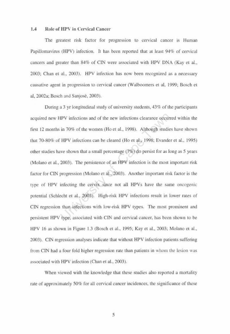
HPV 16 as shown in Figure J.3 (Bosch et al" 1995: Kay N 31" 2003: Mo l:mo el aI .•
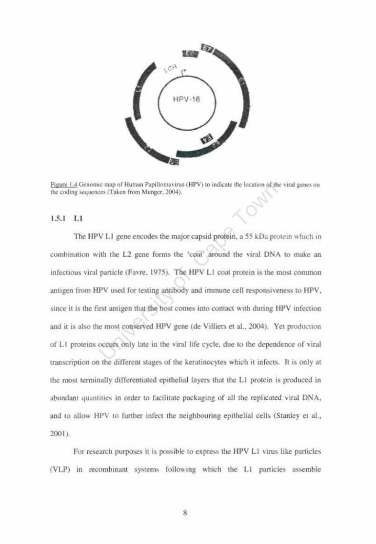
tl,," ! :\ G .... omic ""'I' or Human l'ap'!ilHtlll"iM (HPV ) It) j..Jicao< lI'o: locallOOl 0( lh1: ,,,a! ,<lid On m. ~ool", ~,",<l<U (Ta~~n f'l"Mll Mu"¥,. 2004).
1.5. 1 1.1
'The IIPV LI gcuc encodoes tilt major capsid prolein. a 55 1..0.\ pl"Olem .... tilth m
comhlMtlon ..... llh the L2 selIC fonll5 the ·coat· around the ,·ira] DNA 10 make an
,nfeelLOus I'ir:al pankle (Fane, 1975). 1bc HPV U coal protem is the most common
wlUgen from HPV used for testing antibody and irnnwnc ccll re"ponslVcntsS to ttPV.
since i! is lilt. Jirst antigen thal the host comes into contaet ..... ith during HPV lnfeclion
and It is also !he IDOS! tonSCr.'ed HPV gt:ne (de Villiers et 31 .. 200-\ ). Yet production
of L t pl'Olelns occurs ooly laIc in !he 'or'" life eydc, due 10 the dependence of I'irnl
uunscripuon on the differen! Slagcs of tile ker:ltinocytcs ..... h,Ch it infects. It IS only lit
tbe IllIbt tcnninally differentiated cpithehullayers that the LI protein is produced in
abundanl qUJntni~ in o~r to rfICil it ~tc pacltaglng of all the replicated I'irul DNA,
and 10 aUO\\ HPV 10 funher infect the neighbouring epithelial ceJb (SL:ll\ley et al..
lOOt).
For re-earch purposes it IS pos~ib~ to expre"s the HPV Ll virus like panicles
(VLP) in fe(:Ombmllnt 5)"tem, follOlllng IIhieh the LJ panicles as.>emble
,
automatically into conformationally correct VLPs (Zhang et al., 2000). Therefore
HPV Ll VLPs can be successfully used to induce HPV specific antigen responses in
host immune cells, allowing assessment of host immune function to HPV infection.
I.S.2 E6 and E7
The oncogenic early genes, E6 and E7, are transcribed into proteins which
bestow on HPV the ability to cause abnormal cell proliferation by facilitating
immortalisation of the infected cell (Munger et al., 2004). The E6 protein complexes
to a host cell protein called p53. The function of p53 is to halt cell cycling if DNA
damage has occurred, and thereby facilitate the repair of the DNA (Brenna and
Syrjanen, 2003). When E6 binds to p53 it prevents it from performing its vitally
important function. The E7 gene product binds the retinoblastoma tumour suppressor
protein (pRB), a nuclear protein that regulates gene expression (Brenner and Syrjanen,
2003). In its dephosphorylated state, pRB is a negative regulator of cell growth by
inhibiting a cellular transcription factor (E2F-l). E7 preferentially binds the
dephosphorylated pRB and therefore allows the cell to continue cycling (Howley et
al.,1996).
In non-productive HPV infections (Le. infections which are not progressing
through the epithelial layers, but rather causing abnormal cellular growth of the basal
keratinocytes) a large quantity of the early E7 protein accumulates in the basal
infected cells as they undergo repeated cell cycling (Middleton et al., 2003).
Therefore in late stage eIN and early cervical cancers, E7 proteins are abundant in the
basal epithelial layer and these proteins could then induce priming of the immune
system in order to elicit a response to the HPV infection.
9
1.5.3 Diversity of HPV Genotypes
HPV belongs to the Papillomaviridae family of which 118 different
papillomavirus (PV) types have thus far been sequenced and grouped according to the
relatedness of their genome sequences (de Villiers et al., 2004). HPV types are host
species and tissue specific. They are primarily grouped as either mucosal or
cutaneous depending on the location of their preferred site of infection; mucosal HPV
types infect the genital and oro-pharynx area whereas the cutaneous HPV types infect
the skin (Frazer, 2004).
It is the mucosal HPV types which cause infections at the cervical mucosal
epithelium, but not all of those HPV types are associated with cervical cancer. The
mucosal HPV types have been classified as either high risk (HPV type 16, 18,26,31,
33,35,39,45,51. 52, 55, 56,58,59,68,82,83 and 73) or low risk (HPV type 6, II,
40,42, 53, 54, 57, 66 and 84) (Gravitt et al., 1998). The high-risk types are so called
since they are more likely to cause cellular transformation and progression to
cancerous lesions (zur Hausen, 2(02).
There are some functional differences between the high-risk HPV types and
the low-risk types. Firstly the E6 protein transcribed off the low risk DNA can not
bind the p53 protein and secondly, the E7 proteins from low risk types bind the pRB
with ten fold less affinity compared with the high risk types (Wemess et al., 1990;
Gage et al., 1990). Another difference between the HPV types is that low-risk viral
DNA remains extra-chromosomal, whereas the high-risk HPV genomes become
integrated into the host cell's chromosome, which allows the increased and persistent
expression of the HPV viral genes, thereby leading to HPV persistence and lesion
progression (Tjiong, 2(01).
10
Studies have shown that the HPV antigens were entirely type specific and
elicited only responses from patients who were previously infected with that HPV
type, especially in serum antibody responses (de Gruil et al., 1996, 1998; Gill et al,
1998). This strict type specificity in immune responses to HPV is not absolute since
there is some evidence for cross-reactivity of both serum antibodies and T cell
responses to HPV types other than the HPV type with which the patient was infected
(Hopfl et al., 2000; Kadish et al., 2(02). This could imply that immunity to one HPV
type is cross-protective against infection by other HPV types, or it could suggest that
those patients who had responses to an HPV type that they were not currently infected
with, might have had a previous infection with that specific HPV type.
1.5.4 HPV: Infection Mechanism and Viral Lifestyle
Papillomaviruses are host species and tissue specific (Stanley, 2(01).
Therefore HPV only infects humans and the site of infection (skin or mucosal
epithelial cells) depends on the specific HPV type causing infection. Initial infection
by HPV is at the basal epithelial cells (Shah et al., 1996). Epithelial cells
continuously undergo differentiation until they become terminally differentiated (at
the outermost surface of the epithelium) after which they are shed. HPV utilizes the
continuous cell division of keratinocytes (subtype of epithelial cells) in order to
vegetatively increase the original viral copy number at infection (of 10 copies) to 50
copies per cell (Figure 1.5). HPV is fully dependant on the differentiation stages of
the keratinocyte and only in the late stages of differentiation can the virus initiate
expression of 'late' genes and exponentially increase the copies of viral DNA in the
cell (Oriel, 1971).
11
This dependence on the differentiation stages of the epithelial keratinocytes
has prevented the culturing of HPV in the lab, since it is difficult to make a tissue
culture in which the cells gradually become more differentiated. Therefore it is not
possible to collect HPV antigens through culturing, it is necessary to express the
genes of interest recombinantly, as HPV genes expressed in other vectors.
Eventually, the dead keratinocyte laden with viral particles is sloughed off the
epithelium and the infectious viral particles are released and able to infect a new host,
or to infect neighbouring cells (Tindle, 2(02). Infection with HPV is a long process -
approximately 3 weeks from time of infection to virus particle release, since this is the
time it takes for a keratinocyte to fully differentiate (Stanley, 2(01).
Due to the nature of HPV infection which was described above, it has several
advantages over the immune system. Firstly, HPV causes only a localized infection at
the outermost periphery of the host where immune cells are less abundant (Tindle,
2002; Frazer, 2003; Stanley 2(03). This selective location, combined with the ability
of HPV to prevent apoptosis of infected cells thereby preventing inflammation of the
infected area, protects HPV antigens from coming into contact with cells of the
immune system (Stanley, 2001; Tindle, 2(02). Secondly, at the early stages of
infection, only low levels of viral transcription occur and it is only once the infected
cell is almost fully differentiated that the virus initiates high viral gene expression
(Stanley et al., 2(01). Finally, studies have shown that viral proteins may modulate
the immune response, by interfering with cytokine production or through modulation
of antigen presentation, in order to disrupt any immune response that might have been
initiated (Frazer et al., 1999; Arany et al., 2002; Woodworth, 2(02). It is almost as
though HPV was designed to avoid immune surveillance.
12
EpilbeJiu.'l1
•
Replication cycle of HPV
t
Dead superficial o:ells laden wlIh virus
::::-- ., •• &mplifie51O lOOO go:nom<: eopies pu ceIL in non dIviding ~cll
Vll'U$ infe<:1S bas.1.I epnhelial cellJ II iboul 10 virus genomes percell men lmpl i fie~ 10 aboul '0 gcnornes per ceIL
EiWU!l: I 'Th~ life c)dc of the P"pi//I:im41, in .. 'J'CCi .. " ilh",ral'"M 11>1: ,n"ial ,nfCC1ion <lCCunin8 in ,lie b,.,.1 kC'r.lm''''')lc, and 11ie" 11>1: dcp<>nd,nce of Ihe ....... 1 hf. cycle on 11>1: d,ff"","uanon Sla8c< (If Ihe <pilr"h .. 1 kcr.llino<yl .... II is ,n lho uppcrn1O\o1 la)".", <If 1r" skin lha, 11'00 VIIU. beg,n. 10 lr~n'<flbc ,lie 1010 gene' LI and 12 in abund"",,-,. ,n onkr '" pao."kagc v,ml DNA .. nJ form v,,,,) pn:>geny TIl< fully d,f(c"r<nli.ufd kralin"'")1e IS laden "ilh IIPV v,ral p.vude; re:>tly 10 ",uP<> lilt dc:><J ",II and onr,,,, nCl< b"",,,1 .",I""1>3.l ",,11. ,"thn from Stanley" 200 I)"
All IlJis illformatiOIl raises Ihe qlll'Slioll: .. "hal j~" Ihe jllvo/'"emmf 0/ fhe host's
imlll/lIlt sJ..,em ill prt'>"elllioJl Or t:iearollce 0/ II/'V ill/eelioll aud regrt!JSirlll of erN
det"elopmrllf? TIll" foNoK-rug scctiolls .. "m go illto IIIOrt! detail at to Ihe expen"",ellfs
Iltal /10'"" bl"t'II performed 10 dclumfllc the correlaus of protcrtiol/ ill 111'1' illfeetioll
Due to the nature of HPV, infection usually remains localized to the original
site of entry of the HPV virion into the basal epithelium (Frazer, 2003; Stanley 2(03).
This is especially so since the various HPV types are highly tissue specific (infecting
either skin or mucosal epithelium). Unlike some other viruses, which can be found
systemically throughout the body after the initial replication stages have occurred (e.g.
HIV), it is unlikely for HPV infection to disseminate and cause a lesion at another
site.
One would expect that any immune responses that are elicited by HPV
infection at the cervix would only be found locally in the immune cells situated in the
mucosal epithelial strata of the cervix. It follows then that one would not expect a
strong immune response to the HPV infection in the systemic T cells which patrol the
body, circuiting in the blood. Yet many studies have shown that there are significant
responses to HPV antigens in the PBMC isolated from the blood of patients suffering
with HPV infections.
1.6.1 Antibodies: the mediators of Humoral Immunity
In most viral infections, neutralizing antibodies are the first line of defense to
protect the host from the pathogen. Antibodies have been shown to play a very
important role in the immunity of the cervix and are an effective protective
mechanism in the host's defense against Papillomavirus (PV) infection (reviewed by
Stanley, 1997; Kutteh, 1999; Quayle, 2(02).
Studies of antibody responses in humans to HPV show clearly that systemic
HPV16 specific IgG responses to HPV 16 VLP are associated with HPV16
persistance and late stage CIN lesions whereas systemic IgA HPV16 VLP specific
14
antibodies seem to be associated to viral clearance (Luxton et al., 1997; Marais et aI.,
1997; Bontkes et al, 1999; de Gruil et al., 1997, 1999a). Local HPV16 specific
antibodies at the cervix were not correlated with viral clearance (Bontkes et al., 1997).
These studies showed that patients with HPV 16 specific IgG reactivity to HPV VLP
Ll in their serum had an increased risk of cervical cancer or late stage eIN 3.
There is also clear evidence that serum HPV specific IgA antibodies to the
HPV16 E7 antigen are significantly associated with HPV clearance at the cervix,
whereas the HPV specific IgG antibodies to E7 antigen seem to be more
representative of previous immune response activity to HPV (de Gruil et al., 1996a,
1996b).
From these results, it is now apparent that using antibodies as a marker for
cleared or persistent viral infection depends on which HPV antigen the HPV specific
antibodies are reactive against. Antibodies to VLP seem to indicate a persistant HPV
infection whereas antibodies to E7 might be the result of an effective immune
response (which responded to the virus when it was in the early stages producing E7
in the basal keratinocytes) and therefore perhaps indicates viral clearance.
1.6.2 Cell Mediated Immunity at the Female Reproductive Tract
The host defense system for the female reproductive tract has evolved to be
highly advanced, involving sophisticated interaction between many different parts of
the immune system. This need has arisen due to the complicated immune
requirements placed on the reproductive tract. It must facilitate the entry of 'foreign'
sperm cells and allow the attachment and unhindered growth of a foetus. Yet any
pathogens which could potentially cause infections must be prevented from accessing
the completely sterile uterus and fallopian tubes (Quayle, 2(02). The lower genital
15
tract, comprising the vagina and ectocervical areas, is particularly interesting in that it
is able to facilitate the growth of commensal bacteria which in tum prevent the
survival of any pathogenic microbes (Hillier, 1999).
1.6.2.1 Antigen Presentation at the Cervix and the CeUs Facilitating the Process
At the cervix there are many different cells, some structural (squamous and
columnar epithelial cells and keratinocytes) and some for protection. The cells which
serve to protect the cervical epithelium include the CD4 and CD8 T cells, antigen
presenting cells (APC), B cells and natural killer (NK) cells (AI-Saleh et al., 1998;
Bell et al., 1995; Jacobs et al., 2003; Johansson et al., 1999).
In the mucosal epithelium there are two main types of cells that can present
foreign antigen to the T cells in order to initiate an immune response; these are
langerhans cells and keratinocytes. The langerhans cells (LC) are a subset of
immature dendritic cells (DC). Dendritic cells are professional antigen presenting
cells (APC) that patrol the mucosal epithelium throughout the body, and after
encountering foreign antigen, can initiate a primary immune response by presenting
the antigen to T cells. In comparison with the exocervix, the epithelium of the
transformation zone (TZ) has decreased numbers of LC and these have been shown to
generally have impaired function, which implies that the TZ is highly vulnerable to
pathogen challenge (Giannini et al., 2(02).
Prior experiments on DC activation using HPV VLP showed that DC successfully
bind and take up the VLP, following which induce a Thl immune response at the site
of the infection (Lenz et al., 2001; Rudolf et al., 2(01). But the DC subset at the
cervix, LC were shown to have significantly reduced functional abilities in
comparison to other DC (Fausch et al., 2(02). Since LC are the primary APC at the
16
cervix, these results indicate that there is a serious deficit in the immunity of the
cervix in relation to HPV infection.
Keratinocytes (or epithelial cells) are the primary target of HPV infection,
therefore it is highly beneficial that these cells are able themselves to present foreign
antigens to the T helper cells through the use of the major histocompatibility complex
n (MHC m complexes. Unfortunately, in order to fully induce activation of the T
cells, when presenting antigen the connection needs to be strengthened and the
activation signals enhanced through the involvement of co-stimulatory molecules
(Abbas et al., 1994). Since keratinocytes are not professional antigen presenting cells,
they are unlikely to have costimulatory molecules on their surface. Keratinocytes
presenting HPV antigens have been shown to induce anergy (a state of antigen
tolerance) in T cells and allow lesion progression (Nickoloff and Turka, 1994;
Nickoloff et al., 1995). Yet after induction of B7 costimulatory molecule expression,
the keratinocytes were able to present effectively to the T cells resulting in an immune
response and lesion regression.
The fact that LC and keratinocytes are the most abundant cells presenting
HPV antigens at the cervix and both show varying inability to successfully activate T
cells to induce immune responses, suggests that there may be even lower numbers of
antigen specific T cells to elicit immune responses to pathogens at the cervix than
elsewhere in the epithelium. Fortunately there are other cells involved in HPV
clearance and lesion regression, for example the natural killer (NK) cells and the
macrophages. These cells have both been shown to cause apoptosis of the infected
keratinocytes, upregulate adhesion molecules and MHC class I expression and also to
secrete IFN-y and TNF-a to initiate a THI immune response (Woodworth, 2002).
17
1.6.2.2 T cell Responses to HPV Infection
Probably the most important host immune cells involved in viral clearance,
found both locally (at the cervix) and peripherally (in the blood) are CD4 T Helper
cells and the CD8 Cytotoxic T Lymphocytes (CTL). The importance of both CD4
and CD8 T cell infiltration into areas of HPV infection has been thoroughly
investigated. Studies which have looked at HPV associated warts, and analysed the
lymphoctes which infiltrated those warts in healthy but HPV infected women, have
begun to define the type of immune responses and/or cytokine profiles that are
associated with the clearance of an HPV infection (Coleman et al., 1994; Nicholls et
al., 2001; Stanley, 2(01). Spontaneously-resolving genital warts were compared with
those that did not regress, and it was found that the non-regressing warts did not show
any immune infiltration while the regressing warts showed an infiltration of CD4 T
cells (and CD8 T cells) into both the stroma and epithelium of the lesion (Nicholls et
al., 2(01). A Thl dominant response was found in these regressing warts with
detectable levels of pro-inflammatory cytokines (IFN-y, TNF-a and IL-12). These
wart infiltrating lymphocytes were activated memory cells, which supports previous
evidence that the CTL responses from patients who could clear their HPV infections,
lasted for up to 20 months (Nakagawa et al., 2(02). These studies suggest that
effective immune responses result in immunological memory and long term immune
protection.
In response to an attack of the immune system (e.g. invasion of the host by a
pathogen) the T cells that recognize the pathogen's foreign antigens will induce
cytokine production and/or kill the infected target cell. CD4 T cells produce
cytokines that activate local immune cells and recruit other types of immune cells
(e.g. phagocytes) to the local area of infection in order to eliminate the pathogen
18
(Abbas, 1994). Similarly to the CD4 T cell, CDS T cells are capable of producing
cytokines, but the CDS cells also have the ability to directly kill cells that they
recognize are infected (Barry and Bleackley, 2002).
1.6.2.3 Cytotoxic T cell responses to HPV antigens
The actual cytolytic functions mediated by CTLs involve either a granule
dependant or a ligand to ligand induced cellular death. The granule dependent death
is more complicated, involving the recruitment of pre-formed cytotoxic granules to
the cell surface membrane location of the activated T cell receptor and then the
exocytosis of the contents of the granule into the immunological synapse between the
CTL and the infected cell (Peters et al., 1991). The granules contain compounds that
are toxic and cause damage to the contents of a cell, including perforin and granzyme
B. Perforin polymerizes and polyperforin complexes create holes in the cell surface
membrane which act as ion channels (Abbas, 1994). These holes allow the infiltration
of the cell by granzyme B which specifically cleaves a family of caspase protein
which subsequently cause damage to the DNA (Barry and Bleackley, 2002). The
resultant permeabilisation of the cell membrane and entry of toxic compounds into the
cell, induces apoptosis of the infected cell and subsequent death of the pathogen.
The method used for testing the HPV specific cytolytic functional ability of T
cells is the chromium release assay (Nakagawa et al., 1997). This assay requires 1-3
week stimulation and culturing of the patient's T cells. Simultaneously antigen
presenting target cells have to be prepared, which express HPV gene products (e.g.
VLP Ll, E6 or E7) and are labeled with radioactive chromium. Upon completion of
the culturing period, the target cells are added to the cultured effector cells and the
cellular mixture is left to incubate. The amount of chromium which is released during
19
antigen specific CTL activity is measured as a reflection of the ability of the CD8 T
cells to elicit cytotoxic functions against HPV antigens (Nimako et al., 1997).
Although the cytolytic activity is a function of CD8 T cells, the responses detected
through chromium release assays are unable to clearly differentiate CD8 and CD4 T
cell involvement (Nakagawa et al., 1999).
HPV16 specific cytotoxic T lymphoctye responses were found to be
significantly higher in disease free women than in those women suffering from CIN
(Nakagawa et al., 1996, 1997). Furthennore, in a longitudinal study of 8 HPV16 +
women who became HPV negative, CTL responses could be detected in all women up
to 20 months after clearance of their HPV infection. In some cases a second detection
of HPV infection occurred during follow up but this was cleared within 4 months. It
was suggested that this might be evidence of immunological memory against HPV
infection (Nakagawa et al., 2(02).
1.6.2.4 Proliferation of T cells to HPV antigens
Proliferation assays determine whether cells are responding to the antigen by
stimulating the cells in a culture medium to which radioactive thymidine is added for
the last part of the stimulation period. As the cells proliferate in response to antigen,
they incorporate radioactive thymidine into their daughter cell's DNA strands (Luxton
et al., 2(03). The relative amount of proliferation is determined by the amount of
radioactivity incorporated into the final proliferated cell population. Another method
of measuring T cell proliferation is by measurement of IL-2 production by T cells
following antigenic stimulation (de Gruil et al., 1998).
Proliferation studies have reported conflicting results. In some studies of
patients suffering with CIN, proliferation of PBMC to HPV16 E7 antigens was
20
significantly associated to HPV clearance and HPV associated lesion regression
(Kadish et al., 2002; Hopfl et al., 2(03).
Other studies have shown that percentages of responding PBMC were higher
in patients with persistent disease than in those patients in whom disease was cleared
or fluctuating (Luxton et al., 2(03). It was also reported that 75% of women who
acquired disease also seemed to acquire a response to the E7 antigen and that
increased magnitude and breadth of proliferation responses by PBMC to HPV 16 E7
antigen is associated to increased lesion progression at the cervix (de Gruil et al.,
1998; Luxton et al., 1996, 2(03). It has also been suggested that CIN 1 patients
elicited the highest responses to HPV E5 antigen, compared with both patients with
more severe disease or less dysplasia at the cervix (Gill et al., 1995).
This phenomenon of proliferation responses to HPV E7 antigen in patients
who are diseased but seem unable to clear their disease might indicate that those
patient's T cells are proliferating following stimulation by the HPV E7 antigen, but
they are not actually eliciting an effective response to clear the virus and induce lesion
regression. Another reason for the increased responses to E7 in patients who are
suffering with late stage CIN could be due to the fact that in a non-productive HPV
infection, the type that leads to abnormal cell proliferation and CIN development, it is
the E6 and E7 proteins that are abundantly expressed. Therefore, in HPV -associated
CIN there are probably much higher levels of E7 antigen to prime the T cells whereas
in HPV infections without CIN, where the virus is progressing with the keratinocyte
to a fully differentiated level, there are very low levels of the early E7 gene product
and the predominantly expressed antigen is rather the late gene Ll capsid protein
(Middleton et al, 2(03).
21
1.6.2.5 Cervical Cancer Patients Have Impaired Immune Responses
Immune cells isolated from the blood of patients with cervical cancer have
been shown to exhibit impaired functional abilities in comparison with PBMC from
healthy women or patients with CIN (Clerici et al., 1997; Luxton et al., 1996; Hopfl et
al., 2000). These studies showed that both the non antigen specific responses to
PMNionomycin and the HPV specific responses of PBMC were reduced in cervical
cancer patients.
Perhaps the lack of T cell responses to HPV specific antigen is due to the state
of disease in a cervical cancer patient. Cervical cancer is usually the end result of a
long-lasting chronic HPV infection. At that stage only low levels of the HPV antigen
are expressed, which could result in T cell anergy, immune tolerance and therefore a
lack in the ability to kill infected cells, allowing the tumour cells to proliferate
unchecked.
1.6.2.6 CD4 Responses in HPV Immunity: the Thl versus Th2 Paradigm
T helper cells are CD3+ (T cells), which also display the CD4 co-receptor on
their surface. Following stimulation by specific antigen, the T cells secrete soluble
cytokine molecules to signal to and induce activation of other immune cells. The T
helper cells are divided into three groups; T helper 1 (Th1), T helper 2 (Th2) and
Naive T cells (ThO), each defined by the cytokines they produce. Th1 cells produce
primarily inflammatory cytokines (IFN-y and interleukin-12 [ll.,-12]) and Th2 cells
produce higher levels of ll.,-4 and ll.,-5 cytokines (to induce an anti-inflammatory
response) (Cousins et al., 2(02). It must be noted that both inflammatory and immune
inhibitory cytokines can be produced simultaneously by a T cell, therefore the T cell
is assigned as Th1 or Th2 depending on which cytokine is predominant. ThO cells are
22
called 'naIve' since they haven't yet been exposed to antigen and produce both types
of cytokines (Thl and Th2) in equal amounts due to their undifferentiated state
(Openshaw et al., 1995). Therefore a population of T cells is defined to be type 1 if
IFN-y is being produced in the absence (or relative absence) of IL-4.
Studies have shown that a Thl cytokine response to HPV infection is
preferential for clearance of the pathogen and regression of lesions (Al-Saleh et al.,
1998; Luxton et al., 1997). Women displaying predominantly Thl inflammatory
cytokines at the cervical mucosa have been shown to be more likely to clear their
HPV infections than women with Th2 or ThO profiles (Luxton et al., 1997; Scott et
al., 1999).
Following stimulation with HPV E7 antigen, only CD4 T cells were found to
produce IL-2 and not CD8 T cells (de Gruil et al., 1998). Although it was
demonstrated that these cells did produce IL-2 following stimulation of PBMC, no IL-
2 was detected in cervical biopsies of the patients (de Gruil et al., 1999). Therefore,
although the assays and cells used to detect IL-2 differed, it is tempting to speculate
that PBMC in these women were capable of producing this cytokine, but cells at the
site of pathology were not.
1.6.2.7 Impact of HIV Infection on Progression of HPV associated CIN
Probably the ideal cohort of women in which to study the role of CD4 T cells
in HPV lesion progression, would be one in which their CD4 cells are suppressed, for
example a cohort of HIV+ women. Currently this is an area of great interest and
much research, since HN infection has been shown to be associated to increased
infection with high-risk HPV types. This is particularly true in those high risk women
with CD4 counts < 500 cellslmm2, where rapid progression to late stage CIN and
23
cervical cancer is clearly noted (Hawes et al, 2003; Schuman et al., 2003; Lee et al,
1999). In HN +HPY + women, the time from HPY infection to subsequent CIN and
cervical cancer detection is significantly shorter, in comparison with HPY infected but
otherwise healthy women (Schuman et al, 2(03). This indicates that perhaps the lack
of immunocompetent CD4 T cells in HIV+ women allows for rapid progression of the
HPY associated lesion and one would therefore deduce that CD4 T cells are integral
to the cell mediated immunity (CMI) against HPY infection and lesion progression.
But the study by Schuman et al. (2003) revealed that this conclusion might not be
correct, by analyzing HIV+HPY+ patients receiving highly active anti-retroviral
therapy (HAART) in whom the number of CD4 cells should have increased to > 500
cells/mm2 and observed that there was no associated decrease in risk of CIN
development or progression as the T cell numbers increased. Perhaps this is due in
part to functional impainnent of the CD4 T cells that survived the infection of the
immune system by HIV (McCune, 2(01). The above-mentioned studies suggest that
CD4 T cells play an important role, but are not exclusively involved in HPY
immunity.
1.6.2.8 Cytokines Play an Integral Role in the Progress of HPV Infection
The cytokine microenvironment is a crucial factor when initiating an immune
response. At the cervix, it is maintained through the constant release of cytokines
from both keratinocytes and the local immune cells (Malejczyk et al, 1997). Most
studies have suggested a role for Th 1 cytokines in HPY clearance and a role for Th2
cytokines in CIN development (Scott et al, 1999; de Gruil, 1999).
The cytokine profile of tissues from sections of cervical biopsies of normal or
diseased women showed that IL-12 (inflammatory) and IL-tO (anti-inflammatory)
24
cytokine levels were increased in the eIN biopsies as compared to the normal
exocervix tissue (Giannini et al., 1998). Yet interestingly the IL-12 levels peaked in
low-grade biopsies and began to decrease again in high-grade eIN, which could
suggest (since this is an inflammatory cytokine) that in the early eIN 1 lesions, the
immune system is attempting to initiate a response to kill the infected tumour cells,
but as the eIN progressively worsens the immune system can somehow no longer
fight the onslaught and therefore allows (or can no longer prevent) the switch to a Th2
environment. Of importance, they also observed in the study that the region of the
cervix where most eIN occur, the transformation zone, was on average associated
with higher levels of the immuno-suppressive cytokine, IL-IO (Giannini et al., 1998;
Jacobs et al., 2(03).
These studies suggest how a skewing of the cytokines towards a Th2 profile
does result in a more immunosuppressive and antigen-tolerant cervix and therefore
could make the cervical cells more susceptible to infection by pathogens including
HPV and less able to prevent persistence of the infection, which could in turn lead to
lesion formation and cervical cell dysplasia.
In a longitudinal study to analyse the effects of the cytokines on HPV status,
100% of the women who cleared their HPV infection had a Th1 response at the visit
preceding clearance (Scott et al., 1999). None of these women's samples showed any
presence of IL-4 (the immunosuppressive cytokine). This situation was not apparent
in the HPV negative patients, in which equal percentages of IL-4 and IFN-y mRNA
were detected in the samples. This suggests that the Th2 cytokines might be
employed by the immune system once the virus is cleared to then modulate and
decrease the inflammatory response in order to prevent excess damage to the
surrounding tissues.
25
Cytokines can have more specific roles in HPV immunity than the broad
skewing of the host's immune response between inflammatory and suppressive.
Studies have shown that IFN-y actually reduces HPV gene expression, especially
expression of E7 RNA, simultaneously preventing the immortalisation of
keratinocytes by interfering with production of the E7 protein. Yet, in defense, HPV
E6 and E7 proteins are able to inhibit and decrease the IFN-y signaling (Woodworth,
2(02). 1NF-a is also able to perform an anti-viral function; by repressing HPVl6
early gene transcription and therefore expression where IFN-y can not (Kyo et al.,
1994).
The anti-inflammatory IL-IO works against the host, increasing HPV16 E7
mRNA levels significantly (by upregulating the transcription rate) thereby enhancing
the progression of the CIN (Arany et al, 2002). This is an area for concern since the
levels of IL-I0 at the cervix were found to increase, with increasing severity of the
cervical lesion (Giannini et al, 1998). But the cytokine balance between pro- and anti
inflammatory is a complicated one. In contrast to the many studies that have labelled
IL-IO as an anti-inflammatory cytokine, a possible role has been observed for IL-l 0 in
promoting inflammatory responses (Santin et al., 2000). PBMC were incubated with
a combination of IL-IO and IL-2 (a growth cytokine and inducer of T cell
proliferation) before stimulation. Results were compared with those elicited by
PBMC incubated with just IL-2 or IL-IO alone. The results showed both a significant
increase in the proliferation ability of the T cells and a significant CTL response when
the cells were exposed to specific antigen in the presence of both cytokines in
combination. Interestingly, the cells that had been incubated with both IL-2 and IL-IO
were also expressing significantly higher levels of IFN-y.
26
1.6.2.9 Conclusion of T cell Mediated Immunity to HPV Infection
From previous studies it is evident that a Thl inflammatory type of cell
mediated response at the cervix of HPV infected individuals is a necessary factor in
the clearance of the HPV infection and regression of cervical lesion. Yet, there is
much confusion surrounding the optimal T cell proliferation response needed to
induce lesion regression and HPV clearance. Most published work to date used either
the ability of T cells to proliferate in response to HPV antigens (proliferation assay) or
the ability of cytotoxic T cells to lyse HPV antigen expressing target cells (Chromium
release cytotoxicity assay). The major difficulty with both of these approaches is that
it is not possible to determine from the results which T cell subsets are eliciting the
strong antigen specific responses, and thereby inducing the disease regression.
Therefore it could be that the different studies are finding conflicting results since
they are detecting responses from different T cell subsets. Therefore, more techniques
for analysing T cell responses need to be developed in order to (i) determine the
intracellular cytokine (ICC) responses of the individual T cell subsets (CD4 vs CDS)
and (ii) to compare the ability of the T cells to elicit cytotoxic activity as well as ICC
production, in response to the HPV 16 specific antigens.
Most importantly, many published reports of CMI responses during HPV
infection have focused on responses in the peripheral blood of infected or diseased
women. It is well recognized that HPV types that infect the genital mucosa (such as
HPV-16) do not cause systemic infection but viral replication is localized, highly
tissue specific (only infecting basal keratinocytes) and tightly controlled. The value
of studies of systemic T cell responses to such a localized infection is questionable
without a representative comparison from T cells isolated from the site of pathology,
the genital mucosa.
27
1.7 Objectives of Project
Studies which have used chromium release based cytotoxicity assays have
been able to indicate a strong role for CD8 cytotoxic T cells in HPV immunity.
Proliferation studies and IL-2 assays also suggest an important role for the T helper
cells in clearing HPV infections and associated lesions. Yet none of these studies
were able to specifically distinguish between the T cell populations and determine
which T cell subset is more involved in inducing a response to the HPV antigens.
Therefore this study proposes to use the technique of flow cytometry, which
allows individual analysis of the cytokine profiles and cytotoxic ability of each cell in
a sample, thereby allowing accurate and sensitive analysis of the immune responses
from T cells and facilitating distinct separation of which responses were elicited by
the CD4 or CD8 cells.
Because cervical immunity to genital HPV infections is rarely studied and
investigations of this nature add significant value to our collective knowledge of HPV
correlates of protection, the major focus of this dissertation was to study cervical T
cell responses in women with active HPV infections and HPV -associated cervical
disease. This was done using a cervical cytobrush to non-invasively obtain a sample
of the cells present at the cervical transfonnation zone for investigation into cervical T
cell responses to HPV. In order to relate these findings back to published reports, all
studies on cervical T cell responses to HPV were compared with T cells responses in
peripheral blood.
Since HPV-16 is the major high-risk type associated with CIN and cervical
cancer in South Africa (Kay et al., 2(03), this study focused exclusively on responses
to this oncogenic type. Because the major capsid protein Ll and the major oncogenic
28
protein E7 have demonstrated some degree of immunodominance in published
reports, these 2 gene products were selected for this study.
1.7.1 Development of Techniques for Investigating Cervical T cell Responses
Because only a single HPV study (Scott et al., 1999) and a very small number
of HIV studies (Musey et al., 1997; 2003; Kaul et al., 200 1) have used this approach
to investigate T cell responses from the cervix, the first objective was to develop
approaches and technology to obtain and functionally assess cervical cytobrush
derived T cell responses (Chapter 2). This involved detennining the viability of the
cellular sample following collection by cytobrush method. The reliability of various
counting methods were compared, to detennine which would provide a quick and
accurate estimate of the approximate number of T cells per cervical samples. Flow
cytometry was also used to develop an accurate method to quantify the actual
peripheral blood cell contamination of cervical samples. Finally a statistical model
was used to establish the validity of the cervical cellular sample and whether the T
cell populations were sufficiently large to be used in the subsequent ICC assay.
1.7.2 Determination of cervical versus peripheral blood T cell intracellular
cytokine responses to HPV16 specific antigens
This project used flow cytometry (as described by Passmore et al., 2(02) to
analyse the Thl or Th2 responses from cervical and PBMC T cells isolated from
women with CIN and genital HPV infections (Chapter 3). The T cells were
stimulated with HPV16 specific VLP Ll and E7 in order to induce T cell activation
and allow the detection of HPV antigen specific responses. Because immune
responses at the genital mucosa are likely to be influenced by a whole variety of host
29
and pathogen determined factors, the intracellular cytokine T cell responses detected
in this study were interpreted in light of these confounding variables. The major
factors that were compared in this study are: (i) Impact of HPV type actively infecting
the cervical tissues (as determined by Roche Reverse Line Blot); (ii) influence of
HPV viral load (as determined by relative light units from Digene Hybrid Capture II);
(iii) impact of previous HPV-16 infection (as determined by seropositivity in patients
to HPV-16 VLPs); and (iv) effect of cervical inflammation on T cell responses and
disease severity (as determined using BD Cytometric Bead Array analysis of cervIcal
washes by flow cytometry).
1.7.3 Determination of the cytotoxic ability of HPV specific T cells
Finally, this project aimed to develop a flow cytometry-based cytotoxicity
assay in order to accurately determine the level cytolytic T cell activity in a cervical T
cell population in response to HPV 16 specific stimulation (Chapter 4). A previous
study had reported the use of the molecule CD107a (LAMP 1) to act as a marker of
degranulation (Betts et al., 2(03). Initially various markers of degranulation (e.g.
Perf orin, CD107a) were compared and the CD107a based assay was favoured since it
yielded optimal results. PBMC T cells isolated from twenty three women with
varying grades of cervical disease and HPV-16 infection status were then investigated
for CD107a expression (indicative of cytotoxicity) following stimulation with HPV-
16 L1 and E7.
30
CHAPTER Two: DEVELOPMENT OF TECHNIQUES FOR PROCESSING
CERVICAL CELL SAMPLES
2.1 Introduction
It is known that HPV -16 is the most prevalent HPV type associated with CIN and
progression to cervical cancer in Western Cape (Kay et al., 2(03). HPV preferentially
infects cells of the transformation zone of the cervix because this is the zone most rapidly
dividing (Crompton, 1976). Therefore, several techniques have been developed to evaluate
cells or immune responses directly from the cervix. The most common include cervical
biopsy, cervical lavage, weck cel and cervical cytobrush (NCI Workshop, 1989; Snowhite
et al., 2002; Musey et al., 1997; 2(03). This chapter focused on cervical cytobrush
sampling to evaluate mucosal HPV-16-specific T cell responses direct ex vivo.
It was necessary to determine the most efficient method for collecting T cells from
the cervical epithelium and transformation zone of patients with cervical disease (CIN), so
that these cells would be both competent for use in direct ex vivo functional studies and
sufficient in number for the results to be statistically meaningful. Phenotypic
characterization by immunohistochemical staining of immune cells at the cervical
epithelium have mostly been studied through biopsies of cervical tissues, which were
obtained following standard hysterectomy in both healthy and diseased individuals (Bell et
al., 1995; AI-Saleh et al., 1998). The major advantage of this approach is that it yields very
high numbers of cells for study, but an obvious disadvantage of this approach is that study
participants are restricted to those undergoing hysterectomy. This can be avoided by using
small tissue biopsy sections (Jacobs et al., 2(03), which requires the removal of only a 2-
4mm3 area of the cervical epithelium in order to obtain sufficient cells for further analysis.
31
The least invasive method for collecting cervical cell samples which makes use of a
cone-shaped brush inserted into the cervical os and rotated 360°C in order to dislodge and
obtain cells from the cervical epithelium, was described by Musey et al. (1997). This
cytobrush method has been successfully utilised in other studies. although further
development and refinement of the subsequent processing of the cervical samples is still
required (Koelle et al., 2000; Passmore et al., 2002; Milner, 2(03).
Factors of importance when collecting cells for subsequent analysis include (i)
viability of cellular sample, (ii) the number of mucosal T cells available for analysis and
(iii) establishing a reliable screen and cut-off level for T cell number in a sample in order
for the subsequent functional assays to be statistically meaningful. The first aim of this
study was to develop the cytobrush method of cervical sample collection through
optimisation of basic techniques and determination of reliable checks to ensure that
samples were sufficient in T cell number and were not contaminated with peripheral blood
cells.
32
!.2
and Methods
ng a Digene Cervical cytobrush sampler
~d according to the method described by Musey
0-., g .,
i g'
lnal speculum examination, a Digene Cervical
)s and gently rotated 360°. The brush was then
Ibe containing 3ml transport media (10% human
g/ml streptomycin and 2.5 J.1g/ml amphotericin B
lting microbial organisms). Patients who were
reported discharge (and therefore other potential
mpact on the immune microenvironment) were
e stored on ice and transported to the laboratory
m cytobrush specimens
)rush samples were incubated for 15 min in a 37°C
samples collected where significantly contaminated
lponent of the samples had to be broken down using
lY trapped T cells. This was done by the addition of
lOJ.1l1ml DTT (500rilM Dlthiomrenol; Sigma-Aldreich, Germany) and the cells were
incubated for a further 15 min. Thereafter, the cervical cytobrush was vigorously rotated
against the sides of the tube in order to dislodge all of the cervical cells and a pipette was
utilised to flush the media through the cytobrush bristles to ensure all cells were
resuspended in the medium. The cytobrush was then discarded and the transport media
33
containing cervical cytobrush-derived cells was then transferred to a conical bottom 15ml
centrifuge tube and centrifuged for 10 min at 200 x g in order to pellet the cells. The
supernatant was removed (and stored at -20°C for evaluation of cytokine content), the cells
were washed once in 10ml PBS and then finally resuspended in Iml PBS from which two
50111 aliquots of sample were removed to 2 BD Falcon FACS tubes for the CD3 Screen and
determination of RBC contamination (section 2.2.3 and 2.2.4.3 below). The remaining
sample containing cervical cells was stored on ice to await further processing.
2.2.3 CD3+ Screen to accurately determine the quantity of T cells in specimens
The technique of flow cytometry enables the study of intracellular cytokines (ICC),
allowing sensitive and individual analysis of the cellular phenotype and cytokine contents
of each cell in a sample population. Briefly, monoclonal antibodies to markers of interest
(e.g. receptors: CD3, CDS or cytokines: IFNy, n..-13) are supplied pre-conjugated to
fluorochromes. These fluorochromes emit light of specific wavelengths when they become
activated through laser beam excitation. The monoclonal antibodies are used to 'stain' the
cells in order to determine whether the cells are carrying the markers of interest. Thereafter
the samples are analysed on a flow cytometer to allow detection of the antibodies. Flow
cytometers are able to channel individual cells through a stream of fluid in the flow cell,
which is intersected perpendicularly at one point with a laser beam. As cells pass through
that point, the laser beam activates any fluorochromes bound to the cellular surface or the
intracellular constituents, and the emitted light is captured by various photo-detectors,
which then translate to the computer the relative quantity of each marker on each cell.
Flow cytometery can be used on many different cellular samples to detect many
different cellular characteristics or events. In this development part of the study, flow
34
cytometry was used to determine more accurately the numbers of CD3+ cells in the
cervical samples. This was necessary since cervical cytobrush specimens generally yield
very low levels of CD3+ T cells for analysis (-104 cellslbrush; Prakash et al., 200 1;
Passmore et al., 2002), and the responses I was interested in characterizing were likely to
be present at an extremely low frequency (Waldrop et al., 1997). Therefore a minimum
level of statistical power and hence cell number had to be determined to allow for
statistically meaningful results.
In order to determine the absolute number of T cells present in the cervical sample,
cells were stained with a monoclonal directed against CD3. CD3+ T cells in the cervical
samples were then evaluated using a Becton Dickinson FACS Caliber Flow cytometer.
Briefly, from the Iml sample of cervical cells, an aliquot of 50Jll was removed for CD3+
screening. The cells were incubated in the dark on ice for 15 min in the presence of anti
CD3-APC conjugated antibody (BD Biosciences). The sample was washed with 2ml PBS
(PCS) for 5 min at 200x g to remove unconjugated antibody and the cells were finally fixed
in 400Jll BD CellFix solution (BD Biosciences). The CD3+ screen samples were acquired
on a BD FACS Caliber Flow Cytometer within 24 hours of staining and analyzed using BD
Cell Quest software.
2.2.4 Determination of Red Blood Cell (RBC) contamination of cervical specimens
When using the cervical cytobrush, the disturbance of the epithelium through the
brushing can sometimes result in bleeding and hence potential contamination of cervical
immune cells from those derived from peripheral blood. Prevention of this potential
contamination is another important consideration in cervical specimen collection. This is
especially important when cervical T cell responses to HPV are to be compared with the
35
responses elicited by the peripheral blood T cells, as is the case in this study. The results
are not reliable if there is blood contamination of the cervical sample.
Previous studies (Museyet al., 1997; 2003) have excluded cervical samples on the
basis of RBC contamination by visually assessing contamination. This study evaluated the
usefulness and sensitivity of this visual assessment method compared to an objective
quantification method based on glycophorin A (CD235) staining) (Data not shown).
CD235 or glycophorin A is expressed on RBC but not on peripheral blood mononuclear
cells (BD Pharmingen Technical Catalogue, 2004; Chasis and Mohands, 1992) so may
serve as a useful marker for RBC contamination.
2.2.4.1 Sensitivity of CD235 staining for use on cervical specimens
Once the cervical T cell sample had been processed (as described in section 2.2.2), a
50J..lI aliquot of the cellular sample (which had been resuspended in Iml PBS) was
transferred to a BD FACS Falcon tube and incubated for 15 min at 4°C in the presence of
anti-CD235a-CChr antibody. The sample was washed once with 2ml PBS for IOmin at
200 x g and fixed with 400J..lI BD CellFix (BD Biosciences). Percentage contamination of
cervical samples with CD235a expressing cells was determined using a BD FACS Caliber
Flow Cytometer and the BD CellQuest software.
2.2.5 Trypan Counting method using haemcytometer
The number of T cells in a cervical sample was also quantified using Trypan Blue
staining and counting on a Nubaur haemcytometer. Once cervical cytobrush specimens
had been processed to release T cells from cervical mucous, a 10J.!l aliquot of the sample
containing cervical cells was stained with 0.4% Trypan Blue stain (Sigma; equal volume of
36
cells to Trypan stain). The stained cells were placed onto a haemcytometer slide and
viewed under a light microscope (lOOx magnification). The dead or apoptotic cells are
permeable to the trypan stain (Shapiro, 1988), therefore these cells will become pigmented
with a dark blue colour which clearly distinguishes them from the translucent viable cells
(which can not be permeated by the stain).
2.2.6 7 AAD Staining to measure cell viablllty of cervical cells by FACS analysis
Due to the DNA-binding nature of 7-Amino Actinomycin D (7AAD) , which
intercalates between the guanine and cytosine bases in the DNA of dying cells, this stain is
a simple and accurate method for determining the percentage of non-viable cells in a
sample (Philpott et al., 1996). The stain is taken up readily by dead cells, faintly by
apoptotic cells and is not taken up by healthy living cells (since their membranes are not
permeable to the stain), 7 AAD is capable of emitting a fluorescence which can be detected
through the use of a flow cytometer, therefore on a flow plot of forward scatter (FSC)
versus 7 AAD fluorescence, the cells are depicted as three distinct populations. The dead
population is that which has the highest fluorescence intensity and the healthy living cells
are the 7 AAD negative population, therefore the percentages of viable cells can easily be
determined (Figure 2.2).
7AAD (Sigma) stocks were dissolved in acetone at 5mglml and stored at -20°C.
Working stocks were made up in PBS with 7 AAD at a concentration of 200J.1g1ml. This
test was performed on 50J.11 of the processed cervical cellular sample which had been
stained with monoclonal antibodies to the CD3 marker (anti-CD3-APC). Cells were
washed with 2ml of PBS and then resuspended in 4OOJ.11 of PBS. A volume of 4OJ.11 7 AAD
stain was added to the 4OOJ.Il cellular sample and this was incubated for 20min at 4°C
37
followed by inunediate analysis using the BD FACS Caliber Flow Cytometer. Samples
have to be analysed within 20min of staining in order to obtain maximum fluorescence
readings before the stain begins to fade (Philpott et al., 1996). Unstained cellular sample
was used as a negative control.
Dead (7AAD Dript)
7AAD(FL3)1
1----+---: Apoptoti.c but Live (7AAD Dim)
Live (7AAD Neptive)
Forward Scatter
Figure 2.1 Diagram of Facs plot of forward scatter (FSC) vs 7 AAD stain showing the three positions of the distinct populations relative to their viability. The uppennost population with brightest 7AAD fluorescence are where the dead cells are expected to sit, below that the apoptotic cells which take up stain but not as readily as dead cells, and finally in the 7 AAD negative area, the healthy and viable cellular population.
38
2.3 Results
2.3.1 Viability of cervical cell sample after collection using cytobrush method
Unlike peripheral blood samples, where the isolated PBMC is made up primarily of T
cells, cervical cytobrush samples have a much more varied cellular composition (with the
majority of cells being epithelial in origin) where the frequency of T cells in the sample is
always low and varies drastically between donors. Since the diverse range of cells found in
a cervical sample constitutes the environment in which the T cells will be stimulated ex
vivo, it was necessary to check the viability of all cells in the cervical samples, since dead
cells emit toxins, which at high concentrations could inhibit the function of the T cells.
To determine the viability of the cervical samples. the cells were stained with 7 AAD.
As shown in Figure 2.2a. the results show that 95% of the CD3+ were viable and excluded
7 AAD stain, while 2% of the CD3 population was dead and a further 3% were apoptotic.
Similarly. if 7 AAD staining was assessed on an ungated plot (Figure 2.2b; representing all
cells present in the cervical sample including epithelial cells). then 70% were alive, 11 %
were apoptotic and 19% were dead. This confirms that the cells collected through the
cytobrush technique were viable after processing and supports previously published
provided by Or Neil ChrhU:nS()n. The Jake GIil1en Canl'Cr Re..earch In,cuu tc) aDd J4
(a mooocional directed agtllllSl Iinc.Jf cpilOpc. .... nho pro\'idcd by Dr Nell Chri>lcnson).
The ELISA t~hniqllC u<.ed VLP-16 of tr:no"'" cOl'lCcntr.Jlion (provided by
McdImlllull('j :b lhe "and;ud again ... ",hieh IU calculate our In Hou.-.e. VLP ... J6
conr:cnlr3tion. Figure 3.1 show~ IhI: quality control checks of the preparalion o f VLP-
16 used In this study.
"
(A)
(11) eC)
" , , .. • , • " 8 , ..
• • , , • vlPc .... (L.9'ml
1::i&lm_J.J An ~e of Ill< -....~ 01 VLP-16 prq>anIlion """" in thl' Rod,. VLP-16 quoJil~ and concentrllion "'= cvIIIIIIW by 1M cIo;;tron mi<rooropy; (0) ~ 1>1 .. "linl _lonaI ..... ibod) iii deto:I L I (lITO'" indicate, """,ilion <II' L 1 band); and (e) VL!'·16 [USA usinl Vj _l<:nol l nliboJi .. l<> dcICCI LI (P;'-'" sq""'" indi<:ak Vj deIo:tioo of McdImmLdl< VL.I' and bilK dia_ i.ulical<: .....,g_icy 10 VLp·16 produoed. ucn.
3.2.2.4.2 l'rtpMnllillll IIl1d f'ucitialioa of IIl'V. 16 £7
HPV-16 107 protcm .... ere: kind ly prepared and purified by Dr Inga Becker
(Dept. Mol«:ular and Cell Uiology. University of Cape Town). Uriefly. IIPY 16 101
gene was amplified willi PCR using IIPV 16 L2E2E7 gene obfail>ed from John
Schiller (l ..... boralOry of Cellular Oncology. Nat ional Cancer Institute, Ikthcsda) •• II
lcmpla~. It was cloned inlo pI'ToEx'" IH i'rokaryotic Exp.ession Vector (life
Technologies, GIOCOHRL). Competent DH50. ",ere tnmsfonned with lhe above
57
DNA and histidine tagged E7 protein was induced with O.6mM IPTG for 3h at 37°C
and purified utilizing the batch wise purification protocol with Ni-NT A resin
according to manufactures instructions.
3.2.2.4.3 Stimulation of cervical and PBMC·derived T cells with Ll and E7
Isolated cervical cells and PBMC, adjusted to 2 x 10⁶ cells/ml, were
transferred in 200111 aliquots into 4 wells (per sample) of a 96-well round-bottom
plate. Each sample was stimulated with either (i) no antigen, (ii) the positive control
PMAllonomycin (PMA 25nglml, Sigma; Ionomycin Illglml, Sigma) for 5 hr or
HPV16 specific antigens (iii) VLP (lOllglml) and (iv) E7 antigen (9Ilglml) for 21 hr.
The optimal length of stimulation for these HPV specific antigens was determined in a
previous study (Milner, 2(03). Brefaldin A (lOllglml, Sigma) was added to the
culture from the for the last 3 hours of the 4 hour PMNionomycin stimulation and for
the last 5 hours of the unstimulated, E7 and Ll-stimulations. All stimulations
occurred in the presence of co-stimulatory antibodies, anti-CD28 and anti-CD49d
(lmglml; BD Pharmingen), since previous studies have shown that when antigen is
presented by an MHC molecule to the corresponding T cell receptor (TCR), it is vital
that the interaction between the two cells is stabilised and supported by various other
costimulatory molecules. If the induction of the TCR lacks costimulation it has been
shown to result in T cell anergy and subsequently tolerance of that T cell in response
to the antigen (Nickoloff et al., 1994, 1995). Therefore it is essential to add antibodies
to the co-stimulatory molecules found on the T cells. Cells were incubated at 37°C,
5%C02•
58
3.2.2.5 Staining of stimulated cell populations
Following completion of the staining protocol, cells were thoroughly
resuspended and each well transferred to BD Falcon FACS tubes. The cells were
washed once with 5% FCS PBS containing 0.01 % Azide (Stain Buffer) and then
immediately incubated for 10 min at room temperature in 500J.lI BD
CytofixlCytoperrn solution (BD Pharrningen) which simultaneously fixes the cells and
their contents and perrneabilises the cellular surface membrane for access of
antibodies to intracellular cytokines. It is important that the fixation occurs prior to or
at the same time as the perrneabilisation in order to prevent the leakage of cellular
contents out of the porous cellular membrane.
Fixed cells were pelleted at 200 x g for 5 min and then washed with 2ml 0.1 %
Saponin (0.01% Azide) (at 200 x g for 5 min). Saponin is a detergent
perrneabilisation agent which will maintain the porous state of the cellular membrane.
Washed cells were resuspended in saponin solution (± l00J.lI) and the following
antibodies (obtained from BD Pharrningen) were added to all tubes: anti-CD3-APC,
anti-CD8-FITC, anti-IFN-y-PE and anti-n...-13-biotin (which needed a secondary
streptavidin antibody conjugated to CyChrome for detection). Staining was allowed
to occur at 4 DC for 30 min protected from light (which could result in bleaching of the
fluorochrome dye and reduction of the fluorochrome's intensity). Cells were then
washed again in 2ml saponin and then stained with the secondary antibody for n...-13-
biotin detection, streptavidin conjugated CyChrome (BD Pharrningen). The
streptavidin bound the biotinylated antibody and therefore resulted in amplification of
each marker and improved detection of n...-13 production. Percentage CD4 T cell
responses were extrapolated from the CD8-CD3+ population.
59
Stained cells were washed once again with Iml saponin and thereafter the
pellets were fixed in 400JlI BD CellFix reagent. Fixed cells were acquired on the BD
FACS Caliber Flow Cytometer within 24 hr of staining, and analysed using the BD
Cell Quest software.
3.2.2.6 Digene Cytobrush for evaluating HPV DNA infection, HPV typing and
relative viral load
The second cervical cytobrush specimen was taken using a Digene Cervical
Sampler for use in determining the HPV types and the viral load at the cervix of
patients participating in the study. The Digene Cervical Sampler includes a Digene
Cervical Cytobrush and a tube of Specimen Transport Medium. The cytobrush was
inserted into the cervical os and rotated one 360° rotation and then placed into the
specimen transport medium and stored at -20°C until they could be processed as
described in section 3.3 below. The Digene cytobrush used for detection of HPV
infection and typing was always done after the cytobrush used for obtaining cervical T
cells to minimize the chance of blood contamination of the first sample.
3.2.3 Testing for active cervical HPV infection, HPV typing and Viral Load
Determination
It was obviously important to determine for each patient the HPV types
causing infection at the cervix, and to obtain an indication of the quantity of HPV
infection present at the cervix (viral load). The experiments to yield these results
were the Digene Hybrid Capture® II HPV Test (to determine presence of high risk
HPV DNA present at the cervix and an the relative viral load) and the Roche Reverse
60
Line blot assay (to identify the types of HPV infecting the cervix). The Digene
Hybrid Capture II Test was kindly performed by Mr Bruce Allen (Dept. Medical
Virology, University of Cape Town) and the Roche Reverse Line Blot assay was
kindly performed by Ms Candice Sampson (Medical Virology Department, UCT).
3.2.3.1 Digene Hybrid Capture® n HPV Test
The Digene Hybrid Capture@ II Test allows the qualitative determination of
how much viral DNA there is in each sample to give an idea of the viral load of each
patient. In this study the Digene kit was used to detect DNA from the following high
risk HPV types; 16, 18, 31, 33, 35, 39, 45, 51, 52, 56, 58, 59 and 68. All reagents
used were supplied by the manufacturer. Briefly, the samples which had been stored
at -20·C, were thawed to RT and an equal volume of Denaturation Reagent was
added to each sample (including a negative and a positive calibration control). The
samples were mixed well by vortexing and this was followed with a 45min incubation
at 65°C. The probe to the high-risk HPV DNA types were aliquoted into new
eppendorfs (25~1) with 75~1 of the denatured sample and thoroughly mixed. The
samples were then shaken on a rotary shaker (Thermolyne Maxi-Mix m, Type 65800)
at 1100 r.p.m. for 3min to ensure samples were completely homogenous. Thereafter
they were incubated at 65°C for 60min. Samples were then transferred to respective
wells on the capture microplate and covered with a plate sealer to prevent spilling of
the well contents through the next 60 min 1100 r.p.m. mixing period on the rotary
shaker. Thereafter the samples were decanted and the plate was blotted welL
Detection Reagent 1 was added to each well (75~1), the plate resealed and a further
incubation commenced for 30min. After a second decanting of the well contents, the
plate was washed 6 times in Wash Buffer and drained for 5min on paper towelling.
61
Finally each well was immersed in Detection Reagent 2 (75J.11) and the plate was
allowed to incubate for 15min before the OD was determined through the use of a
Luminometer. The OD reading was converted by the computer into a relative light
units (RLU) value, which is indicative of the viral load of the patient.
In order to standardise the test so that the samples from one experiment can be
compared to those from another independent HC test, the test includes various internal
controls. Firstly, the assay includes a positive and negative control, which are
performed in triplicate and their results averaged. The positive control mean is used
to determine the cut off value in each experiment. The relative light units (RLU) from
each sample are then displayed as a ratio, in proportion to the cut off (RLU/CO). This
allows standardisation of the experiment. The tests also include a positive and
negative calibration control, the OD readings of which must fall within 10% of the
means of the controls in order to validate the assay. The RLU/CO results are
interpreted as positive if they are greater than or equal to 1 according to the FDA
approved interpretation method. This 1 RLU/CO is approximately equivalent to Ipg
of HPV DNA per Iml of sample media (Iftner and Villa, 2003).
3.2.3.2 HPV Consensus PCR and Genotyping utilising Reverse Line Blots
In order to extract and purify the DNA from inside the cervical cells which
were collected with the Digene Cervical Sampler, the QIAamp® DNA Mini Kit
(QIAGEN) was used. In order to perform HPV genotyping it was necessary to
increase the quantity of viral DNA in the purified DNA sample by PCR. The PCR
reagents for these experiments were supplied by Roche Molecular Systems, Inc., and
the protocols followed were described by Mullis & Faloona (1987).
62
Reverse line blots (RLB) are a simple and efficient method of determining the
actual HPV DNA types in a sample. The technique for using RLB was described by
Gravitt et al. (1998). Each genotyping blot is a strip supplied by Roche Molecular
Sytems, Inc, which has an array of immobilised oligonucleotide probes set at specific
positions relative to an ink reference line. Two of the probes are control probes
designated for 2 concentrations of the ~-globin PCR product. The rest of the probes
are specific for various HPV types. The Roche RLB used in this study detected the
following HPV types: high risk HPV 16, 18,26,31,33,35,39,45,51,52,55,56,58,
59,68, MM4, MM7, MM9 and low risk HPV 6,11,40,42,53,54,57,66, MM8. The
principle of the technique is that the amplified DNA will bind the oligonucleotides,
which are complementary to that specific HPV type. The positions on the strip where
hybridisation has occurred is visualised through labelling the primers (PGMY09111
which were used to amplify the HPV DNA through PCR, section 3.3.2.2) with biotin
and conjugating enzymes to the biotinylated fragment. Those enzymes are then able
to break down the substrate which is added to the reaction well and this resulted in the
formation of a dye at that position which stains the strip at the position of the
hybridised DNA (Figure 3.3).
63
tI' biolin·labeIed B_PC04IB_GH20
NaOH --......
denaturanl,p;; If:=::::''1
A6d denatured peR produet 10 ptObe strip in hybridization buffer
Figure 3.3 Illustrated method depicting the technique of reverse line blot detection of HPV types (taken from Gravitt et al., 1998). Initially HPV DNA is amplified using L1 consensus peR primers, thereafter the HPV DNA mixture is allowed to bind the corresponding oligonucleotide probes on the RLB strips, and the bound complexes are visualised through the addition of enzymes which cause substrate reactions to produce colour pigment at the position of the DNA complex on the strip.
The methods followed were those described by Gravitt et al. (1998). Initially
it was necessary to denature the PCR product (to allow hybridisation to occur between
HPV DNA strands and their complementary oligonucleotides) by addition of an equal
volume of Denaturation Solution (40J1I, Arnplicor). HPV genotyping strips (Roche
Molecular Systems, Inc.) were prepared by placement into wells of typing tray and
addition of 3rnl pre-warmed (53·C) hybridisation solution. Thoroughly mixed PCR
product was added to each respective well (75J1l) and the strips were incubated in
53·C waterbath with shaking for 30min. Vacuum aspiration was utilised to remove
the hybridisation solution from each well to allow washing of the strips with 3rnl RT
wash solution. This washing step involved a brief swirling of the wash solution over
the strip to remove any extra hybridisation solution that may have been left behind
after the previous aspiration step. Wash solution was removed and the stringency of
the hybridisation was tested thr,?ugh another 15 min, 53·C incubation in 3rnl of pre-
64
warmed wash solution (53°C). After careful aspiration of the second wash buffer
solution, 3ml of SA-HRP solution was added to each well and the tray was shaken at
70 r.p.m. for 30 min to allow complete conjugation of the horse radish peroxidase
(HRP) enzyme to the biotinylated primers which were bound to the strip. The
conjugate solution was then aspirated and the wells were rinsed with 3ml RT wash
solution as before. Thereafter, two lOmin incubations on the platform shaker were
performed with strips immersed in 3ml wash solution. After the second removal of
wash buffer, the strips were incubated for 5min in 3ml citrate buffer and then
immersed in 3ml colour development solution (comprising 4:1; Substrate A to B) for
5min with shaking at 70 r.p.m. Finally strips were thoroughly rinsed with dH20 and
stored in citrate buffer at 4°C to await manual interpretation using the specially made
overlay. The overlay was a diagram of the strip which was printed on a transparent
sheet and therefore could be placed over each strip, lining up the reference lines and
used to determine what positions on the strip had been stained and therefore bound by
a specific type of HPV DNA. Interpretation of RLB results is only qualitative and can
not give an indication of the actual amount of viral DNA present in each sample since
primers do not amplify the viral DNA equally and often have preference for certain
viral types (e.g. HPV 16, in which case the HPV 16 position is stained much darker
than other positions because it had a much greater amount of biotinylated hybridised
DNA and therefore a higher number of conjuagated enzymes to break down the
substrate and cause more staining to occur at that position).
65
3.2.4 Enzyme Linked Immunosorbent Assays (ELISA) to Assess HPV-16
specific antibody (lgG) reactivity to HPV VLPs
It has been shown that the presence of HPV -16 specific antibodies in the
serum of individuaIs could indicate that they have had a previous infection and
previous exposure to the HPV antigens (de GruU. 1996). Therefore in studies of
immune responses to HPV it is of great interest as to whether the patients have an
antibody response or not. However, looking at serum antibodies is not necessarily a
good predictor of HPV infection, as there is a significant time lag between infection
and sero-conversion. Serum antibodies specific for HPV-16 Ll have been shown to
develop only -6 to 18 months after infection (Carter et aI., 2(00), with -40-60% of
women failing to seroconvert at ail (Carter et aI., 2000; Kirnbauer et aI., 1994; Le
Cann et aI., 1995).
To test for antibodies in serum samples the simplest way is to utilise the
method of enzyme linked immunosorbent assays (ELISA). This technique relies on
the ability of the antibodies in the serum to bind to a specific antigen which the
ELISA plate is pre-coated with. If serum antibodies bind and are 'captured' on the
plate, then those antibodies are 'detected' by the addition of a secondary antibody
(synthesised by the manufacturer to bind to for example human immunoglobulin)
which has been conjugated to an enzyme that is capable of breaking down substrates
resulting in either luminescence or colour changes in the well of the plate where the
enzyme is located. Accordingly, the greater the amount of light or colour change
detected, the higher the concentration of HPV specific antibodies in the patient's
serum.
There are many different uses for ELlS As so each one is tailored to the
samples that it is testing for. In this experiment the protocols followed were
66
described by Studentsov et aL (2002). Since the unknown variable is the specificity
of the IgG in the serum of the donors (i.e. whether there is an IgG specific to the HPV
antigen), HPV VLP (lJlglml in PBS) was used to coat the of a 96-well flat-bottomed
microwell plate overnight at 4°C wells (l00J.1l per well). The following morning the
wells were washed twice with PBS and then blocked for 3hr at RT with 200JlI
blocking solution (0.5% Polyvinyl Alcohol in PBS, ph 7.4). Thereafter the plate was
washed 3 times with PBS and serum samples were added to each well (in dulicate) at
a 1: 100 dilution with blocking solution. Both control and serum samples were loaded
(lOOJlllwell) and the plate was then incubated for Ihr at 37QC in a waterbath. The
three control samples are predetermined high, medium and low (negative) responders,
obtained from a random batch of sera supplied by the National Healthy Laboratory
Service (NHLS) in order to calibrate the experiment and control for interassay
variability. Following incubation the plate was washed 6 times and then incubated
with lOOJ.1l rabbit anti-human IgG secondary antibody (1:6000 dilution in blocking
solution with 0.8% polyvinyl pyrrolidone) for 30 min at 37QC. The plate was then
washed again and the substrate OPD solution (O-phenylenediamine dihydrochoride,
2mg tablets; Dakocytomation, Denmark) was pi petted into each well (lOOJlI). The
secondary antibody was conjugated to the horse radish peroxidase (HRP) enzyme
therefore if any of the secondary antibodies had bound there would be a relative
amount of HRP in the wells to break down the substrate and create the colour. The
enzymatic reaction was stopped after 30min by the addition of lOOJlI 0.5M H2S04.
The OD levels were detected at a wavelength of 492nm and 620nm (reference
wavelength) by a VERSAmax ELISA Plate Reader.
Before the results could be interpreted, it was necessary to check the assay's
reliability in terms of the calibration controls. The means and ranges of the responses
67
from the three calibration controls, determined previously through multiple
independent ELISA results, were as follows: high OD = 2.5 (3.0 - 2.0), Medium OD
= 0.8 (0.94 - 0.62) and Low OD = 0.17 (0.21 - 0.13). Therefore to check that the
assay has worked reliably those three controls should have values residing in their
respective expected ranges. The seropositivity cutoff levels were calculated by
Marais et al. (unpublished data) as 0.42 from the average OD value of children control
serum tests against HPV VLP (minus outliers, plus 3 standard deviations). These
experiments were kindly carried out by Ms Candice Sampson (Dept. Medical
Virology, VCT).
3.2.5 CBA Bead Kit to test for Inflammation at the Cervix
Cervical cell supernatants from 23/33 patients from whom both cervical and
PBMC samples had successfully been obtained and analysed, were tested for signs of
inflammation at the cervix. This was made possible through the use of a BD
Cytometric Bead Array (CBA) Kit (BD Pharmingen), which detects the presence of
six cytokines all involved in the mediation of inflammatory responses: IL-6
(costimulator of T cells), IL-8 (pro-inflammatory chemokine), IL-lO (inhibition of
phagocytes), IL-1~ (activator of T cells and endothelial cells), IL-12 (most potent
stimulator and activator of T and NK cells, inducing growth and differentiation into
mature functional T cells) and TNF-a (activation of neutrophils, endothelials and T
cells) (Abbas, 1994).
The BD CBA kit comprises six bead populations that have distinct
fluorescence intensities (in the FL3 detect~on spectrum). Each set of beads is coated
in a single type of capture antibody to bind one specific cytokine. The six bead
populations are mixed together and thereafter added to the test sample along with PE-
68
conjugated capture antibodies (which are able to bind any of the six cytokines if they
are bound to the beads) in order to fonn a sandwich complex attached to the beads.
The beads are thereafter analysed using a flow cytometer, which separates the beads
based on their FL3 fluorescence intensity and then detennines the respective PE (FL2)
fluorescence intensity of each of the six populations. The BD CBA Analysis
Software provided allows analysis of the final FL2 fluoresence intensities of the bead
populations and through the standard curves set by the provided Human Inflammation
Cytokine standards of known concentrations, it determines the relative concentrations
of each of the six cytokines in the tested sample.
The protocols to perfonn these experiments were supplied by the manufacturer
(BD Biosciences). Briefly, the Human Inflammation Cytokine standards were
prepared by doubling dilution from the Top Standard (1/10 dilution of stock) to the
final 1:256 dilution. The assay diluent was used as a negative control. Next, the
capture beads were prepared by removing a 10,....1 aliquot of beads for each test to be
perfonned (including standards, samples and a negative control) and mixing together
well the aliquots from the six individual bead populations. Aliquots of mixed capture
beads (50,....1) were then transferred into each experimental BD Falcon FACS tube
along with 50,....1 of PE-Detection Reagent. Thereafter 50,....1 of each test, standard or
control sample was added to each tube and were incubated for 3hr at RT, protected
from light.
In parallel to the incubation, the Cytometer Setup beads were prepared. These
were three tubes of 50,....1 of Cytometer Setup Beads which were mixed with (A) no
additives, (B) 50!J.l of FITC Positive Control Detector and (C) 50!J.l of PE Positive
Control Detector. The tubes were incubated at RT for 30min protected from light
69
exposure, and thereafter resuspended in 450j.l1 (tube A) or 400j.l1 of wash buffer
respectively.
Once the test sample staining was complete, the beads were washed with 1 ml
Wash Buffer at 200x g centrifugation for 5 min. The wash buffer was carefully
aspirated using a Gilson pipette and the bead pellet was resuspended in 300j.l1 of Wash
Buffer and tubes were immediately analysed using the BD FACS Caliber Flow
Cytometer and BD CBA Analysis Software using templates provided by Becton
Dickenson.
3.2.6 Statistical Analysis
Where indicated, results were analysed for statistical significance using either
the Mann-Whitney U test for unpaired non-parametric data or the Wilcoxon Rank
Test for paired non-parametric data (Statistica®).
70
3.3 Results
3.3.1 Description of women with varying grades of CIN attending the Groote
Schuur Hospital Outpatients Colposcopy clinic
3.3.1.1 CIN status of the study participants
This study recruited 100 women who were presenting with abnormal cytology
at the cervix and who had been referred to the Groote Schuur Outpatients Colposcopy
Clinic run by Dr Lynnette Denny (Dept. Obstetrics and Gynaecology, Groote Schuur
Hospital). Of the 100 patients, 9 could not be analysed due to blood contamination of
the cervical specimen sample, another 8 could not be sampled when the doctor
diagnosed them as they were suffering from micro-invasive carcinoma of the cervix,
and the samples from 50 patients had to be discarded due to insufficient cervical T
cell numbers (CD3 Screen cut off was set at >500 events, section 2.3.2.1). The
remaining 33 patients were suitable for study and complete cervical and peripheral
blood samples were obtained. The details of these 33 patients have been listed in
Table 3.1. Seven of the thirty three (21.2%) women had histologically confirmed CIN
I, 5/33 (15.2%) had CIN 2 and 9/33 (27.2%) had CIN 3. The remaining 12/33
(36.4%) women were found to be negative for disease at the cervix upon presentation
at the Colposcopy clinic. This group of women shall henceforth be referred to as the
CIN negative popUlation, but it is very important to remember that these women are
not true negative controls due to the fact that they were referred to the colposcopy
clinic with a pap smear diagnosis of dysplasia at the cervix. Therefore, they are more
likely to be patients in whom the CIN has regressed, than patients in whom no
cervical lesion had occurred recently.
In order to prevent peripheral T cell contamination of the cervical samples,
any sample which was contaminated with RBC had to be discarded. The cut off for
71
RBC contamination was set at 30%, which correlated to a contamination of 0.03%
white blood cells. The numbers of cervical samples which had to be discarded due to
RBC contamination ranged from 1130 (3.3%) in the group of CIN negative women,
4114 (28.6%) in the CIN 1 group, 3114 (21.4%) in the CIN 2 group and 1116 (6.3%) in
the group of women with CIN 3.
It was also necessary to discard samples that had too low T cell yields,
therefore a CD3+ screen was implemented. The numbers of cervical specimens
discarded due to low T cell quantities from each disease grade group were: 18/30
(60%) in the CIN negative group, 6114 (42.9%) in the CIN 1 group, 7114 (50%) in the
CIN 2 group and 7116 (43.8%) in the CIN 3 group. This study did not find that
women with increasing disease severity were associated with lower cervical T cell
recovery from their cytobrush specimens and were hence more likely to be excluded
from the study.
3.3.1.1 Antibody Seropositivity of the study participants
Experiments to determine the serum antibody responses to HPV16 VLP were
kindly performed by Ms Candice Sampson, Dept. Medical Virology, VCT. For the
purposes of this study, it is appropriate to include this data in order to more
comprehensively analyse the immune responses to HPV infection. since seropositivity
could indicate previous infection. Seropositivity was detected in 13/33 (39.4%) of the
women, with the highest number of antibody responses being detected in the disease
free group (7112; 58.3%) and no responses (015) in the CIN 2 group. Interestingly. of
the patients who were found to be antibody positive to HPV antigens, 2/13 were
currently infected with HPV16 at the cervix, and 11113 were not infected with
HPVI6. Of those 11 patients their current HPV status ranged from infection with no
72
HPV types to infection with 5 HPV types other than HPV 16. There were no
significant associations between the seropositivity of patients and their subsequent
ICC responses to HPV antigens.
Table 3.1 Description of the women recruited into this study showing severity of cervical disease. type
of HPV infecting the cervix. viral load and HPV -specific antibody resPOnses
Mean (±SD) N=9 40.4 ± 9.7 8/9 infected with HPV 4/9 infected with multiple types
aRLUlCO = Relattve Light Uruts/Control measured by Hybnd Capture II "values in brackets are negative for HPV DNA at the cervix in the HC test end = not done d? = equivocal * = data kindly provided by Ms C. Sampson, Dept Medical Virology, UCT t = data kindly provided by Mr B. Allan, Dept Medical Virology, UCT
73
Viral Loadt Antibody (RLU/COt Status·
(0.25t + 16.93 + 56.03 -(0.25) -4.44 +
73.46 -39.72 ?d
709.83 + (0.33) + 6.39 +
406.33 + n~ -
119.3 ± 229 7/12
10.08 + nd ? nd -nd +
(0.34) -2236.26 + 64.49 -
577.6 ± 1106 3n
490.63 -(0.47) -11.26 -
745.02 ? 3.58 ?
250± 347 0/5
121.64 -270.79 ? 25.39 -272.34 -695.43 +
nd + 80.46 + 2.75 -
231.37 -212.5 ± 222 3/9
JJ. tJ HPV Genlllyping II r III I' "ully plrtidpMnlS
Experiments til o:ktennine the HPV ~llOtypes Infe.:tmg each pahent's cerv,x
were kindly perfonncd by Ms Candice SampSOfl. Depl. Mo:dirnl Virology. ucr. For
the purposes of thIS study. II was nccCS5a1)' 10 melude this data m orocr to more
3.3.2 In l r" ..... lIul"r Cytokln" (IFN-y .. nd !L-JJ) Production Follo,,;ng Stimulation of C~n·k .. 1 and Peripheral T c-eHs using HPV1 6 Spedri c An t!gt'ns
3.3.2,1 Indi\'K!uall'a tlenls ICC Respon.;es
Cervical nnd PBMC T cells from 33 patients "'ere stimulated with IIPVI6
specifie antigens (VLP L I and E7) and then stained for intracellular lFN-yand lL-J3
eytokines responses. Figure 3.7 shows representative plolS from srimulllllon of donor
JP088's cervical ce ll~ ilIld PBMC. It is clearly evi(icm that while cervical T cells and
PIl.\IC both respond readily to PMNiollOmycin (0 non-specific stimulus). the
frequency of their resPOllseS to HPV llllt igens is of a much lower magnitude. TIle r.lW
data from the antigen specific stimulations of allthc patients' C<lrviea] and peripbcral
T ce ll ,. which was acquired through the now cytometer. is listcd in Tablc A.I aoo A.2
In f\p~ndix A. 'There were <I donors woose ICC responses III pantcular, stood out
from the data: jp().l7 (eIN negative). JP041 (CIN negative). lP049 (CIN I), and
JI'043 (CIN 3). Because each donO£ shov.·oo varied bockground c)10kine production.
results !lavc been nomtalized per donor by clIpressi ng tbe pcrccmage response per
donor as fold over unstimulated cells; thereFore each intraccllullll' cytokine (ICC)
percentage lias been div ided by the percentage of positl\'c events in tile unstimulated
popuJution from Ihat respective donor. This a!lowed stlllldardiZa! ion of the results so
that me responses from different individuals coole! be compared.
111e HI'SI donor of intM'CSt is If''O.*7. from the CIN negative group. This donor
had un active HI'V 16 mfeclion allne cervix, and HPV l6 specific antibodies in their
serum. Out of all the dollOl1l in the study. tile CD4 PBMC T cells From donor JP047
el icited the largest (fold over tmckgroond) production of (FNy (9.5 fold and 78.5 Fold
over background following stimulation with LI alld E7 respectively). 'The responses
to both HPVI6 VLP LI and E7 3t the cervix of JP047, howcver, ""ere definitely
78
sLe ... .:.J to Th2 cytokine production.. 1lie CI).UIL-lJ production ... a, 6.03 and 3.118
fold O,'tt background and the CD8/lL-IJ .... as 13 nud 7.5 fold over bockgroond. in
respon~ to \l;muL:llion .... ith HPV I6 VLP LJ or E7 anti~n n:~pcct ively, The IFNy
production in I'\"~ponse 10 LI or E7 .... as barely dctc:ctnble (al Ics~ Ihan 0,3 fold over
bac~ground in both CD-' and CDS poptJ lalion~).
Another C IN negative patient, JPC»I. .... ho was n04 infceted with HPVI6 III
the ~'enl.'l, but .... ho did have ant ibody responses to HPVI6 VLP. >c..-med to hal'e
n:mar~llbJy high Thl rC$poIl'>l:s 10 Ihe HPVI6 VLP Ll antige-n III the cervix Ilnd
PBMC, The PBMC CD-' and CDS populations produced II and 7 limes the amount
of IL- 13 compared to the background un<;timu):lIcd populalions, This was renect,,-d aI
an even higher fold In Inc cervical T cell~ ..... here C~ cells produced 75.4 (fold Oler
bac~groundland the CDS cells 8O.7S (fold over background) IL-1J in response 10 Ll
antigen, The cervical T cell~ froln Ihis pllilent elicited ~ highest Th2 responses (fold
nbO\'e bac~ground) to the HPVJ6 VLP LJ IlIltigens in comparison ",'ilh the Th2
responses of aJlthe donor<o.
JP049. ~ patient willi CIN I. h:td the higheSi number of HPV typc~ infecting
the cer.'icaJ tissues. Nil'll: HPV typc~ ,,'en: dcte-ctcd through RLR including HPV 16,
This patient had good IFNy teSJXNlSC!l to HPV 16 VLP L I and 1:7 III the PBMC. CD4
T cell, pmdu=.i lfN-y respon)C5 o f .5 and 6 fold over backsround (followlI1g
st:imulation with LI and E7 antigens n:Speclll'e]y). and the CDS T cells produced .5
and 14 fold Ol'cr background (L1 and £7 antigens n:sptttivcl~), The.~e resu lt$ were
reflected at the cervi:l:. '" here cerv~aj CD-1 cells produced IFNl re." ponses o f II and .5
fold o"er background, al tbough the cervical CDS cells wen: less effective tMn the
PB~C CD3+ T cells. producing only 2 folJ llbov~ background 1FN1 when scimulated
with Lland no more IFNllhan the background populntions when stimulaled with E7.
79
1lJe only Thl r<!s[lOO<;Cs wllicll \lcre grealer Ihan 2 fold O\'cr IxIckgroond. were lOOse
elicited by Ille PBMC und cervical C DS T celiS in response to E7 stimulmion.
Finally. in donor Jf'O.t3 woo was sufrering witll CIN 3 and had an ac tive
HPVI6 infection at the cervix. II was interesting 10 obsef\.'e that there was remarkably
high Thl eylOkine production in the pa lient's PB MC. bul tile patient's (.'e",·ical ce!ls
dId nol el icit any detectable rcspon).CS (e ithe r IFNl Of IL·13 produetlonj ubo\'e
background. fo!lowing stimulation with lhe HPVI6 specific anligen~ 1lJe PS;\-1C
CI)l. cells produced 48 and 46 t imes lbe amount of 1L·13 detected in the unstimulated
populatioo (10 Ll and E7 antigens respectively) and the CDS ce ll s produced 10 and 9
ti nJe.O; mon: IJ...J3 thllll Ille unsti mulated population. The CD4 Hnd C DS ce!ls did
prodocc deleclable levels of TFNy. bUI these were low in comparison to the IL·13
production (CD4: 3 and 5 fold above ~kground and CD8: L5 and :2 fold above
background 10 L I and E1 respectively). The antibody ~1aIilS of tbis patien! was ncar
the: cut-off for positive. which implies that. Ihe re~ult was not negative but it could not
be re liably considered pos itive either.
figure' 7. R"pn:,., ........ ·c fACS pl"" IIR ""',",'n on lhe roll ...... '''' 2 pales. dopicti", the popIllauon, nr CD3+C.'01! ... nd CD3+CD8. (CI)t .. ) T celli fro", (A) <:,,,,,eal 'I'edmcns and (B) PBMC 'pe(:ImoM or oonor JPO!!8 sl;mul .. e<J ,,-ilb PMAII (Shr; first """"I). IIPV\6 VLP L.I (second "" ... n. HPV 16 E7 .nl;~n (I/urd "" .. I) or ltn unSl imul,1N (21 hr: 1>S1 I](I""!) In 1110 pn::sel>OO of C(lSllmulalory roolecuks and Bi'A (for II", 1.'1 Shr). c,,11 .... ere swned fo< CD3·AI'C. C[)I:l·ATC. IFN·y·PI! and IL·l)· C)Chro~ 10 allow JifrerenliailOll ofTh I .1Id lhl "'sron""s '0 ibe T ...:1\, lJOPU!auool- V31.cs sbo" n in .orh qW>dranl re~nl J>I'"",nI 3gcs of CD:J.+ T ce lls respMd'''~ m IIIc i"<:" SitmulllS.
80
i J !
C ..... kal l ln. II ... I. '«I:
" - ---, 0.91°0
r ,
< , - , , --C'n"icaII'MAllonD",,<in:
-, ---,-19-:-210/.
-.
"L..,l __ -' , ~- "
ClI-ll WNy
CN"o"I<!1 V\.. I' \ .1:
-, - "'or-m, -,
f; • .. ,
" .,~ • "
Cc[!l<olI:7:
-, - -.,-",." -,
• r' " .. ,
" r •
" --, 0.54° .. , 'I • ,
~, --ClI-lIl t.,. 1J
-, -!!!"---0',
• ~, •
• , • ,,, ...
CIWIIA)
-, --~, "" 1.52°,.
-,
~ • -. -;.r-~~ ":;'-.'
-. •
< l • -
•
.. , ... , ClWII ~ I.I
-_ .. -0",.
• " r
(·nolll.~r CD-llIIA.I
Log ~ Inlenalty
"
,
---,., .. , 0.54""-,
'I f 'r " " • " ~,.
-, -,---56.80/. ,
• " -, I----- ,
-, . ' • , -'. C[)8IIFN,
-, -_."" .... 4.6(;'.'.
-,
• " -, ~
-. -" rt-::I r --CD31IFs-,.
-·,'-'1'-",'1'1-",,, "'". -. ~ -. f-f-c.. ----1
-. _' __ M..,
"'. -,
~ -'1 " -·t • • " " .'.
COSIllr lJ
" r08l1l ... 1l
--""-• 0'. ,
, -- •
" " " , ....
CIMII t.,.1.1
• C08/It.,.I.1
( 1~IIF"IjT rll\IC ~~"'II ... ~ .. ,.<Ioo:
""
CIUJI~'~T
tl!ll.!:. VLP 1. 1:
, r~"l~"""'-L, , 0.(1..1· •
• " 1--+
, ~."
", "0
< , " , ",
• • , ,
CDIIlfNT
-0,06".
16.19".
• 0
CDIIIFNr
O.05~.
C P-4I1 I.r U
--,,-0.010'
• 0 0
CP4II ~'NT (IHIII.rIJ ClWlt"Nl
Log F"--noIo In~ ---------' ••
81
CIIIIII, U
.\.3.2.2 COnlll"riwn or ICC I(l'!o l)l}n"..." froUi 1111 ~tud.l PlHlidpants according 10
diSt-use grud~
In II study orlllis l llld. II i~ of illl~n:SI whether there an: any signmCllllt trends
between tIM: responscs from paticnb witll varying grodes of discuse. Unfonunatcly,
the number of patiems included in the study from whom it wa.~ possible to lUIal~
both cen 'leal and PBMC T cells was small nod therefore ~trong sigmficant
lI."-OCialions between tIM: vnrious ICC re~ponsc, and the patients ClN statu, "'lIS
limned. Due to the amount of row dala from the 33 p.1licnts ( It ~ted in Table A.I and
A.2 in Append;.' AJ. the re~uJts !lave been .ummarised in order to compare the
possible trends tl\;l1 could bo: occurring nnd whIch might becOme 1110C'e ~ignificnl1l in a
Iaf£~r "lilly popul~tlnn .
The PMAll stimlilatioo wus included ao. B po~itive control to ensure that the
cclb we re \ iable and cap.lble of producing immune responscs Wld also tn \'crify that
the a,say was valid and working COntttly, llr dllla ~hown in Figure 3,& illuSlnl1c,
that PMNI stimulation suece)5fully induced Thl e)1okioc production In boIh C04
and C DS cells iMllated from either the ccrvilt Of the pcriplll"ral blood, PMAII IS
pnmarilyan inducer of lbl cytokillC!o lherefore the: result, for the Th2 (IL·I3)
eylOkines are faJ lower than fOf tbe Thl, In onier 10 ensure tbat the ItlCk of detection
of IL-J3 Ic\'cls in the positive control was due to it DOl being produced and not due to
a~say faihlrc to deTecl il. a Hick 2 cell hne wa.~ pureba-wd front BcckTOIl DictinMJn
BIOIIienr:es, Thl< Hick 2 cell hoc IS primed TO produce: ",ell characterized levels of
1lt2 eytotioes (slIC'h ao. TL-4 and IL- 131. thercfon: the cells wen: stained and analysed
by our Slandard method and .... ~ found That the C'ytokiocs ..... en: cffectivel)' detected and
Ie\'els of dcte.;(iQll compBl'ed wuh those repol1cd by the manufOClurer (data not
,.oown), Therefore if IL· 13 wa. being produced il should with confidence be delecta!.
f ioun; 3 IS Do • ."d V.'kioktr r",", """"111 the ,"""nee of pn>- and ...., .. nH.ntmI1<>ry <:yIOI<ines (U.
S. 11.-6 and 1l ... 1 ~) .. the """' .. of ClIO ncsad'"e. ClIO I. CIN2 and CIN 1 '",men. Con<enuaIl"". nf 1M <)'lOki ..... 11.-6. 11.-111 """ It -I i~ eKh pol"'" sample (Palmi). were delcm!ined lIIrooach 11M: .... or • no ellA bead kit ond • DO fACS Caliber now <:yI<>meI<O" with no CIIA ..... 61) CcIlQ.oe .. ...n,,-. • il>licala _~ i. outillically lilP'ifocom.
.00
Sirn:c HPV infcclions do 001 disseminate, the major aim of tim ~Iud)' was to
inl"estigate HPV-16 LI and E7 immullC mpons.cs Ut the eervi~ in women wilh
cen-ical HPV tnfCCl ions and/or HPV-associ:.ted cer.'k:.1 disease and then to compare
these responSC$ 10 tOOse detected in ~npheral blood. This was done by detemdning
tile ~nOl)"JIC of T cells mporwling (CD.:t "c~us CD8) 0.11'.1 tlleir ability to pnxillt"e
cylokines [Thl (IFN1): lhl (IL-IJ)] in respon o;c to IIPV 'pecific stimulus After
recruiting 100 women into tile stl.llly from tile Groote Schuur Hospital Outpatients
Colposcopy Clinic, only 33 women willi cervical discase r'.tnging from negJilil'e to
CIN3 were suitable for study. This study has slJo>l..n tllnt tilt cylobrush ITICIIIOd of
obt~ining ccrvical Iympbocyles combined with intlllCCllular c)1okine analysis and
now cylonl('try in a oon-invasi~"C and pOIentially uscful approach 10 s.:udying immu!lC
responses in tile genital tmet.
Allhough ~"1llioos HPV-specific T cell rcspomc:s emerged II! being significaQt.
til( mostll()lable re~pono;es ,,"'Cre women with CIN I eonsislently h:!llIIg the strongest
CD4 IFN-y (but not lICCe5sanly CDS T cell) responses at lhe cervix 10 HPV. 16
antigens compared to ""omen ,,"' lIh no cervical oeopla;;ia or those w;lh more severe
d'iCa5e KIN 2(3), Thb was observed particularly if one focused on women with
"",i \ e llrov 16 Inf«lIon bul also if One looked al I~ grotJp as :a ",hoi" (im:spoc:cli VI! of
lilt type of IIPV causing inrcction).
Theo: was a significant 10:00 IOwnn:is decreasing 1'112 responses (lL-13
production) whh increa~inl! disease $C,'cnty, in een-'leal CD4 and CDS T cells to I>OIh
HPV antigens (LI Dnd E7) if ooe looked ut Ihe group as a wOOle (and duin'( 51r'Jlify
accon!ing 10 activc infeclion wi th HPV ·16). Conve!1;Cly. this [rend was revcrsed wi[h
101
increasing Th2 responses with increasing disease severity in the women with active
HPV-16 infection (although the correlation coefficients for both increasing and
decreasing Th2 responses were not particularly strong).
When PBMC responses from women with HPV -16 DNA at the cervix were
compared with those that were infected with other HPV types, the HPV-16 DNA+
women generally produced a Th1 dominant response (more IFN-yand less IL-13)
which changed to a Th2 dominant response with increasing disease severity
(particularly for E7 antigen). In contrast, the HPV-16 negative women (infected with
other HPV types) showed a complete reversal of this profile with increasing IFN-y
responses and decreasing IL-13 responses with increasing disease grade. The only
cervical immune response that correlated with disease grade in this study was that
both CD4 and CD8 T cell IL-13 production decreased with increasing disease severity
but this was observed in both women infected with HPV -16 and those infected with
other HPV types. Although evidence of HPV -16 specificity is lacking, the results do
imply that Th2 dominant responses are associated with a "healthier" disease state and
IL-13 responses (possibly driving a protective antibody-mediated response) diminish
with increasing disease grade. Surprisingly, the magnitude of Th1 responses elicited
by cervical T cells was generally lower than those produced by T cells from
peripheral blood.
3.4.1 Does age correlate with a decrease in T cells in the cervical epithelium?
During menopause, the epithelial cells of the transformation zone have been shown to
migrate upwards from the ecto-cervical area into the endo-cervical region through the
cervical os (Crompton, 1976; Cartier, 1984). Therefore, one would expect lower
numbers of cervical T cells to be recovered from the more mature cervices of older
102
women, since there is less of the transformation zone visible to the eye and within
reach of the cervical cytobrush. Most studies therefore use age exclusion to ensure
that the cervices of the patients being sampled are able to yield sufficient numbers of
cervical T cells (Nakagawa et al., 2000; de Gruil et al., 1997). In this study a
comparison of age versus cervical T cell did not give a significant correlation, but the
reason for that might be because we had already excluded post-menopausal women
above the age of 40.
3.4.2 Lack of HPV16 Prevalence in Study Population
HPV DNA was associated with 91 % of CIN in this study, which is slightly
higher than the prevalence of HPV in CIN observed by other recent studies (Kay et al,
2003; Chan et al., 2003; Molano et al., 2003). HPV16 was found to be the most
common type of HPV infecting the cervix of patients in this study. Yet the results
from previous studies have indicated that the prevalence of HPV16 in women
suffering from CIN is in the region of 50% (Bosch et al., 1995; Kay et al., 2003). The
27% HPV16 prevalence observed in this study was far lower than what we had
expected. This could be due to the fact that the specimens used for determining the
HPV type infecting the cervix was different in the previous studies. Cervical biopsy
specimens of CIN diseased women were obtained and the HPV DNA extracted and
typed whereas in this study we extracted and typed HPV DNA from cervical
cytobrush specimens. A biopsy yields a much higher quantity of infected tissue
(usually from deeper layers as well) than a cytobrush, therefore there may have been a
much higher quantity ofHPV16 DNA available in the biopsy specimens for analysing
and therefore higher levels of HPV16 positive patients.
103
In this study 39% of women were infected with 2 or more types of HPV. This
is considerably high compared to other studies that have shown a multiple infection
rate of 12% (Kayet al., 2(03). According to a follow up study of cytologically normal
women infected with HPV at the cervix, it was shown that multiple HPV types were
unlikely to put women at higher risk of lesion progression since clearance of multiple
HPV types occurred at the same rate as clearance of single HPV types (Molano et al.,
2(03). Perhaps the numbers of HPV infections are increased in this study due to a
decreased immune response in patients who might be immuno-compromised due to
co-infection with human immunodeficiency virus (HIV). HIV is currently highly
prevalent in the local South African population, but we did not have ethical approval
to determine the HIV status of the patients. When interpreting the above data,
however, it should be recognized that the small study size is not conducive to
discussing HPV prevalence in the larger female popUlation.
3.4.3 Type of cytokine microenvironment at the cervix
The Cytometric Bead Array (CBA) kit allowed some insight into the cytokine
microenvironment present at the cervix when the cytobrush specimens were taken. It
was observed that the cytokine present at the highest concentrations at the cervix was
IL-8 followed by IL-6 and then IL-l~. Interestingly, the women suffering from grade
1 CIN had the highest amounts of IL-8 cytokine present in their cervical specimens
compared with women with higher disease grades (CIN2 or 3) and those without
disease. Since IL-8 is an inflammatory chemokine, this could imply that the immune
cells at the cervix of women with CIN 1 lesions are eliciting large inflammatory
responses to induce lesion regression. This is supported by data which has shown that
85% of CIN 1 lesions actually regress back to CIN negative status (latrakis et al.,
104
2004). Yet recent studies have shown a role for IL-8 in cancer progression, and have
shown that cancer cells produced higher levels of IL-8, which correlated with tumour
progression (Yuan et al., 2(05). In view of these developments this could suggest that
it is the actively proliferating tumour cells that are inducing higher levels of IL-8 and
perhaps IL-8 is not indicative of a strong immune response to the cervical lesion.
The concentration of IL-8 was significantly higher at the cervix of all
patients, in comparison to IL-6 and IL-l~, perhaps because all of the women in the
study are either presently suffering or were previously suffering with CIN, therefore
all of the women are likely to have inflamed cervical epitheliums due to the original
disease. This hypothesis could not be tested, however, because no healthy HPV
infected women were included in the study.
The concentrations of IL-12p70, TNF and IL-1O were lower than results
shown in previous studies, which show a peak of IL-12 cytokine at the cervices of the
CIN 1 patients (Giannini at al., 1998). The supernatant in which the cytobrush was
transported and washed vigorously is probably not ideal for measuring cervical
cytokines concentrations (although it did contain the entire mucolytic component of
the cytobrush sample and all cervical components other than cells). Future studies
aiming at measuring cytokines from the cervix would have to investigate alternative
collection methods and compare these with the approach used above. It would
probably be more suitable for the samples of the cytokines present at the cervix to be
collected by cervical lavage (where the top of the vagina and the cervix are flushed
repeatedly with PBS). This might have improved the detection of the IL-I0, IL-12p70
and TNF cytokines.
105
3.4.4 Individual ICC Responses of four interesting patients
This study analysed a total of 33 women's cervical and PBMC responses to
HPV specific antigens. Four donors who appeared to have the strongest and most
interesting immune responses were isolated and their results described in detail. Two
patients were from the CIN negative women; the patient with an active HPV 16
infection at the cervix elicited a strong PBMC CD4IIFNy response to both HPV16
VLP L 1 and E7 antigens. Yet the responses from the patient's cervical T cells were
marked by a strong production of Th2 cytokine (IL-13), This suggests that perhaps
following clearance of a lesion at the cervix this patient is attempting to regulate and
balance the cervical environment, by inducing Th2 responses in the cervical T cells
(reflected in the presence of serum antibodies to HPV16 VLP), yet in the PBMC
population, there is still an immunological memory against the HPV antigens since
there is still HPV16 infection at the cervix which needs to be cleared.
The second CIN negative patients was not infected with HPV 16 but still had
antibodies, which suggests that perhaps this patient had successfully cleared a
previous HPV 16 infection. In this patient, both PBMC and cervical T cell responses
were skewed towards a Th2 profile, which suggests that after successful clearance of
HPV infection and regression of CIN, the patient is left with antibody immunity to
prevent future HPV 16 infections.
The third patient was from the CIN 1 group of women. This patient was
actively infected with HPVI6, but was seronegative at the time of specimen
collection. Interestingly, this patient had a high response in the cervical CD4/IFNy
population to both the HPV16 specific antigens, which suggests that this CIN 1
patient is currently attempting to induce lesion regression and viral clearance at the
cervix through inflammatory methods.
106
Finally, the fourth patient was of interest because the cervical T cell responses
of this patient were no higher than the background unstimulated population. In the
PBMC T cells of this patient there was high production of Th2 IL-13 cytokines. This
patient was infected with HPV 16 at the cervix, but their antibody status was
equivocal, and could not be reliably determined. Therefore this patient might be an
example of the type of immune response (or lack thereof), which facilitates
progression of CIN lesions to CIN 3 and can not induce clearance of the HPV
infection.
3.4.5 Women with CIN 1 consistently showed the strongest responses to HPV
antigens
Women with CIN 1 consistently showed stronger CD4 IFN-y responses at the
cervix but also in peripheral blood to HPV-16 antigens in all women with HPV
infection as well as in women with active HPV -16 infection. This was not a function
of increased HPV viral load, CD3 cervical count or any of the other criteria measured
in this study. This significantly elevated recall response to HPV -16 antigens in
women with CIN 1 was also mirrored in direct measurement of inflammatory
cytokines (IL-8 and IL-6) in the cervical washing. It is clear from recent publications
that progression from persistent HPV-infection to CINI occurs with much higher
frequency than progression of CINI to CIN2/3 (Schlecht et al., 2003; Brenner and
Syrjanen, 2(03). This indicates that the majority of CIN 1 lesions spontaneously
regress following an effective immune response. Consistent with previous evidence
from wart infiltrating lymphocytes that show CD4 T cell production of IFN-y
correlates with lesion regression (Nicholls et al., 2001; Stanley, 2(01), this study also
107
found significantly higher levels of cervical CD4-mediated IFN-y production in CIN 1
than in either CIN2/3 or women with no lesions.
3.4.6 Trends in T helper responses between patients with varying grades of
cervical disease
The type of T helper response predominating in the environment where the
host immune cells encounter a foreign organism, can influence the type of response
that the host immune defence system chooses to fight the pathogen - i.e. anti
inflammatory or inflammatory (Openshaw et al., 1995; Counsins et al., 2(02).
Therefore it is of interest to determine whether the cervical or blood T cells are
predisposed to induce a Thl or Th2 environment (or type of response) when
stimulated with HPV antigens following HPV infection.
The trends in the data indicate that the numbers of Thl responses at the cervix
were far fewer than that in the blood and this was found to be significant for CD4+ T
cells producing IFNy in particular. It seemed that cervical T cells from patients
suffering with late stage cervical dysplasia (CIN 3) were significantly less responsive
to HPV16 specific VLP Ll antigens than their PBMCs. Since there was no
significant difference between the magnitudes of the T cell responses elicited by
PBMC and cervical cells isolated from women with no lesion, CIN 1 or CIN 2, this
could imply that these patients had equally effective cervical and PBMC responses.
This suggests that patients with weak cervical CD4 T cell IFNy responses are unable
to control their HPV infection and therefore are likely to progress to CIN 3. This
supports the data found in previous studies of a correlation between Th 1 cytokines at
the cervix and subsequent lesion regression (Al-Saleh et al., 1998; Luxton et al.,
1997). Although an alternative (and equally likely) explanation would be that
108
women with CIN 3 have higher frequencies of HPV -responsive cells in their PBMC
than women with lower grade disease and this increased frequency results in
significantly elevated PBMC responses compared with those at the cervix. The
reasons for comparatively depressed responses in CIN3 women at the cervix
compared to PBMC are unknown but very interesting. HPV causes localized
infections that do not induce an inflammatory response and hence, it is generally
believed that the systemic immune system remains ignorant of the infection. The
fmding here then that peripheral blood responses are greater in magnitude than
cervical HPV responses is difficult to reconcile with this hypothesis of peripheral
immunological ignorance. The female genital tract is however recognized as a
necessary "immuno-privileged" or tolerogenic environment to ensure reproductive
success and this could largely account for the observed weak cervical responses
measured in this study.
The differing cervical and PBMC T cells responses found in this study are
important to bear in mind when interpreting the data from other studies only analyzing
PBMC responses to HPV specific antigens. Since it is clear from this study that
PBMC responses to HPV antigens do not necessarily imply that there are T cell
responses to HPV at the cervix of the patient, were active infection is occurring.
The data from this study highlight a potential correlation of decreased Th2
responses of both cervical and peripheral blood T cells with increased severity of
cervical disease. Yet the difference between the PBMC and cervical populations was
that the cervical cells isolated from women with no CIN had the highest Th2
responses, whereas in the PBMC, it was the women with the most severe CIN 3
disease who had on average the largest Th2 responses to HPV16 VLP. These results
imply that a CD4IIL-13 response to HPV16 VLP L1 at the cervix might be optimal
109
for prevention of lesion formation, whereas a CD4IIL-13 response detected in the
peripheral blood T cells of patients could be a risk factor for disease progression to
late stage cervical dysplasia. Another important factor is that perhaps responses in
CIN negative women are due to an effective immunological memory, which in
previous studies has been shown to last for 20 months (Nakagawa et al., 2002).
3.4.7 Impact of HPVa 16 infection on local and systemic T cell responses
As would be expected, patients actively infected with HPV16 at the cervix
were found generally to exhibit higher PBMC responses to the HPV16 specific
antigens. This was not reflected in the cervical T cell populations, which is
interesting to note since the cervical T cells are located at the site of active HPV
replication. Therefore one would expect the cervical T cells to be more responsive to
HPV-16-specific antigens since they are the immune cells closest to the cervical
infection to receive priming of the HPV16 antigens through presentation by the
infected keratinocytes. Because it is well recognized that epithelial cells do not have
the necessary co-stimulatory molecules to enhance the antigen presenting synapse (by
virtue of the fact that they are not antigen-presenting cells) and that T cell responses
inadequately primed by poor costimulation are often termed tolerant or anergic, the
results from this study must be interpreted with this in mind (Maljezyk et al.,1997).
Interestingly, women infected with HPV types other than HPV-16 were also
able to elicit responses to the HPV16 specific antigens which imply that the T cell
primed to other HPV types may be able to cross react with HPV 16 VLP and E7. This
phenomenon has been observed in previous studies (Kadish et al., 2002). This
information is valuable for future vaccine considerations, since it suggests that
vaccination against HPV16 could cross protect the cervix from other types of HPV
110
infection. More likely, however, is that the T cell responses detected in women with
HPV infections other than HPV -16 may reflect that they had been infected with HPV-
16 at some time point prior to this study (supported by detection of HPV-16 specific
antibodies in 39.4% of women) but had subsequently cleared this infection and had
readily detectable HPV-16-specific memory T cell responses in the absence of active
infection. There was, however, no significant difference observed between the T cell
responses of patients who were HPV -16 seropositive versus those that were
seronegative.
In the comparisons of responses from HPV 16 infected women and those
infected with other HPV types according to the severity of their cervical disease, there
was a strong positive correlation between IL-13 production from CD8 T cells in
PBMC and severity of the cervical lesion. These responses were induced by the E7
protein stimulations only, which suggests that perhaps due to the nature of a non
productive HPV infection (where E6 and E7 are produced abundantly in comparison
to Ll and L2), the further the patient's CIN has progressed, the more E7 would be
found in the region of the CIN (Middleton et al., 2(03). This also supports evidence
ofTh2 responses allowing lesion progression (Scott et al., 1999; de Gruil et al., 1999).
The results also showed that the CD4 T cells from the cervical specimens of
the HPV16 infected CIN 3 patients produced significantly higher levels of IFNy than
the CD4 cells of the CIN negative group. Either this suggests that a CD4 IFNy
response at the cervix is not a correlate of protection for patients exposed to HPV or it
implies that CIN3 women induce a Thl type immune response at their cervices in
order to defend the host against both the pathogenic invasion and neoplastic changes.
This latter idea is supported by the results from women with non-HPV16 type
infections that showed significantly lower levels of IL-13 cytokine production in the
111
CIN 3 group than the CIN negative group. This could be due to a skewing of the
immune profile at the highly dysplastic cervix to a Th1 inflammatory response, in an
attempt to clear the HPV infection (too little too late). Since many studies have
shown the importance of the Th1 inflammatory profile in clearance of HPV and
regression of lesions, it seems that the latter explanation is more likely. This is also
supported by the finding that CIN negative women have a more balanced Th1:Th2
ratio at the cervix than CIN 1 patients (who all had high levels ofTh1 cytokines at the
cervix; Scott et al .• 1999).
3.4.8 EtTect of HPV viral load on T cell responses at the cervix
Detection of the quantity of HPV DNA in cervical specimens by Digene
Hybrid Capture can be used as a useful correlate of HPV viral load (Pretet. 2004).
The limitations to this assay are that the viral load is not absolute but more a relative
guide. The main disadvantage of this approach is that there is no control for the
number of cells collected by the cytobrush. Therefore if there were a higher number
of cells collected for one patient, the resultant viral load could be confounded if this
result were compared directly to the same result from a patient with low cervical cell
count
No correlation between grade of cervical disease and HPV viral load was
found in this study. This was important to note, because if viral load was associated
with CIN status this might skew the T cell responses being investigated. A
significant correlation between high viral loads and high levels of IFNy cytokine
production by CD4 T cells was observed following stimulation of the T cells with
HPV16 VLP Ll. This result is important since the significance held true for CD4
IFNy production in both the cervical and the PBMC specimens. These results suggest
112
that high levels of viral DNA at the cervix results in CD4+ T cells undergoing
constant priming by the capsid proteins of the viral particles as the virus replicates and
produces much viral progeny. This shows that in response to an active HPV16
infection, the immune system elicits primarily Thl CD4 responses both locally and
systemically and that this is directly associated with the amount of antigen present
(viral load).
3.4.9 Summary of the cellular immune environment in response to HPV
infection at the cervix of women with varying grades of CIN
Even though there were many interesting trends in both cervical and systemic
T cell responses to HPV antigens that associated with grade of cervical dysplasia, this
study did not have the statistical power to significantly confirm these trends and this
was primarily due to the fact that so few of the 100 women enrolled had sufficient T
cell numbers in their cervical specimens for in depth analysis.
Therefore for future studies, the following three points should be taken into
consideration. Firstly, it is recommended that the study recruits the largest number of
women possible. If only approximately one third of the women recruited are eligible
for study, then at least 300 women would have to be evaluated to give 100 eligible
study participants.
Secondly, this study found that PBMC and cervical T cell responses to a given
HPV antigen are often significantly different in matched samples from the same
woman. This shows that future studies can not only study PBMC responses to HPV
antigens as being representive of the cervical immune response to HPV infection. It is
necessary to study both the cervical and the PBMC responses in order to obtain a
113
more accurate picture of the complete immune response elicited by a patient to HPV
infection.
Thirdly, it was also noted that the eIN 3 patients elicited significantly higher
Th1 cervical responses to HPV antigens, whereas the eIN negative patients produced
significantly higher Th2 responses to HPV antigens. In terms of correlates of
protection, this implies that Th1 type responses are not protective but are associated
with increasing disease severity. In contrast, responses from the eIN negative patient
populations appear to be associated most strongly with Th2 cytokine production,
which could be the result of the immune system switching from Th1 responses (which
might have induced previous lesion regression) to Th2 responses to HPV antigens,
since there is no longer a lesion and therefore no longer a need for inflammatory
action in response to HPV antigens.
Finally, the most significant points of interest which emerged from analysis of
the data collected in this study were that patients with early cervical dysplasia (eIN 1)
elicited the highest and largest overall responses to HPV antigens, whereas patients
with late stage cervical lesions (eIN 3) elicited the fewest and lowest responses to
HPV 16 specific antigens.
114
CHAPTER FOUR:
HPV-16 SPECIFIC CDS T CELL MEDIATED CYTOTOXICITY IN WOMEN
WIm CIN: PERFORIN AND CDI07 A AS MARKERS OF CYTOLYTIC
POTENTIAL AND FUNCTION
4.1 Introduction
Host immune responses to viral pathogens rely on a combination of different
mechanisms to elicit protection. One of the most important mechanisms is antigen
specific cytotoxicity elicited by the CD8 cytotoxic T lymphocytes (CTLs) (Barry and
Bleackley, 2(02). These T cells are vital in response to viral infection since they are
able to specifically kill the cells which are infected with virus, and therefore interrupt
the viral lifecycle and abort viral reproduction. Cytolytic activity in CD8 T cells is
induced when the T cell is activated by antigen that it recognizes as foreign to the
host. CD8+ T cell receptors interact with the MHC I antigen presenting molecules
which are displayed ubiquitously on all cells (Abbas et al., 1994). If a CTL
recognizes that the peptides displayed by MHC I on antigen presenting cells are
foreign it will induce cytokine production and cytotolytic activity in order to induce
apoptosis of the infected cell. Direct cytotoxicity involves recruitment of cytotoxic
granules to the membrane location of the activated T cell receptor (Peters et al., 1991).
These lytic granules contain lytic proteins perforin and granzymes. The membrane of
cytotoxic granules (enclosing these cytotoxic proteins) contain lysosomal associated
membrane glycoproteins (LAMPs; also known as CD107), which are not usually
found on the outer surface membrane of the T cells (Peters et al., 1991). CDI07 or
115
LAMP is further divided into CD107a (LAMP-I) and CD107b (LAMP-2). On
stimulation of the CTL, these granules fuse to the cell surface membrane and secrete
their contents via exocytosis into the synapse between the T cell and the target celL
Perf orin molecules polymerize and form ion channels through the surface membrane
of the target cell (Abbas et al., 1994). This permeabilisation of the infected cell's
outer membrane, allow various granzyme proteins into the cell and these trigger a
cascade of nuclear destruction and subsequent cellular apoptosis by cleaving a family
of caspase proteins (Barry and Bleackley, 2(02).
Since the functional capability of CD8 T cells is an important factor in the
strength of an immune response again viral pathogen, many studies of HPV infection
have used assays to directly measure the amount of target cell lysis by primed CTL
(measured by Chromium release). Previous studies of cytotoxic response to HPV
infection in women suffering from CIN have revealed conflicting results, although it
appears that there are significantly more cytotoxic responses to HPV antigens in
disease free women, than in patients suffering with CIN (Nimako et al., 1997;
Nakagawa et al., 1996, 1997). CTL responses from HPV16+ patients seem to be
associated with clearance of the infection and have been shown to last for up to 20
months post clearance (Nakagawa, 2000, 2(02). These results indicate the
importance of a CD8 T cell cytolytic response in the defense against HPV infection
and associated CIN. Therefore in order to more comprehensively understand the
abilities of the immune system in defense of HPV infections, it was decided that this
study would include the development of an assay to assess the cytolytic ability of T
cells. The assay was developed on PBMC with the intention for future use on cervical
T cell specimens.
116
Traditionally the method used for testing the cytolytic functional ability of T
cells was the chromium release assay (Nakagawa et al., 2(00). This is a complicated
assay and the process involves many steps. Firstly, it requires 1-3 week stimulation
and culturing of the patient's T cells. Secondly, it in necessary to prepare target cells
which are infected with modified viral genomes to induce production of HPV specific
antigens (e.g. VLP Ll, E6 or E7) and which are labeled with radioactive chromium.
Thirdly, these target cells are added to the cultured effector CD8 T cells and the
amount of chromium which is released during the assay is measured as a reflection of
the ability of the CD8 T cells to elicit cytotoxic functions against HPV infection
(Nimako et al., 1997). Apart from being time consuming and involving potent
radioactivity, a further disadvantage is the need for in vitro bulking up of responsive
CD8 T cells in order to obtain an activity level which is detectable. This step is
necessary since the assay is not sensitive enough to detect responses directly ex vivo.
Another method of studying CTL activity involves the use of flow cytometry
in order to measure the various cytotoxic markers inside T cells (such as perforin,
granzyme A and B, or LAMP) (Appay et al., 2000; Sandberg et al., 2001; Betts et al.,
2(03). Perforin is stored inside CTLs in granules and following activation of the T
cell by antigen presented by an antigen presenting cell, these granules are released
into the synapse between the two cells in order to cause damage to the infected cell's
surface membrane, and thereby cause the cell to undergo apoptosis. Because perforin
is expressed by CTL and is one of the main cytolytic molecules involved in lysing
infected target cells, it could potentially be a useful surrogate marker for either
cytolytic potential or function. There are two main complications when using perforin
as a marker of cytotoxic activity: firstly, constitutive levels of perforin vary widely
between different donors and are often very low (Lichtenheld et al., 1988; Appay et
117
al., 2000; Sandberg et al., 2001) and secondly, perforin is released when CTLs
degranulate to facilitate direct cytotoxicity. One must therefore measure perforin both
before and after stimulation in order to determine how much perforin was released
and from this infer how cytolytically active the cell was. Perf orin is instead a very
useful marker of cytolytic potential (how much cytotoxicity can be generated by a
particular CD8 cell) rather than cytol ytic function.
A recently published paper has established a new technique for the ex vivo
study of cytotoxic function in T cells using extracellular surface expression of LAMP
or CD107 as a marker (Betts et al., 2003). CD107 are found in the membranes of
cytotoxic granules, therefore when cytolytic granules move to the cell surface during
CTL degranulation in order to release their cytotoxic contents, the CD107 molecules
are transiently exposed on the outer surface of the cell (Peters et al., 1991). The
transient exposure of the CDI07 molecule is sufficient to allow antibody conjugation
and staining of the CD107a molecule. In this way it is possible to determine precisely
how much cytotoxic activity was mediated by each T cell following stimulation.
In this chapter, the use of CD 107 a staining as a marker of cytotoxic activity of
T cells has been investigated on PBMC with the aim that the assay could at a later
stage be applied to cervical cells. Both CD107a and CD107b can be used as markers
of cytolytic function by T cells but Betts et al. (2003) demonstrated that CDI07a
staining consistently gave the most reliable results. Development of the CD107a
Cytotoxicity Assay involved (i) comparing markers of degranulation (perf orin versus
CD107a) and determining which marker provides the most efficient and accurate
representation of the degree to which degranulation has occurred, (ii) establishing the
optimal positive control for CD107a expression, and (iii) determining the optimal
period of stimulation with whole protein antigens for CD107a expression. The
118
CDI07a Cytotoxicity Assay was used to determine the cytotoxic ability of HPV16 Ll
and E7 responsive T cells isolated from the peripheral blood of women with HPV
associated CIN.
119
4.2 Materials and Methods
4.2.1 Isolation ofPBMC
Donor PBMC used for the development of the cytotoxicity assay were
obtained from the Western Province Blood Transfusion Service (WPBTS), but once
the assay was optimised, it was performed on PBMC isolated from heparinized whole
blood samples from 23 consenting women who were attending the Groote Schuur
Colposcopy Clinic. Essentially all PBMC were isolated using the Hypaque-Ficoll
(Sigma) density centrifugation method described in Chapter 3 (Section 3.2.2.2). This
method relies on the weights of the constituents of the blood sample in order to
differentiate and separate the mononuclear cells (lymphocytes and monocytes) from
the more dense red blood cells and granulotcytes.
LeucoSep® tubes were used as described in detail in Chapter 3. Isolated
PBMC were stored at a concentration of 20 x 106/rnl per cryovial in 10%
Dimethylsulphoxide (DMSO) Fetal Calf Serum (FCS) freezing medium at -80·C. In
order to ensure the temperature of the freezing medium was decreased at a constant
rate, thereby preventing excessive rupturing of the cellular membrane by the
formation of ice crystals, a Mr Frosty container (Sigma) was used, which uses
isopropanol to regulate the freezing temperature to decrease at an average of l·C per
minute.
When it was necessary to thaw PBMC, the cryovial containing the frozen cells
was thawed in a 37·C waterbath. Then lrnl of 1O%FCS RPMI was carefully added
dropwise whilst agitating the sample. This step was performed cautiously since the
difference in osmotic potential between the two types of media (freezing and
120
culturing) could result in cell death if it was adjusted too rapidly. Cells were then
washed twice with lO%FCS RPMI for lOmin at 200 x g. Cells were usually adjusted
to a concentration of 2 x 10⁶/ml.
4.2.2 Analysis of Perforin as a marker of cytotoxic activity
4.2.2.1 Stimulation of PBMC to determine constitutive and post-stimulation
levels of perforin
PBMC from eight anonymous donors from Western Province Blood
Transfusion Services (WPBTS) were thawed (as described in section 4.2.1), adjusted
to 2x10⁶cells/ml and transferred in 200JlI aliquots in duplicate into a 96-well round
bottom plate for incubation either unstimulated or with PMAllonomycin (PMA
25nglml; Ionomycin 1 Jlglml). Cells were incubated at 37°C, 5%C02 for 1 hr (in the
presence of BFA, lOJlglml) or 4hr (with last 3hr in presence of BFA).
4.2.2.2 Intracellular Staining for Perforin
Following stimulation, it is necessary to stain the cells with antibodies to the
various phenotypic and functional markers. Cell surface markers (e.g. CD3 and CD8)
can easily be stained since these are displayed on the surface of the cell. For
intracellular markers, such as perforin, the staining process involves an additional step
- permeabilisation of the cellular membrane. This is necessary since it creates
openings through which the antibodies to intracellular molecules are able to enter the
cell and bind to their respective targets. It is also important when performing
intracellular staining to first 'fix' the cells, to prevent the target intracellular molecules
from being released through the same permeabilised membrane openings.
121
Stimulated cells were transferred to individual BD Falcon FACS tubes and
fixed and permeabilised simultaneously in the dark for 1000n in 500~1 BD
CytofixlCytoPerm solution (BD Pharmingen). The cells were pelleted through
centrifugation at 200 x g in a Labofuge 400R centrifuge (Heraeus Instruments) and
washed with Iml 0.1% Saponin PBS (containing 0.01% Sodium Azide). Pellets were
resuspended in the dead volume of saponin (approx. 50-100~1) and antibodies were
added to the tubes as follows; anti-CD3-APC, anti-CD8-FITC and anti-Perforin-PE.
Staining was performed at 4°C, protected from light for 30min. Thereafter the cells
were washed with 1 ml 0.1 % Saponin, fixed in 400~1 BD CellFix reagent (BD
Pharmingen) and stored at 4°C to be analysed on the Becton Dickenson FACS Caliber
flow cytometer using the BD Cellquest Aquistion software.
4.2.3 Staining of PBMC for CDI07a expression
4.2.3.1 Intracellular staining for CDI07a
Freshly thawed PBMC from three independent donors were adjusted to
lx106cells/ml and viability was confirmed to be above 90% using trypan staining and
counting on a haemocytometer. Cells were stimulated in 96-well round-bottom plates
(in 200~) as described above with either no antigen (unstimulated) or with SEB
(lO~glml) in the presence of Monensin (lO~glml). Monenesin is a Na+/H+ ionophore
which prevents the secretion of intracellular molecules but does not block CTL
degranulation. It also neutralizes the pH of the intracellular endosomes and
lysosomes which is important since acidic conditions within granule compartments
may quench the fluorescence from the tagged intracellular antibody to CD107
(Mollenhauer et aL, 1990). Anti-CDI07a-CyChrome was added at the beginning of
the incubation period as Betts et al. (2003) had previously demonstrated that CD107a
122
surface expression is transient and its incorporation right from the beginning of
stimulation substantially increased the level of sensitivity of the assay. Once
stimulation was initiated the cells were incubated at 37°C, 5%C02• After 4 hours,
unstimulated and stimulated cells were transferred to BD Falcon FACS tubes and
stained for cell surface antigens using anti-CD3-APC, anti-CD8-FITC. CD107a
would have been stained during the incubation period. Cells were analysed on a BD
FACS Caliber Flow Cytometer within 24hr of staining and data analysed using BD
Cell Quest software.
4.2.3.2 Kinetics of cell surface CDI07a versus intracellular Perforin expression
following stimulation
In order to compare the kinetics of perforin release versus CTL degranulation
(indicated by cell surface CDI07a expression), PBMC were stimulated with SEB and
levels of intracellular perf orin and exposed labeled CD107a molecules were
compared. Again freshly thawed PBMC from three independent donors were
adjusted to IxlcfceUS/ml and viability was confirmed to be above 90%. Cells were
stimulated in 96-well round-bottom plates (in 2ooJ.1I) as described above with either
no antigen (unstimulated) or with SEB (lOJ.1g1ml) in the presence of Monensin
(lOJ.1g1ml). Anti-CD107a-CyChrome was added at the beginning of the incubation
period as described by Betts et al. (2003). Once stimulation was initiated the cells
were incubated at 3TC, 5%C02• At 30 minutes, 2hr, and 4hr, stimulated cells were
transferred to BD Falcon FACS tubes and immediately incubated in the dark for
lOmin in the presence of 5ooJ.11 BD CytofixCytoperm reagent. Thereafter they were
stained following the protocols described in section 4.2.2.2, using anti-CD3-APC,
anti-CD8-FITC and anti-Perforin-PE. Cells were analysed on a BD FACS Caliber
123
Flow Cytometer within 24hr of fixing and data analysed using BD Cell Quest
software.
4.2.3.3 Comparison of PHA, PMAlIonomycin and SED-stimulation for induction
of CDI07a expression
To further optimize the CD107a cytotoxicity assay, various stimulants were
investigated for use as a positive control. The stimulants compared were (i) a
mitogenic agent, phytohaemagglutinin (PHA) which non-specifically interacts with
the T cell receptor (TCR) through cross linking thereby mediating T cell activation
and subsequent degranulation (Baran et al., 2001), (ii) a pharmacological agent;
phorbyl myristate ester (PMA) which activates a Ca+ dependant protein kinase C to
phosphorylate the CD4 co-receptor and induce signaling cascades through the TCR
with the assistance of a calcium ionophore (Ionomycin) [Imboden and Stobo. 1985];
and (iii) a superantigen, Staphylococcus enterotoxin B (SEB), which is a potent T cell
stimulator (Herman et al., 1991). It was necessary to compare the stimulants, since
each mediates T cell activation through a different method and this study aimed to
find the stimulation agent which generated the highest levels of degranulation of the T
cell. PMAII has been shown to be the most potent stimulator of T cells (Baran et al.,
200 1). yet it also results in down regulation of the CD4 receptor molecule (Petersen et
al., 1992). This down regulation does not happen following stimulation of T cells
with PHA (Baran et al., 2001). Superantigens. such as SEB, are antigens from
bacteria or viruses. but unlike the HPV16 specific antigens being used in this study,
superantigens are capable of inducing massive T cell responses in most T cells since
they stimulate the T cell through a different and more generic interaction with the T
cell receptor than the specific antigens which can only induce responses in T cells
124
which have been primed to recognise a specific element of the antigen (Herman et al.,
1991).
Freshly thawed PBMC (2x10⁶/ml) were transferred in 200J.Ll aliquots into 96-
well round-bottom plates and incubated at 37°C, 5% CO2 for 5hr in the presence of (i)
no antigen (unstimulated), (ii) PHA (5J,lglml), (iii) PMA (25nglml)IIonomycin
(lJ,lglml), or (iv) SEB (lJ,lglml). Antibody to the CD107a molecule (anti-CD107a
CChr) was added to each well for the full incubation period. Additionally, the
microtubule inhibitor Brefeldin A (BFA; IOllglml) was added to each well to block
the endoplasmic reticulum and golgi transport apparatus, thereby preventing the
secretion of intracellular cytokines. In this instance, BFA was used instead of the
secretion inhibitor monensin since it had subsequently come t9 my attention that
studies have shown that monensin can induce IL-l expression in monocytes (Yewdell
and Bennink, 1989). Since IL-l acts as a mediator of immune responses and
costimulator of T cells, the resultant T cell responses might be skewed by the
unnatural production of the IL-l cytokine (Abbas et al., 1994).
After 5 hours, cells were transferred to BD Falcon FACS tubes and stained
following the protocols described in section 4.2.2.2. Briefly, the cells were fixed,
permeabilised and washed with 0.1 % Saponin. Pellets were resuspended in the dead
volume (approximately 50-100J.Ll) of saponin and antibodies to the cell surface
markers CD3 and CD8 (anti-CD3-APC and anti-CD8-FITC) were added to each tube
and incubated at 4°C in the dark for 30 min. Cells were then washed and finally fixed
in 400J,l1 BD CellFix solution. Stained cells were acquired using a BD F ACS Caliber
Flow cytometer and analyzed using BD Cell Quest software.
125
4.2.4 HPV·16 Ll and E7 specific cytotoxicity (CDI07a) Assay on PBMC from
women with HPV ·associated CIN
4.2.4.1 Colposcopy Clinic Study Population
Twenty three women from the larger cohort of 100 women who had been
referred to the Groote Schuur Colposcopy Clinic following diagnosis by cytology of
CIN disease at the cervix were recruited into this study. The details of the 23 women
recruited into this part of the study are described in Table 4.1. In accordance with the
Research Ethics Committee of the University of Cape Town all participants were
informed regarding the study and their written consent was obtained. From each
patient, a 10 ml anti-coagulated (Lithium Heparin) peripheral blood sample was taken
for PBMC isolation (as described previously) and a Digene cervical cytobrush was
taken for HPV typing.
4.2.4.2 HPV Typing using Roche Reverse Line Blots
It was of interest to determine for each patient the types of HPV infecting their
cervical tissues and this was done using Roche Reverse Line Blot strips. The
experiments were performed by Candice Sampson from the Medical Virology
Department, UCT. The methods used for HPV typing cervical specimens using
Roche Reverse Line Blot technology were exactly the same as described in detail in
Chapter 3, Section 3.2.3.
4.2.4.3 CDI07a Cytotoxicity Assay following stimulation with HPV·16 Ll and E7
The final cytotoxicity assay that was used on the PBMC from patients of the
Colposcopy clinic combined the methods used for the 5hr positive control stimulation
and the 21hr antigen specific stimulation (sections 4.2.3.3 and 4.2.3.4). PBMC
126
(2x10⁶celllml) from the 23 patients were aliquoted into 96-well round-bottom plates
and subjected to the following stimulation conditions: (i) no antigen (unstimulated),
21 hours; kindly provided by Mr E. van der Walt) and HPV16 E7 antigen (9J.1g1ml; 21
hours; kindly provided by Dr I. Becker). Antibodies against costimulatory molecules
(anti-CD2S/49d, Imglml) were added to each well along with anti-CD107a-CChr.
BFA (lOJ.1g1ml, Sigma) was added to each well for the last 5hr of stimulation in order
to inhibit release of the intracellular cytokines. Once incubation at 37°C, 5%C02 was
complete, the cells were transferred to 5 ml BD Falcon FACS tubes and stained using
the protocols described in section 4.2.2.2, with antibodies to CD3, CDS and IFNy.
Following fixation of the cells, they were immediately acquired and analysed on the
BD FACS Caliber flow cytometer using BD CellQuest software.
4.2.5 Statistical analysis
Where indicated, results were analysed for statistical significance using either the
Mann-Whitney U Test for unpaired non-parametric data or the Wilcoxon Rank Test
for paired non-parametric data (Statistica ®).
127
4.2 Results
4.2.1 Heterogeneity in the level of intracellular Perf orin expression in CTLs
from ditTerent donors
In order to investigate donor variability in the intracellular levels of perforin in
CD8+ CTL, perf orin expression in 8 different PBMC donors (from WP Blood
Transfusion Services) was assessed. In these donors, perforin expression in CD8 T
cells ranged from 0.1-26.2% (Figure 4.1 panel A and B). When the same donor was
assessed in independent experiments, the level of perforin positive T cells varied
widely (ranging from 0.1% - 2.77%; Fig 4.1C). This indicates that not only is there
substantial inter-donor variability in perforin expression, but there is also significant
inter-assay variability. This is an important indicator that perf orin might not be an
accurate and reliable marker of CTL activity or functional capability. The high levels
of inter-assay variability is particularly concerning as it implies that either (i) the
assay itself is error prone, (ii) the assay is technically poorly standardized; or (iii) that
depending on thawing and general culturing conditions, constitutive levels of perforin
may really vary.
128
A
r 8.66¥.
II:!
j CDS FITC • "
Vt<IoobIII, .. r a.._ l'o,rOtIn l .... 10 In tTL 110m I
11110'''' Do .... ..
, , • , • , •
c.
i II:! j '-.l----+
CDS FITC ----•• EjKLm' ~, I HCI<"JI.eneily in COl T ""II po1or1n ~'ion in PB).1C from B ","""", rU\1C "'= incubatod "ilhoul IlII,ii"";n the """"""" of Ur<:f.ldin A (BFA) for JC min, ,hen fi><d. pom>eobW .. d and:ll&incd ";0. ... ibodin 10 c m , CDI.nil I'trforln "",1c..'lJk:;. (AI~"" pluu dopic<i"i ,-.n oble porforin I",d. brt ... "" Ill"", diITL~ dMors. ,II) fuJi> dcpOctl"i Iho het=II""";'y of pori"". bd. in 8 Indq><ndm. <kJn<Jr.L (e) F_ plots lkpicIina flllCl .... inll ~c< of paC";. pooili"" ell! T tdl> in...,,~..,...,. baworn Ilw'« scPO"'"'" "I'<rimcnu. All c~mq, ......... n In oJl plw...., p(<<l r ... CI)3+Cll!+ <><:11. 001). Volues u, Upptl riGhl 4....nnt iR<licalc pcr<:aII&ll" pufor1n positiv~ el}A l' <d"
129
-1,2.1 ImpaN of T cellll ~ti, " ti , on ""d dcgr"nu I:o riOi I un in rr"""Uul:.or Pc n ur;n
upl"1$.~ion
In order ro Invrstlg:Nt whether ao;m:uion of T C"Cll~ wilh PMAlionomycin or
SEB o;-ause-. hlghcr ]('Ic]s 0( t1cgJ'Ulluhuion IlI1U release of prrforin Ihan In Ihe
uru.ilmulllk"d cell., mtmccllular prrforin lclel~ lI>'cre ~s.scd hdon: :md following S
hours of -.timulation I"th PMA (or SED) (FigUl't' -1,2), The data sho .. s thai 11M.>
~line lc\'cl~ of perfonn posi lile en. for donor.. I,) lI>erc 18 . 2~. ~6.2<;t ;moJ 22_1~
rc~pecli vcly {22.17 ± ,j 0 'l; mC3n ± S D J before SllmulauOfl, follOWing !>lImulation.
release of prrforin resulted In an pverngc reduction of 56% In the prrforin Jl'Olollil"C
CfL population (dccn:u'>Cs of 58%,63% and 45'l- fotcach donor re.'>pcctll·eJy).
CTL~ from di ffcrent donors ha"'" been shown to e~hibit large "wiabllily In
!be,r bib(' Ic,els of perfonn (>«lion 4.2.1). This is an i mport~nl f~ctor .. hen usIng
perforin release as II marl.:~ of o;-ytotmic {unction. smcc the arnoom of perforin
relea!>Cd by en. in respon,.e 10 :lmigc:n specific stimulus might differ significlltllly
betll>CC'n donor.. bUI might not be indic3ljle of a ~ign ifielllli differencc between cuch
donot'~ C)1otOlHe ability,
130
A. I'erfona t.ndll in Uaslimula loo crt.:
Donor J I)o;>nor "'
IS.31 ·/O 14.81 '10
B. Perrorin u>'els after PMAIlollomycin sli mula lion:
Donor J
• o
o •
• ..
DODor "'
t>oN>r4 ..... PMoV! 0" 8.3%
, " . , ,
.. , , • ..
FilJllrc 4.2. Caml'"ri_ of porfcrin .'preMl,.. in ern· C"ll frum tWl> dilJ"crm' don<n (hi b<f~ :IIimullllion ond {B) full"",,"& .'Ih, ";m"l. ti,.. with I'MAil f'U\1C "''<J"< eik, Idl unSlimr,lla1<:<l at Il\CUtIa1ed in In.: ~ "r BFA f<.O" Sir .. ·illr ['MAlI <li ..... lotl,... 1hnr odls ....... ';multanoou$ly ,hed ond penneabill...-d bd,,", OO"lo !. ••• med with """bodies'" CDl. crn ond porfcrin. 1: ...... <II""", are &O'C<l r ... CD3+CD8+ T <:cIt. ond , .. I""" In ufIPOI rillla q"""""'l Indicae fie,", ... ,,,,,. "r !""'fcrin positive CTL
'31
The~ are sc'·cra.i complicaliOllS when uSing perforin as a marker of CyIOlO~lc
activity and it Wlb I'\'eenlly suggested Ihat CDI07a might be a lilOft" repre<.cntativc
nwrker of c)101ytic function (Beus et al .. 2003). COJ07a m()kcule~ arc: found on 1m:
membrane of vacooles containing cytOioxic granules (Peters CI a1 .. [991), therefore
Whcll the vlICuole containing cytolytic granules fu~c:s with the T cel! membr.me in
order to release its cytotoxic contents (degranu[ates). the CO [07u molecules are
tmnsiemly t"~posed on the OUler surface of the cell. This transient c:~posurc of the
CO[07a mo[ecok i~ SlJfficient to allow nmibody conjugation and ~t:!ining of the
COI0711 molet:ule and in thi s way it is possible to detennine Itow much c)1otoxic
llCtivity is elicited by each T cell following stimulation .
. U ,3. 1 ~iation be,ween CDI07a expnossion and perfonn release supports Ihe
use of CDI07a as an elT~li"e mark ... r of cylolo .• ic aell"ity
A comparhon of COI07a eJlpre>Sion and perforin re[c~ during stimulation
ww; perfomled (Figure 4,3). The results for three donors sho'" II progressive increa!)C'
In COI07a exprc~ion com:[otecl "ith a decre~c: in perforin [ev('l~ in the ceUs, Thi ~
provides e~idence to ~upport the theory that CO[07a markers are exposed lb tile cells
degronu[ale and release p!'rforin in respoll.'Ill to st i mu[u~. C OI07a expl'CSllion for
Donor I iocrcased from 0.2" % 100.94% (J.9-fold increm;", at )0 minUlC'$ versus 4
hours). Donor 2 increased from 0.[8% 10 1.59% (S.8·fold inr:rea.e): and Donor 3
illCrcased from 0.91 % to 1.14% ([.9- foJd incrca'iC). In compari!oOn the percentage!; of
cells posuive for ~rforin stain were initially 0.13%. 026% and 0.1% respecnvely in
132
the unstlmulru~d porulruioo:ll. the: 30min tIme poin! Folio .. ing Slimuilltioo .. uh SEll
the perc('JUa~ of ",,"orin pJ5lh\'e ccll~ In 1111 donor samples had decrc~d 100%.
From this mt:! It ;~ e\ idem m:u lhc'rc ;~ an associlll.ion between mcrc:lSmg
CDl07a expression and loss of Intracellular ]Xrforin. Tllercforc il was concluded th:1t
CDI07a expression coold reliably be used as a marker of C)101OXic fU/1C1lon.
fllIU 4.11}, C'OIIIpw;1oCWI bot ......... !he ryd.;ne IlrN..,) .nd C)1OIOlIk: (CD 1070) rapC1115eS d id'''' b) \hi: COl T , .. U. or III'V 16 Info<:l<:d IHPVI6+) _on ... IIPV 16 1IotpI;"" oJo.wn. f<lllowi", ocrmullotlorl "illl UPVI6 sp«ifi .... u ..... PBMC _ a.hnolalocl f ... 2 1 hr In 11>0....-_ 01 ...... VlJ' 1. 1 .,.. £ 7 ."d .... cootomul>lOry ""*""'100 .. d wltb B",roldLn " ooldcd tOr !he I~ ,hr. A .. lbo.:Ie:o 10 COI07' ... "'" oddod (tr emre Rim"1abon period ond alkr .. imula.1on <ells "' ..... n><td. .,..,....,.bll....., -' ..... oN "'illl .,"bool..,,., cn. COI ... d IFN_y. Slained....:l fixed cells _ ... 01)"l<1li on .... IlC fACS Coiibcr now q_""';", SO Cd~ soli .......... Loch bar 0111>0 p:1Optl <Ioplw!he..-_ .... oc..donI ...... 0It'-' _ <>I"pooiti_e ........ ~ for ci""" COI07 ... ~ ~~).,.. I .~~ .... (red.-pm). 11' .. ...r .... Ii ...... hI~ 110m JodjIlOltd f..-~ !I .. , ""'~ pooid .. ..... !he lINIionulalod sample hubcu..tMroc\nl from 1Ior",", oamp,lo)
'"
4.3 Discussion
The ability of T cells to elicit cytotoxic acitivity in response to viral pathogens is
a highly important part of the host's immune system. Viral pathogens use the cells
they have infected both for producing viral offspring and also to hide from the
immune system. In order to engage in effective defense of the host it is necessary for
the immune cells to be able to specifically identify and kill cells which are infected
with viral pathogens. Therefore cytotoxic functional abilities of T cells are always of
interest to immunologists studying immune responses to viral pathogens.
Many studies analyzing the T cell immune response to HPV have utilised
Chromium release CTL assays (Nakagawa, 2000). These allow determination of the
cytotoxic ability of CTL through the use of target HPV infected cells that release
radioactive chromium (C~l) when lysed. Therefore when the HPV specific CTL
recognize the HPV antigen presented by the infected target cells, they will attempt to
lyse the target cell and if they are successful it will result in release of radioactive
chromium into the culture medium which can then be analysed using a 'Y-radiation
counter. This technique is laborious requiring an 1 - 3 week in vitro stimulation
period and preparation of HPV antigen expressing, antigen presenting cells (APC)
which must then be labeled with radioactive chromiun before the actual CTL assay
commences (Nakagawa et al., 1999; Nimako, 1997).
In this chapter, the development of a CTL assay which will allow direct ex vivo
determination of the cytotoxic ability of HPV specific T cells, using the technique of
flow cytometry and fluorochrome conjugated antibodies to cytotoxic markers has
been described. The aim of this chapter was to develop this assay and use it in
conjunction with the ICC assay on cervical T cell samples. Previous studies have
148
•
utilised fluorochrome-conjugated antibodies to label cytotoxic markers of interest
(such as perforin and granzyme B) for analysis through fluorescent microscopy
(Bontkes et al., 1997). This allowed determination of whether cells in cervical
biopsies had the potential to elicit a 'kill' response, by analysis of whether cytotoxic
molecules were present in the cells. But this does not indicate whether those cells
actually induce a cytotoxic response to antigens. Recently papers have been
published which describe the use of flow cytometry to analyse the functional
cytotoxic ability of PBMC in response to antigen (Appay et al., 2000; Sandberg et al.,
2001; Betts et al., 2003). This technique uses fluorochrome conjugated antibodies to
various cytotoxic markers to stain PBMC, following stimulation with antigen. The
labeled cells were analysed using flow cytometry to determine whether a cytotoxic
response had been initiated.
The markers of interest when studying cytotoxic responses in T cells are located
within cytotoxic granules (Peters et al., 1991). Following stimulation of a T cell
receptor and induction of cytotoxic signals, these granules are recruited to the location
of the activated T cell receptor, where they fuse with the cell surface membrane and
exocytose their cytolytic contents (including granzyme B and perforin) into the
immunological synapse between the T cell and the infected cell. During this process,
lysosomal associated membrane glycoproteins (LAMPs, also known as CD107a1b)
which are found only in the lipid bilayer of the cytotoxic granules, become transiently
exposed on the outer surface of the T cell. Thereafter the LAMPs are endocytosed
and return to their original location in the lipid bilayer of the cytotoxic granules.
Perf orin, Granzyme B and CD107a are the markers of interest when analyzing
cytotoxic activity in T cells.
149
For the purposes of this study, the efficiency of CDI07a and perforin as markers
of cytotoxicity were compared. The results show that immediate disadvantages of
using perforin as a marker of cytotoxicity are (i) the high variability in the levels of
intracellular perf orin between different donors (as much as 0.1 - 26.2%) and (ii) the
inter-assay variability in the intracellular levels of perforin in one individual donor
(assayed in 3 independent experiments) (0.1 - 2.6%). These results indicate that the
reproducibility of the cytotoxicity results when using perforin as a marker could be
compromised. Finally, since cytolytic activity involves perforin release, the final
percentage perf orin levels in the cells have to be compared to the original levels in
order to determine the relative amount of activity which occurred during stimulation.
This is an important factor when using perforin release as a marker of cytotoxic
function, since the amount of perforin released by CIL in response to antigen specific
stimulus might differ significantly between donors but might not be indicative of a
significant difference between each donor's cytotoxic ability.
The advantages of labeling CD 1 07 a While it is transiently expressed during the
process of degranulation are 0) it is a direct marker of cytotoxic potential in the cell
and (ii) the labeled CD107a molecules accumulate inside the T cell allowing
subsequent analysis of cytotoxic activity in individual cells with the flow cytometer.
Comparison of the data from three independent experiments, where PBMC from the
same donor were stimulated identically, shows that CD107a is a relatively reliable
marker capable of producing reproducible results. CDI07a was also shown to be an
accurate marker of the degranulation process. since the percentage of CD 107 a
positive cells increased as the percentage of perforin positive cells decreased.
Therefore it was concluded that CD107a could be reliably used as an effective and
accurate marker of the cytolytic activity occurring in CDS T cells.
150
The first stage of developing any assay is to determine the optimal positive
control - in other words the protocol control test that can be included in every
experiment to verify that the assay itself had worked and the results should be
reliable. This assay involves the stimulation of PBMC with an HPV specific antigen
and then the staining of the cells to determine whether any of the T cells were able to
mount a cytotoxic immune response. Therefore the positive control in this situation
needed to be an antigen that would non-specifically cause CD8 T cell stimulation and
therefore reveal whether the donors T cells were capable of reacting to any stimulus at
all. Previous experiments have utilised various positive controls including: mitogenic
agents (PHA), pharmacologic agents (PMAII) and superantigens (SEB). Comparison
of these agents revealed PMAII to be the optimal positive control for determining
inherent cytotoxic ability in CD8 T cells. Interestingly the cells stimulated with SEB,
which should induce massive CTL responses (Herman et al., 1991), actually only
expressed 5 fold less CD107a than the PHA stimulated cells. A possible explanation
for this vast difference in degranulation capabilities is that SEB induces less
intracellular calcium during the T cell stimulation process than other antigens, which
results in an inability of the CTL to perform cytoloysis (Fuller and Braciale, 1998).
When developing assays for use with specific antigens, to which only 'rare
event' responses are expected, it is necessary to consider other factors; for example
the optimal length of stimulation period in order to generate a detectable response. In
this case, the optimal stimulation for the HPV16 antigens (VLP L1 and E7) was 21hr,
therefore it was necessary to establish at which point in the stimulation period the
antibodies to CD 107 a should be added to the culture medium and whether the
CyChrome fluorochrome would be sufficiently stable to withstand 21hr of incubation
at 37°C, 5%C02 without becoming bleached and losing fluorescence intensity. The
151
results revealed that the anti-CD107a-CChr antibody was stable enough to be used for
the duration of the 21hr stimulation period, and it was preferable to add the antibody
for the entire 21hr stimulation period (as opposed to just for the last 5hr as was done
for the positive control), which suggests that cytotoxicity is not a cumulative event
(such as cytokine production) but that the cell responds immediately to the T cell
receptor stimulus with release of most of its cytotoxic granules.
The optimised cytotoxicity assay was successful in assessing the cytotoxic
function of CD8 T cells when stimulated with either the positive control or the
HPV16 specific antigens. The benefit of this flow cytometry based assay for
cytotoxic activity is that unlike previous assays which can only quantify the amount
of cytolytic activity which has occurred by determining target cell lysis, this assay
facilitates the analysis of the functional characteristics of the cells which are
performing the cytolytic activity. Therefore with this cytotoxicity assay it is possible
to determine which cells are capable of 'killing' the infected cells and what other
functional characteristics these cells possess (e.g. cytokine production).
Due to the small size of the study population in this pilot study of cytotoxic
responses, it is not possible to draw any statistically significant conclusions. Yet, this
assay has still revealed some interesting points. It was noticed that HPV16 E7 was a
much more effective antigen than VLP L1, inducing higher levels of CD107a
expression (i.e. degranulation) across a larger number of patient samples. The
production of IFN-y cytokine and degranulation process seemed to be mutually
exclusive events in the PBMC responding to E7, and observing double positive
responses (IFN-y+CD107a+) to E7 antigen was rare. This was unexpected since in all
the positive control stimulated PBMC samples, most of the CD107a+ cells were also
producing IFN-y, which would seem to be the natural process of immune response
152
events in T cells. Perhaps the unusual nature of the T cell response to E7 is due to the
fact that HPV infected patients the infection might cause a disturbance in the function
of the T cells and results in impaired immune responses to the HPV antigens
(Nickoloff et al., 1994, 1995). This observation may however be an artifact of the
low numbers of positive events following stimulation with HPV antigen. So perhaps
if responses to HPV specific antigens occurred more often or if a greater number of
PBMC could be analyzed to improve the significance of each positive event, this
would result in a greater number of double positive cells (IFN-y+CD107a+) in the
HPV antigen stimulated populations.
The majority of cytotoxic responses were elicited by women infected with
HPV16 at the cervix which supports previous studies that have shown that patients
with CTL responses were likely to be suffering from persistant HPV infections (de
Gruil et al., 1996). Also, the majority of the cytotoxic responses were elicited by CIN
negative women (in whom cervical lesions are likely to have regressed since these
women were referred to the clinic with CIN but on arrival at the clinic were found to
be disease free). This concurs with previous studies, which have shown that T cells
from healthy women elicited more effective CTL responses to HPV antigens than T
cells from CIN diseased women (Nakagawa et al., 1997).
Despite extensive setup and validation of the assay on PBMC samples, the low
frequency of CDI07a expression to HPV antigens and the low yields of cervical T
cells from patient cytobrush samples made it impossible to perform this assay on
cervical samples as originally intended.
In conclusion, this chapter has described the development and optimization of
a cytotoxicity assay which may prove invaluable in the analysis of CTL responses to
antigen specific stimuli. It has been shown how CD107a is a reliable and direct
153
marker of the degree of degranulation in CD8 T cells, with reproducible results across
independently performed experiments. Since CTL defense in the face of viral
pathogens is a crucial part of the immune response, this is a highly important area in
immune response research. This assay has described a simple, direct ex vivo method
for accurately quantifying the magnitude of potential CTL activity and assessing
various other phenotypic or functional characteristics on each individual cell of the
experimental population.
154
CHAPTER 5:
DISCUSSION
5.1 Overall Objectives of this Project
First and foremost, the main aim of this project was to investigate the type of
cellular immune response elicited by cervical T cells in women with HPV -associated
cervical disease (CIN 1-3) in response to HPV infection and to compare these cervical
T cell responses with those from peripheral blood. Many published reports of CMI
responses during HPV infection have focused on responses in the peripheral blood of
infected or diseased women. It is well recognized that HPV types that infect the
genital mucosa (such as HPV-16) do not cause systemic infection but rather, viral
replication is localized, highly tissue specific (only infecting basal keritinocytes) and
tightly controlled. The value of studies of systemic T cell responses to such a
localized infection is questionable without a representative comparison from T cells
isolated from the site of pathology, the genital mucosa.
Secondly, the fact that almost half (47-58%) of CIN2I3 lesions and 85% of
CINI lesions are likely to regress completely (Chan et al., 2003; Schlecht et al., 2003;
Brenner and Syrjanen, 2003; Iatrakis et al., 2(04) highlights the value in determining
(through this cross-sectional study) the type of immune responses which were elicited
by T cells from women with CIN. Therefore it was of interest in this project to
compare the ICC responses from CIN negative women to those suffering with
cervical dysplasia, to determine whether there were any significant differences
between their responses and whether those significant responses were potential
correlates of protection to HPV infection and associated cervical disease.
155
This project also aimed to develop techniques in order to (i) improve the
efficiency of the current protocols for processing cervical cytobrush samples and (ii)
to analyse the functional cytotoxic ability of T cells using a newly described marker
of degranulation in combination with the technique of flow cytometry.
5.2 Development of Methods to Process Cervical Samples
Since this study was interested in the actual immune response at the site of
mucosal HPV infection, it was necessary to isolate cervical T cells for further analysis
and process them in such a way that these cells would be both competent for use in
direct ex vivo functional studies and sufficient in number for the results to be
statistically meaningful. The published protocols on the processing of cervical
cytobrush samples needed to be refined in several areas (Musey et al., 1997).
Of primary importance, it was necessary to determine whether a cervical
cytobrush sample had yielded enough CD3+ T cells in order to continue with the
stimulation and analysis of the sample. This is because, when T cells undergo ex vivo
stimulation with various virus specific antigens, the frequency of positive events that
occur to those specific antigens is low (Waldrop et al., 1997), therefore the number of
events evaluated by flow cytometry need to be as large as possible so that the strength
of numbers might lend significance to the few positive events above the background
unstimulated population. From these experiments it was determined that a CD3+
Screen could be utilised to determine the validity of the cellular sample and whether it
should be analysed or discarded. It was established that a CD3+ event number >500
in the CD3+ screen would serve as an optimal cut off, ensuring sufficient T cell yield
in the sample such that in the final analysis the number of T cells would be sufficient
to support differences between the antigen specific stimulation and a background of
156
0.85% to 1.7%. The differences between antigen stimulated responses and
unstimulated responses which were observed in this study were, however, rarely large
enough, with the frustrating result that many of the individual low positive responses
could not be considered significant.
5.3 Analysis of HPV -specific cervical and peripheral blood T cell Responses
by intracellular cytokine staining and Dow cytometry
This project aimed to use flow cytometry to analyse the type of cytokine (Th 1
versus Th2) responses elicited by the CD4 and the CD8 T cells. There are many
benefits of the use of flow cytometry to determine the T cell responses to HPV
specific antigens in comparison with techniques previously used such as proliferation
(Luxton et al., 2003) or Chromium release assays (Nakagawa et al., 2001). Firstly,
other assays require a 1-3 week in vitro culturing period in order to induce expansion
of the T cells and facilitate subsequent detection of the positive responses. Flow
cytometry has higher sensitivity to detect positive responses, therefore it allows the
direct ex vivo analysis of T cells and eliminates the need for a potentially problematic
and misrepresentative culturing period.
Secondly, flow cytometry has the added advantage of allowing detection of
multiple parameters individually on each single cell of a cellular population.
Therefore, through this method it is possible to determine the exact numbers of
responding T cells, whether they belong to the CD4 or CD8 subset, and whether the
responding cells are capable of producing one or more cytokines of interest.
Therefore flow cytometry facilitates a comprehensive and accurate analysis of the
responding populations of T cells following stimulation with specific antigens.
157
Due to the sample size of this study, where only one third of the 100 recruited
women had sufficient cervical T cell numbers to be fully analysed, it was found that
statistical power of the results were limited. This being so, analysis of the data was
continued and subsequently many interesting trends were observed.
Interestingly, it was found that often the types of responses eilicted by
matched PBMC and cervical T cells samples from a single patient, to stimulation with
the same HPV specific antigen, were highly different. The differences between
cervical T cell responses, and those elicited by the PBMC were most significant in the
CIN 3 women, whose responses were significantly reduced at the cervix. This could
imply that patients with weak cervical CD4 T cells IFNy responses are unable to
control their HPV infection and therefore are likely to progress to CIN 3. It is
recommended that an important consideration for future studies, especially when
interpreting results from PBMC responses, would be that the detection of responses to
a specific antigen in a patient's PBMC specimen does not necessarily imply that there
are T cell responses at the cervix of that same patient.
The most consistently high and significant responses were elicited by the
PBMC and cervical cells of women with CIN 1. These women had the strongest CD4
IFN-"( responses at the cervix to HPV-16 antigens compared to women with no
cervical neoplasia or those with more severe disease (CIN 2/3). This is consistent with
previous evidence from wart infiltrating lymphocytes studies (Nicholls et al., 2001;
Stanley, 2001), which show that CD4 T cell production of IFN-"( correlates with
lesion regression. These results also support those reported in other studies where a
correlation was found between Th 1 cytokines at the cervix, and subsequent lesion
regression (AI-Saleh et al., 1998; Luxton et al., 1997). The strong inflammatory Th1
responses detected were also reflected by the detection of inflammatory cytokines in
158
cervical washing, which revealed significantly higher inflammation at the cervix of
the women with CIN 1 (IL-8) than any of the other patient groups. This implies that
the immune cells at the cervix of women with CIN 1 lesions might be eliciting large
inflammatory responses to induce lesion regression and HPV clearance. This
supports the results of previous studies, which have shown that the vast majority
(85%) of CIN 1 lesions spontaneously regress (Iatrakis et al., 2004) as opposed to
only half (47-58%) of CIN2 and CIN3 lesions (Schlecht et al., 2(03). Finally, a
significant correlation was observed between high viral loads and high levels of IFNy
cytokine production by CD4 T cells following stimulation of the T cells with HPV16
VLP L1 demonstrating increased immune responsiveness driven by increased antigen
load. This shows that in response to an active HPV16 infection, the immune system
elicits primarily Th1 CD4 responses both locally and systemically.
As expected, patients actively infected with HPV16 at the cervix were found
generally to exhibit higher PBMC responses to the HPV16 specific antigens, although
women infected with HPV types other than HPV -16 were also able to elicit responses
to the HPV16 specific antigens. In the absence of no true negative control group (a
group of women without previous history of HPV infection; both HPV DNA and
antibody negative), the magnitude of responses in women with active HPV-16
infection compared with women infected with non-HPV-16 responses is difficult to
interpret as is the potential cross-recognition of HPV -16 antigens by T cells elicited
during non-HPV -16 infections.
In the comparisons of responses from HPV 16 infected women and those
infected with other HPV types according to the severity of their cervical disease, there
was a strong positive correlation between IL-13 production from PBMC CD8 T cells
and severity of the cervical lesion in women infected with HPV 16, when cells were
159
exposed to HPV16 E7 antigen. This data indicates that in women with active HPV 16
infections at the cervix, the immune response to the HPV antigens is predominantly
anti-inflammatory (Th2) and this supports the hypothesis that an inadequate
inflammatory immune response could result in progression of the cervical lesion.
Yet it has also emerged from this study that CIN 3 patients appear to elicit Thl
IFNy responses (possibly as a last attempt to rid the host of the infection), which
could be interpreted as a contradiction to the hypothesis that lesion progression is
associated to a Th2 type of cytokine profile. However, overall the CIN 3 Thl type
responses were noted to be smaller in magnitude and number than the responses
elicited by other patient groups. In comparison, the CIN 1 patients elicited the highest
number and largest responses to HPV specific antigens and it is the data from these
patients that suggest that a Thl inflammatory (CD4IIFNy) response at the cervix of
HPV infected women is the strongest correlate of protection against HPV infection
and subsequent cervical disease.
5.4 Markers of HPV-16 specific Cytolytic Activity by peripheral blood T
cells: CDI07a versus perf orin
One of the most important immune response mechanisms is the antigen
specific kill mechanism elicited by the CD8 cytotoxic T lymphocytes (CTLs). In
HPV infected women, these responses were found to be highly associated with the
regression of CIN (Nakagawa et al., 1996; 1997). CTL responses from HPVI6+
patients have been shown to last for up to 20 months post clearance (Nakagawa, 2000,
2002). These results indicate the importance of a CD8 T cell cytolytic response in the
defense against HPV infection and associated CIN.
160
There are various methods of detecting the cytotoxic function in T cells. Most
HPV immune response studies have utilised the chromium release CTL assay. But
due to a lack of sensitivity, this method requires a 1-3 week culturing of the T cells
prior to the assay. in order to amplify responding cells in order to facilitate detection
of the positive responses. It also involves generating a line of HPV antigen
expressing antigen presenting cells to serve as targets for the HPV specific T cells. It
is through the lysis of the target cells by the CD8 T cell population that the relative
amount of cytotoxic activity is determined. This highlights another disadvantage of
this method. it does not measure the actual cytolytic activity of CD8 T cells. rather it
measures the death of target cells, and therefore indirectly the relative amount of
cytotoxic activity. A recently published method used the flow cytometer to determine
directly on antigen stimulated CD8 T cells how much degranulation had occurred in
response to the stimulus (Betts et al., 2(03). It reported that a fluorochrome
conjugated antibody to the CD107a molecule could be used as a marker of
degranulation, since CDI07a is located only on cytolytic granules, and is transiently
exposed on the surface of a degranulating cell during exocytosis of the cytotoxic
granules contents (e.g. perforin and granzyme) (Betts et al., 2(03).
Therefore, this study sought to develop an assay that could analyse the
cytotoxic ability of T cells in response to HPV16 specific antigens. through the use of
the flow cytometer. Initially cytotoxic T cell responses to HPV-16 Ll and E7
antigens were investigated in PBMC from twenty three women with HPV -associated
cervical disease (CIN) , with the intention to develop the assay for use on cervical
cytobrush samples. Although the study population was small and statistical power
limited, I observed that higher levels of CD107a expression (Le. degranulation) were
elicited much more frequently to HPV16 E7 than VLP Ll. Yet in comparison to the
161
PMAII stimulated PBMC where all CD107a+ CTL were also producing IFNy in
response to the mitogenic stimulation, PBMC which were stimulated with HPV16
specific E7 antigen showed a completely different profile with the production of IFN
y cytokine and degranulation process seeming to be mutually exclusive events (Le. to
observe double positive responses (IFN-y+CD107a+) to E7 antigen was rare).
Whether this is an artifact of the very low frequencies of these events or indicative
that cytokine production and the cytotoxic ability of the T cells are induced by
different and independent T cells, remains to be determined.
In this study, the majority of cytotoxic responses were elicited by CIN
negative women. This supports previous studies, which have shown that T cells from
healthy women elicited more effective CTL responses to HPV antigens than T cells
from CIN diseased women (Nakagawa et al., 1997), and also confinns that this assay
could be a reliable method for detecting HPV specific CTL activity in HPV infected
women.
Despite extensive set up and validation on PBMC, due to the low frequencies
of CDI07a expression in response to HPV antigens and the low yields of cervical T
cells from cytobrush samples, it was impossible to perfonn the developed CD107a
assay on cervical cytobrush specimens.
5.5 Future Considerations for the analysis of HPV -specific cervical T cell
responses
Through the experiments perfonned in this thesis, some fundamental
considerations have been established, which will significantly assist future studies.
Firstly, it is crucial for a sample to have sufficient numbers of T cells for further
analysis. It is beneficial in the early stages of the study to determine a CD3 screen
162
and appropriate cut off value in order to prevent the loss of time, money, valuable
resources and intensive labour on the stimulation, staining and subsequent attempt at
analysis of samples with insufficient T cell yields.
Secondly, due to the heterogenous nature of cervical cytobrush specimens,
which yield such variable quantities of T cells, and of which so many samples had to
be discarded due to insufficient T cell populations, the following options are highly
recommended. When commencing applications to undertake a project which analyses
cells through collection using cervical cytobrush, the applicant should request ethical
pennission for as large a study population as is physically possible to process. Yet
even if this is taken into consideration, an inclusion rate of 33% (33/100) as described
in this study is very low. Since screening and processing of the 66% of samples,
which were ultimately not suitable for inclusion into the study, also required
significant personnel and financial resources, enrolling more women to increase
suitable cervical specimen inclusion might not be the most viable option.
Perhaps a more suitable and efficient option would be to increase the numbers
of responding T cells using in vitro culturing and repeated stimulation with either
mitogenic stimuli, or HPV specific antigens (Luxton et al., 2(03). This would greatly
improve the numbers of responsive T cells in the total population and thereby prevent
the need for a CD3 screen, allowing the inclusion of many more cervical T cell
samples into the study population. The later option will be investigated in future
studies of HPV -specific T cell responses from cervical cytobrush specimens.
163
Appendix A
T hI AIR D f a e aw ata rom ICCR f ·alT 11 esoonses 0 cervlc ce s stlmu ate d . h PMAII HPV16 VLP L1 d E7a WIt an Stimulation % Above Aba Fold % Above % Above % Above
Donor CIN Condition %CD4+IFN+ Unstlm (StlmlUnstlm) %CD8+IFN+ Unstlm Aba Fold %CD4+1L-13+ Unstlm Aba Fold % CD8+IL-13+ Unstlm Aba Fold
Ingredient Final Concentration KCI 2.7mM KH2P04 l.2mM NaCI 138mM final Na2HP04.7H20 8.1mM
r Adjust pH to 7.2 Make up Water to 11 Autoclave and store at 4°C.
Amount Added 0.20g 0.20g 8.0g 2.16g 900ml
1640 Medium with Glutamax-l and 25mM HEPES (Invitrogen Corporation, UK)
To all RPMI, the following were added: Penicillin (1 x 10⁶iulml) Iml to lOOml RPMI Streptomycin (0.5 ~glml) Iml to lOOml RPMI Amphotericin B Iml to lOOml RPMI (supplied by Invitrogen Corporation UK)
10% FCSRPMI Fetal Calf Serum (Fetal Bovine Serum) was heat inactivated at 56°C and then added to RPMI (l0ml to lOOml RPMI).
10% ABRPMI Heat inactivated serum collected from AB positive blood donors, was heat inactivated and filtered before addition to RPMI (10ml per l00ml RPMI)
Staining Solutions
Staining Solution 5%FCS 0.01% Azide made up to volume with PBS.
Abbas, A.K., A.H. Lichtman and J.S. Pober. 1994. Cellular and Molecular Immunology. 2nd Edition. W.B. Saunders Company, Pensylvannia. USA. Chp 13. P262-276.
AI-Saleh, W., S.L. Giannini, N. Iacobs, M. Moutschen, I. Doyen, I. Boniver and P. Delvenne. 1998. Correlation ofT-Helper Secretory Differentiation and Types of Antigen-Presenting Cells in Squamous Epithelia] Lesions of the Uterine Cervix. J Pathology. 184: 283-290.
Appay, V., D.E Nixon, S.M. Donahoe, G.M.A. Gillespie, T. Dong, A.King, G.S. Ogg, H.M.L. Spiegel, C. Conlon, C.A. Spina, D.V. Havlir, D.D. Richman, A.Water, P. Easterbrook, A.I. McMichael and S.I. Rowland-Iones. 2000. HIV-Specific CD8+ T Cells Produce Antiviral Cytokines but are Impaired in Cytolytic Function. J Exp Med.192(1):63-75.
Arany, I., K.G. Grattendick and S.K. Tyring. 2002. Interleukin-IO Induces Transcription of the Early Promoter of Human Papillomavirus Type 16 (HPVI6) Through the 5' -Segment of the Upstream Regulatory Region (URR). Antiviral Research. 55: 331-339.
Baran, I., D. Kowalczyk, M. Ozog and M. Zembala. 200 I, Three-Color Flow Cytometry of Intracellular Cytokines in peripheral Blood Mononuclear Cells: Comparative Analysis ofPhorbol myristate AcetateIonomycine and Phytohemagglutinin Stimulation. Clinical and Diagonostic Laboratory Immunology. 8 (2):303-313
Barry, M. and RC. Bleackley. 2002. Cytotoxic T Lymphocytes: All Roads Lead to Death. Nature Reviews Immunology. 2:401-408.
Bell, M.C., R.P. Edwards, E.E. Partridge, K. KuykendaU, W. Conner, H. Gore, E. Turbat-Herrara and P.A. Crowley-Nowick. 1995. CD8+ T Lymphocytes are Recruited to Neoplastic Cervix. J Clinical Immunology. 15 (3):130-136.
Betts, M.R., I.M. Brenchley, D.A. Price, S.C. De Rosa, D.C. Douek, M. Roederer and RA. Koup. 2003. Sensitive and Viable Identification of Antigen-Specific CD8+ T cells by a Flow Cytometric Assay for Degranulation. J Immunol Methods. 281 :65-78.
Bontkes, H.I., T.D. de Gruil, I.M.M. Walboomers, A.I.C. van den muysenberg, A.W. Gunther, RI. Scheper, C.I.L.M. Meijer and I.A. Krummer. 1997. Assessment of Cytotoxic T-Lymphocyte Phenotype using the Specific Markers Granzyme Band TIA-l in Cervical Neoplastic Lesions. British Journal of Cancer. 76(10):1353-1360.
Bontkes, H.I., T.D. de Gruit, I.M.M. Walboomers, I.T. Schiller, I. Dillner, T.J.M. Helmerhorst, R.H.M.Verheijen, R.J. Scheper and C.J.L.M. Meijer. 1999. Immune Responses Against Human Papillomavirus (HPV) Type 16 Virus-Like Particles in a Cohort Study of Women with Cervical Intraepithelial Neoplasia n. Systemic But Not Local IgA Responses Correlate With Oearance of HPV-16. J Gen Virology. 80: 409-417.
Bosch, EX .• M.M. Manos, N. Munos, M. Sherman, A.M. Jansen, J. Peto, M.H. Schiffman. Vi. Moreno. R Kurman, K.V. Shah. IBSCC Study Group. 1995. Prevalence of Human Papillomavirus in Cervical Cancer: A Worldwide Perspective. J Natl Cancer Inst. 87: 796-802.
Bosch. EX., A. Lorincz. N. Munoz, C.J. Meijer and K.V. Shah. 2002. The Causal Relation Between Human Papillomavirus and Cervical Cancer. J Clin Parhol. 55: 244-265.
Bosch, F.X. and S. de Sanjose. 2003. Human Papillomavirus and Cervical Cancer - Burden and Assessment of Causality. Journal of the National Cancer Institute Monographs. 31: 3-13.
174
Brenner, S.M.F. and K.J. Syrjanen. 2003. Regulation of cell cycles is of key importance in human papillomavirus (HPV)-associated cervical carcinogenesis. Sau Paulo Medical Journal. 121(3): 128-132.
Carter U., L.A. Koutsky, J.P. Hughes, S.K. Lee, J. Kuypers, N. Kiviat and D.A. Galloway. 2000. Comparison ofHPV type 16, 18, and 6 capsid antibody responses following incident infection. J Infect Diseases. 181: 1911-1919.
Cartier, R 1984. Anatomical and Histopathological Basis of Colposcopy and the Transformation Zone in Practical Colposcopy. 2nd Edition. Laboratoire Cartier, France. Pg 46-60.
Chambers MA, S.N. Stacey, J.R Arrand and M.A. Stanley. 1994. DTH response to HPV type 16 E6 protein in a mouse model. J General Virology. 75: 165-169.
Chan, lK., B.J. Monk, C. Brewer, K.A. Keefe, K. Osann, S. McMeekin, G.S. Rose, M. Youssef, S.P. Wilczynski, F.L. Meyskens and M.L. Berman. 2003. HPV Infection and Number of Lifetime Sexual Partners are Strong Predictors for 'Natural' Regression of CIN 2 and 3. Br J Cancer. 89(6): 1026-1066.
Chasis, J.A. and N. Mohands. 1991. Red Blood Cell Glycophorins.Blood.80(8):1869-1879.
Clerici, M., M. Merola, E. Ferrario, D. Trabattoni, M.L. Villa, B. Stefanon, D.l Venzon, G. M. Shearer, G. D. Palo and E. Clerici. 1997. Cytokine Production Patterns in Cervical Intraepithelial Neoplasia: Association with Human Papillomavirus Infection. J Natl Cancer Institute. 89:245-250.
Coleman N, H.D. Birley, AM. Renton, N.F. Hanna, B.K. Ryiat, M. Byrne, D. Taylor-Robinson and M.A Stanley. 1994. Immunological events in regressing genital warts. Amer J Clinical Pathology. 102: 768-74
Crompton, AE. 1976, Cervical Colposcopic Changes Associated with the Menopause in The Management of the Menopause. MTP Press Limited, United Kingdom.
Cousins, D.J., T.K. Lee and D.Z. Staynov. 2002. Cytokine Coexpression During Human ThlfTh2 Cell Differentiation: Direct Evidence for Coordinated Expression ofTh2 Cytokines. J Immunol. 169:2498-2506.
Cripe, T.P., T.H. Haugen, lP. Turk et al. 1987. Transcriptional regulation of the human papillomavirus-16 E6-E7 promoter b a keratinocyte-dependent enhancer, and by viral E2 trans-activator and repressor gene products: Implications for cervical carcinogenesis. EMBO J. 6:3745-3753.
De Gruil, T.D., H.J. Bontkes, J.M.M. Walboomers, M.J. Stukart, A.A.J.P. Robbesom, B.M.E. von Blomber-van der Flier, P. Herbrink, A.J. Remmink, H.M. Verheijen, T.lM. Helmerhorst, C.lL.M. Meijer and RJ. Scheper. 1996a. Analysis oflgG Reactivity Against Human Papillomavirus Type 16 E7 in Patients with Cervical Intraepithelial Neoplasia Indicates an Association with Clearance of Viral Infection: Results of a Prospective Study.lnt J Cancer. 68: 731-738.
De Gruil, T.D., HJ. Bontkes, MJ. Stukart, J.M.M.Walboomers, AJ. Remink, RH.M. Verheijen, TJ.M. Helmerhorst, C.J.L.M. Meijer and RJ. Scheper. 1996b. T Cell Proliferative Responses Against Human Papillomavirus Type 16 E7 Oncoprotein Are Most Prominent in Cervical Intraepithelial Neoplasia Patients with a Persistent Viral Infection. J Gen Virology. 77: 2183-2191.
De Gruil, T.D., HJ. Bontkes, J.M.M. Walboomers, J.T. Schiller, M.J. Stukart, B.S. Groot, M.M.R Chabaud, AJ. Remmink, RH.M. Verheijen, T.J.M. Helmerhorst, C.J.L.M. Meijer and RJ. Scheper. 1997. Immunoglobulin G Responses Against Human Papillomavirus Type 16 Virus-Like Particles in a Prospective Nonintervention Cohort Study of Women With Cervical Intraepithelial Neoplasia. J Natl Cancer Inst. 89(9): 630-638.
175
De Gruit, T.D., H.J. Bontkes, J.M.M. Walboomers, Marij J. Stukart, F.S. Doekhie, A.J. Remmink, T.J.M. Helmerhorst. RH.M. Verheijen, M.F. Duggan-Keen, P.L. Stern, C.J.L.M. Meijer and RJ, Scheper. 1998. Differential T Helper Cell Responses to Human Papillomavirus Type 16 E7 Related to Viral Clearance or Persistance in Patients with Cervical Neoplasia: A Longitudinal Study. Cancer Research. 58: 1700-1706.
De Gruil. T.D., H.J. Bontkes, J.M.M. Walboomers, P. Corsaget, M.J. Stukart, C. Dupuy, E. Kueter, RH.M. Verheijen, T.J.M. Helmerhorst, M.F. Duggen-Keen. P.L. Stern, C.J.L.M. Meijer and RJ. Scheper. 1999a. Immune Responses Against Human Papillomavirus (HPV) Type 16 Virus-Like Particles in a Cohonrt Study of Women With Cervical Intraepithelial Neoplasia I. Differential T-Helper and IgG Responses in Relation to HPV Infection and Disease Outcome. J Gen Virology. 80: 399-408.
De Gruil, T.D., HJ. Bontkes, AJ.C. van den Muysenberg, J.W. van Oostveen, MJ. Stukart, R.H.M. Verheijen, N. van der Vange, P.J.F. Snijders, C.J.L.M. Meijer, J.M.M. Walboomers and R.J. Scheper. 1999b. Differences in Cytokine mRNA Profiles Between Premalignant and Malignant Lesions of the Uterine Cervix. European Journal of Cancer. 35(3):490-497.
4? De Villiers, E.-M., c. Fauquet, T.R. Broker, H.-U. Bernard and H zur Hausen. 2004. Classification of Papillomaviruses. Virology. Article in Press.
Donnelly, J.J., D. Martinez, K.U, Jansen. RW. Ellis, D.L. Montgomery and M.A Liu. 1996. Protection against papillomavirus with a polynucleotide vaccine. J Infect. Diseases. 173:314-320.
Evander, M., K. Edlund. A Gustafsson, M. Jonsson, R. Karlsson, E. Rylander and G. WadelL 1995. Human Papillomavirus Infection is Transient in Young Women: A Population Based Cohort -study. The Journal ofInfectious Diseases. 171: 1026-1030.
Fausch, S.C., D.M. da Silva. M.P. Rudolf and W.M. Kast. 2002. Human Papillomavirus Virus-Like Particles Do Not Activate Langerhans Cells: A Possible Immune Escape Mechanism Used by Human Papillomaviruses. J Immunol. 169:3242-3249. .
Favre. M. 1975. Structural polypeptides of rabbit. bovine. and human papillomaviruses. J Virology. 15(5):1239-1247.
Ferlay, J .• F. Bray, P. Pisani and D.M. Parkin. GLOBOCAN 2002: Cancer Incidence, Mortality and Prevalence Worldwide. Version 2.0. IARC CancerBase No.5. Lyon, IARCPress, 2004. (Limited version available from: URL: http://www-depdb.iarc.fr/globocan2002.htm)
Frazer, L.H., R Thomas, J. Zhou, G.R Leggatt, L. Dunn, N. McMillan, RW. Tindle, L. Filgueira. P.Manders, P. Barnard and M. Sharkey. 1999. Potential strategies utilised by Papillomavirus to evade host immunity.lmmunol Rev. 168:131-142 .
.. Frazer. 2004. Prevention of Cervical Cancer Through Papillomavirus Vaccination. Nature Reviews Immunology. 4: 46-54.
Fuller, C.L. and V.L. Braciale. 1998. Selective Induction of CD8+ Cytotoxic T Lymphocyte Effector Function by Staphyloccocus Enterotoxin B. J Immunology. 161:5179-5186.
Gage, J.R, C. Meyers and F.O. Wettstein. 1990. The E7 proteins ofthe nononcogenic human papillomavirus type 6b (HPV-6b) and of the oncogenic HPV16 differ in retinoblastoma protein binding and other properties. J Virology. 64:723-730.
Giannini, S.L .• W. AI-Saleh, H.Piron, N. Jacobs, J. Doyen, J. Boniver and P. Delvenne. 1998. Cytokine Expression in Squamous Intraepithelial Lesions of the Uterine Cervix: Implications for the Generation of Local Immunosuppression. Clin Exp Immunol. 113: 183-189.
176
Giannini. S.L.. P. Hubert, J. Doyen, J. Boniver and P. Delvenne. 2002. Influence ofthe Mucosal Epithelium Microenvironment on Langerhans Cells: Implications for the Development of Squamous Intraepithelial lesions of the Cervix. International Journal of Cancer. 97: 654-659.
Gill. D.K., J.M. Bible, C. Biswas, B. Kell, J.M. Best, N.A. Punchard and J. Cason. 1998. Proliferative Tcell Responses to Human Papillomavirus type 16 E5 are Decreased Amongst Women with High-Grade Neoplasia. J Gen Virology. 79: 1971-1976 .
... Gravitt, P.E., C. L. Peyton, R.J. Apple and C.M. Wheeler. 1998. Genotyping of 27 Human papillomavirus Types by using L1 Consensus PCR Products by a Single-Hybridization, Reverse Line Blot Detection Method. J. CUn. Microbiology. 36(10):3020-3027.
Hawes. S.E., C.W. Critchlow, MAF. Niang, M.B. Diouf, A. Diop, P. Toure, A.A. Kasse. B. Dembele. P.S. Sow. A.M. Coli-Seck, J.M. Kuypers and N.B. Kiviat. 2003. Increased Risk of High-Grade Cervical Squamous Intraepithelial Lesions and Invasive Cervical Cancer among African Women with Human Immunodeficiency Virus Type 1 and 2 Infections. J Infectious Diseases. 188: 555-563.
Herman, A., J.W. Kappler. P. Marrck and A.M. Pullen. 1991. Superantigens: Mechanism ofT cell Stimulation and Role in Immune Responses. Annual Review Immunology. 9:745.
Hillier, S.L. 1999. Normal vaginal flora. in Sexually transmitted Diseases. McGraw-Hili, New York, pp.191-204.
Ho, G.Y.F., R. Bierman, L. Beardsley, C.J. Chang and R.D. Brink. 1998. Natural History ofCervicovaginal papillomaviurs infection in young women. New England Journal of Medicine. 338: 423-428.
Hong K, C.E. Greer, N. Ketter, G. Van Nest and X. Paliard. 1997. Isolation and characterization of human papilloma virus type 6-specific T cells infiltrating genital warts. J Virology. 71(9):6427-32.
Hopfl, R., K. Heim, N Christensen, K. Zumbach, U. Wieland, B. Volgger, A. Widschwendter, S. Haimbuchner, E. Miiller-Holzner, M. Pawlita. H. Pfister and P. Fritsch. 2000. Spontaneous Regression of CIN and Delayed -Type Hypersensitivity to HPV-16 Oncoprotein E7. The Lancet. 356:1985-1986.
Howley, P.M. 1996. Papillotnavirinae: The Viruses and Their Replication. in Fields Virology. 3rd Ed. Lippincott-Raven Publishers, Philadelphia. Chp 65:2045-2071
Iatrakis, G., G. Kourounis, N. Georgopoulos and J. Karachotzitis. 2004. Treatment Delay and Pathological Results in women with low grade squamous intraepitheliallesions. A Preliminary Study. EurJ Gynaecol. 25(3):376-378.
Iftner, T. and L.L. Villa. 2003. Chapter 12: Human Papillomavirus Technologies. J Natl Cancer Inst. Monogr.31:80-88.
Imboden, J.B. and J.D. Stobo. 1985. Transmembrane signaling by the T cell antigen erceptor. J Exp Medicine. 161:446.
International Association of Cancer Registries (www-dep.iarc.fr)
Jacobs, N., I. Renard, W. AI-Saleh, P. Hubert, J. Doyen, W. Kedzia, J. Boniver and P. Delvenne. 2003. Distinct T Cell Subsets and Cytokine Production in Cultures Derived From Transformation Zone and Squamous Intraepithelial Lesion Biopsies of the Uterine Cervix. American J Reproductive Immunol. 49: 6-13.
Johansson, E.-L., A. Rudin, L. Wassen and J. Holmgren. 1999. Distribution of lymphocytes and adhesion molecules in human cervix and vagina. Immunology. 96: 272-277.
177
Kadish. A.S .• P. Timmins. Y. Wang. G. Y.F. Ho, RD. Burk, J. Ketz, W. He, S.L. Romney, A. Johnson, R. Angeletti, M. Abadi and the Albert Einstein Cervix Dysplasia Clinical Consortium. 2002. Regression of Cervical Intraepithelial Neoplasia and Loss of Human Papillomavirus (HPV) Infection Is Associated with Cell-mediated Immune Responses to an HPV Type 16 E7 Peptide. Cancer Epidemiology. 11 :483-488.
Kaul, R, F.A. Plummer, J. Kimani, T. Dong, P. Kiama, T. Rostron, E. Njagi, K.S. MacDonald, U. Bwayo, A.J. McMichael and S.L. Rowland-Jones. 2000. HIV-l Specific Mucosal CD8+ Lymphocyte Responses in the Cervix ofHIV-l Resistant Prostitutes in Nairobi. Journal of Immunology. 164:1602-1611.
Kay, P., R Soeters, J. Nevin, L. Denny, C.M.C. Dehaeck and A.-L. Williamson. 2003. High Prevalence of HPV 16 in South African Women With Cancer of the Cervix and Cervical Intraepithelial Neoplasia. J. Med. Virol. 71: 265-273.
Kirnbauer R, N.L. Hubbert, C.M. Wheeler, T.M. Becker, D.R Lowry and J.T. Schiller. 1994. A Virus Like Particle Enzyme Linked Immunosorbent Assay detects serum antibodies in a majority of women infected with HPV -16. J Natl Cancer Inst. 86: 494-499.
Koelle, D.M., M. Schomogyi and L. Corey. 2000. Antigen-Specific T Cells Localize to the Uterine Cervix in Women with Genital Herpes Simplex Virus Type 2 Infection. J Infectious Diseases. 182:662-670.
Kutteh, W.H. 1999. Mucosal Immunity in the Human Female Reproductive Tract. in Mucosal Immunology. Academic Press. Chp 90: 1423-1432.
Kyo, S., M. loue,. Hayasaka, T. Inoue, M. Yutsudo, O. Tanizawa and A. Hakura. 1994. Regulation of Early Gene Expression of Human Papillomavirus Type 16 by Inflammatory Cytokines. Virology. 200 (1): 130-139.
Le Cann, P., A. Touze, N. Enogat, D. Leboulleux, C. Mougin, M.e. Legrand, e. Cal vet, J.M. Afoutou and P. Coursaget. 1995. Detection of Antibodies to Human Papillomavirus (HPV) type 16 virions by enzymelinked immunosorbent assay using recombinant HPV16 L1 capsids produced by recombinant baculovirus. J Clinical Microbiology. 33(5):1380-1382. .r
Lee, B.-N., M. Follen, G. Tortolero-Luna, N. Eriksen, A. Helfgott, H. Hammill, W.T. Shearer and J.M. Reuben. 1999. Synthesis ofIFN-y by CD8+ T Cells Is Preserved in HIV-Infected Women with HPVRelated Cervical Squamous Intraepithelial Lesions. Gynecologic Oncology. 75: 379-386.
Lenz, P., P.M. Day, Y.S. Pang, S.A. Frye, P.N. Jensen, D.R Lowy and J.T. Schiller. 2001. Papillomaviruslike Particles Induce Acute Activation of Dendritic Cells. The Journal ofImmunology. 166: 5346-5355.
Lichtenheld, M.G., K.J. Olsen, P. Lu, D.M. Lowrey, A. Hameed, H. Hengartner and E.R Podack. 1988. Structure and Function of Human Perforin. Nature. 335:448-451.
Longobardi Givan, A. 2001. Flow Cytometry First Principles. Second Edition. Wiley-Liss, Canada.
Luxton, J.C., A.l Rowe, J.C. Cridland, T. Coletart, P. Wilson and P.S. Shepherd. 1996. Proliferative T cell Responses to the Human Papillomavirus type 16 E7 Protein in Women with Cervical Dysplasia and Cervical Carcinoma and in Healthy Individuals. J Gen Virology. 77: 1585-1593.
Luxton, lC., R Nath, N. Derias, A. Herbert and P.S. Shepherd. 2003. Human Papillomavirus type 16-specific T cell Responses and their Association with Recurrence of Cervical Disease following Treatment. J Gen Virology. 84:1063-1070.
Malejczyk. J., S. Majewski and S. Jablonska. 1997. Cellular Immunity in Cutaneous and Genital HPV Infections. Clinics in Dermatology. 15:261-274.
178
Marais, DJ., R.C. Rose and A.L. Williamson. 1997. Age Distribution of antibodies to HPV in children, women with CIN, and blood donors from South Africa. J. Med. Virol.51:126-131.
McCune, J.M. 2001. The Dynamics ofCD4+ T-cell Depletion in HIV Disease. Nature. 410: 974-979.
McLean CS, J.S. Sterling, J. Mowat, A.A. Nash and M.A. Stanley. 1993. Delayed Type Hypersensitivity responses to the HPV type 16 E7 protein in a mouse model. J Gen Virology. 239-245.
Middleton, K., W. Peh, S. Southern, H. Griffin, K. Sodar, T. Nakahara, A. EI-Sherif, L. Morris, R. Seth, M. Hibma, D. Jenkins, P. Lambert, N. Coleman and J. Doorbar. 2003. Organization of Human Papillomavirus Productive Cycle During Neoplastic Progression Provides a Basis for Selection of Diagnostic Markers. J Virology. 77(19):10186-10201.
Milner, M.C. 2003. Cellular Immune Responses to Human Papillomavirus (HPV) at the Cervix of Women with Genital HPV Infections. Honours Thesis. Dept. Medical Virology, University of Cape Town.
Molano, M., A. van den Brule, M. Plummer, E. Weiderpass, H. Posso, A.Arslan, CJ.L.M. Meijer, N. Munoz, S. Franceschi and the HPV Study Group. 2003. Determinants of Clearnace of Human Papillomavirus Infections in Colombian Women with Normal Cytology: A Population-based, 5-Y ear Follow-Up Study. Am J Epidemiology. 158:486-494.
Mollenhauer, H.H., D.J. Morre and L.D. Rowe. 1990. A1theration of intracellular traffic by monensin; mechanism, specificity and relationship to toxicity. Biochim Biophys Acta. 1031 :225-246.
Moscicki, A-B., S.D. Hunter, S.Garland, M.Quinn, S.M. Crowe, K. Shortman and D. Stites. 1995. A Simple Method for the Propagation of Cervical Lymphocytes. Clinical and Diagnostic lAboratory Immunology. 2(1):40-43.
Munch Petersen, C., E.I. Christensen, B. Storstein Andresen and B.K. Moller. 1992. Internalization, Lysosomal Degradation and New Synthesis of surface Membrane CD4 in Phorbol Ester-Activated TLymphocytes and U-937 Cells. Experimental Cell Research. 201:160-173.
Mullis, K.B. and Faloona, F.A. 1987. Specific synthesis of DNA in vitro via a polymerase-catalyzed chain reaction. Methods Enzymology. 155; 335-350
o Munger, K., A. Baldwin, D.M. Edwards, H. Hayakawa, C. L. Nguyen, M. Owens, M. Grace and K.W. Huh. 2004. Mechanisms of Human Papilloma virus-Induced Oncogenesis. J Virology. 78(21): 11451-11460.
Musey, L., Y. Hu, L. Eckert, M. Christensen, T. Karchmer and M.J. McElrath. 1997. HIV-l Induces Cytotoxic t Lymphocytes in the Cervix of Infected Women. J. Exp. Med. 185(2): 293-303.
Musey, L., Y. Ding, J. cao, J. Lee, C. Galloway, A. Yuen, K.R. Jerome and M.J. McElrath. 2003. Ontogeny and Specificities of Mucosal and Blood Human Immunodeficiency Virus Type I-specific CD8+ Cytotoxic T Lymphocytes. J Virology. 77(1):291-300.
National Cancer Institute Workshop. 1989. The Bethesda System for Reporting CervicalNaginal Cytological Diagnoses. JAMA. 262 (7):931-934.
Nakagawa, M., D. P. Stites, S. Farhat, A. Judd, A.-B. Moscicki, A.J. Canchola, J.F. Hilton and J.M. Palefsky. 1996. T-Cell Proliferative Responses to Human Papillomavirus Type 16 Peptides: Relationship to Cervical Intraepithelial Neoplasia. Clinical and Diagnostic Laboratory Immunology. 3(2):205-210.
Nakagawa, M., D.P. Stites, S. Farhat, F.R. Sisler, et al. 1997. Cytotoxic T lymhocyte responses to E6 and E7 proteins of human papillornavirus type 16: relationship to cervical intraepithelial neoplasia. J Infectious Diseases. 175:927-931.
179
Nakagawa, M., D.P. Stites, 1.M. Palefsky, Z. Kneass and A.-B. Moscicki. 1999. CD4-Postive and Cd8-Positive Cytotoxic T Lymphocytes Contribute to Human Papillomaviurs Type 16 E6 and E7 Responses. Clinical and Diagnostic lAboratory Immunology. 6(4):494-498.
Nakagawa. M .• D.P. Stites, S. Patel, S. Farhat, M. Scott, N.K. Hills, 1.M. Palefsky and A.-B. Moscicki. 2000. Persistence of Human Papillomavirus Type 16 Infection is Associated with Lack of Cytotoxic T Lymphocyte Response to the E6 Antigens. J Infect. Diseases. 182:595-598.
Nakagawa, M., R. Viscidi, I. Deshmukh, M. da Costa. M. Palefsky. S. Farhat and A.-B. Moscicki. 2002. Time Course of Humoral and Cell-Mediated Immune Responses to Human Papillomavirus Type 16 in Infected Women. Clinical and Diagnostic lAboratory Immunology. 9(4):877-882.
Nickoloff. BJ. and L.A. Turka. 1994. Immunologic functions of non-professional antigen -presenting cells. New insights from studis of T-cell interaction with keratinocytes. Immunol Today. 15:464-469.
Nickoloff. BJ., L.A. Turka, R.S. Mitra. et al. 1995. Direct and indirect control ofT-cell activation by keratinocytes. J Invest Dermatology. 105:25S-29S.
Nicholls PK, P.F. Moore. D.M. Anderson, R.A Moore, N.R. Parry. G.W. Gough and M.A. Stanley. 2001. Regression of canine oral papillomavirus is associated with infiltration of CD4 and CD8 lymphocytes. Virology. 283: 31-39.
Nimako, M., A.N. Fiander, G.W.G. Wilkinson, L.K. Borysiewicz and S. Man. 1997. Human Papilomavirus-specific Cytotoxic T Lymphocytes in Patients with Cervical Intraepithelial Neoplasia Grade III. Cancer Research. 57:4855-4861.
Oriel,l.D. 1971. Natural History of genital warts. British Journal of Venereal Diseases. 47: 1-13.
Openshaw, P., E.E. Murphy, N.A. Hosken, V. Maino, K. Davis, K. Murphy and A. O'Garra. 1995. Heterogeneity ofIntracellular Cytokine Synthesis at the Single-Cell level in Polarized T Helper 1 and T Helper 2 Populations. J. Exp. Medicine. 182:1357-1367.
Passmore, 1.S., V.C. Burch, E.G. Shepherd. DJ. Marais, B. Allan, P. Kay, R.C. Rose and A.-L. Williamson. 2002. Single-Cell Cytokine Analysis Allows Determination of Cervical T-Cell Responses Against Human papiUomavirus Type 16 Ll in Women Infected with Genital HPV. J Medical Virology. 67:234-240.
Peters, PJ., 1. Borst. V. Oorschot, M. Fukuda, O. Krahenbuhl. 1. Tschopp, 1.W. Slot and HJ. Geuze. 1991. Cytotoxic T Lymphocyte Granules Are Secretory Lysosomes, Containing Both Perforin and Granzymes. J Exp Medicine. 173:1099-1109.
Philpott, NJ., A.l.e. Turner, 1. Scopes. M. Westby, 1.e.W. Marsh, E.e. Gordon-Smith, A.G. Dalgleish and EM. Gibson. 1996. The Use of 7-Smino Actinomycin D in identifying Apoptosis: Simplicity of Use and Broad Specturm of Application Compared with Other Techniques. Blood. 87(6):2244-2251.
Prakash. M .• S. Patterson and M.S. Kapembwa. 2001. Evaluation of the cervical cytobrush sampling technique for the preparation ofCD45+ mononuclear cells from the human cervix. J Immunological Methods. 258:37-46.
Pretet. J.L., V. Dalstein, S. Monnier-Benoit, S. Delpeut and e. Mougin. 2004. High risk HPV load estimated by Hybrid Capture II® correlates with HPV 16 load measured by real-time PCR in cervical smears of HPVl6-infected women. J Clinical Virology. 13:140-147.
Prussin, C. 1997. Cytokine Flow Cytometry: Understanding Cytokine Biology at the Single-Cell Level. J Clinincal Immunology. 17 (3): 195-204.
180
Quayle, AJ. 2002. The innate and early immune responses to pathogen challenge in the female genital tract and the pivotal role of epithelial cells. J Reproductive Immunology. 57:61-79.
Roederer, M., J.M. Brenchley, M.R Betts and S.C. De Rosa. 2004. Flow Cytometric Analysis of Vaccine Responses: how many colours are enough? J Clinical Immunology. 110(3)199-205.
Ruldolf, M.P., S.C. Fausch, D.M. Da Silva and W.M. Kast. 2001. . Human Dendritic Cells Are Activated by Chimeric Human Papillomavirus Type-16 Virus-Like Particles and Induce Epitope-Specific Human T Cell Responses In Vitro. The Journal of Immunology. 166: 5917-5924.
Sandberg, J.K., N.M. Fast and D.F. Nixon. 2001. Functional Hetergeneity of Cytokines and Cytolytic Effector Molecules in Human Cd8+ T Lymphocytes. Journal of Immunology. 167:181-187.
Santin, AD., P.L. Hermonat, A Ravaggi, S. Bellone, S. Pecorelli, 1.. Roman, G. P. Parham and M.J. Cannon. 2000. Interleukin-l0 Increases Thl Cytokine Production and Cytotoxic Potential in Human Papillomavirus-Specific CD8+ Cytotoxic T Lymphocytes. J Virology. 74 (10): 2429-4737.
Schlecht, N.F., RW. Platt, E. Duarte-France, M.C. Costa, J.P. Sobrinho, J.C. Prado, A Ferenczy, T.E. Rohan, L.L. Villa and E.L. Franco. 2003. Human Papillomavirus Infection and Time to Progression and Regression of Cervical Intraepithelial Neoplasia. J Natl Cancer Inst. 95( 17): 1336-1343.
Schuman, P., S.E. Ohmit, R.S. Klein, A Duerr, S. Cu-Uvin, D.J. Jamieson, J. Anderson and K.V. Shah. 2003. Longitudinal Study of Cervical Squamous Intraepithelial Lesions in Human Immunodeficiency Virus (HIV) - Seropositive and At-Risk HIV Seronegative Women. J Infectious Diseases. 188: 128-136.
Scott, M., D.P. Stites and A-B. Moscicki. 1999. Thl Cytokine Patterns in Cervical Human Papillomavirus Infection. Clinical and Diagnostic Laboratory Immunology. 6(5):751-755.
Scott., M., M. Nakagawa and A-B. Moscicki. 2001. Cell-Mediated Immune Response to Human Papillomavirus Infection. Clinical and Diagnostic Laboratory Immunology. 8(2):209-220.
Shah, K.V. and P.M. Howley. 1996. Papillomaviruses in Fields Virology, 3n:1 Ed. Lippicott-Raven Publishers, Philadelphia. Chp 66: 2077-2102.
Shapiro, H.M. 1988. Practical Flow Cytometry, 2nd Edition. John Wiley and Sons. New York. Pg 129.
Shepherd, P. and J. Luxton. 1999. T cell responses to HPV in cervical dysplasia. Papilloma virus Rep. 10: 53-59.
Snowhite, LV., W.E. Jones, J. Dumestre, K. Dunlap, p,s. Braly and M.E. Hagensee. 2002. Comparative Analysis of methods for collection and measurement of cytokines and immunoglobulins in cervical and vaginal secretions of HIV and HPV infected women. J Immunol Methods. 263:85-95.
Stanley, M.A 1997. Genital Papillomaviruses - prospects for vaccination. Current Opin. Infect. Dis. 10: 55-61.
.. Stanley, M. 2001. Immunobiology of papilloma virus infections. J Reprod Immunology. 52: 45-59.
Studenstov,Y.Y., M. Schiffman, H.D. Strickler, G.Y.F. Ho, Y.-Y.S. Pang, ISchiller, R Herrero and RD. Burk. 2002. Enhanced Enzyme Linked Immunosorbent Assay for Detection of Antibodies to Virus-Like Particles of Human Papillomavirus. J Clinical Microbiology. 40(5): 1755-1760.
Tjiiong, M.Y., T.A Out, J. Schegget, M.P.M. Burger and N. Van der Vange. 2001. Epidemiologic and mucosal immunologic aspects ofHPV infection and HPV-related cervical neoplasia in the lower female genital tract: A review. Int J Gynecol Cancer.lt :9-17.
181
Tindle, R.W. 2002. Immune Evasion in human papil1omavirus-associated cervical cancer. Nature Reviews Cancer. 2:1-7
Trigona, W.L., J.H. Clair, N. Persaud, K. Punt, M. Bachinsky, U. Sadasivan-Nair, S. Dubey, L. Tussey, T.M. Fu and J. Shiver. 2003. Intracellular Staining for HN-Specific IFN-y Production: Statistical Analyses Establish Reproducibility and Criteria for Distinguishing Positive Responses. J Inteiferon and Cytokine Research. 23:369-377.
Walboomers, 1M., M.V. Jacobs, M.M. Manos, EX. Bosch, 1A Kummer, K.V. Shah, P.J. Snijders, J. Peto, C.J. Meier and N. Munoz. 1999. Human Papillomavirus is a necessary cause of invasive cervical cancer worldwide. J Pathol. 189:12-19.
Waldrop, S.L., G.J. Pitcher, D.M. Petersen, V.C. Maine and L.J. Picker. 1997. Detennination of Antigen Specific Memory or Effector CD4+ T cel1 Frequencies by flow cytometry: Evidence for a novel, antigenspecific homeostatic mechanism in HIV -associated immunodeficiency. J Clinical Investigations. 99: 1739-1750.
Wemess, B.A., A.J. Levine and P.M. Howley. 1990. Association of Human Papillomavirus types 16 and 18 E6 proteins with p53. Science. 248:76-79.
Woodworth, C.D. 2002. HPV Innate Immunity. Frontiers in Bioscience. 7:2058-2071.
World Health Organisation. 1988. Diagnosis, Treatment and Fol1ow Up in Cytological Screening in the Control of Cervical Cancer: technical guidelines. Macmillan/Clays. Geneva. Pg 29-35.
Yewdell, J. W. and J.R. Bennink. 1989. Brefeldin A specifically inhibits presentaion of protein antigen to cytotoxic T lymphocytes. Science. 239:637-640.
Yuan, A, J.J. Chen, P.L. Yao and P.C. Yang. 2005. The Role ofInterleukin-8 in Cancer Cells and Microenvironment Interaction. Frontiers in Bioscience. 10:853-865.
Zhang, L.F., J. Zhou. S. Chen, L.L. Cai, Q.Y. Bao, EY. Zheng, J.Q. Lu, J. Padmanabha, K. Hengst, K. Malcolm and I.H. Frazer. 2000. HPV6B Virus Like Particles are Potent Immunogens Without Adjuvant in Man. Vaccine 18:1051-1058.
Zur Hausen, H. 2002. Papillomaviruses and Cancer: From Basic Studies to Clinical Application. Nature Reviews Cancer. 2: 342-350.