Synthetic Double-Stranded RNAs Are Adjuvants for the Induction of T Helper 1 and Humoral Immune Responses to Human Papillomavirus in Rhesus Macaques Christiane Stahl-Hennig 1 , Martin Eisenbla ¨ tter 2 , Edith Jasny 2 , Tamara Rzehak 3 , Klara Tenner-Racz 4 , Christine Trumpfheller 5 , Andres M. Salazar 6 , Klaus U ¨ berla 7 , Karen Nieto 8 , Ju ¨ rgen Kleinschmidt 8 , Reiner Schulte 1 , Lutz Gissmann 8,9 , Martin Mu ¨ ller 8 , Anna Sacher 8 , Paul Racz 4 , Ralph M. Steinman 5 , Mariagrazia Uguccioni 3 , Ralf Ignatius 2¤ * 1 Laboratory of Infection Models, German Primate Center, Go ¨ ttingen, Germany, 2 Institute of Microbiology and Hygiene, Department of Infection Immunology, Charite ´– University Medicine Berlin, Campus Benjamin Franklin, Hindenburgdamm, Berlin, Germany, 3 Institute for Research in Biomedicine, Bellinzona, Switzerland, 4 Bernhard Nocht Institute for Tropical Medicine, Hamburg, Germany, 5 Laboratory of Cellular Physiology and Immunology, The Rockefeller University, New York, New York, United States of America, 6 Oncovir Inc., Washington, D.C., United States of America, 7 Department of Molecular and Medical Virology, Ruhr-University Bochum, Bochum, Germany, 8 Infection and Cancer Research Program, German Cancer Research Center (DKFZ), Heidelberg, Germany, 9 Department of Botany and Microbiology, King Saud University, Riyadh, Saudi Arabia Abstract Toll-like receptor (TLR) ligands are being considered as adjuvants for the induction of antigen-specific immune responses, as in the design of vaccines. Polyriboinosinic-polyribocytoidylic acid (poly I:C), a synthetic double-stranded RNA (dsRNA), is recognized by TLR3 and other intracellular receptors. Poly ICLC is a poly I:C analogue, which has been stabilized against the serum nucleases that are present in the plasma of primates. Poly I:C 12 U, another analogue, is less toxic but also less stable in vivo than poly I:C, and TLR3 is essential for its recognition. To study the effects of these compounds on the induction of protein-specific immune responses in an animal model relevant to humans, rhesus macaques were immunized subcutaneously (s.c.) with keyhole limpet hemocyanin (KLH) or human papillomavirus (HPV)16 capsomeres with or without dsRNA or a control adjuvant, the TLR9 ligand CpG-C. All dsRNA compounds served as adjuvants for KLH-specific cellular immune responses, with the highest proliferative responses being observed with 2 mg/animal poly ICLC (p = 0.002) or 6 mg/animal poly I:C 12 U (p = 0.001) when compared with immunization with KLH alone. Notably, poly ICLC—but not CpG-C given at the same dose—also helped to induce HPV16-specific Th1 immune responses while both adjuvants supported the induction of strong anti-HPV16 L1 antibody responses as determined by ELISA and neutralization assay. In contrast, control animals injected with HPV16 capsomeres alone did not develop substantial HPV16-specific immune responses. Injection of dsRNA led to increased numbers of cells producing the T cell–activating chemokines CXCL9 and CXCL10 as detected by in situ hybridization in draining lymph nodes 18 hours after injections, and to increased serum levels of CXCL10 (p = 0.01). This was paralleled by the reduced production of the homeostatic T cell–attracting chemokine CCL21. Thus, synthetic dsRNAs induce an innate chemokine response and act as adjuvants for virus-specific Th1 and humoral immune responses in nonhuman primates. Citation: Stahl-Hennig C, Eisenbla ¨tter M, Jasny E, Rzehak T, Tenner-Racz K, et al. (2009) Synthetic Double-Stranded RNAs Are Adjuvants for the Induction of T Helper 1 and Humoral Immune Responses to Human Papillomavirus in Rhesus Macaques. PLoS Pathog 5(4): e1000373. doi:10.1371/journal.ppat.1000373 Editor: John T. Schiller, National Cancer Institute, United States of America Received October 14, 2008; Accepted March 10, 2009; Published April 10, 2009 Copyright: ß 2009 Stahl-Hennig et al. This is an open-access article distributed under the terms of the Creative Commons Attribution License, which permits unrestricted use, distribution, and reproduction in any medium, provided the original author and source are credited. Funding: This work was supported by grants QLRT-PL-1999-01215, QLK2-CT-2002-00882, and LSHP-CT-2005-018685 (to CS-H, KT-R, KU, PR, RMS, MU, and RI), and LSHB-CT-2005-518167 to MU from the European Union, by the Foundation for the NIH through the Grand Challenges in Global Health initiative (to CS-H, KT-R, PR, RMS, and RI), by the Bill and Melinda Gates Foundation (GCGH to LG), by grants from the Swiss National Science Foundation 3100A0-104237/1 and 118048/1 to MU, and by the DFG (KFO 104 and SFB 633) and the H. W. & J. Hector Foundation to RI. The funders had no role in study design, data collection and analysis, decision to publish, or preparation of the manuscript. Competing Interests: AMS is the CEO of Oncovir, Inc., which provided the poly ICLC for the current study. He was not involved in the acquisition and analysis of the data. All other authors declare they have no competing interests. * E-mail: [email protected]¤ Current address: Institute of Tropical Medicine, Charite ´ –Universita ¨tsmedizin Berlin, Berlin, Germany Introduction Effective vaccines against infections caused by intracellular pathogens including HIV infection, malaria, or tuberculosis most likely will need to induce strong cellular and humoral immune responses [1]. Current vaccine strategies under develop- ment are based on prime-boost immunizations, such as vaccina- tion with plasmid DNA followed by booster injections with replication-incompetent viral vectors (e.g., adenoviruses or poxvi- ruses), with both DNA and viruses encoding immunogenic proteins of the pathogen [2]. There is concern that these strategies may be insufficiently immunogenic and protective, so alternative vaccine approaches are under development [3,4]. While protein based vaccines allow the delivery of large amounts of immuno- genic vaccine antigens, particularly when targeted to antigen presenting dendritic cells (DCs) [5], these vaccines require the PLoS Pathogens | www.plospathogens.org 1 April 2009 | Volume 5 | Issue 4 | e1000373
Transcript
Synthetic Double-Stranded RNAs Are Adjuvants for theInduction of T Helper 1 and Humoral Immune Responsesto Human Papillomavirus in Rhesus MacaquesChristiane Stahl-Hennig1, Martin Eisenblatter2, Edith Jasny2, Tamara Rzehak3, Klara Tenner-Racz4,
Christine Trumpfheller5, Andres M. Salazar6, Klaus Uberla7, Karen Nieto8, Jurgen Kleinschmidt8, Reiner
Schulte1, Lutz Gissmann8,9, Martin Muller8, Anna Sacher8, Paul Racz4, Ralph M. Steinman5, Mariagrazia
Uguccioni3, Ralf Ignatius2¤*
1 Laboratory of Infection Models, German Primate Center, Gottingen, Germany, 2 Institute of Microbiology and Hygiene, Department of Infection Immunology, Charite–
University Medicine Berlin, Campus Benjamin Franklin, Hindenburgdamm, Berlin, Germany, 3 Institute for Research in Biomedicine, Bellinzona, Switzerland, 4 Bernhard
Nocht Institute for Tropical Medicine, Hamburg, Germany, 5 Laboratory of Cellular Physiology and Immunology, The Rockefeller University, New York, New York, United
States of America, 6 Oncovir Inc., Washington, D.C., United States of America, 7 Department of Molecular and Medical Virology, Ruhr-University Bochum, Bochum,
Germany, 8 Infection and Cancer Research Program, German Cancer Research Center (DKFZ), Heidelberg, Germany, 9 Department of Botany and Microbiology, King Saud
University, Riyadh, Saudi Arabia
Abstract
Toll-like receptor (TLR) ligands are being considered as adjuvants for the induction of antigen-specific immune responses, asin the design of vaccines. Polyriboinosinic-polyribocytoidylic acid (poly I:C), a synthetic double-stranded RNA (dsRNA), isrecognized by TLR3 and other intracellular receptors. Poly ICLC is a poly I:C analogue, which has been stabilized against theserum nucleases that are present in the plasma of primates. Poly I:C12U, another analogue, is less toxic but also less stable invivo than poly I:C, and TLR3 is essential for its recognition. To study the effects of these compounds on the induction ofprotein-specific immune responses in an animal model relevant to humans, rhesus macaques were immunizedsubcutaneously (s.c.) with keyhole limpet hemocyanin (KLH) or human papillomavirus (HPV)16 capsomeres with orwithout dsRNA or a control adjuvant, the TLR9 ligand CpG-C. All dsRNA compounds served as adjuvants for KLH-specificcellular immune responses, with the highest proliferative responses being observed with 2 mg/animal poly ICLC (p = 0.002)or 6 mg/animal poly I:C12U (p = 0.001) when compared with immunization with KLH alone. Notably, poly ICLC—but notCpG-C given at the same dose—also helped to induce HPV16-specific Th1 immune responses while both adjuvantssupported the induction of strong anti-HPV16 L1 antibody responses as determined by ELISA and neutralization assay. Incontrast, control animals injected with HPV16 capsomeres alone did not develop substantial HPV16-specific immuneresponses. Injection of dsRNA led to increased numbers of cells producing the T cell–activating chemokines CXCL9 andCXCL10 as detected by in situ hybridization in draining lymph nodes 18 hours after injections, and to increased serum levelsof CXCL10 (p = 0.01). This was paralleled by the reduced production of the homeostatic T cell–attracting chemokine CCL21.Thus, synthetic dsRNAs induce an innate chemokine response and act as adjuvants for virus-specific Th1 and humoralimmune responses in nonhuman primates.
Citation: Stahl-Hennig C, Eisenblatter M, Jasny E, Rzehak T, Tenner-Racz K, et al. (2009) Synthetic Double-Stranded RNAs Are Adjuvants for the Induction of THelper 1 and Humoral Immune Responses to Human Papillomavirus in Rhesus Macaques. PLoS Pathog 5(4): e1000373. doi:10.1371/journal.ppat.1000373
Editor: John T. Schiller, National Cancer Institute, United States of America
Received October 14, 2008; Accepted March 10, 2009; Published April 10, 2009
Copyright: � 2009 Stahl-Hennig et al. This is an open-access article distributed under the terms of the Creative Commons Attribution License, which permitsunrestricted use, distribution, and reproduction in any medium, provided the original author and source are credited.
Funding: This work was supported by grants QLRT-PL-1999-01215, QLK2-CT-2002-00882, and LSHP-CT-2005-018685 (to CS-H, KT-R, KU, PR, RMS, MU, and RI), andLSHB-CT-2005-518167 to MU from the European Union, by the Foundation for the NIH through the Grand Challenges in Global Health initiative (to CS-H, KT-R, PR,RMS, and RI), by the Bill and Melinda Gates Foundation (GCGH to LG), by grants from the Swiss National Science Foundation 3100A0-104237/1 and 118048/1 toMU, and by the DFG (KFO 104 and SFB 633) and the H. W. & J. Hector Foundation to RI. The funders had no role in study design, data collection and analysis,decision to publish, or preparation of the manuscript.
Competing Interests: AMS is the CEO of Oncovir, Inc., which provided the poly ICLC for the current study. He was not involved in the acquisition and analysis ofthe data. All other authors declare they have no competing interests.
tion receptors that are expressed by many cell types and are
involved in anti-viral immune responses [20].
In mice, polyriboinosinic-polyribocytoidylic acid (poly I:C) has
long been known as a strong IFN-a inducer and provides anti-viral
and adjuvant activity [21,22]. Poly I:C also works as a mucosal
adjuvant for the induction of humoral and cell-mediated immune
responses [23–25]. MDA-5 is important for the IFN response
induced by poly I:C [26,27].
In primates, poly I:C is a less effective IFN-a inducer, most
likely due to nucleases, which reduce the biostability of poly I:C
and are reported to be more prevalent in the serum of primates
than rodents [28]. A complex of poly I:C with poly-L-lysine and
carboxymethylcellulose (poly ICLC), however, is five to 10 times
more resistant to hydrolysis by RNAse in primate serum than the
parent poly I:C and induces significant levels of interferon in
monkeys under conditions in which poly I:C itself induces no
interferon [29,30]. Poly ICLC possesses anti-viral activity against a
variety of viruses in monkeys [31–33] and chimpanzees [34], and
also inhibits malaria infection of macaques [35]. Furthermore, it
has shown potent adjuvant activity on the induction of humoral
immune responses in the nonhuman primate models of Venezue-
lan equine encephalomyelitis virus and swine influenza virus
[36,37]. In humans, dose-dependently, mild to moderate side
effects of poly ICLC were observed in a number of phase I and II
studies conducted in children and adults [38–45]. Another
synthetic dsRNA, poly I:C12U (Ampligen), supports the induction
of broad antiviral immune responses in mice [46,47], shows low
toxicity in humans [48], and should therefore also be considered as
an adjuvant in human vaccine trials. To date, no studies have been
reported on the potential of synthetic dsRNA to augment cellular
immunity in primates.
We therefore have performed studies in rhesus macaques to
address the impact of dsRNA on the induction of protein-specific
immune responses. As a prelude to studies with protein based
vaccines, we selected keyhole limpet hemocyanin (KLH). In
contrast to a previous study where TLR7/8 and TLR9 ligands
have been used as adjuvants for cellular immunity in rhesus
macaques [49], we injected the dsRNA plus KLH in aqueous
solution without additional emulsification in water-in-oil adju-
vants, such as Montanide, to minimize the risk of undesired side-
effects at the site of injection. To confirm that the adjuvant effect
of dsRNA is also manifest in the context of the injection of viral
proteins, we injected some animals with the major capsid protein
(L1) of HPV16 with or without poly ICLC. HPV16 is the major
carcinogenic genotype of HPV in most countries and involved in
about 50% of the cases of cervical cancer worldwide [50].
Recently, prophylactic vaccines against HPV16 have been
marketed that consist of L1 virus-like particles (VLPs) and induce
neutralizing antibodies that efficiently protect against persistent
HPV infection and premalignant cervical lesions [51]. However,
therapeutic vaccines for the use in individuals who are already
infected will need to induce cellular immunity, most likely against
the E6/E7 antigens of HPV. Subunits of VLPs (pentameric
capsomeres) have potential advantages over VLPs, i.e., higher
stability and reduced production costs but their immunogenicity
has not yet been evaluated in nonhuman primates.
To monitor also the innate response to dsRNAs, we
concentrated on the rapid innate production of CXCL9 (MIG)
and CXCL10 (IP-10) chemokines, which are induced by dsRNA
[52] as well as CCL21 (SLC), which attracts naıve T lymphocytes
and DCs [53]. Here we show that dsRNAs act as adjuvants for the
induction of innate and adaptive cellular and humoral immunity
in nonhuman primates.
Results
Synthetic dsRNAs are effective adjuvants for theinduction of protein-specific cellular immune responses
Poly ICLC has adjuvant activity on the induction of humoral
immunity at doses as low as 0.1 mg/kg [37]. Since we assumed
that higher doses might be required for the induction of cellular
immune responses, we immunized rhesus macaques subcutane-
ously (s.c.) with KLH and either poly ICLC (0.5 mg/kg body
weight; 6 animals), poly I:C (0.5 mg/kg; 4 animals), or without
adjuvant (4 animals). To monitor the development of T cell
immunity, we cultured peripheral blood mononuclear cells
(PBMC) with or without KLH and determined whether
immunization resulted in T cell proliferative responses to the
Author Summary
Novel adjuvants that facilitate the induction of strongcellular immunity could be of help in the design of vaccinestrategies to combat infections such as HIV or tuberculosis.Our immune cells possess archaic receptors recognizingstructures of infectious pathogens, and the interaction ofthese receptors with their ligands results in an activation ofthe immune system. Here we exploited synthetic forms ofone of these ligands, i.e., dsRNA, to define an adjuvant forthe induction of cellular immune responses in primates.We injected model and viral proteins together with threedifferent forms of dsRNA subcutaneously (s.c.) in rhesusmacaques, and all compounds served as adjuvants for theinduction of cellular immunity without the incidence ofmajor side effects. These adjuvant effects depended on theadjuvant dose and coincided with profound alterations inthe chemokine production in the draining lymph nodes.dsRNA also helped to induce cellular and humoral immuneresponses against capsomeres of low immunogenicityderived from the human papillomavirus 16, the causativeagent in about 50% of all cases of cervical cancerworldwide. Therefore, formulations involving syntheticdsRNA are promising candidates for development of novelvaccines.
poly ICLC also induced higher titers than an equal dose of CpG-C
(p,0.05 for week 4, p,0.01 for weeks 8 and 10). The individual
antibody titers of all animals are shown for all points in time in the
Table S2. In addition, we performed neutralization assays using
the serum samples collected 12 weeks after first immunization and
HPV16 pseudovirions as targets. Sera of the animals from both
adjuvant groups showed considerable neutralizing activity while
samples from the control animals were not able to neutralize the
activity of the pseudovirions in our assay (Figure 3B). Poly ICLC
injected animals showed stronger responses than monkeys that had
received CpG-C (p = 0.03 for serum dilutions of 1:1000). There
was a good correlation between ELISA and neutralization titers in
the sera of the individual animals (Figure S3). Therefore, while
CpG-C mainly affects the induction of antibodies, poly ICLC acts
as adjuvant for both humoral and cellular immunity.
Injection of KLH plus synthetic dsRNA induces an innatechemokine response in the T cell areas of draining lymphnodes and serum
Since we have previously observed that poly I:C activates
monkey DCs [58], immunohistochemistry was performed to
determine the number and activation status of DCs present in
lymph nodes taken prior to immunization and at 18 h after
injection of poly ICLC. The numbers of phenotypically immature
(CD1a+) and mature (CD83+ or CD208+) DCs varied between
animals but did not show a clear decrease or increase after
immunization (Figure S4).
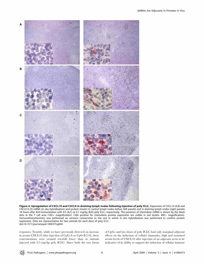
Draining inguinal lymph nodes were also analyzed for
CXCL10, CXCL9, and CCL21 by in-situ hybridization and
immunohistochemistry. In comparison to control lymph nodes
removed before immunizations, elevated expression of CXCL10
(Figure 4A and 4B) and CXCL9 (Figure 4C and 4D) was detected
in the T cell areas of draining lymph nodes at 18 hours after
immunization. Chemokine mRNA expression correlated with
protein expression detected by immunohistochemistry (insets in
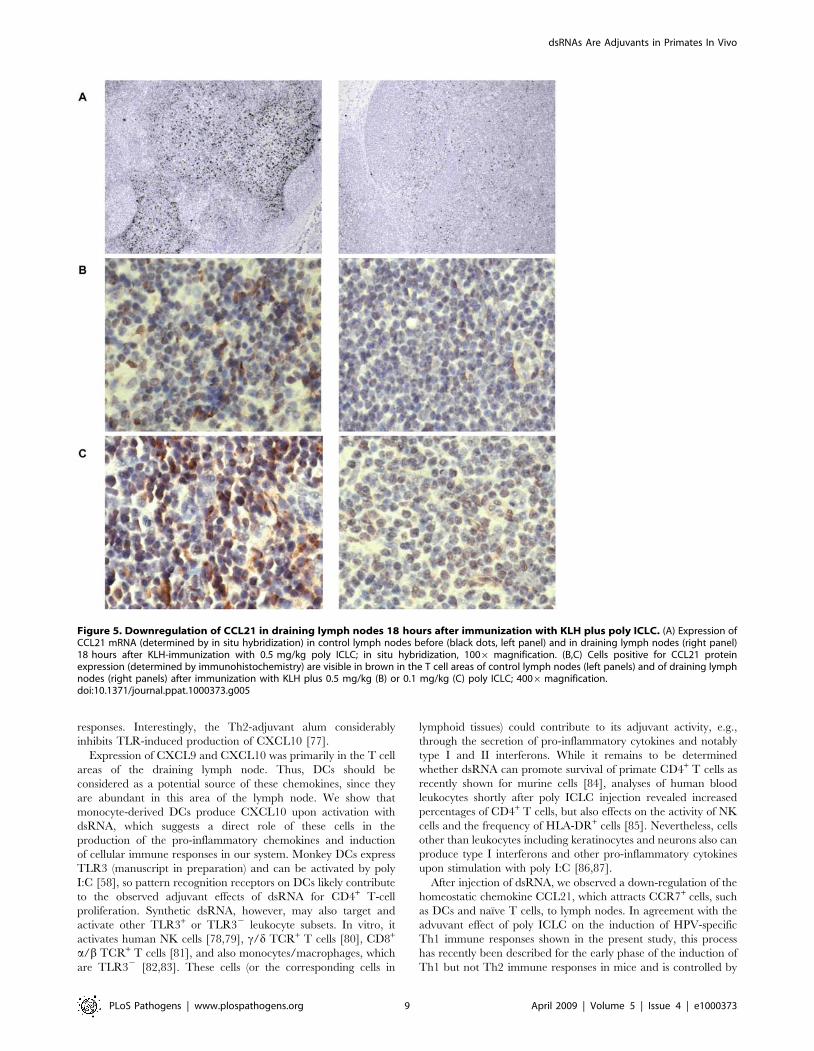
Figure 4). Expression of CCL21 mRNA (Figure 5A) and protein
(Figure 5B and 5C) 18 hours after immunization was markedly
decreased in draining lymph nodes compared with control lymph
nodes obtained before immunization. Thus, the innate response to
dsRNA is detectable in lymph node cells.
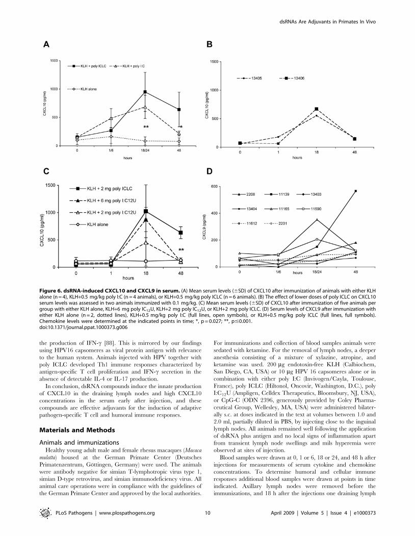
The administration of poly ICLC or poly I:C together with
KLH led to a significant increase of serum levels of CXCL10
(Figure 6A; p = 0.001 for both compounds at 18 or 24 h).
Furthermore, 48 h after immunization, serum levels of CXCL10
were significantly higher in poly ICLC- than in poly I:C-injected
monkeys (p = 0.027). Like poly I:C at 0.5 mg/kg, poly I:C12U or
lower doses of poly ICLC (0.1 mg/kg) induced increased CXCL10
levels, which were less sustained (Figure 6B and 6C).
We detected increased CXCL9 serum levels in animals injected
with poly I:C or poly ICLC (0.5 mg/kg), and there were minor
changes in CXCL9 concentrations in monkeys receiving KLH
alone (Figure 6D). No changes of CXCL9 serum concentrations
were observed when 0.1 mg/kg poly ICLC were administered
(data not shown). At 6, 24, or 48 h after infection, no significant
differences in serum levels of IFN-a, IFN-c, TNF, IL-12p40, and
CCL3 (MIP-1a) were observed between groups receiving KLH
alone or together with dsRNA (data not shown). We were not able
to detect considerable serum concentrations of IFN-a at any point
in time including 1 h post injection.
CXCL10 production by rhesus macaque DCs activatedthrough dsRNA
Since immunohistochemistry and in-situ hybridization revealed
that CXCL10 was mainly produced in the T cell-areas of the
draining lymph nodes (Figure 4), we considered DCs as a potential
source for this chemokine in vivo. Unfortunately, double-labeling
with DC identifying mAbs was not possible on formalin-fixed
specimens. We therefore tested whether dsRNA may directly
induce CXCL10 secretion by highly purified rhesus macaque DCs
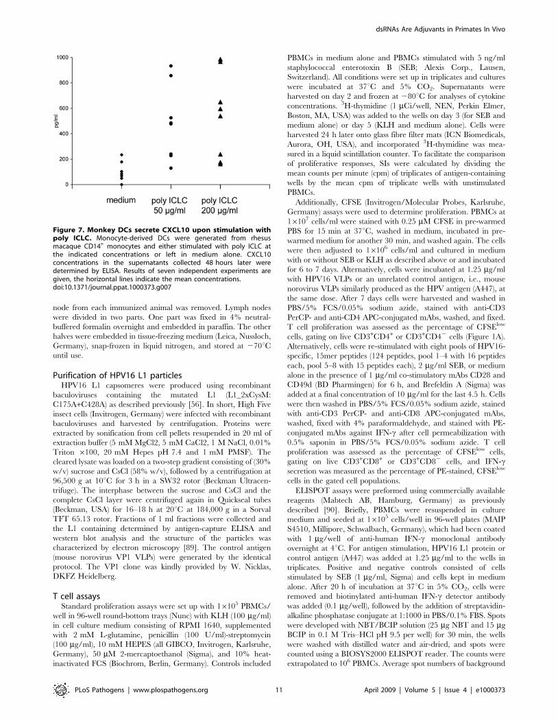
in vitro. When monocyte-derived monkey DCs were incubated
with poly ICLC at two different concentrations (50 and 200 mg/
ml), significantly elevated CXCL10 concentrations were detecta-
ble 48 h later in the cell culture supernatants (p = 0.002 compared
to un-stimulated controls), and both doses of of poly ICLC
induced comparable levels of CXCL10 (Figure 7). Thus, primate
DCs produce CXCL10 upon stimulation with synthetic dsRNA,
making DCs one of the candidate sources of CXCL10 observed in
the draining lymph nodes.
Discussion
This study shows that s.c. injection of synthetic dsRNA, i.e., poly
I:C, poly ICLC, or poly I:C12U supports the induction of cellular
immune responses to protein antigens in nonhuman primates. These
responses could also be boosted by a second injection of antigen
together with dsRNA. We observed antigen-specific T cell
proliferation of CD3+CD4+ and CD3+CD42 T cells. High but
nontoxic doses (toxicity starts in M. mulatta at i.v. doses .2 mg/kg,
i.m. or s.c. injections are better tolerated than i.v. injections;
unpublished observations) of poly ICLC (0.5 mg/kg or 2 mg/
animal) might be more potent than lower doses (#0.1 mg/kg). Using
HPV16 capsomeres at low doses (10 mg/animal) as a relevant viral
antigen with low immunogenicity, we also showed that poly ICLC,
but not CpG-C (which supported the induction of humoral
responses, however), supports the induction of HPV16-specific
Th1 responses. The lack of effect of CpG-C in our system compared
to other studies where the same compound helped to elicit cellular
immunity in nonhuman primates is most likely due to the fact that
we injected the antigens in PBS, while others injected CpG-C and
antigens in the synthetic water-in-oil emulsion, Montanide [49].
Amongst the three different formulations of synthetic dsRNA, poly
ICLC appears to possess the most potent adjuvant activity on the
induction of cellular immune responses. Subsequent studies will
show whether it will help to induce protective immune responses
against other pathogens, e.g., SIV.
Both adjuvants supported the induction of humoral
immune responses, including neutralizing antibodies. Therefore,
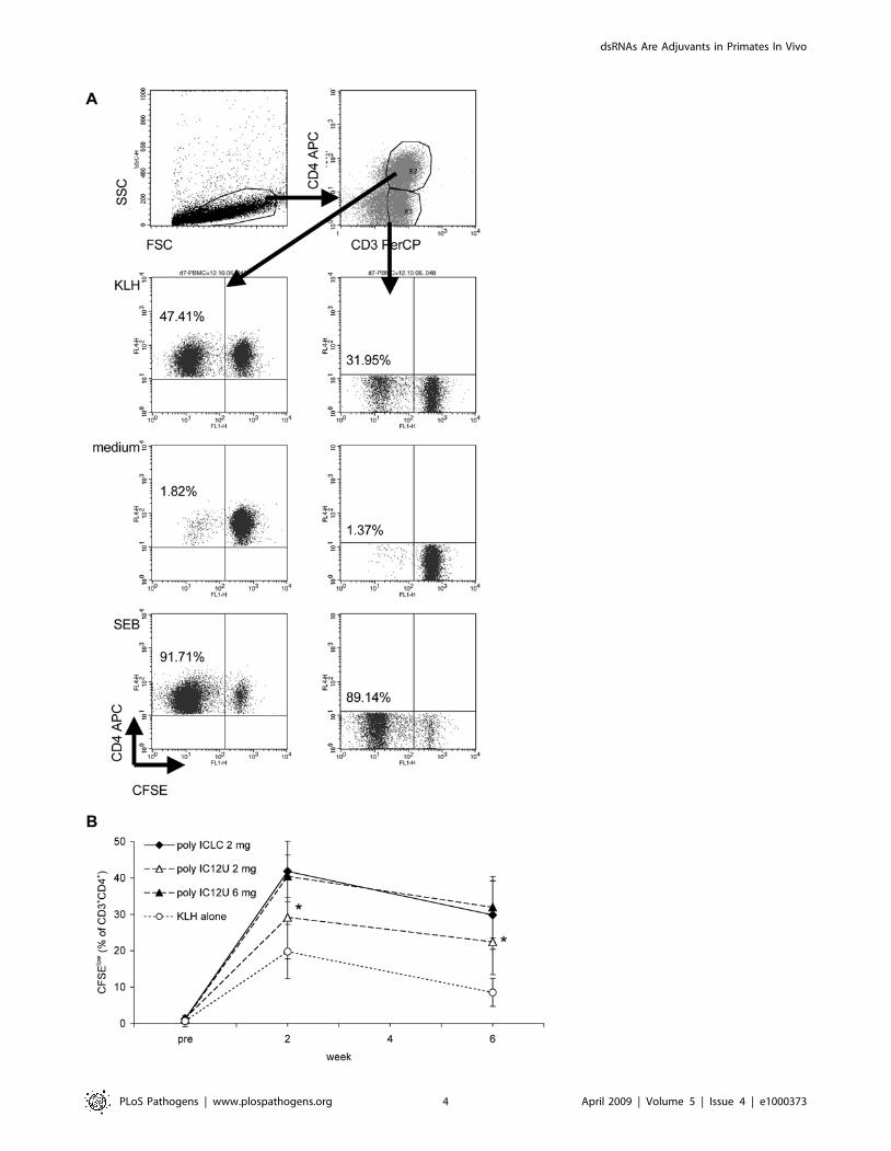
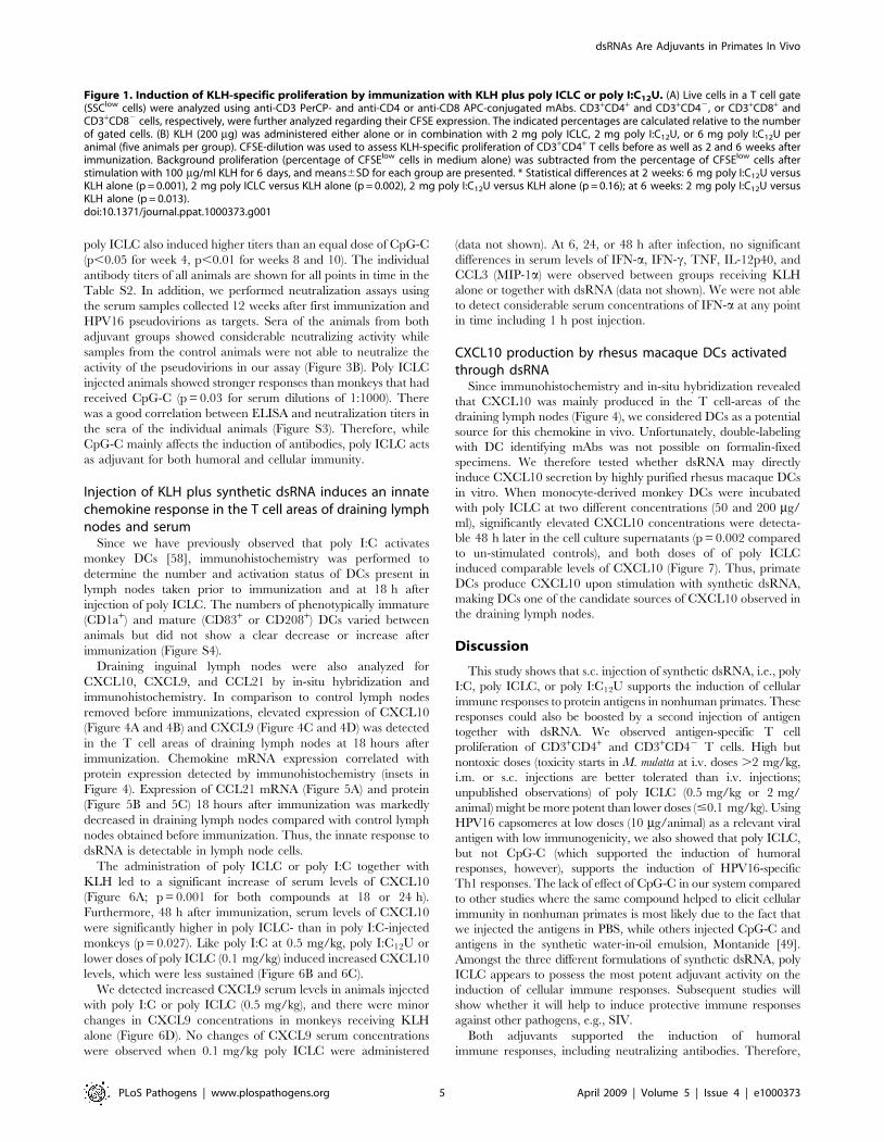
Figure 1. Induction of KLH-specific proliferation by immunization with KLH plus poly ICLC or poly I:C12U. (A) Live cells in a T cell gate(SSClow cells) were analyzed using anti-CD3 PerCP- and anti-CD4 or anti-CD8 APC-conjugated mAbs. CD3+CD4+ and CD3+CD42, or CD3+CD8+ andCD3+CD82 cells, respectively, were further analyzed regarding their CFSE expression. The indicated percentages are calculated relative to the numberof gated cells. (B) KLH (200 mg) was administered either alone or in combination with 2 mg poly ICLC, 2 mg poly I:C12U, or 6 mg poly I:C12U peranimal (five animals per group). CFSE-dilution was used to assess KLH-specific proliferation of CD3+CD4+ T cells before as well as 2 and 6 weeks afterimmunization. Background proliferation (percentage of CFSElow cells in medium alone) was subtracted from the percentage of CFSElow cells afterstimulation with 100 mg/ml KLH for 6 days, and means6SD for each group are presented. * Statistical differences at 2 weeks: 6 mg poly I:C12U versusKLH alone (p = 0.001), 2 mg poly ICLC versus KLH alone (p = 0.002), 2 mg poly I:C12U versus KLH alone (p = 0.16); at 6 weeks: 2 mg poly I:C12U versusKLH alone (p = 0.013).doi:10.1371/journal.ppat.1000373.g001
subsequent in vivo studies should compare poly ICLC with the
adjuvants currently used in vaccine formulations, e.g., alum,
and investigate whether its co-application might allow fewer
injections than required today for the currently licensed vaccine
formulations.
In order to understand the activity of dsRNA, we examined the
innate response since this includes events that can improve the
function of antigen presenting DCs and T cells. Surprisingly, we
did not detect the expected increase of serum IFN-a shortly after
injection of poly I:C or poly ICLC. This might be due to the s.c.
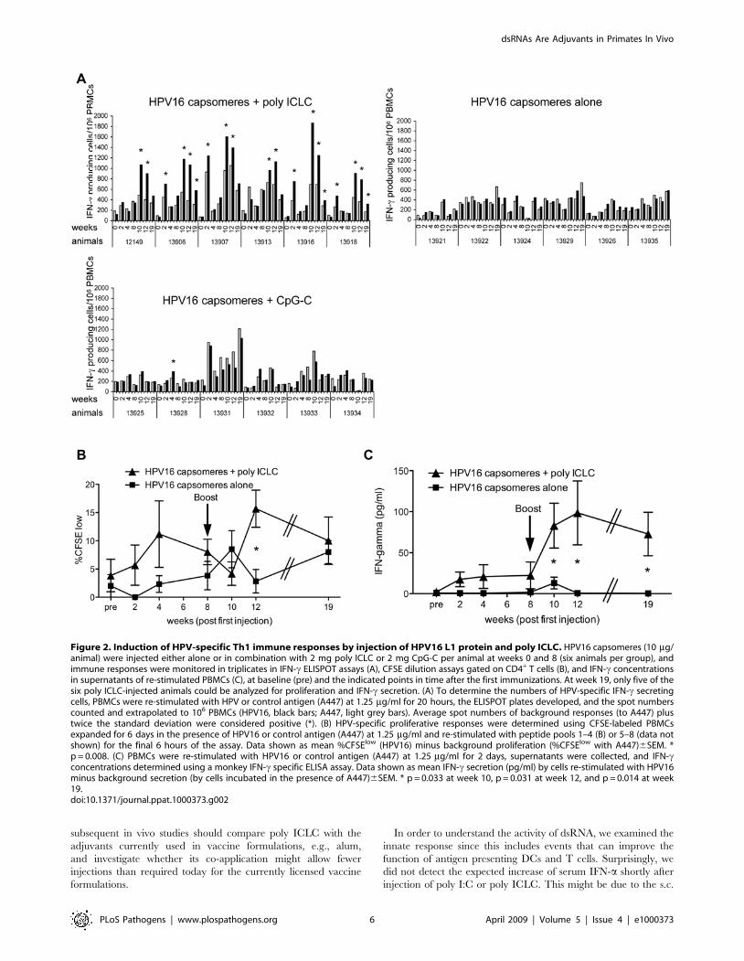
Figure 2. Induction of HPV-specific Th1 immune responses by injection of HPV16 L1 protein and poly ICLC. HPV16 capsomeres (10 mg/animal) were injected either alone or in combination with 2 mg poly ICLC or 2 mg CpG-C per animal at weeks 0 and 8 (six animals per group), andimmune responses were monitored in triplicates in IFN-c ELISPOT assays (A), CFSE dilution assays gated on CD4+ T cells (B), and IFN-c concentrationsin supernatants of re-stimulated PBMCs (C), at baseline (pre) and the indicated points in time after the first immunizations. At week 19, only five of thesix poly ICLC-injected animals could be analyzed for proliferation and IFN-c secretion. (A) To determine the numbers of HPV-specific IFN-c secretingcells, PBMCs were re-stimulated with HPV or control antigen (A447) at 1.25 mg/ml for 20 hours, the ELISPOT plates developed, and the spot numberscounted and extrapolated to 106 PBMCs (HPV16, black bars; A447, light grey bars). Average spot numbers of background responses (to A447) plustwice the standard deviation were considered positive (*). (B) HPV-specific proliferative responses were determined using CFSE-labeled PBMCsexpanded for 6 days in the presence of HPV16 or control antigen (A447) at 1.25 mg/ml and re-stimulated with peptide pools 1–4 (B) or 5–8 (data notshown) for the final 6 hours of the assay. Data shown as mean %CFSElow (HPV16) minus background proliferation (%CFSElow with A447)6SEM. *p = 0.008. (C) PBMCs were re-stimulated with HPV16 or control antigen (A447) at 1.25 mg/ml for 2 days, supernatants were collected, and IFN-cconcentrations determined using a monkey IFN-c specific ELISA assay. Data shown as mean IFN-c secretion (pg/ml) by cells re-stimulated with HPV16minus background secretion (by cells incubated in the presence of A447)6SEM. * p = 0.033 at week 10, p = 0.031 at week 12, and p = 0.014 at week19.doi:10.1371/journal.ppat.1000373.g002
route of injection. While i.v. injections of poly ICLC give rise to
high serum interferon levels [30], the s.c. application of dsRNA
may lead to a more protracted release from the site of injection
and a delayed bioavailability. In mice, type I interferon induced by
poly I:C has been shown to be essential for its adjuvant effect on
humoral immunity and isotype switching [59], and it also seems
essential for TLR3-mediated cross-priming of CD8+ T cells [60–
62]. Likewise, type I interferon is critical for the CD8+ T cell
expansion induced by TLR agonists in combination with CD40
[63]. Poly I:C and poly ICLC induce proliferation of CD8+ T
cells, both have been shown to be effective as an adjuvant for the
induction of specific CD8+ T cell responses in mice [64–66], and
this effect partially depends on NK cells [67]. Thus, poly I:C, and
most likely also poly ICLC, support the induction of CD8+ T cell
responses, and the KLH-specific responses expressed by
CD3+CD42 T cells observed by us might reflect true CD8
responses.
In contrast to our inability to detect IFN-a in the serum in
response to dsRNA, we did detect enhanced levels of CXCL10.
These were sustained over 48 hours in animals injected with
0.5 mg/kg poly ICLC but decreased more rapidly in monkeys
following injection of lower concentrations of poly ICLC, 0.5 mg/
kg poly I:C, or a comparable dose (2 mg/animal) of poly I:C12U.
This may reflect the reduced biostability of the nonstabilized poly
I:C and poly I:C12U compared with that of poly ICLC as
described before [29,30]. CXCL10 is known for its activity to
attract effector Th1 cells through interaction with its receptor
CXCR3 at sites for the expression of Th1 immune responses [68],
e.g., rejection of allografts or the inflammatory response upon
mycobacterial infection [69,70]. CXCL10 is also required for
resistance to protozoan or viral pathogens [71,72]. Studies in mice
revealed additionally that CXCL10 is secreted early (e.g., earlier
than CXCL9, which we did not detect at the same levels in the
serum as CXCL10) [73], and stimulates T cell proliferation [74].
In fact, CXCL10-deficient mice have impaired T cell responses
following primary immunization with exogeneous protein antigen
indicating a role for CXCL10 in effector T cell generation [75].
Since CXCR3 also is induced early in CD4 T lymphocyte
differentiation [76], the literature suggests an enhancing role for
CXCL10 in both the expression and induction of Th1 immune
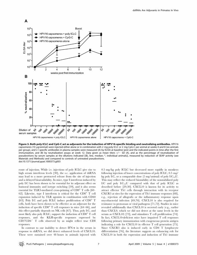
Figure 3. Both poly ICLC and CpG-C act as adjuvants for the induction of HPV16-specific binding and neutralizing antibodies. HPV16capsomeres (10 mg/animal) were injected either alone or in combination with 2 mg poly ICLC or 2 mg CpG-C per animal at weeks 0 and 8 (six animalsper group), and L1-specific antibodies in plasma samples were measured (A) by ELISA at baseline (pre) and the indicated points in time after the firstimmunization, and (B) by neutralization assays at week 12. Data given as mean titers +/2 SD (A), and as the percentage of neutralization ofpseudovirions by serum samples at the dilutions indicated [(B), line, median; *, individual animals)], measured by reduction of SEAP activity (seeMaterials and Methods) and compared to controls of untreated pseudovirions.doi:10.1371/journal.ppat.1000373.g003
responses. Notably, while we have previously detected an increase
in serum CXCL10 after injection of CpG-A or CpG-B [14], these
concentrations were around ten-fold lower than in animals
injected with 0.5 mg/kg poly ICLC. Since both the two forms
of CpGs and low doses of poly ICLC had only marginal adjuvant
effects on the induction of cellular immunity, high and sustained
serum levels of CXCL10 after injection of an adjuvant seem to be
indicative of its ability to support the induction of cellular immune
Figure 4. Upregulation of CXCL10 and CXCL9 in draining lymph nodes following injection of poly ICLC. Expression of CXCL10 (A,B) andCXCL9 (C,D) mRNA (in situ hybridization) and protein (insets) in control lymph nodes before (left panels) and in draining lymph nodes (right panels)18 hours after KLH-immunization with 0.5 (A,C) or 0.1 mg/kg (B,D) poly ICLC, respectively. The presence of chemokine mRNA is shown by the blackdots in the T cell area (1006 magnification). Cells positive for chemokine protein expression are visible in red (insets: 4006 magnification).Immunohistochemistry was performed on sections consecutive to the one in which in situ hybridization was performed to confirm proteinexpression. Data are representative for two animals for each dose of poly ICLC.doi:10.1371/journal.ppat.1000373.g004
responses. Interestingly, the Th2-adjuvant alum considerably
inhibits TLR-induced production of CXCL10 [77].
Expression of CXCL9 and CXCL10 was primarily in the T cell
areas of the draining lymph node. Thus, DCs should be
considered as a potential source of these chemokines, since they
are abundant in this area of the lymph node. We show that
monocyte-derived DCs produce CXCL10 upon activation with
dsRNA, which suggests a direct role of these cells in the
production of the pro-inflammatory chemokines and induction
of cellular immune responses in our system. Monkey DCs express
TLR3 (manuscript in preparation) and can be activated by poly
I:C [58], so pattern recognition receptors on DCs likely contribute
to the observed adjuvant effects of dsRNA for CD4+ T-cell
proliferation. Synthetic dsRNA, however, may also target and
activate other TLR3+ or TLR32 leukocyte subsets. In vitro, it
activates human NK cells [78,79], c/d TCR+ T cells [80], CD8+
a/b TCR+ T cells [81], and also monocytes/macrophages, which
are TLR32 [82,83]. These cells (or the corresponding cells in
lymphoid tissues) could contribute to its adjuvant activity, e.g.,
through the secretion of pro-inflammatory cytokines and notably
type I and II interferons. While it remains to be determined
whether dsRNA can promote survival of primate CD4+ T cells as
recently shown for murine cells [84], analyses of human blood
leukocytes shortly after poly ICLC injection revealed increased
percentages of CD4+ T cells, but also effects on the activity of NK
cells and the frequency of HLA-DR+ cells [85]. Nevertheless, cells
other than leukocytes including keratinocytes and neurons also can
produce type I interferons and other pro-inflammatory cytokines
upon stimulation with poly I:C [86,87].
After injection of dsRNA, we observed a down-regulation of the
homeostatic chemokine CCL21, which attracts CCR7+ cells, such
as DCs and naıve T cells, to lymph nodes. In agreement with the
advuvant effect of poly ICLC on the induction of HPV-specific
Th1 immune responses shown in the present study, this process
has recently been described for the early phase of the induction of
Th1 but not Th2 immune responses in mice and is controlled by
Figure 5. Downregulation of CCL21 in draining lymph nodes 18 hours after immunization with KLH plus poly ICLC. (A) Expression ofCCL21 mRNA (determined by in situ hybridization) in control lymph nodes before (black dots, left panel) and in draining lymph nodes (right panel)18 hours after KLH-immunization with 0.5 mg/kg poly ICLC; in situ hybridization, 1006 magnification. (B,C) Cells positive for CCL21 proteinexpression (determined by immunohistochemistry) are visible in brown in the T cell areas of control lymph nodes (left panels) and of draining lymphnodes (right panels) after immunization with KLH plus 0.5 mg/kg (B) or 0.1 mg/kg (C) poly ICLC; 4006magnification.doi:10.1371/journal.ppat.1000373.g005
or CpG-C (ODN 2396, generously provided by Coley Pharma-
ceutical Group, Wellesley, MA, USA) were administered bilater-
ally s.c. at doses indicated in the text at volumes between 1.0 and
2.0 ml, partially diluted in PBS, by injecting close to the inguinal
lymph nodes. All animals remained well following the application
of dsRNA plus antigen and no local signs of inflammation apart
from transient lymph node swellings and mils hyperemia were
observed at sites of injection.
Blood samples were drawn at 0, 1 or 6, 18 or 24, and 48 h after
injections for measurements of serum cytokine and chemokine
concentrations. To determine humoral and cellular immune
responses additional blood samples were drawn at points in time
indicated. Axillary lymph nodes were removed before the
immunizations, and 18 h after the injections one draining lymph
Figure 6. dsRNA-induced CXCL10 and CXCL9 in serum. (A) Mean serum levels (6SD) of CXCL10 after immunization of animals with either KLHalone (n = 4), KLH+0.5 mg/kg poly I:C (n = 4 animals), or KLH+0.5 mg/kg poly ICLC (n = 6 animals). (B) The effect of lower doses of poly ICLC on CXCL10serum levels was assessed in two animals immunized with 0.1 mg/kg. (C) Mean serum levels (6SD) of CXCL10 after immunization of five animals pergroup with either KLH alone, KLH+6 mg poly IC12U, KLH+2 mg poly IC12U, or KLH+2 mg poly ICLC. (D) Serum levels of CXCL9 after immunization witheither KLH alone (n = 2, dotted lines), KLH+0.5 mg/kg poly I:C (full lines, open symbols), or KLH+0.5 mg/kg poly ICLC (full lines, full symbols).Chemokine levels were determined at the indicated points in time; *, p = 0.027; **, p#0.001.doi:10.1371/journal.ppat.1000373.g006
Germany) assays were used to determine proliferation. PBMCs at
16107 cells/ml were stained with 0.25 mM CFSE in pre-warmed
PBS for 15 min at 37uC, washed in medium, incubated in pre-
warmed medium for another 30 min, and washed again. The cells
were then adjusted to 16106 cells/ml and cultured in medium
with or without SEB or KLH as described above or and incubated
for 6 to 7 days. Alternatively, cells were incubated at 1.25 mg/ml
with HPV16 VLPs or an unrelated control antigen, i.e., mouse
norovirus VLPs similarly produced as the HPV antigen (A447), at
the same dose. After 7 days cells were harvested and washed in
PBS/5% FCS/0.05% sodium azide, stained with anti-CD3
PerCP- and anti-CD4 APC-conjugated mAbs, washed, and fixed.
T cell proliferation was assessed as the percentage of CFSElow
cells, gating on live CD3+CD4+ or CD3+CD42 cells (Figure 1A).
Alternatively, cells were re-stimulated with eight pools of HPV16-
specific, 15mer peptides (124 peptides, pool 1–4 with 16 peptides
each, pool 5–8 with 15 peptides each), 2 mg/ml SEB, or medium
alone in the presence of 1 mg/ml co-stimulatory mAbs CD28 and
CD49d (BD Pharmingen) for 6 h, and Brefeldin A (Sigma) was
added at a final concentration of 10 mg/ml for the last 4.5 h. Cells
were then washed in PBS/5% FCS/0.05% sodium azide, stained
with anti-CD3 PerCP- and anti-CD8 APC-conjugated mAbs,
washed, fixed with 4% paraformaldehyde, and stained with PE-
conjugated mAbs against IFN-c after cell permeabilization with
0.5% saponin in PBS/5% FCS/0.05% sodium azide. T cell
proliferation was assessed as the percentage of CFSElow cells,
gating on live CD3+CD8+ or CD3+CD82 cells, and IFN-csecretion was measured as the percentage of PE-stained, CFSElow
cells in the gated cell populations.
ELISPOT assays were preformed using commercially available
reagents (Mabtech AB, Hamburg, Germany) as previously
described [90]. Briefly, PBMCs were resuspended in culture
medium and seeded at 16105 cells/well in 96-well plates (MAIP
S4510, Millipore, Schwalbach, Germany), which had been coated
with 1 mg/well of anti-human IFN-c monoclonal antibody
overnight at 4uC. For antigen stimulation, HPV16 L1 protein or
control antigen (A447) was added at 1.25 mg/ml to the wells in
triplicates. Positive and negative controls consisted of cells
stimulated by SEB (1 mg/ml, Sigma) and cells kept in medium
alone. After 20 h of incubation at 37uC in 5% CO2, cells were
removed and biotinylated anti-human IFN-c detector antibody
was added (0.1 mg/well), followed by the addition of streptavidin-
alkaline phosphatase conjugate at 1:1000 in PBS/0.1% FBS. Spots
were developed with NBT/BCIP solution (25 mg NBT and 15 mg
BCIP in 0.1 M Tris–HCl pH 9.5 per well) for 30 min, the wells
were washed with distilled water and air-dried, and spots were
counted using a BIOSYS2000 ELISPOT reader. The counts were
extrapolated to 106 PBMCs. Average spot numbers of background
Figure 7. Monkey DCs secrete CXCL10 upon stimulation withpoly ICLC. Monocyte-derived DCs were generated from rhesusmacaque CD14+ monocytes and either stimulated with poly ICLC atthe indicated concentrations or left in medium alone. CXCL10concentrations in the supernatants collected 48 hours later weredetermined by ELISA. Results of seven independent experiments aregiven, the horizontal lines indicate the mean concentrations.doi:10.1371/journal.ppat.1000373.g007
72. Hsieh MF, Lai SL, Chen JP, Sung JM, Lin YL, et al. (2006) Both CXCR3 and
CXCL10/IFN-inducible protein 10 are required for resistance to primaryinfection by dengue virus. J Immunol 177: 1855–1863.
73. Widney DP, Xia YR, Lusis AJ, Smith JB (2000) The murine chemokine
CXCL11 (IFN-inducible T cell alpha chemoattractant) is an IFN-gamma- andlipopolysaccharide-inducible glucocorticoid-attenuated response gene expressed
in lung and other tissues during endotoxemia. J Immunol 164: 6322–6331.74. Whiting D, Hsieh G, Yun JJ, Banerji A, Yao W, et al. (2004) Chemokine
monokine induced by IFN-gamma/CXC chemokine ligand 9 stimulates T
lymphocyte proliferation and effector cytokine production. J Immunol 172:7417–7424.
75. Dufour JH, Dziejman M, Liu MT, Leung JH, Lane TE, et al. (2002) IFN-gamma-inducible protein 10 (IP-10; CXCL10)-deficient mice reveal a role for
IP-10 in effector T cell generation and trafficking. J Immunol 168: 3195–3204.76. Rabin RL, Alston MA, Sircus JC, Knollmann-Ritschel B, Moratz C, et al. (2003)
CXCR3 is induced early on the pathway of CD4+ T cell differentiation and
bridges central and peripheral functions. J Immunol 171: 2812–2824.77. Li H, Nookala S, Re F (2007) Aluminum hydroxide adjuvants activate caspase-1
and induce IL-1beta and IL-18 release. J Immunol 178: 5271–5276.78. Sivori S, Falco M, Della Chiesa M, Carlomagno S, Vitale M, et al. (2004) CpG
and double-stranded RNA trigger human NK cells by Toll-like receptors:
induction of cytokine release and cytotoxicity against tumors and dendritic cells.Proc Natl Acad Sci U S A 101: 10116–10121.
79. Schmidt KN, Leung B, Kwong M, Zarember KA, Satyal S, et al. (2004) APC-independent activation of NK cells by the Toll-like receptor 3 agonist double-
stranded RNA. J Immunol 172: 138–143.80. Wesch D, Beetz S, Oberg HH, Marget M, Krengel K, et al. (2006) Direct
costimulatory effect of TLR3 ligand poly(I:C) on human gamma delta T
lymphocytes. J Immunol 176: 1348–1354.81. Tabiasco J, Devevre E, Rufer N, Salaun B, Cerottini JC, et al. (2006) Human
effector CD8+ T lymphocytes express TLR3 as a functional coreceptor.J Immunol 177: 8708–8713.
82. Stevenson HC, Dekaban GA, Miller PJ, Benyajati C, Pearson ML (1985)
Analysis of human blood monocyte activation at the level of gene expression.Expression of alpha interferon genes during activation of human monocytes by
poly IC/LC and muramyl dipeptide. J Exp Med 161: 503–513.
83. Pirhonen J, Siren J, Julkunen I, Matikainen S (2007) IFN-{alpha} regulates Toll-
like receptor-mediated IL-27 gene expression in human macrophages. J LeukocBiol 82: 1185–1192. E-pub August 7.
84. Gelman AE, Zhang J, Choi Y, Turka LA (2004) Toll-like receptor ligands
directly promote activated CD4+ T cell survival. J Immunol 172: 6065–6073.85. Bever CT Jr, Jacobson S, Mingioli ES, McFarland HF, McFarlin DE, et al.
(1991) Changes in leukocyte recirculation, NK cell activity, and HLA-DRexpression in peripheral blood mononuclear cells of MS patients treated with
Poly ICLC. Int J Immunopharmacol 13: 613–618.
86. Lebre MC, Antons JC, Kalinski P, Schuitemaker JH, van Capel TM, et al.(2003) Double-stranded RNA-exposed human keratinocytes promote Th1
responses by inducing a Type-1 polarized phenotype in dendritic cells: role ofkeratinocyte-derived tumor necrosis factor alpha, type I interferons, and
interleukin-18. J Invest Dermatol 120: 990–997.87. Prehaud C, Megret F, Lafage M, Lafon M (2005) Virus infection switches TLR-
3-positive human neurons to become strong producers of beta interferon. J Virol
79: 12893–12904.88. Mueller SN, Hosiawa-Meagher KA, Konieczny BT, Sullivan BM,
Bachmann MF, et al. (2007) Regulation of homeostatic chemokine expressionand cell trafficking during immune responses. Science 317: 670–674.
89. Muller M, Zhou J, Reed TD, Rittmuller C, Burger A, et al. (1997) Chimeric
papillomavirus-like particles. Virology 234: 93–111.90. Stahl-Hennig C, Eisenblatter M, Franz M, Stoiber H, Tenner-Racz K, et al.
(2007) A single vaccination with attenuated SIVmac 239 via the tonsillar routeconfers partial protection against challenge with SIVmac 251 at a distant
mucosal site, the rectum. Front Biosci 12: 2107–2123.91. Gasparic M, Rubio I, Thones N, Gissmann L, Muller M (2007) Prophylactic
DNA immunization against multiple papillomavirus types. Vaccine 25:
4540–4553.92. Manzo A, Paoletti S, Carulli M, Blades MC, Barone F, et al. (2005) Systematic
microanatomical analysis of CXCL13 and CCL21 in situ production andprogressive lymphoid organization in rheumatoid synovitis. Eur J Immunol 35:
1347–1359.
93. Mazzucchelli L, Blaser A, Kappeler A, Scharli P, Laissue JA, et al. (1999) BCA-1is highly expressed in Helicobacter pylori-induced mucosa-associated lymphoid
tissue and gastric lymphoma. J Clin Invest 104: R49–54.