Design and feasibility of a novel program ofcervical screening in Nigeria: self-sampledHPV testing paired with visual triageKanan T. Desai1,2†, Kayode O. Ajenifuja3*†, Adekunbiola Banjo4, Clement A. Adepiti3, Akiva Novetsky5, Cathy Sebag6,Mark H. Einstein5, Temitope Oyinloye3, Tamara R. Litwin1, Matt Horning7, Fatai Olatunde Olanrewaju3,Mufutau Muphy Oripelaye3, Esther Afolabi3, Oluwole O. Odujoko3, Philip E. Castle8, Sameer Antani9, Ben Wilson7,Liming Hu7, Courosh Mehanian7, Maria Demarco1, Julia C. Gage1, Zhiyun Xue9, Leonard R. Long9, Li Cheung1,Didem Egemen1, Nicolas Wentzensen1 and Mark Schiffman1
Abstract
Background: Accelerated global control of cervical cancer would require primary prevention with humanpapillomavirus (HPV) vaccination in addition to novel screening program strategies that are simple, inexpensive, andeffective. We present the feasibility and outcome of a community-based HPV self-sampled screening program.
Methods: In Ile Ife, Nigeria, 9406 women aged 30–49 years collected vaginal self-samples, which were tested for HPV inthe local study laboratory using Hybrid Capture-2 (HC2) (Qiagen). HPV-positive women were referred to the colposcopyclinic. Gynecologist colposcopic impression dictated immediate management; biopsies were taken when definiteacetowhitening was present to produce a histopathologic reference standard of precancer (and to determine final clinicalmanagement). Retrospective linkage to the medical records identified 442 of 9406 women living with HIV (WLWH).
Results: With self-sampling, it was possible to screen more than 100 women per day per clinic. Following an audio-visual presentation and in-person instructions, overall acceptability of self-sampling was very high (81.2% womenpreferring self-sampling over clinician collection). HPV positivity was found in 17.3% of women. Intensive follow-upcontributed to 85.9% attendance at the colposcopy clinic. Of those referred, 8.2% were initially treated with thermalablation and 5.6% with large loop excision of transformation zone (LLETZ). Full visibility of the squamocolumnarjunction, necessary for optimal visual triage and ablation, declined from 68.5% at age 30 to 35.4% at age 49. CIN2+ andCIN3+ (CIN- Cervical intraepithelial neoplasia), including five cancers, were identified by histology in 5.9 and 3.2% of theHPV-positive women, respectively (0.9 and 0.5% of the total screening population), leading to additional treatment asindicated. The prevalences of HPV infection and CIN2+ were substantially higher (40.5 and 2.5%, respectively) amongWLWH. Colposcopic impression led to over- and under-treatment compared to the histopathology reference standard.
* Correspondence: [email protected]†Kanan T. Desai and Kayode O. Ajenifuja contributed equally to this work.3Department of Obstetrics and Gynecology, Obafemi Awolowo UniversityTeaching Hospital, Ile Ife, NigeriaFull list of author information is available at the end of the article
Desai et al. Infectious Agents and Cancer (2020) 15:60 https://doi.org/10.1186/s13027-020-00324-5
Conclusion: A cervical cancer screening program using self-sampled HPV testing, with colposcopic immediatemanagement of women positive for HPV, proved feasible in Nigeria. Based on the collected specimens and images, weare now evaluating the use of a combination of partial HPV typing and automated visual evaluation (AVE) of cervicalimages to improve the accuracy of the screening program.
IntroductionNearly 85% of the annual 570,000 cervical cancer casesand almost 90% of the 311,000 related deaths occur inlower-resource countries [1, 2] due to the lack of effectivecervical cancer prevention programs [3]. The COVID-19(coronavirus disease of 2019) pandemic threatens to re-duce elective procedures even further, including cervicalscreening and related diagnostic procedures, especially inlower-resource settings, and will likely worsen cervicalcancer health disparities. As a major advantage comparedwith cytology, the specimen for HPV (human papillomavi-rus) testing can be collected by the woman herself using avaginal self-collection device, yielding sensitivity for HPVinfection that is similar to clinician-collected specimenswhen target-amplification methods like PCR (polymerasechain reaction) are used [4, 5]. Use of HPV testing will re-quire a second diagnostic modality for positives, becausemajority of HPV infections are cleared within 1–2 years ofinitial detection. Only HPV infections that persist cancause precancer and invasive cancer [6]. Thus, an im-proved screening program must include triage methods tofocus treatment safely on the small fraction of HPV-positive women with precancer, the general term we useto refer to lesions at substantial risk of invasion withouttreatment [7]. Ideal characteristics of a triage test forHPV-positive women, for use in low-resource regions,would include excellent risk discrimination (high precan-cer risk in positives, low risk in negatives), low-cost, sim-plicity, and point-of-care use.A new candidate for triage of HPV-positive women is
automated visual evaluation (AVE) of cervical imagesusing a deep-learning algorithm. A proof-of-principleevaluation of AVE of Cervigrams (NTL Worldwide, Fen-ton, Missouri) for the diagnosis of cervical precancer,demonstrated higher accuracy of AVE than expertgynecologist visual assessment or cytology [8].Although these are promising results, the Cervigram
cervical images were based on film and a discontinuedexpensive custom camera, and the technology is obso-lete. Thus, it is essential to advance the transfer of themethod to modern image acquisition devices (e.g.,smartphones) [9]. There is also a need to evaluate theperformance of AVE specifically for the triage of HPV-positive women, as HPV-positive controls [<CIN2 (CIN-
cervical intraepithelial neoplasia)] tend to look less nor-mal and are consequently harder to differentiate fromprecancer cases than HPV-negative controls.This paper describes the field methods, feasibility, and
preliminary descriptive results of Project Itoju in Ile-Ife,Nigeria, designed to evaluate ultimately the combinedstrategy of self-sampled HPV typing combined withAVE triage on three different image capture devices.
Materials and methodsStudy design and overviewWomen aged 30–49 years (comprising ~ 8% of the totalpopulation [10]) residing in the catchment area of Oba-femi Awolowo University Teaching Hospitals Complex(OAUTHC) in Ile-Ife, Nigeria were invited by a publicmessage campaign for self-sampled HPV testing.Women who screened HPV-positive were referred tothe colposcopy clinic and invited to participate in a re-search study (examining triage methods) under informedconsent. Standard colposcopic examination, includingcolposcopic images and treatment, if indicated, was of-fered to all HPV-positive women regardless of participa-tion in the triage methods research. In addition, cervicalimages were collected with a cellphone and the EVA(enhanced visual assessment) system (MobileODT,Israel), and a cervical sample was collected for subse-quent HPV typing, from all the participants in the study.The study was approved by National Cancer Institute
(NCI) and OAUTHC ethical Institutional ReviewBoards.
Study timelineThe screening period was from November 2018 to De-cember 2019. The colposcopy period was from Decem-ber 2018 to March 2020.
Screening visitThe screening visit started when a woman attended oneof the three screening clinics (no appointment needed).A nurse-administered short screening questionnaire de-termined eligibility [age 30–49 years and not pregnant(self-reported)] for screening. If a woman was menstru-ating and was uncomfortable undergoing screening, shewas advised to return later. Known pregnant women
Desai et al. Infectious Agents and Cancer (2020) 15:60 Page 2 of 13
were excluded due to an “abundance of caution”, mainlyto prevent any possibility that an unrelated adverse preg-nancy outcome could be due, or even perceived to bedue, to the self-sampling for screening. The lower limitof age for screening was set at 30 years since youngerwomen have a high prevalence of HPV but a very lowrisk of cancer [11]. The upper age limit was restricted to49 years due to age-related repositioning of the squamo-columnar junction (SCJ), where cancers arise, into theendocervical canal, limiting the ability of any existingvisual screening or triage method to diagnose precanceraccurately [12]. An informed consent at screening wasobtained from eligible women seeking permission tostore the left-over sample after HPV testing for futureresearch and to be contacted in future for a follow-upstudy. However, the screening effort was a public healthintervention and not an experimental study.After enrollment, eligible women were provided with
an HPV self-sample collection kit containing a cervicalbrush and a Specimen Transport Medium (STM) tube(Qiagen, USA) [13]. Women were shown a 5-min ani-mated video on how to collect a vaginal self-sample[https://www.youtube.com/watch?v=JiNqrDntbTc] [14]while waiting. Women went into a private self-samplecollection area one at a time to self-sample. After col-lecting the sample, each woman left the brush in theSTM vial in a rack. A nurse helped to break the stem ofthe brush, closed and labelled the vial, and cleaned theoutside of the vial and rack with an alcohol wipe. Thenurse was available to assist women in specimen collec-tion upon request. The collection room was cleaned be-tween collections. Before leaving the clinic, participantscompleted an anonymous feedback form regarding theirexperience with self-sampling. The specimens werestored at room temperature and transferred to the HPVlaboratory at the end of the day, to be stored at 2–80 Cuntil testing.
HPV testingThe primary HPV test used in the study was the DigeneHybrid Capture-2 (HC2) HPV DNA (deoxyribose nu-cleic acid) Test (Qiagen, USA), which is a US Food andDrug Administration approved nucleic acidhybridization assay with signal amplification using mi-croplate chemiluminescence targeting 13 high-risk typesof HPV DNA in cervical and vaginal specimens, withoutdistinguishing between them [15].Trained nurses called participants by phone, when
their HPV test results were available (within two weeksof collection for most of the study period). HPV-negative women were informed and educated about thetest result over the phone, and their questions were an-swered. HPV-positive women were asked to visit the col-poscopy clinic to receive their test results. A minimum
of five contact attempts were made to approach and ad-vise a woman to attend the colposcopy clinic before awoman was declared lost to follow-up.
Colposcopy clinic visitAt the clinic, a nurse communicated the positive HPVtest result and its clinical meaning to the woman, ensur-ing privacy. The woman was counseled to undergo a col-poscopy examination, preferably during that same visitor later.Each woman was registered and interviewed to deter-
mine eligibility for the triage methods research study(analyses in progress to be reported separately). Anyonewith a history of cervical cancer, hysterectomy, or whowas pregnant at the time of enrollment (confirmed withrapid pregnancy test) was excluded from the study. A fe-male nurse took informed consent from all the eligiblewomen for participation in a research study (examiningtriage methods). An anonymized picture of the cervixwas shown to the woman at the time of consent in orderto reassure her about confidentiality and privacy ofimage collection and to minimize refusals.Following the interview, a colposcopy examination was
performed by one of the study gynecologists (KOA,CAA). Cervical images for research were collected oneminute after applying 5% acetic acid for each device, se-quentially with three different devices: 1) a SamsungGalaxy S8 [16] smartphone; 2) a MobileODT EVA de-vice that provided lighting and magnification for a Sam-sung Galaxy J5 smartphone [17]; and 3) a Zeiss FC150colposcopic image captured via a beam splitter by aDSLR (digital single-lens reflex) camera [18, 19]. Afterimage collection, a cervical specimen was collected usinga cervical sample collection kit containing a cervicalbrush and a STM tube (Qiagen, USA) [13] and stored at2-8 °C. We plan to test this sample along with the re-sidual sample from screening for HPV typing using theTypeSeq HPV test [20] at the NCI and report the resultsin future publications.At the beginning of the study, a dry swab sample was
collected at colposcopy, before collecting images and ap-plying acetic acid, for the two-type OncoE6 test (ArborVita, USA). The test is known to have high positive pre-dictive value; in fact, three of the five positives (all forHPV 16) were diagnosed with CIN2+. However, the testwas dropped after testing 373 samples due to rare posi-tivity and low yield.Finally, a standard colposcopy examination was per-
formed to assess the presence and possible nature of cer-vical lesions and to take biopsies of acetowhite lesions,up to a maximum of four biopsies. In addition, endocer-vical curettage (ECC) was performed in cases where theSCJ was not fully visible, even in the absence of aceto-whitening. All women with acetowhite lesions were
Desai et al. Infectious Agents and Cancer (2020) 15:60 Page 3 of 13
offered immediate treatment without waiting for histo-pathology results, following the American Society ofColposcopy and Cervical Pathology 2012 Consensusguidelines [21], leaning towards the more clinically-aggressive options for women at risk of being lost tofollow-up. Either thermal ablation or large loop excisionof the transformation zone (LLETZ) was performed, de-pending on the colposcopy examination findings. Abla-tion was performed only if the SCJ was fully visible, theentire lesion was visible, the lesion did not cover > 75%of the ectocervix, and cervix architecture was appropri-ate for the ablation probe [22].
Histopathology and final diagnosisHistopathological confirmation of CIN2 or CIN3 wasused as the reference standard for the presence of pre-cancer, against which other experimental tests were eval-uated and final clinical review decisions were made.Even though from the clinical management purposes, allhigh-grade (CIN2+) lesions were treated equivalently;for the true yield of the screening effort, we reported theprevalence of CIN2+ and CIN3+ lesions separately toavoid the ambiguity of equivocal CIN2 lesions (a mixtureof HPV infections, true precancers, and an error in his-topathologic diagnosis) [23]. The study pathologist (AB)at the University of Lagos performed all pathologydiagnoses.
Quality assurance review and treatment recallsAll cases were reviewed for adequate clinical manage-ment by a US gynecologic oncologist (AN). The morecomplex cases were discussed in a case conference call.Recall was recommended for women needing furthermanagement and such cases were re-reviewed once therecall was completed until the case was determined tobe adequately treated. We planned to reach all womenneeding recall for additional management, a minimumof seven times. However, we restricted our attempts, andrecalled only those women at the highest immediate riskof invasive cancer because of the spread of the COVID-19 pandemic in March 2020.
Screening and management project softwareAll data and images were collected with a HIPAA(Health Insurance Portability and Accountability Act)compliant smartphone application ‘EVA for research’(MobileODT, Israel). The data platform was custom de-signed for the project (led by CS) using an advanced bar-code scanning system to limit human error from manualkey-in. The data and images from different sources onstudy assigned smartphones were held locally until inter-net connectivity was available, at which point all dataautomatically transferred and aggregated to cloud
servers and portal for analysis and remote qualityassurance.
Data analysisThe preliminary data were analyzed using SPSS 20 (Stat-istical Package for the Social Sciences) [24] and Epi Info[25]. Descriptive results were presented as frequenciesand percentages. Chi-square tests were used to comparethe yield of disease between different subgroups. In fu-ture analyses to evaluate the triage tests, the area underthe curve (AUC) on a ROC (Receiver Operating Charac-teristic) curve will be used.Additional details on study methods (i.e. study site,
organization of the clinics, training of staff, and imagecollection protocol) are provided in the Additional file 1.
ResultsA total of 9625 women came for screening, of which9406 (97.7%) eligible women aged 30–49 years werescreened; and 219 (2.3%) ineligible women (1.7% due toage restrictions) were not screened (Details on enroll-ment and exclusions are provided in the Additional file 1).A total of 442 (4.7%) of 9406 women living with HIV(WLWH) were noted when cross referenced with theHIV clinic of OAUTHC, whereas HIV status was un-known for the remaining 8964 (95.3%) women.With self-sampling, we were able to screen an average
of 20 women per working day (with a peak of up to 100women in a single day per clinic with two self-samplecollection areas). Roughly 9065 participants providedwritten feedback. Of those, 80.8% women said that theywere able to collect the self-sample without any helpfrom a nurse and 81.2% women said that they wouldprefer self-sampling to provider’s sampling in the future.Asked to score their impressions, 78.5% women werevery confident in their ability to collect the self-sampling, 91% found it very easy, 88.5% found it verycomfortable (not painful at all), 95.1% found the video tobe very helpful in guiding how to collect the sample, and97.8% said they are very likely to recommend self-sampling to others. The difficult components of self-sampling reported (one or more issues for 2322 respon-dents) were: the decision on how deep to insert thebrush into the vagina for 52% of 2322, how to insert thebrush into the vagina for 49.1%, identifying the vaginalopening for 39.6%, rotating the brush inside the vaginafor 23.3%, and proper handling to put the brush into thetube after collection for 18.9% women.A total of 1630 (17.3%) [95% confidence interval (CI):
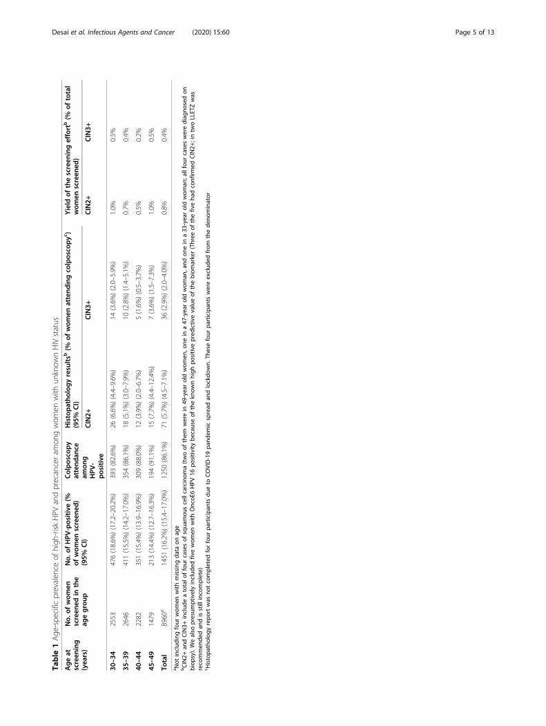
16.6–18.1%] of 9406 screened women were HC2-positive. The rate of HC2 positivity was 16.2% amongthe 8964 women with unknown HIV status and 40.5%among the 442 women living with HIV (P < 0.001, inde-pendent chi-square) (Table 1, Table 2). The strong
Desai et al. Infectious Agents and Cancer (2020) 15:60 Page 4 of 13
Table
1Age
-spe
cific
prevalen
ceof
high
-riskHPV
andprecanceram
ongwom
enwith
unknow
nHIV
status
Ageat
screen
ing
(yea
rs)
No.
ofwom
enscreen
edin
the
agegroup
No.
ofHPV
-positive(%
ofwom
enscreen
ed)
(95%
CI)
Colposco
py
attend
ance
amon
gHPV
-positive
Histopatho
logyresultsb
(%of
wom
enattend
ingco
lposco
pyc)
(95%
CI)
Yield
ofthescreen
ingeffortb(%
oftotal
wom
enscreen
ed)
CIN2+
CIN3+
CIN2+
CIN3+
30–3
42553
476(18.6%
)(17.2–20.2%)
393(82.6%
)26
(6.6%)(4.4–9.6%
)14
(3.6%)(2.0–5.9%)
1.0%
0.5%
35–3
92646
411(15.5%
)(14.2–17.0%)
354(86.1%
)18
(5.1%)(3.0–7.9%
)10
(2.8%)(1.4–5.1%)
0.7%
0.4%
40–4
42282
351(15.4%
)(13.9–16.9%)
309(88.0%
)12
(3.9%)(2.0–6.7%
)5(1.6%)(0.5–3.7%)
0.5%
0.2%
45–4
91479
213(14.4%
)(12.7–16.3%)
194(91.1%
)15
(7.7%)(4.4–12.4%)
7(3.6%)(1.5–7.3%)
1.0%
0.5%
Total
8960
a1451
(16.2%
)(15.4–17.0%)
1250
(86.1%
)71
(5.7%)(4.5–7.1%
)36
(2.9%)(2.0–4.0%)
0.8%
0.4%
a Not
includ
ingfour
wom
enwith
missing
data
onag
ebCIN2+
andCIN3+
includ
eatotalo
ffour
casesof
squa
mou
scellcarcinom
a(twoof
them
werein
49-yearoldwom
en,o
nein
a47
-yearoldwom
an,and
onein
a33
-yearoldwom
an;allfour
caseswerediag
nosedon
biop
sy).Wealso
presum
ptivelyinclud
edfivewom
enwith
OncoE
6HPV
16po
sitiv
itybe
causeof
thekn
ownhigh
positiv
epred
ictiv
evalueof
thebiom
arker(Three
ofthefiveha
dconfirm
edCIN2+
;intw
oLLETZwas
recommen
dedan
disstillincomplete)
c Histopa
tholog
yrepo
rtwas
notcompleted
forfour
participan
tsdu
eto
COVID-19pa
ndem
icspread
andlockdo
wn.
Thesefour
participan
tswereexclud
edfrom
thede
nominator
Desai et al. Infectious Agents and Cancer (2020) 15:60 Page 5 of 13
differences in HPV positivity between the two groupspersisted in all age groups.Out of 1630 HPV-positive women, 1400 (85.9%) en-
rolled for the study at colposcopy, of which seven caseswere excluded due to unsatisfactory colposcopy and dif-ficulty in image collection due to various reasons out-lined in the Additional file 1. Out of the 1400 whoenrolled, 709 (50.6%) women came for colposcopy after
only one scheduling contact. An additional 299 (21.4%),166 (11.9%), 85 (6.1%), 77 (5.5%) and 64 (4.6%) cameafter two, three, four, five, and more than five contactattempts.Following the protocol of immediate management by
colposcopic impression, without awaiting histopathologydiagnosis of the biopsies taken, 114 (8.2%) women weretreated with thermal ablation and 78 (5.6%) with LLETZ.
Table 2 Age-specific prevalence of high-risk HPV and precancer among women living with HIV (WLWH)
Age atscreening(years)
No. ofwomenscreenedin theagegroup
No. of HPV-positive(% of womenscreened) (95% CI)
Colposcopy attendanceamong HPV-positive
Histopathology resultsa (% of womenattending colposcopyb) (95% CI)
Yield of the screeningeffortb (% of totalwomen screened)
Total 442 179 (40.5%) (35.9–45.2%) 143 (79.9%) 11 (7.8%) (4.0–13.5%) 9 (6.4%) (3.0–11.8%) 2.5% 2.0%aCIN2+ and CIN3+ includes one case of squamous cell carcinoma in a 49-year old womanbHistopathology report was not completed for two participants due to COVID-19 pandemic spread. These two participants were excluded from the denominator
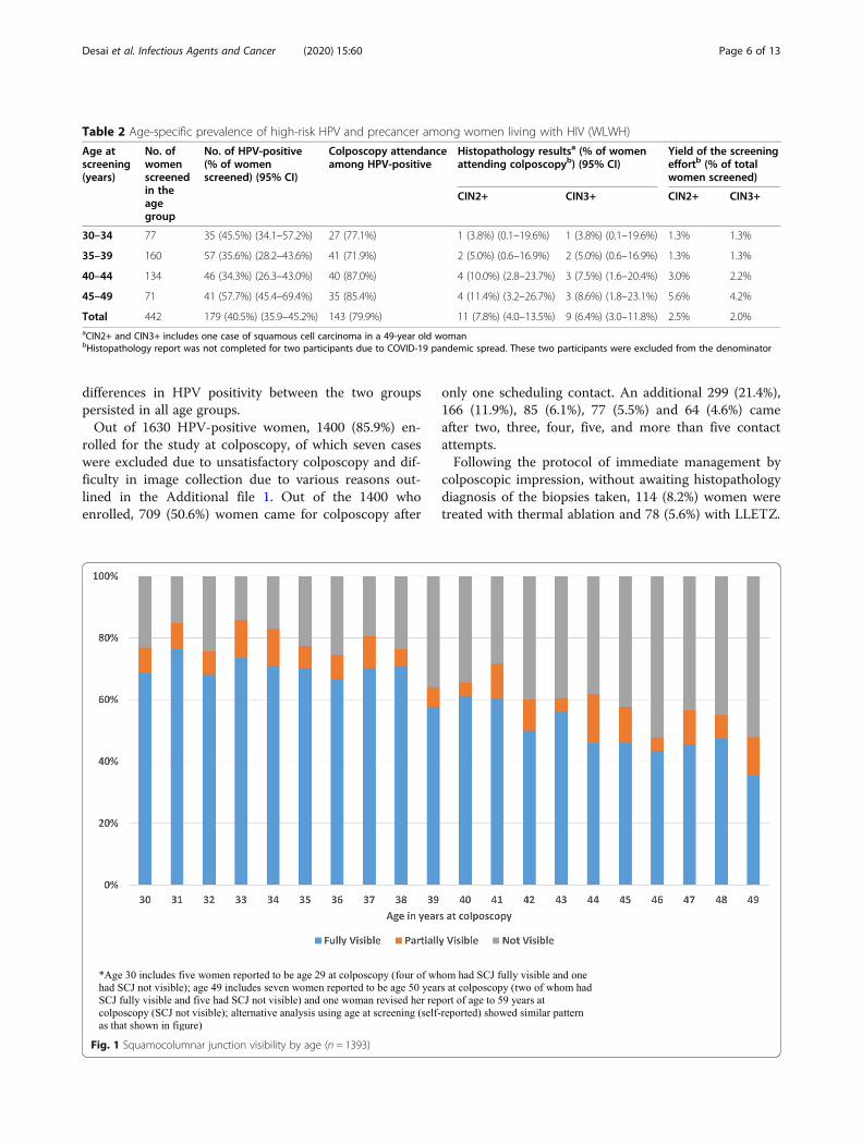
Fig. 1 Squamocolumnar junction visibility by age (n = 1393)
Desai et al. Infectious Agents and Cancer (2020) 15:60 Page 6 of 13
Overall, CIN2+ and CIN3+ (including five cancers),were detected in 5.9% (95% CI:4.7–7.3%) and 3.2% (95%CI:2.4–4.3%) of the HPV-positive women attending col-poscopy (85% attendance among the 1630), respectively.Thus, overall yield of the screening effort was 0.9% of9406 women for CIN2+ and 0.5% for CIN3 + .The prevalences of CIN2+ and CIN3+ among the
HPV-positive women with unknown HIV status attend-ing colposcopy were 5.7 and 2.9%, respectively, whereasthe overall yields of the screening effort in these groupswere 0.8 and 0.4%, respectively (Table 1). The age-specific prevalences of high-risk HPV positivity andprecancer among women with unknown HIV statusare shown in Table 1. The prevalence of HPV de-creased from 18.6% at age 30–34 years to 14.4% atage 45–49 years (P = 0.0003, chi-square for trend). Nomeaningful trend was observed in the prevalence ofprecancer by age (P = 0.87, chi-square for trend forCIN2+), except for a very high 24.1% (seven CIN2+including three cancers) overall prevalence at self-reported age of 49 years.The prevalences of CIN2+ and CIN3+ among the
HPV-positive WLWH attending colposcopy were 7.8
and 6.4%, respectively (P = 0.31 for CIN2+ and P = 0.03for CIN3+, independent chi-square test in comparisonto women with unknown HIV status) (Table 2). Theyield of the screening effort among women living withHIV was 2.5% for CIN2+, of which all except two wereactually CIN3+ (P < 0.001 for CIN2+ and CIN3+, inde-pendent chi-square in comparison to women with un-known HIV status) (Table 2). Relatively small age-specific numbers preclude any conclusions regardinga trend in the prevalence of HPV or precancer byage. The proportion of HPV-positive women in-creased with decrease in CD4 (cluster of differenti-ation 4) count (64% in CD4 < 200/mm3 vs 31.1% inCD4 > 500/ mm3, P = 0.001, chi-square for trend) andincrease in HIV viral load (36.3% in <=20 copies/mlvs 50% in > 20 copies/ml, P = 0.02, independent chi-square), however small numbers precluded trend ana-lysis of precancer/cancer.Out of a total of 75 histopathologically confirmed
cases, 59 (78.7%) were diagnosed through biopsy orLLETZ of acetowhite lesions at colposcopy visit, 35 ofwhich (59.3% of 59) had a high-grade colposcopic im-pression. QA review of colposcopic images revealed
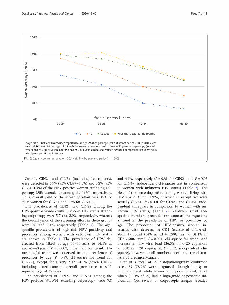
Fig. 2 Squamocolumnar junction (SCJ) visibility, by age and parity (n = 1380)
Desai et al. Infectious Agents and Cancer (2020) 15:60 Page 7 of 13
Table
3Managem
entof
wom
enby
histop
atho
logicdiagno
sis
Histopatho
logicdiagno
sis
Man
agem
ent
Row
total
Notrea
tmen
tindicated(row
%)
(colum
n%)
Immed
iate
man
agem
entbased
onco
lposco
pic
impression(row
%)(colum
n%)
Delayed
excision
altrea
tmen
ton
arecallvisit
(row
%)(colum
n%)
Withab
lation
WithLLET
ZCom
pleted
Pend
inga
<CIN2
1158
(88.7%
)(100.0%)
90(6.9%)(78.9%
)54
(4.1%)(69.2%
)3c
(0.2%)(12.0%
)0
1305
(100%)
CIN2
Diagn
osed
onbiopsy/LLETZ
010
(40.0%
)(8.8%)
11(44.0%
)(14.1%)
2(8.0%)(8.0%)
2(8.0%)(16.7%
)25
(100%)
Diagn
osed
onEC
C0
00
8(66.7%
)(32.0%
)4(33.3%
)(33.3%
)12
(100%)
CIN3
Diagn
osed
onbiopsy/LLETZ
013
(38.2%
)(11.4%
)12
(35.3%
)(15.4%)
5(14.7%
)(20.0%
)4(11.8%
)(33.3%
)34
(100%)
Diagn
osed
onEC
Cb
01(16.7%
)(0.9%)
03(50.0%
)(12.0%
)2(33.3%
)(16.7%
)6(100%)
Can
cer
Diagn
osed
onbiopsy/LLETZ
00
1d(20.0%
)(1.3%
)4(80.0%
)(16.0%
)0
5(100%)
Colum
ntotal
1158
(100%)
114(100%)
78(100%)
25(100%)
12(100%)
Grand
total=
1387
a Recalla
ttem
ptsaretempo
rarilypa
used
dueto
COVID-19pa
ndem
icspread
bInclud
estw
owom
enwith
OncoE
6HPV
16po
sitiv
e,in
who
mLLETZwas
recommen
dedon
recallan
disstillincomplete
c Includ
eson
ecase
with
colposcopicim
pression
ofcancer
treatedwith
hysterectomyof
wha
teven
tually
turned
outto
be<CIN2;
othe
rtw
ocaseswererecalledforLLETZdu
eto
repo
rtingerror
dInvasive
squa
mou
scellcarcinom
awith
CIN3at
margins
was
diag
nosedon
on-site
LLETZlead
ingto
arecallforarepe
atLLETZwith
CIN3diag
nosislead
ingto
a2n
drecallforahy
sterectomy
Desai et al. Infectious Agents and Cancer (2020) 15:60 Page 8 of 13
potential under-biopsying of more subtle acetowhite le-sions. 16 (21.3%) of 75 histopathologic precancer caseswere identified, in the absence of visible acetowhitening,only on ECC (1.2% of colposcopy examinations and 0.2%of the screening population). This is important in lightof the fact that even amongst women as young as age30, the SCJ was only partially visible in 8.3% and not vis-ible in 23.1% of women (31.5% total), rising to 12.5 and52.1% (64.6% total) by age 49 years (P < 0.001, chi-squarefor trend) (Fig. 1). Interestingly, multiple vaginal deliver-ies were found to increase full SCJ visibility (Fig. 2).Among 192 women treated on-site based on colpo-
scopic impression, without awaiting histopathology re-sults, only 48 (25%) were eventually diagnosed withCIN2+ on histopathology (Table 3). Viewing overtreat-ment from another perspective, 11% of women with his-topathologic <CIN2 were treated based on colposcopicimpression. On the other hand, 43.2% of CIN2 (16 outof 37) and 35% of CIN3 (14 out of 40) were not treatedimmediately; either because of underdiagnosis [diag-nosed later on ECC] or a variety of programmatic issuessuch as equipment failure. A total of 12 (1.1% of colpos-copy population) precancer cases needed more than onetreatment visit in order to obtain clear margins free ofhigh-grade findings.Out of 1138 (82%) patients with full clinical quality as-
surance review completed, 143 (12.6%) cases were rec-ommended for recall; mainly (113, or 9.9% of total) forrepeat ECC; commonly because the tissue was insuffi-cient for diagnosis on the prior ECC (100, or 8.8% oftotal) or because the ECC required dilatation/sedationdue to stenotic os or tethered cervix (13, or 1.1% oftotal). However, the overall yield of precancer from re-call for ECC was only 0.2% for the colposcopy popula-tion and 0.02% for the general population of womenaged 30–49 years.
DiscussionOur results showed the feasibility and acceptability ofself-sampled HPV testing and smartphone-based cervicalimage collection in Nigeria. Nevertheless, the combin-ation of higher HPV prevalence (17.4%) with a muchlower risk of precancer (0.9%) suggests the need for tri-age to improve the accuracy of the screening program.In addition, any visual triage method would requirerestricting the upper age limit of inclusion to increaseprogram effectiveness.There was a very positive response and clear accept-
ability for vaginal self-sampling for HPV testing. The dif-ficult aspects of self-sampling reported by someparticipants included identifying the vaginal opening, de-termining depth of the insertion of the brush into thevagina, rotating the brush inside the vagina, and puttingthe brush back into the tube after collection; these need
to be explained more clearly in future self-sampling pro-grams. Compliance with follow-up for colposcopy wasalso very high.The two previous phases of Project Itoju established
the epidemiology of HPV infection in rural Nigeria [26],validated a low-cost HPV test (careHPV, Qiagen) [27](phase 1) and attempted (ultimately unsuccessfully) todetermine whether immunosuppression due to soil-borne helminth infections or other parasitoses was thecause of high HPV prevalence among older women inthe region (phase 2). In the current phase of ProjectItoju, we reconfirmed the overall high prevalence ofHPV infections even after restricting the age of screen-ing to 30–49 years of age [28]. Although the prevalenceof HPV decreased to 16% at age 45–49 years from 19%at age 30–34 years, it was still much higher than the glo-bal average of around 5% for women age 45–54 yearsand consistent with what is observed for the women insub-Saharan Africa [29]. Despite the high prevalence ofHPV infection, the prevalence of precancer was less than1% in the general population of mostly unscreenedwomen 30–49 years old. One reason for the low preva-lence of precancer could be under biopsy of more subtleacetowhite lesions [30]. It also underscores the need tofurther study the type-specific natural history in the re-gion of HPV acquisition, clearance, persistence, and pro-gression, particularly in the setting of HIV infection. Inthis current study, we obtained samples for HPV geno-typing from women at screening and again at colposcopyvisit. We will be testing and reporting the results ofthese tests elaborating the prevalence of various HPV ge-notypes and short-term clearance of infection in subse-quent papers. We also hope to retest the women in thefuture to explore the long-term persistence of HPVinfection.The prevalence of HPV and precancer (particularly
CIN3+) was markedly high among WLWH as also re-ported by others [28]. This was observed despite thesuppressed viral load with a documented average of < 20copies/ml. This finding was expected since HPV andHIV can be co-transmitted and women with HIV tendto have lower clearance of acquired HPV infections. Themanagement of HPV in WLWH is an important topicbeyond the scope of this article.According to the World Health Organization (WHO),
Nigeria has only four physicians per 10,000 population[31]. At present, only < 10% of women > 15 years haveever been screened for cervical cancer in Nigeria [32]. In aresource-constrained setting with a shortage of expert gy-necologic providers and infrastructure, avoidable referralto colposcopy and substantial overtreatment are not sus-tainable. On the contrary, in settings with once in a life-time screening opportunity for a majority of women,substantial missed diagnosis and undertreatment is not
Desai et al. Infectious Agents and Cancer (2020) 15:60 Page 9 of 13
acceptable either. It is therefore essential that simple yetaccurate triage tests, separately or in combination, areavailable to stratify risk of precancer/cancer among HPV-positive women such that treatment intensity can be tai-lored to risk of cancer and sustained with local resources.In the future, we will be assessing the machine-learningbased AVE of cervical images using three different imagecollection methods. We will be reporting the results of theassessment of the combination of AVE with HPV geno-typing for triage of HPV-positive women in subsequentpapers. However, preliminary analysis of the cervical
images from three different devices (Fig. 3a) has raised animportant research question regarding the device portabil-ity of deep-learning based AVE algorithms due to vari-ation in color, brightness, reflection, glare imparted byeach device camera, light source and image processing ap-plication. Training or automation to capture an in-focus,non-blurry good quality cervical image, capturing entireSCJ, for evaluation by AVE, is yet another challenge.It is important to note that AVE, like other visual as-
sessment methods, requires that the cervical SCJ be vis-ible. In the current study population, we found that the
Fig. 3 Effect of image capture method on cervical appearance and limitation of visual triage method. a. Cervical images of the same cervixshowing histopathologic <CIN2 (left trio) and CIN2+ (right trio), captured with (clockwise starting from upper left) a Samsung S8 smartphonecamera and its flashlight, a MobileODT EVA device (Samsung J5 phone with an extra light source and a zoom lens), and a Zeiss FC150colposcope with a beam splitter and DSLR camera (see supplement). b. Cervical images of histopathologic CIN2+ cases with squamocolumnarjunction (clockwise from upper left) fully visible, partially visible, and not visible (diagnosed on ECC); captured with a Zeiss FC150 colposcope witha beam splitter and DSLR camera
Desai et al. Infectious Agents and Cancer (2020) 15:60 Page 10 of 13
SCJ was not fully visible for almost 64.6% of women byage 49 years. We also corroborated an earlier poorlyunderstood observation that women with multiple vagi-nal deliveries were more likely to have fully visible SCJs[33]. No visual screening methods or ablative treatmentmethods work when the transformation zone, where cer-vical cancer typically arises, is not visible (Fig. 3b) [12,34]. This could partially explain the decrease in theprevalence of precancer with age despite the high HPVprevalence, found in this study. It is, therefore, particu-larly crucial to emphasize restricting visual approachesfor screening or triage of older women to avoid givingfalse reassurance to women in these age groups and toavoid identifying high-risk HPV-positive women, par-ticularly with HPV 16, 18/45, with no available means offurther triage. It is worth noting however that even atage 30, the SCJ was not fully visible in up to 30% ofwomen, thus age-restriction to women <=49 years doesnot eliminate inadequacies in visual triage. The currentstandard practice is to collect an ECC sample wheneverthe SCJ is not fully visible, to rule out cancer within theendocervical canal. But an ECC needs an interpretationby an expert histopathologist, which is challenging inlow resource settings. Difficulty in collecting ECC inwomen with a stenotic cervical os and insufficient tissuein the ECC sample to rule out carcinoma are other chal-lenges with ECC. Thus, the development of low-costsimple triage alternatives for ECC remains an unsolvedchallenge for improving cervical cancer screening pro-grams. Research is also needed on simpler alternativesfor LLETZ, that could be performed easily by a generalphysician or a nurse without the need of an expertgynecologist, particularly in low-resource settings wherethe needed resources are in short supply especially inrural underserved areas.It is worth noting that even though the yield of pre-
cancer from ECC at colposcopy visit was 3.2% (21% oftotal precancers diagnosed on ECC, possibly due to nottaking enough biopsies of subtle acetowhite lesions), theadditional yield of precancer from the women recalledfor insufficient ECC or difficult ECC collection was only0.02% for the overall screened population. Another studyhas also noted overall low yield of ECC with an increasein proportionate additional yield of ECC when fewer bi-opsies are taken [35]. It is worth exploring the cost-effectiveness of recalls for ECC to avert a very low riskof adenocarcinoma in low-resource settings where noorganized follow-up is available. Any recommendationfor follow-up outside the study in these settings is likelyto remain a theoretical reassurance. In this regard, therewere a total of 12 women (1.1% of colposcopy popula-tion) who needed multiple rounds of treatment in orderto obtain clear margins on excision. These are womenwith confirmed high-grade lesions and hence also at
high risk of progression to invasion. Even though in thestudy we managed to achieve a relatively higher rate ofcompliance with follow-up, the compliance in the real-world setting needs to be monitored and factored in theeffectiveness of the screening programs.We had to halt the recall activities under the screening
program as the global spread of the COVID-19 pan-demic led to pausing of field efforts in Nigeria, includinga very few treatment visits still pending, and changed therisk-benefit ratio for cervical cancer screening. When wereopen, the first step will be to complete these treatmentvisits. Then, moving forward in the COVID-19 era, weare considering the importance of self-sampled HPVtesting with a sterile kit at a household level, avoidingany mass gathering and minimizing the need forspeculum examination. A small percentage of HPV-positive women with high-risk types could be triagedand treated, spacing community clinic appointments in aCOVID conscious manner [36].The limitations of this study should be noted. The
population selected for the study was a volunteer popu-lation of women residing in the university town of Ile-Ife, which may have a lower HPV prevalence than thegeneral population in Nigeria. Also, HC2 is not generallyused for self-sampling, as it is slightly less sensitive fordetection of precancer than PCR-based HPV testmethods [37]. A moderate amount of cross-reactivityagainst other genetically related but less oncogenic HPVtypes with HC2 is well-documented. This may partly ex-plain the low precancer to HPV ratio found in this study.Despite the known limitations of HC2, its operationalsimplicity, and easy trainability, as supported by virtuallytrouble-free operation of HC2 throughout the study sup-ported its use.In future work we will assess misclassification by
retesting residual screening samples with a more sensi-tive whole-genome sequencing method [20]; review andrecall women with highest risk HPV types and potentialmissed biopsies of subtle acetowhite lesions; anddigitalize the histopathology slides for a second review,particularly for borderline cases.
ConclusionA cervical cancer screening program using self-sampledHPV testing, with colposcopic immediate managementof women positive for HPV, is feasible in Nigeria but re-sults in both over-and under-treatment. There are newerHPV tests entering the marketplace that cost less thanten dollars per test, take less than an hour to perform,and provide genotyping, suggesting that a self-sampledHPV based screening program would be feasible in thispopulation in the future [38]. Having proven feasibility,we are now evaluating the accuracy and efficacy in diag-nosing CIN2+ of smartphone-based automated visual
Desai et al. Infectious Agents and Cancer (2020) 15:60 Page 11 of 13
evaluation of cervical images combined with HPV geno-typing as an assistive strategy to improve visual triage.
Supplementary informationSupplementary information accompanies this paper at https://doi.org/10.1186/s13027-020-00324-5.
Additional file 1.
AbbreviationsAUC: Area under the curve; AVE: Automated visual evaluation; CD4: Cluster ofdifferentiation 4; CI: Confidence interval; CIN: Cervical intraepithelialneoplasia; COVID-19: Coronavirus disease of 2019; DNA: Deoxyribose nucleicacid; DSLR: Digital single lens reflex; ECC: Endocervical curettage;EVA: Enhanced visual assessment; HC2: Hybrid capture-2; HIPAA: HealthInsurance portability & accountability act; HPV: Human papillomavirus;HIV: Human immunodeficiency virus; LLETZ: Large loop excision of thetransformation zone; NCI: National Cancer Institute; OAUTHC: ObafemiAwolowo University Teaching Hospitals Complex; OAU: Obafemi AwolowoUniversity; PCR: Polymerase chain reaction; RLU/CO: Relative light unit/cut-off;RNJMS: Rutgers New Jersey Medical school; ROC: Receiver operatingcharacteristic; SCJ: Squamocolumnar junction; SPSS: Statistical package forsocial studies; STM: Specimen transport media; US: United States;WLWH: Women living with HIV
AcknowledgementsThis research was supported in part by an appointment to the NationalCancer Institute Research Participation Program. This program isadministered by the Oak Ridge Institute for Science and Education throughan interagency agreement between the U.S. Department of Energy and theNational Institutes of Health. The video on self-sampling was designed byLivinghealth International, Nigeria.
Authors’ contributionsMS, KOA, KTD, CAA, AN, MHE, PC contributed substantially to the conceptionand design of the study. KOA, AB, CAA, TO, TOO, MMO, KTD, AN, MHE, andMS contributed to acquisition of data or the analysis and interpretation. MS,KOA, and KTD drafted the manuscript. All authors provided critical revision ofthe article and provided final approval of the version to publish.
FundingThe study was funded by the NCI intramural research program and GlobalGood (Now, Global Health Labs) (Bellevue, USA).
Availability of data and materialsThe datasets used and/or analysed during the current study are availablefrom the corresponding author on reasonable request.
Ethics approval and consent to participateThe study was approved by National Cancer Institute Special StudiesInstitutional Review Board (NCI-SSIRB) [iRIS reference number: 376424; IRBnumber: 09CN045; Version date: 12/20/2017] and OAUTHC Ethics andResearch Committee (ERC) [Protocol number: ERC/2016/05/08 and ERC/2018/09/10; Registration number international: IRB/IEC/0004553, national: NHREC/27/02/2009a].Written informed consent was take from all the participants.
Consent for publicationN/A
Competing interestsThe HC2 test kits were donated by Qiagen. The OncoE6 test kits weredonated by Arborvitae. The MobileODT EVA system devices and datamanagement software were donated by MobileODT. None of the companieshad any role in design, analysis, interpretation, and finalization of themanuscript.
Author details1Division of Cancer Epidemiology and Genetics, National Cancer Institute,NIH, Rockville, USA. 2Oak Ridge Institute of Science and Education, OakRidge, USA. 3Department of Obstetrics and Gynecology, Obafemi AwolowoUniversity Teaching Hospital, Ile Ife, Nigeria. 4Lagos University TeachingHospital, Lagos, Nigeria. 5Rutgers New Jersey Medical School and CancerInstitute of New Jersey (CINJ), Newark, USA. 6Hela Health, Tel Aviv, Israel.7Global Health Labs, Bellevue, USA. 8Albert Einstein College of Medicine, NewYork City, USA. 9National Library of Medicine, NIH, Bethesda, USA.
Received: 28 July 2020 Accepted: 22 September 2020
References1. Arbyn M, Weiderpass E, Bruni L, de Sanjosé S, Saraiya M, Ferlay J, et al.
Estimates of incidence and mortality of cervical cancer in 2018: a worldwideanalysis. Lancet Glob Heal. 2020;8(2):e191–203.
2. Ferlay J, Soerjomataram I, Dikshit R, Eser S, Mathers C, Rebelo M, et al.Cancer incidence and mortality worldwide: sources, methods and majorpatterns in GLOBOCAN 2012. Int J Cancer. 2015;136(5):E359–86.
3. Vaccarella S, Lortet-Tieulent J, Plummer M, Franceschi S, Bray F. Worldwidetrends in cervical cancer incidence: impact of screening against changes indisease risk factors. Eur J Cancer. 2013;49(15):3262–73.
4. Polman NJ, Ebisch RMF, Heideman DAM, Melchers WJG, Bekkers RLM, MolijnAC, et al. Performance of human papillomavirus testing on self-collectedversus clinician-collected samples for the detection of cervical intraepithelialneoplasia of grade 2 or worse: a randomised, paired screen-positive, non-inferiority trial. Lancet Oncol. 2019;2045(18):1–10.
5. Porras C, Hildesheim A, González P, Schiffman M, Rodríguez AC, WacholderS, et al. Performance of Self-collected Cervical Samples in Screening forFuture Precancer using Human Papillomavirus DNA Testing. J Natl CancerInst. 2015;107(1):1–9 Available from: http://www.ncbi.nlm.nih.gov/pubmed/25479804. [cited 2019 Jun 13].
6. Rodríguez AC, Schiffman M, Herrero R, Hildesheim A, Bratti C, Sherman ME,et al. Longitudinal Study of Human Papillomavirus Persistence and CervicalIntraepithelial Neoplasia Grade 2/3: Critical Role of Duration of Infection. JNatl Cancer Inst. 2010;102(5):315–24 Available from: https://www.ncbi.nlm.nih.gov/pmc/articles/PMC2831050/. [cited 2020 Mar 17].
7. Wentzensen N, Schiffman M, Palmer T, Arbyn M. Triage of HPVpositive women in cervical cancer screening. J Clin Virol. 2016;76:S49–55.
8. Hu L, Bell D, Antani S, Xue Z, Yu K, Horning MP, et al. An observationalstudy of deep learning and automated evaluation of cervical images forCancer screening. J Natl Cancer Inst. 2019;11(9):923–32.
9. Xue Z, Novetsky AP, Einstein MH, Marcus JZ, Befano B, Guo P, et al. Ademonstration of automated visual evaluation (AVE) of cervical imagestaken with a smartphone camera. Int J Cancer. 2020:ijc.33029 Availablefrom: https://onlinelibrary.wiley.com/doi/abs/10.1002/ijc.33029. [cited2020 May 14].
10. Nigeria Data Portal. Population Distribution by Age 2006-OsunDemographic pyramid. National Bureau of Statistics. 2006. Available from:https://nigeria.opendataforafrica.org/xlomyad/population-distribution-by-age-2006?state=Osun. [cited 2020 Sep 11].
11. Schiffman M, Castle PE, Jeronimo J, Rodriguez AC, Wacholder S. Humanpapillomavirus and cervical cancer. Lancet. 2007;370(9590):890–907Available from: http://www.ncbi.nlm.nih.gov/pubmed/17826171. [cited 2018Feb 16].
12. Pierre V, Raluca N, Catarino P. Anatomy of the cervix, squamocolumnarjunction, metaplastic change and transformation zone. Geneva. Availablefrom: http://www.gfmer.ch/vic/. [cited 2020 Mar 26].
13. Qiagen. digene® HC2 DNA Collection Device. Germantown: Qiagen; 2015.14. Livinghealth TV. HPV cervical Cancer self testing video. Nigeria: NCI/
OAUTHC; 2018. Available from: https://www.youtube.com/watch?v=JiNqrDntbTc.
19. Colposkop 150 FC. Carl Zeiss Meditec, Inc. Available from: https://www.zeiss.com/meditec/us/products/gynecology-/colposcopy/colposkop-150-fc.html#technical-data. [cited 2018 Mar 12].
20. Wagner S, Roberson D, Boland J, Yeager M, Cullen M, Mirabello L, et al.Development of the TypeSeq Assay for Detection of 51 HumanPapillomavirus Genotypes by Next-Generation Sequencing. 2019; Availablefrom: https://doi.org/. [cited 2020 Mar 26].
21. MobileODT. How to get better images with a colposcope. MobileODT.Available from: https://www.mobileodt.com/blog/get-better-images-with-a-colposcope/. [cited 2020 Apr 10].
22. World Health Organization (WHO). WHO Guidelines for Treatment ofCervical Intraepithelial Neoplasia 2–3 and Adenocarcinoma in situ - NCBIBookshelf. Geneva. Available from: https://www.ncbi.nlm.nih.gov/books/NBK206769/. [cited 2020 May 22].
23. Carreon JD, Sherman ME, Guillén D, Solomon D, Herrero R, Jerónimo J, et al.CIN2 is a much less reproducible and less valid diagnosis than CIN3: resultsfrom a histological review of population-based cervical samples. Int JGynecol Pathol. 2007;26(4):441–6.
24. IBM Corporation. IBM SPSS statistics. New York: IBM Corporation; 2011.25. Centers for Disease Control and Prevention (CDC). Epi Info. Atlanta: Centers
for Disease Control and Prevention (CDC); 2020.26. Gage JC, Ajenifuja KO, Wentzensen NA, Adepiti AC, Eklund C, Reilly M, et al.
The age-specific prevalence of human papillomavirus and risk of cytologicabnormalities in rural Nigeria: implications for screen-and-treat strategies. IntJ cancer. 2012;130(9):2111–7 Available from: http://www.ncbi.nlm.nih.gov/pubmed/21630264. [cited 2019 Jun 11].
27. Gage JC, Ajenifuja KO, Wentzensen N, Adepiti AC, Stoler M, Eder PS, et al.Effectiveness of a simple rapid human papillomavirus DNA test in ruralNigeria. Int J cancer. 2012;131(12):2903–9 Available from: http://www.ncbi.nlm.nih.gov/pubmed/22473652. [cited 2018 may 29].
28. Bruni L, Albero G, Serrano B, Mena M, Gómez D, Muñoz J, et al. HumanPapillomavirus and Related Diseases in Nigeria. Summary Report 2019.Available from: https://www.hpvcentre.net/statistics/reports/NGA.pdf?t=1593107314338.
29. ICO/IARC Information Centre on HPV and cancer. Human Papillomavirusand Related Diseases Report WORLD. Barcelona. Available from: www.hpvcentre.net. [cited 2020 Apr 11].
30. Wentzensen N, Walker JL, Gold MA, Smith KM, Zuna RE, Mathews C, et al.Multiple biopsies and detection of cervical cancer precursors at colposcopy.J Clin Oncol. 2015;33(1):83–9.
31. World Health Organization (WHO). Nigeria- Countdown to 2015 DecadeReport. Available from: https://www.who.int/workforcealliance/countries/Nigeria_En.pdf. [cited 2020 Apr 9].
32. ICO/IARC Information Centre on HPV and cancer. Nigeria: HumanPapillomavirus and Related Cancers, Fact Sheet 2018. Barcelona; 2019.Available from: www.hpvcentre.net. [cited 2020 Apr 9].
33. Autier P, Coibion M, Huet F, Grivegnee AR. Transformation zone location andintraepithelial neoplasia of the cervix uteri. Br J Cancer. 1996;74(3):488–90.
34. Doorbar J, Griffin H. Refining our understanding of cervical neoplasia and itscellular origins. Papillomavirus Res. 2019;7:176–9.
35. Liu A, Walker J, Gage JC, Gold MA, Zuna RE, Dunn ST, et al. Diagnosis ofcervical Precancers by Endocervical curettage at colposcopy of women withabnormal cervical cytology Angela. Obstet Gynecol. 2017;130(6):1218–25.
36. Ajenifuja KO, Belinson J, Goldstein A, Desai K, de Sanjosé S, Schiffman M.Designing low-cost, accurate cervical screening strategies that take intoaccount COVID-19: a role for self-sampled HPV typing. Infect Agents Cancer.2020.
37. Arbyn M, Smith SB, Temin S, Sultana F, Castle P. Detecting cervicalprecancer and reaching underscreened women by using HPV testing onself samples: updated meta-analyses. BMJ. 2018;363:k4823.
38. Zhang W, Du H, Huang X, Wang C, Duan X, Liu Y, et al. Evaluation of anisothermal amplification HPV detection assay for primary cervical cancerscreening. Infect Agents Cancer. 2020.
Publisher’s NoteSpringer Nature remains neutral with regard to jurisdictional claims inpublished maps and institutional affiliations.
Desai et al. Infectious Agents and Cancer (2020) 15:60 Page 13 of 13