Ophiuroids Discovered in the Middle Triassic Hypersaline Environment Mariusz A. Salamon 1 *, Robert Niedz ´ wiedzki 2 , Rafal Lach 1 , Tomasz Brachaniec 3 , Przemyslaw Gorzelak 4 1 Department of Palaeontology and Biostratigraphy, University of Silesia, Faculty of Earth Sciences, Sosnowiec, Poland, 2 Institute of Geological Sciences, Wroclaw University, Wroclaw, Poland, 3 Department of Geochemistry, Mineralogy, and Petrography, University of Silesia, Sosnowiec, Poland, 4 Department of Biogeology, Institute of Paleobiology, Polish Academy of Sciences, Warsaw, Poland Abstract Echinoderms have long been considered to be one of the animal phyla that is strictly marine. However, there is growing evidence that some recent species may live in either brackish or hypersaline environments. Surprisingly, discoveries of fossil echinoderms in non-(open)marine paleoenvironments are lacking. In Wojkowice Quarry (Southern Poland), sediments of lowermost part of the Middle Triassic are exposed. In limestone layer with cellular structures and pseudomorphs after gypsum, two dense accumulations of articulated ophiuroids (Aspiduriella similis (Eck)) were documented. The sediments with ophiuroids were formed in environment of increased salinity waters as suggested by paleontological, sedimentological, petrographical and geochemical data. Discovery of Triassic hypersaline ophiuroids invalidates the paleontological assumption that fossil echinoderms are indicators of fully marine conditions. Thus caution needs to be taken when using fossil echinoderms in paleoenvironmental reconstructions. Citation: Salamon MA, Niedz ´wiedzki R, Lach R, Brachaniec T, Gorzelak P (2012) Ophiuroids Discovered in the Middle Triassic Hypersaline Environment. PLoS ONE 7(11): e49798. doi:10.1371/journal.pone.0049798 Editor: Christopher Fulton, The Australian National University, Australia Received July 9, 2012; Accepted October 12, 2012; Published November 19, 2012 Copyright: ß 2012 Salamon et al. This is an open-access article distributed under the terms of the Creative Commons Attribution License, which permits unrestricted use, distribution, and reproduction in any medium, provided the original author and source are credited. Funding: National Science Centre grant no. UMO-2011/01/B/ST10/02639, Institute of Geological Sciences of Wroclaw University grant no. 1017/S/ING/11-IV, the UPGOW project for financial support (Z481,2011/2012). This work was partially performed in the NanoFun laboratory co-financed by the European Regional Development Fund within the Innovation Economy Operational Programme POIG.02.02.00-00-025/09. The funders had no role in study design, data collection and analysis, decision to publish, or preparation of the manuscript. Competing Interests: The authors have declared that no competing interests exist. * E-mail: [email protected]Introduction Numerous studies dealing with salinity level and its impact on modern echinoderms showed that this parameter is important in terms of their spatial distribution and size [1–8]. In general, modern echinoderms have a more limited salinity range than any other invertebrates because they have a permeable body wall and lack differentiated osmoregulatory or excretory organs [2–9]. This limit can especially be drawn for fossil echinoderms, among which only stenohaline taxa have been documented. Although, some authors recently described Messinian echinoids from non-normal saline deposits deposited during evaporitic episode of the Mediterranean region [10], [11]; however, it seems that these echinoids [Brissopsis gr. lyrifera (Forbes)] occurred in interbedded horizons with rather normal(?) salinity (personal communication, A. Kroh, Naturhistorisches Museum Wien). Intriguingly, in situ and labolatory experiments demonstrated that some extant echinoderms, in particular ophiuroids, asteroids and holothurians, can tolerate a great range of salinities [12]. So far, an ophiuroid Ophiophragmus filograneus (Lyman) constitutes an echinoderm species living in the lowest salinity in the field [13]. This form was found in the brackish facies of salinity level of 7.7%. Remarkable salinity tolerance of this species has been also confirmed under laboratory conditions [14], [15]. Likewise, many authors draw attention to the fact that certain species of echinoderms can tolerate very low salinity [7–16]. Additionally, it has been pointed out that low salinity may lead to size abnormalities (dwarfism) [17–20]. On the other hand, echinoderms can tolerate hypersaline conditions (i.e.,.35.5 psu according to [21]). For example, Price [22] reported two ophiuroid [Amphipholis squamata (Chiaje) and Amphiura fasciata Mortensen), and one holothurian (Leptosynapta chela Mortensen) species in Arabia Gulf with salinity ranging form 52 to 55%. Furthermore, the latter author documented two stunted asteroid species (Astropecten polyacanthus phragmorus Fisher and Asterina burtowi Gray) in lagoons with salinity exceeding 60% which to date constitute the highest record for salinity tolerance by echinoderms in the wild. In this paper, we report for the first time well-preserved ophiuroids Aspiduriella similis (Eck) from the Middle Triassic sediments of Poland that were deposited in hypersaline conditions. Materials and Methods Geological Setting Abandoned quarry ‘‘Wojkowice’’ is situated in the so-called Silesian-Cracow Monocline, in Upper Silesia, Southern Poland (Figure 1). This monocline contains mainly Triassic sediments deposited in the Germanic Basin on the northern margin of the Tethys Ocean [23]. In Wojkowice, carbonate deposits of the Upper Buntsandstein (Ro ¨t or Roetian) as well as the lowermost part of the Lower Muschelkalk (the Lower Gogolin Beds and lowermost part of the Upper Gogolin Beds; lithostratigraphic division of the Lower Muschelkalk after [24]) are exposed (detailed description of the lithostratigraphy and paleontology of the Wojkowice Quarry see PLOS ONE | www.plosone.org 1 November 2012 | Volume 7 | Issue 11 | e49798
Transcript
Ophiuroids Discovered in the Middle Triassic HypersalineEnvironmentMariusz A. Salamon1*, Robert Niedzwiedzki2, Rafał Lach1, Tomasz Brachaniec3, Przemysław Gorzelak4
1Department of Palaeontology and Biostratigraphy, University of Silesia, Faculty of Earth Sciences, Sosnowiec, Poland, 2 Institute of Geological Sciences, Wroclaw
University, Wroclaw, Poland, 3Department of Geochemistry, Mineralogy, and Petrography, University of Silesia, Sosnowiec, Poland, 4Department of Biogeology, Institute
of Paleobiology, Polish Academy of Sciences, Warsaw, Poland
Abstract
Echinoderms have long been considered to be one of the animal phyla that is strictly marine. However, there is growingevidence that some recent species may live in either brackish or hypersaline environments. Surprisingly, discoveries of fossilechinoderms in non-(open)marine paleoenvironments are lacking. In Wojkowice Quarry (Southern Poland), sediments oflowermost part of the Middle Triassic are exposed. In limestone layer with cellular structures and pseudomorphs aftergypsum, two dense accumulations of articulated ophiuroids (Aspiduriella similis (Eck)) were documented. The sedimentswith ophiuroids were formed in environment of increased salinity waters as suggested by paleontological,sedimentological, petrographical and geochemical data. Discovery of Triassic hypersaline ophiuroids invalidates thepaleontological assumption that fossil echinoderms are indicators of fully marine conditions. Thus caution needs to betaken when using fossil echinoderms in paleoenvironmental reconstructions.
Citation: Salamon MA, Niedzwiedzki R, Lach R, Brachaniec T, Gorzelak P (2012) Ophiuroids Discovered in the Middle Triassic Hypersaline Environment. PLoSONE 7(11): e49798. doi:10.1371/journal.pone.0049798
Editor: Christopher Fulton, The Australian National University, Australia
Received July 9, 2012; Accepted October 12, 2012; Published November 19, 2012
Copyright: � 2012 Salamon et al. This is an open-access article distributed under the terms of the Creative Commons Attribution License, which permitsunrestricted use, distribution, and reproduction in any medium, provided the original author and source are credited.
Funding: National Science Centre grant no. UMO-2011/01/B/ST10/02639, Institute of Geological Sciences of Wroclaw University grant no. 1017/S/ING/11-IV, theUPGOW project for financial support (Z481,2011/2012). This work was partially performed in the NanoFun laboratory co-financed by the European RegionalDevelopment Fund within the Innovation Economy Operational Programme POIG.02.02.00-00-025/09. The funders had no role in study design, data collectionand analysis, decision to publish, or preparation of the manuscript.
Competing Interests: The authors have declared that no competing interests exist.
Numerous studies dealing with salinity level and its impact on
modern echinoderms showed that this parameter is important in
terms of their spatial distribution and size [1–8]. In general,
modern echinoderms have a more limited salinity range than any
other invertebrates because they have a permeable body wall and
lack differentiated osmoregulatory or excretory organs [2–9]. This
limit can especially be drawn for fossil echinoderms, among which
only stenohaline taxa have been documented. Although, some
authors recently described Messinian echinoids from non-normal
saline deposits deposited during evaporitic episode of the
Mediterranean region [10], [11]; however, it seems that these
echinoids [Brissopsis gr. lyrifera (Forbes)] occurred in interbedded
horizons with rather normal(?) salinity (personal communication,
A. Kroh, Naturhistorisches Museum Wien).
Intriguingly, in situ and labolatory experiments demonstrated
that some extant echinoderms, in particular ophiuroids, asteroids
and holothurians, can tolerate a great range of salinities [12]. So
far, an ophiuroid Ophiophragmus filograneus (Lyman) constitutes an
echinoderm species living in the lowest salinity in the field [13].
This form was found in the brackish facies of salinity level of 7.7%.
Remarkable salinity tolerance of this species has been also
confirmed under laboratory conditions [14], [15]. Likewise, many
authors draw attention to the fact that certain species of
echinoderms can tolerate very low salinity [7–16]. Additionally,
it has been pointed out that low salinity may lead to size
abnormalities (dwarfism) [17–20].
On the other hand, echinoderms can tolerate hypersaline
conditions (i.e.,.35.5 psu according to [21]). For example, Price
[22] reported two ophiuroid [Amphipholis squamata (Chiaje) and
Amphiura fasciata Mortensen), and one holothurian (Leptosynapta chela
Mortensen) species in Arabia Gulf with salinity ranging form 52 to
55%. Furthermore, the latter author documented two stunted
asteroid species (Astropecten polyacanthus phragmorus Fisher and Asterina
burtowi Gray) in lagoons with salinity exceeding 60% which to date
constitute the highest record for salinity tolerance by echinoderms
in the wild.
In this paper, we report for the first time well-preserved
ophiuroids Aspiduriella similis (Eck) from the Middle Triassic
sediments of Poland that were deposited in hypersaline conditions.
Materials and Methods
Geological SettingAbandoned quarry ‘‘Wojkowice’’ is situated in the so-called
Silesian-Cracow Monocline, in Upper Silesia, Southern Poland
(Figure 1). This monocline contains mainly Triassic sediments
deposited in the Germanic Basin on the northern margin of the
Tethys Ocean [23].
In Wojkowice, carbonate deposits of the Upper Buntsandstein
(Rot or Roetian) as well as the lowermost part of the Lower
Muschelkalk (the Lower Gogolin Beds and lowermost part of the
Upper Gogolin Beds; lithostratigraphic division of the Lower
Muschelkalk after [24]) are exposed (detailed description of the
lithostratigraphy and paleontology of the Wojkowice Quarry see
PLOS ONE | www.plosone.org 1 November 2012 | Volume 7 | Issue 11 | e49798
[25]; Figure 2A). Two ophiuroid accumulations were found in the
Cellular Limestones Unit (the so-called ‘‘Zellenkalk 20) - upper-
most lithological unit of the Lower Gogolin Beds. Its thickness in
Wojkowice is up to ca. 1.3 m. This unit represents Aegean [26].
The Cellular Limestones Unit is considered equivalent of the
Grenzgelbkalk and Liegende Dolomite in Germany [23], [27] and
crops out in the whole area in the Upper Silesia where the
sediments of Lower Muschelkalk are present. The thickness of
these sediments varies from 0.8 to 2.0 m depending on the locality
[24]. In contrast to other units of the Gogolin Beds, body fossils are
extremely rare in the Cellular Limestones Unit of Silesia. Only
occasionally, bivalves: Hoernesia socialis (Schlotheim), Gervilleia
mytiloides (Schlotheim), Myoconcha gastrochaena Dunker and isolated
crinoid ossicles were recorded [24]. In numerous outcrops of this
unit, body fossils are absent [28], [29]. Similarly, ichnofossils were
not documented whereas they are numerous in the Lower
Muschelkalk (e.g., [23]).
In Wojkowice, the Cellular Limestones Unit is represented by
yellow dolomitic limestones and dolomitic marls. Lower part of
this unit consists of dolomitic limestones. Above, a few (45 cm
thick) layers are exposed [23]: palisade calcite layer, dolocrete
layer, rauhwacke, cellular limestones and rauhwacke layer,
respectively. In the upper part of the Cellular Limestones Unit,
dolomitic limestones and marls are exposed. Two dense
accumulations of ophiuroids have been found on the upper
surface of the cellular limestone layer that was covered by a thin
(ca. 2 mm) muddy layer.
SamplingDuring the field work about 28 square metres of the sediments
of the Celullar Limestones Unit (including 4 square meters layer of
cellular dolomitic limestone) were investigated (Figure 2). Fossils
are extremely rare in this unit: we recorded only three poorly
preserved bivalve molds and surprisingly two accumulations of
ophiuroids (26 and 6 specimens) that were found within one
horizon near the upper surface of the cellular dolomitic limestone
layer (Figure 2C,D). Both accumulations were separated from each
other by a distance of ca. 80 cm. Additionally, three bulk samples
from the cellular dolomitic limestone (each ca. 2 kg) were taken
and transported to the laboratory of the Department of Earth
Sciences of University of Silesia. No specific permissions were
required for collection of fossils form this location. Field studies
were carried out at an abandoned quarry with public right-of-way
and did not involve endangered or protected species.
Slabs with ophiuroids were initially cleaned with hot water.
Later the samples were slightly treated using peroxide in order to
clean them off from the remnants of the thin layer of muddy
sediments and again washed with hot water. Slabs were dried and
watched under a binocular microscope for taphonomic studies. All
ophiuroids from the Celullar Limestones Unit were examined
carefully for evidence of breakage, abrasion, dissolution, re-
generation traces and evidence of bite marks. Three selected bulk
samples of dolomitic limestone were dissolved using glauber salt (8
cycles of boiling-freezing procedure). Later they were washed
using hot tap water and sieved using Ø 0.5 mm, 0.315 mm and
Figure 1. Fossil locality and geological setting. Map of Poland with investigated area indicated and enlargement of Upper Silesia with thesampled Wojkowice Quarry (circle). Figure slightly modified from [25].doi:10.1371/journal.pone.0049798.g001
Triassic Ophiuroids from Hypersaline Environment
PLOS ONE | www.plosone.org 2 November 2012 | Volume 7 | Issue 11 | e49798
Triassic Ophiuroids from Hypersaline Environment
PLOS ONE | www.plosone.org 3 November 2012 | Volume 7 | Issue 11 | e49798
0.1 mm mesh widths. After drying in 180uC, samples were
screened under a binocular microscope.
Petrographic and Geochemical AnalysesPolished and carbon-coated thin sections of the samples from
the Celullar Limestones Unit were examined with cathodolumi-
nescence microscope equipped with a hot cathode integrated with
spectograph and linked to a Kappa video camera for recording
digital images, at the Institute of Paleobiology of the Polish
Academy of Sciences in Warsaw. Integration times for CL-
emission spectra of luminescent samples were 50 s. The cath-
odoluminescent properties of sections were additionally examined
using the Cambridge Image Technology Ltd CCL 8200 mk3
model system with 12–15 kV beam potential and 400 mA beam
current attached to a NIKON ECLIPSE E400 Pol optical
microscope at the Department of Stratigraphical Geology of
Wrocław University. The petrographical investigations were
carried out at the Institute of Geological Sciences of Wrocław
University and at the Institute of Paleobiology of the Polish
Academy of Sciences in Warsaw.
Seven samples (two from limestones and five from dolomitic
limestones) were selected for geochemical analysis (in particular
boron content) using PGNAA (Pulsed Gamma Neutron Activation
Analysis) method (Figure 2B). 1 g samples are encapsulated in
a polyethylene vial and placed in a thermalized beam of neutrons
produced from a nuclear reactor. Samples are measured for the
doppler broadened prompt gamma ray at 478 KeV using a high
purity GE detector. Samples are compared to certified reference
materials used to calibrate the system. A minimum of four
standards are analyzed with every work order. Duplicates were
analyzed to check method stability. The detection limit (0.5 ppm)
reported is a function of the counting times required for each.
The collections are housed at the Department of Palaeontology
and Biostratigraphy of the University of Silesia, Sosnowiec, Poland
(catalogue number GIUS 7-3601– Geological Institute of the
University of Silesia).
Results
PaleontologyMacerated samples did not contain any macro- or microfauna.
However, surprisingly two dense ophiuroid accumulations were
found in the field on the surface of two slabs. These ophiuroids
were scattered across the area of 15 and 30 cm2, respectively. The
first accumulation yielded 26 specimens whereas the second
yielded only 6 specimens. All specimens were represented by
complete or nearly complete forms (disc plus arms or partly
preserved arms; see details below). Only 6% of total number of
ophiuroids (2 specimens) were preserved in situ, i.e., with oral side
directing towards the bottom (more details in discussion).
Ophiuroid specimens lack clear evidences of abrasion, extensive
dissolution, regeneration or bite mark traces (Figure 2E,F,G).
Three specimens yield (likely post-diagenetic) evidence of breakage
(specimens no. 10 and 23 on the slab no. 1; specimens no. 2 on
the slab no. 2; see Table 1,2). Most speciemens possess complete
disc with partly preserved arms, i.e., 70% of speciemens lack
distalmost part of arms. Two states of preservations can be
distinguished:
1. Well-preserved ophiuroids (Taphonomic Group 1), with only
minor signs of disarticulation (e.g., lack of distal arm portions);
2. More disarticulated specimens (Taphonomic Group 2) com-
prising central disc with attached less than five and partly
preserved arms (having only proximal and median portions of
their arms).
Aspiduriella DescriptionThe ophiuroid assemblages from Wojkowice consist of mono-
specific and multiindividual accumulation with small ophiuroids
Aspiduriella similis (Eck) with narrow range size 2.7–3.4 mm
(Table 1,2). This species is thought to have been a slow-moving,
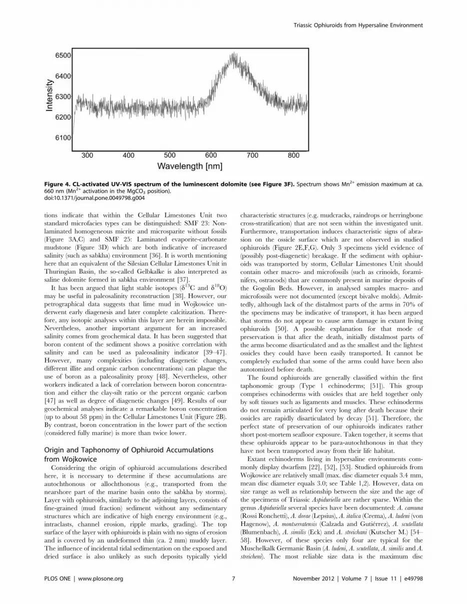
luminescent areas are due to changing diagenetic fluid chemistry
leading to Mn2+incorporation (‘‘activator ion’’). The CL emission
spectrum of an orange luminescing Mn2+-activated showed
emission maximum at about 660 nm (Figure 4, Mn2+activation
in the MgCO3 position).
Geochemical analyses revealed that the boron is non-uniformly
distributed within a part of investigated section. The main feature
of boron distribution is a relatively low B concentration in the
lower part of the section (10–23 ppm) and much higher
Figure 2. Stratigraphical section of Triassic sediments with ophiuroids. (A) Section of the northern and southern part of the WojkowiceQuarry (from [25]). 1, dolomitic limestones and marls; 2, cellular dolomitic limestones; 3, organodetrital limestones with bivalve detritus andcolumnals; 4, marly limestones; 5, pelitic limestones with abundance shells of bivalves; 6, pelitic limestones; 7, wavy limestones; 8, nodular limestones;9, vertebrate remains; 10, Dadocrinus columnals; 11, encrinid columnals; 12, intraclasts; 13, regurgitalites; 14, Rhizocorallium commune; 15, numerousgastropods; 16, numerous Plagiostoma; 17, numerous Pseudocorbula sp.; 18, numerous Gervillia sp.; 19, Thalassinoides; 20, Holocrinus columnals; 21,layer(s) with presently recorded ophiuroids. I, Roetian; II, ‘‘limestones with Entolium and Dadocrinus unit’’; III, ‘‘first wavy limestones unit’’; IV, ‘‘cellularlimestones unit’’; V, ‘‘thick-bedded limestones’’ and ‘‘wavy limestones unit’’. (B) Investigated section of ‘‘first wavy limestones unit’’ and ‘‘cellularlimestones unit’’; S1–S7 = rock samples for boron content analyses. Scale bar equals 1 m. (C) Enlargement of the ophiuroid layer. Arrows show theplace of accumulations. Scale bar = 10 cm. (D) Slab (described in the paper as no. 1) with ophiuroid accumulation. (E) SEM micrographs of the oralview of the ophiuroid disc. (F) SEM micrographs of the aboral view of the near complete ophiuroid specimen. (G) SEM micrographs of the contact ofarm plates showing relicts of the stereom microstructure. Scale bar = 10 mm.doi:10.1371/journal.pone.0049798.g002
Triassic Ophiuroids from Hypersaline Environment
PLOS ONE | www.plosone.org 4 November 2012 | Volume 7 | Issue 11 | e49798
concentration in upper part of the section (44–58 ppm) (see
Figure 2B).
Discussion
Depositional Paleoenvironment of Cellular LimestonesUnit
The Silesian Cellular Limestones Unit (Zellenkalk 2) was
formed at the end of the highstand system tracts phase, when
significant shallowing of the basin occurred [23]. This led to the
temporarily emergence of the bottom and strong evaporation
which is supported by isotopic data of d13C and d18O [23], [31].
The Cellular Limestones Unit has no evidence of marine
intercalations and was presumably deposited in sabkha environ-
ment with a dominant sedimentation of lime mud [31]. Analogical
cellular structures (cells) in cellular limestone in Silesian Roetian
(Zellenkalk 1) are interpreted as voids after dissolved and removed
gypsum and halite crystals [32]. Similar voids from Zellenkalk 2 of
the same origin are observed (Figure 3 C).
Supra-normal salinity during deposition of this unit is supported
by low abundance and diversity of fossils (3 bivalve molds and two
opiuroid accumulations on 28 square metres investigated) and lack
Table 1. Taphonomic features of Aspiduriella similis from Celullar Limestones Unit (accumulation no 1).
Specimen number Disc diameter [in mm] Life position: oral side up [u], down [d] Breakage Taphonomic group
1 2.7 d no 1
2 2.9 u no 1
3 3.0 u no 2
4 3.0 u no 2
5 3.1 u no 2
6 2.8 u no 2
7 3.2 u no 2
8 3.3 u no 2
9 3.2 u no 2
10 2.9 u yes 2
11 3.0 u no 2
12 2.9 u no 2
13 3.4 u no 1
14 3.1 u no 2
15 3.2 u no 2
16 2.8 u no 2
17 2.8 u no 2
18 2.9 u no 2
19 3.1 d no 1
20 3.3 u no 1
21 3.2 u no 1
22 3.1 u no 2
23 3.0 u yes 1
24 2.8 u no 1
25 3.0 u no 2
26 3.1 u no 1
doi:10.1371/journal.pone.0049798.t001
Table 2. Taphonomic features of Aspiduriella similis from Celullar Limestones Unit (accumulation no 2).
Specimen number Disc diameter [in mm] Life position: oral side up [u], down [d] Breakage Taphonomic group
1 3.0 u no 2
2 2.8 u yes 2
3 2.7 u no 1
4 2.8 u no 2
5 2.9 u no 2
6 3.0 u no 2
doi:10.1371/journal.pone.0049798.t002
Triassic Ophiuroids from Hypersaline Environment
PLOS ONE | www.plosone.org 5 November 2012 | Volume 7 | Issue 11 | e49798
of ichnofossils. By contrast, fossils (especially crinoids) are
numerous in the adjacent lithological units [25], [33]. It is
noteworthy that previous studies have documented three bivalve
species within the Cellular Limestones Unit: Hoernesia socialis
Dunker [24]. According to the literature [34], all these species are
euryhaline. Especially, Hoernesia has been commonly reported in
hypersaline deposits of Europe and Canada [34], [35].
The occurrence of some postevaporatic structures (including
layer of palisade calcite and the rauhwacke layers) is also consistent
with increased salinity interpretation [23] although according to
some authors [31] the Cellular Limestones Unit comprises also
early diagenetic evaporites. Additionally, our microfacial observa-
Figure 3. Petrographical and cathodoluminescent features of the layer with ophiuroids. (A) Fine-grained dedolomites under transmittedlight and (B) in crossed-nicoles. (C) Laminated dedolomites with irregulary distributed voids under transmitted light. (D) Laminated and microfoldeddedolomites under cathodoluminescence. (E) Pseudospar crystals and rhombohedral crystals of calcitized dolomites and pseudomorphs aftergypsum (arrows) in crossed-nicoles. (F) Rhombohedral crystals of calcitized dolomites under cathodoluminescence showing zoned crystal growth.Dolomite rhomboids have non-luminescent and bright orange luminescent zones. Non-luminescent areas are due to Fe rich diagenetic fluids and theincorporation of Fe in the dolomite lattice (‘‘quench ion’’) during crystal gwowth, bright orange luminescent areas indicates changes in the diageneticfluid chemistry into Mn enrichment and the incorporation of Mn in the dolomite lattice (‘‘activator ion’’) during crystal growth.doi:10.1371/journal.pone.0049798.g003
Triassic Ophiuroids from Hypersaline Environment
PLOS ONE | www.plosone.org 6 November 2012 | Volume 7 | Issue 11 | e49798
tions indicate that within the Cellular Limestones Unit two
standard microfacies types can be distinguished: SMF 23: Non-
laminated homogeneous micrite and microsparite without fossils
(Figure 3A,C) and SMF 25: Laminated evaporite-carbonate
mudstone (Figure 3D) which are both indicative of increased
salinity (such as sabkha) environment [36]. It is worth mentioning
here that an equivalent of the Silesian Cellular Limestones Unit in
Thuringian Basin, the so-called Gelbkalke is also interpreted as
saline dolomite formed in sabkha environment [37].
It has been argued that light stable isotopes (d13C and d18O)
may be useful in paleosalinity reconstruction [38]. However, our
petrographical data suggests that lime mud in Wojkowice un-
derwent early diagenesis and later complete calcitization. There-
fore, any isotopic analyses within this layer are herein impossible.
Nevertheless, another important argument for an increased
salinity comes from geochemical data. It has been suggested that
boron content of the sediment shows a positive correlation with
salinity and can be used as paleosalinity indicator [39–47].
However, many complexities (including diagenetic changes,
different illite and organic carbon concentrations) can plague the
use of boron as a paleosalinity proxy [48]. Nevertheless, other
workers indicated a lack of correlation between boron concentra-
tion and either the clay-silt ratio or the percent organic carbon
[47] as well as degree of diagenetic changes [49]. Results of our
geochemical analyses indicate a remarkable boron concentration
(up to about 58 ppm) in the Cellular Limestones Unit (Figure 2B).
By contrast, boron concentration in the lower part of the section
(considered fully marine) is more than twice lower.
Origin and Taphonomy of Ophiuroid Accumulationsfrom Wojkowice
Considering the origin of ophiuroid accumulations described
here, it is necessary to determine if these accumulations are
autochthonous or allochthonous (e.g., transported from the
nearshore part of the marine basin onto the sabkha by storms).
Layer with ophiuroids, similarly to the adjoining layers, consists of
fine-grained (mud fraction) sediment without any sedimentary
structures which are indicative of high energy environment (e.g.,
intraclasts, channel erosion, ripple marks, grading). The top
surface of the layer with ophiuroids is plain with no signs of erosion
and is covered by an undeformed thin (ca. 2 mm) muddy layer.
The influence of incidental tidal sedimentation on the exposed and
dried surface is also unlikely as such deposits typically yield
characteristic structures (e.g. mudcracks, raindrops or herringbone
cross-stratification) that are not seen within the investigated unit.
Furthermore, transportation induces characteristic signs of abra-
sion on the ossicle surface which are not observed in studied
ophiuroids (Figure 2E,F,G). Only 3 specimens yield evidence of
(possibly post-diagenetic) breakage. If the sediment with ophiur-
oids was transported by storm, Cellular Limestones Unit should
contain other macro- and microfossils (such as crinoids, forami-
nifers, ostracods) that are commonly present in marine deposits of
the Gogolin Beds. However, in analysed samples macro- and
microfossils were not documented (except bivalve molds). Admit-
tedly, although lack of the distalmost parts of the arms in 70% of
the specimens may be indicative of transport, it has been argued
that storms do not appear to cause arm damage in extant living
ophiuroids [50]. A possible explanation for that mode of
preservation is that after the death, initially distalmost parts of
the arms become disarticulated and as the smallest and the lightest
ossicles they could have been easily transported. It cannot be
completely excluded that some of the arms could have been also
autotomized before death.
The found ophiuroids are generally classified within the first
taphonomic group (Type 1 echinoderms; [51]). This group
comprises echinoderms with ossicles that are held together only
by soft tissues such as ligaments and muscles. These echinoderms
do not remain articulated for very long after death because their
ossicles are rapidly disarticulated by decay [51]. Therefore, the
perfect state of preservation of our ophiuroids indicates rather
short post-mortem seafloor exposure. Taken together, it seems that
these ophiuroids appear to be para-autochthonous in that they
have not been transported away from their life habitat.
Extant echinoderms living in hypersaline environments com-
monly display dwarfism [22], [52], [53]. Studied ophiuroids from
Wojkowice are relatively small (max. disc diameter equals 3.4 mm,
mean disc diameter equals 3.0; see Table 1,2). However, data on
size range as well as relationship between the size and the age of
the specimens of Triassic Aspiduriella are rather sparse. Within the
genus Aspiduriella several species have been documented: A. camuna
(Rossi Ronchetti), A. dorae (Lepsius), A. italica (Crema), A. ludeni (von
Hagenow), A. montserratensis (Calzada and Gutierrez), A. scutellata
(Blumenbach), A. similis (Eck) and A. streichani (Kutscher M.) [54–
58]. However, of these species only four are typical for the
Muschelkalk Germanic Basin (A. ludeni, A. scutellata, A. similis and A.
streicheni). The most reliable size data is the maximum disc
Figure 4. CL-activated UV-VIS spectrum of the luminescent dolomite (see Figure 3F). Spectrum shows Mn2+ emission maximum at ca.660 nm (Mn2+ activation in the MgCO3 position).doi:10.1371/journal.pone.0049798.g004
Triassic Ophiuroids from Hypersaline Environment
PLOS ONE | www.plosone.org 7 November 2012 | Volume 7 | Issue 11 | e49798
diameter. A. ludeni occurring commonly in the Lower Muschelkalk
of the Polish part of the Germanic Basin is the largest form with
disks reaching up to 8.2 mm. This form was also noted from the
Lower Muschelkalk of the eastern Germany (Rudersdorf near
Berlin) [57], [59–61]. A. streichani is similarly-sized to A. ludeni and
its disk reached up to ca. 8 mm. This species is common only in
the vicinity of the Lower Muschelkalk of Rudersdorf [62].
However, it should be pointed out that the morphology of this
species is very similar to the previously mentioned A. ludeni. Stoll
[63] ascribed this taxon to A. ludeni with a question mark. A.
scutellata with its maximum disk diameter equaling 6.9 mm occurs
in the Lower and Upper Muschelkalk of Poland and Germany
[54], [55], [62–64]. This species is the only one that also has been
reported from the Tethyan Realm of Italy [65]. The maximum
disk diameter of A. similis commonly occurring in the Lower
Muschelkalk of Poland equals 5 mm [66]. Unfortunately, data on
range size as well as mean size are sparse. Previous workers [30],
[66] have mentioned that ophiuroids from various localities in
Poland have mean disc diameter commonly not exceeding 3 mm.
However, these ophiuroid taphofacies are connected with
transportation from their life habitat due to storm-related obrution
events. In such cases, segregation of the skeletons is likely. Thus,
data on mean disc diameter of these ophiuroid populations are
probably underestimated. Therefore, given our current state of
knowledge on ontogenesis of A. similis it is impossible to clearly
state if ophiuroids from Wojkowice underwent dwarfism or not.
Good state of preservation (see taphonomic groups in Table 1,2)
and an overall sedimentological context indicate that the death of
ophiuroids was not caused by high-energy (storm or tidal) event.
Predation is also not a likely cause of their death as ophiuroids do
not display evidence of damages of their discs and arms (such as
bite marks comparable to those observed in crinoids from the
lower part of this section; [33], [67]). Furthermore, the Cellular
Limestone Unit in the Wojkowice lacks potential body predatory
fossils (e.g., cidaroids, crabs, hybodontid sharks) or ichnofossils
(e.g., Thalassinoides, Rhizocorallium) that are ascribed to predatory
decapods. Such fossils do not occur also in other outcrops of this
unit in the Upper Silesia [23], [24], [68], [69].
One possible explanation is that the ophiuroids from Wojkowice
died due to the gradual deterioration of environmntal conditions.
In the unstable sabkha environment, progressive evaporation
might have been stressful for ophiuroids. For example, subaerially
weathered dolomites indicate that this area might have been
episodically lifted up [23]. In such drying sabkha, elevated
temperatures, increasing salinity, as well as progressive oxygen
depletion might have been responsible for the ophiuroid death.
The fact that nearly all ophiuroids are preserved with their oral
side turned up is consistent with such interpretation as studies on
modern ophiuroids from the Northern Adriatic Sea [70] indicate
that oxygen depletion commonly leads to their arm-tipping and
the accompanying uplifted disc [71–72]. This behaviour is
generally interpreted as an attempt to reach higher oxygen
concentrations. In such humped postures, ophiuroids are prone to
overturn. Similar arm-tipping behaviour have been recorded
elsewhere during hypoxia in modern opiuroid species, including
Ophiura texturata (Linnaeus) [73], [74], O. albida (Forbes) [75], or
Amphiura chiajei (Forbes) and A. filiformis (Muller) [76], [77].
Alternatively, other factors may have also contributed to such an
inverted position. It is noteworthy that extant ophiuroids can
change body posture to an upside down position, with oral side of
the disc facing upward and arms raised above the disc [78].
Furthermore, these organisms can curl into a ball-shaped
configuration leading to the overturn due to shift of the center
of gravity. Finally, the inverted position of extant ophiroids has
been also reported in ophiuroids escaping from sediments [79].
After the overturn of ophiuroids, initially distalmost parts of the
arms became disarticulated (if not earlier autotomized) and as the
smallest and the lightest ossicles could have been easily
transported. Then, within several? days the speciemens must have
been covered by thin ca. 2 mm thick layer of the sediment
allowing their near-complete preservation [51].
ConclusionsFossil echinoderms are generally considered ideal indicators of
fully marine conditions. Thus they have been commonly used in
paleoenvironmental reconstructions. For example, echinoderm
fossils (in particular asterozoan traces) have recently warranted
a re- interpretation of the depositional environment of Stuttgart
Formation (Middle Keuper) from the non-open marine into fully
marine environment [80]. Discovery of ophiuroid accumulations
from the Middle Triassic of Poland constitutes the first and the
oldest fossil record to our knowledge of echinoderms found in
hypersaline environment. Our discovery suggests that some
echinoderm species might have been euryhaline and imply that
adaptation to increased salinity might have already appeared in
the Middle Triassic. Therefore, great care needs to be exercised
when using fossil echinoderm as paleoenvironmental proxies.
Acknowledgments
We would like to especially thank Dr. Monika Kowal-Linka (Adam
Mickiewicz University) for useful comments and access to valuable
bibliographic data, and Dr. Frederick Hotchkiss (The Marine and
Paleobiological Research Institute, Massachusetts) for improving the
English of this paper. Constructive comments by three anonymous
reviewers greatly helped us to improve the manuscript.
Author Contributions
Conceived and designed the experiments: MAS RN PG. Performed the
experiments: MAS RN RL TB PG. Analyzed the data: MAS RN RL TB
PG. Wrote the paper: MAS RN PG.
References
1. Smith GFM (1940) Factors limiting distribution and size in the starfish. J FishRes Board Can 5: 84–103.
2. Binyon J (1961) Salinity tolerance and permeability to water of the starfishAsterias rubens. J Mar Biol Assoc U.K. 41: 161–174.
3. Binyon J (1962) Ionic regulation and mode of adjustment to reduced salinity of
the starfish Asterias rubens. J Mar Biol Assoc U.K. 42: 49–64.
4. Binyon J (1972a) Physiology of Echinoderms. Oxford: Pergamon Press. 264 p.
5. Binyon J (1972b) The effects of diluted seawater upon podial tissues of thestarfish Asterias rubens. Comp Biochem Physiol 41: 1–6.
6. Shumway SE (1977) The effects of fluctuating salinities on four species ofasteroid echinoderms. Comp Biochem Physiol 58: 177–179.
7. Pagett RM (1980) Tolerance to brackish water by ophiuroids with special
reference to a Scotish sea loch, Loch Etive. In: Jangoux M, editor. Echinoderms:
Present and Past. Rotterdam: A.A. Balkema. 223–229.
8. Byrne RH, Kaltenbacher E, Waterbury R (1999) Autonomous in situ analysis of
the upper ocean: construction of a compact, long pathlength absorbance
spectrometer aimed at order-of-magnitude improvements in the sensitivity of
9. Meglitsch PA (1972) Invertebrate Zoology, second edition. London: Oxford
University Press. 834 p.
10. Lacour D, Neraudeau D (1999) Evolution of the biodiversity of Mediterranean
irregular echinoids between the Late Miocene and the Present-Day. In: Camoin
GF, Dullo WC, editors. Paleoceanology of reefs and carbonate platforms:
Miocene to Modern. Publication de lAssociation des Sedimentologistes francais
133–135.
11. Lacour D, Neraudeau D (2000) Evolution de la diversite des Brissopsis
(Echinoidea, Spatangoida) en Mediterranee depuis la ‘‘crise messinienne’’:
Triassic Ophiuroids from Hypersaline Environment
PLOS ONE | www.plosone.org 8 November 2012 | Volume 7 | Issue 11 | e49798
application paleoecologique aux B. lyrifera intragypses de Sorbas (SE Espagne):Geodiversitas 22: 509–523.
12. Boolootian RA (1966) Physiology of Echinodermata. New York: Interscience
Publishers. 822 p.
13. Thomas LP (1961) Distribution and salinity tolerance in the amphiurid
brittlestar Ophiophragmus filograneus (Lym, 1875). Bull Mar Sci 11: 158–160.
14. Turner RL, Meyer CE (1980) Salinity tolerance of the brackish-water
echinoderm Ophiophragmus filograneus (Ophiuroidea). Mar Ecol Prog Ser 2:249–256.
15. Talbot TD, Lawrence JM (2002) The effect of salinity on respiration, excretion,regeneration and production in Ophiophragmus filograneus (Echinodermata:
Malni (Slask Opolski): Annales Universitatis Mariae Curie –Skłodowska. Sectio
B. Geographia, Geologia, Mineralogia et Petrographia 22: 191–217.
29. Siedlecki S (1949) Zagadnienia stratygrafii morskich osadow triasu krakows-
kiego. Rocz Pol Tow Geol 18: 191–272.
30. Zaton M, Salamon MA, Boczarowski A, Sitek S (2008) Taphonomy of denseophiuroid accumulations from the Middle Triassic of Poland. Lethaia 41: 47–58.
31. Kowal-Linka M (2010) Origin of cone-in-cone calcite veins during calcitizationof dolomites and their subsequent diagenesis: A case study from the Gogolin
33. Gorzelak P, Salamon MA, Baumiller TK (2012) Predator-induced macroevo-lutionary trends in Mesozoic crinoids. Proc Natl Acad Sci U S A 109: 7004–
7007.
34. Senkowiczowa H (1962) Wpływy fauny alpejskiej w osadach retu i wapienia
muszlowego na obszarach Polski. In: Ksiega Pamiatkowa ku czci prof. JanaSamsonowicza. Polska Akademia Nauk, Warszawa. 239–255.
35. Gibson DW, Poulton TP (1994) Field guide to the Triassic and Jurassic
stratigraphy and depositional environments. In: The Rocky Mountain Foothills
and Front Ranges in the Banff, Jasper and Cadomin Areas, Alberta. GeologicalSurvey of Canada, Open File Report No.2780. 1–85.
36. Flugel E (2004) Microfacies of Carbonate Rocks. Analysis, Interpretation andApplication. Berlin: Springer-Verlag. 976 p.
37. Assaruri M, Langbein R (1990) Dolomitische Gelbkalke des Unteren
Muschelkalks (Mittlere Trias) im Thuringer Becken. Zeit Geolog Wissensch
18: 1011–1016.
38. Keith ML, Weber JN (1964) Carbon and oxygen isotopic composition ofselected limestones and fossils. Geochim Cosmochim Acta 28: 1787–1816.
39. Landergren S (1945) Distribution of boron in some Swedish sediments, rocksand iron-ores. Arkiv For Kemi, Mineralogi och Geologi 19A: 1–31.
40. Landergren S (1958) On the distribution of boron on different size classes inmarine clay sediments. Geologiska Foreningens Stockholm Forhandlinger 80:
104–107.
41. Degens ET, Williams EG, Keith ML (1957) Environmental studies of
carboniferous sediments: Part I. Geochemical criteria for differentiating marineand fresh water shales. Am Assoc Petrol Geol Bull 41: 2427–2455.
42. Degens ET, Williams EG, Keith ML (1958) Environmental studies ofcarboniferous sediments: Part II. Application of geochemical criteria. Am Assoc
Petrol Geol Bull 42: 981–997.
43. Ernst W (1963) Diagnoze der Salinitatfazies mit Hilfe des Bors. Fortschritte in
der Geologie von Rheinland und Westfalen 10: 253–266.
44. Walker CT, Price NB (1963) Departure curves for computing paleosalinity from
boron in illites and shales. Am Assoc Petrol Geol Bull 47: 833–841.
45. Ernst W, Krejcl-Graf K, Werner H (1958) Farallelisering von Leithorizonten im
Ruhrkaroon mit hilf des Bor-Gehaltes. Geochim Cosmochim Acta 14: 211–222.
46. Potter PE, Shimp NF, Witters J (1963) Trace elements in marine and fresh-water
Post-Paleozoic crinoid radiation in response to benthic predation preceded the
Mesozoic marine revolution. Procl Natl Acad Sci USA 107: 5893–5896.
68. Assmann P (1937) Revision der Fauna der Wirbellosen der oberschlesischen
Trias. Abhandlungen der Preußischen Geologischen Landesanst alt, Neue Folge
170: 1–134.
69. Hagdorn H, Głuchowski E (1993) Palaeobiogeography and Stratigraphy of
Muschelkalk Echinoderms (Crinoidea, Echinoidea) in Upper Silesia. In:
Hagdorn H, Seilacher A, editors. Muschelkalk. Schontaler Symposium 1991.
Stuttgart: Sonderbande der Gesellschaft fur Naturkunde Wurttemberg, Gold-
schneck. 165–176.
70. Riedel B, Zuschin M, Haselmair A, Stachowitsch M (2008) Oxygen depletion
under glass: Behavioural responses of benthic macrofauna to induced anoxia in
the Northern Adriatic. J Exp Mar Biol Ecol 367: 17–27.
71. Diaz RJ, Rosenberg R (1995) Marine benthic hypoxia: a review of its ecological
effects and the behavioural responses of benthic macrofauna. Oceanogr Mar
Biol Annu Rev 33: 245–303.
72. Hagerman L (1998) Physiological flexibility; a necessity for life in anoxic and
sulphidic habitats. Hydrobiol 375/376: 241–254.
73. Dethlefsen V, Westernhagen H (1983) Oxygen deficiency and effects on bottom
fauna in the eastern German Bight. Meeresforschung 30: 42–53.
74. Dries RR, Theede H (1974) Sauerstoffmangelresistenz mariner Bodeneverteb-
raten aus der Westlichen Ostsee. Mar Biol 25: 327–333.
75. Baden SP, Loo L, Pihl L, Rosenberg R (1990) Effects of eutrophication on
benthic communities including fish. Swedish west coast: Ambio 19: 113–122.
76. Rosenberg R, Hellman B, Johansson B (1991) Hypoxic tolerance of marine
benthic fauna. Mar Ecol Prog Ser 79 127–131.
Triassic Ophiuroids from Hypersaline Environment
PLOS ONE | www.plosone.org 9 November 2012 | Volume 7 | Issue 11 | e49798
77. Vistisen B, Vismann B (1997) Tolerance to low oxygen and sulfide in Amphiura
filiformis and Ophiura albida (Echinodermata: Ophiuroidea). Mar Biol 128: 241–
246.
78. Shroat-Lewis R A (2007) Taphonomy of a Pliocene ophiuroid mass mortality
lagerstatte in the Tirabuzon Formation, Baja California sur. Master Thesis of the
University of North Carolina at Wilmington. 70 p.
79. Ishida Y, Fujita T (2000) Escape behavior of epibenthic ophiuroids buried in the
sediment: observations of extant and fossil Ophiura sarsii sarsii. In: Barker M,editor. Proceedings of the 10th International Echinoderm Conference.
Dunedin.pp. 285–292.
80. Schlirf M (2012) Heliophycus seilacheri n. isp. and Biformites insolitus linck, 1949(trace fossils) from the Late Triassic of the Germanic Basin: their taxonomy and
palaeoecological relevance. N Jb Geol Palaont Abh 263: 185–198.
Triassic Ophiuroids from Hypersaline Environment
PLOS ONE | www.plosone.org 10 November 2012 | Volume 7 | Issue 11 | e49798