+ All Categories
Home > Documents > Pyrosequencing of Bacterial Symbionts within Axinella corrugata Sponges: Diversity and Seasonal...

Pyrosequencing of Bacterial Symbionts within Axinella corrugata Sponges: Diversity and Seasonal...

Date post: 16-Nov-2023
Category:
Upload: independent
View: 0 times
Download: 0 times
Share this document with a friend
12
Pyrosequencing of Bacterial Symbionts within Axinella corrugata Sponges: Diversity and Seasonal Variability James R. White 1 , Jignasa Patel 1 , Andrea Ottesen 2 , Gabriela Arce 2 , Patricia Blackwelder 1,3 , Jose V. Lopez 1 * 1 Nova Southeastern University Oceanographic Center, Dania Beach, Florida, United States of America, 2 Food and Drug Administration Office of Regulatory Science, Division of Microbiology, College Park, Maryland, United States of America, 3 University of Miami Center for Advanced Microscopy and Marine Geology and Geophysics, Rosenstiel School of Marine and Atmospheric Science, University of Miami, Miami, Florida, United States of America Abstract Background: Marine sponge species are of significant interest to many scientific fields including marine ecology, conservation biology, genetics, host-microbe symbiosis and pharmacology. One of the most intriguing aspects of the sponge ‘‘holobiont’’ system is the unique physiology, interaction with microbes from the marine environment and the development of a complex commensal microbial community. However, intraspecific variability and temporal stability of sponge-associated bacterial symbionts remain relatively unknown. Methodology/Principal Findings: We have characterized the bacterial symbiont community biodiversity of seven different individuals of the Caribbean reef sponge Axinella corrugata, from two different Florida reef locations during variable seasons using multiplex 454 pyrosequencing of 16 S rRNA amplicons. Over 265,512 high-quality 16 S rRNA sequences were generated and analyzed. Utilizing versatile bioinformatics methods and analytical software such as the QIIME and CloVR packages, we have identified 9,444 distinct bacterial operational taxonomic units (OTUs). Approximately 65,550 rRNA sequences (24%) could not be matched to bacteria at the class level, and may therefore represent novel taxa. Differentially abundant classes between seasonal Axinella communities included Gammaproteobacteria, Flavobacteria, Alphaproteo- bacteria, Cyanobacteria, Acidobacter and Nitrospira. Comparisons with a proximal outgroup sponge species (Amphimedon compressa), and the growing sponge symbiont literature, indicate that this study has identified approximately 330 A. corrugata-specific symbiotic OTUs, many of which are related to the sulfur-oxidizing Ectothiorhodospiraceae. This family appeared exclusively within A. corrugata, comprising .34.5% of all sequenced amplicons. Other A. corrugata symbionts such as Deltaproteobacteria, Bdellovibrio, and Thiocystis among many others are described. Conclusions/Significance: Slight shifts in several bacterial taxa were observed between communities sampled during spring and fall seasons. New 16 S rDNA sequences and concomitant identifications greatly expand the microbial community profile for this model reef sponge, and will likely be useful as a baseline for any future comparisons regarding sponge microbial community dynamics. Citation: White JR, Patel J, Ottesen A, Arce G, Blackwelder P, et al. (2012) Pyrosequencing of Bacterial Symbionts within Axinella corrugata Sponges: Diversity and Seasonal Variability. PLoS ONE 7(6): e38204. doi:10.1371/journal.pone.0038204 Editor: Melanie R. Mormile, Missouri University of Science and Technology, United States of America Received January 9, 2012; Accepted May 3, 2012; Published June 12, 2012 Copyright: ß 2012 White et al. This is an open-access article distributed under the terms of the Creative Commons Attribution License, which permits unrestricted use, distribution, and reproduction in any medium, provided the original author and source are credited. Funding: PI Lopez is funded through National Science Foundation grant DEB-0829271, and an internal NSU President’s Faculty Research Development Grant. The funders had no role in study design, data collection and analysis, decision to publish, or preparation of the manuscript. Competing Interests: The authors have declared that no competing interests exist. * E-mail: [email protected] Introduction Recognition that many biological processes often involve multiple organismal partners continues to grow, yet symbiosis research remains a relatively understudied field – compared to cancer biology or genomics. Symbiosis between eukaryotic hosts and microbes can affect whole organismal (‘‘holobiont’’) health, encompasses complex microbial community interactions and can lead to construction of large three-dimensional structures such as coral reefs [1,2,3]. Sponges live on many types of reefs and represent the oldest metazoan phylum, having existed since the Cambrian period 500 million years ago [4,5]. With regard to diverse microbial microcosms, marine sponges can be viewed as a microbial niche, incubator and nurturing host par excellence. In some sponge species, microbes may reach over 50% of the total system biomass [6,7]. Due to its filter-feeding lifestyle, a 1 kg sponge can filter up to 24,000 L of seawater per day, which will include some bacterioplankton [8,9,10]. However, recent ‘‘next generation’’ DNA sequencing data indicate that many of these water column- derived bacteria do not colonize very well [11], perhaps due to the pre-adapted symbiont complexes already present in the sponge mesohyl. Over the past two decades, the sponge research community has identified a large number of the microbial taxa that reside and appear to be symbiotic within this unique marine invertebrate [12,13,14]. Since Wilkinson’s pioneering papers on the culture of sponge-associated microbes, numerous studies have emerged, applying modern molecular tools and culture-independent meth- ods based on 16 S rRNA gene sequences to characterize sponge microbial communities [15,16,17,18,19,20]. Recent next genera- tion DNA sequencing studies have shown up to 3000 microbial PLoS ONE | www.plosone.org 1 June 2012 | Volume 7 | Issue 6 | e38204
Transcript

Pyrosequencing of Bacterial Symbionts within Axinellacorrugata Sponges: Diversity and Seasonal VariabilityJames R. White1, Jignasa Patel1, Andrea Ottesen2, Gabriela Arce2, Patricia Blackwelder1,3, Jose V. Lopez1*

1 Nova Southeastern University Oceanographic Center, Dania Beach, Florida, United States of America, 2 Food and Drug Administration Office of Regulatory Science,

Division of Microbiology, College Park, Maryland, United States of America, 3 University of Miami Center for Advanced Microscopy and Marine Geology and Geophysics,

Rosenstiel School of Marine and Atmospheric Science, University of Miami, Miami, Florida, United States of America

Abstract

Background: Marine sponge species are of significant interest to many scientific fields including marine ecology,conservation biology, genetics, host-microbe symbiosis and pharmacology. One of the most intriguing aspects of thesponge ‘‘holobiont’’ system is the unique physiology, interaction with microbes from the marine environment and thedevelopment of a complex commensal microbial community. However, intraspecific variability and temporal stability ofsponge-associated bacterial symbionts remain relatively unknown.

Methodology/Principal Findings: We have characterized the bacterial symbiont community biodiversity of seven differentindividuals of the Caribbean reef sponge Axinella corrugata, from two different Florida reef locations during variable seasonsusing multiplex 454 pyrosequencing of 16 S rRNA amplicons. Over 265,512 high-quality 16 S rRNA sequences weregenerated and analyzed. Utilizing versatile bioinformatics methods and analytical software such as the QIIME and CloVRpackages, we have identified 9,444 distinct bacterial operational taxonomic units (OTUs). Approximately 65,550 rRNAsequences (24%) could not be matched to bacteria at the class level, and may therefore represent novel taxa. Differentiallyabundant classes between seasonal Axinella communities included Gammaproteobacteria, Flavobacteria, Alphaproteo-bacteria, Cyanobacteria, Acidobacter and Nitrospira. Comparisons with a proximal outgroup sponge species (Amphimedoncompressa), and the growing sponge symbiont literature, indicate that this study has identified approximately 330 A.corrugata-specific symbiotic OTUs, many of which are related to the sulfur-oxidizing Ectothiorhodospiraceae. This familyappeared exclusively within A. corrugata, comprising .34.5% of all sequenced amplicons. Other A. corrugata symbiontssuch as Deltaproteobacteria, Bdellovibrio, and Thiocystis among many others are described.

Conclusions/Significance: Slight shifts in several bacterial taxa were observed between communities sampled during springand fall seasons. New 16 S rDNA sequences and concomitant identifications greatly expand the microbial communityprofile for this model reef sponge, and will likely be useful as a baseline for any future comparisons regarding spongemicrobial community dynamics.

Citation: White JR, Patel J, Ottesen A, Arce G, Blackwelder P, et al. (2012) Pyrosequencing of Bacterial Symbionts within Axinella corrugata Sponges: Diversity andSeasonal Variability. PLoS ONE 7(6): e38204. doi:10.1371/journal.pone.0038204

Editor: Melanie R. Mormile, Missouri University of Science and Technology, United States of America

Received January 9, 2012; Accepted May 3, 2012; Published June 12, 2012

Copyright: � 2012 White et al. This is an open-access article distributed under the terms of the Creative Commons Attribution License, which permitsunrestricted use, distribution, and reproduction in any medium, provided the original author and source are credited.

Funding: PI Lopez is funded through National Science Foundation grant DEB-0829271, and an internal NSU President’s Faculty Research Development Grant. Thefunders had no role in study design, data collection and analysis, decision to publish, or preparation of the manuscript.

Competing Interests: The authors have declared that no competing interests exist.

* E-mail: [email protected]

Introduction

Recognition that many biological processes often involve

multiple organismal partners continues to grow, yet symbiosis

research remains a relatively understudied field – compared to

cancer biology or genomics. Symbiosis between eukaryotic hosts

and microbes can affect whole organismal (‘‘holobiont’’) health,

encompasses complex microbial community interactions and can

lead to construction of large three-dimensional structures such as

coral reefs [1,2,3].

Sponges live on many types of reefs and represent the oldest

metazoan phylum, having existed since the Cambrian period 500

million years ago [4,5]. With regard to diverse microbial

microcosms, marine sponges can be viewed as a microbial niche,

incubator and nurturing host par excellence. In some sponge

species, microbes may reach over 50% of the total system biomass

[6,7]. Due to its filter-feeding lifestyle, a 1 kg sponge can filter up

to 24,000 L of seawater per day, which will include some

bacterioplankton [8,9,10]. However, recent ‘‘next generation’’

DNA sequencing data indicate that many of these water column-

derived bacteria do not colonize very well [11], perhaps due to the

pre-adapted symbiont complexes already present in the sponge

mesohyl.

Over the past two decades, the sponge research community has

identified a large number of the microbial taxa that reside and

appear to be symbiotic within this unique marine invertebrate

[12,13,14]. Since Wilkinson’s pioneering papers on the culture of

sponge-associated microbes, numerous studies have emerged,

applying modern molecular tools and culture-independent meth-

ods based on 16 S rRNA gene sequences to characterize sponge

microbial communities [15,16,17,18,19,20]. Recent next genera-

tion DNA sequencing studies have shown up to 3000 microbial

PLoS ONE | www.plosone.org 1 June 2012 | Volume 7 | Issue 6 | e38204

operational taxonomic units (OTUs) across several sponge species,

including Ianthella basta, Ircinia ramosa and Rhopaloeides odorabile

[11,21]. Moreover, a sponge-specific bacterial phylum, ‘‘Poribac-

teria’’, has been proposed due to its association and presence in

several sponge species [22].

In spite of the recent progress, many questions regarding the

specific ecological roles and mechanisms of individual microbes or

communities within sponge microcosms remain unanswered,

partly due to the paucity of robust sponge models. How and

why has the sponge-microbial symbiosis system persisted for

hundreds of millions of years? How stable are these symbioses even

within shorter time frames such as years or decades?

In order to address some of these gaps in knowledge and to

expand a model for sponge symbioses, biomedicine and natural

products chemistry, the marine sponge Axinella corrugata has been

chosen for a deep temporal bacterial community profile. A.

corrugata has wide distribution in the Gulf of Mexico, Florida, and

east coast of the United States from Georgia to North Carolina

[23,24,25] (and see http://porifera.lifedesks.org/pages/1080).

This sponge is found in the southern Caribbean along the

Venezuelan and Colombian coasts, as well as off Curacao,

Dominican Republic, and the Bahamas [23]. A. corrugata also

produces the secondary metabolite ‘stevensine’, an alkaloid

metabolite that has function as a protective measure to deter

predatory reef fishes [26], as well as antitumor and weak

antimicrobial properties [27,28]. Previous research has character-

ized its cell culture, aquaculture, sponge-specific genetic markers,

and begun the characterization of A. corrugata-specific microbial

communities [24,29,30,31]. Although marine microbes do not

appear directly responsible for stevensine production [32], the

microbial communities within A. corrugata likely have important

functions for the host.

In this context, the present study encompasses several goals.

Firstly we aim to obtain a comprehensive profile of the typical

A. corrugata bacterial symbiont community by applying high

throughput, ‘‘next-generation’’ sequencing methods. Secondly,

this effort represents a survey of in situ sponge microbial

community diversity over time, by testing the null hypothesis that

predominant A. corrugata microbial symbiont profiles do not

change according to seasonal and temperature variations. Third,

the data generated from these advanced technologies will be

analyzed with state-of-the-art next-generation bioinformatics

software and algorithms, including the Quantitative Insights Into

Microbial Ecology (QIIME) package and the CloVR virtual

machine.

Results

Overall Diversity and Taxonomic Composition ofSponge Microbiota

Sequenced 16 S rRNA amplicons were rigorously assessed for

quality as well as contaminant and putative chimeras (see Methods).

Table 1 provides an overview of the results of quality filtering,

chimera detection and analysis.

To assess the shared diversity among samples, the high-quality

chimera-filtered dataset of 265,512 16 S sequence fragments was

clustered into species-level OTUs using a 97% pairwise-identity

threshold. The average read length across all the sponge samples

was 423 bp. A total of 9,444 OTUs were generated, 2,728 and

1,613 of which contained $5 and $10 sequences, respectively.

Figure 1 displays rarefaction plots for each sample. At least

1000 OTUs were observed in each sample, indicating the sponge

symbiont community is highly complex. The outgroup Amphimedon

community (Amp-May) revealed the least overall species-level

diversity, significantly less than all other samples (95% confidence).

This was supported by Ace and Shannon ecological diversity

measures (Fig. 2). Additionally, the Ax-May2 sample resulted in

the most diverse OTU structure, significantly more than all other

samples (95% confidence). This is interesting given that the

associated Ax-May1 sample maintained roughly 30% less OTU

diversity (at equivalent sampling depth).

To compare community compositions across samples, we used

the unweighted UniFrac metric in QIIME to assess beta-diversity.

The principal coordinate analysis plot of the UniFrac distance

matrix (Fig. 3) easily distinguishes Amphimedon and Axinella samples.

However the variability among the A. corrugata samples is also

noteworthy, especially for the highly diverse Ax-May2 sample.

Representative sequences from each OTU were assigned to a

taxonomic lineage using the RDP Bayesian classifier. Examining

reads with phylum-level assignments, we observed that Cyano-

bacteria and Proteobacteria dominated all samples. Among the

latter, Gamma- . Delta- . Alphaproteobacteria in overall

abundance, while Epsilonproteobacteria were found in trace

numbers. Figure 4 displays results of unsupervised clustering of

samples based on relative abundances of taxonomic groups at the

class and order levels. The distinction between the Amphimedon and

Axinella communities is immediately recognized by the relatively

low levels of Nitrospira, Deltaproteobacteria and high levels of

Betaproteobacteria and Bacilli in Amphimedon. The single geo-

graphical outlier sample from Summerland Key (Ax-June-Key)

exhibited microbial profiles that mostly conformed to Broward

county A. corrugata samples, except for taxa such as a) Nitrospira and

b) Clostridia and Sphingobacteria that appear at lower and elevated

levels in Summerland Key, respectively (Fig. 4).

Examining which OTUs were shared between communities, we

discovered 377 OTUs were present in all seven A. corrugata

samples, 331 of which were specific to A. corrugata (i.e. not observed

in the Amphimedon sample). These A. corrugata specific OTUs

spanned multiple phyla including: Proteobacteria (130), Bacteroi-

detes (3), Cyanobacteria (5), and Nitrospira (6). Nitrospira comprised a

ubiquitous and diverse group within A. corrugata at around 2% total

composition. Interestingly Nitrospira sequences did not appear in

the single Amphimedon sample, as it had in a previous study [33].

One prevalent OTU assigned to Ectothiorhodospiraceae (OTU

118) had at least 850 observations in all Axinella samples, but none

in Amphimedon, which had zero OTUs assigned to Ectothiorho-

dospiraceae. Phylogenetic analysis of selected bacterial groups

such as OTU 118 was performed to determine intra-clade

variation. Fifty to sixty random OTU 118 sequences from each

Table 1. Overview of the results of quality filtering, chimeradetection and analysis for all sponge samples.

Total sponge samples 8

Total raw sequences 300,801

Sequences below length requirement 29,314

Sequences violating homopolymer limit 36

Sequences passing quality filtering 271,451

Putative chimeric reads 5,939

Final high-quality sequence count 265,512

Avg. reads per sample 33,189

Final number of OTUs 9,444

Unique phyla detected 18

doi:10.1371/journal.pone.0038204.t001

Axinella corrugata Sponge Microbial Communities

PLoS ONE | www.plosone.org 2 June 2012 | Volume 7 | Issue 6 | e38204

of the A. corrugata hosts were analyzed with MEGA, resulting in

uncorrected and Kimura-2N corrected mean distances that were

,1.0%. This finding included sequences from the Florida Keys

sample (Ax-June-Key), indicating high sequence conservation

within this clade across geographical distances. Neighbor joining

and maximum parsimony reconstructions with up to 63 OTU 118

sequences were generally polytomous, as there were only 51

variable sites out of 521, with 13 of these being parsimony

informative (Fig S1).

Strikingly, 187 A. corrugata -specific OTUs could not be

confidently assigned to any bacterial phylum. On average these

unassigned OTUs made up over 36% of 16 S fragments from A.

corrugata samples. The remaining 46 of the 377 OTUs observed in

all seven A. corrugata samples, were also observed in the Amphimedon

sample. These OTUs represented on average 13.9% of 16 S

fragments, and over 46% of all sequences observed from the

Amphimedon community alone (see Table 2). Taxonomic assign-

ments of these OTUs included: 21 Cyanobacteria, 7 Proteobacteria,

and 10 Bacteroidetes, as well as 8 OTUs that could not be

confidently assigned to a phylum.

The large number of unidentifiable OTUs led us to create a

secondary taxonomic assignment procedure in which we queried

all 16 S fragments against the SILVA SSU rRNA database using

BLASTN (minimum e-value threshold of 1e-5). In the interest of

finding the nearest known species for each sequence, we reduced

the SILVA database to only references with taxonomic identifi-

cations. Using the best-BLAST-hit of each read, we were able to

give 99.9% of the sequences a secondary taxonomic assignment at

the species level. Table S1 displays the most abundant species

assignments in the A. corrugata samples. Overall only 18 species

were assigned with an average relative abundance greater than

1%, suggesting a substantial number of low frequency members in

these communities. We discovered remarkable differences in

assignments between the Amphimedon and Axinella samples, most

notably the dominance of purple sulfur bacteria Ectothiorhodospira

sp. in A. corrugata communities, and its virtual nonexistence in

Amphimedon. Transmission Electron Microscopy (TEM) analyses

reveals some cells with multiple lamellar type internal membranes

(Fig. 5A and 5D), that appear distinct from possible Cyanobacteria

(Fig. 5C). Also notable albeit few in number, ten 16 S rRNA

sequences matching to potentially pathogenic Vibrio and Legionella

spp. were detected. Other interesting A. corrugata-specific taxa not

typically highlighted in previous sponge symbiont surveys include

Parvularcula sp., Sedimentiocola-like endosymbionts of Ridgeia piscesae.

and iodide-oxidizers, (Table S1). Among the most common

Deltaproteobacteria were matches to unidentified clone

‘‘Sh765B-TzT-290.

The presence of sulfur-metabolizing bacteria across two time

periods alluded to a stable sulfur metabolism, as well as possible

alkaline and ultrahaline microhabitats within the A. corrugata

sponge. A sequence that appeared conspicuous by its low

abundance in the current datasets is the sponge-specific taxon

Figure 1. Rarefaction plots of OTU diversity for each sample. The right plot is a subset of the left plot with equal sampling depth across allsamples. Significantly fewer OTUs were observed in the Amphimedon sample relative to the Axinella communities (at equivalent sampling depths,95% confidence).doi:10.1371/journal.pone.0038204.g001

Figure 2. Ace and Shannon diversity measures. To prevent biasdue to sampling depth, all samples were first rarefied to 18,000sequences per sample. The Amphimedon community appears lessdiverse relative to the Axinella samples. All Axinella samples weresignificantly more diverse according to Ace and Shannon measures(95% confidence intervals).doi:10.1371/journal.pone.0038204.g002

Axinella corrugata Sponge Microbial Communities

PLoS ONE | www.plosone.org 3 June 2012 | Volume 7 | Issue 6 | e38204

Poribacteria [34]. Missing taxa may likely reflect differences in

universal rRNA primers applied in specific studies or DNA

extraction as well as sponge host (see Discussion).

Temporal Variability in Community CompositionAlthough temperatures between May and December timepoints

varied by only about 56C in Broward county, other non-

temperature related factors could contribute to seasonal differ-

ences in S. Florida coastal waters and affect change in community

profiles. To compare different taxonomic classes between sample

groups, we used the Metastats program at each phylogenetic level

(phylum down to OTU assignments). One limitation of the

Metastats method is poor estimation of the false discovery rate

(FDR) in cases where ,100 features are present. As a solution to

these cases, we use an earlier approach to estimate the total FDR

of a set by Benjamini and Hochberg [35].

We identified 11 differentially abundant class–level groups

between spring and fall season A. corrugata samples (FDR , 0.1%)

(see Table 3). Differentially abundant classes included Gamma-

proteobacteria, Alphaproteobacteria, Cyanobacteria, Acidobacter

and Nitrospira. Cumulatively, these differentially abundant classes

made up over 99% of sequences with taxonomic assignments in

both spring and fall A. corrugata communities, suggesting potentially

high seasonal variability between the dominant bacterial members.

Different groups of Thioalkalivibrio also seemed to have incongruent

patterns between time points. For example Thioalkalivibrio

thiocyanodenitrificans-like sequences, which made up 1.5% of total

observed sequences, appeared more prevalent in December than

in May, while Thioalkalivibrio sp. K90mix strain had the opposite

pattern.

Additionally, in samples from both seasons, a large number of

sequences could not be assigned to any phylum (.50%). We

examined these unknown groups in more detail by comparing

OTU abundances between seasonal samples. Of the 8,000

considered OTUs from A. corrugata seasonal samples, 268 were

detected as differentially abundant (FDR , 1%); 112 and

156 OTUs were enriched in spring and fall populations,

respectively (see Table S2). There were 114 differentially abundant

OTUs with no confident assignment to a phylum. Twenty-eight

OTUs were assigned to the Cyanobacteria genus GpIIa. Overall

these differentially abundant OTUs made up on average 67% and

70% of spring and fall samples, respectively.

Comparison of Sponge and Environmental CompositionsTo provide broader comparisons to our dataset, we further

sequenced 16 S amplicons from two samples of associated

sedimentary and planktonic-based microbial communities, gener-

ating a total of 8,905 high-quality pyrosequences. We submitted

Figure 3. Principal coordinate analysis (PCoA) plot of samples using the unweighted UniFrac distance metric. The variance explainedby each principal coordinate axis is shown in parentheses. Datasets were subsampled to equal depth prior to the UniFrac distance computation.doi:10.1371/journal.pone.0038204.g003

Axinella corrugata Sponge Microbial Communities

PLoS ONE | www.plosone.org 4 June 2012 | Volume 7 | Issue 6 | e38204

these sequences to the CloVR-16 S pipeline for taxonomic

assignment using the same processing as our original dataset.

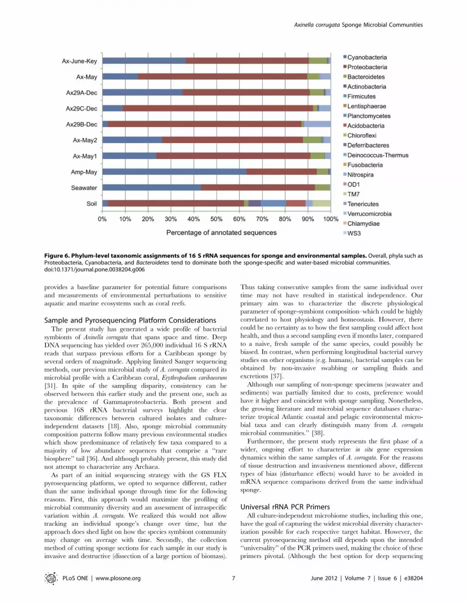
Figure 6 displays the overall phylum-level distribution of sequence

assignments for all samples including the sediment and seawater

samples. Across all samples, Proteobacteria was a dominant

member, representing 26–48% of sequences from each sample.

We observe that the sediment sample has several well-represented

phyla that are not abundant in the sponge communities including

Planctomycetes (9%), Acidobacteria (7%), and the TM7 candidate

division (6%). Phyla present in the sponge communities that were

not observed in the seawater or sediment samples included

Lentisphaerae and Firmicutes. Given the abundance of Nitrospira

observations across all A. corrugata communities, we expected to

recover members of this phylum in the associated environmental

samples. Intriguingly, we did discover Nitrospira members within

the sediment sample (2.5% of sequences), but not a single member

in seawater. The lack of Nitrospira may be due to its low abundance

in the surrounding seawater population (too low given our

sequencing depth), but may also suggest an environmental niche

shared between the A. corrugata microbiome and nearby sediment

communities. Class level assignments for the environmental

samples appear in Table S4.

Figure 4. Unsupervised cluster analysis of taxonomic assignments using CloVR. The assignments are either at the class (A) or order (B)levels. Values in the heatmap reflect the log of the relative abundance within each sample (e.g. -1 , 10%, -2 , 1%). We observe that Amphimedon isconsistently an outlier relative to the other samples, in part due to a lack of Deltaproteobacteria species and Nitrospira and a larger abundance ofBetaproteobacteria, Bacilli, and Bacteroidetes.doi:10.1371/journal.pone.0038204.g004

Table 2. Relative abundance of sequences within universally observed OTUs across all sponge samples.

Sample Name Percentage of sequences in OTUs universally found in all samples

Amp-May 46.25%

Ax-May1 8.04%

Ax-May2 8.65%

Ax-May 7.08%

Ax29A-Dec 14.73%

Ax29B-Dec 0.96%

Ax29C-Dec 3.38%

Ax-June-Key 21.88%

doi:10.1371/journal.pone.0038204.t002

Axinella corrugata Sponge Microbial Communities

PLoS ONE | www.plosone.org 5 June 2012 | Volume 7 | Issue 6 | e38204

Finally, we examined the consistency of the current sponge-

associated dataset with that of data from a previously sequenced

library of A. corrugata microbial symbionts [31]. Querying the

current pyrosequenced 16 S rRNA dataset reveal 72,115 sequenc-

es that match at least one of the 111 earlier accessions with $98%

identity along at least 95% of its length. The vast majority (.95%)

appear as significant hits to Axinella-samples and not Amphimedon

(see Table S3). Thus, we find stable community membership

between earlier A. corrugata samples and this study.

Discussion

The extensive pyrosequencing effort described here reveals that

similar microbiomes are harbored within different individual

Axinella corrugata samples and locales, providing a comprehensive

profile of microbial diversity within this unique sponge species.

Comparisons between two seasons indicate measureable shifts but

an overall stability among most microbial community members.

Also, certain class level similarities are seen among the microbial

consortia of A. compressa and A. corrugata, but these different sponge

species collected at the same location also have distinct symbiotic

communities. This data contributes to the growing database of

sponge symbiont biodiversity [12,14,17,21], which in turn

Figure 5. Representative TEM micrographs of Axinella corrugata sponge mesohyl. A) Wide angle view showing potentially aggregatedbacteria (b), possible phage (Ph) and spicule –forming cells (Sp). Scale bar = 1 mm; B) One of several unidentified pear-shaped bacteria within Axinellacorrugata sponge mesohyl. Scale bar = 0.2 mm; C) Possible Cyanobacteria, Scale bar = 1 mm; D) Possible Ectothiorhodospiraceae microbial symbiontwithin Axinella corrugata. Scale bar = 0.5 mm.doi:10.1371/journal.pone.0038204.g005

Table 3. Differentially abundant taxonomic classes detectedbetween spring and fall Axinella corrugata bacterialcommunities.*

May December

Taxon mean std. err. mean std. err. p-value

Alphaproteobacteria 16.97% 0.18% 11.82% 0.17% 7.067E-94

Flavobacteria 6.41% 0.12% 3.37% 0.10% 6.525E-86

Cyanobacteria 26.95% 0.21% 21.06% 0.21% 9.121E-84

Deltaproteobacteria 14.11% 0.16% 19.17% 0.21% 6.908E-83

Gammaproteobacteria 30.38% 0.22% 36.46% 0.25% 2.229E-74

Nitrospira 4.16% 0.09% 6.82% 0.13% 1.784E-62

Actinobacteria 0.11% 0.02% 0.53% 0.04% 7.270E-28

Betaproteobacteria 0.05% 0.01% 0.18% 0.02% 3.085E-07

Clostridia 0.26% 0.02% 0.11% 0.02% 1.323E-06

Sphingobacteria 0.27% 0.02% 0.12% 0.02% 5.520E-06

Acidobacteria 0.00% 0.00% 0.04% 0.01% 2.251E-04

*False discovery rate , 0.01%.doi:10.1371/journal.pone.0038204.t003

Axinella corrugata Sponge Microbial Communities

PLoS ONE | www.plosone.org 6 June 2012 | Volume 7 | Issue 6 | e38204

provides a baseline parameter for potential future comparisons

and measurements of environmental perturbations to sensitive

aquatic and marine ecosystems such as coral reefs.

Sample and Pyrosequencing Platform ConsiderationsThe present study has generated a wide profile of bacterial

symbionts of Axinella corrugata that spans space and time. Deep

DNA sequencing has yielded over 265,000 individual 16 S rRNA

reads that surpass previous efforts for a Caribbean sponge by

several orders of magnitude. Applying limited Sanger sequencing

methods, our previous microbial study of A. corrugata compared its

microbial profile with a Caribbean coral, Erythropodium caribaeorum

[31]. In spite of the sampling disparity, consistency can be

observed between this earlier study and the present one, such as

the prevalence of Gammaproteobacteria. Both present and

previous 16S rRNA bacterial surveys highlight the clear

taxonomic differences between cultured isolates and culture-

independent datasets [18]. Also, sponge microbial community

composition patterns follow many previous environmental studies

which show predominance of relatively few taxa compared to a

majority of low abundance sequences that comprise a ‘‘rare

biosphere’’ tail [36]. And although probably present, this study did

not attempt to characterize any Archaea.

As part of an initial sequencing strategy with the GS FLX

pyrosequencing platform, we opted to sequence different, rather

than the same individual sponge through time for the following

reasons. First, this approach would maximize the profiling of

microbial community diversity and an assessment of intraspecific

variation within A. corrugata. We realized this would not allow

tracking an individual sponge’s change over time, but the

approach does shed light on how the species symbiont community

may change on average with time. Secondly, the collection

method of cutting sponge sections for each sample in our study is

invasive and destructive (dissection of a large portion of biomass).

Thus taking consecutive samples from the same individual over

time may not have resulted in statistical independence. Our

primary aim was to characterize the discrete physiological

parameter of sponge-symbiont composition–which could be highly

correlated to host physiology and homeostasis. However, there

could be no certainty as to how the first sampling could affect host

health, and thus a second sampling even if months later, compared

to a naive, fresh sample of the same species, could possibly be

biased. In contrast, when performing longitudinal bacterial survey

studies on other organisms (e.g. humans), bacterial samples can be

obtained by non-invasive swabbing or sampling fluids and

excretions [37].

Although our sampling of non-sponge specimens (seawater and

sediments) was partially limited due to costs, preference would

have it higher and coincident with sponge sampling. Nonetheless,

the growing literature and microbial sequence databases charac-

terize tropical Atlantic coastal and pelagic environmental micro-

bial taxa and can clearly distinguish many from A. corrugata

microbial communities.’’ [38].

Furthermore, the present study represents the first phase of a

wider, ongoing effort to characterize in situ gene expression

dynamics within the same samples of A. corrugata. For the reasons

of tissue destruction and invasiveness mentioned above, different

types of bias (disturbance effects) would have to be avoided in

mRNA sequence comparisons derived from the same individual

sponge.

Universal rRNA PCR PrimersAll culture-independent microbiome studies, including this one,

have the goal of capturing the widest microbial diversity character-

ization possible for each respective target habitat. However, the

current pyrosequencing method still depends upon the intended

‘‘universality’’ of the PCR primers used, making the choice of these

primers pivotal. (Although the best option for deep sequencing

Figure 6. Phylum-level taxonomic assignments of 16 S rRNA sequences for sponge and environmental samples. Overall, phyla such asProteobacteria, Cyanobacteria, and Bacteroidetes tend to dominate both the sponge-specific and water-based microbial communities.doi:10.1371/journal.pone.0038204.g006

Axinella corrugata Sponge Microbial Communities

PLoS ONE | www.plosone.org 7 June 2012 | Volume 7 | Issue 6 | e38204

strategies would be complete independence from gene specific

primers). We applied two universal eubacterial primers (27F and

533r), previously proven to amplify a wide diversity of the eubacterial

spectrum in many past culture-independent studies [39]. These

primers span theV2regionwhichhasalsobeenshowntobeoneof the

most phylogenetically informative rRNA regions for eubacteria [40].

In this context, it is curious that this study shows a deficiency of the

Chloroflexi, since only 4 OTUs were found, and Chloroflexi have been

shown to be a major taxon across many sponge species [16,21].

However, previous community profiling with a different universal

16 S rRNA primer pair also did not detect any Chloroflexi sequences in

multiple A. corrugata samples [31], and indicated many Chloroflexi

sequences occurred more often in deeper rather than shallow water

sponges [41]. These results contrast with a recent sponge symbiont

pyrosequencing survey by Schmitt et al, [21] that applied a modified,

slightly more degenerate 533r primer. Their results designate a fairly

small number of ‘‘core’’ flora for Phylum Porifera, which included

Chloroflexi and Proteobacteria, sometimes excluding Poribacteria.

Our results re-emphasize the tangible differences between diverse

sponge hosts and the additional variables that can affect small subunit

rRNA censuses: PCR primer sequences, alternative DNA extraction

methods, geographical source or distinct features of host microcosm

and identity. We are currently testing alternative 16S rRNA primers

that may be established as standard primers for accessing an even

wider number of taxa and habitats as part of a burgeoning ‘‘earth

microbiome’’ project (http://www.earthmicrobiome.org/) [42,43].

The Unique Host Sponge Axinella corrugataA. corrugata possesses many interesting traits justifying its

elevation as a model sponge. For example, its distribution is fairly

widespread across the Western Atlantic, Caribbean and Gulf of

Mexico [23,24]. Secondly, the species has been applied in

aquaculture studies [30]. Moreover, marine sponges continue to

attract attention due to their production of many chemically

diverse marine natural products [44] which still have great

potential in pharmacological research [6,45]. Retention of the

ability to produce stevensine in antibiotic-supplemented A. corrugata

cell culture supported a sponge biosynthetic origin [32]. Stevensine

also has weak antimicrobial activity at a concentration of 50 to

200 mg ml-1 against certain marine microbial strains, and thus

could be a regulator of the microbial community.

As mentioned, there have been long standing efforts to establish

permanent A. corrugata in vitro cell lines for cell biology research

[29,32]. The characterization of a fairly stable core bacterial

community, that includes phototrophic Cyanobacteria, may

explain why permanent cell culture of A. corrugata remains a goal

difficult to attain. It may be likely that the apparently stable and

intimate sponge-microbial associations demonstrated in this study,

can make permanent axenic growth of pure sponge cell lines

difficult to establish in vitro.

Axinella corrugata Microbial SymbiontsAlthough preliminary ultrastructure analysis suggests that A.

corrugata may be a relatively low microbial abundance (LMA) host,

this designation contrasts with high taxonomic diversity observed

from the 16 S rRNA data. Unknown taxonomic identities are

reflected in the unusual morphologies of several bacteria such as

those in Fig. 5B. Its also possible that some specific bacterial

species may be relegated to specific locations or structures within

the sponge similar to the aggregate formations observed in another

local reef sponge, Agelas tubulata by FISH [33] but not yet detected

by our current TEM surveys.

The finding of a large number of A. corrugata symbionts that

encompass a wide range of heterotrophs, chemoautolithotrophs.

phototrophic Cyanobacteria and purple sulfur bacteria adds another

interesting dimension to this sponge’s physiology. All characterized

A. corrugata microbial communities showed no deficiency in

Cyanobacteria which comprised over 7.2% of all sequences. The

transfer of photosynthetic nutrients and byproducts may be vital

between the host and these bacteria. A. corrugata has been found at

depths no lower than 71 m [23], a distribution which still fits well

within the photic zone. Curiously, Cyanobacteria seemed to be

only sparsely detected in TEM micrographs (Fig. 5A and 5C). One

interpretation of this is that the identification of Cyanobacteria

from A. corrugata samples represented transient cells within the

mesohyl or seawater.

Although we do not have definitive evidence for low aeration

within the mesohyl, this would be consistent with an anaerobic or

microaerophilic environment that favors the sulfur and nitrogen

metabolism of many A. corrugata microbial community members.

Clear presence of multiple sulfur oxidizing and reducing taxa are

detected in this dataset: Desulfovibrio, Thiocystis, and Thioalkalivibrio

sp. Extremely haloalkaliphilic, obligate chemolithoautotrophic and

sulfur-oxidizing species belonging to genus Thioalkalivibrio can

efficiently oxidize sulfur in alkaline conditions (,pH 10).

Among the several Chromotiales taxa found in A. corrugata,

sequences were found similar to Halorhodospira halophila (formerly

Ectothiorhodospira halophila), which is a motile, alkalophilic, sulfide-

oxidizing extreme halophile, whose whole genome has been

sequenced (DOE Project ID: 15767). Halorhodospira halophila has

garnered attention for biotechnological applications due to its

production of a) blue sensor Photoactive Yellow Proteins (PYP)

and b) hydrogen via photosynthetic pathways [46]. The latter has

significance for sustainable energy initiatives [47]. All members of

Ectothiorhodospiraceae form and excrete elemental sulfur [48].

H. halophila is one of the most halophilic eubacteria of the genus

Halorhodospira found in hypersaline environments that contain

sulfide [49], and similar bacteria are known to oxidize sulfur from

natural gas and refineries [50]. An interesting question is how

these bacteria survive within sponges growing at normal salinity, as

we did not determine whether hypersaline microhabitats exist

within the sponge. It is possible that sulfur compounds cycle as

potential electron donors which can be used by the various

chemotrophic and phototrophic bacteria found in A. corrugata

communities.

Even if not directly coupled with sulfur, nitrogen metabolism of

some A. corrugata symbionts, such as Nitrospira, probably occurs and

is consistent with the evidence of sulfur metabolism. Atmospheric

nitrogen fixing symbiotic Cyanobacteria, ammonia-oxidizing

Gamma- and Betaproteobacteria and nitrite-oxidizing Nitrospira

have been previously recovered from sponges and marine habitats

[51,52]. Anaerobic denitrification (reduction of nitrate to nitrogen)

is a crucial process in the release of global atmospheric nitrogen

which occurs primarily on the seafloor [53] with bacteria as

significant contributors. Anaerobic microbial processes such as

sulfate reduction have been detected in the sponge, Geodia barreti

[52]. Genus Thioalkalivibrio also includes nitrate-reducers, faculta-

tive alkaliphiles and denitrifiers among its nine identified species

[54]. A. corrugata may be a suitable candidate to study interspecific

interactions and rates of complex nitrogen cycling in sponges

owing to the presence of Nitrospira sp., Thioalkalivibrio sp. and

Cyanobacteria.

Another interesting, though rare, taxon found at around 0.06%

total abundance in A. corrugata communities matches the genus

Rubellimicrobium, a member of the family Rhodobacteraceae in the

Roseobacter clade, which is associated with an oil tolerant microbe

Wenxinia marina [55]. This finding is relevant in the wake of the

2010 BP Deepwater Horizon oil spill and the increased focus on

Axinella corrugata Sponge Microbial Communities

PLoS ONE | www.plosone.org 8 June 2012 | Volume 7 | Issue 6 | e38204

microbial taxa that may be involved in metabolizing polycyclic

aromatic hydrocarbons (PAH) and petroleum based hydrocarbons

and possible remediation [56,57].

Many bacterial species are known to give a sponge its color,

derived from specific pigments. A. corrugata, is a reddish orange

hue. Photoacclimation by cyanobacterial symbionts and different

phycobiliprotien ratios have also been suspected as a reason for

maroon, brown or yellow colors of other sponges. In this context,

carotenoid and yellowish pigments are produced by Parvularcula

lutaonensis and several Thioalkalivibrio strains found in A. corrugata,

respectively [58]. Members of the family Flavobacteriaceae have

been found in the current dataset and other pigmented sponges.

Many marine taxa belonging to family Flavobacteriaceae are also

known to have carotenoid or flexirubin pigments or both which

cause a yellow coloration [59].

In the context of the above metabolic considerations, in the

future it would be interesting to test hypotheses regarding sponge

symbiont interactions and membership in discrete ‘‘networks’’

[60]. Community models can classify organisms according to

trophic level or degrees of ecological specialization. Habitat

complexity or the apparently high level of metabolic distinctive-

ness found in some A. corrugata symbionts can explain why invasion

from transient environmental bacteria may be difficult.

Physical Habitat of A. corrugataAlthough a discrete, sponge specific community has been

characterized, we cannot discount any transient effects of the local

marine habitat. Water quality monitoring is important for ocean

and human health, especially in the context of local habitats such

as coral reefs and highly populated beach areas [61,62]. Although

we had a statistically low number of environmental samples, many

A. corrugata-specific taxa did not appear abundant in surrounding

seawater and sediments (6,472 and 2,433 16S rRNA sequences,

respectively). Ectothiorhodospiraceae sequences were not detected in

our single seawater sample, but some were observed in Broward

county sediment and Key West seawater samples deposited in

MG-RAST. Future sponge symbiont profiling may fit into these

monitoring schemes, due to possible taxonomic overlap of

transient, planktonic microbial taxa.

Understanding the basic functions and activities of a diverse

microbial consortium begins with the basic cataloging of relevant

taxa. This study now nearly completes this process, and highlights

some unique bacterial symbionts which set A. corrugata apart from

its proximal eukaryotic neighbors. Lastly, the current data

establishes a foundation for more in depth functional, metabolic

and metagenomic analyses that will further elaborate interactions

and networks of a complex microbial microcosm.

Materials and Methods

Sponge and Environmental SamplesSponge samples were collected during the day by SCUBA at the

same location off the third reef in the Broward County, Florida

shown in Table 4 in December 2009 and May 2010. Three A.

corrugata samples (designated with ‘‘Ax’’ prefix) each were collected

from this site in each season. Additionally, in May 2009,

Amphimedon compressa (Amp) was collected at the same site and in

June 2010, one Axinella corrugata (Ax-June-Keys) was collected at

Summerland Key, Florida. (Table 4). After collections from the

reef, live sponges were placed in a bucket of ambient seawater for a

15 min transport back to the laboratory. At the laboratory,10–

20 gm subsamples of each sponge was preserved with each of the

following methods: a) placement in RNAlater (Ambion) according

to the manufacturer’s directions, b) snap freezing within a plastic

storage bag placed in an ethanol-dry ice bath, followed by storage–

in a 280uC freezer, and c) placement in a 50 ml conical centrifuge

tube with 3 volumes of 75% ethanol, thrice changed at 15, 60 min

and 12 hour intervals. The primary goal of these methods was for

total RNA (see text), and thus each was more than sufficient for

preserving genomic DNA. Environmental samples were obtained

at the same Broward reef locations, but at different timepoints

than the sponges. Overall, no specific permits were required for

the described field studies, since collections did not involve

endangered species and did not occur within a designated marine

protected area, private reserve or park.

Sponge taxonomic identifications were confirmed with gene

markers [24], morphology and spicule analysis. It should also be

reiterated that taxonomic re-designations have synonymized the

former species name of Teichaxinellaxinella morchella with Axinella

corrugata [23,27,63].

Genomic DNA ExtractionFor total DNA extractions from the sponge, prokaryotic and

eukaryotic cells were ‘‘squeezed’’ out of each sponge sample by

mincing 3–5 gm sections of tissue in L buffer [10 mM Tris-Cl,

pH 8.0; 0.1 M Ethylenediaminetetraacetic acid 487 (EDTA),

pH 8.0; 0.5% (w/v) ] [64]. This buffer preserved cell integrity

which could be viewed with light microscopy, while DNA

degradation was prevented via high EDTA concentrations. When

a 2–3 gm cell pellet was obtained, it was processed with a

UltraCleanH Genomic DNA Isolation Kit (MoBio) according to kit

instructions. Purity and concentration of DNA was measured

using a NanoDrop 1000 spectrophotometer (Thermo Scientific)

and DNA gel electrophoresis. A260/A280 ratio of approximately

1.8 and clearly visible bands on the gel, confirmed isolation of pure

DNA [64]. DNA quality and size of the fragment was visually

confirmed using the Agilent 7500 Bioanalyzer DNA chip, which

assessed DNA integrity and base-pair length.

Preparation of DNA for PyrosequencingTotal sponge genomic DNA was PCR amplified using conserved

16 S SSU rRNA primers that were fused to Roche Fusion primers

(FusionPrimerA:59CGTATCGCCTCCCTCGCGCCATCAG3’

and Fusion Primer B: 59CTATGCGCCTTGCCAG

CCCGCTCAG 39) each having a unique 10 base DNA bar code

according to the thermocycling parameters in Rapid Library

Preparation Manual. These were attached to universal 16S small

subunit (SSU) rRNA primer sequences - Forward primer 27F GTT

TGA TCCTGGCTCAG3’andReverseprimer533r 5’TTA CCG

CGG CTG CTG GCA C 3’. Primers were annealed 55uC for 60 sec,

after initial denaturation at 95uC for 300 s, denaturing at 95u for 60s,

and extension at 72uC for 60 s (with 29 additional cycles) and final

extension at 72u for 300 s. The resulting 16S rRNA fragments

spanned about 350–400 bp including hypervariable V1–V3 regions

[38]. Quality and quantity of the amplicons was checked using the

DNA 7500 Bioanalyzer chip and Fluorescence-based Quantification

Assay (Qubit) respectively. DNA sequencing was carried out by using

a GS FLX, Roche pyrosequencer (454 Life Sciences, Maryland,

USA).

BioinformaticsPreprocessing. Barcoded multiplex pyrosequences generat-

ed using the 454 Titanium platform were initially trimmed for

quality using standard the sff software tools from Roche/454.

Sequences were preliminarily assessed for possible host contam-

inant from the amplification process by performing a BLASTN

[65] search of sequences against the 16 S rRNA homologous gene

in Axinella; no evidence of contaminant was detected. All sequences

Axinella corrugata Sponge Microbial Communities

PLoS ONE | www.plosone.org 9 June 2012 | Volume 7 | Issue 6 | e38204

in this study have been submitted to MG-RAST, and assigned the

following ID numbers: 4479837.3, 4479836.3, 4479835.3,

4479834.3, 4479833.3, 4479832.3, 4479831.3 4479830.3,

4479829.3 and 4479828.3 (Table 4). We have also deposited the

raw sequence data in the NCBI Sequence Read Archive under the

project accession: SRP010086.

Processing. Reads were input to the CloVR-16 S automated

pipeline which uses a comprehensive automated protocol for

comparative 16 S sequence analysis. Briefly, the CloVR-16 S

pipeline employs several popular tools for phylogenetic analysis of

16S rRNA data including: QIIME [66] and Mothur [67] for

sequence processing and diversity analysis, the RDP Bayesian

classifier [39] for taxonomic assignment, UCHIME (http://www.

drive5.com/uchime/) for chimera detection and removal, Meta-

stats [68] for statistical comparisons, and various R scripts for

visualization and unsupervised clustering. The CloVR-16 S

pipeline initially screens sequences using a minimum length of

100 bp, a maximum homopolymer run requirement of 8 bp, and

also removes sequences containing ambiguous base calls. High-

quality sequences are subsequently assessed for chimeras using

UCHIME with a reference database of 16 S sequences from

known species. The resulting chimera-free reads are clustered into

OTUs using the UCLUST module from QIIME with a pairwise

identity threshold of 97%. OTU representatives are assigned to a

phylogenetic lineage using the RDP classifier with a minimum

confidence threshold of 80%. Default settings were used in the

CloVR analysis. A full description of the CloVR-16 S standard

operating protocol is available online at http://clovr.org. [69].

Additional processing of sequence data was performed using the

standardized outputs from the CloVR-16 S pipeline. ‘‘Rarefied’’

datasets (with equivalent sampling depths) were generated in

QIIME by randomly subsampling 18,000 sequences from each

sample in the full dataset. PCoA plots were visualized using the

KiNG software package [70]. Unsupervised clustering of taxo-

nomic groups and samples was performed using the Skiff program

in CloVR. Within Skiff, relative abundances of taxa within each

sample are log-transformed and clustered using a Euclidean

distance metric and furthest-neighbor clustering. To detect

differentially abundant taxa between May and December sample

time points, we used Metastats with default parameters at each

phylogenetic level (phylum down to OTU assignments). For

comparisons with less than 100 features (e.g. phyla, classes), the

false discovery rate was controlled using a method by Benjamini

and Hochberg [35].

To assign each 16 S fragment to it’s closest matching known

species, BLASTN searches were performed against the SILVA

rRNA database (SSURef_106_tax_silva) [71] (reduced to reference

sequences with full species-level information) with a minimum e-

value requirement of 1e-5. Sequences were assigned to the

taxonomy of the best BLAST hit under this criterion. Due to

their known presence in the sponge holobiont system, uncultured

Poribacteria reference sequences were also included in the reduced

SILVA database. To assess degrees of relatedness, subsets of the

most common bacterial sequences were analyzed phylogenetically

using MEGA5 [72].

Electron MicroscopyImmediately after collection, small sponge sections of 3–6 cm3

were fixed in 2% gluteraldehyde sodium caccodylate buffered sea

water, posted-fixed in 1% osmium tetroxide, dehydrated in a series

of ethanols, and embedded in SpurrTM low viscosity resin. Blocks

were then sectioned, stained with lead citrate and uryanl acetate

and examined in a JEOL 100X TEM.

Supporting Information

Figure S1 Neighbor-joining phylogenetic tree of repre-sentative OTU 118 sequences. The optimal tree with the sum

of branch length = 1.95291608 is shown. The percentage of

replicate trees in which the associated taxa clustered together in

the bootstrap test (500 replicates) are shown next to the branches.

The tree is drawn to scale, with branch lengths in the same units as

those of the evolutionary distances used to infer the phylogenetic

tree. The evolutionary distances were computed using the

Maximum Composite Likelihood method and are in the units of

the number of base substitutions per site. The rate variation

among sites was modeled with a gamma distribution (shape

parameter = 1). The analysis involved 34 nucleotide sequences. All

positions containing gaps and missing data were eliminated. There

were a total of 225 positions in the final dataset. Evolutionary

analyses were conducted in MEGA5 [72]. The same topology was

observed with maximum parsiomony and minimum evolution

reconstructions. Reference sequences for Thioalkalivibrio

(343202513 -NR_042855.1) and Methylomicrobium album

Table 4. Collection site and dates for sponge and environmental samples characterized in this study.

Collection DateSpecies orsample type

Sample IDnames Temp (C) Location Depth (m) MG-RAST Nos.

December 2009 Axinella corrugata(sponge)

Ax29A-Dec, Ax29B-Dec,Ax29C-Dec

21.1 26 09.104N,80 04.659W

20 4479833.3, 4479834.3,4479835.3

May 2010 Axinella corrugata(sponge)

Ax-May1,Ax-May2,Ax-May

26.6 26 09.104N,80 04.659W

20 4479830.3, 4479831.3,4479832.3

June 2010 Axinella corrugata(sponge)

Ax-June-Key 28.8 24.80N,80.76W

13 4479829.3

May 2009 Amphimedon compressa(sponge)

Amp-May 25.0 26 09.104N,80 04.659W

16.7 4479828.3

July 2011 Seawater BR-4C 29 26 09.618N,80 04.554W

18 4479836.3

December2010

Reef sediment Sed 2010 21.0 26 09.104N,80 04.659W

9 4479837.3

doi:10.1371/journal.pone.0038204.t004

Axinella corrugata Sponge Microbial Communities

PLoS ONE | www.plosone.org 10 June 2012 | Volume 7 | Issue 6 | e38204

(265678936 -NR_029244.1) were included for reference and

rooting.

(TIFF)

Table S1 Representative unique A. corrugata-specificsymbionts (as percentages of total). * - Average occurrence

is based only the six Broward county A. corrugata- samples

(XLS)

Table S2 Differentially abundant OTUs identified be-tween May and December A. corrugata samples. OTUs

are ordered by relative abundance of May samples. OTU

abundances were input to Metastats using default parameters. A

total of 268 were detected as differentially abundant (with a

corresponding false discovery rate , 1%). The May samples

contained 112 enriched OTUs relative to the December group,

while 156 OTUs were relatively enriched in the December

population. No confident phylum assignment could be made for

114 of these OTUs using the RDP Bayesian classifier.

(XLS)

Table S3 Summary of BLAST query matches in currentdataset to previously characterized A. corrugata 16SrDNA clones. 72,115 sequences hit at least one previous

reference sequence from the set of accessions you wanted with

at least 98% identity along at least 95% of their length.

(XLS)

Table S4 Class level assignments of 16S rRNA sequenc-es. Assignments were made using the RDP classifier with a

minimum confidence threshold of 80%.

(XLS)

Acknowledgments

We are grateful to Dr. Marc Allard and Dr. Eric Brown of the FDA Food

and Drug Administration Office of Regulatory Science Division of

Microbiology for early assistance with the Roche pyrosequencing platform.

We thank Dr. Shirley Pomponi for helpful comments on Axinella biology,

Dr Malcolm Hill for collection assistance at Summerland Key, Dr.

Alexander Ereskovsky for help with ultrastructural interpretations, and

Alexandra Campbell for assistance with genomic DNA extractions. This

manuscript is the National Coral Reef Institute publication #149, and is

dedicated to the memory and works of Professor Lynn Margulis, who was

instrumental in promoting the importance of symbiosis in modern biology.

Author Contributions

Conceived and designed the experiments: JVL JP JRW. Performed the

experiments: JRW JP GA AO. Analyzed the data: JRW JVL JP PB.

Contributed reagents/materials/analysis tools: JVL AO. Wrote the paper:

JRW JVL JP PB AO.

References

1. Moran NA (2007) Symbiosis as an adaptive process and source of phenotypic

complexity. Proceedings of the National Academy of Sciences 104: 8627–8633.

2. Margulis L (1993) Symbiosis in cell evolution: microbial communities in the

Archean and Proterozoic eons: Freeman.

3. Knowlton N (2001) The future of coral reefs. Proceedings of the National

Academy of Sciences 98: 5419–5425.

4. Simpson TL (1984) The cell biology of sponges: Springer-Verlag.

5. Rutzler K (2004) Sponges On Coral Reefs: A Community Shaped By

Competitive Cooperation. Bollettino dei Musei e degli Istituti Biologici

dell’University di Genova 68: 85–148.

6. Munro MHG, Blunt JW, Lake RJ, Litaudon M, Battershill CN, et al. (1994)

From seabed to sickbed: What are the prospects? R. W. M. Van Soest, T. Van

Kempen, Braekman JC, editors. Balkema, Rotterdam.

7. Santavy DL, Willenz P, Colwell RR (1990) Phenotypic study of bacteria

associated with the caribbean sclerosponge, Ceratoporella nicholsoni. Appl

Environ Microbiol 56: 1750–1762.

8. Reiswig HM (1971) In situ pumping activities of tropical Demospongiae. Marine

Biology 9: 38–50.

9. Vogel S (1977) Current-induced flow through living sponges in nature.

Proceedings of the National Academy of Sciences 74: 2069–2071.

10. Pile A, Patterson M, Witman J (1996) In situ grazing on plankton ,10 mm by the

boreal sponge Mycale lingua Marine Ecology Progress Series 141: 95–102.

11. Webster NS, Taylor MW, Behnam F, Lucker S, Rattei T, et al. (2010) Deep

sequencing reveals exceptional diversity and modes of transmission for bacterial

sponge symbionts. Environmental Microbiology 12: 2070–2082.

12. Taylor MW, Radax R, Steger D, Wagner M (2007) Sponge-Associated

Microorganisms: Evolution, Ecology, and Biotechnological Potential. Microbiol

Mol Biol Rev 71: 295–347.

13. Webster NS, Blackall LL (2008) What do we really know about sponge-microbial

symbioses[quest]. ISME J 3: 1–3.

14. Webster NS, Taylor MW (2011) Marine sponges and their microbial symbionts:

love and other relationships. Environmental Microbiology: no-no.

15. Wilkinson CR, Nowak M, Austin B, Colwell RR (1981) Specificity of Bacterial

Symbionts in Mediterranean and Great Barrier Reef Sponges. Microbial

Ecology 7: 13–21.

16. Lopez JV, McCarthy PJ, Janda KE, Willoughby R, Pomponi SA (1999)

Molecular techniques reveal wide phyletic diversity of heterotrophic microbes

associated with the sponge genus Discodermia (Porifera:Demospongiae).

Proceedings of the 5th International Sponge Symposium Memoirs of the

Queensland Museum Birsbane 44.

17. Hill R (2004) Microbes from marine sponges: A treasure trove of biodiversity for

natural products discovery. In: Ball A, editor. Microbial Diversity and

Bioprospecting. Washington DC: American Society for Microbiology, ASM

Press.

18. Sfanos K, Harmody D, Dang P, Ledger A, Pomponi S, et al. (2005) A molecular

systematic survey of cultured microbial associates of deep-water marine

invertebrates. Systematic and Applied Microbiology 28: 242–264.

19. Hill M, Hill A, Lopez N, Harriott O (2006) Sponge-specific bacterial symbionts

in the Caribbean sponge, Chondrilla nucula (Demospongiae, Chondrosida).

Marine Biology 148: 1221–1230.

20. Mohamed NM, Colman AS, Tal Y, Hill RT (2008) Diversity and expression of

nitrogen fixation genes in bacterial symbionts of marine sponges. Environmental

Microbiology 10: 2910–2921.

21. Schmitt S, Tsai P, Bell J, Fromont J, Ilan M, et al. (2011) Assessing the complex

sponge microbiota: core, variable and species-specific bacterial communities in

marine sponges. ISME J.

22. Fieseler L, Horn M, Wagner M, Hentschel U (2004) Discovery of the Novel

Candidate Phylum ‘‘Poribacteria’’ in Marine Sponges. Applied and Environ-

mental Microbiology 70: 3724–3732.

23. Alvarez B, van Soest RWM, Ruetzler K (1998) A Revision of Axinellidae

(Porifera: Demospongiae) of the Central West Atlantic Region. Smithsonian

contributions to zoology 598.

24. Lopez JV, Peterson CL, Willoughby R, Wright AE, Enright E, et al. (2002)

Characterization of Genetic Markers for In Vitro Cell Line Identification of the

Marine Sponge Axinella corrugata. Journal of Heredity 93: 27–36.

25. Messing CG, Diaz MC, Kohler KE, Reed JK, Ruetzler K, et al. (2010) Taxon

page for Axinella corrugata (George & Wilson, 1919). Porifera LifeDesk, http://

poriferalifedesksorg/pages/1080.

26. Wilson D, Puyana M, Fenical W, Pawlik J (1999) Chemical Defense of the

Caribbean Reef Sponge Axinella corrugata Against Predatory Fishes. Journal of

Chemical Ecology 25: 2811.

27. Andrade P, Willoughby R, Pomponi SA, Kerr RG (1999) Biosynthetic studies of

the alkaloid, stevensine, in a cell culture of the marine sponge Teichaxinella

morchella. Tetrahedron Letters 40: 4775–4778.

28. Wright AE, Chiles SA, Cross SS (1991) 3-Amino-1-(2-Aminoimidazolyl)-prop-1-

ene from the Marine Sponges Teichaxinella morchella and Ptilocaulis walpersi.

Journal of Natural Products 54: 1684–1686.

29. Willoughby R, Pomponi S (2000) Quantitative assessment of marine sponge cells

in vitro: Development of improved growth medium. In Vitro Cellular &

Developmental Biology - Animal 36: 194–200.

30. Duckworth A, Samples G, Wright A, Pomponi S (2003) In Vitro CuIture of the

Tropical Sponge Axinella corrugata (Demospongiae): Effect of Food Cell

Concentration on Growth, Clearance Rate, and Biosynthesis of Stevensine.

Marine Biotechnology 5: 519.

31. Lopez JV, Ranzer L, Ledger A, Schoch B, Duckworth A, et al. (2010)

Comparison of bacterial diversity within the Coral Reef Sponge, Axinella

corrugata, and the Encrusting Coral, Erythropodium caribaeorum and adjacent

environmental samples Proceedings of the 11th International Coral Reef

Symposium (IRCS); Ft. Lauderdale, Florida. 1362–1366.

32. Pomponi S, Willoughby R, Kaighn M, Wright A (1997) Development of

techniques for in vitro production of bioactive natural products from marine

sponges.

33. Negandhi K, Blackwelder P, Ereskovsky A, Lopez J (2010) Florida reef sponges

harbor coral disease-associated microbes. Symbiosis 51: 117–129.

Axinella corrugata Sponge Microbial Communities

PLoS ONE | www.plosone.org 11 June 2012 | Volume 7 | Issue 6 | e38204

34. Lafi FF, Fuerst JA, Fieseler L, Engels C, Goh WWL, et al. (2009) Widespread

Distribution of Poribacteria in Demospongiae. Appl Environ Microbiol 75:5695–5699.

35. Benjamini Y, Hochberg Y (1995) Controlling the false discovery rate: a practical

and powerful approach to multiple testing. Journal of Royal Statistical Society57.

36. Sogin M, Morrison H, Huber J, Mark Welch D, Huse S, et al. (2006) Microbialdiversity in the deep sea and the underexplored "rare biosphere". Proc Natl

Acad Sci USA 103: 12115–12120.

37. Turnbaugh PJ, Hamady M, Yatsunenko T, Cantarel BL, Duncan A, et al.(2009) A core gut microbiome in obese and lean twins. Nature 457: 480–484.

38. Rusch DB, Halpern AL, Sutton G, Heidelberg KB, Williamson S, et al. (2007)The Sorcerer II Global Ocean Sampling Expedition: Northwest Atlantic

through Eastern Tropical Pacific. PLoS Biol 5: e77.39. Weisburg WG, Barns SM, Pelletier DA, Lane DJ (1991) 16S ribosomal DNA

amplification for phylogenetic study. Journal of Bacteriology 173: 697–703.

40. Wang Q, Garrity GM, Tiedje JM, Cole JR (2007) Naive Bayesian Classifier forRapid Assignment of rRNA Sequences into the New Bacterial Taxonomy. Appl

Environ Microbiol 73: 5261–5267.41. Cassler M, Peterson C, Ledger A, Pomponi S, Wright A, et al. (2008) Use of

Real-Time qPCR to Quantify Members of the Unculturable Heterotrophic

Bacterial Community in a Deep Sea Marine Sponge, Vetulina sp. MicrobialEcology 55: 384–394.

42. Galkiewicz JP, Kellogg CA (2008) Cross-Kingdom Amplification UsingBacteria-Specific Primers: Complications for Studies of Coral Microbial

Ecology. Applied and Environmental Microbiology 74: 7828–7831.43. Walters WA, Caporaso JG, Lauber CL, Berg-Lyons D, Fierer N, et al. (2011)

PrimerProspector: de novo design and taxonomic analysis of barcoded

polymerase chain reaction primers. Bioinformatics 27: 1159–1161.44. Braekman JC, Daloze D, Moussiaux B, Stoller C, Deneubourg F (1989) Sponge

secondary metabolites: new results. Pure and Applied Chemistry 61: 509–512.45. Sipkema D, Osinga R, Schatton W, Mendola D, Tramper J, et al. (2005) Large-

scale production of pharmaceuticals by marine sponges: Sea, cell, or synthesis?

Biotechnology and Bioengineering 90: 201–222.46. van Wilderen LJGW, van der Horst MA, van Stokkum IHM, Hellingwerf KJ,

van Grondelle R, et al. (2006) Ultrafast infrared spectroscopy reveals a key stepfor successful entry into the photocycle for photoactive yellow protein.

Proceedings of the National Academy of Sciences 103: 15050–15055.47. Tsuihiji H, Yamazaki Y, Kamikubo H, Imamoto Y, Kataoka M (2006) Cloning

and characterization of nif structural and regulatory genes in the purple sulfur

bacterium, Halorhodospira halophila. Journal of Bioscience and Bioengineering101: 263–270.

48. Imhoff JF (1984) Reassignment of the Genus Ectothiorhodospira Pelsh 1936 to aNew Family, Ectothiorhodospiraceae fam. nov., and Emended Description of

the Chromatiaceae Bavendamm 1924. International Journal of Systematic

Bacteriology 34: 338–339.49. Imhoff JF, Suling J (1996) The phylogenetic relationship among Ectothiorho-

dospiraceae: a reevaluation of their taxonomy on the basis of 16S rDNAanalyses. Archives of Microbiology 165: 106–113.

50. Foti M, Ma S, Sorokin DY, Rademaker JLW, Kuenen JG, et al. (2006) Geneticdiversity and biogeography of haloalkaliphilic sulphur-oxidizing bacteria

belonging to the genus Thioalkalivibrio. FEMS Microbiology Ecology 56: 95–101.

51. Diaz M, Akob D, Cary C (2004) Denaturing gradient gel electrophoresis ofnitrifying microbes associated with tropical sponges. Boll Mus 1st Biol Univ

Genova 68: 279–289.52. Hoffmann F, Radax R, Woebken D, Holtappels M, Lavik G, et al. (2009)

Complex nitrogen cycling in the sponge Geodia barretti. Environmental

Microbiology 11: 2228–2243.53. Middelburg JJ, Soetaert K, Herman PMJ, Heip CHR (1996) Denitrification in

marine sediments: A model study. Global Biogeochem Cycles 10: 661–673.

54. Sorokin DY, Kuenen JG (2005) Haloalkaliphilic sulfur-oxidizing bacteria in soda

lakes. FEMS Microbiology Reviews 29: 685–702.

55. Ying J-Y, Wang B-J, Dai X, Yang S-S, Liu S-J, et al. (2007) Wenxinia marina gen.

nov., sp. nov., a novel member of the Roseobacter clade isolated from oilfield

sediments of the South China Sea. International Journal of Systematic and

Evolutionary Microbiology 57: 1711–1716.

56. Hazen TC, Dubinsky EA, DeSantis TZ, Andersen GL, Piceno YM, et al. (2010)

Deep-Sea Oil Plume Enriches Indigenous Oil-Degrading Bacteria. Science 330:

204–208.

57. Lu Z, Deng Y, Van Nostrand JD, He Z, Voordeckers J, et al. (2011) Microbial

gene functions enriched in the Deepwater Horizon deep-sea oil plume. ISME J.

58. Cho J-C, Giovannoni SJ (2003) Parvularcula bermudensis gen. nov., sp. nov., a

marine bacterium that forms a deep branch in the a-Proteobacteria.

International Journal of Systematic and Evolutionary Microbiology 53: 1031–

1036.

59. Bernardet JF, Nakagawa Y, Holmes B (2002) Proposed minimal standards for

describing new taxa of the family Flavobacteriaceae and emended description of

the family. International Journal of Systematic and Evolutionary Microbiology

52: 1049–1070.

60. Montoya JM, Pimm SL, Sole RV (2006) Ecological networks and their fragility.

Nature 442: 259–264.

61. Abdelzaher AM, Wright ME, Ortega C, Solo-Gabriele HM, Miller G, et al.

(2010) Presence of Pathogens and Indicator Microbes at a Non-Point Source

Subtropical Recreational Marine Beach. Applied and Environmental Microbi-ology 76: 724–732.

62. Banks KW, Riegl BM, Richards VP, Walker BK, Helmle KP, et al. (2008) The

Reef Tract of Continental Southeast Florida (Miami-Dade, Broward and Palm

Beach Counties, USA) In: Riegl BM, Dodge RE, editors. Coral Reefs of the

USA: Springer Netherlands. 175–220.

63. Hooper JNA, Levi C (1993) Axinellida (Porifera: Demospongiae) from the New

Caledonia Lagoon. Invertebrate Taxonomy 7: 1395–1472.

64. Sambrook J, Russell DW (2001) Molecular Cloning: A Laboratory Manual Cold

Spring Harbour, New York.: Cold Spring Harbor Laboratory Press.

65. Altschul S, Madden T, Schaffer A, Zhang J, Zhang Z, et al. (1997) Gapped

BLAST and PSI-BLAST: a new generation of protein database search

programs. Nucleic Acids Res 25: 3389–3402.

66. Caporaso JG, Kuczynski J, Stombaugh J, Bittinger K, Bushman FD, et al. (2010)

QIIME allows analysis of high-throughput community sequencing data. Nature

Methods 7: 335–335–336.

67. Schloss PD, Westcott SL, Ryabin T, Hall JR, Hartmann M, et al. (2009)

Introducing mothur: Open-Source, Platform-Independent, Community-Sup-ported Software for Describing and Comparing Microbial Communities. Appl

Environ Microbiol 75: 7537–7541.

68. White JR, Nagarajan N, Pop M (2009) Statistical Methods for Detecting

Differentially Abundant Features in Clinical Metagenomic Samples. PLoS

Comput Biol 5: e1000352.

69. Angiuoli S, Matalka M, Gussman A, Galens K, Vangala M, et al. (2011) CloVR:

A virtual machine for automated and portable sequence analysis from the

desktop using cloud computing. BMC Bioinformatics 12: 356.

70. Chen VB, Davis IW, Richardson DC (2009) KING (Kinemage, Next

Generation): a versatile interactive molecular and scientific visualization

program. Protein science : a publication of the Protein Society 18: 2403–2409.

71. Pruesse E, Quast C, Knittel K, Fuchs BM, Ludwig W, et al. (2007) SILVA: a

comprehensive online resource for quality checked and aligned ribosomal RNAsequence data compatible with ARB. Nucleic Acids Research 35: 7188–7196.

72. Tamura K, Peterson D, Peterson N, Stecher G, Nei M, et al. (2011) MEGA5:

Molecular Evolutionary Genetics Analysis Using Maximum Likelihood,

Evolutionary Distance, and Maximum Parsimony Methods. Molecular Biology

and Evolution 28: 2731–2739.

Axinella corrugata Sponge Microbial Communities

PLoS ONE | www.plosone.org 12 June 2012 | Volume 7 | Issue 6 | e38204


Recommended